Article

BAP1 Malignant Pleural Mesothelioma Mutations in Caenorhabditis elegans Reveal Synthetic Lethality between ubh-4/BAP1 and the Proteasome Subunit rpn-9/PSMD13

1
Modeling Human Diseases in C. elegans Group, Genes, Diseases, and Therapies Program, Institut d’Investigació Biomèdica de Bellvitge (IDIBELL), L’Hospitalet de Llobregat, 08908 Barcelona, Spain
2
Medicum, Department of Biochemistry and Developmental Biology, Faculty of Medicine, University of Helsinki, Haartmaninkatu 8, 00290 Helsinki, Finland
3
Department of Medical Oncology, Institut Català d’Oncologia (ICO), L’Hospitalet de Llobregat, 08908 Barcelona, Spain
4
Preclinical and Experimental Research in Thoracic Tumors (PReTT), Institut d’Investigació Biomèdica de Bellvitge (IDIBELL), L’Hospitalet de Llobregat, 08908 Barcelona, Spain
*
Author to whom correspondence should be addressed.
Cells 2023, 12(6), 929; https://doi.org/10.3390/cells12060929
Submission received: 16 January 2023 / Revised: 28 February 2023 / Accepted: 2 March 2023 / Published: 18 March 2023

Abstract

:
The deubiquitinase BAP1 (BRCA1-associated protein 1) is associated with BAP1 tumor predisposition syndrome (TPDS). BAP1 is a tumor suppressor gene whose alterations in cancer are commonly caused by gene mutations leading to protein loss of function. By CRISPR-Cas, we have generated mutations in ubh-4, the BAP1 ortholog in Caenorhabditis elegans, to model the functional impact of BAP1 mutations. We have found that a mimicked BAP1 cancer missense mutation (UBH-4 A87D; BAP1 A95D) resembles the phenotypes of ubh-4 deletion mutants. Despite ubh-4 being ubiquitously expressed, the gene is not essential for viability and its deletion causes only mild phenotypes without affecting 20S proteasome levels. Such viability facilitated an RNAi screen for ubh-4 genetic interactors that identified rpn-9, the ortholog of human PSMD13, a gene encoding subunit of the regulatory particle of the 26S proteasome. ubh-4[A87D], similarly to ubh-4 deletion, cause a synthetic genetic interaction with rpn-9 inactivation affecting body size, lifespan, and the development of germ cells. Finally, we show how ubh-4 inactivation sensitizes animals to the chemotherapeutic agent Bortezomib, which is a proteasome inhibitor. Thus, we have established a model to study BAP1 cancer-related mutations in C. elegans, and our data points toward vulnerabilities that should be studied to explore therapeutic opportunities within the complexity of BAP1 tumors.

1. Introduction

BRCA1-associated protein-1 (BAP1) was identified as a tumor suppressor gene in 2008 [1]. Germline pathogenic variants in BAP1 are associated with a variety of tumors in the context of the BAP1-tumor predisposition syndrome (TPDS) characterized by familial occurrence of cutaneous melanocytic tumors, uveal melanoma, malignant mesothelioma of the pleura and the peritoneum, renal cell carcinoma and specific non-malignant neoplasms of the skin [2]. In a study of 350 patients affected by BAP1-TPDS, 84.3% inherited a BAP1 mutant allele and developed one or more malignancies during their lifetime [3]. Thus, although most BAP1 mutations are germline mutations, somatic mutations also occur. A genomic study identified somatic inactivating BAP1 mutations in 23% of the 53 MPM tumor samples analyzed [4]. In our study, we have mimicked two of the somatic mutations cited in that article (BAP1 F81V and A95D). Since cancer alterations in BAP1 are typically caused by genetic mutations leading to protein loss of function [5,6], we have also generated deletion alleles of its ubh-4 C. elegans ortholog.
Malignant Pleural Mesothelioma (MPM), the most common type of malignant mesothelioma, is particularly aggressive, affecting about one and four individuals in 100,000 people in the US and UK, respectively [7]. The only FDA-approved first-line treatment regimen for patients with advanced, unresectable MPM combines cisplatin with either pemetrexed or raltitrexed [7,8]. Cisplatin plus pemetrexed has been the standard of care treatment for several decades and the addition of bevacizumab, an antibody for a vascular endothelial growth factor (VEGF), modestly improved the overall survival [9]. More recently in the CheckMate-743 trial, frontline dual immune checkpoint blockade with nivolumab plus ipilimumab improved the overall survival compared with platinum-based chemotherapy [10]. This immune checkpoint combination became the new standard of care in many countries. However, only a minority of patients with MPM are long-term survivors and novel therapeutic approaches are needed.
BAP1 is part of the ubiquitin proteasome system and encodes a deubiquitinase (DUB) that has a Ubiquitin Carboxy-terminal Hydrolase (UCH) domain and nuclear localization signals (NLS) that are essential for its nuclear function [11]. BAP1 received its name because it is a BRCA1-associated protein. Although several functional interactions have been described for these two proteins [12,13], it is still controversial whether BAP1 and BRCA1 (the latter being a tumor suppressor gene with a ubiquitin E3 ligase activity) physically interact with each other. BAP1 has been associated with diverse nuclear and cytoplasmic functions including cell death regulation, cell pluripotency maintenance, DNA damage response and replication, cell cycle progression, histone modification, and modulation of metabolism [14,15,16,17,18,19].
Given the conservation of this protein in C. elegans ortholog ubh-4, we built an in vivo model to study BAP1 inactivating mutations and to gain insight into BAP1 potential interaction with other factors. The efficiency and speed of CRISPR-Cas in C. elegans can help to mimic human BAP1 mutations in nematodes to better understand BAP1-TPDS and its complex genetic landscape, and ultimately to unveil drug targets for the development of new treatments for MPM patients with BAP1 mutations.

2. Materials and Methods

2.1. Caenorhabditis elegans Strains

C. elegans strains were maintained using standard procedures [20]. Before conducting the experiments, strains were grown for at least two generations at the experimental temperature. Worms were synchronized using sodium hypochlorite [21]. N2 was used as a wild-type strain. Used strains for this study were generated by CRISPR-Cas or provided by the Caenorhabditis Genetics Center (CGC) and genotyped by PCR (Supplementary File S2).

2.2. CRISPR Generation of Strains

Guide RNAs were designed using both Benchling and CCTop online tools. CRISPR-Cas9 mutants and reporter strains were obtained following a co-CRISPR strategy [22] using dpy10 as a marker to enrich for genome-editing events [23]. RNP complexes containing crRNA, tracrRNA, and commercial Cas9 (IDT) were annealed at 37 °C for 10 min and later, the remaining reagents were added. CRISPR mixes were centrifuged for 5 min at 13,000 RCF and injected into the gonads of young adult hermaphrodites using the XenoWorks Microinjection System. F1 progeny was screened by PCR using specific primers and F2 homozygotes were confirmed by Sanger sequencing. crRNAs, repair templates and the composition of the injection mixes are specified in a supplementary excel file (Supplementary File S3).

2.3. Body Length

A synchronized population of L1-arrested larvae was cultured on NGM plates containing fresh OP50 and 60 µg/mL of cisplatin. The body length of ≥50 worms for each condition was measured at 72 h or 96 h at 20 °C on the stereomicroscope using the NIS-Elements 3.2 imaging system. Experiments were conducted at 1 °C, the body length was measured after 5 days of incubation. Each assay was done in duplicate, and at least two biological replicates were performed.

2.4. Brood Size

A synchronized population of L1-stage worms was grown into NGM plates, with fresh OP50 until L4 stage at 20 °C. An average of 12 animals from each genotype were seeded individually into separated plates. Animals were passed to a new plate after few days until total progeny were laid; progeny was counted after each pass.

2.5. RNA Interference

For the RNAi screen, ubh-4 knockout (cer27) was used and N2 as the wild-type strain. A 150-gene sublibrary was generated to perform the screen. RNAi clones were obtained from the ORFeome library [24] or the Ahringer library [25] and insert size was validated by PCR. The classification and description of these genes are collected in Supplementary File S1. Bacterial cultures of different RNAi clones were grown overnight at 37 °C in LB with 50 µg/mL ampicillin and 12.5 µg/mL tetracycline. RNAi 24 multiwell plates (NGM media was supplemented with 3 mM IPTG, 50 µg/mL ampicillin, and 12.5 µg/mL tetracycline) were seeded with 30 µL of bacterial cultures of different RNAi clones. dsRNA synthesis was induced overnight at room temperature. Synchronized L1-arrested worms were placed onto RNAi plates and different phenotypes were scored at every day at 20 °C for 168 h. The screen was conducted in triplicate for each condition and was performed two different times, using fresh batches of RNAi plates and bacterial cultures. In all the cases, a gfp(RNAi) clone was used as a negative control. Hits were further validated in 55 mm RNAi plates seeded with 300 µL the corresponding RNAi clone. For rpn-9 deletion mutants, individual animals were plated separately when they reached L4 stage and were passed into a new RNAi plate every two days until they stopped laying eggs. Hatched larvae, dead embryos and animals presenting cuticle abnormalities were scored.

2.6. Bortezomib Treatment and Survival Assay

Animals at the L4 stage were picked to NGM seeded plates containing Bortezomib (Merck KGaA, Darmstadt (Germany), Cat# 5043140001) at concentrations of 5 µM, 10 µM, 15 µM, 20 µM or 30 µM, or DMSO in corresponding volumes. Animals were closely monitored after 24 h for three days and scored at 1-day, 2-day, and 3-day intervals of adulthood as normal, sick (defective movement and appearance), very sick (highly defective movement and appearance) and dead.

2.7. Immunofluorescence with Dissected Animals

Age-synchronized 1-day adult animals were harvested in an M9 buffer. Animals were transferred onto a glass dish and immobilized prior to dissection using 1 mM levamisole hydrocloride (Merck KGaA, Darmstadt, Germany, Cat# T7660). Incisions with a 27-gauge syringe needles were made close to the pharynx forcing the intestine and gonad to extrude. Dissected animals were fixed with 2×RFB (160 mM KCL, 40 mM NaCl, 20 mM EGTA, 10 mM spermidine, 30 mM PIPES pH 7.4, 50% methanol, and 1% formaldehyde), followed by a short 100% methanol fixation. The fixed dissected animals were permeabilized with 0.5 or 1% Triton X-100 in PB before mounting with SlowFade Diamond Antifade Mountant (Thermo Fisher Scientific, Waltham, MA, USA, Cat# S36967). The antibodies against the proteasome 20S alpha subunits (Enzo Life Sciences, New York, NY, USA, Cat# BML-PW8195,) were used in 1:200 dilution. Alexa fluor 594 conjugated anti-mouse IgM (Thermo Fisher Scientific, Cat# A-21044) and IgG (Thermo Fisher Scientific, Cat# A-11005, or Cat# R37121) secondary antibodies in 1:100 dilution were used for visualization. DNA was stained with 4 ug/mL Hoechst 33342 (Merck KGaA, Darmstadt, Germany, Cat# B2261).

2.8. Immunohistochemical Analysis

Age-synchronized 1-day adult animals were collected in M9 and fixed with 10% (v/v) phosphate-buffered formalin. Formalin-fixed animals were embedded in 2% agar, and paraffin-embedded agar blocks were cut into 4 µm sections. Immunohistochemical staining was performed using Dako REALTM EnVisionTM Detection System, Peroxidase/DAB+, Rabbit/mouse kit (Agilent Dako, Santa Clara, CA, Cat# K500711-2) and the slides were immunostained using the anti-20S alpha antibody (Enzo Life Sciences, BML-PW8195) in 1:1000 dilution following the protocol previously reported [26].

2.9. Graph Plotting and Statistical Analysis

Data representation and statistical analysis were performed using GraphPad Prism 8.

3. Results

3.1. ubh-4 Is the C. elegans Ortholog of Human Proteins UCHL5 and BAP1

Sequence similarity findings indicate that UBH-4 is the ortholog of human UCHL5 and BAP1, sharing 47.13% and 33.14% identity with these two proteins, respectively (Figure 1A) [27]. The two proteins belong to the Ubiquitin C-terminal Hydrolase (UCH) family, which is part of the Ubiquitin Cysteine Peptidase C12 superfamily. UCHL5 and BAP1 contain an N-terminal catalytic domain with cysteine peptidase activity, which is conserved in UBH-4 (Figure 1A). The C-terminal region, including the C-terminal Binding Domain (CTD) and Nuclear Location Signals (NLSs), also present a high level of conservation among the three proteins. The C-terminal end of BAP1 and UCHL5 is involved in interactions with other proteins and regulatory functions [28]. Thus, given the identity at the amino acids level, it is expected that UBH-4 performs the ancestral functional roles of UCHL5 and BAP1, pointing C. elegans as a suitable model for studying the effect of mutations in BAP1 orthologues.

3.2. ubh-4 Is Ubiquitously Expressed in All Developmental Stages

First, to investigate the cellular and subcellular distribution of UBH-4, we generated an endogenous fluorescent reporter for ubh-4 by Nested CRISPR [29]. We observed ubiquitous UBH-4 expression at physiological levels in all developmental stages (Figure 1B). In the hermaphrodite C. elegans germline, the proliferative region (mitotic zone) is at the distal part, followed by a meiotic region with cells at different meiotic stages until they form oocytes, which are self-fertilized after crossing the spermatheca. Interestingly, UBH-4::EGFP in the adult germline displays a more intense fluorescent signal at the meiotic region, suggesting a relevant role for UBH-4 in meiotic progression (Figure 1B).

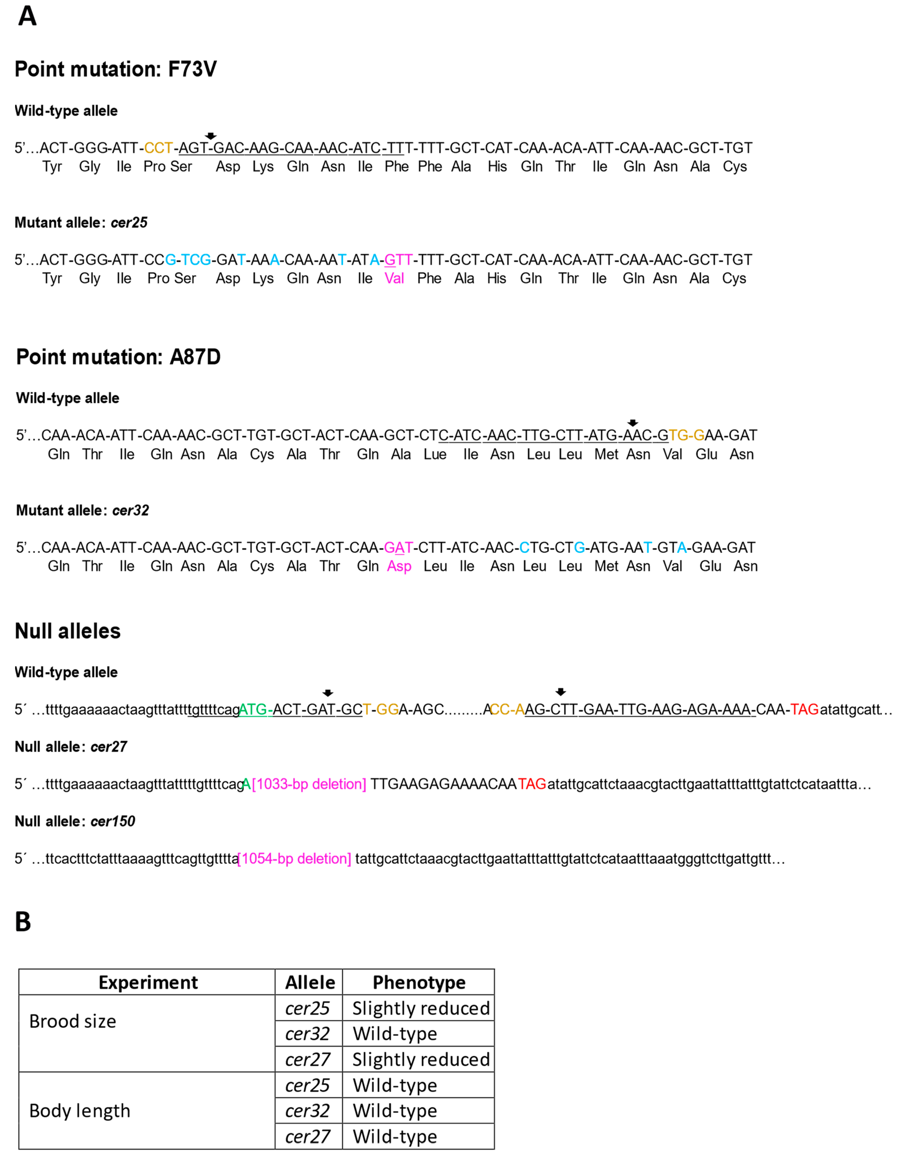
3.3. Mimicking Human BAP1 Mutations in C. elegans ubh-4

Thanks to the high level of conservation between BAP1 and UBH-4 at the N-terminal, we mimicked BAP1-like cancer-related missense mutations in C. elegans to explore their functional impact on this pluricellular organism. We created mutations affecting two residues close to C91, a residue functionally relevant at the active site [1], which are also conserved in C. elegans UBH-4 (Figure 1A), and correspond to a functionally relevant residue for BAP1 activity [2,3]. These BAP1 cancer-related missense mutations were p.F81V and p.A95D, which correspond to C. elegans p.F73V and p.A87D, respectively. The CRISPR-Cas genome editing in C. elegans (Figure 2A) resulted in animals with genotypes ubh-4(cer25[F73V]) and ubh-4(cer32[A87D]) that do not display any overt phenotype.
Then, since other BAP1 somatic mutations include frame-shift mutations that may result in the inactivation of BAP1 functions [4], we investigated the potential consequences of UBH-4 loss-of-function by producing null alleles. The ubh-4 gene produces two transcripts, C08B11.7.1 and C08B11.7.2, both with a 966 nucleotides coding sequence differing only at their 3′ UTR. We generated null alleles for ubh-4 by non-specific NHEJ (non-homologous end joining) repair of a CRISPR-Cas9-induced DSB (allele cer27), and by HDR (homology-directed repair) using a repair template (allele cer150), resulting in the depletion of 1033 (cer27) and 1054 (cer150) base pairs, respectively (Figure 2A). As a result of this editing, both alleles lost their start codons and did not produce any transcripts (confirmed by RT-PCR). Therefore, these two ubh-4 deletion alleles were considered null or without function. Surprisingly, despite ubh-4 being expressed ubiquitously and throughout the C. elegans lifecycle, no overt phenotypes were observed, just a slight reduction of the progeny in animals harboring the deletion allele (cer27) and the missense mutation F73V(cer25) (Figure 2B). Accordingly, in the absence of penetrant phenotypes, we observed that mutations in ubh-4 do not alter the 20S proteasome tissue levels (Figure S1), despite encoding a proteasome-associated DUB [30].

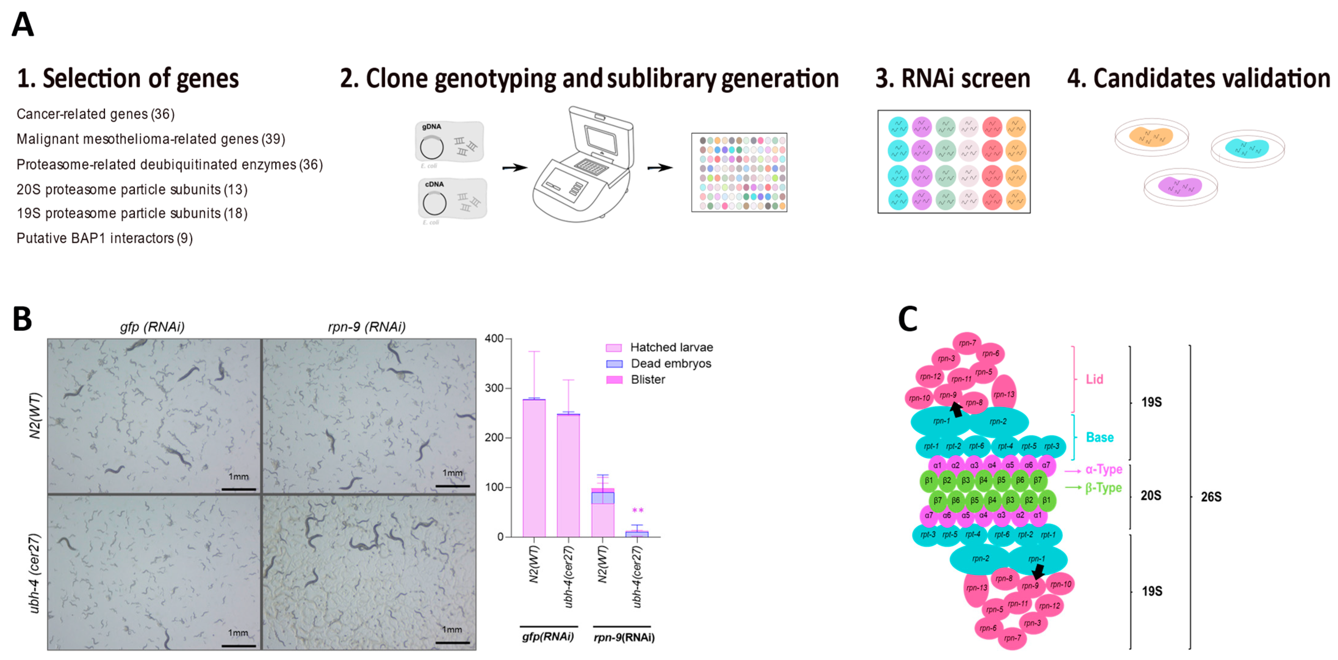
3.4. RNAi-Based Screen Reveals rpn-9 as Genetic Interactor of ubh-4

Since ubh-4 null mutants were viable and did not display overt phenotypes, we wondered whether the lack of ubh-4 function would influence the activity of other genes or genetic pathways. Thus, we performed an RNAi screen in ubh-4(cer27) mutants to identify synthetic genetic interactions with ubh-4. Using the two existing C. elegans RNAi libraries as sources of clones [24], we selected 150 RNAi clones whose identities were verified by PCR (by checking the size of the insert in each clone) (Supplementary Table S1). This RNAi sublibrary included clones that inactivate proteasome subunits, proteasome-related enzymes, orthologs of known and putative BAP1 interactors, genes frequently mutated in cancer, and malignant BAP1-TPDS-related genes [31,32,33,34]. We also added potential UBH-4-protein interactors according to information in the STRING 11.0 database [35].
The 150-gene RNAi library was screened in 24-well plates at 20 °C. Clones producing synthetic interactions were further validated in larger RNAi plates (Figure 3A). As a result, we concluded that RNAi inactivation of rpn-9, a non-ATPase subunit of the 19S proteasome (a regulatory particle of the 26S proteasome) [36], caused a robust and significant reduction of the brood size in ubh-4(cer27) animals (Figure 3B). Therefore, rpn-9 was identified as an ubh-4 genetic interactor. Since PSMD13, the RPN-9 human ortholog, is required for proteasome-mediated protein degradation [37,38] (Figure 3C), the proteasome subunit RPN-9 and UBH-4 may cooperate to regulate the degradation of ubiquitinated proteins.

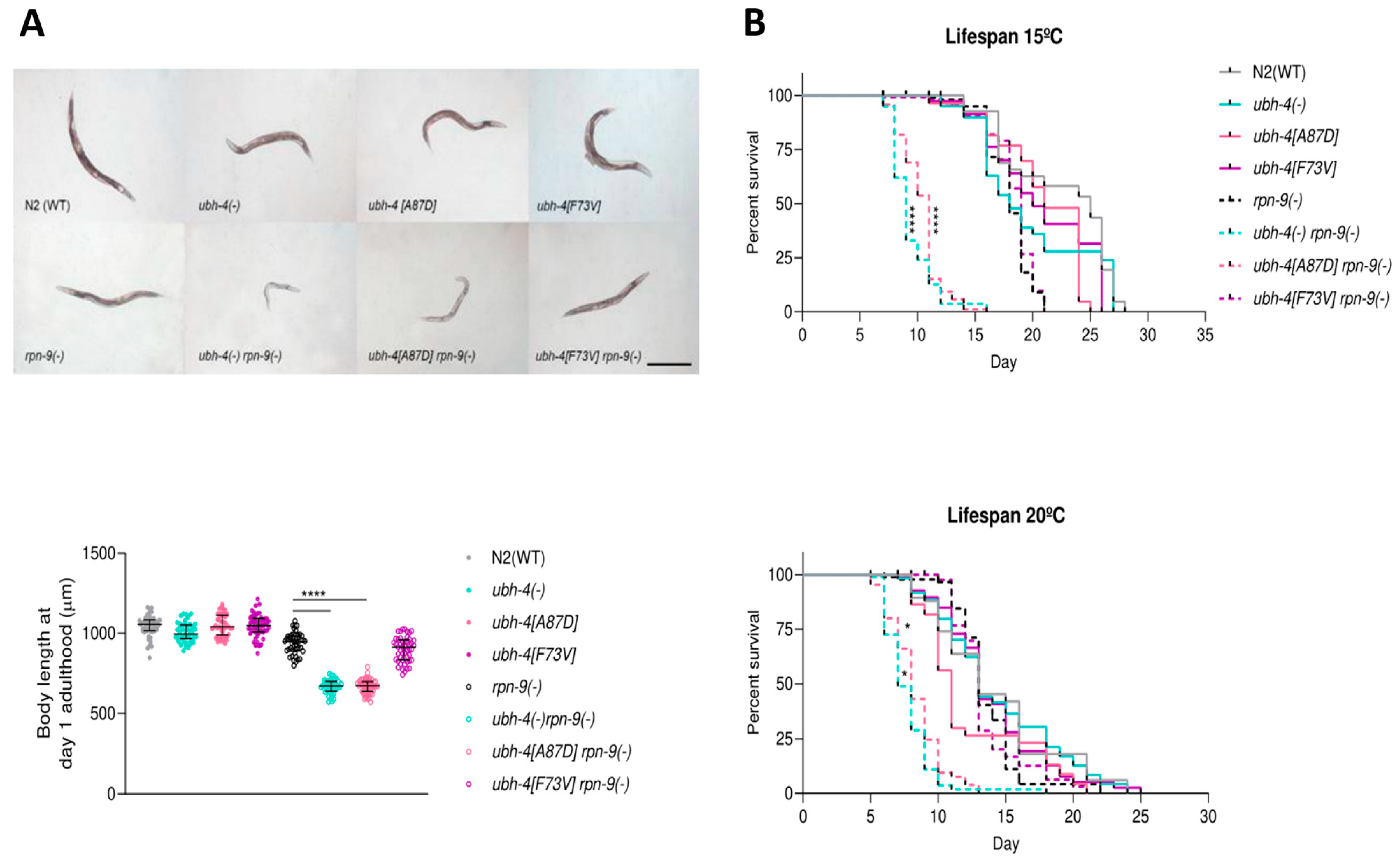
3.5. BAP1 Cancer-Related Mutation A87D Mimics ubh-4 Deletion Phenotypes in rpn-9(gk140) Background

To validate the hit resulting from the RNAi screen, we studied the synthetic phenotype between ubh-4 and rpn-9 by using genetic mutants. Since ubh-4 and rpn-9 are located on chromosome II, we performed CRISPR-Cas9 gene editing in rpn-9(gk401) mutants to have both mutations in the same strain. rpn-9(gk401) is a deletion allele causing sterility in homozygosis, thus only heterozygous animals (balanced with the balancer chromosome mIn1 containing myo-2::GFP as a marker) were microinjected to generate double-mutant strains with the ubh-4 deletion allele (cer140) and the ubh-4 missense mutations cer195[F73V] and cer198[A87D]. We observed that although rpn-9(gk401) mutants present a reduced body length compared with wild-type animals, this rpn-9 mutation produces even smaller animals in the ubh-4(cer140) and ubh-4(cer198[A87D]) backgrounds (Figure 4A).
Since adult animals harboring the rpn-9(gk401) in combination with ubh-4(cer140) and ubh-4(cer198[A87D]) seemed sicker, we monitored their lifespans at 15 °C and 20 °C. Interestingly, there was a dramatic decrease in these double-mutant strains compared to wild-type and the double-mutant rpn-9(gk401); ubh-4(cer195[F73V]) (Figure 4B). This decrease was more abrupt at 15 °C than 20 °C. These two described phenotypes suggest that the missense mutation ubh-4(cer198[A87D]) is a loss-of-function mutation that phenocopies the ubh-4 deletion allele.

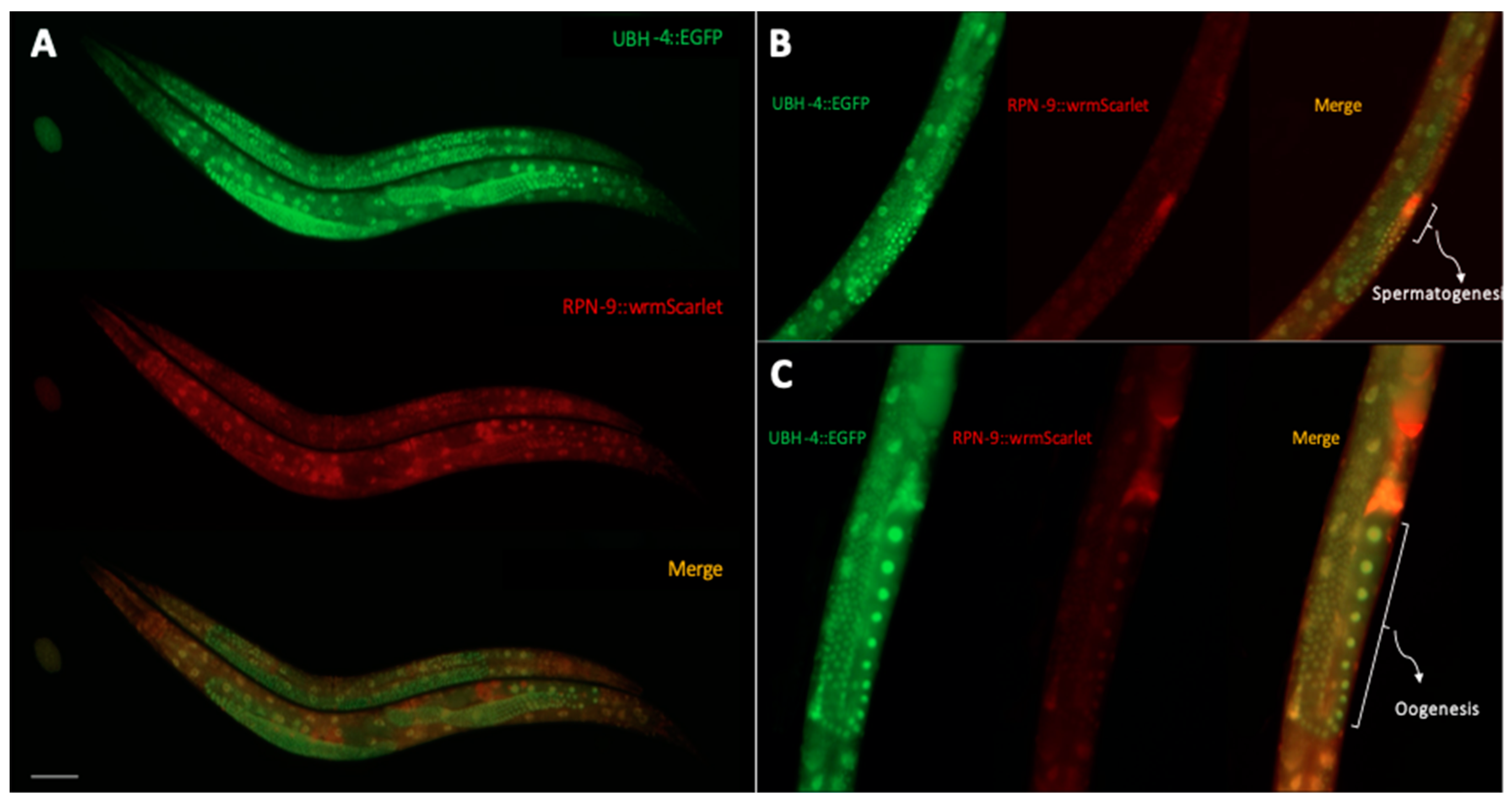
3.6. UBH-4 and RPN-9 Present Ubiquitous and Overlapping Expression Patterns

If ubh-4 and rpn-9 mutations functionally interact, these two genes may be co-expressed in some cells. Taking advantage of C. elegans transparency, we aimed to explore both the expression pattern and the endogenous levels of UBH-4 and RPN-9 in a living animal. Thus, we generated a double fluorescent endogenous reporter by Nested CRISPR [29] in the ubh-4(cer68[ubh-4::EGFP]) background. We introduced the wrmScarlet sequence to tag the rpn-9 locus obtaining the double endogenous fluorescent reporter strain CER620: ubh-4(cer68[ubh-4::EGFP]); rpn-9(cer203[rpn-9::wrmScarlet])II. Using fluorescence microscopy, we observed that UBH-4 is ubiquitously expressed from embryo to adult stages (Figure 5A). A similar expression pattern was detected for RPN-9 (Figure 5A). Interestingly, we noticed that both proteins were highly expressed in the germline, predominantly in meiotic regions (Figure 5B,C), suggesting a cooperating role for UBH-4 and RPN-9 in germline development.

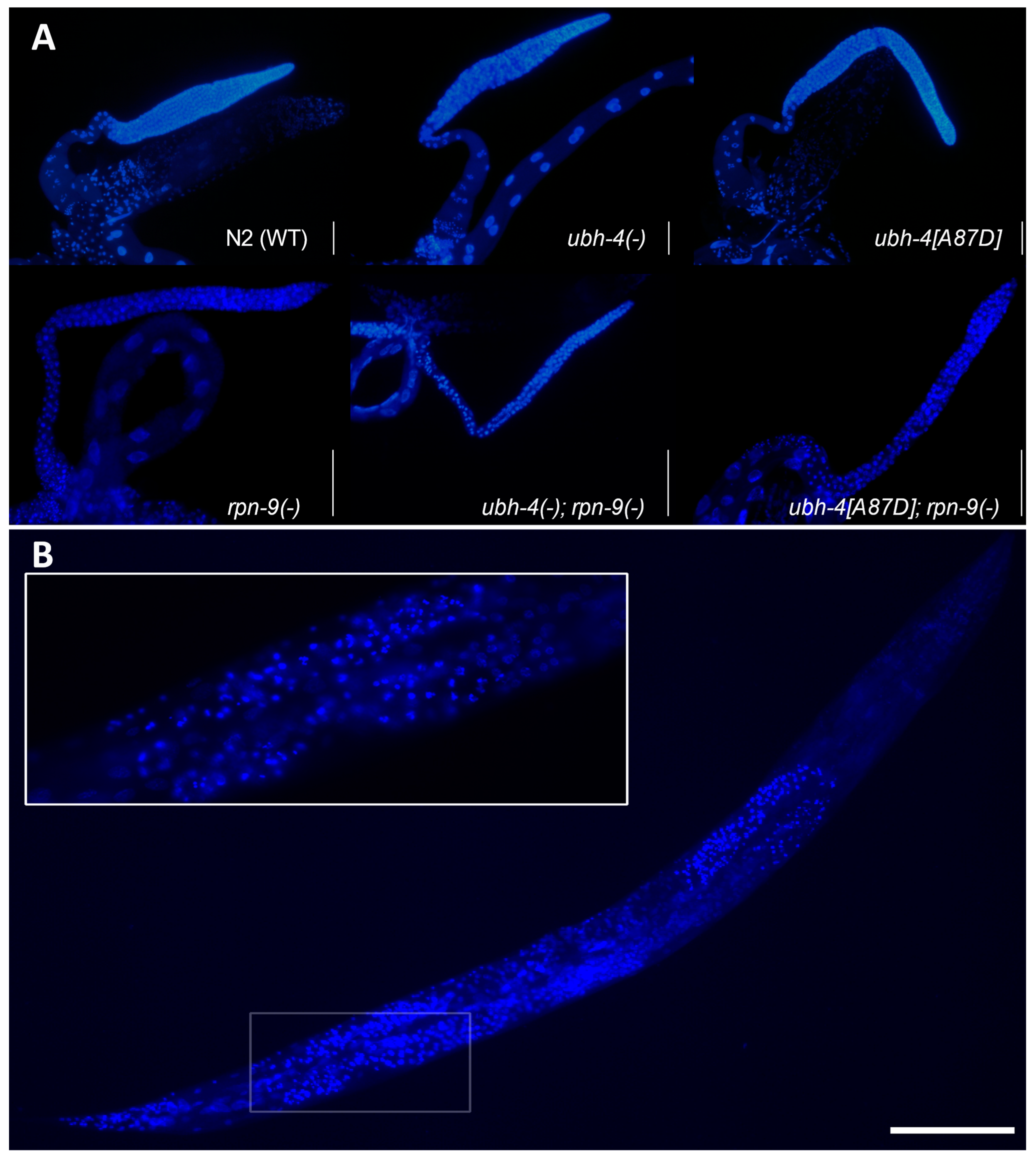
3.7. ubh-4 Deletion and A87D Missense Mutation Enhance the Germline Phenotype of the rpn-9 Deletion Mutant

Since ubh-4 and rpn-9 colocalize in the germline and we detected a synthetic reduced brood size between rpn-9(gk401) and ubh-4(cer140) or ubh-4(cer198[A87D]), we studied the dissected germline of these animals after DAPI staining. We observed thinner and smaller germlines in these double mutants than in any single mutant, suggesting a synthetic effect of rpn-9 and ubh-4 mutations on germline development (Figure 6A). We detected condensed nuclei in the germline of these double mutants, particularly abundant at the meiotic zone, but we did not quantify these aberrant nuclei since germlines were difficult to compare among themselves (Figure 6B).

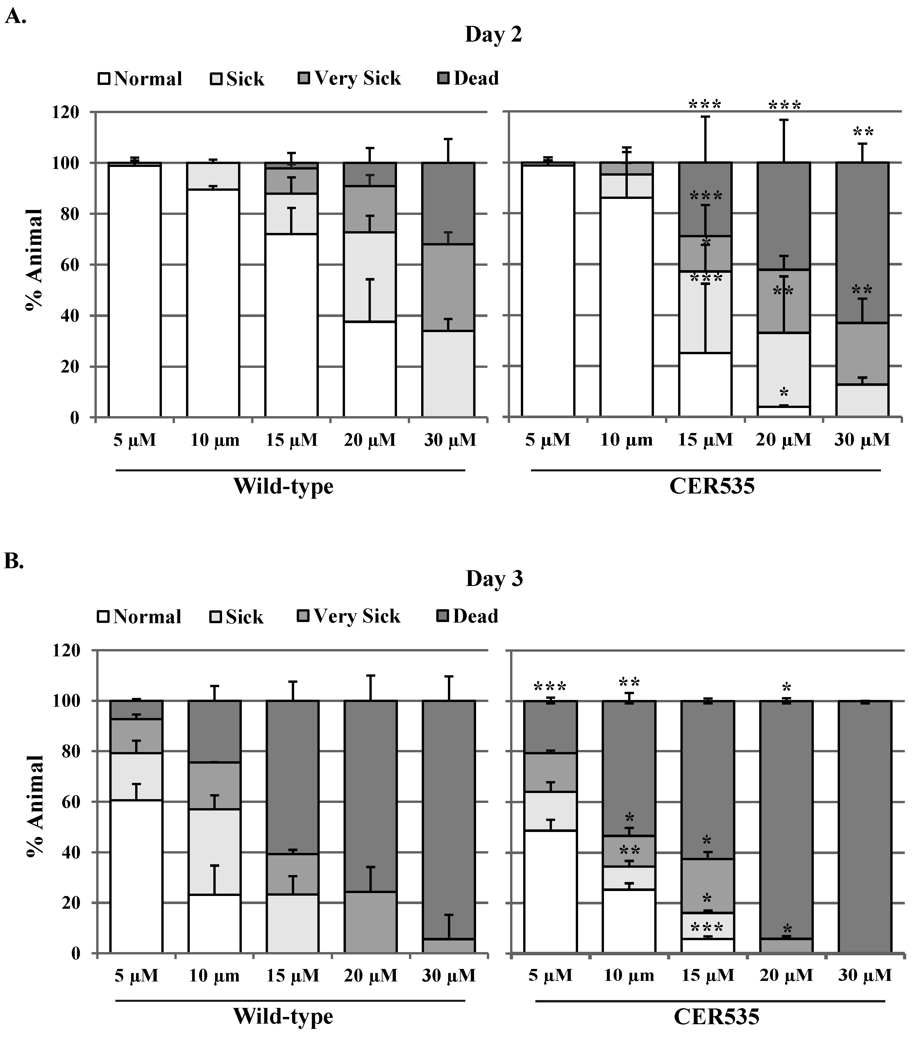
3.8. Animals Defective in ubh-4 Are Sensitive to the Proteasome Inhibitor Bortezomib

Since animals without a functional UBH-4 do not display strong phenotypes but sensitivity to the inactivation of rpn-9/PSMD13, which is a core proteasome subunit, we asked whether ubh-4 defective mutants were sensitive to the proteasome inhibitor Bortezomib. Thus, we transferred L4 animals harboring the ubh-4 deletion allele cer150 to plates with distinct concentrations of Bortezomib (5 µM, 10 µM, 15 µM, 20 µM, 25 µM, 30 µM) and the effect of this drug was scored at the first days of adulthood. Interestingly, we found that ubh-4 deletion mutants were sensitive to Bortezomib at days two and three of adulthood (Figure 7).

4. Discussion

We established an in vivo model in C. elegans to study mutations in the BAP1 ortholog ubh-4. Since ubh-4 is not essential for viability, ubh-4 null mutants do not display overt phenotypes and are convenient for RNAi screens in the search of genetic interactions, which, in the context of cancer, may help to investigate potential drug targets. An RNAi screen revealed a synthetic interaction between ubh-4 and rpn-9, a gene encoding a proteasome regulatory subunit. RPN-9 is the ortholog of human PSMD13, a component of the regulatory unit (19S) of the 26S proteasome [37,38]. The moderate inactivation produced by RNAi facilitated the identification of this synthetic interaction since rpn-9 loss-of-function causes a strong phenotype by itself. The yeast ortholog of rpn-9 is a non-essential subunit for cell viability [40]. However, in worms, rpn-9 is crucial for proteasome assembly, protein homeostasis, and proper development [41,42,43]. Moreover, rpn-9 and other proteasome-forming subunits have been identified as essential for both the lifespan and immunity of germline-deficient animals [44].
Supporting this genetic interaction, endogenous fluorescent reporters for UBH-4 and RPN-9 present an overlapping expression pattern in soma but also in germ cells, where the expression of both genes seems higher. UBH-4 physically interacts with the proteasome and binds the RPN-13 subunit, as well as affects proteasome activity [30]. RPN-9, along with other 19S regulatory subunits, is required a proper proteolytic activity of the proteasome [43]. Since ubiquinated proteins are degraded by the proteasome, the hypothetical mechanisms behind the functional interaction between UBH-4/BAP1 and RPN-9/PSMD13 is unclear. We hypothesize that the inactivation of UBH-4 deubiquitanase activity cooperates with RPN-9 somehow to alter levels of certain proteins that escape the regulated control of degradation in dynamic developmental processes.
The role of the proteasome during germline development, particularly in meiosis, is well-studied [45,46,47,48,49,50]. Thus, the mitotic and meiotic progression of the germline would be a convenient developmental context to further investigate the genetic interaction between ubh-4 and rpn-9. Moreover, as C. elegans has orthologs for BRCA1 and BRAD1 (brc-1 andbrd-1, respectively) [51,52], our model would help to study the mechanisms of controversial functional links between BAP1 and the BRCA1-BRAD1 complex, which play a critical role in DNA repair and homologous recombination, both in mammals [13] and C. elegans [53,54]. Since inactivation of the proteasome by RNAi or use of inhibitors produces aberrant nuclei in C. elegans germline [43,50] and BAP1 has a regulatory function on ubiquitination of histones [55], genome instability would be the “hallmark of cancer” behind the BAP1 alterations. Interestingly, although the specific mechanism of carcinogenesis induced by asbestos in MPM is still unclear, it has been proposed that asbestos causes genome instability [56] and chromosomal segregation defects [57]. Given the limited effective therapeutic intervention in MPM [58], and that about 50% of cases of MPM present alteration in BAP1 [59], the development of animal models such as the one presented here would open new venues to detect vulnerabilities of cells with mutations in BAP1.
We mimicked two missense mutations affecting the catalytic region of BAP1, F81V, and A95D. These variants have been associated with diverse types of cancer and both produce structural destabilization of BAP1 [4,60,61]. However, only A95D provoked a phenotype when mimicked in C. elegans ubh-4. Previous in vitro studies have suggested that F81V disturbs the catalytic activity of BAP1 [1,4]. However, ubh-4[F73V]; rpn-9(gk104) animals did not show abnormal lifespan, body length, or germline development. Therefore, according to our results, F73V missense mutation does not compromise UBH-4 functions. Thus, although C. elegans would be a good tool to study the functional impact of mutations and variants of uncertain significance (VUS), there are residues that, despite being conserved, may not present the same functional relevance in worms and humans.
Patients with MPM carry three molecular aberrations in distinct genes on average, more frequently affecting BAP1, NF2, and CDKN2A/B genes [31,60,62]. At lower frequencies, missense mutations in PSMD13 have also been reported in MPM tumors [60,61] and in whole-exome sequencing studies, including 2680 melanoma, mesothelioma, and clear-cell renal carcinoma samples (Figure S2) [62]. Interestingly, BAP1 and PSMD13 genetic alterations do not co-occur in any of the samples reported in these studies (Figure S2), suggesting that the presence of mutations in both genes compromise cell viability, not only in MPM, but also in BAP1-TPDS-related cancer types. Therefore, we hypothesize that the precise targeting of PSMD13 could be a potential therapeutic strategy in BAP1 mutant tumors.
Here we show that animals carrying ubh-4 genetic aberrations (complete deletion and A87D missense mutation) are more sensitive to proteasome stresses caused by genetic (rpn-9 partial or complete silencing) or drug (Bortezomib) targeting. Therefore, gene-based therapeutics focused on disturbing the 19S regulatory complex or PDMS13 specifically may reduce the proteasome functionality in BAP1-affected tumors. With this strategy, it may be possible to re-sensitize MPM tumors to proteasome inhibitors-induced apoptosis (such as Bortezomib), considering that according to a previous study, Bortezomib-sensitive MPM lines display lower proteasome activity [63]. However, since the ubiquitin proteasome system is involved in several key cellular pathways, therapeutic interventions centered on the proteasome would be cautious to avoid adverse secondary effects. Despite Bortezomib failing to demonstrate convincing antitumor activity in clinical trials, either in monotherapy in pretreated patients with MPM or in combination with cisplatin and pemetrexed in the frontline, these clinical trials did not stratify patients based on their genomic profile [64,65,66].
Our study shows that C. elegans is a valuable model to explore the functional role of BAP1 genetic aberrations recurrent in MPM and to investigate drug targets for developing new treatments for BAP1-related MPM.

Supplementary Materials

The following supporting information can be downloaded at: https://www.mdpi.com/article/10.3390/cells12060929/s1, Figure S1: 20S proteasome levels in ubh-4 mutants; Figure S2: Genetic alterations in BAP1 and PSMD13 do not cooccur in BAP1-TPDS related cancer. Table S1: 150 genes for the proteasome RNAi sublibrary. File S2: Strains and primers. File S3: CRISPR reagents and injection mixes.

Author Contributions

Conceptualization, J.C. and E.N.; methodology, C.M.-F., S.J. and E.A.; software, C.M.-F.; validation, C.M.-F. and S.J.; formal analysis, J.C., C.M.-F., E.N., S.J. and C.I.H.; investigation, C.M.-F. and S.J.; resources, J.C., E.N. and C.I.H.; data curation, J.C. and C.M.-F.; writing—original draft preparation, J.C. and C.M.-F.; writing—review and editing, J.C., C.M.-F., E.N. and C.I.H.; visualization, C.M.-F.; supervision, J.C. and C.I.H.; project administration, J.C.; funding acquisition, J.C., E.N. and C.I.H. All authors have read and agreed to the published version of the manuscript.

Funding

This study was supported by grants from Instituto de Salud Carlos III (grants PI14/01109 and PI18/00920) (co-funded by European Regional Development Fund. ERDF, a way to build Europe) and the Spanish Society of Medical Oncology grant for emerging research groups. J.C. is supported by the Generalitat de Catalunya through the SGR research group 2021 SGR 00184. C.M.-F. was supported by the European Cooperation in Science and Technology GENiE with a Short Term Mission (STSM), C.I.H. was supported by grants from by grants the Academy of Finland (297776), and Medicinska Understödsföreningen Liv och Hälsa r.f. S.J. was supported by the Doctoral Programme in Biomedicine of University of Helsinki and by grants from the Magnus Ehrnrooth Foundation.

Informed Consent Statement

Not applicable.

Data Availability Statement

Raw data is available upon request.

Acknowledgments

We thank CERCA Programme/Generalitat de Catalunya for institutional support. We thank the Biomedicum Imaging Unit (BIU) core facility of University of Helsinki for microscopy support, and Professor Caj Haglund for immunohistochemistry facilities.

Conflicts of Interest

The authors declare not conflict of interest.

References

  1. Ventii, K.H.; Devi, N.S.; Friedrich, K.L.; Chernova, T.A.; Meir, E.G.V.; Wilkinson, K.D. BAP1 Is a Tumor Suppressor That Requires Deubiquitinating Activity and Nuclear Localization. Cancer Res. 2008, 68, 6953–6962. [Google Scholar] [CrossRef] [PubMed] [Green Version]
  2. Walpole, S.; Pritchard, A.L.; Cebulla, C.M.; Pilarski, R.; Stautberg, M.; Davidorf, F.H.; De La Fouchardière, A.; Cabaret, O.; Golmard, L.; Stoppa-Lyonnet, D.; et al. Comprehensive Study of the Clinical Phenotype of Germline BAP1 Variant-Carrying Families Worldwide. J. Natl. Cancer Inst. 2018, 110, 1328–1341. [Google Scholar] [CrossRef]
  3. Carbone, M.; Yang, H.; Pass, H.I.; Krausz, T.; Testa, J.R.; Gaudino, G. BAP1 and Cancer. Nat. Rev. Cancer 2013, 13, 153–159. [Google Scholar] [CrossRef]
  4. Bott, M.; Brevet, M.; Taylor, B.S.; Shimizu, S.; Ito, T.; Wang, L.; Creaney, J.; Lake, R.A.; Zakowski, M.F.; Reva, B. The Nuclear Deubiquitinase BAP1 Is Commonly Inactivated by Somatic Mutations and 3p21.1 Losses in Malignant Pleural Mesothelioma. Nat. Genet. 2011, 43, 668–672. [Google Scholar] [CrossRef]
  5. Harbour, J.W.; Onken, M.D.; Roberson, E.D.O.; Duan, S.; Cao, L.; Worley, L.A.; Council, M.L.; Matatall, K.A.; Helms, C.; Bowcock, A.M. Frequent Mutation of BAP1 in Metastasizing Uveal Melanomas. Science 2010, 330, 1410–1413. [Google Scholar] [CrossRef] [Green Version]
  6. Murali, R.; Wiesner, T.; Scolyer, R.A. Tumours Associated with BAP1 Mutations. Pathology 2013, 45, 116–126. [Google Scholar] [CrossRef]
  7. Wald, O.; Sugarbaker, D.J. New Concepts in the Treatment of Malignant Pleural Mesothelioma. Annu. Rev. Med. 2018, 69, 365–377. [Google Scholar] [CrossRef] [PubMed]
  8. Baas, P.; Fennell, D.; Kerr, K.M.; Van Schil, P.E.; Haas, R.L.; Peters, S.; Committee, G. Malignant Pleural Mesothelioma: ESMO Clinical Practice Guidelines for Diagnosis, Treatment and Follow-up. Ann. Oncol. 2015, 26, v31–v39. [Google Scholar] [CrossRef]
  9. Zalcman, G.; Mazieres, J.; Margery, J.; Greillier, L.; Audigier-Valette, C.; Moro-Sibilot, D.; Molinier, O.; Corre, R.; Monnet, I.; Gounant, V.; et al. Bevacizumab for Newly Diagnosed Pleural Mesothelioma in the Mesothelioma Avastin Cisplatin Pemetrexed Study (MAPS): A Randomised, Controlled, Open-Label, Phase 3 Trial. Lancet 2016, 387, 1405–1414. [Google Scholar] [CrossRef] [PubMed]
  10. Scherpereel, A.; Antonia, S.; Bautista, Y.; Grossi, F.; Kowalski, D.; Zalcman, G.; Nowak, A.K.; Fujimoto, N.; Peters, S.; Tsao, A.S.; et al. First-Line Nivolumab plus Ipilimumab versus Chemotherapy for the Treatment of Unresectable Malignant Pleural Mesothelioma: Patient-Reported Outcomes in CheckMate 743. Lung Cancer 2022, 167, 8–16. [Google Scholar] [CrossRef] [PubMed]
  11. Mashtalir, N.; Daou, S.; Barbour, H.; Sen, N.N.; Gagnon, J.; Hammond-Martel, I.; Dar, H.H.; Therrien, M.; Affar, E.B. Autodeubiquitination Protects the Tumor Suppressor BAP1 from Cytoplasmic Sequestration Mediated by the Atypical Ubiquitin Ligase UBE2O. Mol. Cell 2014, 54, 392–406. [Google Scholar] [CrossRef] [Green Version]
  12. Carbone, M.; Harbour, J.W.; Brugarolas, J.; Bononi, A.; Pagano, I.; Dey, A.; Krausz, T.; Pass, H.I.; Yang, H.; Gaudino, G. Biological Mechanisms and Clinical Significance of BAP1 Mutations in Human Cancer. Cancer Discov. 2020, 10, 1103–1120. [Google Scholar] [CrossRef]
  13. Nishikawa, H.; Wu, W.; Koike, A.; Kojima, R.; Gomi, H.; Fukuda, M.; Ohta, T. BRCA1-Associated Protein 1 Interferes with BRCA1/BARD1 RING Heterodimer Activity. Cancer Res. 2009, 69, 111–119. [Google Scholar] [CrossRef] [PubMed] [Green Version]
  14. Balasubramani, A.; Larjo, A.; Bassein, J.A.; Chang, X.; Hastie, R.B.; Togher, S.M.; Lähdesmäki, H.; Rao, A. Cancer-Associated ASXL1 Mutations May Act as Gain-of-Function Mutations of the ASXL1-BAP1 Complex. Nat. Commun. 2015, 6, 7307. [Google Scholar] [CrossRef] [PubMed] [Green Version]
  15. Baughman, J.M.; Rose, C.M.; Kolumam, G.; Webster, J.D.; Wilkerson, E.M.; Merrill, A.E.; Rhoads, T.W.; Noubade, R.; Katavolos, P.; Lesch, J. NeuCode Proteomics Reveals Bap1 Regulation of Metabolism. Cell Rep. 2016, 16, 583–595. [Google Scholar] [CrossRef] [Green Version]
  16. Dey, A.; Seshasayee, D.; Noubade, R.; French, D.M.; Liu, J.; Chaurushiya, M.S.; Kirkpatrick, D.S.; Pham, V.C.; Lill, J.R.; Bakalarski, C.E. Loss of the Tumor Suppressor BAP1 Causes Myeloid Transformation. Science 2012, 33, 1541–1546. [Google Scholar] [CrossRef] [PubMed] [Green Version]
  17. Jensen, D.E.; Proctor, M.; Marquis, S.T.; Gardner, H.P.; Ha, S.I.; Chodosh, L.A.; Ishov, A.M.; Tommerup, N.; Vissing, H.; Sekido, Y. BAP1: A Novel Ubiquitin Hydrolase Which Binds to the BRCA1 RING Finger and Enhances BRCA1-Mediated Cell Growth Suppression. Oncogene 1998, 16, 1097–1112. [Google Scholar] [CrossRef] [Green Version]
  18. Ruan, H.B.; Han, X.; Li, M.D.; Singh, J.P.; Qian, K.; Azarhoush, S.; Zhao, L.; Bennett, A.M.; Samuel, V.T.; Wu, J. O-GlcNAc Transferase/Host Cell Factor C1 Complex Regulates Gluconeogenesis by Modulating PGC-1α Stability. Cell Metab. 2012, 16, 226–237. [Google Scholar] [CrossRef] [Green Version]
  19. Zarrizi, R.; Menard, J.A.; Belting, M.; Massoumi, R. Deubiquitination of γ-Tubulin by BAP1 Prevents Chromosome Instability in Breast Cancer Cells. Cancer Res 2014, 74, 6499–6508. [Google Scholar] [CrossRef] [Green Version]
  20. Stiernagle, T. Maintenance of C. elegans. In WormBook; OUP: Oxford, UK, 2006; pp. 1–11. [Google Scholar]
  21. Porta-de-la-Riva, M.; Fontrodona, L.; Villanueva, A.; Cerón, J. Basic Caenorhabditis Elegans Methods: Synchronization and Observation. J. Vis. Exp. 2012, e4019. [Google Scholar] [CrossRef] [Green Version]
  22. Kim, H.; Ishidate, T.; Ghanta, K.S.; Seth, M.; Conte, D.; Shirayama, M.; Mello, C.C. A Co-CRISPR Strategy for Efficient Genome Editing in Caenorhabditis Elegans. Genetics 2014, 197, 1069–1080. [Google Scholar] [CrossRef] [PubMed] [Green Version]
  23. Arribere, J.A.; Bell, R.T.; Fu, B.X.H.; Artiles, K.L.; Hartman, P.S.; Fire, A.Z. Efficient Marker-Free Recovery of Custom Genetic Modifications with CRISPR/Cas9 in Caenorhabditis Elegans. Genetics 2014, 198, 837–846. [Google Scholar] [CrossRef] [PubMed] [Green Version]
  24. Rual, J.F.; Ceron, J.; Koreth, J.; Hao, T.; Nicot, A.S.; Hirozane-Kishikawa, T.; Vandenhaute, J.; Orkin, S.H.; Hill, D.E.; Heuvel, S. Toward Improving Caenorhabditis Elegans Phenome Mapping with an ORFeome-Based RNAi Library. Genome Res. 2004, 14, 2162–2168. [Google Scholar] [CrossRef] [Green Version]
  25. Kamath, R.S.; Fraser, A.G.; Dong, Y.; Poulin, G.; Durbin, R.; Gotta, M.; Kanapin, A.; Le Bot, N.; Moreno, S.; Sohrmann, M. Systematic Functional Analysis of the Caenorhabditis Elegans Genome Using RNAi. Nature 2003, 421, 231–237. [Google Scholar] [CrossRef] [PubMed]
  26. Mikkonen, E.; Haglund, C.; Holmberg, C.I. Immunohistochemical Analysis Reveals Variations in Proteasome Tissue Expression in C. Elegans. PLoS ONE 2017, 12, e0183403. [Google Scholar] [CrossRef] [PubMed] [Green Version]
  27. Camacho, C.; Coulouris, G.; Avagyan, V.; Ma, N.; Papadopoulos, J.; Bealer, K.; Madden, T.L. BLAST+: Architecture and Applications. BMC Bioinform. 2009, 10, 421. [Google Scholar] [CrossRef] [PubMed] [Green Version]
  28. Sanchez-Pulido, L.; Kong, L.; Ponting, C.P.; Valencia, A. A Common Ancestry for BAP1 and Uch37 Regulators. Bioinforma. Discov. NOTE 2012, 28, 1953–1956. [Google Scholar] [CrossRef] [Green Version]
  29. Vicencio, J.; Martínez-Fernández, C.; Serrat, X.; Cerón, J. Efficient Generation of Endogenous Fluorescent Reporters by Nested CRISPR in Caenorhabditis Elegans. Genetics 2019, 211, 1143–1154. [Google Scholar] [CrossRef]
  30. Matilainen, O.; Arpalahti, L.; Rantanen, V.; Hautaniemi, S.; Holmberg, C.I. Insulin/IGF-1 Signaling Regulates Proteasome Activity through the Deubiquitinating Enzyme UBH-4. Cell Rep. 2013, 3, 1980–1995. [Google Scholar] [CrossRef] [Green Version]
  31. Kato, S.; Tomson, B.N.; Buys, T.P.H.; Elkin, S.K.; Carter, J.L.; Kurzrock, R. Genomic Landscape of Malignant Mesotheliomas. Mol. Cancer Ther. 2016, 15, 2498–2507. [Google Scholar] [CrossRef] [Green Version]
  32. De Rienzo, A.; Archer, M.A.; Yeap, B.Y.; Dao, N.; Sciaranghella, D.; Sideris, A.C.; Zheng, Y.; Holman, A.G.; Wang, Y.E.; Dal Cin, P.S.; et al. Gender-Specific Molecular and Clinical Features Underlie Malignant Pleural Mesothelioma. Cancer Res. 2016, 76, 319–328. [Google Scholar] [CrossRef] [PubMed] [Green Version]
  33. Poulin, G.; Nandakumar, R.; Ahringer, J. Genome-Wide RNAi Screens in Caenorhabditis Elegans: Impact on Cancer Research. Oncogene 2004, 23, 8340–8345. [Google Scholar] [CrossRef] [PubMed] [Green Version]
  34. Soura, E.; Eliades, P.J.; Shannon, K.; Stratigos, A.J.; Tsao, H. Hereditary Melanoma: Update on Syndromes and Management Genetics of Familial Atypical Multiple Mole Melanoma Syndrome. J. Am. Acad. Dermatol. 2016, 74, 411–420. [Google Scholar] [CrossRef] [PubMed] [Green Version]
  35. Szklarczyk, D.; Gable, A.L.; Lyon, D.; Junge, A.; Wyder, S.; Huerta-Cepas, J.; Simonovic, M.; Doncheva, N.T.; Morris, J.H.; Bork, P.; et al. STRING V11: Protein-Protein Association Networks with Increased Coverage, Supporting Functional Discovery in Genome-Wide Experimental Datasets. Nucleic Acids Res. 2019, 47, D607–D613. [Google Scholar] [CrossRef] [PubMed] [Green Version]
  36. Papaevgeniou, N.; Chondrogianni, N. The Ubiquitin Proteasome System in Caenorhabditis Elegans and Its Regulation. Redox Biol. 2014, 2, 333–347. [Google Scholar] [CrossRef] [PubMed] [Green Version]
  37. Bi, W.; Zhu, L.; Zeng, Z.; Jing, X.; Liang, Y.; Guo, L.; Shi, Q.; Xu, A.; Tao, E. Investigations into the Role of 26S Proteasome Non-ATPase Regulatory Subunit 13 in Neuroinflammation. Neuroimmunomodulation 2014, 21, 331–337. [Google Scholar] [CrossRef]
  38. Li, S.; Izumi, T.; Hu, J.; Jin, H.H.; Siddiqui, A.A.A.; Jacobson, S.G.; Bok, D.; Jin, M. Rescue of Enzymatic Function for Disease-Associated RPE65 Proteins Containing Various Missense Mutations in Non-Active Sites. J. Biol. Chem. 2014, 289, 18943–18956. [Google Scholar] [CrossRef] [Green Version]
  39. Kanehisa, M.; Goto, S. KEGG: Kyoto Encyclopedia of Genes and Genomes. Nucleic Acids Res. 2000, 72, 27–30. [Google Scholar] [CrossRef]
  40. Takeuchi, J.; Fujimuro, M.; Yokosawa, H.; Tanaka, K.; Toh-e, A. Rpn9 Is Required for Efficient Assembly of the Yeast 26S Proteasome. Mol. Cell. Biol. 1999, 19, 6575–6584. [Google Scholar] [CrossRef] [Green Version]
  41. Hu, Y.; Wu, Y.; Li, Q.; Zhang, W.; Jin, C. Solution Structure of Yeast Rpn9: Insights into Proteasome Lid Assembly. J. Biol. Chem. 2015, 290, 6878–6889. [Google Scholar] [CrossRef] [Green Version]
  42. Wang, J.; Robida-Stubbs, S.; Tullet, J.M.A.; Rual, J.-F.; Vidal, M.; Blackwell, T.K. RNAi Screening Implicates a SKN-1–Dependent Transcriptional Response in Stress Resistance and Longevity Deriving from Translation Inhibition. PLoS Genet. 2010, 6, 1001048. [Google Scholar] [CrossRef] [PubMed]
  43. Fernando, L.M.; Quesada-Candela, C.; Murray, M.; Ugoaru, C.; Yanowitz, J.L.; Allen, A.K. Proteasomal Subunit Depletions Differentially Affect Germline Integrity in C. Elegans. Front. Cell Dev. Biol. 2022, 10, 901320. [Google Scholar] [CrossRef] [PubMed]
  44. Sinha, A.; Rae, R. A Functional Genomic Screen for Evolutionarily Conserved Genes Required for Lifespan and Immunity in Germline-Deficient C. Elegans. PLoS ONE 2014, 9, e101970. [Google Scholar] [CrossRef] [Green Version]
  45. Burger, J.; Merlet, J.; Tavernier, N.; Richaudeau, B.; Arnold, A.; Ciosk, R.; Bowerman, B.; Pintard, L. CRL2LRR-1 E3-Ligase Regulates Proliferation and Progression through Meiosis in the Caenorhabditis Elegans Germline. PLoS Genet. 2013, 9, e1003375. [Google Scholar] [CrossRef] [PubMed] [Green Version]
  46. Shimada, M.; Kanematsu, K.; Tanaka, K.; Yokosawa, H.; Kawahara, H. Proteasomal Ubiquitin Receptor RPN-10 Controls Sex Determination in Caenorhabditis Elegans. Mol. Biol. Cell 2006, 17, 5356–5371. [Google Scholar] [CrossRef] [PubMed] [Green Version]
  47. Phillips, C.M.; McDonald, K.L.; Dernburg, A.F. Cytological Analysis of Meiosis in Caenorhabditis elegans. Methods Mol. Biol. 2009, 558, 171–195. [Google Scholar]
  48. Sanford, C.; Perry, M.D. Asymmetrically Distributed Oligonucleotide Repeats in the Caenorhabditis Elegans Genome Sequence That Map to Regions Important for Meiotic Chromosome Segregation. Nucleic Acids Res. 2001, 29, 2920–2926. [Google Scholar] [CrossRef] [Green Version]
  49. Kumar, G.A.; Subramaniam, K. PUF-8 Facilitates Homologous Chromosome Pairing by Promoting Proteasome Activity during Meiotic Entry in C. elegans. Development 2018, 145, dev.163949. [Google Scholar] [CrossRef] [Green Version]
  50. Ahuja, J.S.; Sandhu, R.; Mainpal, R.; Lawson, C.; Henley, H.; Hunt, P.A.; Yanowitz, J.L.; Valentin Börner, G. Control of Meiotic Pairing and Recombination by Chromosomally Tethered 26S Proteasome. Science 2017, 355, 408–411. [Google Scholar] [CrossRef]
  51. Adamo, A.; Montemauri, P.; Silva, N.; Ward, J.D.; Boulton, S.J.; Volpe, A.L. BRC-1 Acts in the Inter-Sister Pathway of Meiotic Double-Strand Break Repair. EMBO Rep. 2008, 9, 287–292. [Google Scholar] [CrossRef] [Green Version]
  52. Boulton, S.J.; Martin, J.S.; Polanowska, J.; Hill, D.E.; Gartner, A.; Vidal, M. BRCA1/BARD1 Orthologs Required for DNA Repair in Caenorhabditis Elegans. Curr. Biol 2004, 14, 33–39. [Google Scholar] [CrossRef]
  53. Li, Q.; Saito, T.T.; Martinez-Garcia, M.; Deshong, A.J.; Nadarajan, S.; Lawrence, K.S.; Checchi, P.M.; Colaiacovo, M.P.; Engebrecht, J.A. The Tumor Suppressor BRCA1-BARD1 Complex Localizes to the Synaptonemal Complex and Regulates Recombination under Meiotic Dysfunction in Caenorhabditis Elegans. PLoS Genet. 2018, 14, e1007701. [Google Scholar] [CrossRef] [Green Version]
  54. Janisiw, E.; Dello Stritto, M.R.; Jantsch, V.; Silva, N. BRCA1-BARD1 Associate with the Synaptonemal Complex and pro-Crossover Factors and Influence RAD-51 Dynamics during Caenorhabditis Elegans Meiosis. PLoS Genet. 2018, 14, e1007653. [Google Scholar] [CrossRef] [PubMed]
  55. Caporali, S.; Butera, A.; Amelio, I. BAP1 in Cancer: Epigenetic Stability and Genome Integrity. Discov. Oncol. 2022, 13, 117. [Google Scholar] [CrossRef] [PubMed]
  56. Kuroda, A. Recent Progress and Perspectives on the Mechanisms Underlying Asbestos Toxicity. Genes Environ. Off. J. Jpn. Environ. Mutagen. Soc. 2021, 43, 46. [Google Scholar] [CrossRef] [PubMed]
  57. Zhang, T.; Lv, L.; Huang, Y.; Ren, X.; Shi, Q. Chromosome Nondisjunction during Bipolar Mitoses of Binucleated Intermediates Promote Aneuploidy Formation along with Multipolar Mitoses Rather than Chromosome Loss in Micronuclei Induced by Asbestos. Oncotarget 2017, 8, 11030–11041. [Google Scholar] [CrossRef] [PubMed] [Green Version]
  58. Nicolini, F.; Bocchini, M.; Bronte, G.; Delmonte, A.; Guidoboni, M.; Crinò, L.; Mazza, M. Malignant Pleural Mesothelioma: State-of-the-Art on Current Therapies and Promises for the Future. Front. Oncol. 2019, 9, 1519. [Google Scholar] [CrossRef] [PubMed]
  59. Hmeljak, J.; Sanchez-Vega, F.; Hoadley, K.A.; Shih, J.; Stewart, C.; Heiman, D.; Tarpey, P.; Danilova, L.; Drill, E.; Gibb, E.A.; et al. Integrative Molecular Characterization of Malignant Pleural Mesothelioma. Cancer Discov. 2018, 8, 1549–1565. [Google Scholar] [CrossRef] [Green Version]
  60. Sugarbaker, D.J.; Richards, W.G.; Gordon, G.J.; Dong, L.; De Rienzo, A.; Maulik, G.; Glickman, J.N.; Chirieac, L.R.; Hartman, M.L.; Taillon, B.E.; et al. Transcriptome Sequencing of Malignant Pleural Mesothelioma Tumors. Proc. Natl. Acad. Sci. USA 2008, 105, 3521–3526. [Google Scholar] [CrossRef] [Green Version]
  61. Torricelli, F.; Lococo, F.; Di Stefano, T.S.; Lorenzini, E.; Piana, S.; Valli, R.; Rena, O.; Veronesi, G.; Billè, A.; Ciarrocchi, A. Deep Sequencing Analysis Identified a Specific Subset of Mutations Distinctive of Biphasic Malignant Pleural Mesothelioma. Cancers 2020, 12, 2454. [Google Scholar] [CrossRef]
  62. Cerami, E.; Gao, J.; Dogrusoz, U.; Gross, B.E.; Sumer, S.O.; Aksoy, B.A.; Jacobsen, A.; Byrne, C.J.; Heuer, M.L.; Larsson, E.; et al. The CBio Cancer Genomics Portal: An Open Platform for Exploring Multidimensional Cancer Genomics Data. Cancer Discov. 2012, 2, 401–404. [Google Scholar] [CrossRef] [Green Version]
  63. Cerruti, F.; Jocollè, G.; Salio, C.; Oliva, L.; Paglietti, L.; Alessandria, B.; Mioletti, S.; Donati, G.; Numico, G.; Cenci, S.; et al. Proteasome Stress Sensitizes Malignant Pleural Mesothelioma Cells to Bortezomib-Induced Apoptosis. Sci. Rep. 2017, 7, 17626. [Google Scholar] [CrossRef] [Green Version]
  64. Gordon, G.J.; Mani, M.; Maulik, G.; Mukhopadhyay, L.; Yeap, B.Y.; Kindler, H.L.; Salgia, R.; Sugarbaker, D.J.; Bueno, R. Preclinical Studies of the Proteasome Inhibitor Bortezomib in Malignant Pleural Mesothelioma. Cancer Chemother. Pharmacol. 2008, 61, 549–558. [Google Scholar] [CrossRef]
  65. Fennell, D.A.; McDowell, C.; Busacca, S.; Webb, G.; Moulton, B.; Cakana, A.; O’Byrne, K.J.; Meerbeeck, J.V.; Donnellan, P.; McCaffrey, J.; et al. Phase II Clinical Trial of First or Second-Line Treatment with Bortezomib in Patients with Malignant Pleural Mesothelioma. J. Thorac. Oncol. 2012, 7, 1466–1470. [Google Scholar] [CrossRef] [Green Version]
  66. O’Brien, M.E.R.; Gaafar, R.M.; Popat, S.; Grossi, F.; Price, A.; Talbot, D.C.; Cufer, T.; Ottensmeier, C.; Danson, S.; Pallis, A.; et al. Phase II Study of First-Line Bortezomib and Cisplatin in Malignant Pleural Mesothelioma and Prospective Validation of Progression Free Survival Rate as a Primary End-Point for Mesothelioma Clinical Trials (European Organisation for Research and Treatment of Cancer 08052). Eur. J. Cancer 2013, 49, 2815–2822. [Google Scholar] [CrossRef]
Figure 1. C. elegans BAP1 ortholog UBH-4 homology and expression pattern. (A) UBH-4 is the ortholog of human BAP1. Scheme of UBH-4, UCHL5 and BAP1 protein sequences (black lines) and conserved residues (purple bars), predominantly at the N-terminal and C-terminal domains. In the black square are the catalytic domains of these proteins. The bottom scheme shows with more detail conserved residues between UBH-4 and BAP1. Pink shadows indicate MPM-related residues modelled in this study. Table shows BLASTP analysis of C. elegans UCHs proteins, UCHL5 and BAP1. Alignments were illustrated by CLC Sequence Viewer 8.0. (B) UBH-4 is ubiquitously expressed during C. elegans development. Representative images of UBH-4::EGFP signal in developing embryo (1), L3 (2), and adult stages (3). Scale bars represent 10, 100, and 100 µm, respectively. (4) Posterior arm of the adult gonad at higher magnification (scale bar represents 100 µm) and its diagram. (5) (2) pointing out distal mitotic (a), transition (b), and meiotic zones (pachytene (c), diplotene (d) and diakinesis (e)), and spermatheca (f).
Cells 12 00929 g001
Figure 2. CRISPR-Cas generation and characterization of UBH-4 mutant alleles. (A) Molecular designs to generate the ubh-4 cancer-related alleles cer25 and cer32 by CRISPR-Cas9. Sense strands are represented. PAM sequences are shown in yellow; crRNA sequences are underlined and Cas9 cut sites are indicated by black arrows; silent mutations are labelled in light blue; mutated codons are shown in pink; nucleotide changes (T > G (cer25) and C > A (cer32)) are underlined. Aminoacidic sequences are represented below nucleotide sequences, highlighting the mutated residues in pink. (B) Phenotypes observed for indicated alleles after brood size, body length and proteasome amount and activity characterization.
Cells 12 00929 g002
Figure 3. RNAi for synthetic lethal interactions with ubh-4 mutants. (A) RNAi screen workflow is illustrated. After candidate genes are selected (1), clones from RNAi libraries were picked aleatorily and genotyped to generate a 150-gene RNAi sublibrary for screening (2). For the RNAi screen, 150-RNAi cultures were IPTG-induced in RNAi 24-well plates. Synchronized L1 animals were seeded and checked every day for 120 h at 20 °C, to score both wild-type and cer27 animals (3). Finally, candidate genes were validated in larger (55 mm-diameter) RNAi plates (4). (B) Representative images of reduced brood size in ubh-4(cer27) background after 120 h rpn-9(RNAi) treatment (20 °C). The chart quantifies hatched larvae, dead embryos, and blister animals under different RNAi conditions. Statistics were performed by one-way ANOVA (Kruskal–Wallis and Dunn’s tests). ** p < 0.01 comparing hatched larvae between wild-type and cer27 under rpn-9(RNAi). No significant differences were found between the other observed phenotypes. An average of 20 animals were scored. The experiment was performed once. (C) Schematic model of the C. elegans 26S proteasome. 26S complex is formed by two regulatory particles 19S and one core particle 20S [39]. Structure subunit forms are represented by colors and detailed subunit proteins by coding gene names. Rings conforming to the 20S core and 19S structure are represented linearly. Black arrows point out rpn-9 subunits. Illustrations were drawn using Inkscape 1.2.2. (Retrieved from https://inkscape.org).
Cells 12 00929 g003
Figure 4. ubh-4 deletion and [A87D] significantly reduces body length and survival in the rpn-9 mutant background. (A) Representative images showing smaller body length in ubh-4 and rpn-9 single and double mutants (i, ii, iii, or use top and bottom rows). Pictures were taken at day 1 of adulthood growth at 1 °C. Scale bar represents 0.5 mm. Below, the chart quantifies the body length in wild-type (WT), single, and double mutants. Bars represent median and interquartile range, and dots the measured length of individual animals. The same experiment was performed three times, measuring an average of 100 animals per replicate. **** mean p < 0.0001. Statistics were analyzed by one-way ANOVA (Kruskal–Wallis and Dunn’s tests). (B) Survival curves of WT, ubh-4, and rpn-9 simple and double mutants at 1 °C and 2 °C. Percent survival is indicated for each allele. An average of 80 animals were censored through two independent experiments for lifespan analysis at both temperatures. Statistical analysis by Log-rank (Mantel–Cox) test was performed to compare rpn-9(gk401) and double mutants. * and **** indicate p < 0.1 and p < 0.0001, respectively.
Cells 12 00929 g004
Figure 5. UBH-4 and RPN-9 expression patterns suggest these proteins cooperate, particularly in the germline. (A) Single and merged UBH-4::EGFP and RPN-9::wrmScarlet signals evidence ubiquitously co-expression of both proteins at different stages (embryo, young adult and adult stages). Scale bar represents 100 µm. Representative images showing endogenous levels of UBH-4 and RPN-9 during spermatogenesis (B) and oogenesis (C).
Cells 12 00929 g005
Figure 6. DAPI-stained nuclei in gonads at day one adulthood. (A) DAPI staining of dissected gonads from WT, simple and double ubh-4 and rpn-9 mutants. (-) indicates deletion. Scale bars represent 50 µm. (B) Whole-body DAP staining of a double mutant ubh-4(cer150); rpn-9(gk104) showing abundant fragmented nuclei in the germline. The highlighted region is shown at higher magnification (63× lens) at the upper part. Scale bar represents 100 µm.
Cells 12 00929 g006
Figure 7. Dose-dependent effect of proteasome inhibitor Bortezomib on ubh-4 deletion mutant. Graphs represent the health status of both wild-type and CER535 animals under different Bortezomib dosages after two (A) and three (B) days of exposure at 20 °C. Animals were transferred to Bortezomib-containing plates at the L4 stage and were observed on day 2 and day 3 of adulthood. Animals were scored on basis of mobility and visual appearance as normal, sick (defective movement and appearance), very sick (highly defective movement and appearance), and dead. Results are the mean of three independent experiments (~30 animals in each experiment). Statistical analysis by t-test were preformed between wild-type and ubh-4 mutant (CER535) within each corresponding phenotypic group (normal, sick, very sick, and dead). Error bars, STD, * p < 0.05, ** p < 0.01 and *** p < 0.001.
Cells 12 00929 g007
Disclaimer/Publisher’s Note: The statements, opinions and data contained in all publications are solely those of the individual author(s) and contributor(s) and not of MDPI and/or the editor(s). MDPI and/or the editor(s) disclaim responsibility for any injury to people or property resulting from any ideas, methods, instructions or products referred to in the content.

