
Amplification of Signal on Cell Surfaces in Molecular Cascades

Abstract
:1. Introduction
2. Results and Discussion
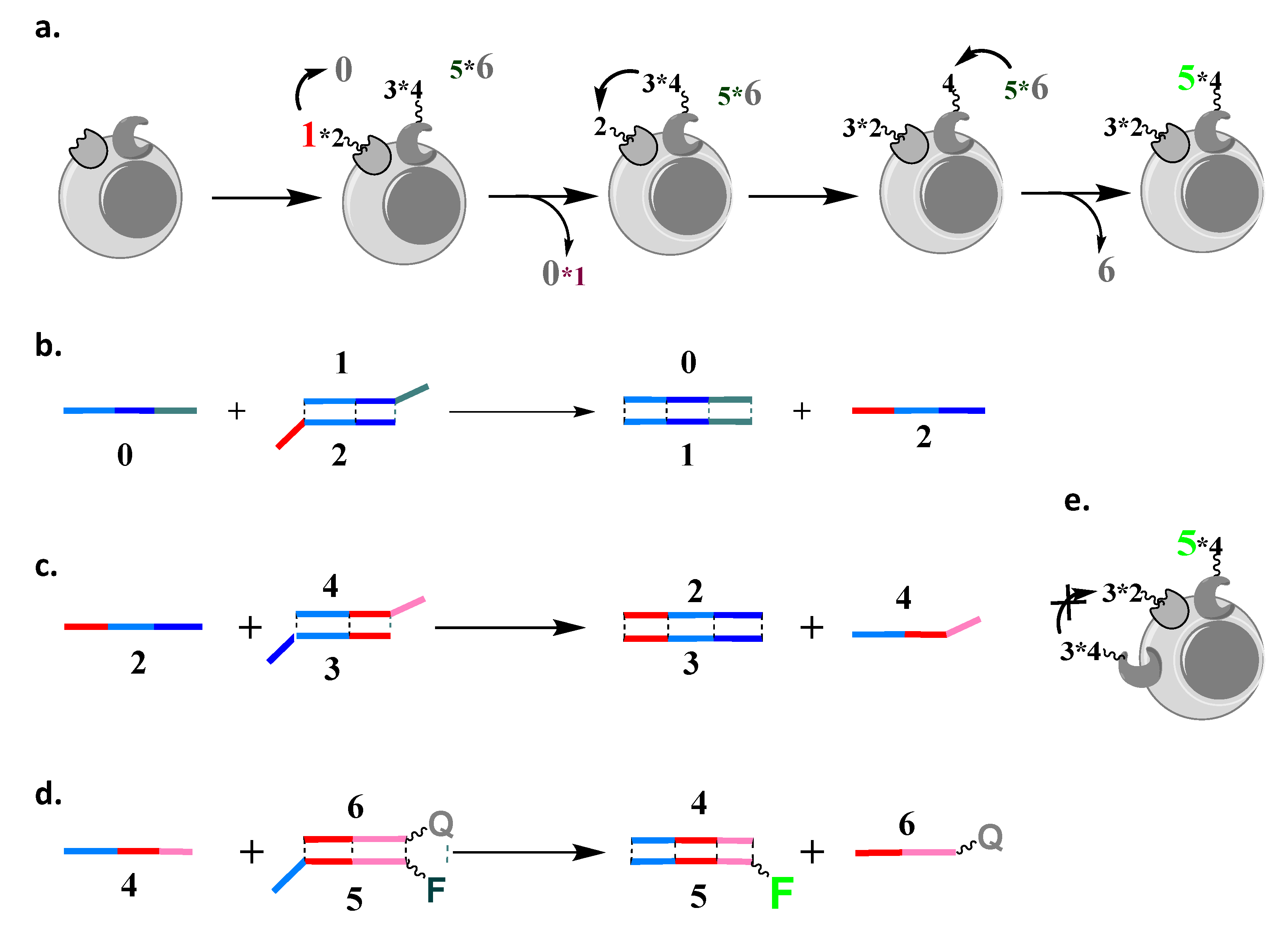
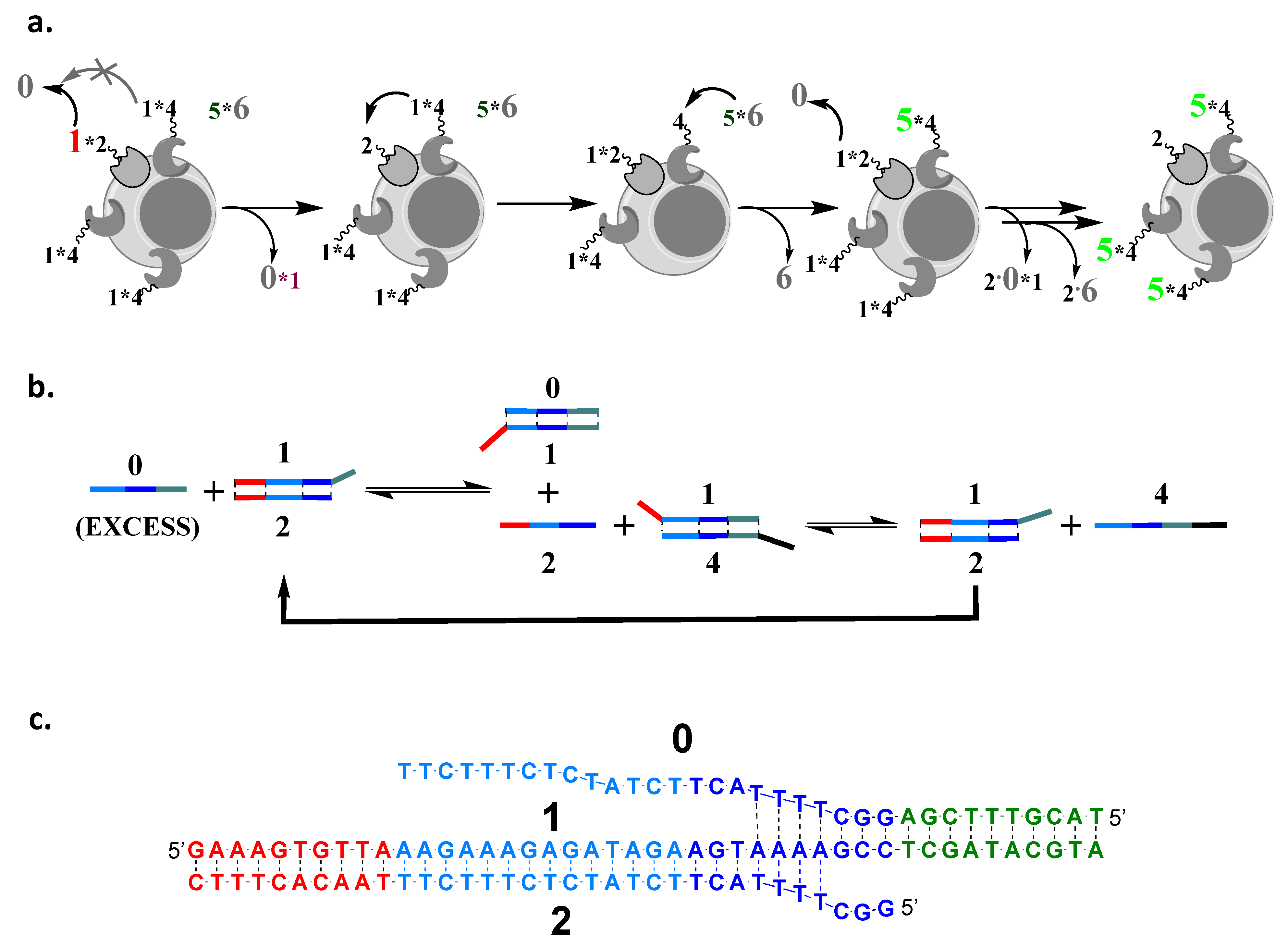
2.1. Design Considerations for Amplification Cascade Reaction
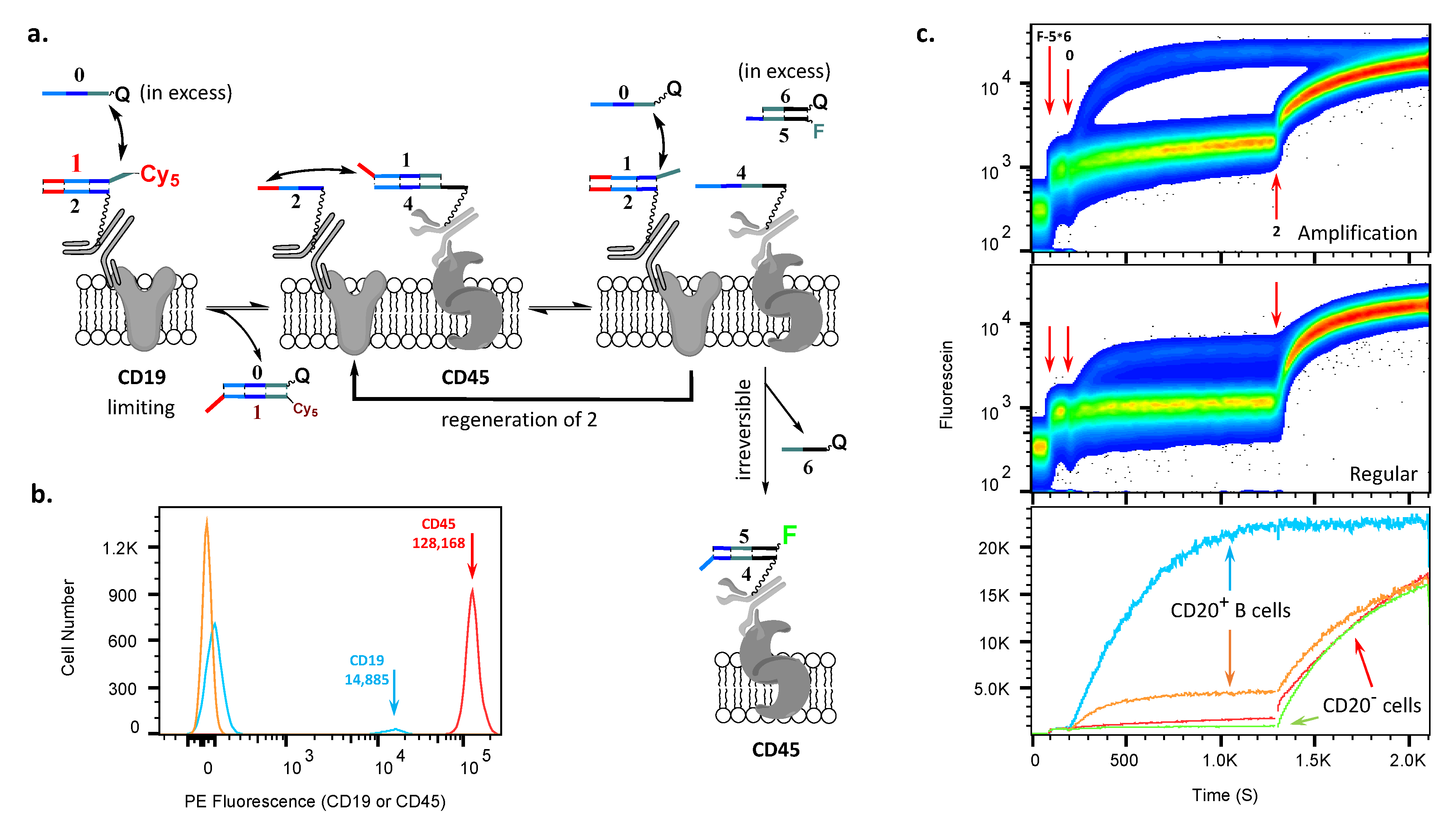
2.2. AMPLIFY(YESCD19YESCD45) Cascade Reaction
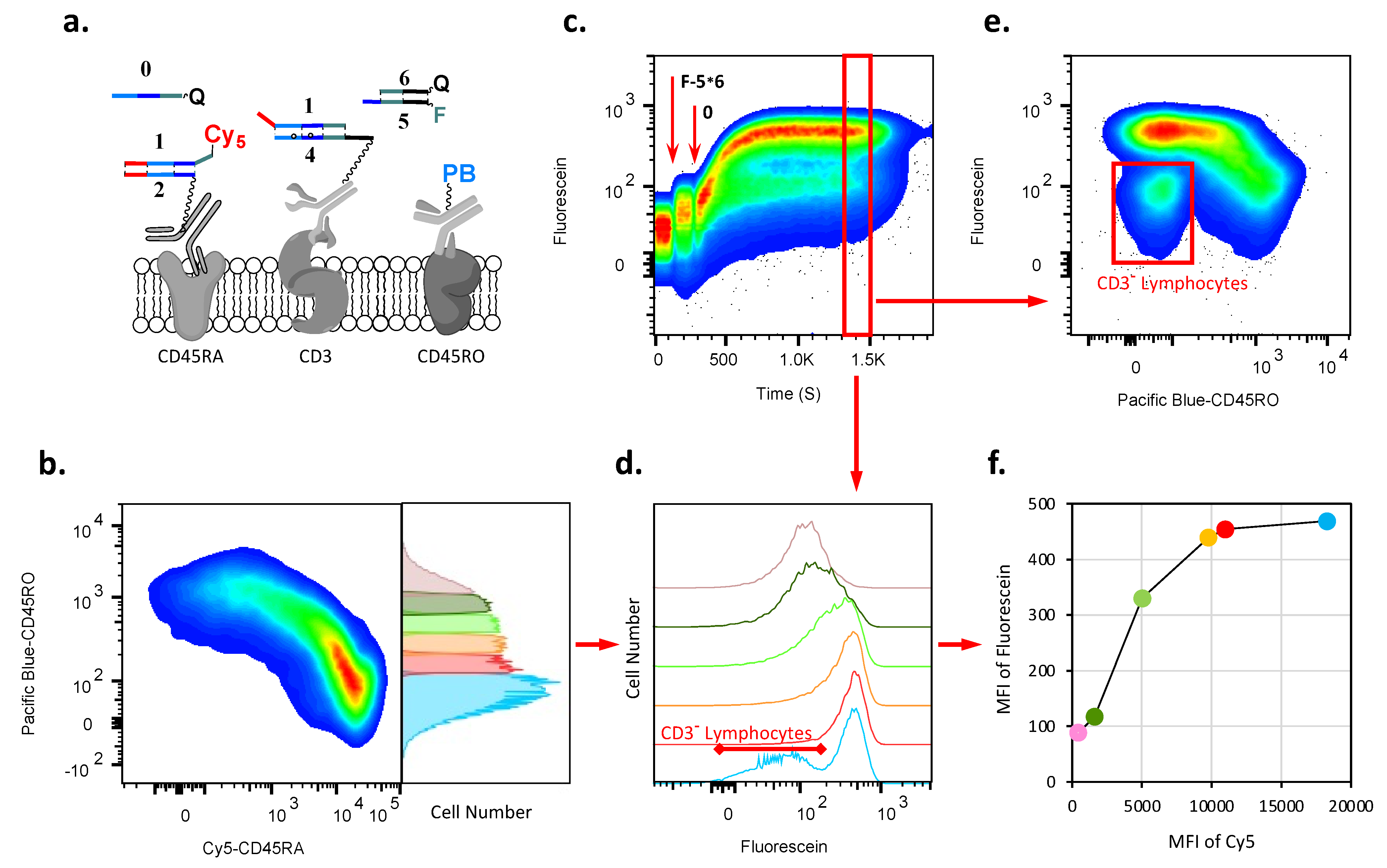
2.3. AMPLIFY(YESCD45RAYESCD3) Cascade Reaction
3. Conclusions
4. Materials and Methods
4.1. Materials
- 0 =/5IAbRQ/TACGTATCGAGGCTTTTACTTCTATCTCTTTCTT;
- 1 = GAAAGTGTTAAAGAAAGAGATAGAAGTAAAAGCCTCGATACGTA;
- 1(Cy5) = GAAAGTGTTAAAGAAAGAGATAGAAGTAAAAGCCTCGATAC
- GTA/3Cy5Sp/;
- 2 =/5ThioMC6-D/GGCTTTTACTTCTATCTCTTTCTTTAACACTTTCCTATACTT
- ATGCACTT;
- 3 = GAAAGTGTTAAAGAAAGAGATTGAAGTAAATGCCTC;
- 4 =/5ThioMC6-D/CTTTACGATTTGGTTACGTATCGAGGCATTTACTTCAATCT
- CTTTCTT;
- 5 =/5BioTinTEG/AGTAAATGCCTCGATACGTAACCAAATCGTTAAGCC/36-FAM/;
- 6 =/5IABkFQ/GGCTTAACGATTTGGTTACGTATCGA.
4.2. Methods
- PBMCs (25 × 106/mL) were pre-incubated with anti-CD32 antibodies (12.5 ng/mL, Clone IV.3, STEMCELL Technologies, Vancouver, BC, Canada) and Shredded Salmon Sperm DNA (125 ug/mL, Ambion, Austin, TX, USA) in buffer (PBS supplemented with 0.5% (w/v) BSA and 2 mM EDTA) at room temperature for 10 min.
- The conjugates of antibodies participated in the cascade reaction together with the corresponding Pacific Blue conjugated antibody were added to PBMCs in an amount that was determined after previous titration, to be sure that all antigens on the cell surface would be saturated with minimal non-specific binding registered. The mixture was incubated on ice for 30 min with periodic gentle mixing.
- Samples were washed twice with up to 15 mL buffer by centrifugation (350 g × 10 min, at 5 °C).
- The final pellet was resuspended to obtain a concentration of cells of 50 × 106 PBMCs per ml of buffer at room temperature.
- About 5 × 10⁶ PBMCs (100 µL) were resuspended in a final volume of 500 µL in a 5 mL tube for flow cytometry and the sample subjected to flow cytometry at a rate 12 µL/min. After the monitoring of the baseline during the first 100 s, the duplex F-5∗6 was added to sample to reach a final concentration of about 700 nM; after the second 100 s, the strand 0 was added to reach a final concentration about 200 nM. In some experiments, after the next 1100 s, strand 2 was added to obtain a final concentration of about 400 nM.
- PBMCs (25 × 106/mL) were pre-incubated with anti-CD32 antibodies (12.5 ng/mL, Clone IV.3, STEMCELL Technologies) and Shredded Salmon Sperm DNA (125 ug/mL, Ambion) in PBS supplemented with 0.5% (w/v) BSA and 2 mM EDTA (Buffer) at room temperature for 10 min.
- The CD45-(1∗4) conjugate was added to PBMCs in an amount that was determined after previous titration, to be sure that all antigens on the cell surface would be saturated, with minimal non-specific binding registered. The mixture was incubated on ice for 30 min with periodic gentle mixing.
- Samples were washed twice with up to 15 mL of buffer by centrifugation (350 g × 10 min, at 5 °C).
- Cells were resuspended in a concentration of 25 × 106/mL in buffer and strand 2 was added to obtain a final concentration 400 nM with subsequent incubation at room temperature for 30 min.
- Samples were washed twice with up to 15 mL of buffer by centrifugation (350 g × 10 min, at 5 °C).
- Cells were resuspended at a concentration of 25 × 106/mL in buffer and strand 3 was added to obtain a final concentration of 400 nM with subsequent incubation on ice for 30 min.
- Samples were washed twice with up to 15 mL buffer by centrifugation (350 g × 10 min, at 5 °C).
- The CD19-(1∗2) conjugate together with Pacific Blue anti-CD20 antibodies was added to PBMCs in an amount that was determined after previous titration, to be sure that all antigens on the cell surface would be saturated with minimal non-specific binding registered. The mixture was incubated on ice for 30 min with periodic gentle mixing.
- Samples were washed twice with up to 15 mL of buffer by centrifugation (350 g × 10 min, at 5 °C).
- The final pellet was resuspended to obtain a concentration of cells at 50 × 106 PBMCs per ml of buffer at room temperature.
- About 5 × 10⁶ PBMCs (100 µL) were resuspended in a final volume 500 µL in a 5 mL tube for flow cytometry and the sample subjected to flow cytometry at a rate of 12 µL/min. After the monitoring of the baseline during the first 100 s, the duplex F-5∗6 was added to the sample to obtain a final concentration of about 700 nM; after the second 100 s, the strand 0 was added to obtain a final concentration of about 200 nM. After the next 1100 s, strand 2 was added to obtain final concentration of about 400 nM.
Supplementary Materials
Author Contributions
Funding
Institutional Review Board Statement
Informed Consent Statement
Data Availability Statement
Conflicts of Interest
References
- Stojanovic, M.N.; Stefanovic, D.; Rudchenko, S. Exercises in molecular computing. Acc. Chem. Res. 2014, 47, 1845–1852. [Google Scholar] [CrossRef] [PubMed]
- De Silva, A.P. Molecular-Logic-Based Computation; RSC Publishing: Cambridge, UK, 2013. [Google Scholar]
- Chen, L.; Chen, W.; Liu, G.; Li, J.; Lu, C.; Li, J.; Tan, W.; Yang, H. Nucleic acid-based molecular computation heads towards cellular applications. Chem. Commun. 2021, 50, 12551–12575. [Google Scholar] [CrossRef] [PubMed]
- Shapiro, E.; Benenson, Y. Bringing DNA computers to life. Sci. Am. 2006, 294, 44–51. [Google Scholar] [CrossRef] [PubMed]
- Pei, R.; Matamoros, E.; Stefanovic, D.; Stojanovic, M.N. Teaching a Molecular Automaton to Play a Game. Nat. Nanotechnol. 2010, 5, 773–777. [Google Scholar] [CrossRef] [PubMed]
- Douglas, S.M.; Bachelet, I.; Church, G.M. A logic-gated nanorobot for targeted transport of molecular payloads. Science 2012, 335, 831–834. [Google Scholar] [CrossRef]
- Amir, Y.; Ben-Ishay, E.; Levner, D.; Ittah, S.; Abu-Horowitz, A.; Bachelet, I. Universal computing by DNA origami robots in a living animal. Nat. Nanotechnol. 2014, 9, 353–357. [Google Scholar] [CrossRef]
- Rudchenko, M.; Taylor, S.; Pallavi, P.; Deschovskaia, A.; Khan, S.; Butler, V.P., Jr.; Rudchenko, S.; Stojanovic, M.N. Autonomous molecular cascades for evaluation of cell surfaces. Nat. Nanotechnol. 2013, 8, 580–586. [Google Scholar] [CrossRef]
- You, M.; Peng, L.; Shao, N.; Zhang, L.; Qiou, L.; Cui, C.; Tan, W. DNA nano-claw: Logic-based autonomous cancer targeting and therapy. J. Am. Chem. Soc. 2014, 136, 1256–1259. [Google Scholar] [CrossRef]
- You, M.; Zhu, G.; Chen, T.; Donovan, M.J.; Tan, W. Programmable and Multiparameter DNA-based Logic Platform for Cancer Recognition and Targeted Therapy. J. Am. Chem. Soc. 2015, 137, 667–674. [Google Scholar] [CrossRef]
- Dosio, F.; Stella, B.; Cerioni, S.; Gastaldi, D.; Arpicco, S. Advances in anti-cancer antibody-drug conjugates and immunotoxins. Recent Pat. Anticancer Drug Discov. 2014, 9, 35–65. [Google Scholar] [CrossRef]
- Bornstein, G.G. Antibody Drug Conjugates: Preclinical Considerations. APPS J. 2015, 17, 525–534. [Google Scholar] [CrossRef] [PubMed]
- FitzGerald, D.J.; Wayne, A.S.; Kreitman, R.J.; Pastan, I. Treatment of hematologic malignancies with immunotoxins and antibody-drug conjugates. Cancer Res. 2011, 71, 6300–6309. [Google Scholar] [CrossRef] [PubMed]
- Frankel, S.R.; Baeuerle, P.A. Targeting T cells using bispecific antibodies. Curr. Opin. Chem. Biol. 2013, 17, 385–392. [Google Scholar] [CrossRef] [PubMed]
- Przewoznik, M.; Hoemberg, N.; Naujoks, M.; Poetzl, J.; Muenchmeier, N.; Brenner, C.D.; Anz, D.; Bourquin, C.; Nelson, P.J.; Roecken, M.; et al. Recruitment of natural killer cells in advanced stages of engogenously arising B-cell lymphoma: Implications for therapeutic cell transfer. J. Immunother. 2012, 35, 217–222. [Google Scholar] [CrossRef]
- Torikai, H.; Cooper, L.J.N. Translational Implications for Off-the-shelf Immune Cells Expressing Chimeric Antigen Receptors. Mol. Ther. 2016, 24, 1178–1186. [Google Scholar] [CrossRef]
- Bleakley, M.; Heimfeld, S.; Jones, L.A.; Turtle, C.; Krause, D.; Riddell, S.R.; Shlomchik, W. Engineering human peripheral blood stem cell grafts that are depleted of naïve T cells and retain functional pathogen-specific memory T cells. Biol. Blood Marrow Transplant. 2014, 20, 705–716. [Google Scholar] [CrossRef]
- Seelig, G.; Soloveichik, D.; Zhang, D.Y.; Winfree, E. Enzyme-Free Nucleic Acid Logic Circuits. Science 2006, 314, 1585–1588. [Google Scholar] [CrossRef]
- Qian, L.; Winfree, E. Scaling Up Digital Circuit Computation with DNA Strand Displacement Cascade. Science 2011, 332, 1196–1201. [Google Scholar] [CrossRef]
- Soderberg, O.; Gullberg, M.; Jarvius, M.; Ridderstrale, K.; Leuchowius, K.-J.; Jarvius, J.; Wester, K.; Hydrbing, P.; Bahram, F.; Larsson, L.-G.; et al. Direct observation of individual endogenous protein complexes in situ by proximity ligation. Nat. Methods 2006, 3, 995–1000. [Google Scholar] [CrossRef]
- Zhang, D.Y.; Winfree, E. Control of DNA Strand Displacement Kinetics Using Toehold Exchange. J. Am. Chem. Soc. 2009, 131, 17303–17314. [Google Scholar] [CrossRef]
- Qian, L.; Winfree, E. A simple DNA gate motif for synthesizing large-scale circuits. J. R. Soc. Interface 2011, 8, 1281–1297. [Google Scholar] [CrossRef] [PubMed]
- Ginaldi, L.; De Martinis, M.; D’Ostilio, A.; Marini, L.; Quaglino, D. Changes in antigen expression on B lymphocytes during HIV infection. Pathobiology 1998, 66, 17–23. [Google Scholar] [CrossRef] [PubMed]
- Poncelet, P.; Bikque, A.; Lavabre, T.; Poinas, G.; Parant, M.; Duperray, O.; Sampol, L. Quantitative expression of human lymphocytes membrane antigens: Definition of normal densities measured in immune-cytometry with the QIFI assay. Cytometry Suppl. 1991, S5, 82–83. [Google Scholar]
Disclaimer/Publisher’s Note: The statements, opinions and data contained in all publications are solely those of the individual author(s) and contributor(s) and not of MDPI and/or the editor(s). MDPI and/or the editor(s) disclaim responsibility for any injury to people or property resulting from any ideas, methods, instructions or products referred to in the content. |
© 2023 by the authors. Licensee MDPI, Basel, Switzerland. This article is an open access article distributed under the terms and conditions of the Creative Commons Attribution (CC BY) license (https://creativecommons.org/licenses/by/4.0/).
Share and Cite
Rudchenko, S.; Taylor, S.; Milosavic, N.; Rudchenko, M.; Wedderhoff Tissi, B.; Mapara, M.Y.; Stojanovic, M.N. Amplification of Signal on Cell Surfaces in Molecular Cascades. Cells 2023, 12, 2858. https://doi.org/10.3390/cells12242858
Rudchenko S, Taylor S, Milosavic N, Rudchenko M, Wedderhoff Tissi B, Mapara MY, Stojanovic MN. Amplification of Signal on Cell Surfaces in Molecular Cascades. Cells. 2023; 12(24):2858. https://doi.org/10.3390/cells12242858
Chicago/Turabian StyleRudchenko, Sergei, Steven Taylor, Nenad Milosavic, Maria Rudchenko, Betina Wedderhoff Tissi, Markus Y. Mapara, and Milan N. Stojanovic. 2023. "Amplification of Signal on Cell Surfaces in Molecular Cascades" Cells 12, no. 24: 2858. https://doi.org/10.3390/cells12242858
APA StyleRudchenko, S., Taylor, S., Milosavic, N., Rudchenko, M., Wedderhoff Tissi, B., Mapara, M. Y., & Stojanovic, M. N. (2023). Amplification of Signal on Cell Surfaces in Molecular Cascades. Cells, 12(24), 2858. https://doi.org/10.3390/cells12242858