Review

Physiological Action of Progesterone in the Nonhuman Primate Oviduct

1
Division of Reproductive & Developmental Sciences, Oregon National Primate Research Center, 505 NW 185th Ave., Beaverton, OR 97006, USA
2
Department of Obstetrics and Gynecology, Health & Science University, Portland, OR 97239, USA
3
Department of Animal and Rangeland Sciences, College of Agricultural Sciences, Oregon State University, Corvallis, OR 97331, USA
*
Author to whom correspondence should be addressed.
Cells 2022, 11(9), 1534; https://doi.org/10.3390/cells11091534
Submission received: 31 March 2022 / Revised: 28 April 2022 / Accepted: 30 April 2022 / Published: 3 May 2022
(This article belongs to the Special Issue Progesterone Receptor Signaling)

Abstract

:
Therapies that target progesterone action hold potential as contraceptives and in managing gynecological disorders. Recent literature reviews describe the role of steroid hormones in regulating the mammalian oviduct and document that estrogen is required to stimulate epithelial differentiation into a fully functional ciliated and secretory state. However, these reviews do not specifically address progesterone action in nonhuman primates (NHPs). Primates differ from most other mammals in that estrogen levels are >50 pg/mL during the entire menstrual cycle, except for a brief decline immediately preceding menstruation. Progesterone secreted in the luteal phase suppresses oviductal ciliation and secretion; at the end of the menstrual cycle, the drop in progesterone triggers renewed estrogen-driven tubal cell proliferation ciliation secretory activity. Thus, progesterone, not estrogen, drives fallopian tube cycles. Specific receptors mediate these actions of progesterone, and synthetic progesterone receptor modulators (PRMs) disrupt the normal cyclic regulation of the tube, significantly altering steroid receptor expression, cilia abundance, cilia beat frequency, and the tubal secretory milieu. Addressing the role of progesterone in the NHP oviduct is a critical step in advancing PRMs as pharmaceutical therapies.

1. Introduction

Several reviews address the action of ovarian steroid hormones on the mammalian reproductive tract [1,2,3,4,5,6]. It is well documented that estrogen (estradiol; E2) is essential for the normal development of reproductive tract organs and regulating cell proliferation and metabolism of tract tissues. Progesterone (P4) is also a crucial reproductive hormone with roles in the cyclic regulation of the fallopian tube [7,8,9,10,11], uterus [4,12], and cervix [13], as well as in supporting embryo implantation [14] and the maintenance of pregnancy [15]. Specific progesterone receptors mediate these actions, and synthetic progesterone receptor modulators (PRMs) are a class of drugs that alter P4 effects and have clinical utility as contraceptives and in treating P4-associated disorders [16].
The menstrual cycles of women and nonhuman primates (NHPs) are strikingly different from the estrous cycles of most laboratory animals [17]. Therefore, NHPs provide valuable animal models for translational studies relating to women’s health [18]. However, most literature reviews overlook the specific actions of P4 on the fallopian tube of NHPs. Therefore, our focus here is to describe the effect of P4 on the oviduct, highlighting the current state of knowledge on NHPs.
We begin this discussion with a brief overview of primate oviductal anatomy and histology during the menstrual cycle because the cyclic pattern of ovarian steroid secretion in women and NHPs is strikingly different from many mammals [19]. Our goal is not to discuss all historical studies, as these were addressed in previous reviews [4,11,20]. Like most mammals, primates have elevated levels of E2 in the follicular phase and elevated P4 in the luteal phase of the cycle. However, the primate corpus luteum also expresses high aromatase levels and secretes E2 in addition to P4 in the luteal phase [2]. Therefore, in primates, the cyclic changes in P4, not E2, drive cycles of epithelial differentiation in the fallopian tube (Figure 1).

2. NHP Oviductal Anatomy

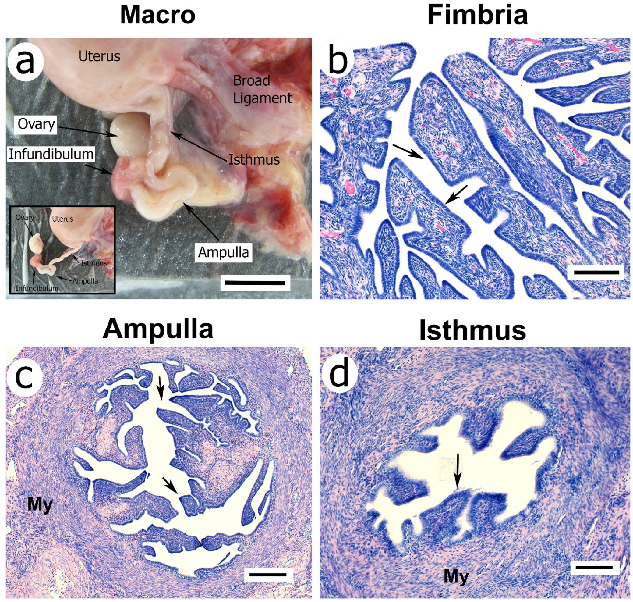
The oviduct of women [20] and NHPs [4] is a tubular, seromuscular organ supported by the mesosalpinx, a portion of the broad ligament that anchors the uterus and oviduct to the body wall (Figure 2a). It is attached distally to the ovary and proximally to the lateral aspect of the uterine fundus. The fallopian tube consists of four anatomical regions based on tubular anatomy and distinct lumen histology [7]. These include the infundibulum (Figure 2a,b), ampulla (Figure 2a,c), and isthmus (Figure 2a,d), as well as the interstitial portion that passes through the myometrium wall. The infundibulum transitions into the ampulla, the longest part, and then to the isthmus of the oviduct. The inner mucosal layer of the tube is the endosalpinx, and the muscular layers are collectively called the myosalpinx. The lumen of the endosalpinx is continuous from the abdominal tubal ostium near the ovary to the uterine cavity.
The most distal portion of the oviduct, the infundibulum, is funnel-shaped with a fimbriated end surrounding the abdominal tubal ostium. Unlike rodents and domestic species, primates have no distinct ovarian bursa, and one of the fimbrial folds is usually attached to the ovary. The endosalpinx of the infundibulum and ampulla is extensively folded (Figure 2b,c) and contains ciliated and secretory endothelial cells (Figure 3). The myosalpinx of the ampulla consists of a thin inner circular layer of smooth muscle surrounded by an equally thin longitudinal muscle layer. The oviductal ampulla transitions into the most proximal portion of the oviduct, the isthmus. The isthmus has a thick circular smooth muscle layer that is continuous with the uterine myometrium. The cellular integrity of the endosalpinx in primates is dependent on ovarian hormones; ovariectomy results in almost complete atrophy of the endosalpinx epithelium of the infundibulum and ampulla. Interestingly, the isthmus portion of the tube is less responsive to hormonal cycles than the more distal ampulla and fimbria.
The ciliated cells of the endosalpinx and smooth muscle of the myosalpinx contribute to the movement of gametes along the oviductal canal [21]. Folding and hypertrophy of the fimbria and ampulla are dependent on E2. Co-administration of E2 with P4 in ovariectomized monkeys suppresses E2-driven tubal hypertrophy [11,22]. The ciliated cells of the infundibular fimbria create a current of peritoneal fluid toward the oviductal ostium, facilitating oocyte passage into the tube. The binding of spermatozoa to the ciliated epithelium occurs in many species, including primates [23,24,25,26], to provide a reservoir for fertilization [27,28]. In contrast to the ampulla, the isthmic and intramural epithelium contains only four primary epithelial folds and is strikingly less ciliated and therefore appears less sensitive to the actions of E2 and P4. Myosalpinx contractions move the oocyte to the site of fertilization in the ampulla and the developing embryo toward the uterus [29].

3. The Primate Menstrual Cycle

Old World nonhuman primates experience approximately 28–31-day ovarian cycles with prolonged follicular and luteal phases similar to women. These cycles are a striking contrast to many other mammalian species [17]. Laboratory rodents, for instance (e.g., mice and rats), display a short 4–5-day estrous cycle due to the formation of short-lived corpora lutea [30]; domestic livestock species (sheep, goats, and cattle) display estrous cycles featuring a short follicular phase followed by a long luteal phase [31]; and some mammals (e.g., rabbits) have induced ovulation [32]. Some species of New World monkeys do not reliably menstruate. However, Old World NHPs (e.g., apes [33], baboons [34], macaques [35], and vervet [36]) display actual menstruation with cyclic shedding of the endometrial lining [2,17,19]. By convention, the start of the cycle (day 1) is the first day of detectable menses, marking the beginning of the cycle’s follicular phase. The average luteal phase in macaques, baboons, and women is 10 days long, with P4 declining 24–48 h before menses.
Historical studies extensively characterized circulating concentrations of ovarian steroid hormones throughout the menstrual cycle in women and NHPs, including the vervet, macaque, baboon, and chimpanzee [17,37,38,39]. Assay technologies have undergone remarkable advancements since many of these early studies. Over the past 50 years, our research center has assayed E2 and P4 levels in macaques with techniques including radioimmunoassay [40,41], automated electro-chemoluminescent assays [42,43,44], and liquid chromatography–tandem mass spectrometry (LC-MS/MS) [45]. Comparing these methods reveals that LC-MS/MS can provide greater assay sensitivity and thus improve hormone detection at low concentrations. However, at normal cycling levels, the patterns described in early studies [17,39,43] remain reasonably accurate and are worth briefly describing here.
In macaques, early follicular phase (menstrual cycle day 1–8) serum levels of E2 average approximately 50 pg/mL and gradually rise to 100 pg/mL 2–4 days before the day of peak luteinizing hormone (LH) preceding ovulation. There is a surge in E2 to levels >350 pg/mL in response to the LH peak, followed by a rapid fall to about 25 pg/mL. Then, as the luteal phase proceeds, there is a second rise in E2 back to approximately 50 pg/mL. As the corpus luteum regresses, P4 and E2 levels fall to near the detection threshold for most assays (10–20 pg/mL). Human and chimpanzee E2 levels follow a similar pattern but trend slightly higher, with mid-luteal phase levels in the 100 pg/mL range [17]. This pattern of P4 secretion is very similar among all primate species.
Therefore, there is essentially a constant E2 > 50 pg/mL level in primates throughout almost all of the menstrual cycle, except for a brief mid-cycle E2 surge and a brief decline immediately preceding menstruation. Before ovulation in the cycle’s follicular phase, P4 levels range from 0.1 to 1.0 ng/mL. A small but significant rise in serum P4 coincides with the LH peak; then, as luteal formation occurs, levels in macaques rise to maximal values of 3–8 ng/mL. As observed with E2, in humans and chimpanzees, the luteal phase peak of P4 can be three to four times higher than in macaques. In all Old World primates, three days before the onset of menstruation, serum levels of P4 fall sharply to <1 ng/mL. This rapid fall in P4 triggers menstruation [46].

4. Cytologic Changes in the Oviduct during the Menstrual Cycle

Brenner and coworkers [11] rigorously examined the NHP oviduct in naturally cycling cynomolgus [35] and rhesus macaques [47] and compared the animals to ovariectomized monkeys treated with implants releasing E2 and P4 to produce controlled artificial cycles [48]. To characterize tubal morphology, they assessed epithelial cell height, the percentage of secretory and ciliated cells, and the abundance of mitotic cells and apoptotic cells, including the phagocytic macrophages containing apoptotic nuclear fragments. Cycle phase-associated apoptosis was also described for the epithelium of primates (macaques, baboons, and women) and non-primate mammals [49,50,51]. Brenner and colleagues examined the formation of cilia and cytologic features including extension secretory tips and the deciliation process in which the apical portions of the ciliated cells pinch off the cell bodies [52]. Their studies revealed that E2 and P4 are the only ovarian factors required to recapitulate regular cyclic changes in tubal histology identical to the natural menstrual cycle [4,11]. Ovariectomized animals displayed almost complete atrophy and loss of cilia in the epithelium of fimbriae and ampulla. Treatment of ovariectomized monkeys with E2 alone stimulated epithelial differentiation into a ciliated and secretory state. However, the sequential exposure to E2 followed by E2 + P4 resulted in epithelial regression to a non-ciliated and non-secretory state similar to ovariectomized, untreated animals. They defined eight morphological conditions associated with epithelial ciliation and secretory activity [11]. These stages are summarized in Table 1.
Figure 3 shows examples of the histology of the oviductal fimbria in the late follicular phase and the late luteal phase of the cycle. Luteal phase P4 acts as a master regulator of the cyclic oviductal differentiation against a background of continuous E2. At the end of the luteal phase of the natural menstrual cycle, most of the epithelium of the fimbria and ampulla is cuboidal with very few ciliated and secretory cells. Then, in the follicular phase’s post-menstrual period, epithelium hypertrophies become columnar and ciliated, and secretory cells develop to dominate in the infundibular fimbria and ampulla. These cells increase to a maximum height near mid-cycle and then shrink to a minimal height again by the mid-late luteal phase. Ciliated cells appear to shrink more rapidly than the secretory cells, and the apices of the latter are projected well beyond the tips of the cilia during the latter part of the cycle [22,53]. In ovariectomized animals, 2–3 days of E2 begin the process of oviductal differentiation into a ciliated and secretory state. The first evidence of deciliation and suppression of secretion often emerged within 48 to 72 h of the onset of P4 treatment.
The work of Verhage et al. [54] supported the view that the tubal epithelium of women undergoes cyclic changes similar to those of NHPs, and the epithelial cells attain their maximum height and degree of ciliation during the late follicular phase in both the fimbriae and the ampulla. It is noteworthy that some reports indicate a minimal change in the percentage ciliation during women’s cycle [55]. Those prior reports may not have fully appreciated the role of fallopian tube anatomy in the ciliogenic cycle and did not examine all tube sections. More recent studies in women confirmed these conclusions and added that an increase in epithelial mitotic activity occurred during the follicular phase when P4 was almost undetectable. Moreover, in macaques, the timing of the oviductal stages is not precise (Table 1). There is variability associated with tubal anatomy. These stages appear most intense in the oviductal fimbria, which is either the first to enter or the quickest to complete the ciliogenic process. The ampulla responds slightly slower to changes in the hormonal milieu when entering and completing the ciliated secretory state. When most cells near the lumen of the ampulla were regressing, a few ciliated and secretory cells could be identified that lagged behind the main population near the muscle wall.
Oviductal secretions have long been proposed to support fertilization and early embryo development [56]. These secretions and the regulation by steroid hormones in non-primate species have been recently reviewed [7]. Verhage and coworkers were among the first investigators to address steroid hormone-dependent oviductal secretions in primates [57]. Their studies reported the presence of secretory granules at the apical tips of secretory cells in the baboon. The same group then characterized an oviduct-specific glycoprotein (OVGP1) as an estrogen-dependent secretory protein synthesized by non-ciliated oviduct epithelial cells in various species, including macaque baboon and human [58,59,60]. While OVGP1 has recently been reported for other tissues, including the macaque cervix [61] and ovarian cancer [62,63], it remains a primary hormonally regulated secretory product of oviductal epithelial cells. OVGP1 appears to be an estrogen-upregulated protein that P4 and other pure progestins suppress, and pure PRAs reverse the effect of P4 treatment. During the normal menstrual cycle, OVGP1 is highly expressed in the secretory epithelial cells of the oviduct during the proliferative phase and is significantly reduced after ovulation [64]. In artificially cycled macaques, OVGP1 is reduced in P4 and levonorgestrel in the cervix [61] and oviduct. Expression of OVGP1 is a marker of P4 action and conditions including endometriosis that results in P4 resistance result in persistent oviductal OVGP1 expression [65]. Moreover, treatment of macaques with contraceptive levels of ZK137-316, a PRA compound similar to mifepristone, significantly increased OVGP1 protein in oviductal fluid [66].

5. Progesterone Receptors

Actions of steroid hormones to influence cellular function fall into two classifications: the slower classical genomic response and the rapid non-genomic response. The genomic actions of P4 in target tissues are mediated through interactions with intracellular progesterone receptors (PGR; also called PR), ligand-activated transcription factors that belong to the nuclear receptor family [67,68,69]. This family of transcription factors includes estrogen receptors (ER, ESR1, and ESR2), androgen receptors (AR), mineralocorticoid receptors (MR, nuclear receptor subfamily 3 group C member 2/NR3C2), and glucocorticoid receptors (GR, nuclear receptor subfamily 3 group C member 1/NR3C1). These are referred to as “classical” steroid receptors in which steroid binding leads to a long-lasting but slowly emerging response [68]. The transcription factor PGR is expressed in all P4-responsive organs, including the reproductive tract, mammary glands, cardiovascular system, and the oviduct [70,71,72,73,74]. To stimulate this “classical” response, binding of P4 to the ligand-binding domain of the PGR induces a conformational change that transforms the receptor from a static, non-DNA-binding configuration into one that activates gene transcription. This occurs by loss of associated (heat shock) proteins and dimerization of receptor moieties. The activated receptor–ligand complex can then activate the transcriptional machinery by direct action on regulatory motifs, most commonly at PGR response elements (PRE) sites, or by direct association of ligand-bound PGR with other transcription factors and coactivators [69,75,76,77,78].
Differences among fixation methodologies can greatly affect the outcome of ER and PGR localization by immunohistochemistry (IHC). For instance, studies of OCT-embedded cryosections of oviductal fimbria and ampulla of ovariectomized macaques revealed staining for ER and PGR localized to the nuclei of epithelial, underlying stromal cells and smooth muscles. However, staining of paraffin-embedded sections produced variable cytoplasmic plus nuclear localization. Since binding assays revealed that most of the ER and PR were recovered from the cytosol, IHC on cryosections was interpreted as indicating that PGR was rapidly translocated from the cytoplasm to the nucleus regardless of the hormonal state of the animal and interacted strongly with chromatin in the nucleus. It is noteworthy to mention that ESR-2 (ERβ) expression is reported for the oviduct of several mammalian species [7], but the role of ESR-2 during cyclic regulation in NHPs in unknown.
It is worth mentioning that PGR exists in two primary isoforms (A and B) encoded by a single gene but with different initiation sites that permit transcription of either a large or short isoform [71,79,80,81]. The larger (PR-B) isoform contains an N-terminal fragment of 164 amino acids that is absent from the short (PR-A) isoform. Thus, PR-B exhibits three transcription-activating domains (AF-1, AF-2, and AF-3), whereas PRA contains only two (AF-1 and AF-2) [82]. The two PR isoforms have similar steroid hormone and DNA binding activities but have distinct functions depending on the cell type and context of the target gene promoter. PRB appears to be a stronger transcription activator than PRA [80]. Due to the structural overlap of the two PGR isoforms, assessing the localization of the two PGR isoforms in the oviduct has been challenging. One approach is to use differential immunostaining with antibodies directed against PR-B and PR-A plus PR-B as well as specific differential PCR approaches. Using this approach, researchers at the University of Edinburgh reported attenuated PR-B in human fallopian tubes during the luteal phase of the menstrual cycle and during ectopic pregnancy [83]. However, cyclic regulation of oviductal PR-A/PR-B isoforms has not been confirmed in NHP studies.
Rapid, nongenomic actions of P4 are also reported for the oviduct. These are mainly attributed to so-called “membrane” receptors or “non-classical” progesterone receptors that appear to activate cellular second messenger pathways [68,84,85,86]. Among the rapid actions of P4 in the oviduct are the effects on ciliary beat frequency [87,88,89] and rapid alteration to spermatozoa motility [90]. Non-classical PRs include a family of membrane progestin receptors (mPRs) as well as the G-protein-coupled receptor (GPCR) family, which includes progesterone receptor membrane component (PGRMC), PGRMC1, and PGRMC2 [6,67]. The PGRMC family shares properties not associated with P4, including a heme-binding domain related to some cytochromes. The mPRs were first reported in fish [85,91,92], and subsequently, five mPR subtypes (α, β, γ, δ, and ε) were identified [91] in a wide array of cell types in many mammalian species, including primates. The mPRs have no known homologies with GPCRs or nuclear PGRs, but are structurally related to adiponectin receptors and are classified as the progestin and adipoQ receptor (PAQR) superfamily. They display a predicted seven-transmembrane region and bind small steroid molecules, resulting in G-protein activation. However, the function of mPRs remains less clearly defined than that of the nuclear receptors [68]. This is largely due to a lack of data on the mPR steroid binding domains [93] and the absence of well-defined mPR modulators.

6. Cyclic Regulation of Steroid Responsiveness

It is well recognized that the oviduct is an estrogen-responsive organ that expresses ERα (ESR1) and PGR. Interestingly, ESR2 (ERβ) is also expressed in human fallopian tube ciliated cells, but the role of ERβ in NHPs remains to be determined. ER (not specific to ESR1 or ESR2) and PGR abundance have been assayed in naturally cycling NHPs as well as in NHPs treated sequentially with E2 and P4 to create artificial menstrual cycles. The earliest research characterizing the abundance of PGR in the primate oviduct utilized radiolabeled steroid binding on human fallopian tube [94]. These were followed by NHP studies that employed steroid binding and exchange assays to estimate levels of estrogen receptor and PGR (e.g., specific binding) in tissue homogenates [11,95]. These assays often used radiolabeled R2858 (a nuclear ER ligand) and R5020 (a nuclear PGR ligand) to avoid the metabolism of estrogen and P4. The sum of specifically bound steroids to the nuclear and cytosolic fractions from the homogenates represented an estimate of total receptor abundance. Binding of labeled R2858 and of R5020 were found to be significantly elevated in ovariectomized animals treated with E2 (or at mid-menstrual cycle) compared to hormone-depleted animals. This technique revealed that the oviduct’s differentiation into a fully ciliated and secretory endosalpinx epithelium was accompanied by significant increases in total ER and PGR [11]. Treatment of E2-primed monkeys with E2 in combination with P4 similar to the luteal phase resulted in significantly reduced levels of ER and PGR. In the case of ER, levels were reduced below those of ovariectomized untreated animals. Thus, ER and PGR expression were dependent on E2 action. Moreover, average ER and PGR levels were lower in animals treated with a combination of E2 and P4 than those observed in ovariectomized untreated monkeys. Because treatment with P4 alone failed to stimulate either ER or PGR, it was proposed that P4 acted to antagonize the effects of E2 on oviductal differentiation by suppressing ER levels below the threshold required to facilitate E2 action.
The overall relationship provided by classical binding assays appears to be more complex than was initially proposed. In concert with biochemical binding assays, cellular localization of ER and PGR by IHC on cryosections revealed that both cell and tissue type affected P4 suppression of ER and PGR. In support of binding assay results, the abundance of cells with strong nuclear staining for both ER and PGR increased in the follicular phase (and after E2 treatment) and decreased in the luteal phase (or after E2 plus P4 treatment). However, specific staining for epithelial ER and PGR were localized to the secretory epithelial cells, not the ciliated cells (Figure 3). This represents a paradox in that E2 and P4 strongly affect the ciliated phenotype, but staining is minimal in the ciliated cells. How can the dramatic effects of both E2 and P4 on the ciliated cells occur when the ciliated cells lack or express minimal receptors for both steroids? Moreover, PGR staining was almost completely absent in the epithelium during the luteal phase or after P4 treatment. This produces the question: How does P4 maintain its effects while suppressing its own receptor?
IHC revealed that strong ERα and PGR staining were present in stromal, smooth muscle, and secretory epithelial cells, suggesting that the effects of P4 on ciliated cells may be indirect. In the luteal phase (or after P4 treatment), ERα staining is retained in all the undifferentiated epithelial cells and in the underlying stromal cells, whereas PGR is minimal in the epithelium and retained (but noticeably less intense) in the stromal compartment. Therefore, one possibility is that the state of differentiation of the oviductal epithelium is mediated indirectly through soluble growth factors (or other unidentified mediators) secreted by ERα- and PGR-positive stromal cells. Moreover, stromal cells are separated from the epithelium by a definitive basement membrane, which could reduce the influence of soluble factors.
One potential mediator of P4 progesterone action, particularly in PR-negative cells, is the presence of specific mPRs or other non-classical PRs reported for human, murine, bovine, and canine oviducts, as well as ovarian cancers that may be of tubal origin. Oviductal mPR (beta and gamma) have been localized to bovine, human, and mouse ciliated epithelial cells [96] and may mediate the rapid effect of P4 on cilia beat frequency. However, localization of mPRs to oviductal cilia does not appear to reflect expected cyclic changes in ciliated cell abundance [96], as observed in nonhuman primates. Cyclic PGRMC1 and PGRMC2 expression and localization are reported for the macaque endometrium, but cyclic regulation in the NHP oviduct has not been extensively studied. The absence of reliable mPR/PGRMC modulators has significantly limited the study of these pathways in NHP models. In contrast, the expression and cellular action of nuclear PGR in the mammalian oviduct have been studied extensively.

7. Progesterone Receptor Modulators

The characterization of nuclear PGR isoforms has prompted the development of synthetic compounds called progesterone receptor modulators (PRMs) [97]. These include synthetic P4 analogs (progestins) and P4 antagonists (anti-progestins; PRAs) that bind to PGR and either stimulate or block PGR function [98,99,100]. It is noteworthy that long-term treatment with P4 and synthetic progestins reduces the abundance of ciliated cells in NHPs [101], and short-term treatment decreases cilia beat frequency in human oviductal cultures [102].
Mifepristone (RU486), the first well-characterized PRA, acts as a glucocorticoid receptor antagonist in the primate uterus, opposing various estrogen effects. The action of PRMs is often unique to the target organ, cell type, and sometimes the animal model examined. This has led to tissue-selective or physiologically selective PRMs [98]. The nuclear action of PRMs on classical (genomic) action of P4 has been most extensively evaluated because of the pharmaceutical potential of these compounds to treat gynecological disorders [97,98,99,103].
Compared to nuclear receptors, the action of PRM on mPRα, mPRβ, and mPRγ appears less clearly defined, with reports ranging from the minimal binding of mPRs to synthetic PRMs, especially classical PGR antagonists such as mifepristone [93,104], to putative or predictable actions [105]. Moreover, much of the studies on the nuclear action of PRMs have been conducted on non-primate models with strikingly different hormone profiles. In vivo assessments of mPR actions are confounded by co-expression of nuclear PGRs in many of the responsive cell types. However, differential binding of synthetic ligands offers the potential development of mPR-selective agonists and antagonists [93].
We have treated rhesus macaques with an array of potent PRAs including mifepristone [106,107], ZK 137 316 [108,109], ZK 230 211 [110], and CDB 2914 (Ulipristal) [111]. However, the primary experimental goal of these studies was to evaluate the action of these compounds on the uterine endometrium. Driving these studies was the observation that some PRA compounds, including mifepristone, have been reported to have unexpected anti-estrogenic actions on the endometrium. However, in the oviduct, pure PRA compounds such as ZK 230-211 appear to lack antiestrogen effects and block the genomic action of P4 [106,107]. In this condition, the estrogen action is unopposed, and the oviducts appear in a fully differentiated and ciliated secretory state.
PRMs can also provide contraception. Levonorgestrel is a contraceptive progestin that has inhibitory actions on oviductal cilia beat frequency and ovulation. Ulpristral and levonorgestrel have potential as emergency contraception, presumably by blocking ovulation. However, a secondary target may include oviductal cilia function. In vitro, progesterone can decrease human oviductal ciliary beat frequency (CBF) and muscular contractions, and the inhibitory effect of progesterone on CBF can be antagonized by mifepristone, a progesterone receptor (PR) modulator. Treatment of cycling rhesus monkeys with low-dose ZK 137-316, a compound very similar to mifepristone [112], prevented pregnancy at low doses that allowed menstrual cycles [113]. However, low-dose ZK 137-316 did not block ovulation and failed to alter oviductal differentiation and sperm passage but did significantly increase oviductal fluid levels of OVGP1 [66]. In contrast, ulipristal acts as a mixed agonist–antagonist compound and could disrupt gamete passage. This outcome on sperm passage may be PRA dose-dependent because other reports indicate that both ulipristal and mifepristone reduce ciliary beat frequency and contractility in human oviductal explants [114].
As indicated above, blockade of P4 action in NHPs is not associated with well-defined tubal abnormalities. Treatments with pure PRA, including ZK137-316 [112], mifepristone, and ZK 230-211, result in a fully ciliated and secretory tubal epithelium. This is not abnormal for the proliferative phase of the cycle. However, tubal abnormalities such as ectopic pregnancy are almost nonexistent in NHPs compared to women. Reproductive tract infections occur in NHPs and appear to be affected by estrogen and P4 action on the cervix, endometrium, and oviduct [115]. It can be speculated that treatment with mixed-action PRM therapy could alter normal cyclic changes. However, this represents a knowledge gap and further studies are required to assess the impact of P4 modulation on tubal dysfunction in NHP models.

8. Conclusions

The role of P4 in modulating oviductal morphology and physiology is indisputable. Cyclic changes in circulating P4 against a background of E2 stimulate changes in ciliary beating, muscular contraction, and oviductal fluid volume and composition, and, over time, suppress oviductal differentiation. These actions are mediated via intracellular nuclear receptors and via novel membrane receptors. However, the specific roles for membrane receptors remain to be resolved. Complicating these action mechanisms is that both classical nuclear receptors and fast-acting membrane receptors may be present in the same target cells. Thus, P4 can have rapid and long-lasting actions by stimulating paracrine factors that mediate hormone responsiveness. There is a large gap in our knowledge regarding P4-regulated effectors in the oviduct, although prostaglandins, endothelins, and growth factors may have roles as critical secondary mediators. Further development of selective PRMs that specifically target membrane receptors versus nuclear receptor isoforms may be required to elucidate the complex cyclic regulation of the primate oviduct.

Author Contributions

Conceptualization, O.D.S., F.L. and C.V.B.; data curation, O.D.S.; writing—original draft and outline preparation, O.D.S., F.L. and C.V.B.; writing—review and editing, O.D.S., F.L. and C.V.B.; visualization, O.D.S. and F.L.; funding acquisition, O.D.S. All authors have read and agreed to the published version of the manuscript.

Funding

This work was supported by the Office of the Director, National Institutes of Health (NIH/OD) Support for National Primate Research Center 5P51OD011092.

Institutional Review Board Statement

Not applicable.

Informed Consent Statement

Not applicable.

Data Availability Statement

Not applicable.

Conflicts of Interest

The authors declare no conflict of interest. The funders had no role in the design of the study; in the collection, analyses, or interpretation of data; in the writing of the manuscript, or in the decision to publish the results.

References

  1. Gray, C.A.; Bartol, F.F.; Tarleton, B.J.; Wiley, A.A.; Johnson, G.A.; Bazer, F.W.; Spencer, T.E. Developmental biology of uterine glands. Biol. Reprod. 2001, 65, 1311–1323. [Google Scholar] [PubMed]
  2. Saltzman, W.; Tardif, S.D.; Rutherford, J.N. Chapter 13—Hormones and Reproductive Cycles in Primates. In Hormones and Reproduction of Vertebrates; Norris, D.O., Lopez, K.H., Eds.; Academic Press: London, UK, 2011; pp. 291–327. [Google Scholar]
  3. Cline, J.M.; Soderqvist, G.; Register, T.C.; Williams, J.K.; Adams, M.R.; von Schoultz, B. Assessment of hormonally active agents in the reproductive tract of female nonhuman primates. Toxicol. Pathol. 2001, 29, 84–90. [Google Scholar] [PubMed]
  4. Brenner, R.M.; Slayden, O.D. Cyclic Changes in the Primate Oviduct and Endometrium, The Physiology of Reproduction, 2nd ed.; Knobil, E., Neill, J.D., Eds.; Raven Press Ltd.: New York, NY, USA, 1994; Volume 2, pp. 541–569. [Google Scholar]
  5. Hewitt, S.C.; Winuthayanon, W.; Korach, K.S. What’s new in estrogen receptor action in the female reproductive tract. J. Mol. Endocrinol. 2016, 56, R55–R71. [Google Scholar] [CrossRef] [PubMed] [Green Version]
  6. Thomas, P. Characteristics of membrane progestin receptor alpha (mPRalpha) and progesterone membrane receptor component 1 (PGMRC1) and their roles in mediating rapid progestin actions. Front. Neuroendocrinol. 2008, 29, 292–312. [Google Scholar] [PubMed] [Green Version]
  7. Barton, B.E.; Herrera, G.G.; Anamthathmakula, P.; Rock, J.K.; Willie, A.; Harris, E.A.; Takemaru, K.I.; Winuthayanon, W. Roles of steroid hormones in oviductal function. Reproduction 2020, 159, R125–R137. [Google Scholar] [CrossRef]
  8. Binelli, M.; Gonella-Diaza, A.M.; Mesquita, F.S.; Membrive, C.M.B. Sex Steroid-Mediated Control of Oviductal Function in Cattle. Biology 2018, 7, 15. [Google Scholar] [CrossRef] [Green Version]
  9. Akison, L.K.; Boden, M.J.; Kennaway, D.J.; Russell, D.L.; Robker, R.L. Progesterone receptor-dependent regulation of genes in the oviducts of female mice. Physiol. Genom. 2014, 46, 583–592. [Google Scholar] [CrossRef] [Green Version]
  10. Akison, L.K.; Robker, R.L. The critical roles of progesterone receptor (PGR) in ovulation, oocyte developmental competence and oviductal transport in mammalian reproduction. Reprod. Domest. Anim. 2012, 47, 288–296. [Google Scholar] [CrossRef]
  11. Brenner, R.M.; Slayden, O.D. The Fallopian Tube Cycle. In Reproductive Endocrinology, Surgery, and Technology; Adashi, E.Y., Rock, J.A., Rosenwaks, Z., Eds.; Lippincott-Raven Publishers: Philadelphia, PA, USA, 1995; Volume 1, pp. 325–339. [Google Scholar]
  12. Slayden, O.D. Cyclic remodeling of the nonhuman primate endometrium: A model for understanding endometrial receptivity. Semin. Reprod. Med. 2014, 32, 385–391. [Google Scholar] [CrossRef]
  13. Larsen, B.; Hwang, J. Progesterone interactions with the cervix: Translational implications for term and preterm birth. Infect. Dis. Obstet. Gynecol. 2011, 2011, 353297. [Google Scholar] [CrossRef]
  14. Slayden, O.D.; Keator, C.S. Role of progesterone in nonhuman primate implantation. Semin. Reprod. Med. 2007, 25, 418–430. [Google Scholar] [CrossRef] [PubMed]
  15. Lonergan, P.; Forde, N.; Spencer, T. Role of progesterone in embryo development in cattle. Reprod. Fertil. Dev. 2016, 28, 66–74. [Google Scholar] [CrossRef] [PubMed]
  16. Zhang, Z.; Lundeen, S.G.; Slayden, O.; Zhu, Y.; Cohen, J.; Berrodin, T.J.; Bretz, J.; Chippari, S.; Wrobel, J.; Zhang, P.; et al. In vitro and in vivo characterization of a novel nonsteroidal, species-specific progesterone receptor modulator, PRA-910. In Ernst Schering Foundation Symposium Proceedings; Springer: Berlin, Germany, 2007; pp. 171–197. [Google Scholar]
  17. Brenner, R.M.; West, N.B. Hormonal regulation of the reproductive tract in female mammals. Annu. Rev. Physiol. 1975, 37, 273–302. [Google Scholar] [CrossRef] [PubMed]
  18. Stouffer, R.L.; Woodruff, T.K. Nonhuman Primates: A Vital Model for Basic and Applied Research on Female Reproduction, Prenatal Development, and Women’s Health. ILAR J. 2017, 58, 281–294. [Google Scholar] [CrossRef] [PubMed] [Green Version]
  19. Phillips, K.A.; Bales, K.L.; Capitanio, J.P.; Conley, A.; Czoty, P.W.; t Hart, B.A.; Hopkins, W.D.; Hu, S.L.; Miller, L.A.; Nader, M.A.; et al. Why primate models matter. Am. J. Primatol. 2014, 76, 801–827. [Google Scholar] [CrossRef] [PubMed] [Green Version]
  20. Mazur, E.C.; Large, M.J.; DeMayo, F.J. Chapter 24—Human Oviduct and Endometrium: Changes over the Menstrual Cycle. In Knobil and Neill’s Physiology of Reproduction, 4th ed.; Plant, T.M., Zeleznik, A.J., Eds.; Academic Press: San Diego, CA, USA, 2015; pp. 1077–1097. [Google Scholar]
  21. Coy, P.; Garcia-Vazquez, F.A.; Visconti, P.E.; Aviles, M. Roles of the oviduct in mammalian fertilization. Reproduction 2012, 144, 649–660. [Google Scholar] [CrossRef] [Green Version]
  22. Brenner, R.M.; Resko, J.A.; West, N.B. Cyclic changes in oviductal morphology and residual cytoplasmic estradiol binding capacity induced by sequential estradiol—progesterone treatment of spayed Rhesus monkeys. Endocrinology 1974, 95, 1094–1104. [Google Scholar] [CrossRef]
  23. Tollner, T.L.; Vandevoort, C.A.; Yudin, A.I.; Treece, C.A.; Overstreet, J.W.; Cherr, G.N. Release of DEFB126 from macaque sperm and completion of capacitation are triggered by conditions that simulate periovulatory oviductal fluid. Mol. Reprod. Dev. 2009, 76, 431–443. [Google Scholar] [CrossRef]
  24. Tollner, T.L.; Yudin, A.I.; Treece, C.A.; Overstreet, J.W.; Cherr, G.N. Macaque sperm coating protein DEFB126 facilitates sperm penetration of cervical mucus. Hum. Reprod. 2008, 23, 2523–2534. [Google Scholar] [CrossRef] [Green Version]
  25. Tollner, T.L.; Yudin, A.I.; Tarantal, A.F.; Treece, C.A.; Overstreet, J.W.; Cherr, G.N. Beta-defensin 126 on the surface of macaque sperm mediates attachment of sperm to oviductal epithelia. Biol. Reprod. 2008, 78, 400–412. [Google Scholar] [CrossRef]
  26. Rajagopal, M.; Tollner, T.L.; Finkbeiner, W.E.; Cherr, G.N.; Widdicombe, J.H. Differentiated structure and function of primary cultures of monkey oviductal epithelium. In Vitro Cell. Dev. Biol. Anim. 2006, 42, 248–254. [Google Scholar] [CrossRef] [PubMed]
  27. Suarez, S.S.; Pacey, A.A. Sperm transport in the female reproductive tract. Hum. Reprod. Update 2006, 12, 23–37. [Google Scholar] [CrossRef] [PubMed] [Green Version]
  28. Gwathmey, T.M.; Croxatto, H.B.; Ortiz, M.E.; Suarez, S.S. Interactions of human sperm with cervical and oviductal epithelium: Implications for reservoir formation. Biol. Reprod. 2003, 68, 265. [Google Scholar]
  29. Eytan, O.; Jaffa, A.J.; Elad, D. Peristaltic flow in a tapered channel: Application to embryo transport within the uterine cavity. Med. Eng. Phys. 2001, 23, 473–482. [Google Scholar] [CrossRef]
  30. Ajayi, A.F.; Akhigbe, R.E. Staging of the estrous cycle and induction of estrus in experimental rodents: An update. Fertil. Res. Pract. 2020, 6, 5. [Google Scholar] [CrossRef] [Green Version]
  31. Fabre-Nys, C.; Gelez, H. Sexual behavior in ewes and other domestic ruminants. Horm. Behav. 2007, 52, 18–25. [Google Scholar] [CrossRef]
  32. Zerani, M.; Polisca, A.; Boiti, C.; Maranesi, M. Current Knowledge on the Multifactorial Regulation of Corpora Lutea Lifespan: The Rabbit Model. Animals 2021, 11, 296. [Google Scholar] [CrossRef]
  33. Lacreuse, A.; Chennareddi, L.; Gould, K.G.; Hawkes, K.; Wijayawardana, S.R.; Chen, J.; Easley, K.A.; Herndon, J.G. Menstrual cycles continue into advanced old age in the common chimpanzee (Pan troglodytes). Biol. Reprod. 2008, 79, 407–412. [Google Scholar] [CrossRef] [Green Version]
  34. Nyachieo, A.; Chai, D.C.; Deprest, J.; Mwenda, J.M.; D’Hooghe, T.M. The baboon as a research model for the study of endometrial biology, uterine receptivity and embryo implantation. Gynecol. Obstet. Investig. 2007, 64, 149–155. [Google Scholar] [CrossRef]
  35. Brenner, R.M.; Carlisle, K.S.; Hess, D.L.; Sandow, B.A.; West, N.B. Morphology of the oviducts and endometria of cynomolgus macaques during the menstrual cycle. Biol. Reprod. 1983, 29, 1289–1302. [Google Scholar] [CrossRef] [Green Version]
  36. Carroll, R.L.; Mah, K.; Fanton, J.W.; Maginnis, G.N.; Brenner, R.M.; Slayden, O.D. Assessment of menstruation in the vervet (Cercopithecus aethiops). Am. J. Primatol. 2007, 69, 901–916. [Google Scholar] [CrossRef]
  37. Bruggemann, S.; Dukelow, W.R. Characteristics of the menstrual cycle in nonhuman primates. III. Times mating in Macaca arctoides. J. Med. Primatol. 1980, 9, 213–221. [Google Scholar] [CrossRef] [PubMed]
  38. Dukelow, W.R.; Grauwiler, J.; Bruggemann, S. Characteristics of the menstrual cycle in nonhuman primates. I. Similarities and dissimilarities between Macaca fascicularis and Macaca arctoides. J. Med. Primatol. 1979, 8, 39–47. [Google Scholar] [CrossRef] [PubMed]
  39. Hess, D.L.; Hendrickx, A.G.; Stabenfeldt, G.H. Reproductive and hormonal patterns in the African green monkey (Cercopithecus aethiops). J. Med. Primatol. 1979, 8, 273–281. [Google Scholar] [CrossRef] [PubMed]
  40. Lanzendorf, S.E.; Zelinski-Wooten, M.B.; Stouffer, R.L.; Wolf, D.P. Maturity at collection and the developmental potential of rhesus monkey oocytes. Biol. Reprod. 1990, 42, 703–711. [Google Scholar] [CrossRef] [PubMed] [Green Version]
  41. Monfort, S.L.; Hess, D.L.; Shideler, S.E.; Samuels, S.J.; Hendrickx, A.G.; Lasley, B.L. Comparison of serum estradiol to urinary estrone conjugates in the rhesus macaque (Macaca mulatta). Biol. Reprod. 1987, 37, 832–837. [Google Scholar] [CrossRef] [PubMed] [Green Version]
  42. Young, K.A.; Stouffer, R.L. Gonadotropin and steroid regulation of matrix metalloproteinases and their endogenous tissue inhibitors in the developed corpus luteum of the rhesus monkey during the menstrual cycle. Biol. Reprod. 2004, 70, 244–252. [Google Scholar] [CrossRef] [Green Version]
  43. Bethea, C.L.; Mueller, K.; Reddy, A.P.; Kohama, S.G.; Urbanski, H.F. Effects of obesogenic diet and estradiol on dorsal raphe gene expression in old female macaques. PLoS ONE 2017, 12, e0178788. [Google Scholar] [CrossRef] [Green Version]
  44. Bethea, C.L.; Pau, F.K.; Fox, S.; Hess, D.L.; Berga, S.L.; Cameron, J.L. Sensitivity to stress-induced reproductive dysfunction linked to activity of the serotonin system. Fertil. Steril. 2005, 83, 148–155. [Google Scholar] [CrossRef]
  45. Bishop, C.V.; Reiter, T.E.; Erikson, D.W.; Hanna, C.B.; Daughtry, B.L.; Chavez, S.L.; Hennebold, J.D.; Stouffer, R.L. Chronically elevated androgen and/or consumption of a Western-style diet impairs oocyte quality and granulosa cell function in the nonhuman primate periovulatory follicle. J. Assist. Reprod. Genet. 2019, 36, 1497–1511. [Google Scholar] [CrossRef]
  46. Slayden, O.D.; Brenner, R.M. A critical period of progesterone withdrawal precedes menstruation in macaques. Reprod. Biol. Endocrinol. 2006, 4, S6. [Google Scholar] [CrossRef] [PubMed] [Green Version]
  47. Brenner, R.M. Ciliogenesis during the menstrual cycle in rhesus monkey oviduct. J. Cell Biol. 1967, 35, 16A. [Google Scholar]
  48. Brenner, R.M.; Resko, J. Artificial oviductal cycles in the rhesus monkey. Biol. Reprod. 1972, 7, 121. [Google Scholar]
  49. Verhage, H.G.; Mavrogianis, P.; Fazleabas, A.T. The effects of estradiol (E) and progesterone (P) on the morphological and functional state of the baboon (Papio anubis) oviduct. In Proceedings of the Program and Abstracts of 102nd Annual Meeting of American Association of Anatomists, New Orleans, LA, USA, 9–12 April 1989; p. 119A. [Google Scholar]
  50. Verhage, H.G.; Murray, M.K.; Boomsma, R.A.; Rehfeldt, P.A.; Jaffe, R.C. The postovulatory cat oviduct and uterus: Correlation of morphological features with progesterone receptor levels. Anat. Rec. 1984, 208, 521–531. [Google Scholar] [CrossRef]
  51. Steffl, M.; Schweiger, M.; Sugiyama, T.; Amselgruber, W.M. Review of apoptotic and non-apoptotic events in nonciliated cells of the mammalian oviduct. Ann. Anat. 2008, 190, 46–52. [Google Scholar] [CrossRef]
  52. Brenner, R.M.; Anderson, R.G.W. Endocrine control of ciliogenesis in the primate oviduct. In Handbook of Physiology, Section 7: Endocrinology, The Female Reproductive System, Part 2; Greep, R.O., Astwood, E.B., Eds.; Williams and Wilkins: Baltimore, MD, USA, 1973; Volume 2, pp. 123–140. [Google Scholar]
  53. Brenner, R.M. Renewal of oviduct cilia during the menstrual cycle of the rhesus monkey. Fertil. Steril. 1969, 20, 599–611. [Google Scholar] [CrossRef]
  54. Verhage, H.G.; Bareither, M.L.; Jaffe, R.C.; Akbar, M. Cyclic changes in ciliation, secretion and cell height of the oviductal epithelium in women. Am. J. Anat. 1979, 156, 505–521. [Google Scholar] [CrossRef]
  55. Jansen, R.P. Cyclic changes in the human fallopian tube isthmus and their functional importance. Am. J. Obstet. Gynecol. 1980, 136, 292–308. [Google Scholar] [CrossRef]
  56. Bavister, B.D. Role of oviductal secretions in embryonic growth in vivo and in vitro. Theriogenology 1988, 29, 143–154. [Google Scholar] [CrossRef]
  57. Verhage, H.G.; Jaffe, R.C.; Fazleabas, A.T. Steroid-dependent oviduct secretions in the primate. Arch. Biol. Med. Exp. 1991, 24, 301–309. [Google Scholar]
  58. Verhage, H.G.; Mavrogianis, P.A.; Boomsma, R.A.; Schmidt, A.; Brenner, R.M.; Slayden, O.V.; Jaffe, R.C. Immunologic and molecular characterization of an estrogen-dependent glycoprotein in the rhesus (Macaca mulatta) oviduct. Biol. Reprod. 1997, 57, 525–531. [Google Scholar] [CrossRef] [PubMed] [Green Version]
  59. O’Day-Bowman, M.B.; Mavrogianis, P.A.; Reuter, L.M.; Johnson, D.E.; Fazleabas, A.T.; Verhage, H.G. Association of oviduct-specific glycoproteins with human and baboon (Papio anubis) ovarian oocytes and enhancement of human sperm binding to human hemizonae following in vitro incubation. Biol. Reprod. 1996, 54, 60–69. [Google Scholar] [CrossRef] [PubMed]
  60. Verhage, H.G.; Fazleabas, A.T.; Mavrogianis, P.A.; O’Day-Bowman, M.B.; Donnelly, K.M.; Arias, E.B.; Jaffe, R.C. The baboon oviduct: Characteristics of an oestradiol-dependent oviduct-specific glycoprotein. Hum. Reprod. Update 1997, 3, 541–552. [Google Scholar] [CrossRef] [PubMed] [Green Version]
  61. Slayden, O.D.; Friason, F.K.; Calhoun, A.R.; Bond, K.R. Hormonal regulation of oviductal glycoprotein 1 (OVGP1; MUC9) in the macaque cervix: A novel indicator of progestogen action. Fertil. Steril. 2016, 106, e7. [Google Scholar] [CrossRef]
  62. Woo, M.M.; Gilks, C.B.; Verhage, H.G.; Longacre, T.A.; Leung, P.C.; Auersperg, N. Oviductal glycoprotein, a new differentiation-based indicator present in early ovarian epithelial neoplasia and cortical inclusion cysts. Gynecol. Oncol. 2004, 93, 315–319. [Google Scholar] [CrossRef] [PubMed]
  63. Maines-Bandiera, S.; Woo, M.M.; Borugian, M.; Molday, L.L.; Hii, T.; Gilks, B.; Leung, P.C.; Molday, R.S.; Auersperg, N. Oviductal glycoprotein (OVGP1, MUC9): A differentiation-based mucin present in serum of women with ovarian cancer. Int. J. Gynecol. Cancer 2010, 20, 16–22. [Google Scholar] [CrossRef]
  64. Verhage, H.G.; Mavrogianis, P.A.; Boice, M.L.; Li, W.; Fazleabas, A.T. Oviductal epithelium of the baboon: Hormonal control and the immuno-gold localization of oviduct-specific glycoproteins. Am. J. Anat. 1990, 187, 81–90. [Google Scholar] [CrossRef]
  65. Wang, C.; Mavrogianis, P.A.; Fazleabas, A.T. Endometriosis is associated with progesterone resistance in the baboon (Papio anubis) oviduct: Evidence based on the localization of oviductal glycoprotein 1 (OVGP1). Biol. Reprod. 2009, 80, 272–278. [Google Scholar] [CrossRef]
  66. Borman, S.M.; Lawson, M.S.; Sullivan, R.; Chwalisz, K.; Verhage, H.; Zelinski-Wooten, M.B. Sperm transport and oviductal function following chronic low-dose antiprogestin in macaques. Biol. Reprod. 2003, 68, 265. [Google Scholar]
  67. Kowalik, M.K.; Rekawiecki, R.; Kotwica, J. The putative roles of nuclear and membrane-bound progesterone receptors in the female reproductive tract. Reprod. Biol. 2013, 13, 279–289. [Google Scholar] [CrossRef]
  68. Medina-Laver, Y.; Rodriguez-Varela, C.; Salsano, S.; Labarta, E.; Dominguez, F. What Do We Know about Classical and Non-Classical Progesterone Receptors in the Human Female Reproductive Tract? A Review. Int. J. Mol. Sci. 2021, 22, 11278. [Google Scholar] [CrossRef] [PubMed]
  69. Scarpin, K.M.; Graham, J.D.; Mote, P.A.; Clarke, C.L. Progesterone action in human tissues: Regulation by progesterone receptor (PR) isoform expression, nuclear positioning and coregulator expression. Nucl. Recept. Signal. 2009, 7, e009. [Google Scholar] [CrossRef] [PubMed] [Green Version]
  70. Ismail, P.M.; Amato, P.; Soyal, S.M.; DeMayo, F.J.; Conneely, O.M.; O’Malley, B.W.; Lydon, J.P. Progesterone involvement in breast development and tumorigenesis--as revealed by progesterone receptor “knockout” and “knockin” mouse models. Steroids 2003, 68, 779–787. [Google Scholar] [CrossRef]
  71. Conneely, O.M.; Mulac-Jericevic, B.; DeMayo, F.; Lydon, J.P.; O’Malley, B.W. Reproductive functions of progesterone receptors. Recent Prog. Horm. Res. 2002, 57, 339–355. [Google Scholar] [CrossRef]
  72. Conneely, O.M.; Mulac-Jericevic, B.; Lydon, J.P.; De Mayo, F.J. Reproductive functions of the progesterone receptor isoforms: Lessons from knock-out mice. Mol. Cell. Endocrinol. 2001, 179, 97–103. [Google Scholar] [CrossRef]
  73. Lydon, J.P.; Sivaraman, L.; Conneely, O.M. A reappraisal of progesterone action in the mammary gland. J. Mammary Gland Biol. Neoplasia 2000, 5, 325–338. [Google Scholar] [CrossRef]
  74. Conneely, O.M.; Lydon, J.P.; De Mayo, F.; O’Malley, B.W. Reproductive functions of the progesterone receptor. J. Soc. Gynecol. Investig. 2000, 7, S25–S32. [Google Scholar] [CrossRef]
  75. Hall, J.M.; McDonnell, D.P. Coregulators in nuclear estrogen receptor action: From concept to therapeutic targeting. Mol. Interv. 2005, 5, 343–357. [Google Scholar] [CrossRef]
  76. Wu, R.C.; Smith, C.L.; O’Malley, B.W. Transcriptional regulation by steroid receptor coactivator phosphorylation. Endocr. Rev. 2005, 26, 393–399. [Google Scholar] [CrossRef]
  77. DeFranco, D.B. Navigating Steroid Hormone Receptors through the Nuclear Compartment. Mol. Endocrinol. 2002, 16, 1449–1455. [Google Scholar] [CrossRef]
  78. Dinh, D.T.; Breen, J.; Akison, L.K.; DeMayo, F.J.; Brown, H.M.; Robker, R.L.; Russell, D.L. Tissue-specific progesterone receptor-chromatin binding and the regulation of progesterone-dependent gene expression. Sci. Rep. 2019, 9, 11966. [Google Scholar] [CrossRef] [PubMed]
  79. Mulac-Jericevic, B.; Conneely, O.M. Reproductive tissue selective actions of progesterone receptors. Reproduction 2004, 128, 139–146. [Google Scholar] [CrossRef] [PubMed] [Green Version]
  80. Conneely, O.M.; Mulac-Jericevic, B.; Lydon, J.P. Progesterone-dependent regulation of female reproductive activity by two distinct progesterone receptor isoforms. Steroids 2003, 68, 771–778. [Google Scholar] [CrossRef]
  81. Mulac-Jericevic, B.; Mullinax, R.A.; DeMayo, F.J.; Lydon, J.P.; Conneely, O. Subgroup of reproductive functions of progesterone mediated by progesterone receptor-B isoform. Science 2000, 289, 1751–1754. [Google Scholar] [CrossRef] [PubMed]
  82. Edwards, D.P.; Wardell, S.E.; Boonyaratanakornkit, V. Progesterone receptor interacting coregulatory proteins and cross talk with cell signaling pathways. J. Steroid Biochem. Mol. Biol. 2002, 83, 173–186. [Google Scholar] [CrossRef]
  83. Horne, A.W.; King, A.E.; Shaw, E.; McDonald, S.E.; Williams, A.R.; Saunders, P.T.; Critchley, H.O. Attenuated sex steroid receptor expression in fallopian tube of women with ectopic pregnancy. J. Clin. Endocrinol. Metab. 2009, 94, 5146–5154. [Google Scholar] [CrossRef] [Green Version]
  84. Gellersen, B.; Fernandes, M.S.; Brosens, J.J. Non-genomic progesterone actions in female reproduction. Hum. Reprod. Update. 2009, 15, 119–138. [Google Scholar] [CrossRef] [Green Version]
  85. Zhu, Y.; Hanna, R.N.; Schaaf, M.J.; Spaink, H.P.; Thomas, P. Candidates for membrane progestin receptors--past approaches and future challenges. Comp. Biochem. Physiol. Part. C Toxicol. Pharmacol. 2008, 148, 381–389. [Google Scholar] [CrossRef]
  86. Bramley, T. Non-genomic progesterone receptors in the mammalian ovary: Some unresolved issues. Reproduction 2003, 125, 3–15. [Google Scholar] [CrossRef]
  87. Nakahari, T.; Nishimura, A.; Shimamoto, C.; Sakai, A.; Kuwabara, H.; Nakano, T.; Tanaka, S.; Kohda, Y.; Matsumura, H.; Mori, H. The regulation of ciliary beat frequency by ovarian steroids in the guinea pig Fallopian tube: Interactions between oestradiol and progesterone. Biomed. Res. 2011, 32, 321–328. [Google Scholar] [CrossRef] [Green Version]
  88. Nishimura, A.; Sakuma, K.; Shimamoto, C.; Ito, S.; Nakano, T.; Daikoku, E.; Ohmichi, M.; Ushiroyama, T.; Ueki, M.; Kuwabara, H.; et al. Ciliary beat frequency controlled by oestradiol and progesterone during ovarian cycle in guinea-pig Fallopian tube. Exp. Physiol. 2010, 95, 819–828. [Google Scholar] [CrossRef] [PubMed] [Green Version]
  89. Nutu, M.; Weijdegard, B.; Thomas, P.; Bergh, C.; Thurin-Kjellberg, A.; Pang, Y.; Billig, H.; Larsson, D.G. Membrane progesterone receptor gamma: Tissue distribution and expression in ciliated cells in the fallopian tube. Mol. Reprod. Dev. 2007, 74, 843–850. [Google Scholar] [CrossRef] [PubMed]
  90. Losel, R.; Breiter, S.; Seyfert, M.; Wehling, M.; Falkenstein, E. Classic and non-classic progesterone receptors are both expressed in human spermatozoa. Horm. Metab. Res. 2005, 37, 10–14. [Google Scholar] [CrossRef] [PubMed]
  91. Zhu, Y.; Bond, J.; Thomas, P. Identification, classification, and partial characterization of genes in humans and other vertebrates homologous to a fish membrane progestin receptor. Proc. Natl. Acad. Sci. USA 2003, 100, 2237–2242. [Google Scholar] [CrossRef] [PubMed] [Green Version]
  92. Zhu, Y.; Rice, C.D.; Pang, Y.; Pace, M.; Thomas, P. Cloning, expression, and characterization of a membrane progestin receptor and evidence it is an intermediary in meiotic maturation of fish oocytes. Proc. Natl. Acad. Sci. USA 2003, 100, 2231–2236. [Google Scholar] [CrossRef] [PubMed] [Green Version]
  93. Levina, I.S.; Kuznetsov, Y.V.; Shchelkunova, T.A.; Zavarzin, I.V. Selective ligands of membrane progesterone receptors as a key to studying their biological functions in vitro and in vivo. J. Steroid. Biochem. Mol. Biol. 2021, 207, 105827. [Google Scholar] [CrossRef]
  94. Pollow, K.; Inthraphuvasak, J.; Manz, B.; Grill, H.J.; Pollow, B. A comparison of cytoplasmic and nuclear estradiol and progesterone receptors in human fallopian tube and endometrial tissue. Fertil. Steril. 1981, 36, 615–622. [Google Scholar] [CrossRef]
  95. Brenner, R.M.; West, N.B.; McClellan, M.C. Localization and regulation of estrogen and progestin receptors in the macaque oviduct. Arch. Biol. Med. Exp. 1991, 24, 283–293. [Google Scholar]
  96. Nutu, M.; Weijdegard, B.; Thomas, P.; Thurin-Kjellberg, A.; Billig, H.; Larsson, D.G. Distribution and hormonal regulation of membrane progesterone receptors beta and gamma in ciliated epithelial cells of mouse and human fallopian tubes. Reprod. Biol. Endocrinol. 2009, 7, 89. [Google Scholar] [CrossRef] [Green Version]
  97. Bouchard, P.; Chabbert-Buffet, N.; Fauser, B.C. Selective progesterone receptor modulators in reproductive medicine: Pharmacology, clinical efficacy and safety. Fertil. Steril. 2011, 96, 1175–1189. [Google Scholar] [CrossRef]
  98. Benagiano, G.; Bastianelli, C.; Farris, M.; Brosens, I. Selective progesterone receptor modulators: An update. Expert. Opin. Pharmacother. 2014, 15, 1403–1415. [Google Scholar] [CrossRef] [PubMed]
  99. Driak, D.; Sehnal, B.; Svandova, I. Selective progesterone receptor modulators and their therapeutical use. Ceska. Gynekol. 2013, 78, 175–181. [Google Scholar] [PubMed]
  100. Bhathena, R. Progesterone receptor modulators. J. Fam. Plann. Reprod. Health Care 2010, 36, 179. [Google Scholar] [CrossRef] [PubMed] [Green Version]
  101. Slayden, O.D.; Critchley, H.; Carroll, R.; Tsong, Y.; Citruk-Ware, R.; Brenner, R. Low dose mifepristone suppresses breakthrough bleeding induced by Levonorgestrel intrauterine devices in rhesus macaques. In Proceedings of the 54th Annual Society for Study of Gynecologic Investigation Annual Meeting, Reno, NV, USA, 14–17 March 2007; p. 338. [Google Scholar]
  102. Zhao, X.; Tang, X.; Ma, T.; Ding, M.; Bian, L.; Chen, D.; Li, Y.; Wang, L.; Zhuang, Y.; Xie, M.; et al. Levonorgestrel Inhibits Human Endometrial Cell Proliferation through the Upregulation of Gap Junctional Intercellular Communication via the Nuclear Translocation of Ser255 Phosphorylated Cx43. Biomed. Res. Int. 2015, 2015, 758684. [Google Scholar] [CrossRef] [PubMed]
  103. Maybin, J.A.; Critchley, H.O. Medical management of heavy menstrual bleeding. Womens Health 2016, 12, 27–34. [Google Scholar] [CrossRef] [PubMed]
  104. Kelder, J.; Azevedo, R.; Pang, Y.; de Vlieg, J.; Dong, J.; Thomas, P. Comparison between steroid binding to membrane progesterone receptor alpha (mPRalpha) and to nuclear progesterone receptor: Correlation with physicochemical properties assessed by comparative molecular field analysis and identification of mPRalpha-specific agonists. Steroids 2010, 75, 314–322. [Google Scholar] [CrossRef] [PubMed] [Green Version]
  105. Bottino, M.C.; Cerliani, J.P.; Rojas, P.; Giulianelli, S.; Soldati, R.; Mondillo, C.; Gorostiaga, M.A.; Pignataro, O.P.; Calvo, J.C.; Gutkind, J.S.; et al. Classical membrane progesterone receptors in murine mammary carcinomas: Agonistic effects of progestins and RU-486 mediating rapid non-genomic effects. Breast Cancer Res. Treat. 2011, 126, 621–636. [Google Scholar] [CrossRef]
  106. Slayden, O.D.; Brenner, R.M. RU 486 action after estrogen priming in the endometrium and oviducts of rhesus monkeys (Macaca mulatta). J. Clin. Endocrinol. Metab. 1994, 78, 440–448. [Google Scholar]
  107. Slayden, O.D.; Hirst, J.J.; Brenner, R.M. Estrogen action in the reproductive tract of rhesus monkeys during antiprogestin treatment. Endocrinology 1993, 132, 1845–1856. [Google Scholar] [CrossRef]
  108. Zelinski-Wooten, M.B.; Chwalisz, K.; Iliff, S.A.; Niemeyer, C.L.; Eaton, G.G.; Loriaux, D.L.; Slayden, O.D.; Brenner, R.M.; Stouffer, R.L. A chronic, low-dose regimen of the antiprogestin ZK 137 316 prevents pregnancy in rhesus monkeys. Hum. Reprod. 1998, 13, 2132–2138. [Google Scholar] [CrossRef] [Green Version]
  109. Zelinski-Wooten, M.B.; Slayden, O.D.; Chwalisz, K.; Hess, D.L.; Brenner, R.M.; Stouffer, R.L. Chronic treatment of female rhesus monkeys with low doses of the antiprogestin ZK 137 316: Establishment of a regimen that permits normal menstrual cyclicity. Hum. Reprod. 1998, 13, 259–267. [Google Scholar] [CrossRef] [Green Version]
  110. Nayak, N.R.; Slayden, O.D.; Mah, K.; Chwalisz, K.; Brenner, R.M. Antiprogestin-releasing intrauterine devices: A novel approach to endometrial contraception. Contraception 2007, 75, S104–S111. [Google Scholar] [CrossRef] [PubMed] [Green Version]
  111. Brenner, R.M.; Slayden, O.D.; Nath, A.; Tsong, Y.Y.; Sitruk-Ware, R. Intrauterine administration of CDB-2914 (Ulipristal) suppresses the endometrium of rhesus macaques. Contraception 2010, 81, 336–342. [Google Scholar] [CrossRef] [PubMed] [Green Version]
  112. Slayden, O.D.; Zelinski-Wooten, M.B.; Chwalisz, K.; Stouffer, R.L.; Brenner, R.M. Chronic treatment of cycling rhesus monkeys with low doses of the antiprogestin ZK 137 316: Morphometric assessment of the uterus and oviduct. Hum. Reprod. 1998, 13, 269–277. [Google Scholar] [CrossRef] [Green Version]
  113. Borman, S.M.; Schwinof, K.M.; Niemeyer, C.; Chwalisz, K.; Stouffer, R.L.; Zelinski-Wooten, M.B. Low-dose antiprogestin treatment prevents pregnancy in rhesus monkeys and is reversible after 1 year of treatment. Hum. Reprod. 2003, 18, 69–76. [Google Scholar] [CrossRef] [PubMed] [Green Version]
  114. Li, H.W.; Liao, S.B.; Yeung, W.S.; Ng, E.H.; O, W.S.; Ho, P.C. Ulipristal acetate resembles mifepristone in modulating human fallopian tube function. Hum. Reprod. 2014, 29, 2156–2162. [Google Scholar] [CrossRef] [PubMed] [Green Version]
  115. Berry, A.; Hall, J.V. The complexity of interactions between female sex hormones and Chlamydia trachomatis infections. Curr. Clin. Microbiol. Rep. 2019, 6, 67–75. [Google Scholar] [CrossRef]
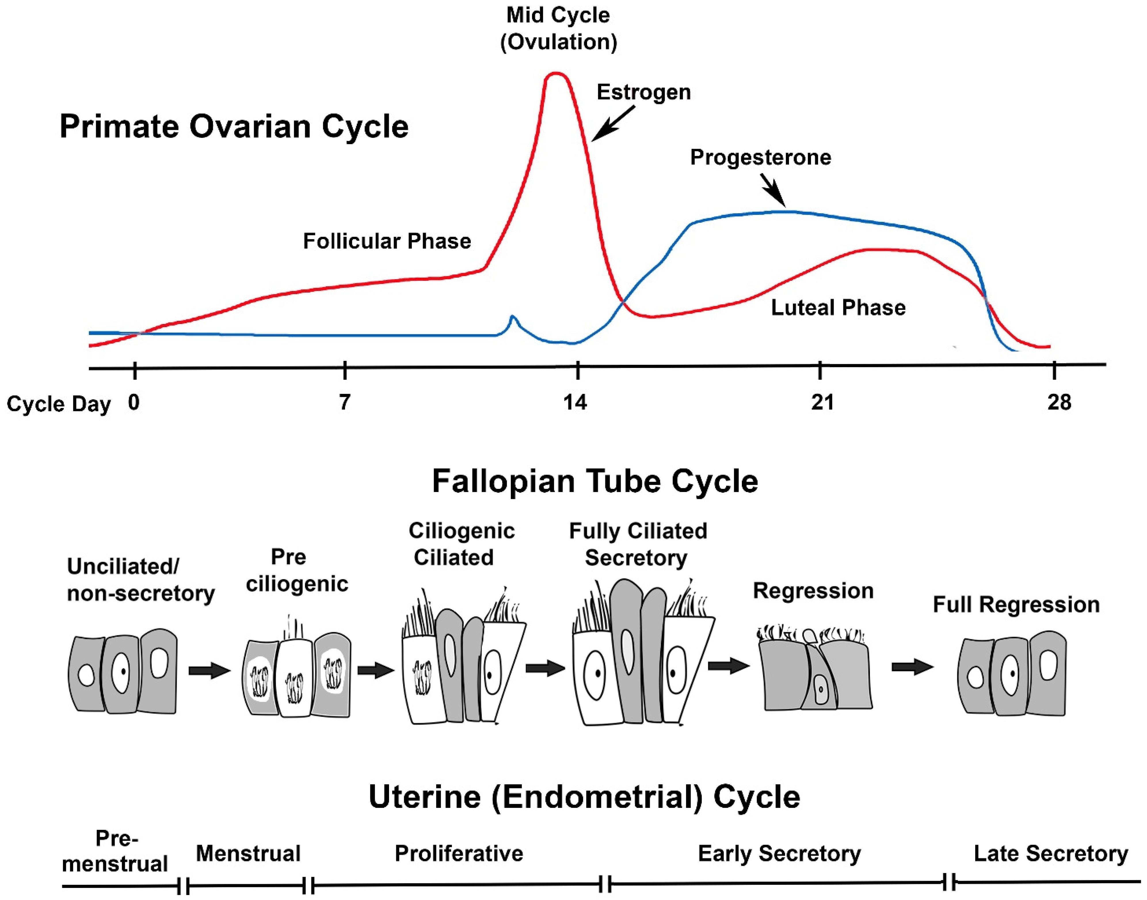
Figure 1. The fallopian tube cycle. Changes in the level of P4 in the presence of E2 drive cytologic changes in the fallopian tube epithelium. The upper panel shows an idealized primate ovarian cycle. In macaques, estrogen levels usually are 30–50 pg/mL at menstruation, rise during the follicular phase, and surge before ovulation. Post-ovulation, E2 declines but remains >50 pg/mL during the luteal phase. Progesterone (P4) levels are minimal in the follicular phase and rise significantly in the luteal phase. Estrogen levels ≥50 pg/mL drive cell proliferation in the pre-ciliogenic and ciliogenic/ciliated phases of the fallopian tube cycle and are necessary for maintaining differentiated ciliated secretory phenotype. The increase in P4 triggers regression of the epithelium despite continued E2 in the luteal phase. The lower panel depicts the phases of the uterine menstrual cycle, where the follicular and luteal phases are referred to as proliferative and secretory phases, respectively.
Cells 11 01534 g001
Figure 2. A photograph of the macaque reproductive tract (a) showing the uterus, oviductal infundibulum, ampulla, and isthmus. The inset shows the fallopian tube dissected free of peri-ovarian adipose and ligaments. Note the attachment of the fimbria to the ovary. Panels (bd) show paraffin-embedded and hematoxylin-eosin stained (H&E) sections of the infundibulum fimbria (b), ampulla (c), and isthmus (d) from a rhesus macaque in the luteal phase of the menstrual cycle. Arrows show the cuboidal epithelium of the endosalpinx. My = myosalpinx. Scale bar in (a) is 1 cm. Scale bars in (bd) represent approximately 100 μm.
Cells 11 01534 g002
Figure 3. Photomicrographs of macaque oviductal fimbria collected in the follicular and luteal phases of the menstrual cycle. Glycol methacrylate sections stained with gills hematoxylin from the follicular phase (a) show differentiation into a fully secretory state. Luteal phase (b) is fully regressed (scale bars in (a,b) are 30 μm). In the luteal phase, presentation of cilia and secretion are blocked by P4. Immunostaining for ERα (c,d) and PGR (e,f) in cryosections revealed that the ciliated cells had minimal receptor staining in the differentiated state, whereas secretory cells stained strongly for both ERα and PGR. In contrast, sections from the luteal phase have strong ERα staining in all the cell types, whereas PGR staining was lost from the epithelium and retained in the stroma (scale bar in (cf) is 50 μm). Together, this staining presentation implies that PGR regulation of oviductal differentiation may be indirect or mediated through non-classical mechanisms. Se = secretory cells; Ci = ciliated cells; S = stromal cells.
Cells 11 01534 g003
Table 1. Cyclic stages of the oviductal fimbria in macaques.
Oviduct StageUterine Cycle StageDescription
Full RegressionLate Luteal/Pre-MenstrualThe epithelium is cuboidal with few ciliated or secretory cells. The epithelial cell nuclei appear shrunken.
Pre-CiliogenicMenstruationThis state is marked by epithelial cellular hypertrophy and mitotic activity. Epithelial cell nuclei swell, smoothing the nuclear contours.
CiliogenicEarly FollicularCellular hypertrophy and mitotic activity continue. Histologically distinct light and dark cells can be identified, and cilia basal bodies are apparent in the apical cytoplasm of the light hypertrophied cells.
Ciliogenic CiliatedMid-FollicularMitotic activity has slowed; abundant ciliated cells and secretory cells are present. The word “ciliogenic” was placed first in the name of this phase to emphasize that ciliogenic cells predominate.
Ciliated CiliogenicLate FollicularThe majority of cells have become ciliated, but secretory cells have become much more prominent and have developed bulbous tips. The word “ciliated” was placed first in the name of this phase to emphasize that ciliated cells predominate over ciliogenic ones.
Ciliated SecretoryPeriovulatoryApproximately 50% of the epithelial cells are ciliated. The remaining epithelial cells are secretory with prominent bulbous tips. There is minimal mitotic activity.
Pre-RegressionEarly LutealThis phase is similar to the ciliated secretory state. There is a striking increase in epithelial apoptotic cells, and macrophages are phagocytosing the apoptotic cells.
RegressionMid-LutealThe epithelium secretion is reduced, and the epithelium is undergoing deciliation. EM studies reveal ciliated cells to be pinching off their tips. Epithelial cell nuclei now appear shriveled. At the end of the luteal phase, the epithelium appears deciliated and cuboidal.
Publisher’s Note: MDPI stays neutral with regard to jurisdictional claims in published maps and institutional affiliations.