Review

New Insights into Adipose Tissue Macrophages in Obesity and Insulin Resistance

Heart Center, Shanghai Chest Hospital, Shanghai Jiaotong University, 241 Huaihai West Road, Shanghai 200030, China
*
Author to whom correspondence should be addressed.
These authors contributed equally to this work.
Cells 2022, 11(9), 1424; https://doi.org/10.3390/cells11091424
Submission received: 26 March 2022 / Revised: 15 April 2022 / Accepted: 20 April 2022 / Published: 22 April 2022
(This article belongs to the Special Issue Adipose Tissue in Cardiovascular Health)

Abstract

:
Obesity has become a worldwide epidemic that poses a severe threat to human health. Evidence suggests that many obesity comorbidities, such as type 2 diabetes mellitus, steatohepatitis, and cardiovascular diseases, are related to obesity-induced chronic low-grade inflammation. Macrophages are the primary immune cells involved in obesity-associated inflammation in both mice and humans. Intensive research over the past few years has yielded tremendous progress in our understanding of the additional roles of adipose tissue macrophages (ATMs) beyond classical M1/M2 polarization in obesity and related comorbidities. In this review, we first characterize the diverse subpopulations of ATMs in the context of obesity. Furthermore, we review the recent advance on the role of the extensive crosstalk between adipocytes and ATMs in obesity. Finally, we focus on the extended crosstalk within adipose tissue between perivascular mesenchymal cells and ATMs. Understanding the pathological mechanisms that underlie obesity will be critical for the development of new intervention strategies to prevent or treat this disease and its associated co-morbidities.

1. Introduction

Obesity is a metabolic disease characterized by abnormal and excessive accumulation of body fat. Obesity increases the risk of developing a wide variety of diseases including but not limited to type 2 diabetes mellitus (T2DM) and cardiovascular diseases and has been strongly associated with increased mortality [1,2]. In the past few decades, the prevalence of obesity has increased dramatically in both developing and developed nations around the world [3,4]. More recent statistics indicate that 39% of adults aged 18 years and over were overweight, and 13% were obese worldwide in 2016 [5]. Obesity is a serious public health problem with major health and economic consequences. Therefore, it is imperative that we polish our understanding of the pathological mechanisms so we can develop effective strategies to prevent or treat obesity and related comorbidities.
Insulin resistance is a key component in the etiology of T2DM, and obesity is clearly the most common cause of insulin resistance in humans [6,7]. With the ongoing worldwide obesity epidemic, there has been a parallel rise in the prevalence of T2DM [8]. It has now been widely recognized that obesity-induced chronic low-grade tissue inflammation, particularly when occurring in adipose tissue, can cause insulin resistance and T2DM [9,10]. Under obese conditions, adipose tissue undergoes a series of dynamic remodeling, including adipocyte hypertrophy, apoptosis, immune cell infiltration, extensive vascularization, and extracellular matrix remodeling [11,12]. Macrophages are the primary immune cells strongly involved in obesity-associated inflammation in both mice and humans [7,13,14,15]. Emerging studies have implicated the crosstalk between adipocytes and adipose tissue macrophages (ATMs) as critical regulators of obesity-associated inflammation and metabolic complications.
In this review, we provide an overview of the distinct subpopulations of ATMs in the context of obesity, with particular attention paid to some novel subsets of ATMs characterized by single-cell or single-nucleus RNA-sequencing (sc/snRNA-seq) technologies. We also review the latest research progress on the extensive interactions between adipocytes and ATMs mediated by miRNA-containing exosomes or mitochondria transfer. Moreover, we also describe the extended crosstalk between perivascular mesenchymal cells and ATMs. Study searches in this review were performed using the PubMed database from the National Library of Medicine.

2. Adipose Tissue Macrophage (ATM) Subpopulations

White adipose tissue (WAT) is comprised of a versatile group of interacting cells, including adipocytes, immune cells, and other cell types. It is evident that ATMs, the most abundant immune cells in WAT, can even represent up to 40–50% of the cells in obese adipose tissue [13]. ATMs are an extraordinarily heterogeneous population of immune cells with varied and diverse functions (as summarized in Table 1), which have been highlighted as important factors contributing to the pathogenesis of obesity and related comorbidities. Historically, ATMs have been categorized into classically activated (M1-like) and alternatively activated (M2-like) macrophages [16]. However, more and more novel ATM subpopulations have been identified [14,15,17,18,19,20,21,22,23].
ATMs were thought to be composed of two main phenotypes: classically activated macrophages and alternatively activated macrophages, which are phenotypically and functionally distinct [16]. The classically activated macrophages represent pro-inflammatory M1-like macrophages, whereas the alternatively activated macrophages are anti-inflammatory M2-like macrophages. M1-like macrophages express F4/80, CD11b, and CD11c and secrete inflammatory factors including tumor necrosis factor-α (TNF-α), interleukin-1β (IL-1β), IL-6, leukotriene B4 (LTB4), and nitric oxide (NO); whereas M2-like macrophages express F4/80, CD11b, CD301, and CD206 and exhibit increased secretion of anti-inflammatory cytokines, such as IL-4 and IL-10 [26,27]. The main roles of ATMs under lean conditions are efferocytosis of dead adipocytes, the production of anti-inflammatory cytokines, the regulation of adipocyte lipolysis, and the restriction of adipocyte progenitor proliferation [28]. Conversely, as obesity progresses, most ATMs are converted from an anti-inflammatory (M2-like) phenotype into a pro-inflammatory (M1-like) phenotype, secreting pro-inflammatory cytokines (such as TNF-α, IL-1β) and causing localized and systemic chronic low-grade inflammation, especially in WAT [29]. The insulin resistance and T2DM would progress under the influence of this inflammatory state [30,31].
ATMs can be further widely divided into adipose tissue-resident macrophages and recruited monocyte-derived macrophages. Tissue-resident macrophages are long-lived and self-renewing cells thought to have originated during embryonic hematopoiesis [32], whereas monocyte-derived macrophages are short-lived cells recruited to adipose tissues during inflammation [33]. Recently, multiple novel populations of adipose tissue-resident macrophages have been discovered in adipose depots [21,22,34]. For example, a new subpopulation of ATMs, TIM4+ adipose tissue-resident macrophages, has recently been revealed to play essential roles in the formation and expansion of adipose tissue during the development and diet-induced obesity [21]. Another novel resident macrophage population (sympathetic neuron-associated macrophages, SAMs) has been discovered in adipose tissue localized around neurons of the sympathetic nervous system (SNS) that mediates noradrenaline clearance and dampens SNS-to-adipocyte communications [22,34].
With the recent advances in scRNA-seq technologies, emerging evidence suggests that ATMs exhibit a wider spectrum of phenotypes and cellular identities than previously described in the context of obesity both in mice and humans. By utilizing scRNA-seq technology, Hill et al. identified three discrete ATM populations (CD11b+ Ly6c+; CD11b+ Ly6c CD9+; CD11b+ Ly6c CD9), two of which (CD11b+ Ly6c+ and CD11b+ Ly6c CD9+) are associated with obesity [17]. CD11b+ Ly6c CD9+ ATMs reside within crown-like structures (CLS) and are lipid-laden and proinflammatory, whereas CD11b+ Ly6c+ ATMs reside outside CLSs and play angiogenic and adipogenic roles [17]. In a more recent study, Jaitin et al. provided a comprehensive single-cell adipose tissue immune atlas in mice and humans and described a novel Trem2+ ATM subpopulation, named lipid-associated macrophages (LAMs), in obese adipose tissue [14]. These LAMs use lipid receptor Trem2 as a sensor of extracellular lipids and play protective functions to counteract adipocyte hypertrophy, inflammation, and metabolic dysfunction [14]. Therefore, obese adipose tissue contains multiple distinct ATM populations with unique origins, tissue distributions, and functions. A comprehensive understanding of ATM heterogeneity in obesity is of great importance for the development of future therapies.

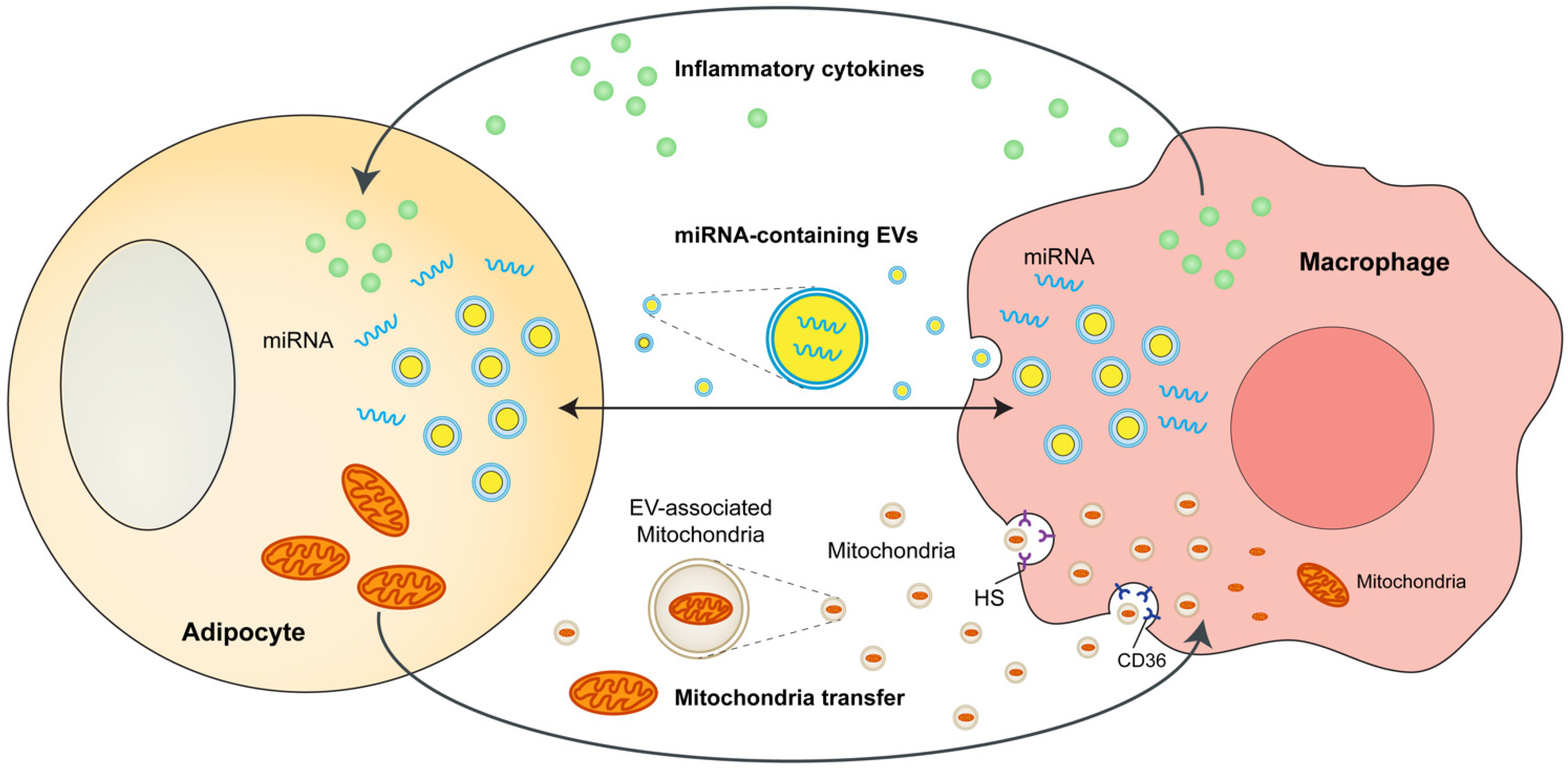
3. Adipocytes and ATMs Crosstalk

The crosstalk between adipocytes and macrophages in adipose tissues is crucial in obesity-induced metabolic complications. Adipocytes and macrophages can interact with each other through a variety of mechanisms, including cytokine and chemokines, microRNA-containing exosomes or microvesicles, and mitochondria transfer. These mechanisms are discussed in this section and partly summarized in Figure 1.

3.1. Cytokines and Chemokines as Mediators of Crosstalk

It is well established that cytokines and chemokines secreted by immune cells ignite localized and systemic inflammation, which builds up a pathogenic connection between obesity and insulin resistance. Moreover, cytokines and chemokines are the major mediators of ATM phenotype and crosstalk between adipocytes and ATMs which play critical roles in the pathogenesis of obesity and associated metabolic complications.
The first evidence for a pathophysiological link between obesity, inflammation, and insulin resistance was provided in 1901, when it was observed that the salicylate, an anti-inflammatory drug as the principal metabolite in aspirin, could beneficially control glucose in diabetics [35]. The concept was revisited, approximately a century later, when Hotamisligil et al. demonstrated that TNF-α was elevated in the adipose tissue of obese mice and neutralization of TNF-α ameliorated insulin resistance [36]. Subsequent studies reported that TNF-α-deficient mice were free from high-fat diet-induced insulin resistance [37]. Increased TNF-α production was also observed in the adipose tissue of obese humans, while the decline of TNF-α level was associated with weight loss in humans [38,39]. A milestone forward in this field was provided by two simultaneous publications that reported independently that obesity was associated with macrophage accumulation in adipose tissue, which was known as the major source of inflammatory mediators (such as TNF-α) [13,40]. Mounting studies have demonstrated that monocyte-derived macrophages are recruited into tissues (particularly into adipose tissue) via C-C chemokine receptor type 2 (CCR2) and secrete inflammatory cytokines such as TNF-α during obesity, thereby causing systemic inflammation and insulin resistance. Therefore, all these studies suggested the central role of macrophage-secreted inflammatory cytokines in the development of obesity and related comorbidities.
Many other inflammatory cytokines and mediators such as IL-1β, IL-6, monocyte chemotactic protein 1 (MCP-1), and macrophage inhibitory factor (MIF) have been implicated in the pathogenesis of insulin resistance [26,41,42,43,44,45]. The production of inflammatory cytokines during obesity has been well demonstrated to be regulated by the signaling pathway of inhibitor of κB kinase-β (IKK-β) and nuclear factor-κB (NF-κB) [46,47], c-Jun N-terminal kinase (JNK) [48,49], and the NLR family pyrin domain containing 3 (NLRP3) inflammasome [50,51,52]. There is enormous and excellent literature available on this topic [53,54,55,56]. It is worth noting that novel pathways were found to regulate the inflammatory processes. For example, Yao et al. demonstrated that the iroquois homeobox gene3 (IRX3) in ATMs functions as a novel transcriptional factor for cytokine expression and therefore accelerates the development of obesity and T2DM [57]. In addition, Fgr tyrosine kinase, which is activated by reactive oxygen species (ROS), has been highlighted as a key regulator for proinflammatory macrophages during diet-induced obesity [58].
Although dramatic progress has been made in our understanding of the role of inflammation in obesity-associated insulin resistance, clinical trials targeting inflammatory mediators of obesity and T2DM (such as TNF-α and IL-1β) to improve glycemic control in T2DM have shown limited benefits [25,59], indicating that there must be other factors contributing to decreased insulin sensitivity. Bu et al. demonstrated that growth/differentiation factor 3 (GDF3) produced from CD11c+ ATMs acts as a ligand of ALK7 in adipocytes to inhibit lipolysis and expand adipose tissue under obese conditions [60]. The GDF3-ALK7 signaling pathway within WAT might represent an important interactive mechanism between adipocytes and ATMs in the modulation of adiposity [23,60]. In a more recent study, Sharma et al. demonstrated that the neuroimmune guidance cue netrin-1 is essential for orchestrating macrophage fate and function that accumulate in the obese adipose tissue [61]. Myeloid-specific netrin-1 deletion reduces the ATM accumulation in adipose tissue and reprograms the ATM phenotype during obesity. Therefore, mice lacking netrin-1 in myeloid cells are protected from diet-induced obesity and metabolic dysfunction [61]. In addition, Wang et al. identified a novel cytokine Slit3 which was secreted by M2-like macrophages under cold exposure and regulated WAT beiging via stimulating norepinephrine secretion [57].

3.2. MicroRNA and Exosomes as Novel Mediators of Crosstalk

MicroRNAs (miRNAs) are small non-coding RNAs consisting of 19–22 nucleotides that govern gene expression at the post-transcriptional level via inhibiting the translation of target mRNA [62,63]. Mature miRNAs are formed inside the cell and perform various functions in the cytoplasm or after being released into the extracellular spaces, blood circulation, and other body fluids, (i.e., urine and saliva) [64]. Notably, miRNAs can be packaged into extracellular vesicles (EVs), including exosomes and microvesicles [65]. These EVs, cell-derived membranous structures, can deliver functional miRNAs to target cells, thereby exerting intercellular communications and interorgan crosstalk [66].
The importance of miRNAs in the immunometabolism field is increasingly recognized. Mounting evidence suggests that in addition to cytokines and chemokines, miRNAs or miRNA-containing exosomes also mediate powerful paracrine functions between ATMs and adipocytes. New mechanistic pathways involving the secretion of miRNA-containing exosomes [67,68] or microvesicles [69] by adipocytes or ATMs have been demonstrated to facilitate metabolic and inflammatory interactions between adipocytes and ATMs as well as distal target tissues, which can regulate insulin sensitivity and modulate metabolic homeostasis. Therefore, miRNAs have now been recognized as a new class of endocrine factors and are strongly implicated in the pathogenesis of obesity and related comorbidities.
Evidence is accumulating that numerous adipocyte-derived miRNAs can modulate macrophage phenotype and function and regulate insulin sensitivity via their paracrine actions. For instance, miR-155 is secreted by adipocyte-derived microvesicles from obese adipose tissue in mice. Adipocyte-derived miR-155 downregulates the protein level of suppressor of cytokine signaling 1 (SOCS1), one of its proven targets [70], and promotes pro-inflammatory M1-like macrophage polarization [69]. MiR-27a, a proven adipogenic miRNA [71,72], is significantly upregulated in the serum of obese mice and humans [73,74]. Adipocyte-derived exosomal MiR-27a facilitates macrophage activation and induces insulin resistance via inhibiting PPARγ during diet-induced obesity [73,74]. Moreover, miR-34a is elevated in the adipose tissue of obese mice and clinical patients. Adipocyte-specific depletion of miR-34a reprograms ATMs from pro-inflammatory M1-like to anti-inflammatory M2-like phenotype and protects mice from obesity-associated inflammation, glucose intolerance, insulin resistance through repressing Krüppel-like factor 4 (KLF4) expression [75]. Taken together, this evidence suggests that adipocyte-derived microvesicles or exosomes can transport miRNAs to mediate adipocyte-macrophage communications and modulate metabolic homeostasis.
ATMs can also regulate in vitro and/or in vivo insulin sensitivity by secretion of miRNA-containing exosomes. Ying et al. found that treatment of lean mice with ATM-derived exosomes harvested from obese mice causes glucose intolerance and insulin resistance, whereas treatment of obese mice with lean ATM-derived exosomes improves insulin resistance [68]. Notably, miR-155 is demonstrated to be one of the miRNAs overexpressed in obese ATM-derived exosomes which mediates the deleterious effects [68]. Moreover, it is demonstrated that mice with miR-155 knockout exhibit improved insulin sensitivity when fed with high fat diet [68]. In another work, Ying et al. treated mouse bone marrow-derived macrophages (BMDMs) with IL-4 and IL-13 to induce M2 BMDMs generation and harvested the released exosomes [76]. Treatment with M2 BMDMs-derived exosomes can improve insulin sensitivity in vivo in obese mice and in vitro (in adipocyte, myocyte, and hepatocyte) [76]. Moreover, as the authors illustrated, miR-690 is highly expressed within M2 BMDM-derived exosomes and acts as a key insulin-sensitizing miRNA which improves insulin sensitivity [76]. In addition, Liu et al. demonstrated that miR-29a is increased in ATM-derived exosomes from obese adipose tissue of mice which can be transferred into adipocytes, myocytes, and hepatocytes causing insulin resistance in vitro and in vivo [77]. These works suggest that ATM-derived exosomal miRNAs can regulate insulin sensitivity and metabolic homeostasis.
In summary, adipocytes and ATMs, residing within adipose tissue, can secrete miRNA-containing exosomes or microvesicles and elicit phenotypes via various mechanisms in obesity and associated complications. Therefore, it is promising that obesity-associated miRNAs are new therapeutic targets for obesity and related diseases.

3.3. Mitochondria Transfer as Novel Mediators of Crosstalk

Mitochondria are essential organelles within eukaryotic cells and play important roles in the processes such as oxidative phosphorylation, energy production, metabolic homeostasis, redox regulation, and apoptosis [78,79]. Therefore, it is not surprising that mitochondrial dysfunction is associated with many diseases, such as metabolic diseases and neurodegenerative disorders. Intercellular mitochondria transfer has attracted more and more attention within the scientific community in recent studies.
The first evidence for functional mitochondria transfer comes from the report that genetic defects in mtDNA of A549 ρ° cells can be rescued by the transfer of healthy mitochondria from human stem/progenitor cells [80]. Emerging evidence now exists suggesting that cells can release functional mitochondria that are transferred to and captured by recipient cells [81,82,83,84]. Notably, mitochondria also can be loaded in EVs and acquired by recipient cells via an EV-cell fusion manner [85,86]. Moreover, it is noted that functional mitochondria, EV-related mitochondria, and mitochondria DNA can circulate in blood both in mice and humans [84,87,88]. The intercellular mitochondrial transfer has been implicated in a variety of physiological and pathological processes, including cardiac homeostasis [89], acute lung injury repair [83], pulmonary hypertension [90], allograft rejection [87], and ischemic stroke [91].
Mitochondria transfer is emerging as a novel mediator of endogenous crosstalk between adipocytes and macrophages within adipose tissue. Brestoff et al. demonstrated that intercellular mitochondria transfer occurs in mice WAT and identified a novel adipocyte-to-macrophage mitochondria transfer axis that modulates energy homeostasis and is impaired in obesity [92]. These findings implicated a decrease in mitochondria transfer in WAT as a sign of obesity, which might be considered a promising therapeutic target [92]. It is demonstrated that intercellular mitochondria transfer is mediated by a heparan sulfates (HS)-dependent mechanism in vitro and in vivo [92]. Moreover, mice with myeloid cell-specific deletion of HS biosynthetic gene Ext1 exhibit impaired mitochondria transfer to macrophages and increased susceptibility to diet-induced obesity [92]. In a more recent study, Rosina et al. reported that thermogenic stimuli induce the EVs released from brown adipocyte which contains damaged mitochondria [93]. These brown adipocyte-derived EVs are captured and removed by brown adipose tissue (BAT)-resident macrophages via the CD36-lysosome pathway, thereby maintaining the BAT thermogenic program [93]. Macrophage depletion causes the accumulation of mitochondria-containing EVs, suppresses the expression of mitochondrial proteins and thermogenic genes, and inhibits BAT’s thermogenic response to cold exposure [93]. These findings highlighted the brown adipocyte-to-macrophage mitochondria transfer axis as an important regulator to maintain brown adipocyte mitochondria quality control and preserve BAT homeostasis [93]. The authors even postulate that failure to remove damaged mitochondria in BAT due to impaired EV production or macrophage phagocytic activity might contribute to the progressive impairment of BAT function during obesity [93].
These findings illustrate a new pattern of immunometabolic crosstalk, adipocyte–macrophage mitochondria transfer, which contributes to maintaining systemic metabolic homeostasis. Further work is needed to elucidate how the mitochondria transfer to macrophages affects metabolism homeostasis. However, treatment targeting intercellular mitochondrial transfer may be a promising strategy for the treatment of obesity and related comorbidities.

4. Adipose Tissue-Resident Macrophages Directly Regulate Adiposity and Energy Storage

In addition to short-lived monocyte-derived macrophages originating from hematopoietic stem cells, there are long-lived tissue-resident macrophages that serve tissue-specific purposes [94]. However, whether adipose tissue-resident macrophages serve their tissue-specific purposes and support the function of energy storage in adipose tissue has not been completely understood.
A new subpopulation of ATMs, CCR2-independent TIM4+ resident macrophages, has recently been reported to modulate adiposity and energy storage in a paracrine manner via the production of platelet-derived growth factor (PDGFcc) in mice WAT [21]. Genetic deletion and pharmacological blockade of PDGFcc reduce adiposity, energy storage, and body weight, and redirect excess lipids mostly toward thermogenesis [21]. This study challenges the outdated M1/M2 macrophage polarization model in which adipose tissue-resident macrophages can serve to sense increased nutritional status and support energy storage, whereas recruited macrophages are responsible for characterizing systemic inflammation of obesity and metabolic complications. Therefore, these data strongly indicate that different developmental subsets of macrophages (including adipose tissue-resident macrophages and recruited monocyte-derived macrophages) exert different functions within adipose tissue and are independent targets of CCR2 and PDGFcc blockade. This study highlighted the additional roles of macrophages beyond classical M1/M2 polarization in obesity development and has the potential to inspire new immunomodulatory therapies that could separately manipulate energy storage and inflammation during obesity.

5. Sympathetic Neuron-Associated Macrophages Indirectly Affect Energy Storage

Adipose tissue is densely innervated by the sympathetic nervous system (SNS), which locally releases noradrenaline into adipose tissue and drives lipolysis and brown or beige adipocyte thermogenesis [24,95,96]. Recent advances in three-dimensional adipose tissue imaging have improved our understanding of how different cell types in adipose tissue are organized and how they interact with one another [97,98]. Moreover, the interwoven relationships between adipocytes, sympathetic nerves, and immune cells in the context of obesity have attracted widespread attention.
In 2017, two landmark studies simultaneously identified noradrenaline-degrading macrophage populations in WAT, which directly modulate the sympathetic innervation of adipocytes [22,23]. Pirzgalska et al. demonstrated that adipose tissue-residing SAMs exhibit specialized morphology for association with SNS neurons in WAT [22]. These SAMs that import and catabolize noradrenaline via noradrenaline transporter (SLC6A2) and degradation enzyme (MAMO) are dramatically increased under obese conditions [22]. Mice with genetic deletion of SLC6A2 from SAMs are resistant to obesity owing to decreased noradrenaline removal, enhanced SNS-to-adipocyte communications, and increased SNS-driven lipolysis [22]. In another study, Camell et al. found that a specialized ATM subpopulation (a SAM-like macrophage population) was activated in aged mice [34]. They further demonstrated these ATMs regulate the age-related reduction in adipocyte lipolysis in adipose tissue by degrading norepinephrine in an inflammasome-dependent manner [34]. In addition, in the more recent study by Wang et al., it is demonstrated that under cold exposure M2-like macrophages secret Slit3, which binds to ROBO1 receptor on sympathetic neurons and stimulates noradrenaline release, leading to enhanced white adipocyte beiging and thermogenesis [99]. These studies identify a sympathetic neuroimmunological role for macrophages in obesity and have opened up a whole new field of neuroimmunometabolism.
It is now well demonstrated that BAT is much more densely innervated by sympathetic nerves than WAT [100]. The triangular relationship between brown adipocytes, macrophages, and SNS has also been recognized. Wolf et al. reported that the nuclear transcription factor MECP2 is an important modulator of BAT function [101]. Mice lacking MECP2 spontaneously develop obesity due to the impairment of BAT function [101]. Further mechanistic investigation indicates that MECP2-deficient macrophage upregulates PlexinA4 expression which prevents the axonal outgrowth of Sema6A+ nerves and diminishes sympathetic innervation of BAT [101].

6. Novel View—Cross-Talk between Perivascular Mesenchymal Cells and ATMs

Although recent research mainly focuses on the role of adipocytes and macrophages in the development of metabolic adipose tissue inflammation, a new study recently highlighted that perivascular mesenchymal cells play a significant role in the regulation of chronic adipose tissue inflammation during obesity.
In the study, Shan et al. utilized scRNA-seq and identified a mouse WAT perivascular cell subpopulation, named fibro-inflammatory progenitors (FIPs) that stimulate pro-inflammatory signaling and modulate accumulation of pro-inflammatory macrophage in the adipose tissue during obesity [102]. These perivascular mesenchymal cells of the adipose tissue are critical “gatekeepers” of macrophage accumulation in obesity. It is also reported that the transcriptional regulator zinc-finger protein 423 (ZFP423) governs the inflammatory response of perivascular mesenchymal cells [102]. Using in vitro studies and in vivo mouse genetic models they determined that ZFP423 modulates NF-κB activity and that expression of ZFP423 in perivascular mesenchymal cells suppresses inflammatory signaling in FIPs and attenuates metabolic inflammation in obesity [102]. These studies highlighted an important role for perivascular mesenchymal cells in the modulation of chronic inflammation in adipose tissue during obesity, and indicate that proinflammatory perivascular mesenchymal cells are potential targets for therapeutic treatment tailored to control obesity and associated co-morbidities.

7. Conclusions

Mounting evidence has revealed that metabolism and immunity are inextricably interwoven. Adipocytes and ATMs are the two most important cells within adipose tissue which mediate obesity-associated inflammation and metabolic compilations. The interactions between adipocytes and ATMs have been implicated to have a key role in the development of obesity. Recently, new evidence has shown that aside from the well-known cytokines and chemokines, miRNA-containing exosomes and mitochondria transfer are also important mediators for cell–cell and organ–organ communications, especially adipocytes and macrophages interaction in obesity. Moreover, it is found that ATMs have additional roles beyond M1-M2 polarization and modulate adiposity and energy storage directly and indirectly. In addition, it is demonstrated that adipose tissue-derived perivascular mesenchymal cells are a significant “safeguard” of macrophage accrual in obesity. In a word, we now have a much deeper understanding of the sophisticated interactions between macrophages and adipocytes during obesity. This progress reveals several advances that may be exploited in treatment to suppress the inflammatory response of effector ATM and treat obesity and related complications.

Author Contributions

The conceptualization, design, and execution of the concept was performed by Z.C. and B.H. Initial literature collection reading and drafting of the review article material was conducted by Z.C. and Y.H. The final draft writing, revision, and updating of the literature and figures was conducted by Z.C. and B.H. The final draft was revised by B.H. All authors have read and agreed to the published version of the manuscript.

Funding

This research was funded by by the National Natural Science Foundation of China (grant numbers: 81830010, 82130012) and Clinical Research Plan of SHDC (grant number: SHDC2020CR1039B).

Conflicts of Interest

The authors declare no conflict of interest.

References

  1. Whitlock, G.; Lewington, S.; Sherliker, P.; Clarke, R.; Emberson, J.; Halsey, J.; Qizilbash, N.; Collins, R.; Peto, R. Body-mass index and cause-specific mortality in 900 000 adults: Collaborative analyses of 57 prospective studies. Lancet 2009, 373, 1083–1096. [Google Scholar] [CrossRef] [PubMed] [Green Version]
  2. Flegal, K.M.; Kit, B.K.; Orpana, H.; Graubard, B.I. Association of all-cause mortality with overweight and obesity using standard body mass index categories: A systematic review and meta-analysis. JAMA 2013, 309, 71–82. [Google Scholar] [CrossRef] [PubMed] [Green Version]
  3. Ng, M.; Fleming, T.; Robinson, M.; Thomson, B.; Graetz, N.; Margono, C.; Mullany, E.C.; Biryukov, S.; Abbafati, C.; Abera, S.F.; et al. Global, regional, and national prevalence of overweight and obesity in children and adults during 1980–2013: A systematic analysis for the Global Burden of Disease Study 2013. Lancet 2014, 384, 766–781. [Google Scholar] [CrossRef] [Green Version]
  4. Afshin, A.; Forouzanfar, M.H.; Reitsma, M.B.; Sur, P.; Estep, K.; Lee, A.; Marczak, L.; Mokdad, A.H.; Moradi-Lakeh, M.; Naghavi, M.; et al. Health Effects of Overweight and Obesity in 195 Countries over 25 Years. N. Engl. J. Med. 2017, 377, 13–27. [Google Scholar] [CrossRef] [PubMed]
  5. World Health Organization. Obesity and Overweight. 2021. Available online: https://www.who.int/news-room/fact-sheets/detail/obesity-and-overweight (accessed on 9 June 2021).
  6. Kahn, S.E.; Hull, R.L.; Utzschneider, K.M. Mechanisms linking obesity to insulin resistance and type 2 diabetes. Nature 2006, 444, 840–846. [Google Scholar] [CrossRef] [PubMed]
  7. Johnson, A.M.; Olefsky, J.M. The origins and drivers of insulin resistance. Cell 2013, 152, 673–684. [Google Scholar] [CrossRef] [PubMed] [Green Version]
  8. World Health Organization. Diabetes. 2021. Available online: https://www.who.int/news-room/fact-sheets/detail/diabetes (accessed on 10 November 2021).
  9. Heilbronn, L.K.; Campbell, L.V. Adipose tissue macrophages, low grade inflammation and insulin resistance in human obesity. Curr. Pharm. Des. 2008, 14, 1225–1230. [Google Scholar] [CrossRef]
  10. Lumeng, C.N.; Saltiel, A.R. Inflammatory links between obesity and metabolic disease. J. Clin. Investig. 2011, 121, 2111–2117. [Google Scholar] [CrossRef] [Green Version]
  11. Marcelin, G.; Silveira, A.L.M.; Martins, L.B.; Ferreira, A.V.; Clement, K. Deciphering the cellular interplays underlying obesity-induced adipose tissue fibrosis. J. Clin. Investig. 2019, 129, 4032–4040. [Google Scholar] [CrossRef]
  12. Choe, S.S.; Huh, J.Y.; Hwang, I.J.; Kim, J.I.; Kim, J.B. Adipose Tissue Remodeling: Its Role in Energy Metabolism and Metabolic Disorders. Front. Endocrinol. 2016, 7, 30. [Google Scholar] [CrossRef] [Green Version]
  13. Weisberg, S.P.; McCann, D.; Desai, M.; Rosenbaum, M.; Leibel, R.L.; Ferrante, A.W., Jr. Obesity is associated with macrophage accumulation in adipose tissue. J. Clin. Investig. 2003, 112, 1796–1808. [Google Scholar] [CrossRef] [PubMed]
  14. Jaitin, D.A.; Adlung, L.; Thaiss, C.A.; Weiner, A.; Li, B.; Descamps, H.; Lundgren, P.; Bleriot, C.; Liu, Z.; Deczkowska, A.; et al. Lipid-Associated Macrophages Control Metabolic Homeostasis in a Trem2-Dependent Manner. Cell 2019, 178, 686–698.e614. [Google Scholar] [CrossRef] [PubMed]
  15. Emont, M.P.; Jacobs, C.; Essene, A.L.; Pant, D.; Tenen, D.; Colleluori, G.; Di Vincenzo, A.; Jorgensen, A.M.; Dashti, H.; Stefek, A.; et al. A single-cell atlas of human and mouse white adipose tissue. Nature 2022, 603, 926–933. [Google Scholar] [CrossRef] [PubMed]
  16. Hill, A.A.; Reid Bolus, W.; Hasty, A.H. A decade of progress in adipose tissue macrophage biology. Immunol. Rev. 2014, 262, 134–152. [Google Scholar] [CrossRef] [Green Version]
  17. Hill, D.A.; Lim, H.W.; Kim, Y.H.; Ho, W.Y.; Foong, Y.H.; Nelson, V.L.; Nguyen, H.C.B.; Chegireddy, K.; Kim, J.; Habertheuer, A.; et al. Distinct macrophage populations direct inflammatory versus physiological changes in adipose tissue. Proc. Natl. Acad. Sci. USA 2018, 115, E5096–E5105. [Google Scholar] [CrossRef] [Green Version]
  18. Burl, R.B.; Ramseyer, V.D.; Rondini, E.A.; Pique-Regi, R.; Lee, Y.H.; Granneman, J.G. Deconstructing Adipogenesis Induced by beta3-Adrenergic Receptor Activation with Single-Cell Expression Profiling. Cell Metab. 2018, 28, 300–309.e4. [Google Scholar] [CrossRef] [Green Version]
  19. Sarvari, A.K.; Van Hauwaert, E.L.; Markussen, L.K.; Gammelmark, E.; Marcher, A.B.; Ebbesen, M.F.; Nielsen, R.; Brewer, J.R.; Madsen, J.G.S.; Mandrup, S. Plasticity of Epididymal Adipose Tissue in Response to Diet-Induced Obesity at Single-Nucleus Resolution. Cell Metab. 2021, 33, 437–453.e435. [Google Scholar] [CrossRef]
  20. Harasymowicz, N.S.; Rashidi, N.; Savadipour, A.; Wu, C.L.; Tang, R.; Bramley, J.; Buchser, W.; Guilak, F. Single-cell RNA sequencing reveals the induction of novel myeloid and myeloid-associated cell populations in visceral fat with long-term obesity. FASEB J. 2021, 35, e21417. [Google Scholar] [CrossRef]
  21. Cox, N.; Crozet, L.; Holtman, I.R.; Loyher, P.L.; Lazarov, T.; White, J.B.; Mass, E.; Stanley, E.R.; Elemento, O.; Glass, C.K.; et al. Diet-regulated production of PDGFcc by macrophages controls energy storage. Science 2021, 373, eabe9383. [Google Scholar] [CrossRef]
  22. Pirzgalska, R.M.; Seixas, E.; Seidman, J.S.; Link, V.M.; Sanchez, N.M.; Mahu, I.; Mendes, R.; Gres, V.; Kubasova, N.; Morris, I.; et al. Sympathetic neuron-associated macrophages contribute to obesity by importing and metabolizing norepinephrine. Nat. Med. 2017, 23, 1309–1318. [Google Scholar] [CrossRef]
  23. Andersson, O.; Korach-Andre, M.; Reissmann, E.; Ibanez, C.F.; Bertolino, P. Growth/differentiation factor 3 signals through ALK7 and regulates accumulation of adipose tissue and diet-induced obesity. Proc. Natl. Acad. Sci. USA 2008, 105, 7252–7256. [Google Scholar] [CrossRef] [PubMed] [Green Version]
  24. Madden, K.S. Sympathetic neural-immune interactions regulate hematopoiesis, thermoregulation and inflammation in mammals. Dev. Comp. Immunol. 2017, 66, 92–97. [Google Scholar] [CrossRef] [PubMed] [Green Version]
  25. Dominguez, H.; Storgaard, H.; Rask-Madsen, C.; Steffen Hermann, T.; Ihlemann, N.; Baunbjerg Nielsen, D.; Spohr, C.; Kober, L.; Vaag, A.; Torp-Pedersen, C. Metabolic and vascular effects of tumor necrosis factor-alpha blockade with etanercept in obese patients with type 2 diabetes. J. Vasc. Res. 2005, 42, 517–525. [Google Scholar] [CrossRef] [PubMed]
  26. Olefsky, J.M.; Glass, C.K. Macrophages, inflammation, and insulin resistance. Annu. Rev. Physiol. 2010, 72, 219–246. [Google Scholar] [CrossRef] [PubMed]
  27. Lumeng, C.N.; Bodzin, J.L.; Saltiel, A.R. Obesity induces a phenotypic switch in adipose tissue macrophage polarization. J. Clin. Investig. 2007, 117, 175–184. [Google Scholar] [CrossRef] [Green Version]
  28. Nawaz, A.; Aminuddin, A.; Kado, T.; Takikawa, A.; Yamamoto, S.; Tsuneyama, K.; Igarashi, Y.; Ikutani, M.; Nishida, Y.; Nagai, Y.; et al. CD206(+) M2-like macrophages regulate systemic glucose metabolism by inhibiting proliferation of adipocyte progenitors. Nat. Commun. 2017, 8, 286. [Google Scholar] [CrossRef]
  29. Hildreth, A.D.; Ma, F.; Wong, Y.Y.; Sun, R.; Pellegrini, M.; O’Sullivan, T.E. Single-cell sequencing of human white adipose tissue identifies new cell states in health and obesity. Nat. Immunol. 2021, 22, 639–653. [Google Scholar] [CrossRef]
  30. Boutens, L.; Stienstra, R. Adipose tissue macrophages: Going off track during obesity. Diabetologia 2016, 59, 879–894. [Google Scholar] [CrossRef] [Green Version]
  31. Russo, L.; Lumeng, C.N. Properties and functions of adipose tissue macrophages in obesity. Immunology 2018, 155, 407–417. [Google Scholar] [CrossRef]
  32. Perdiguero, E.G.; Geissmann, F. The development and maintenance of resident macrophages. Nat. Immunol. 2016, 17, 2–8. [Google Scholar] [CrossRef] [Green Version]
  33. Tsou, C.L.; Peters, W.; Si, Y.; Slaymaker, S.; Aslanian, A.M.; Weisberg, S.P.; Mack, M.; Charo, I.F. Critical roles for CCR2 and MCP-3 in monocyte mobilization from bone marrow and recruitment to inflammatory sites. J. Clin. Investig. 2007, 117, 902–909. [Google Scholar] [CrossRef] [PubMed] [Green Version]
  34. Camell, C.D.; Sander, J.; Spadaro, O.; Lee, A.; Nguyen, K.Y.; Wing, A.; Goldberg, E.L.; Youm, Y.H.; Brown, C.W.; Elsworth, J.; et al. Inflammasome-driven catecholamine catabolism in macrophages blunts lipolysis during ageing. Nature 2017, 550, 119–123. [Google Scholar] [CrossRef] [PubMed]
  35. Williamson, R.T. On the Treatment of Glycosuria and Diabetes Mellitus with Sodium Salicylate. Br. Med. J. 1901, 1, 760–762. [Google Scholar] [CrossRef] [PubMed] [Green Version]
  36. Hotamisligil, G.S.; Shargill, N.S.; Spiegelman, B.M. Adipose expression of tumor necrosis factor-alpha: Direct role in obesity-linked insulin resistance. Science 1993, 259, 87–91. [Google Scholar] [CrossRef]
  37. Uysal, K.T.; Wiesbrock, S.M.; Marino, M.W.; Hotamisligil, G.S. Protection from obesity-induced insulin resistance in mice lacking TNF-alpha function. Nature 1997, 389, 610–614. [Google Scholar] [CrossRef]
  38. Hotamisligil, G.S.; Arner, P.; Caro, J.F.; Atkinson, R.L.; Spiegelman, B.M. Increased adipose tissue expression of tumor necrosis factor-alpha in human obesity and insulin resistance. J. Clin. Investig. 1995, 95, 2409–2415. [Google Scholar] [CrossRef]
  39. Kern, P.A.; Saghizadeh, M.; Ong, J.M.; Bosch, R.J.; Deem, R.; Simsolo, R.B. The expression of tumor necrosis factor in human adipose tissue. Regulation by obesity, weight loss, and relationship to lipoprotein lipase. J. Clin. Investig. 1995, 95, 2111–2119. [Google Scholar] [CrossRef] [Green Version]
  40. Xu, H.; Barnes, G.T.; Yang, Q.; Tan, G.; Yang, D.; Chou, C.J.; Sole, J.; Nichols, A.; Ross, J.S.; Tartaglia, L.A.; et al. Chronic inflammation in fat plays a crucial role in the development of obesity-related insulin resistance. J. Clin. Investig. 2003, 112, 1821–1830. [Google Scholar] [CrossRef]
  41. Feve, B.; Bastard, J.P. The role of interleukins in insulin resistance and type 2 diabetes mellitus. Nat. Rev. Endocrinol. 2009, 5, 305–311. [Google Scholar] [CrossRef]
  42. Donath, M.Y. Targeting inflammation in the treatment of type 2 diabetes: Time to start. Nat. Rev. Drug Discov. 2014, 13, 465–476. [Google Scholar] [CrossRef]
  43. Reilly, S.M.; Saltiel, A.R. Adapting to obesity with adipose tissue inflammation. Nat. Rev. Endocrinol. 2017, 13, 633–643. [Google Scholar] [CrossRef] [PubMed]
  44. McLaughlin, T.; Ackerman, S.E.; Shen, L.; Engleman, E. Role of innate and adaptive immunity in obesity-associated metabolic disease. J. Clin. Investig. 2017, 127, 5–13. [Google Scholar] [CrossRef] [PubMed] [Green Version]
  45. Larabee, C.M.; Neely, O.C.; Domingos, A.I. Obesity: A neuroimmunometabolic perspective. Nat. Rev. Endocrinol. 2020, 16, 30–43. [Google Scholar] [CrossRef] [PubMed]
  46. Yin, M.J.; Yamamoto, Y.; Gaynor, R.B. The anti-inflammatory agents aspirin and salicylate inhibit the activity of I(kappa)B kinase-beta. Nature 1998, 396, 77–80. [Google Scholar] [CrossRef] [PubMed]
  47. Yuan, M.; Konstantopoulos, N.; Lee, J.; Hansen, L.; Li, Z.W.; Karin, M.; Shoelson, S.E. Reversal of obesity- and diet-induced insulin resistance with salicylates or targeted disruption of Ikkbeta. Science 2001, 293, 1673–1677. [Google Scholar] [CrossRef]
  48. Hirosumi, J.; Tuncman, G.; Chang, L.; Gorgun, C.Z.; Uysal, K.T.; Maeda, K.; Karin, M.; Hotamisligil, G.S. A central role for JNK in obesity and insulin resistance. Nature 2002, 420, 333–336. [Google Scholar] [CrossRef]
  49. Sabio, G.; Das, M.; Mora, A.; Zhang, Z.; Jun, J.Y.; Ko, H.J.; Barrett, T.; Kim, J.K.; Davis, R.J. A stress signaling pathway in adipose tissue regulates hepatic insulin resistance. Science 2008, 322, 1539–1543. [Google Scholar] [CrossRef] [Green Version]
  50. Vandanmagsar, B.; Youm, Y.H.; Ravussin, A.; Galgani, J.E.; Stadler, K.; Mynatt, R.L.; Ravussin, E.; Stephens, J.M.; Dixit, V.D. The NLRP3 inflammasome instigates obesity-induced inflammation and insulin resistance. Nat. Med. 2011, 17, 179–188. [Google Scholar] [CrossRef]
  51. Stienstra, R.; van Diepen, J.A.; Tack, C.J.; Zaki, M.H.; van de Veerdonk, F.L.; Perera, D.; Neale, G.A.; Hooiveld, G.J.; Hijmans, A.; Vroegrijk, I.; et al. Inflammasome is a central player in the induction of obesity and insulin resistance. Proc. Natl. Acad. Sci. USA 2011, 108, 15324–15329. [Google Scholar] [CrossRef] [PubMed] [Green Version]
  52. Youm, Y.H.; Nguyen, K.Y.; Grant, R.W.; Goldberg, E.L.; Bodogai, M.; Kim, D.; D’Agostino, D.; Planavsky, N.; Lupfer, C.; Kanneganti, T.D.; et al. The ketone metabolite beta-hydroxybutyrate blocks NLRP3 inflammasome-mediated inflammatory disease. Nat. Med. 2015, 21, 263–269. [Google Scholar] [CrossRef] [Green Version]
  53. Osborn, O.; Olefsky, J.M. The cellular and signaling networks linking the immune system and metabolism in disease. Nat. Med. 2012, 18, 363–374. [Google Scholar] [CrossRef] [PubMed]
  54. Lackey, D.E.; Olefsky, J.M. Regulation of metabolism by the innate immune system. Nat. Rev. Endocrinol. 2016, 12, 15–28. [Google Scholar] [CrossRef] [PubMed]
  55. McNelis, J.C.; Olefsky, J.M. Macrophages, immunity, and metabolic disease. Immunity 2014, 41, 36–48. [Google Scholar] [CrossRef] [PubMed] [Green Version]
  56. Hotamisligil, G.S. Inflammation, metaflammation and immunometabolic disorders. Nature 2017, 542, 177–185. [Google Scholar] [CrossRef]
  57. Yao, J.; Wu, D.; Zhang, C.; Yan, T.; Zhao, Y.; Shen, H.; Xue, K.; Huang, X.; Wang, Z.; Qiu, Y. Macrophage IRX3 promotes diet-induced obesity and metabolic inflammation. Nat. Immunol. 2021, 22, 1268–1279. [Google Scholar] [CrossRef]
  58. Acin-Perez, R.; Iborra, S.; Marti-Mateos, Y.; Cook, E.C.L.; Conde-Garrosa, R.; Petcherski, A.; Munoz, M.D.M.; Martinez de Mena, R.; Krishnan, K.C.; Jimenez, C.; et al. Fgr kinase is required for proinflammatory macrophage activation during diet-induced obesity. Nat. Metab. 2020, 2, 974–988. [Google Scholar] [CrossRef]
  59. Paquot, N.; Castillo, M.J.; Lefebvre, P.J.; Scheen, A.J. No increased insulin sensitivity after a single intravenous administration of a recombinant human tumor necrosis factor receptor: Fc fusion protein in obese insulin-resistant patients. J. Clin. Endocrinol. Metab. 2000, 85, 1316–1319. [Google Scholar] [CrossRef] [Green Version]
  60. Bu, Y.; Okunishi, K.; Yogosawa, S.; Mizuno, K.; Irudayam, M.J.; Brown, C.W.; Izumi, T. Insulin Regulates Lipolysis and Fat Mass by Upregulating Growth/Differentiation Factor 3 in Adipose Tissue Macrophages. Diabetes 2018, 67, 1761–1772. [Google Scholar] [CrossRef] [Green Version]
  61. Sharma, M.; Schlegel, M.; Brown, E.J.; Sansbury, B.E.; Weinstock, A.; Afonso, M.S.; Corr, E.M.; van Solingen, C.; Shanley, L.C.; Peled, D.; et al. Netrin-1 Alters Adipose Tissue Macrophage Fate and Function in Obesity. Immunometabolism 2019, 1, e190010. [Google Scholar] [CrossRef] [Green Version]
  62. Jonas, S.; Izaurralde, E. Towards a molecular understanding of microRNA-mediated gene silencing. Nat. Rev. Genet. 2015, 16, 421–433. [Google Scholar] [CrossRef]
  63. Treiber, T.; Treiber, N.; Meister, G. Regulation of microRNA biogenesis and its crosstalk with other cellular pathways. Nat. Rev. Mol. Cell Biol. 2019, 20, 5–20. [Google Scholar] [CrossRef] [PubMed]
  64. Cortez, M.A.; Bueso-Ramos, C.; Ferdin, J.; Lopez-Berestein, G.; Sood, A.K.; Calin, G.A. MicroRNAs in body fluids—The mix of hormones and biomarkers. Nat. Rev. Clin. Oncol. 2011, 8, 467–477. [Google Scholar] [CrossRef] [PubMed] [Green Version]
  65. Chen, X.; Liang, H.; Zhang, J.; Zen, K.; Zhang, C.Y. Secreted microRNAs: A new form of intercellular communication. Trends Cell Biol. 2012, 22, 125–132. [Google Scholar] [CrossRef] [PubMed]
  66. Lee, Y.; El Andaloussi, S.; Wood, M.J. Exosomes and microvesicles: Extracellular vesicles for genetic information transfer and gene therapy. Hum. Mol. Genet. 2012, 21, R125–R134. [Google Scholar] [CrossRef] [PubMed] [Green Version]
  67. Thomou, T.; Mori, M.A.; Dreyfuss, J.M.; Konishi, M.; Sakaguchi, M.; Wolfrum, C.; Rao, T.N.; Winnay, J.N.; Garcia-Martin, R.; Grinspoon, S.K.; et al. Adipose-derived circulating miRNAs regulate gene expression in other tissues. Nature 2017, 542, 450–455. [Google Scholar] [CrossRef] [PubMed]
  68. Ying, W.; Riopel, M.; Bandyopadhyay, G.; Dong, Y.; Birmingham, A.; Seo, J.B.; Ofrecio, J.M.; Wollam, J.; Hernandez-Carretero, A.; Fu, W.; et al. Adipose Tissue Macrophage-Derived Exosomal miRNAs Can Modulate In Vivo and In Vitro Insulin Sensitivity. Cell 2017, 171, 372–384.e312. [Google Scholar] [CrossRef] [Green Version]
  69. Zhang, Y.; Mei, H.; Chang, X.; Chen, F.; Zhu, Y.; Han, X. Adipocyte-derived microvesicles from obese mice induce M1 macrophage phenotype through secreted miR-155. J. Mol. Cell Biol. 2016, 8, 505–517. [Google Scholar] [CrossRef] [Green Version]
  70. O’Connell, R.M.; Taganov, K.D.; Boldin, M.P.; Cheng, G.; Baltimore, D. MicroRNA-155 is induced during the macrophage inflammatory response. Proc. Natl. Acad. Sci. USA 2007, 104, 1604–1609. [Google Scholar] [CrossRef] [Green Version]
  71. Gan, C.C.; Ni, T.W.; Yu, Y.; Qin, N.; Chen, Y.; Jin, M.N.; Duan, H.Q. Flavonoid derivative (Fla-CN) inhibited adipocyte differentiation via activating AMPK and up-regulating microRNA-27 in 3T3-L1 cells. Eur. J. Pharmacol. 2017, 797, 45–52. [Google Scholar] [CrossRef]
  72. Gu, C.; Xu, Y.; Zhang, S.; Guan, H.; Song, S.; Wang, X.; Wang, Y.; Li, Y.; Zhao, G. miR-27a attenuates adipogenesis and promotes osteogenesis in steroid-induced rat BMSCs by targeting PPARgamma and GREM1. Sci. Rep. 2016, 6, 38491. [Google Scholar] [CrossRef]
  73. Yao, F.; Yu, Y.; Feng, L.; Li, J.; Zhang, M.; Lan, X.; Yan, X.; Liu, Y.; Guan, F.; Chen, L. Adipogenic miR-27a in adipose tissue upregulates macrophage activation via inhibiting PPARgamma of insulin resistance induced by high-fat diet-associated obesity. Exp. Cell Res. 2017, 355, 105–112. [Google Scholar] [CrossRef] [PubMed]
  74. Yu, Y.; Du, H.; Wei, S.; Feng, L.; Li, J.; Yao, F.; Zhang, M.; Hatch, G.M.; Chen, L. Adipocyte-Derived Exosomal MiR-27a Induces Insulin Resistance in Skeletal Muscle Through Repression of PPARgamma. Theranostics 2018, 8, 2171–2188. [Google Scholar] [CrossRef] [PubMed]
  75. Pan, Y.; Hui, X.; Hoo, R.L.C.; Ye, D.; Chan, C.Y.C.; Feng, T.; Wang, Y.; Lam, K.S.L.; Xu, A. Adipocyte-secreted exosomal microRNA-34a inhibits M2 macrophage polarization to promote obesity-induced adipose inflammation. J. Clin. Investig. 2019, 129, 834–849. [Google Scholar] [CrossRef] [PubMed] [Green Version]
  76. Ying, W.; Gao, H.; Dos Reis, F.C.G.; Bandyopadhyay, G.; Ofrecio, J.M.; Luo, Z.; Ji, Y.; Jin, Z.; Ly, C.; Olefsky, J.M. MiR-690, an exosomal-derived miRNA from M2-polarized macrophages, improves insulin sensitivity in obese mice. Cell Metab. 2021, 33, 781–790.e5. [Google Scholar] [CrossRef] [PubMed]
  77. Liu, T.; Sun, Y.C.; Cheng, P.; Shao, H.G. Adipose tissue macrophage-derived exosomal miR-29a regulates obesity-associated insulin resistance. Biochem. Biophys. Res. Commun. 2019, 515, 352–358. [Google Scholar] [CrossRef] [PubMed]
  78. Wallace, D.C. Mitochondrial DNA sequence variation in human evolution and disease. Proc. Natl. Acad. Sci. USA 1994, 91, 8739–8746. [Google Scholar] [CrossRef] [PubMed] [Green Version]
  79. Dyall, S.D.; Brown, M.T.; Johnson, P.J. Ancient invasions: From endosymbionts to organelles. Science 2004, 304, 253–257. [Google Scholar] [CrossRef] [Green Version]
  80. Spees, J.L.; Olson, S.D.; Whitney, M.J.; Prockop, D.J. Mitochondrial transfer between cells can rescue aerobic respiration. Proc. Natl. Acad. Sci. USA 2006, 103, 1283–1288. [Google Scholar] [CrossRef] [Green Version]
  81. Maeda, A.; Fadeel, B. Mitochondria released by cells undergoing TNF-alpha-induced necroptosis act as danger signals. Cell Death Dis. 2014, 5, e1312. [Google Scholar] [CrossRef] [Green Version]
  82. Jiang, D.; Gao, F.; Zhang, Y.; Wong, D.S.; Li, Q.; Tse, H.F.; Xu, G.; Yu, Z.; Lian, Q. Mitochondrial transfer of mesenchymal stem cells effectively protects corneal epithelial cells from mitochondrial damage. Cell Death Dis. 2016, 7, e2467. [Google Scholar] [CrossRef] [Green Version]
  83. Islam, M.N.; Das, S.R.; Emin, M.T.; Wei, M.; Sun, L.; Westphalen, K.; Rowlands, D.J.; Quadri, S.K.; Bhattacharya, S.; Bhattacharya, J. Mitochondrial transfer from bone-marrow-derived stromal cells to pulmonary alveoli protects against acute lung injury. Nat. Med. 2012, 18, 759–765. [Google Scholar] [CrossRef] [PubMed] [Green Version]
  84. Scozzi, D.; Ibrahim, M.; Liao, F.; Lin, X.; Hsiao, H.M.; Hachem, R.; Tague, L.K.; Ricci, A.; Kulkarni, H.S.; Huang, H.J.; et al. Mitochondrial damage-associated molecular patterns released by lung transplants are associated with primary graft dysfunction. Am. J. Transplant. 2019, 19, 1464–1477. [Google Scholar] [CrossRef] [PubMed]
  85. Boudreau, L.H.; Duchez, A.C.; Cloutier, N.; Soulet, D.; Martin, N.; Bollinger, J.; Pare, A.; Rousseau, M.; Naika, G.S.; Levesque, T.; et al. Platelets release mitochondria serving as substrate for bactericidal group IIA-secreted phospholipase A2 to promote inflammation. Blood 2014, 124, 2173–2183. [Google Scholar] [CrossRef] [PubMed] [Green Version]
  86. Torralba, D.; Baixauli, F.; Sanchez-Madrid, F. Mitochondria Know No Boundaries: Mechanisms and Functions of Intercellular Mitochondrial Transfer. Front. Cell Dev. Biol. 2016, 4, 107. [Google Scholar] [CrossRef] [Green Version]
  87. Pollara, J.; Edwards, R.W.; Lin, L.; Bendersky, V.A.; Brennan, T.V. Circulating mitochondria in deceased organ donors are associated with immune activation and early allograft dysfunction. JCI Insight 2018, 3, e121622. [Google Scholar] [CrossRef]
  88. Al Amir Dache, Z.; Otandault, A.; Tanos, R.; Pastor, B.; Meddeb, R.; Sanchez, C.; Arena, G.; Lasorsa, L.; Bennett, A.; Grange, T.; et al. Blood contains circulating cell-free respiratory competent mitochondria. FASEB J. 2020, 34, 3616–3630. [Google Scholar] [CrossRef] [Green Version]
  89. Nicolas-Avila, J.A.; Lechuga-Vieco, A.V.; Esteban-Martinez, L.; Sanchez-Diaz, M.; Diaz-Garcia, E.; Santiago, D.J.; Rubio-Ponce, A.; Li, J.L.; Balachander, A.; Quintana, J.A.; et al. A Network of Macrophages Supports Mitochondrial Homeostasis in the Heart. Cell 2020, 183, 94–109.e123. [Google Scholar] [CrossRef]
  90. Zhou, J.; Zhang, J.; Lu, Y.; Huang, S.; Xiao, R.; Zeng, X.; Zhang, X.; Li, J.; Wang, T.; Li, T.; et al. Mitochondrial transplantation attenuates hypoxic pulmonary vasoconstriction. Oncotarget 2016, 7, 31284–31298. [Google Scholar] [CrossRef] [Green Version]
  91. Hayakawa, K.; Esposito, E.; Wang, X.; Terasaki, Y.; Liu, Y.; Xing, C.; Ji, X.; Lo, E.H. Transfer of mitochondria from astrocytes to neurons after stroke. Nature 2016, 535, 551–555. [Google Scholar] [CrossRef] [Green Version]
  92. Brestoff, J.R.; Wilen, C.B.; Moley, J.R.; Li, Y.; Zou, W.; Malvin, N.P.; Rowen, M.N.; Saunders, B.T.; Ma, H.; Mack, M.R.; et al. Intercellular Mitochondria Transfer to Macrophages Regulates White Adipose Tissue Homeostasis and Is Impaired in Obesity. Cell Metab. 2021, 33, 270–282.e8. [Google Scholar] [CrossRef]
  93. Rosina, M.; Ceci, V.; Turchi, R.; Chuan, L.; Borcherding, N.; Sciarretta, F.; Sanchez-Diaz, M.; Tortolici, F.; Karlinsey, K.; Chiurchiu, V.; et al. Ejection of damaged mitochondria and their removal by macrophages ensure efficient thermogenesis in brown adipose tissue. Cell Metab. 2022, 34, 533–548.e12. [Google Scholar] [CrossRef] [PubMed]
  94. Ginhoux, F.; Guilliams, M. Tissue-Resident Macrophage Ontogeny and Homeostasis. Immunity 2016, 44, 439–449. [Google Scholar] [CrossRef] [PubMed]
  95. Bachman, E.S.; Dhillon, H.; Zhang, C.Y.; Cinti, S.; Bianco, A.C.; Kobilka, B.K.; Lowell, B.B. betaAR signaling required for diet-induced thermogenesis and obesity resistance. Science 2002, 297, 843–845. [Google Scholar] [CrossRef] [PubMed] [Green Version]
  96. Zeng, W.; Pirzgalska, R.M.; Pereira, M.M.; Kubasova, N.; Barateiro, A.; Seixas, E.; Lu, Y.H.; Kozlova, A.; Voss, H.; Martins, G.G.; et al. Sympathetic neuro-adipose connections mediate leptin-driven lipolysis. Cell 2015, 163, 84–94. [Google Scholar] [CrossRef] [Green Version]
  97. Jiang, H.; Ding, X.; Cao, Y.; Wang, H.; Zeng, W. Dense Intra-adipose Sympathetic Arborizations Are Essential for Cold-Induced Beiging of Mouse White Adipose Tissue. Cell Metab. 2017, 26, 686–692.e3. [Google Scholar] [CrossRef] [Green Version]
  98. Chi, J.; Wu, Z.; Choi, C.H.J.; Nguyen, L.; Tegegne, S.; Ackerman, S.E.; Crane, A.; Marchildon, F.; Tessier-Lavigne, M.; Cohen, P. Three-Dimensional Adipose Tissue Imaging Reveals Regional Variation in Beige Fat Biogenesis and PRDM16-Dependent Sympathetic Neurite Density. Cell Metab. 2018, 27, 226–236.e3. [Google Scholar] [CrossRef] [Green Version]
  99. Wang, Y.N.; Tang, Y.; He, Z.; Ma, H.; Wang, L.; Liu, Y.; Yang, Q.; Pan, D.; Zhu, C.; Qian, S.; et al. Slit3 secreted from M2-like macrophages increases sympathetic activity and thermogenesis in adipose tissue. Nat. Metab. 2021, 3, 1536–1551. [Google Scholar] [CrossRef]
  100. Zeng, X.; Ye, M.; Resch, J.M.; Jedrychowski, M.P.; Hu, B.; Lowell, B.B.; Ginty, D.D.; Spiegelman, B.M. Innervation of thermogenic adipose tissue via a calsyntenin 3beta-S100b axis. Nature 2019, 569, 229–235. [Google Scholar] [CrossRef]
  101. Wolf, Y.; Boura-Halfon, S.; Cortese, N.; Haimon, Z.; Sar Shalom, H.; Kuperman, Y.; Kalchenko, V.; Brandis, A.; David, E.; Segal-Hayoun, Y.; et al. Brown-adipose-tissue macrophages control tissue innervation and homeostatic energy expenditure. Nat. Immunol. 2017, 18, 665–674. [Google Scholar] [CrossRef]
  102. Shan, B.; Shao, M.; Zhang, Q.; Hepler, C.; Paschoal, V.A.; Barnes, S.D.; Vishvanath, L.; An, Y.A.; Jia, L.; Malladi, V.S.; et al. Perivascular mesenchymal cells control adipose-tissue macrophage accrual in obesity. Nat. Metab. 2020, 2, 1332–1349. [Google Scholar] [CrossRef]
Figure 1. Interactions between adipocytes and ATMs in obesity. Adipocytes and macrophages interact with each other through a variety of mechanisms, including cytokine and chemokines, microRNA-containing exosomes or microvesicles, and mitochondrial transfer. ATMs, adipose tissue macrophages; miRNA, microRNA; EVs, extracellular vesicles; HS, heparan sulfates.
Cells 11 01424 g001
Table 1. Adipose tissue macrophage (ATM) subpopulations.
Macrophage SubpopulationCharacteristicsFunction
M1-like
(classically activated) [16]
F4/80+, CD11b+, CD11c+Pro-inflammatory phenotype that secrete inflammatory factors including TNF-α, IL-1β, IL-6, and NO
M2-like
(alternatively activated) [16]
F4/80+, CD11b+, CD301+, CD206+Anti-inflammatory phenotype that secrete anti-inflammatory cytokines, such as IL-4 and IL-10
TIM4+ Adipose tissue-resident
Macrophages [21]
F4/80+, CD11b+, TIM4+, CD11c; expressing PDGFccTissue-resident macrophages that modulate adipocyte size and lipid storage
Sympathetic neuron-associated
macrophages [24,25]
expressing the NE transporter Slc6a2 and the NE degradation enzyme MAOA A novel resident macrophage subpopulation that mediates noradrenaline clearance and dampens SNS-to-adipocyte communication
CD9+ ATM [17]CD11b+, Ly6c, CD9+; residing within CLSPro-inflammatory subpopulation
Lipid-associated macrophages [14]CD9+, CD63+, Trem2+Tissue-resident macrophages that
counteract inflammation and adipocyte hypertrophy
Tumor necrosis factor-α (TNF-α), interleukin-1β (IL-1β), interleukin-6 (IL-6), nitric oxide (NO), interleukin-4 (IL-4), interleukin-10 (IL-10), platelet-derived growth factor (PDGFcc), norepinephrine (NE), solute carrier family 6 member 2 (Slc6a2), monoamine oxidase A (MAOA), sympathetic nervous system (SNS), crown-like structure (CLS).
Publisher’s Note: MDPI stays neutral with regard to jurisdictional claims in published maps and institutional affiliations.

