Article

Short-Term Autophagy Preconditioning Upregulates the Expression of COX2 and PGE2 and Alters the Immune Phenotype of Human Adipose-Derived Stem Cells In Vitro

by
Rachel M. Wise
1,2,
Sara Al-Ghadban
2,3,
Mark A. A. Harrison
1,2,
Brianne N. Sullivan
1,2,
Emily R. Monaco
1,
Sarah J. Aleman
1,
Umberto M. Donato
1 and
Bruce A. Bunnell
2,3,4,*
1
Neuroscience Program, Tulane Brain Institute, Tulane University School of Science & Engineering, New Orleans, LA 70118, USA
2
Center for Stem Cell Research & Regenerative Medicine, Tulane University School of Medicine, New Orleans, LA 70112, USA
3
Department of Microbiology, Immunology and Genetics, University of North Texas Health Science Center, Fort Worth, TX 76107, USA
4
Department of Pharmacology, Tulane University School of Medicine, New Orleans, LA 70112, USA
*
Author to whom correspondence should be addressed.
Cells 2022, 11(9), 1376; https://doi.org/10.3390/cells11091376
Submission received: 25 January 2022 / Revised: 28 March 2022 / Accepted: 7 April 2022 / Published: 19 April 2022
(This article belongs to the Special Issue Enhancing Mesenchymal Stem Cells (MSCs) for Therapeutic Purposes)

Abstract

:
Human adipose-derived stem cells (hASCs) are potent modulators of inflammation and promising candidates for the treatment of inflammatory and autoimmune diseases. Strategies to improve hASC survival and immunoregulation are active areas of investigation. Autophagy, a homeostatic and stress-induced degradative pathway, plays a crucial role in hASC paracrine signaling—a primary mechanism of therapeutic action. Therefore, induction of autophagy with rapamycin (Rapa), or inhibition with 3-methyladenine (3-MA), was examined as a preconditioning strategy to enhance therapeutic efficacy. Following preconditioning, both Rapa and 3-MA-treated hASCs demonstrated preservation of stemness, as well as upregulated transcription of cyclooxygenase-2 (COX2) and interleukin-6 (IL-6). Rapa-ASCs further upregulated TNFα-stimulated gene-6 (TSG-6) and interleukin-1 beta (IL-1β), indicating additional enhancement of immunomodulatory potential. Preconditioned cells were then stimulated with the inflammatory cytokine interferon-gamma (IFNγ) and assessed for immunomodulatory factor production. Rapa-pretreated cells, but not 3-MA-pretreated cells, further amplified COX2 and IL-6 transcripts following IFNγ exposure, and both groups upregulated secretion of prostaglandin-E2 (PGE2), the enzymatic product of COX2. These findings suggest that a 4-h Rapa preconditioning strategy may bestow the greatest improvement to hASC expression of cytokines known to promote tissue repair and regeneration and may hold promise for augmenting the therapeutic potential of hASCs for inflammation-driven pathological conditions.

1. Introduction

Adipose-derived mesenchymal stem cells (ASCs) have emerged as a promising cell-based therapeutic agent in regenerative medicine and tissue engineering. They possess significant advantages over their bone marrow-derived (BMSC) counterparts due to their ease of harvest, higher stem cell yield, enhanced secretion of immunomodulatory factors and reduced immunogenicity [1,2,3]. As reviewed by Ceccarelli and colleagues, ASCs’ ability to regulate the immune microenvironment gives them immense translational potential in autoimmune, inflammatory, ischemic, and neurodegenerative disease states [4]. However, the lack of standardized cellular processing, donor-to-donor variability, and low ASC survival in the transplant environment remain major obstacles to translational success. ASCs exert their therapeutic benefit predominantly via the production and secretion of immune-modifying proteins and extracellular vesicles (EVs) [3,5,6,7]. In vitro and in vivo studies demonstrated that ASCs amplify their secretory activity following exposure to hypoxic and inflammatory conditions, which are biomimetic of many disease states. Specifically, these conditions serve to enhance ASC production of molecules like transforming growth factor-beta (TGF-β), indoleamine-pyrrole 2,3-dioxygenase (IDO), prostaglandin E2 (PGE2), and interleukin-10 (IL-10) which suppress T cell proliferation and shift macrophages from pro-inflammatory to anti-inflammatory/pro-regenerative phenotypes [5,8,9,10,11,12,13]. Of particular interest is the eicosanoid PGE2, which is synthesized from arachidonic acid by cyclooxygenase-2 (COX2), and can modify T cell and macrophage populations in several inflammatory [14,15] and autoimmune [16,17,18,19,20,21] diseases. Although preclinical research supports the efficacy of ASC-based therapeutics, the transition from preclinical to clinical testing has had limited success. The focus has therefore shifted toward optimizing strategies that enhance ASC post-transplant survival and immunosuppression with hopes of bridging this gap [22,23].
Autophagy, derived from the Greek words for “self-eating”, is a multifunctional pathway with key roles in stem cell development, homeostatic maintenance, metabolism, secretory pathways, and stress response [24,25]. Under basal conditions, autophagy serves a housekeeping function by degrading damaged organelles and long-lived proteins via trafficking to the lysosome for enzymatic breakdown. However, stress conditions such as nutrient deprivation, hypoxia, and inflammation trigger signaling cascades that activate autophagic flux to maintain protein synthesis and promote survival [26]. Numerous pharmacological agents can be used to manipulate the autophagy pathway. Two of the most common are rapamycin (Rapa), an immunosuppressive drug that induces autophagy by silencing its negative regulator, mammalian target of rapamycin (mTOR); and 3-methyladenine (3-MA), which inhibits the initiation of autophagy through its blockade of vacuolar protein sorting 34 (VPS34) [27,28]. To date, few investigations have explored the correlation between human ASCs (hASCs) autophagy and immunomodulatory capacity [29,30,31]. In hASCs, Li and colleagues demonstrated that autophagy induction with short-term Rapa exposure suppressed activation of caspase 3 and enhanced overall survival in response to oxygen–glucose deprivation (OGD). In contrast, the inhibition of autophagy proteins with an shRNA approach exacerbated cell death [31]. The authors suggested that Rapa may protect hASCs by enhancing their survival rate after therapeutic administration into hypoxic tissue niches. Another study conducted by Kim and collaborators showed that Rapa preconditioning of hASCs increased both mRNA and protein expression of the immunosuppressive cytokines IDO, IL-10, and TGF-β, suppressed T helper 17 (Th17) cells thereby promoting T regulatory cells (Tregs), and prolonged survival in a mouse model of graft-versus-host-disease (GVHD) [29]. Conversely, Javorka et al. demonstrated that Rapa did not enhance the expression of anti-inflammatory markers like TSG-6, IDO, and COX2 in hASCs either at rest or following interferon gamma (IFNγ) stimulation [30]. These contradictory findings suggest that autophagy may play a complicated role in regulating hASC immunomodulatory capacity and may be dependent on undefined variables which must be investigated prior to clinical applications.
In this study, the impact of pharmacological induction and inhibition of autophagy on hASC stemness and immunomodulatory behavior was investigated by using Rapa and 3-MA, respectively. The effects of short-term (4 h) versus long-term (24 h) autophagy preconditioning were also compared to determine the optimal strategy for enhancing production of immune-modifying factors. The data demonstrate that hASCs maintained their stemness regardless of pharmacological agent or duration of exposure. However, expression of pro- and anti-inflammatory mediators differed between autophagy-induced and autophagy-inhibited cells, and also between exposure times, showing that transient versus prolonged manipulation of autophagy has different impacts on hASC physiology. Interestingly, both Rapa and 3-MA-preconditioned hASCs exhibited a time-dependent increase of COX2 gene expression and secretion of PGE2. Overall, our findings demonstrate that short-term preconditioning of hASCs with Rapa represents a novel strategy for enhancement of their immunomodulatory potential. This may prove beneficial in enhancing the translation of ASC-based therapies from animal models to human patients.

2. Materials and Methods

2.1. Cells and Cell Culture

Primary human ASCs (hASCs) were purchased from LaCell LLC (New Orleans, LA, USA). Each hASC cell line underwent full characterization individually prior to being pooled together [32,33,34,35,36]. Pooled hASCs from 5 healthy donors were thawed at passage 3 and maintained in complete culture medium (CCM) consisting of minimum essential medium alpha (Cat #: 12561; Gibco, Grand Island, NY, USA) supplemented with 10% heat-inactivated fetal bovine serum (FBS, Cat #: SH30396.03; Thermo Fisher, Waltham, MA, USA), and 1% penicillin-streptomycin (Cat #: 15140122; 10,000 U/mL, Thermo Fisher) in a humidified 5% CO2 incubator. Media was changed every 2–3 days until cells achieved 80% confluence then harvested with 0.25% trypsin/1 mM EDTA (Cat #: 25200056; Thermo Fisher) and passaged. For all experiments in this study, cells were used at passage 5. Complete donor information is listed in Table 1.

2.2. Western Blot Analysis

hASCs were seeded at a density of 4 × 10³ cells/cm2. After 72 h, cells were preconditioned with either Rapa (500 nM in DMSO, Cat #: 553211; Millipore Sigma, Burlington, MA, USA), or 3-methyladenine (3-MA; 5 mM in CCM, Cat #: M9281; Millipore Sigma) for 1, 4, 12, 24, and 48 h. After autophagy preconditioning, hASCs were washed once with ice-cold 1X phosphate-buffered saline (PBS) and lysed with RIPA lysis buffer (Cat #: 89900; Thermo Fisher) supplemented with 1X protease inhibitor (Cat #: 1862209; Thermo Fisher) and 1X phosphatase inhibitor (Cat #: 1862495; Thermo Fisher). Protein samples were quantified by using the bicinchoninic acid assay (BCA, Cat #: 23225; Thermo Fisher), and 10 µg of protein lysate was resolved with SDS-PAGE on 4–12% Bis-Tris gels (Cat #: NW04122BOX; Thermo Fisher) in NuPAGE LDS sample buffer (Cat #: NP0007; Thermo Fisher) by using the XCell SureLock Mini-Cell Electrophoresis system (Thermo Fisher). Separated proteins were then transferred onto a PVDF membrane (Cat #: IB401032; Thermo Fisher) by using the iBLOT semi-dry transfer system (Thermo Fisher). The membrane was immediately blocked with 5% non-fat milk in TBS-T (150 mM NaCl, 0.1% Tween 20, 25 mM Tris-HCl, pH 7.6) for 1 h at room temperature (RT). The membrane was then probed with primary antibodies for LC3 (1:1000; Cat #: 27755; Cell Signaling Technologies, Danvers, MA, USA), p62/SQSTM1 (1:2000; Cat #: ab56416; Abcam, Cambridge, UK) and β-actin (1:2000; Cat #: 8457S; Cell Signaling Technologies) according to the manufacturer’s protocol. After incubating with HRP-conjugated goat anti-mouse or goat anti-rabbit secondary antibodies (Cat #: 7076P2 or 70745; Cell Signaling Technologies), the membrane was incubated with Clarity Western ECL substrate (Cat #: 1705061; Biorad, Hercules, CA, USA) for 5 min and immediately imaged by using the ImageQuant 4000 Imaging System (GE Healthare, Chicago, IL, USA). Captured images were analyzed by using the ImageJ software (U. S. National Institutes of Health, Bethesda, MD, USA). Expression levels of all proteins were normalized to β-actin.

2.3. Flow Cytometry

For phenotypic analysis of hASC surface marker expression, cells were blocked with 1% CD16/CD32 in 1X PBS supplemented with 1% bovine serum albumin (BSA) and stained with the following fluorochrome-conjugated primary antibodies at 4 °C for 15 min: CD3 (Cat #: 562406, BD Biosciences, San Jose, CA, USA), CD14 (Cat #: IM2640U, Beckman-Coulter, Brea, CA, USA), CD31 (Cat #: 563651, BD Biosciences), CD45 (Cat #: A71117, Beckman-Coulter), CD73 (Cat #: 550257, BD Biosciences), CD90 (Cat #: 11-0909-42, Thermo Fisher, Waltham, MA, USA), and CD105 (Cat #: 17-1057-42, Thermo Fisher). Stained cells were then fixed with 1% paraformaldehyde (PFA) for 5 min at RT and at least 5000 events were captured by using a Gallios flow cytometer (Beckman Coulter) and analyzed by using Kaluza Analysis 2.1 software (Beckman Coulter).

2.4. Colony Forming Unit-Fibroblast (CFU-F) Assay

hASCs were seeded at a density of 250 cells per 10 cm2 and allowed to adhere for 24 h. Cells were autophagy preconditioned for 4 h, washed, then cultured for 14 days to allow for colony formation. The medium was changed on day 7 and on day 14 the cells were washed twice with 1X PBS and stained with 3% crystal violet (MilliporeSigma, St. Louis, MO, USA) in methanol for 30 min at RT. The plates were then washed with deionized water until clear and the number of colonies larger than 2 mm in diameter were manually recorded.

2.5. RNA Isolation and Quantitative Reverse-Transcription PCR (qRT-PCR)

Total RNA was collected from lysed hASCs, and RNA extraction was performed by using the Qiagen RNeasy Plus mini kit (Cat #: 74136, Qiagen, Germantown, MD, USA). A total of 1 µg of mRNA was then used for cDNA synthesis by using the Applied Bioscience High-Capacity cDNA Reverse Transcription kit (Cat #: 4368814, Thermo Fisher). qRT-PCR was performed by using the SsoAdvanced Universal SYBR Green Supermix (Cat #: 1725271, Bio-Rad, Hercules, CA, USA). Exon-spanning human-specific primers were designed by using the Primer-BLAST online tool. Primer sequences used for qRT-PCR are listed in Table 2. All reactions were performed in duplicate. Analysis was performed by using the 2−ΔΔCt method to calculate the relative fold-change in transcript expression after normalization to the reference gene, β-actin.

2.6. Enzyme-Linked Immunosorbent Assay (ELISA)

To determine the concentration of secreted PGE2, hASCs were preconditioned with either control, Rapa, or 3-MA-containing media for 4 or 24 h. Following a wash to remove residual autophagy compounds, cells were exposed to human IFNγ (hIFNγ) (5 ng/mL in sterile water; Cat #: PHC4031; Thermo Fisher). After 24 h, the conditioned medium (CM) was collected, centrifuged to remove cellular debris, and stored at −80°C until use. Levels of PGE2 were then measured by using the Prostaglandin E2 Parameter Assay Kit (Cat #: KGE004B; R&D Systems, Minneapolis, MN, USA) according to the manufacturer’s protocol. Briefly, CM was thawed at RT and equal volumes from three independent experiments were pooled together, diluted three-fold, then incubated in the wells of a 96-well pre-coated plate at room temperature. Wells were incubated with a capture antibody, an HRP-conjugated PGE2 competitor, a substrate solution, and finally a stop solution. Absorbance was then read at 450 nm on a Synergy HTX plate reader (BioTek, Winooski, VT, USA). Each sample was standardized to the appropriate negative controls, and the PGE2 concentration was extrapolated from the standard curve.

2.7. Statistical Analysis

All data are presented as mean ±SEM of at least three independent experiments and GraphPad PRISM 8 was used to perform all statistical analyses. Results were compared by using one-way analysis of variance (ANOVA) followed by a Tukey’s post-hoc test to analyze the differences between multiple groups. Asterisks (*) denote statistical significance: * p< 0.05; ** p < 0.01; and *** p < 0.001.

3. Results

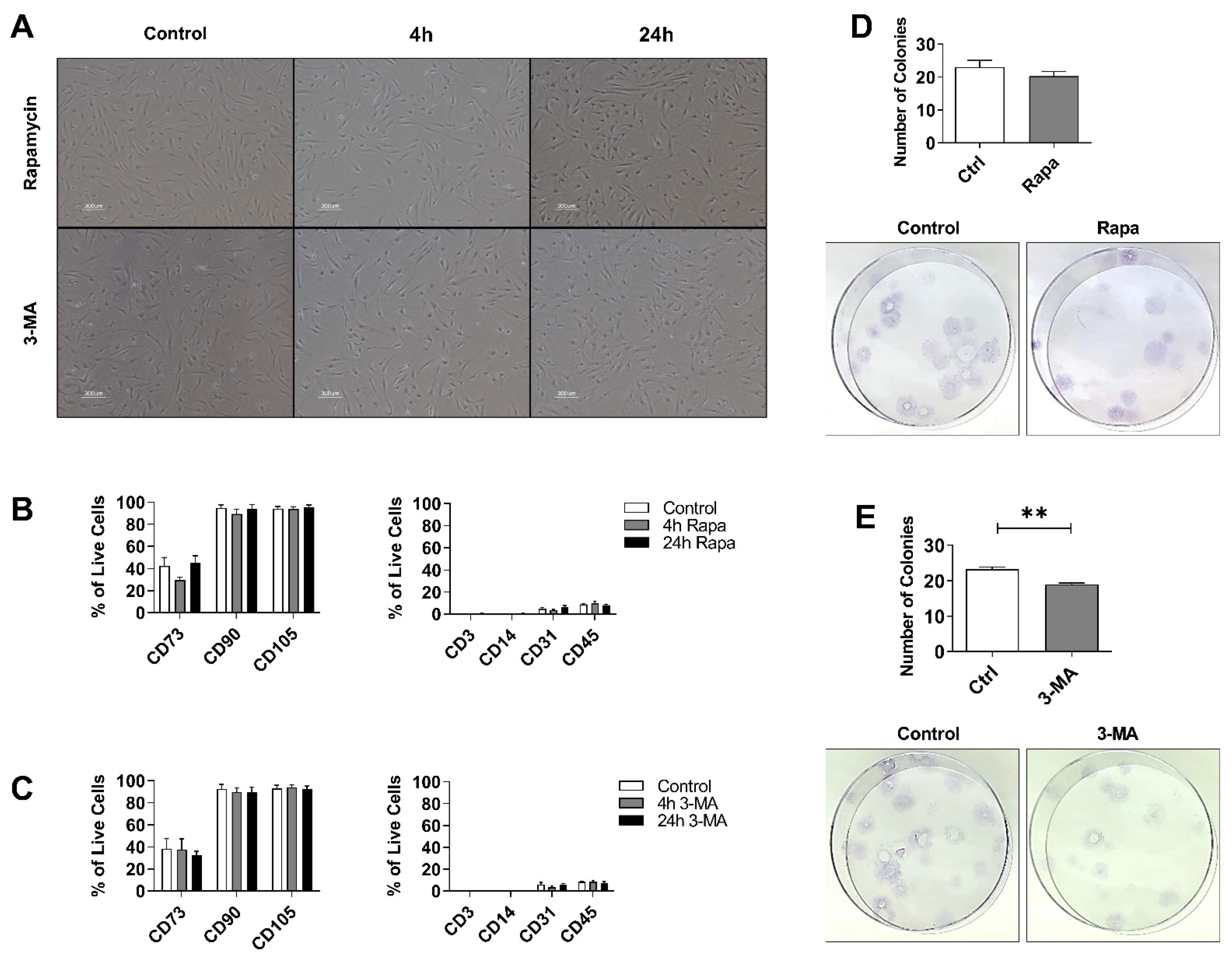
3.1. The mTOR Inhibitor Rapamycin Induces, While the PI3K Inhibitor 3-MA Suppresses, Autophagy in hASCs

In this study, hASCs were treated with rapamycin (Rapa-ASCs) for 1, 4, 12, 24 and 48 h, then examined for the expression of key autophagy genes and proteins. Transcriptional analysis of Rapa-ASCs demonstrated significant upregulation of the autophagy genes ATG7 and LC3B following prolonged, but not short-term exposure to Rapa (Supplementary Figure S1C), suggesting induction of autophagy. A Western blot analysis revealed increased protein levels of LC3-II/β-Actin and decreased protein levels of p62/β-Actin, however this failed to reach significance (Supplementary Figure S1A,B). To examine inhibition of autophagy, hASCs were treated with 3-MA (3MA-ASCs) for 1, 4, 12, and 24 h and then probed for changes in autophagy gene and protein levels. Due to previous reports of autophagy induction with prolonged exposure to 3-MA [37], the longest timepoint (48 h) was not included for this group. The 3MA-ASCs exhibited no change to LC3-II or p62 transcript or protein levels, indicating no induction of autophagosome formation (Supplementary Figure S2A,B).

3.2. Autophagy Preconditioning Does Not Alter hASCs Stem Cell Properties

To characterize the impact of autophagy preconditioning on stem cell characteristics, RapaASCs and 3MA-ASCs were examined for morphology, clonogenicity, and surface marker expression after either short-term (4 h) or long-term (24 h) treatment. Results demonstrated that hASCs maintained their spindle-like morphology with both short-term and long-term exposure to either Rapa or 3-MA (Figure 1A). Autophagy preconditioning did not significantly alter hASCs immunophenotype as measured by flow cytometric analysis of surface proteins (Figure 1B,C). In both Rapa-ASCs and 3MA-ASCs, positive expression of canonical MSC markers CD90 and CD105 ranged from 90–100% across all time points, and expression of CD73 ranged from 29.63% to 45.02% with no significant difference between groups (Figure 1B). Immunophenotype of hASCs was further confirmed by the absence of negative MSC markers including the pan T-cell marker CD3, the monocyte/macrophage marker CD14, the endothelial cell marker CD31, and the broad lymphohematopoietic lineage marker CD45. In both 3MA-ASCs and Rapa-ASCs, expression of these negative markers remained low and was not significantly different from untreated hASCs. Additionally, self-renewal ability of treated ASCs was measured by performing a colony-forming unit-fibroblast assay. Analysis of colony-forming units (CFUs) demonstrated no alteration in self-renewal capacity of Rapa-ASCs (Figure 1D). However, the 3MA-ASCs showed significantly reduced self-renewal capacity (Figure 1E).

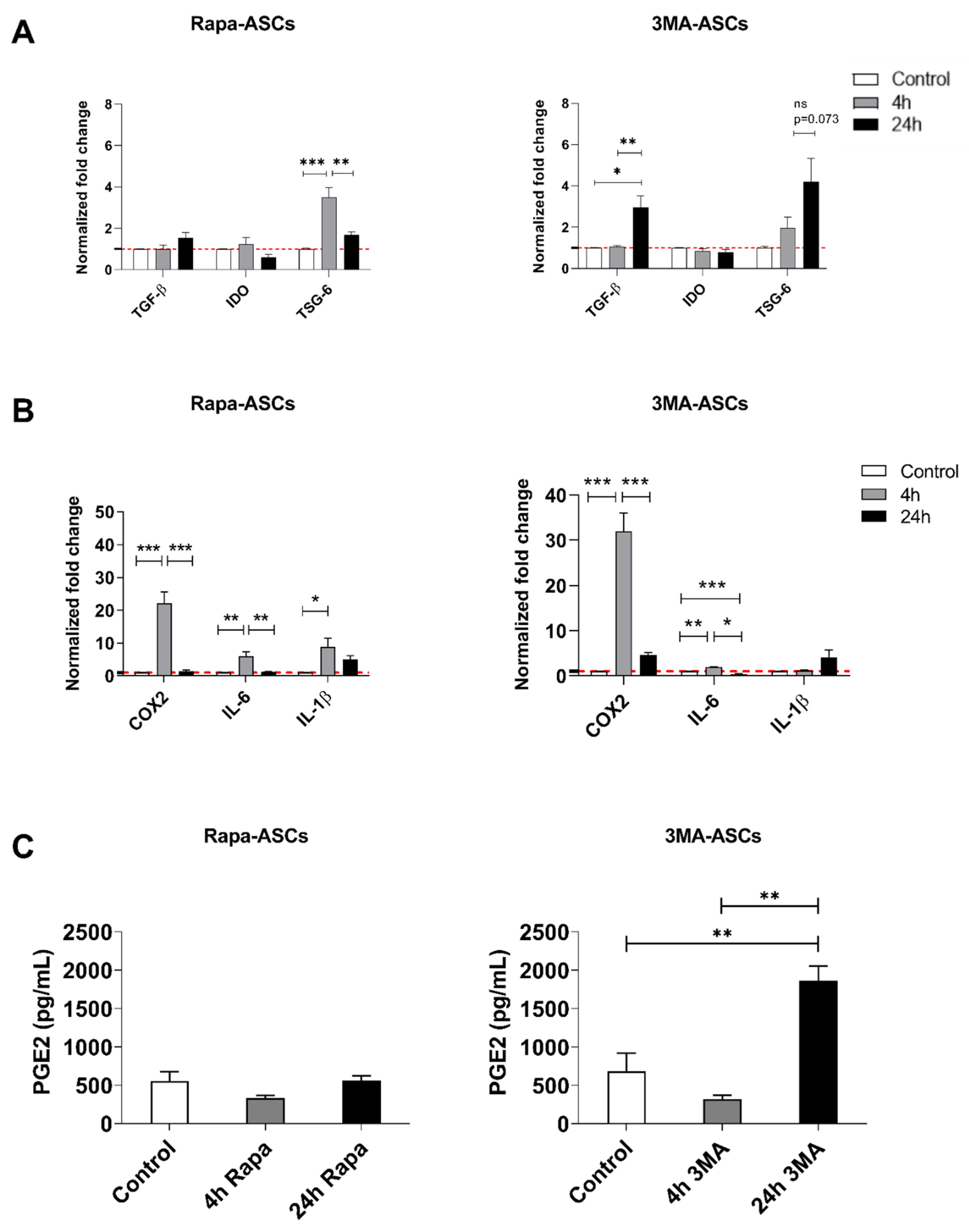
3.3. Autophagy Preconditioning of hASCs Alters Expression of Both Anti-Inflammatory and Pro-Inflammatory Mediators

To determine whether pharmacologically targeting the autophagy pathway alters the immune-modifying abilities of hASCs, cells were treated for either 4 or 24 h with Rapa or 3-MA and then analyzed with RT-qPCR for expression of common immune mediators. In Rapa-ASCs, 4 h, but not 24 h, treatment produced robust enhancement of TSG-6 mRNA (Figure 2A). The opposite result was seen in 3MA-ASCs, with only the 24-h group demonstrating enhanced expression of TGF-β and, to a lesser extent, TSG-6 (p = 0.073). Further analysis revealed similar time-dependent differences in expression of pro-inflammatory mediators (Figure 2B). In both Rapa-ASCs and 3MA-ASCs, IL-6 was upregulated after 4 h, but not 24 h. Moreover, Rapa-ASCs, but not 3MA-ASCs, upregulated IL-1β after 4-h treatment. Interestingly, both 4-h Rapa-ASCs and 3MA-ASCs robustly upregulate COX2 transcription relative to control cells. This effect was not mirrored in secreted protein, as ELISA analysis of conditioned medium revealed elevation of the COX2 metabolite, PGE2, in 24-h treated 3MA-ASCs only (Figure 2C).

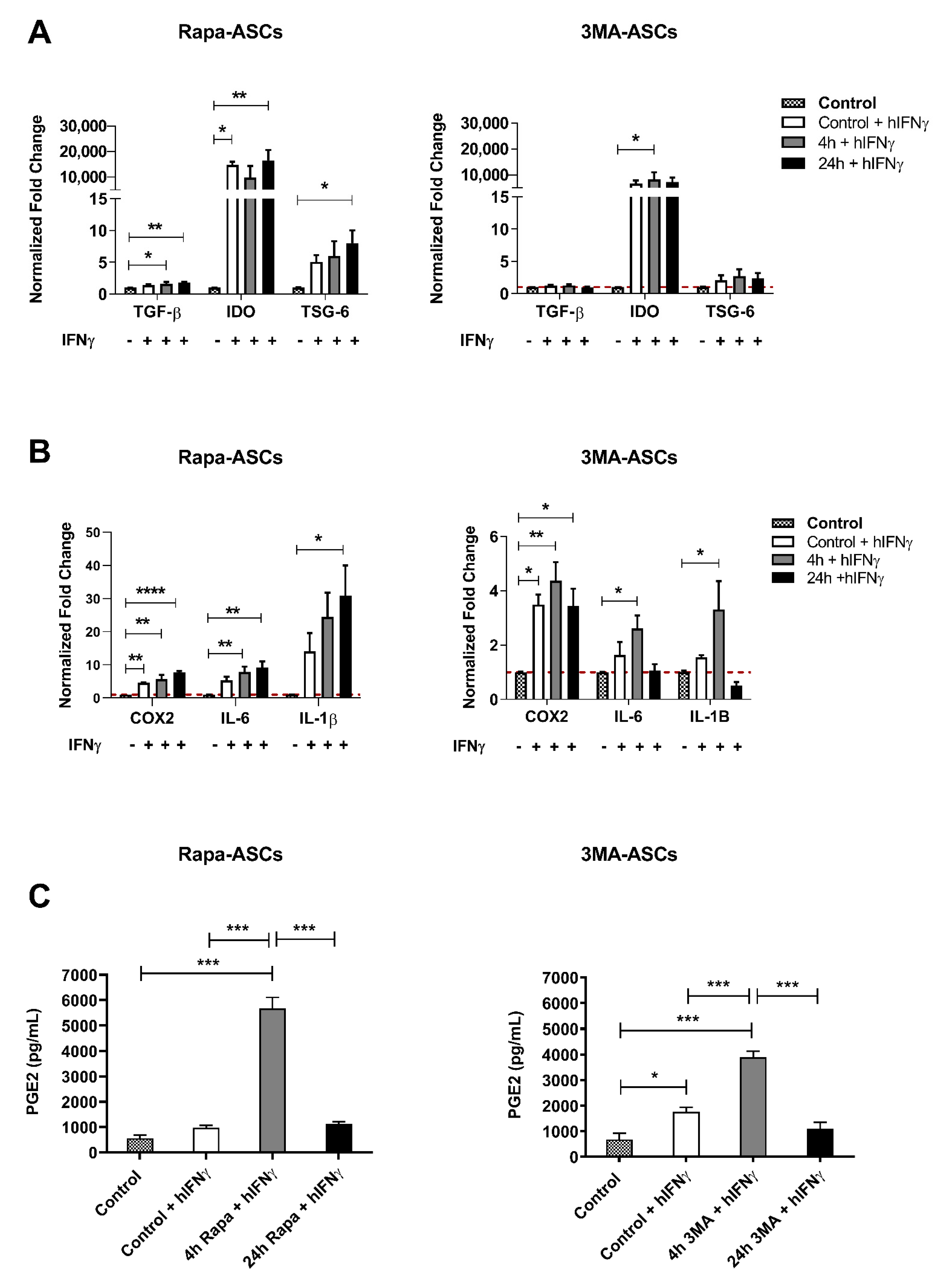
3.4. Autophagy Preconditioning of hASCs Alters Response to Pro-Inflammatory Stimulation with hIFNγ

To investigate the effects of autophagy preconditioning on hASC immunomodulatory response to signals they may encounter following administration into an inflammatory microenvironment, cells were pre-treated for either 4 or 24 h followed by 24 h stimulation with the classic pro-inflammatory cytokine hIFNγ. Our findings demonstrate that, in comparison to unstimulated and non-pretreated control ASCs, 4 h Rapa-preconditioning resulted in significant elevation of TGF-β, COX2, and IL-6, whereas 24 h Rapa-preconditioning resulted in upregulation of TGF-β, IDO, TSG-6, and COX2, as well as the inflammatory cytokines IL-6 and IL-1β. We also found that 4 h 3MA-preconditioning resulted in upregulation of IDO, COX2, IL-6, and IL-1β, whereas24 h 3MA-preconditioning only increased expression of COX2 transcripts (Figure 3A,B). This contrasted with our non-preconditioned control ASCs, which in response to hIFNγ stimulation only displayed significant upregulation of IDO and COX2 transcripts when compared to non- IFNγ-stimulated ASCs. Perhaps most interesting, we demonstrated that secretion of PGE2 was upregulated by both 4 h Rapa and 4 h 3MA, which was significantly higher than both unstimulated control and IFNγ-stimulated control cells, as well as both 24 h preconditioned groups (Figure 3C). Taken together, these findings show that although transcriptional activity is altered based on unique temporal and compound-dependent patterns, the robust upregulation of PGE2 release in both acute Rapa and 3-MA preconditioning strategies suggest the involvement of an autophagy-independent mechanism that is currently unknown.

4. Discussion

ASCs have shown substantial preclinical promise as therapeutic tools in inflammatory, autoimmune, and neurodegenerative diseases [38]. However, the successful application of ASCs in human clinical trials has faced many challenges, including variable clinical efficacy outcomes, low post-transplant viability, and immunosuppressive potency [39]. Preconditioning strategies to improve immunomodulatory potency of MSCs for various anti-inflammatory and regenerative medicine applications is an active focus of the stem cell research field. Rapamycin has emerged as a promising candidate compound, and Rapa-treated MSCs from adipose [29,40], bone marrow [41], and umbilical cord [42] have been examined in preclinical models. In a 2016 report by Kim and colleagues, hASCs were treated with Rapa for 48 h prior to administration in a GvHD mouse model. Authors demonstrate elevated expression of IL-10, IDO, and TGF-β, and correlated this with enhanced production of anti-inflammatory cytokines, modulation of the T cell repertoire, and prevention of GvHD development [29]. However, recent reports have revealed temporally distinct actions of Rapa on mTORC1 and mTORC2 [43], and this has been correlated to dynamic effects on MSC immunomodulatory capacity. Indeed, a wide range of dosage and exposure time has yielded varying success and highlights the importance of defining the unique immunoregulatory consequences of these novel preconditioning strategies depending on experimental conditions (i.e., dose and exposure time), cell type, and therapeutic application. For example, short-term (2 h) treatment of rat BMSCs improved survival and repair of damaged myocardium following transplant into an ischemia/reperfusion model [41]. However, in an animal model of multiple sclerosis, short-term (4 h) preconditioning of human ASCs did not yield any therapeutic benefit and was in fact correlated with worsened disease measures [40]. Thus, to improve translational potential, it is critically important to define the dynamic immunomodulatory response of MSCs and fine-tune these strategies to each pathological situation.
In the present study, the impacts of compounds known to either induce or inhibit autophagy on hASC immunomodulatory potential was assessed. Our data showed that 24 h Rapa treatment increased the expression of the autophagy genes ATG7 and LC3B, whereas the expression of LC3-II protein was upregulated, albeit slightly, suggestive of autophagic induction. Conversely, 3-MA, a class I and class III PI3K inhibitor, obstructed autophagy initiation in hASCs. 3MA-ASCs displayed no significant change to autophagy transcripts or protein levels. Both Rapa-ASCs and 3MA-ASCs maintain characteristic stem cell properties, including plastic adherence, fibroblast-like morphology, and immunophenotypic profile, as determined by flow cytometry. However, because the immunomodulatory strength of ASCs is due to their paracrine activity rather than proliferation and engraftment, this likely has little impact on therapeutic potential. These results denote the retention of basic MSC identifiers in autophagy preconditioned hASCs.
To determine the role of autophagy preconditioning in hASC immunosuppressive potential, both the transcription and secretion of known ASC-derived pro- and anti-inflammatory mediators was examined. Among these, the pleiotropic signaling molecule PGE2 was of particular interest due to its established role in MSC-based therapies for inflammatory and autoimmune diseases including MS [18,19], sepsis [14], inflammatory bowel disease (IBD) [20,21], ischemia-reperfusion injury [15], and arthritis [16,17]. hASC-derived PGE2 exerts its immunomodulatory functions through the inhibition of T cell activation, proliferation and production of pro-inflammatory cytokines [17,44,45,46,47], the generation of IL-10-producing T regulatory cells (Tregs) [17], and the promotion of M2 macrophage polarization [21,48]. PGE2 is synthesized from arachidonic acid by the enzyme COX2 and has been proposed as an important mechanism contributing to the immunoregulatory actions of MSCs. In a mouse model of sepsis, concurrent IV administration of BMSCs pre-stimulated with LPS resulted in prolonged survival, which was correlated with elevated PGE2 secretion and modulation of host macrophage populations [14]. PGE2 has also been shown to be critical for MSCs’ ability to inhibit the proliferation of natural killer (NK) cells [49], the maturation of dendritic cells (DC) from monocytes [45,50], and the proliferation of PHA-stimulated T cells [45].
In this study, 4 h preconditioned Rapa-ASCs and 3MA-ASCs both exhibited robust upregulation of COX2 gene expression. This induction was not seen in secreted PGE2, the protein substrate of COX2, with autophagy preconditioning alone in either 4 h treatment group; however, PEG2 was significantly increased in ASCs treated with 3-MA for 24 h. Following exposure to hIFNγ, elevated COX2 transcription and PGE2 secretion was seen in both Rapa-ASCs and 3MA-ASCs with 4 h, but not 24 h, preconditioning. This indicates that short-term autophagy preconditioning may “prime” hASCs to respond more rapidly and more robustly to an environment mimicking the inflammatory state of autoimmune or neurodegenerative diseases [51,52,53,54].
These unique temporal dynamics of COX2 and PGE2 have previously been shown in BMSCs, and have been suggested to result from activation of the Akt/glycogen synthase kinase 3 Beta (GSK-3β) pathway rather than autophagy [55,56]. mTOR, a serine/threonine kinase which forms the catalytic subunit of the two distinct complexes mTORC1 and mTORC2, regulates a spectrum of cellular processes including cell growth, autophagy, cytoskeletal remodeling, proteostasis, and metabolism (elegantly reviewed here [57]). Rapamycin, an FDA-approved immunosuppressant, rapidly inhibits mTORC1 activity through interaction with the FRB (FKBP12/rapamycin-binding) domain [58]. Under physiological conditions mTORC1 substrates negatively regulate mTORC2 kinase activity [59]. With Rapa inhibition of mTORC1, mTORC2 kinase activity is disinhibited, leading to phosphorylation of Akt at Ser473 and subsequently disrupting the glycogen synthase kinase 3 beta (GSK3B)-dependent blockade of the COX2 promotor region, thereby activating COX2 transcription [37,55,60]. With prolonged Rapa exposure mTORC2 is also inhibited, possibly explaining the rapid yet transient COX2 upregulation [43]. The temporally defined actions of 3-MA may also explain the observed transcriptional changes. 3-MA persistently inhibits class I PI3K whereas its inhibition of the class III PI3K, VPS34, is transient [37]. This was correlated with transient inhibition of autophagy and upregulated COX2 levels in human BMSCs with short-term exposure, but induction of autophagy and suppression of COX2 with long-term treatment. Thus, it may be that the selective inhibition of mTORC1 kinase activity, independent of the effects on autophagy, is responsible for the altered immunophenotype of ASCs.
Wang and colleagues demonstrated that short-term, but not long-term, inhibition of mTORC1 with Rapa resulted in elevated COX2 gene expression, PGE2 secretion, and inhibition of proliferation in PBMCs. However, ASCs represent a more abundant and readily available source of stem cells compared to BMSCs, and possess higher immunomodulatory potential [3]. Thus, the present study investigated whether ASCs demonstrate a similar temporally distinct response to Rapa treatment to determine if this therapeutic strategy may be extended to a novel cell type. Due to the observation that both Rapa and 3-MA elicited some shared immunoregulatory responses in hASCs, it may be that the effects of these preconditioning strategies are independent of autophagy, which was suggested by Chinnadurai and collaborators in 2015 [61]. However, in this study hBMSC were treated with 3-MA for more than 48 h, and based on the temporal dynamics of class I versus class III PI3K inhibition it is possible that the inhibitory effect of 3-MA had weakened or stopped. Our results, consistent with observations in BMSCs, highlight the importance of fully understanding the temporal aspects of autophagy preconditioning to optimize therapeutic potential.
Evidence from two studies indicates that PGE2 is only partially responsible for the immunosuppressive actions of hASC function as inhibition of PGE2 or its receptors does not fully abolish immunosuppressive capacity [46,62]. Other anti-inflammatory mediators, including IDO, TSG-6, and TGF-β1, significantly contribute to hASCs’ suppression of innate and adaptive immune cells. IDO, an enzyme that breaks down the essential amino acid tryptophan, promotes polarization of M2b/c macrophages, induces proliferation and IL-10 production of Tregs, inhibits proliferation of Th cells, and suppresses the cytolytic activity of natural killer (NK) cells [1,5,8,62,63,64]. TSG-6 plays a pivotal role in M2 macrophage polarization and reduction of inflammation in both colitis and acute pancreatitis mouse models [65,66]. Moreover, TSG-6 has displayed autocrine activity which maintains stemness and downregulates IL-6 production in mouse BMSCs [67]. TGF-β1 is necessary for the suppression of dendritic cell maturation and subsequent promotion of FOXP3+ Tregs [68,69]. Additionally, Rapa-treated hBMSCs showed upregulation of TGF-β1, which was indispensable for the inhibition of CD4+ T cell proliferation [70]. The present study showed that IDO was expressed at low levels in resting hASCs and was unaffected by autophagy preconditioning itself. Upon hIFNγ stimulation, both 4 h 3MA-ASCs and 24 h Rapa-ASCs amplified IDO expression compared to control stimulated cells. TSG-6 expression was not significantly changed in resting or hIFNγ-stimulated 3MA-ASCs but was highly upregulated in 4 h preconditioned Rapa-ASCs. Further upregulation of transcription was seen in 24 h preconditioned, hIFNγ-stimulated Rapa-ASCs. Finally, only 24 h preconditioned 3MA-ASCs exhibited upregulation of TGF-β1 transcripts. After hIFNγ stimulation, both 4 h and 24 h Rapa-ASCs elevated TGF-β1 gene expression. Overall, Rapa-ASCs demonstrate more upregulation of anti-inflammatory genes than 3MA-ASCs, suggesting Rapa preconditioning may be a more promising strategy for augmentation of hASC immunomodulatory ability.
The pleiotropic cytokine IL-6 can act as a pro-inflammatory or pro-regenerative agent dependent on its actions on its soluble and membrane-bound receptors, respectively [71,72]. In the context of hASCs, IL-6 production has been linked to repression of Th17 cells, induction of IL-10-producing Tregs, and enhanced recruitment and anti-inflammatory polarization of macrophages [73,74,75,76]. In our study, both 4 h Rapa-ASCs and 3MA-ASCs showed a significant increase in IL-6 transcript levels. Following pro-inflammatory activation with hIFNγ, both 4- and 24 h Rapa-ASCs, and 4 h 3MA-ASCs showed enhanced IL-6 transcription. Finally, we assessed the expression of IL-1β, the potent pro-inflammatory cytokine which negatively correlates with hASCs’ ability to suppress CD4+ T helper, CD8+ T effector, and NK cell proliferation [77]. In our study, 24 h Rapa preconditioning and 4 h 3-MA preconditioning followed by stimulation with hIFNγ resulted in additional upregulation of IL-1β. Taken together, the upregulated IL-6 and unaltered IL-1β expression levels suggest that 4 h Rapa-ASCs may be the most effective autophagy preconditioning strategy.
In summary, these data demonstrate that preconditioning with both the autophagy-inducer Rapa and the autophagy-inhibitor 3-MA had distinct effects on the expression of immunomodulatory factors in hASCs based on the duration of treatment and presence or absence of pro-inflammatory stimuli. The results suggest that the conflicting data in the autophagy literature may be due, at least in part, to varied treatment times, doses, and the unique inflammatory milieu. We also demonstrated that both compounds had similar impact on secretory activity of ASCs in response to inflammatory activation, which strongly suggests a shared molecular mechanism that is independent of their actions on autophagy. Based on our findings, we propose that a short-term preconditioning with Rapa may bestow the most robust enrichment of hASC immunomodulatory potential due to the preservation of stemness qualities, the enhancement of the COX2/PGE2 pathway, and the increase of the anti-inflammatory cytokines TGF-β and IL-6 without concomitant elevation of pro-inflammatory IL-1β. This is consistent with previous investigations which suggest that short-term Rapa treatment in MSCs elicits greater improvement of immunomodulatory function, possibly resulting from its differential inhibition of MTORC1 and MTORC2 [41,55,78]. On the other hand, 3MA-ASCs and Rapa-ASCs both upregulate PGE2 secretion when activated with hIFNγ, indicating that autophagy inhibition with 3-MA may also have therapeutic benefit in certain disease contexts, as was proposed by Dang and colleagues in the experimental autoimmune encephalomyelitis (EAE) mouse model [19]. Future investigations involving co-cultures with innate and adaptive immune cells and administration to preclinical disease models will be needed to determine the influence of these novel preconditioning strategies on the immunosuppressive function of hASCs. These approaches may reveal whether our in vitro findings translate to enhanced immunomodulatory function and therapeutic efficacy in autoimmune, inflammatory, and neurodegenerative diseases in vivo.

5. Conclusions

Adipose-derived stem cells (ASCs) secrete a variety of anti-inflammatory molecules that shift immune cell activity away from driving inflammation and toward tissue regeneration and repair, showing immense therapeutic promise in several inflammatory, autoimmune, and neurodegenerative diseases. However, their efficacy is limited by poor survival and secretory activity after administration, representing a significant hurdle to success in clinical trials. To overcome these hurdles, a novel preconditioning strategy is proposed that targets a cellular pathway involved in stress response, survival, and secretory activity. Drawing on evidence from bone marrow-derived stem cells (BMSCs), we find that compounds commonly used to modify the autophagy pathway result in “primed” ASCs capable of producing higher levels of immunomodulatory genes and proteins. We demonstrate that PGE2, a key contributing factor to stem cell therapeutic efficacy in multiple sclerosis (MS), sepsis, inflammatory bowel disease (IBD), and arthritis, was highly upregulated in ASCs preconditioned with either short-term Rapa or 3-MA, indicating that the immunomodulatory ffects of these compounds may, in fact, derive from mechanisms of action beyond their impact on autophagy. We propose that this represents a promising strategy for enhancing the therapeutic potential and possibly the translational success of ASCs for inflammation-driven disease states that warrants further investigation and delineation of mechanisms involved.

Supplementary Materials

The following supporting information can be downloaded at: https://www.mdpi.com/article/10.3390/cells11091376/s1, Figure S1: Rapamycin induces autophagy in hASCs; Figure S2: 3-MA inhibits autophagy in hASCs.

Author Contributions

Conceptualization and design, R.M.W. and B.A.B.; Data collection, R.M.W., M.A.A.H., B.N.S., E.R.M., S.J.A., and U.M.D.; Data analysis and interpretation, R.M.W. and S.A.-G.; Funding and supervision, B.A.B.; Writing—original draft, R.M.W.; Writing—review & editing, R.M.W., S.A.-G., M.A.A.H., B.N.S., and B.A.B. All authors have read and agreed to the published version of the manuscript.

Funding

The funds used for these studies were private funds from Tulane University.

Institutional Review Board Statement

Not applicable.

Informed Consent Statement

Not applicable.

Data Availability Statement

The original contributions presented in the study are included in the article/supplementary material, further inquiries can be directed to the corresponding author/s.

Acknowledgments

The authors would like to thank Alan Tucker with the Flow Cytometry core for his expertise and assistance with these studies.

Conflicts of Interest

The authors declare that the research was conducted in the absence of any commercial or financial relationships that could be construed as a potential conflict of interest.

References

  1. Zheng, G.; Qiu, G.; Ge, M.; He, J.; Huang, L.; Chen, P.; Wang, W.; Xu, Q.; Hu, Y.; Shu, Q.; et al. Human Adipose-Derived Mesenchymal Stem Cells Alleviate Obliterative Bronchiolitis in a Murine Model via IDO. Respir. Res. 2017, 18, 119. [Google Scholar] [CrossRef] [Green Version]
  2. Gimble, J.M.; Katz, A.J.; Bunnell, B.A. Adipose-Derived Stem Cells for Regenerative Medicine. Circ. Res. 2007, 100, 1249–1260. [Google Scholar] [CrossRef]
  3. Melief, S.M.; Zwaginga, J.J.; Fibbe, W.E.; Roelofs, H. Adipose Tissue-Derived Multipotent Stromal Cells Have a Higher Immunomodulatory Capacity Than Their Bone Marrow-Derived Counterparts. Stem Cells Transl. Med. 2013, 2, 455–463. [Google Scholar] [CrossRef]
  4. Ceccarelli, S.; Pontecorvi, P.; Anastasiadou, E.; Napoli, C.; Marchese, C. Immunomodulatory Effect of Adipose-Derived Stem Cells: The Cutting Edge of Clinical Application. Front. Cell Dev. Biol. 2020, 236. [Google Scholar] [CrossRef] [PubMed]
  5. Serejo, T.R.T.; Silva-Carvalho, A.É.; Braga, L.D.d.C.F.; Neves, F.d.A.R.; Pereira, R.W.; Carvalho, J.L.d.; Saldanha-Araujo, F. Assessment of the Immunosuppressive Potential of INF-γ Licensed Adipose Mesenchymal Stem Cells, Their Secretome and Extracellular Vesicles. Cells 2019, 8, 22. [Google Scholar] [CrossRef] [PubMed] [Green Version]
  6. Yang, D.; Wang, W.; Li, L.; Peng, Y.; Chen, P.; Huang, H.; Guo, Y.; Xia, X.; Wang, Y.; Wang, H.; et al. The Relative Contribution of Paracine Effect versus Direct Differentiation on Adipose-Derived Stem Cell Transplantation Mediated Cardiac Repair. PLoS ONE 2013, 8, e59020. [Google Scholar] [CrossRef]
  7. Fontanilla, C.V.; Gu, H.; Liu, Q.; Zhu, T.Z.; Zhou, C.; Johnstone, B.H.; March, K.L.; Pascuzzi, R.M.; Farlow, M.R.; Du, Y. Adipose-Derived Stem Cell Conditioned Media Extends Survival Time of a Mouse Model of Amyotrophic Lateral Sclerosis. Sci. Rep. 2015, 5, 1–11. [Google Scholar] [CrossRef] [Green Version]
  8. Crop, M.J.; Baan, C.C.; Korevaar, S.S.; IJzermans, J.N.M.; Pescatori, M.; Stubbs, A.P.; van IJcken, W.F.J.; Dahlke, M.H.; Eggenhofer, E.; Weimar, W.; et al. Inflammatory Conditions Affect Gene Expression and Function of Human Adipose Tissue-Derived Mesenchymal Stem Cells. Clin. Exp. Immunol. 2010, 162, 474–486. [Google Scholar] [CrossRef]
  9. Domenis, R.; Cifù, A.; Quaglia, S.; Pistis, C.; Moretti, M.; Vicario, A.; Parodi, P.C.; Fabris, M.; Niazi, K.R.; Soon-Shiong, P.; et al. Pro Inflammatory Stimuli Enhance the Immunosuppressive Functions of Adipose Mesenchymal Stem Cells-Derived Exosomes. Sci. Rep. 2018, 8, 1–11. [Google Scholar] [CrossRef]
  10. Wobma, H.M.; Tamargo, M.A.; Goeta, S.; Brown, L.M.; Duran-Struuck, R.; Vunjak-Novakovic, G. The Influence of Hypoxia and IFN-γ on the Proteome and Metabolome of Therapeutic Mesenchymal Stem Cells. Biomaterials 2018, 167, 226–234. [Google Scholar] [CrossRef] [PubMed]
  11. Roemeling-Van Rhijn, M.; Mensah, F.K.F.; Korevaar, S.S.; Leijs, M.J.; Van Osch, G.J.V.M.; IJzermans, J.N.M.; Betjes, M.G.H.; Baan, C.C.; Weimar, W.; Hoogduijn, M.J. Effects of Hypoxia on the Immunomodulatory Properties of Adipose Tissue-Derived Mesenchymal Stem Cells. Front. Immunol. 2013, 4, 203. [Google Scholar] [CrossRef] [PubMed] [Green Version]
  12. Seo, Y.; Shin, T.-H.; Kim, H.-S. Molecular Sciences Current Strategies to Enhance Adipose Stem Cell Function: An Update. Int. J. Mol. Sci. 2019, 20, 3827. [Google Scholar] [CrossRef] [Green Version]
  13. Hsiao, S.T.; Lokmic, Z.; Peshavariya, H.; Abberton, K.M.; Dusting, G.J.; Lim, S.Y.; Dilley, R.J. Hypoxic Conditioning Enhances the Angiogenic Paracrine Activity of Human Adipose-Derived Stem Cells. Stem Cells Dev. 2013, 22, 1614–1623. [Google Scholar] [CrossRef] [Green Version]
  14. Németh, K.; Leelahavanichkul, A.; Yuen, P.S.T.; Mayer, B.; Parmelee, A.; Doi, K.; Robey, P.G.; Leelahavanichkul, K.; Koller, B.H.; Brown, J.M.; et al. Bone Marrow Stromal Cells Attenuate Sepsis via Prostaglandin E 2-Dependent Reprogramming of Host Macrophages to Increase Their Interleukin-10 Production. Nat. Med. 2009, 15, 42–49. [Google Scholar] [CrossRef] [PubMed] [Green Version]
  15. Liu, L.; He, Y.R.; Liu, S.J.; Hu, L.; Liang, L.C.; Liu, D.L.; Liu, L.; Zhu, Z.Q. Enhanced Effect of IL-1 β-Activated Adipose-Derived MSCs (ADMSCs) on Repair of Intestinal Ischemia-Reperfusion Injury via COX-2-PGE 2 Signaling. Stem Cells Int. 2020, 2020, 2803747. [Google Scholar] [CrossRef] [Green Version]
  16. Bouffi, C.; Bony, C.; Courties, G.; Jorgensen, C.; Noël, D. IL-6-Dependent PGE2 Secretion by Mesenchymal Stem Cells Inhibits Local Inflammation in Experimental Arthritis. PLoS ONE 2010, 5, e14247. [Google Scholar] [CrossRef]
  17. González, M.A.; González-Rey, E.; Rico, L.; Büscher, D.; Delgado, M. Treatment of Experimental Arthritis by Inducing Immune Tolerance with Human Adipose-Derived Mesenchymal Stem Cells. Arthritis Rheum. 2009, 60, 1006–1019. [Google Scholar] [CrossRef]
  18. Matysiak, M.; Orlowski, W.; Fortak-Michalska, M.; Jurewicz, A.; Selmaj, K. Immunoregulatory Function of Bone Marrow Mesenchymal Stem Cells in EAE Depends on Their Differentiation State and Secretion of PGE2. J. Neuroimmunol. 2011, 233, 106–111. [Google Scholar] [CrossRef] [PubMed]
  19. Dang, S.; Xu, H.; Xu, C.; Cai, W.; Li, Q.; Cheng, Y.; Jin, M.; Wang, R.-X.; Peng, Y.; Zhang, Y.; et al. Autophagy Regulates the Therapeutic Potential of Mesenchymal Stem Cells in Experimental Autoimmune Encephalomyelitis. Autophagy 2014, 10, 1301–1315. [Google Scholar] [CrossRef] [Green Version]
  20. Yang, F.Y.; Chen, R.; Zhang, X.; Huang, B.; Tsang, L.L.; Li, X.; Jiang, X. Preconditioning Enhances the Therapeutic Effects of Mesenchymal Stem Cells on Colitis Through PGE2-Mediated T-Cell Modulation. Cell Transplant. 2018, 27, 1352–1367. [Google Scholar] [CrossRef] [Green Version]
  21. Park, H.J.; Kim, J.; Saima, F.T.; Rhee, K.J.; Hwang, S.; Kim, M.Y.; Baik, S.K.; Eom, Y.W.; Kim, H.S. Adipose-Derived Stem Cells Ameliorate Colitis by Suppression of Inflammasome Formation and Regulation of M1-Macrophage Population through Prostaglandin E2. Biochem. Biophys. Res. Commun. 2018, 498, 988–995. [Google Scholar] [CrossRef]
  22. Ceccariglia, S.; Cargnoni, A.; Silini, A.R.; Parolini, O. Autophagy: A Potential Key Contributor to the Therapeutic Action of Mesenchymal Stem Cells. Autophagy 2019, 16, 28–37. [Google Scholar] [CrossRef] [PubMed]
  23. Jakovljevic, J.; Harrell, C.R.; Fellabaum, C.; Arsenijevic, A.; Jovicic, N.; Volarevic, V. Modulation of Autophagy as New Approach in Mesenchymal Stem Cell-Based Therapy. Biomed. Pharmacother. 2018, 104, 404–410. [Google Scholar] [CrossRef]
  24. Glick, D.; Barth, S.; Macleod, K.F. Autophagy: Cellular and Molecular Mechanisms. J. Pathol. 2010, 221, 3–12. [Google Scholar] [CrossRef] [Green Version]
  25. Chen, X.; He, Y.; Lu, F. Autophagy in Stem Cell Biology: A Perspective on Stem Cell Self-Renewal and Differentiation. Stem Cells Int. 2018, 2018, 9131397. [Google Scholar] [CrossRef]
  26. Weiss, D.J.; English, K.; Krasnodembskaya, A.; Isaza-Correa, J.M.; Hawthorne, I.J.; Mahon, B.P. The Necrobiology of Mesenchymal Stromal Cells Affects Therapeutic Efficacy. Front. Immunol. 2019, 10, 1228. [Google Scholar] [CrossRef] [PubMed]
  27. Li, J.; Kim, S.G.; Blenis, J. Rapamycin: One Drug, Many Effects. Cell Metab. 2014, 19, 373–379. [Google Scholar] [CrossRef] [PubMed] [Green Version]
  28. Pyo, J.O.; Nah, J.; Jung, Y.K. Molecules and Their Functions in Autophagy. Exp. Mol. Med. 2012, 44, 73–80. [Google Scholar] [CrossRef] [Green Version]
  29. Kim, K.-W.; Moon, S.-J.; Park, M.-J.; Kim, B.-M.; Kim, E.-K.; Lee, S.-H.; Lee, E.-J.; Chung, B.-H.; Yang, C.-W.; Cho, M.-L. Optimization of Adipose Tissue-Derived Mesenchymal Stem Cells by Rapamycin in a Murine Model of Acute Graft-versus-Host Disease. Stem Cell Res. Ther. 2015, 6, 202. [Google Scholar] [CrossRef] [Green Version]
  30. Javorkova, E.; Vackova, J.; Hajkova, M.; Hermankova, B.; Zajicova, A.; Holan, V.; Krulova, M. The Effect of Clinically Relevant Doses of Immunosuppressive Drugs on Human Mesenchymal Stem Cells. Biomed. Pharmacother. 2018, 97, 402–411. [Google Scholar] [CrossRef]
  31. Li, C.; Ye, L.; Yang, L.; Yu, X.; He, Y.; Chen, Z.; Li, L.; Zhang, D. Rapamycin Promotes the Survival and Adipogenesis of Ischemia-Challenged Adipose Derived Stem Cells by Improving Autophagy. Cell. Physiol. Biochem. 2017, 44, 1762–1774. [Google Scholar] [CrossRef]
  32. Strong, A.L.; Bowles, A.C.; Wise, R.M.; Morand, J.P.; Dutreil, M.F.; Gimble, J.M.; Bunnell, B.A. Human Adipose Stromal/Stem Cells from Obese Donors Show Reduced Efficacy in Halting Disease Progression in the Experimental Autoimmune Encephalomyelitis Model of Multiple Sclerosis. Stem Cells 2016, 34, 614–626. [Google Scholar] [CrossRef] [PubMed] [Green Version]
  33. Al-Ghadban, S.; Diaz, Z.T.; Singer, H.J.; Mert, K.B.; Bunnell, B.A. Increase in Leptin and PPAR-γ Gene Expression in Lipedema Adipocytes Differentiated in Vitro from Adipose-Derived Stem Cells. Cells 2020, 9, 430. [Google Scholar] [CrossRef] [Green Version]
  34. Sabol, R.A.; Villela, V.A.; Denys, A.; Freeman, B.T.; Hartono, A.B.; Wise, R.M.; Harrison, M.A.A.; Sandler, M.B.; Hossain, F.; Miele, L.; et al. Obesity-Altered Adipose Stem Cells Promote Radiation Resistance of Estrogen Receptor Positive Breast Cancer through Paracrine Signaling. Int. J. Mol. Sci. 2020, 21, 2722. [Google Scholar] [CrossRef] [PubMed] [Green Version]
  35. Strong, A.L.; Hunter, R.S.; Jones, R.B.; Bowles, A.C.; Dutreil, M.F.; Gaupp, D.; Hayes, D.J.; Gimble, J.M.; Levi, B.; McNulty, M.A.; et al. Obesity Inhibits the Osteogenic Differentiation of Human Adipose-Derived Stem Cells. J. Transl. Med. 2016, 14, 27. [Google Scholar] [CrossRef] [Green Version]
  36. Scruggs, B.A.; Semon, J.A.; Zhang, X.; Zhang, S.; Bowles, A.C.; Pandey, A.C.; Imhof, K.M.P.; Kalueff, A.V.; Gimble, J.M.; Bunnell, B.A. Age of the Donor Reduces the Ability of Human Adipose-Derived Stem Cells to Alleviate Symptoms in the Experimental Autoimmune Encephalomyelitis Mo. Stem Cells Transl. Med. 2013, 2, 797–807. [Google Scholar] [CrossRef]
  37. Wu, Y.-T.; Tan, H.-L.; Shui, G.; Bauvy, C.; Huang, Q.; Wenk, M.R.; Ong, C.-N.; Codogno, P.; Shen, H.-M. Dual Role of 3-Methyladenine in Modulation of Autophagy via Different Temporal Patterns of Inhibition on Class I and III Phosphoinositide 3-Kinase. J. Biol. Chem. 2010, 285, 10850–10861. [Google Scholar] [CrossRef] [PubMed] [Green Version]
  38. Uccelli, A.; Moretta, L.; Pistoia, V. Mesenchymal Stem Cells in Health and Disease. Nat. Rev. Immunol. 2008, 8, 726–736. [Google Scholar] [CrossRef]
  39. Patrikoski, M.; Mannerström, B.; Miettinen, S. Perspectives for Clinical Translation of Adipose Stromal/Stem Cells. Stem Cells Int. 2019, 2019, 5858247. [Google Scholar] [CrossRef] [PubMed] [Green Version]
  40. Wise, R.M.; Harrison, M.A.A.; Sullivan, B.N.; Al-Ghadban, S.; Aleman, S.J.; Vinluan, A.T.; Monaco, E.R.; Donato, U.M.; Pursell, I.A.; Bunnell, B.A. Short-Term Rapamycin Preconditioning Diminishes Therapeutic Efficacy of Human Adipose-Derived Stem Cells in a Murine Model of Multiple Sclerosis. Cells 2020, 9, 2218. [Google Scholar] [CrossRef]
  41. Li, Z.; Wang, Y.; Wang, H.; Wu, J.; Tan, Y. Rapamycin-Preactivated Autophagy Enhances Survival and Differentiation of Mesenchymal Stem Cells After Transplantation into Infarcted Myocardium. Stem Cell Rev. Rep. 2020, 16, 344–356. [Google Scholar] [CrossRef] [Green Version]
  42. Zheng, J.; Li, H.; He, L.; Huang, Y.; Cai, J.; Chen, L.; Zhou, C.; Fu, H.; Lu, T.; Zhang, Y.; et al. Preconditioning of Umbilical Cord-derived Mesenchymal Stem Cells by Rapamycin Increases Cell Migration and Ameliorates Liver Ischaemia/Reperfusion Injury in Mice via the CXCR4/CXCL12 Axis. Cell Prolif. 2019, 52, 12546. [Google Scholar] [CrossRef]
  43. Sarbassov, D.D.; Ali, S.M.; Sengupta, S.; Sheen, J.H.; Hsu, P.P.; Bagley, A.F.; Markhard, A.L.; Sabatini, D.M. Prolonged Rapamycin Treatment Inhibits MTORC2 Assembly and Akt/PKB. Mol. Cell 2006, 22, 159–168. [Google Scholar] [CrossRef]
  44. Najar, M.; Raicevic, G.; Boufker, H.I.; Kazan, H.F.; De Bruyn, C.; Meuleman, N.; Bron, D.; Toungouz, M.; Lagneaux, L. Mesenchymal Stromal Cells Use PGE2 to Modulate Activation and Proliferation of Lymphocyte Subsets: Combined Comparison of Adipose Tissue, Wharton’s Jelly and Bone Marrow Sources. Cell. Immunol. 2010, 264, 171–179. [Google Scholar] [CrossRef]
  45. Yañez, R.; Oviedo, A.; Aldea, M.; Bueren, J.A.; Lamana, M.L. Prostaglandin E2 Plays a Key Role in the Immunosuppressive Properties of Adipose and Bone Marrow Tissue-Derived Mesenchymal Stromal Cells. Exp. Cell Res. 2010, 316, 3109–3123. [Google Scholar] [CrossRef] [PubMed]
  46. Puissant, B.; Barreau, C.; Bourin, P.; Clavel, C.; Corre, J.; Bousquet, C.; Taureau, C.; Cousin, B.; Abbal, M.; Laharrague, P.; et al. Immunomodulatory Effect of Human Adipose Tissue-Derived Adult Stem Cells: Comparison with Bone Marrow Mesenchymal Stem Cells. Br. J. Haematol. 2005, 129, 118–129. [Google Scholar] [CrossRef]
  47. Cui, L.; Shuo, Y.; Liu, W.; Li, N.; Zhang, W.; Cao, Y. Expanded Adipose-Derived Stem Cells Suppress Mixed Lymphocyte Reaction by Secretion of Prostaglandin E2. Tissue Eng. 2007, 13, 1185–1195. [Google Scholar] [CrossRef]
  48. Manferdini, C.; Paolella, F.; Gabusi, E.; Gambari, L.; Piacentini, A.; Filardo, G.; Fleury-Cappellesso, S.; Barbero, A.; Murphy, M.; Lisignoli, G. Adipose Stromal Cells Mediated Switching of the Pro-Inflammatory Profile of M1-like Macrophages Is Facilitated by PGE2: In Vitro Evaluation. Osteoarthr. Cartil. 2017, 25, 1161–1171. [Google Scholar] [CrossRef] [Green Version]
  49. Spaggiari, G.M.; Capobianco, A.; Abdelrazik, H.; Becchetti, F.; Mingari, M.C.; Moretta, L. Mesenchymal Stem Cells Inhibit Natural Killer-Cell Proliferation, Cytotoxicity, and Cytokine Production: Role of Indoleamine 2,3-Dioxygenase and Prostaglandin E2. Blood 2008, 111, 1327–1333. [Google Scholar] [CrossRef] [PubMed]
  50. Spaggiari, G.M.; Abdelrazik, H.; Becchetti, F.; Moretta, L. MSCs Inhibit Monocyte-Derived DC Maturation and Function by Selectively Interfering with the Generation of Immature DCs: Central Role of MSC-Derived Prostaglandin E2. Blood 2009, 113, 6576–6583. [Google Scholar] [CrossRef] [PubMed]
  51. Liu, J.; Gao, L.; Zang, D. Elevated Levels of IFN-γ in CSF and Serum of Patients with Amyotrophic Lateral Sclerosis. PLoS ONE 2015, 10, e0136937. [Google Scholar] [CrossRef] [PubMed] [Green Version]
  52. Kallaur, A.P.; Oliveira, S.R.; Simao, A.N.C.; De Almeida, E.R.D.; Morimoto, H.K.; Lopes, J.; De Carvalho Jennings Pereira, W.L.; Andrade, R.M.; Pelegrino, L.M.; Borelli, S.D.; et al. Cytokine Profile in Relapsing-Remitting Multiple Sclerosis Patients and the Association between Progression and Activity of the Disease. Mol. Med. Rep. 2013, 7, 1010–1020. [Google Scholar] [CrossRef] [Green Version]
  53. Oke, V.; Gunnarsson, I.; Dorschner, J.; Eketjäll, S.; Zickert, A.; Niewold, T.B.; Svenungsson, E. High Levels of Circulating Interferons Type I, Type II and Type III Associate with Distinct Clinical Features of Active Systemic Lupus Erythematosus. Arthritis Res. Ther. 2019, 21, 107. [Google Scholar] [CrossRef] [PubMed] [Green Version]
  54. Belkhelfa, M.; Rafa, H.; Medjeber, O.; Arroul-Lammali, A.; Behairi, N.; Abada-Bendib, M.; Makrelouf, M.; Belarbi, S.; Masmoudi, A.N.; Tazir, M.; et al. IFN-γ and TNF-α Are Involved during Alzheimer Disease Progression and Correlate with Nitric Oxide Production: A Study in Algerian Patients. J. Interf. Cytokine Res. 2014, 34, 839–847. [Google Scholar] [CrossRef]
  55. Wang, B.; Lin, Y.; Hu, Y.; Shan, W.; Liu, S.; Xu, Y.; Zhang, H.; Cai, S.; Yu, X.; Cai, Z.; et al. MTOR Inhibition Improves the Immunomodulatory Properties of Human Bone Marrow Mesenchymal Stem Cells by Inducing COX-2 and PGE2. Stem Cell Res. Ther. 2017, 8, 292. [Google Scholar] [CrossRef] [PubMed] [Green Version]
  56. Lin, Y.-C.; Kuo, H.-C.; Wang, J.-S.; Lin, W.-W. Regulation of Inflammatory Response by 3-Methyladenine Involves the Coordinative Actions on Akt and Glycogen Synthase Kinase 3β Rather than Autophagy. J. Immunol. 2012, 189, 4154–4164. [Google Scholar] [CrossRef] [Green Version]
  57. Saxton, R.A.; Sabatini, D.M. MTOR Signaling in Growth, Metabolism, and Disease. Cell 2017, 168, 960. [Google Scholar] [CrossRef] [Green Version]
  58. Banaszynski, L.A.; Liu, C.W.; Wandless, T.J. Characterization of the FKBP-Rapamycin-FRB Ternary Complex. J. Am. Chem. Soc. 2005, 127, 4715–4721. [Google Scholar] [CrossRef]
  59. Zhang, J.; Xu, K.; Liu, P.; Geng, Y.; Wang, B.; Gan, W.; Guo, J.; Wu, F.; Chin, Y.R.; Berrios, C.; et al. Inhibition of Rb Phosphorylation Leads to MTORC2-Mediated Activation of Akt. Mol. Cell 2016, 62, 929–942. [Google Scholar] [CrossRef] [Green Version]
  60. Wang, Q.; Zhou, Y.; Wang, X.; Evers, B.M. Glycogen Synthase Kinase-3 Is a Negative Regulator of Extracellular Signal-Regulated Kinase. Oncogene 2005, 25, 43–50. [Google Scholar] [CrossRef] [Green Version]
  61. Chinnadurai, R.; Copland, I.B.; Ng, S.; Garcia, M.; Prasad, M.; Arafat, D.; Gibson, G.; Kugathasan, S.; Galipeau, J. Mesenchymal Stromal Cells Derived from Crohn’s Patients Deploy Indoleamine 2,3-Dioxygenase-Mediated Immune Suppression, Independent of Autophagy. Mol. Ther. 2015, 23, 1248–1261. [Google Scholar] [CrossRef] [PubMed] [Green Version]
  62. Delarosa, O.; Lombardo, E.; Beraza, A.; Mancheño-Corvo, P.; Ramirez, C.; Menta, R.; Rico, L.; Camarillo, E.; García, L.; Abad, J.L.; et al. Requirement of IFN-γ-Mediated Indoleamine 2,3-Dioxygenase Expression in the Modulation of Lymphocyte Proliferation by Human Adipose-Derived Stem Cells. Tissue Eng. Part A 2009, 15, 2795–2806. [Google Scholar] [CrossRef] [PubMed]
  63. Sun, M.; Sun, L.; Huang, C.; Chen, B.; Zhou, Z. Induction of Macrophage M2b/c Polarization by Adipose Tissue-Derived Mesenchymal Stem Cells. J. Immunol. Res. 2019, 2019, 7059680. [Google Scholar] [CrossRef] [PubMed] [Green Version]
  64. DelaRosa, O.; Sánchez-Correa, B.; Morgado, S.; Ramírez, C.; Del Río, B.; Menta, R.; Lombardo, E.; Tarazona, R.; Casado, J.G. Human Adipose-Derived Stem Cells Impair Natural Killer Cell Function and Exhibit Low Susceptibility to Natural Killer-Mediated Lysis. Stem Cells Dev. 2012, 21, 1333–1343. [Google Scholar] [CrossRef] [PubMed] [Green Version]
  65. Song, W.J.; Li, Q.; Ryu, M.O.; Ahn, J.O.; Ha Bhang, D.; Chan Jung, Y.; Youn, H.Y. TSG-6 Secreted by Human Adipose Tissue-Derived Mesenchymal Stem Cells Ameliorates DSS-Induced Colitis by Inducing M2 Macrophage Polarization in Mice. Sci. Rep. 2017, 7, 5187. [Google Scholar] [CrossRef] [PubMed]
  66. Li, Q.; Song, W.J.; Ryu, M.O.; Nam, A.; An, J.H.; Ahn, J.O.; Bhang, D.H.; Jung, Y.C.; Youn, H.Y. TSG-6 Secreted by Human Adipose Tissue-Derived Mesenchymal Stem Cells Ameliorates Severe Acute Pancreatitis via ER Stress Downregulation in Mice. Stem Cell Res. Ther. 2018, 9, 255. [Google Scholar] [CrossRef]
  67. Romano, B.; Elangovan, S.; Erreni, M.; Sala, E.; Petti, L.; Kunderfranco, P.; Massimino, L.; Restelli, S.; Sinha, S.; Lucchetti, D.; et al. TNF-Stimulated Gene-6 Is a Key Regulator in Switching Stemness and Biological Properties of Mesenchymal Stem Cells. Stem Cells 2019, 37, 973–987. [Google Scholar] [CrossRef]
  68. Wang, Y.C.; Chen, R.F.; Brandacher, G.; Lee, W.P.A.; Kuo, Y.R. The Suppression Effect of Dendritic Cells Maturation by Adipose-Derived Stem Cells through TGF-Β1 Related Pathway. Exp. Cell Res. 2018, 370, 708–717. [Google Scholar] [CrossRef]
  69. English, K.; Ryan, J.M.; Tobin, L.; Murphy, M.J.; Barry, F.P.; Mahon, B.P. Cell Contact, Prostaglandin E2 and Transforming Growth Factor Beta 1 Play Non-Redundant Roles in Human Mesenchymal Stem Cell Induction of CD4+CD25Highforkhead Box P3+ Regulatory T Cells. Clin. Exp. Immunol. 2009, 156, 149–160. [Google Scholar] [CrossRef] [Green Version]
  70. Gao, L.; Cen, S.; Wang, P.; Xie, Z.; Liu, Z.; Deng, W.; Su, H.; Wu, X.; Wang, S.; Li, J.; et al. Autophagy Improves the Immunosuppression of CD4+ T Cells by Mesenchymal Stem Cells Through Transforming Growth Factor-Β1. Stem Cells Transl. Med. 2016, 5, 1496. [Google Scholar] [CrossRef] [Green Version]
  71. Scheller, J.; Chalaris, A.; Schmidt-Arras, D.; Rose-John, S. The Pro- and Anti-Inflammatory Properties of the Cytokine Interleukin-6. Biochim. Biophys. Acta Mol. Cell Res. 2011, 1813, 878–888. [Google Scholar] [CrossRef] [PubMed] [Green Version]
  72. Schett, G. Physiological Effects of Modulating the Interleukin-6 Axis. Rheumatology 2018, 57 (Suppl. S2), ii43–ii50. [Google Scholar] [CrossRef] [PubMed] [Green Version]
  73. Vasilev, G.; Ivanova, M.; Ivanova-Todorova, E.; Tumangelova-Yuzeir, K.; Krasimirova, E.; Stoilov, R.; Kyurkchiev, D. Secretory Factors Produced by Adipose Mesenchymal Stem Cells Downregulate Th17 and Increase Treg Cells in Peripheral Blood Mononuclear Cells from Rheumatoid Arthritis Patients. Rheumatol. Int. 2019, 39, 819–826. [Google Scholar] [CrossRef] [PubMed]
  74. Ivanova-Todorova, E.; Bochev, I.; Dimitrov, R.; Belemezova, K.; Mourdjeva, M.; Kyurkchiev, S.; Kinov, P.; Altankova, I.; Kyurkchiev, D. Conditioned Medium from Adipose Tissue-Derived Mesenchymal Stem Cells Induces CD4+FOXP3+ Cells and Increases IL-10 Secretion. J. Biomed. Biotechnol. 2012, 2012, 295167. [Google Scholar] [CrossRef]
  75. Pilny, E.; Smolarczyk, R.; Jarosz-Biej, M.; Hadyk, A.; Skorupa, A.; Ciszek, M.; Krakowczyk, Ł.; Kułach, N.; Gillner, D.; Sokół, M.; et al. Human ADSC Xenograft through IL-6 Secretion Activates M2 Macrophages Responsible for the Repair of Damaged Muscle Tissue. Stem Cell Res. Ther. 2019, 10, 93. [Google Scholar] [CrossRef]
  76. Heo, S.C.; Jeon, E.S.; Lee, I.H.; Kim, H.S.; Kim, M.B.; Kim, J.H. Tumor Necrosis Factor-α-Activated Human Adipose Tissue-Derived Mesenchymal Stem Cells Accelerate Cutaneous Wound Healing through Paracrine Mechanisms. J. Invest. Dermatol. 2011, 131, 1559–1567. [Google Scholar] [CrossRef] [Green Version]
  77. Sempere, J.M.; Martinez-Peinado, P.; Arribas, M.I.; Reig, J.A.; De La Sen, M.L.; Zubcoff, J.J.; Fraga, M.F.; Fernández, A.F.; Santana, A.; Roche, E. Single Cell-Derived Clones from Human Adipose Stem Cells Present Different Immunomodulatory Properties. Clin. Exp. Immunol. 2014, 176, 255–265. [Google Scholar] [CrossRef] [Green Version]
  78. Girdlestone, J.; Pido-Lopez, J.; Srivastava, S.; Chai, J.; Leaver, N.; Galleu, A.; Lombardi, G.; Navarrete, C.V. Enhancement of the Immunoregulatory Potency of Mesenchymal Stromal Cells by Treatment with Immunosuppressive Drugs. Cytotherapy 2015, 17, 1188–1199. [Google Scholar] [CrossRef] [Green Version]
Figure 1. Autophagy preconditioning in hASCs does not alter self-renewal capacity or surface expression of MSC markers. (A) Representative images of hASCs morphology in culture after both short= and long-term exposure to autophagy preconditioning agents. Scale bar = 300 µm. Immunophenotype of Rapa-ASCs (B) and 3MA-ASCs (C) by flow cytometric analysis of surface proteins. Data are presented as mean ± SEM of 3 independent experiments. Quantifaction and images of colony-forming units by Rapa-ASCs (D) and 3MA-ASCs (E). Data are presented as mean ± SEM of 4 independent experiments. All data comparing 3 groups are analyzed by using one-way analysis of variance (ANOVA) with Tukey’s post-hoc multiple comparisons, and CFU-F data are analyzed with unpaired student’s t-test. Abbreviations: Rapa, Rapamycin; 3-MA, 3-methyladenine; CFU-F, colony forming units-fibroblasts. ** p < 0.01.
Cells 11 01376 g001
Figure 2. Autophagy preconditioning in hASCs alters expression of both anti-inflammatory and pro-inflammatory mediators. Rapa-ASCs (left) and 3MA-ASCs (right) were treated for 4 or 24 h, then relative expression levels of anti-inflammatory (A) and pro-inflammatory (B) genes were measured via RT-qPCR by using the ΔΔCt method. Data are presented as mean relative fold-change ± SEM of 4 independent experiments. (C) Secreted PGE2 levels were measured via ELISA in 24 h CM from control and autophagy preconditioned hASCs. Data are presented as means ± SEM of 3 independent experiments. Statistical analysis was performed by using one-way analysis of variance (ANOVA), and differences between the means are indicated with * p < 0.05, ** p < 0.01, *** p < 0.001. Abbreviations: TGF-β, transforming growth factor-beta; IDO, indoleamine 2,3-dioxygenase; TSG-6, TNF Alpha Induced Protein 6, COX2, cyclooxygenase 2; IL-6, interleukin-6; IL-1β, interleukin-1 beta; PGE2, prostaglandin E2.
Cells 11 01376 g002
Figure 3. Autophagy preconditioning in hASCs alters their transcriptional and secretory response to pro-inflammatory stimulation with hIFNγ. Rapa-ASCs (left) and 3MA-ASCs (right) were preconditioned for 4 or 24 h with their respective autophagy compounds, then stimulated with hIFNγ (5 ng/mL) for 24 h. Following stimulation, anti-inflammatory (A) and pro-inflammatory (B) genes were measured by using RT-qPCR. Data are presented as means ± SEM of 4 independent experiments and normalized to controls that were not treated with hIFNγ. (C) Secreted PGE2 levels were measured in 24 h CM from autophagy-preconditioned and hIFNγ-stimulated hASCs. Data are presented as means ± SEM of 3 independent experiments. All statistical significance is determined by using one-way analysis of variance (ANOVA). Statistical differences between the means are indicated with * p < 0.05, ** p < 0.01, *** p < 0.001, **** p < 0.0001. Abbreviations: TGF-β, transforming growth factor-beta; IDO, indoleamine 2,3-dioxygenase; TSG-6, TNF Alpha Induced Protein 6, COX2, cyclooxygenase 2; IL-6, IL-1β, interleukin-6; interleukin-1 beta; PGE2, prostaglandin E2.
Cells 11 01376 g003
Table 1. Donor Demographics.
DonorAgeBMI
13420.34
24021.18
33923.4
42522.0
54021.19
Average ± SD36.5 ± 2.8721.62 ± 0.52
Table 2. Primer Sequences.
GeneForward (5′-3′)Reverse (5′-3′)
Beta-actinACGTTGCTATCCAGGCTGTGCTATTTAATGTCACGCACGATTTCCCGC
ATG7ATGATCCCTGTAACTTAGCCCACACGGAAGCAAACAACTTCAAC
LC3BAAGGCGCTTACAGCTCAATGCTGGGAGGCATAGACCATGT
P62GCACCCCAATGTGATCTGCCGCTACACAAGTCGTAGTCTGG
TGF-βCAGTCACCATAGCAACACTCCCTGGCCTGAACTACTATCT
IDOTCTCATTTCGTGATGGAGACTGCGTGTCCCGTTCTTGCATTTGC
TSG-6AGAATTTGTGAGCAGCCCCTGGCTGCTCGTTCAAGCCATA
IL-1βCATGGGATAACGAGGCTTATGCCACTTGTTGCTCCATATCC
IL-6CCTTCCAAAGATGGCTGAAATGGCTTGTTCCTCACTACT
COX2TTGCTGGCAGGGTTGCTGGTGGTACATCTGCCTGCTCTGGTCAATCGAA
Publisher’s Note: MDPI stays neutral with regard to jurisdictional claims in published maps and institutional affiliations.

