Review

Secretory Immunoglobulin A Immunity in Chronic Obstructive Respiratory Diseases

by
Charlotte de Fays
1,†,
François M. Carlier
1,2,3,†,
Sophie Gohy
1,4,5 and
Charles Pilette
1,4,*
1
Pole of Pneumology, ENT, and Dermatology, Institute of Experimental and Clinical Research, Université Catholique de Louvain, 1200 Brussels, Belgium
2
Department of Pneumology, CHU UCL Namur, Site Mont-Godinne, 5530 Yvoir, Belgium
3
Lung Transplant Centre, CHU UCL Namur, Site Mont-Godinne, 5530 Yvoir, Belgium
4
Department of Pneumology, Cliniques Universitaires Saint-Luc, 1200 Brussels, Belgium
5
Cystic Fibrosis Reference Centre, Cliniques Universitaires Saint-Luc, 1200 Brussels, Belgium
*
Author to whom correspondence should be addressed.
These authors contributed equally to this work.
Cells 2022, 11(8), 1324; https://doi.org/10.3390/cells11081324
Submission received: 9 February 2022 / Revised: 1 April 2022 / Accepted: 8 April 2022 / Published: 13 April 2022
(This article belongs to the Special Issue Mucosal Immunity in Respiratory Diseases Ⅱ)

Abstract

:
Chronic obstructive pulmonary disease (COPD), asthma and cystic fibrosis (CF) are distinct respiratory diseases that share features such as the obstruction of small airways and disease flare-ups that are called exacerbations and are often caused by infections. Along the airway epithelium, immunoglobulin (Ig) A contributes to first line mucosal protection against inhaled particles and pathogens. Dimeric IgA produced by mucosal plasma cells is transported towards the apical pole of airway epithelial cells by the polymeric Ig receptor (pIgR), where it is released as secretory IgA. Secretory IgA mediates immune exclusion and promotes the clearance of pathogens from the airway surface by inhibiting their adherence to the epithelium. In this review, we summarize the current knowledge regarding alterations of the IgA/pIgR system observed in those major obstructive airway diseases and discuss their implication for disease pathogenesis.

1. Introduction

Each breath carries thousands of particles towards the airways, constituting many potential threats to lung integrity. Airway mucosal immunity provides both innate and adaptive responses against these inhaled agents, providing inflammatory responses against harmful antigens and tolerogenic mechanisms towards innocuous ones. Failure to these duties may lead to increased antigen penetration and recurrent infections or exaggerated immune responses towards harmless antigens, both situations potentially resulting in chronic airway inflammation. Immunoglobulin (Ig) A represents the predominant Ig at mucosal surfaces in humans [1], where it predominantly lays in its secretory form (S-IgA). Carried towards the airway lumen by its epithelial transporter, the polymeric Ig receptor (pIgR), S-IgA plays a crucial role in the immune exclusion of inhaled pathogens while IgA also regulates immune cells residing in or attracted to mucosal tissues [2].
The pIgR/IgA system is altered in several chronic respiratory diseases, such as chronic obstructive pulmonary disease (COPD), asthma, and cystic fibrosis (CF). Asthma and COPD are frequent diseases, with a worldwide prevalence of 262 and 212 million people, respectively [3]. While COPD mainly affects adults over 65 years old, asthma also affects children and young adults [3]. CF affects at least 100, 000 people worldwide and is the most common lethal autosomal recessive Caucasian disease, affecting up to 1:1353 new-born in Ireland, the highest prevalence in Europe [4]. Although driven by distinct pathogenic mechanisms, that are further described, these respiratory diseases share common features characterized by chronic airway obstruction and the occurrence of acute episodes of flare-ups called exacerbations [5]. These episodes of acute increased respiratory symptoms are associated with increased medication use, emergency department visits, hospitalization, and death [6,7,8,9]. Viral and bacterial triggers account for a substantial number of these episodes [10]. Numerous factors are involved in mucosal defence, such as epithelial barrier integrity, muco-ciliary clearance, or antimicrobial molecules, along with the pIgR/IgA system, which is altered in these chronic respiratory diseases. Importantly, serum IgA deficiency has been associated with higher exacerbation risk in COPD [11] as well as with increased incidence of allergic diseases [12].
In this review, we summarize the current knowledge regarding the dysregulation of pIgR/IgA mucosal immunity, focusing on the respiratory system. We will then develop how the dysregulation of pIgR/IgA mucosal immunity could impact the pathogenesis and clinical course of chronic airway diseases such as COPD, asthma, and CF.

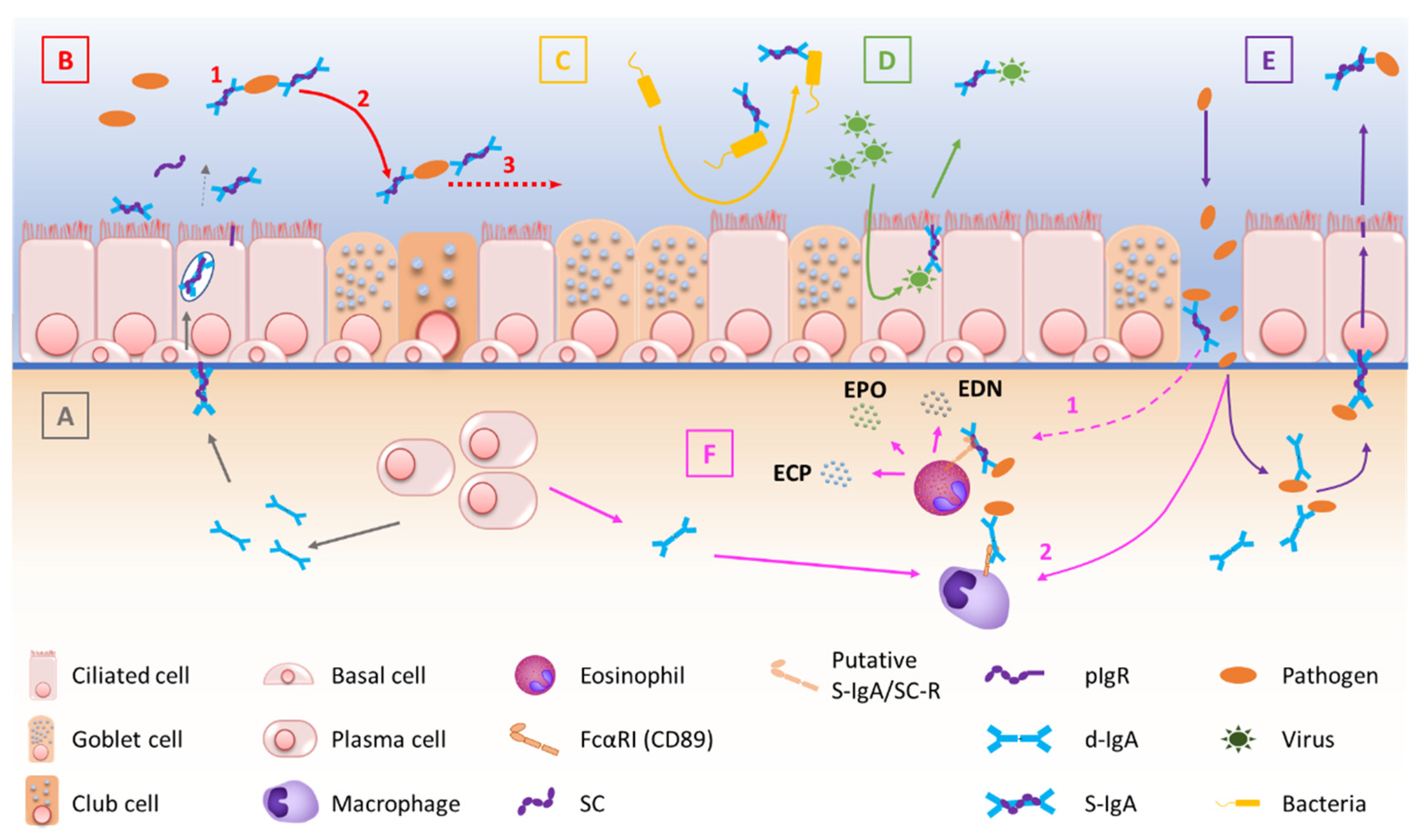
2. The Mucosal S-IgA System in Homeostasis

The mucosal barrier system is constitutively made of several components. The epithelial layer and the intercellular junctions, with the mucus layer on top, and the lamina propria underneath [13,14], form a physical barrier against exogenous antigens and pathogens. Mucosal antimicrobial molecules, cellular innate immunity and mucosal adaptative immunity are complementary lines of defence against aggressions [15]. In the bronchi, the epithelium is pseudostratified, and composed by numerous cell types whose proportions are tightly controlled [16,17]. Most of the bronchial airway epithelium consists of ciliated and goblet cells that together constitute the mucociliary elevator clearing particulates and other irritants out of the airspaces. As well as these cell types, the airway epithelium also comprises basal, club and neuroendocrine cells, along with rare ionocytes [18]. Ciliated cells are prominent and represent more than 50% of all airway epithelial cells. They possess around 300 cilia whose synchronized beating pushes the mucus layer towards the trachea and the larynx [19]. Goblet cells, accounting for 5 to 15% of airway epithelial cells in health, produce mucus and are virtually not present in small airways [19]. Basal cells are multipotent stem cells that both anchor the epithelium to the underlying lamina propria and drive epithelial homeostasis and orderly regeneration after injury [20,21]. They represent 5 to 30% of epithelial cells, their proportion decreasing from the trachea down to the respiratory bronchioles [22]. Club cells are dome-shaped cells involved in host defence, and represent 20% of epithelial cells in small airways, where they also behave as progenitor cells [23,24]. Neuroendocrine cells are rare, innervated cells (< 1% of airway epithelial cells) [25] that are thought to be involved in oxygen sensing, smooth muscle tonus and immune responses [26,27]. Finally, recently discovered ionocytes seem to control the airway surface liquid and mucus viscosity [28]. Among these cell types, ciliated, goblet and club cells have been robustly shown to express the polymeric immunoglobulin receptor (pIgR) and therefore participate in the epithelial transcytosis of dimeric IgA (d-IgA) towards the apical mucus layer (see below) [29,30], while recent single cell transcriptomic data and the emergence of the Human Lung Atlas suggest that ionocytes (but not basal and neuroendocrine cells) also express the pIgR [31].

2.1. Production and Structure of S-IgA and pIgR

IgA constitutes the most prevalent Ig isotype at mucosal sites and the second most prevalent in serum after IgG. Therefore, IgA is the most abundantly produced Ig in the human body, with an average production rate of 66 mg/kg/day [32,33]. IgA largely mediates the adaptive humoral immune defence at mucosal surfaces [34,35], while its role in serum remains relatively unexplored. IgA is produced by B cells both in the systemic and mucosal immune systems, the latter being referred to as mucosa-associated lymphoid tissue (MALT) which includes mucus layers, epithelial cells, lymphoid tissues and immune molecules of the mucosal lamina propria [36]. Structurally, IgA is found as monomers (m-IgA) or polymers, mainly consisting of dimers (d-IgA), although some larger forms also exist. M-IgA is composed of two light chains κ and λ, common to all types of Ig’s, covalently linked to two specific heavy chains α [37].
In serum, IgA represents 6 to 15% of total immunoglobulins [38], and predominates as monomers (85% to 90%). Serum IgA mainly originates from the bone marrow, although the spleen and peripheral lymph nodes could contribute to a lower extent [38]. In the mucosal lamina propria, plasma cells mainly produce IgA as dimers (d-IgA) consisting of two IgA monomers joined to the joining (J) chain at the Fc region by a disulfuric bridge [33,39]. Subepithelial d-IgA molecules can bind to the polymeric immunoglobulin receptor (pIgR) at the basolateral pole of the epithelium [34], ensuring the transcytosis of pIgR/d-IgA complexes across the epithelium. Following transcytosis, the ectodomain of pIgR (called secretory component, SC) is cleaved, allowing the apical release of d-IgA/SC complexes that constitute secretory (S-)IgA [33,35]. S-IgA is the predominant Ig in mucosal secretions such as breast milk, colostrum, tears, saliva, digestive as well as respiratory and genitourinary tract secretions [32]. S-IgA is predominantly found in a dimeric form, although some larger polymers (mainly tetramers) have also been identified [32].
In humans, two IgA subclasses exist, namely IgA1 and IgA2. Each of these IgA subtypes can be found in a monomeric, dimeric, or secretory form [40]. In bronchial secretions, IgA2 accounts for 30% of total IgA, compared to 10% in plasma [40]. This strongly suggests that there is a local production of IgA in the lung, not only spilling from the blood vessels, but also there is a substantial level of IgA2 secreting B cells within the bronchial mucosae [41].
B cells that secrete IgA in the airways are antibody-secreting plasma cells that are generated following cognate interactions between T cells and dendritic cells that have taken up and processed a specific antigen [42,43]. These conventional B(2) cells undergo class switch recombination towards IgA upon a TGF-β-dependent mechanism [44] and produce high affinity IgA antibodies. Alternatively, B1 cells are B cells that produce so-called “natural” IgM and IgA antibodies although those B cells have mainly been described in the serous cavities and in the gut [45]. Thus, the nature and origin of IgA+ B cells in the lung, as well as their regulation, are poorly characterized. Blood B cells are defined into five subsets, namely transitional, naïve, switched memory, unswitched memory and plasma cells, but similar maturation subsets have yet to be defined in the lung [46]. Nevertheless, it is known from studies in bronchial biopsies of patients with asthma that there is a local IgE production with somatic hypermutation within the airway mucosa [47]. Since IgA2 is the only possible switch for IgE+ B cells, as IgA2 heavy chain constant region gene is the only gene located downstream of the IgE gene [48], the hypothesis that IgE+ B cells could switch to IgA2 in the lung has been evoked, and supported by the observation that allergen immunotherapy may induce the induction of antigen-specific IgA2 antibodies in allergic patients [49].
pIgR is an 81 kDa transmembrane protein [50,51] that fulfils two main functions. First, it transports polymeric Ig’s (mainly d-IgA and pentameric IgM) across the mucosal epithelium [51]. Second, it serves as a precursor for SC, that is generated after endoproteolytic cleavage of its extracellular part at the apical pole of epithelial cells [52]. The extracellular, transmembrane and cytoplasmic portions of pIgR are respectively constituted by 620, 23, and 103 amino-acids [50]. The extracellular part is divided into six domains, with domains one to five being Ig-homologous domains, and domain six being a non-homologous domain and possibly representing the site of the proteolytic cleavage [50]. Therefore, SC is composed of the five Ig-like domains of pIgR [50,52].

2.2. Transcytosis of d-IgA and Functions of S-IgA

pIgR-mediated polymeric Ig transcytosis requires four well-orchestrated steps [51]. First, d-IgA (or pentameric IgM) binds to pIgR through a non-covalent interaction between Ig’s J chain and the extracellular part of pIgR. Second, pIgR is internalized in the clathrin-mediated endocytosis process and delivered in basolateral early endosomes, then in common endosomes. Endocytosed pIgR is then either recycled back to the basolateral membrane, or delivered to the apical surface [50]. Third, the extracellular part of the receptor (SC, bound to the transported Ig), undergoes endoproteolytic cleavage by a host serine proteinase. Lastly, S-IgA consisting of d-IgA and SC, crosses the mucus layer by diffusion. Of note is that, unbound pIgR may also undergo epithelial transcytosis and proteolytic cleavage, thereby releasing free SC that may be found in mucosal secretions [50,51].
At mucosal surfaces, S-IgA functions include neutralization, as well as other biological effects through interactions with, and regulation of, immune cells (via specific Fcα receptors) and microbiota components (Figure 1). The first-line mucosal defence is primarily exerted by S-IgA through its binding of soluble or particulate antigens, preventing their adherence to the epithelium. This non-specific immunity process called ‘immune exclusion’ consists of several sub-steps, including agglutination and entrapment as well as clearance at mucosal surfaces [2,53]. First, agglutination occurs when S-IgA non-covalent binding to microorganisms promotes the formation of macroscopic clumps of pathogens [2,36], altering pathogen physiology and gene expression, depending on the epitope recognized by the antibody [2,53]. For instance, S-IgA directed against the O-antigen induces bacterial outer membrane distortion [36,53]. Second, the hydrophily of IgA Fc parts, and even more of the oligosaccharide side chains of SC, favours the entrapment of microorganisms in mucus [36]. Third, muco-ciliary clearance helps to evacuate bacterial clumps [54]. It must be emphasized that immune exclusion can also occur in the submucosa, where d-IgA forms immune complexes with subepithelial trespassing pathogens that are then transcytosed after binding to pIgR [55].
In addition to immune exclusion, S-IgA can directly block toxins and pathogens from adhering to the epithelium. For instance, reovirus type 1 (strain Lang) receptor-binding domain σ1 is recognized by IgA mAbs, preventing its binding to the epithelial cells [36,53]. Moreover, S-IgA may have a direct effect on bacterial virulence [53], while immune neutralization by IgA may also occur within the epithelial cell itself. According to in vitro studies with the measles virus [56], influenza virus [57], and human immunodeficiency virus [58], inactivation of the virus by specific IgA antibodies may even occur during the transcytosis.
In addition to neutralizing properties, IgA and S-IgA also regulate immune responses through the binding of specific Fc-receptors on myeloid leukocytes. FcαRI, also known as CD89, is indeed expressed by neutrophils, eosinophils, monocytes, macrophages, and dendritic cells [59]. In case of pathogen penetration through the mucosal barrier, cross-linking of IgA bound to pathogens or immune complexes to FcαRI can activate immune mechanisms such as phagocytosis of IgA-opsonized particles, release of cytokines and activated oxygen species, as well as antibody-dependent cell-mediated cytotoxicity [36,39,60]. IgA binding to FcαRI also activates human eosinophils [2], whereas S-IgA activates eosinophil degranulation even more efficiently, possibly by binding to a C-lectin-type SC receptor [2,61]. In addition, S-IgA is recognized by DC-SIGN, a lectin receptor mainly expressed by dendritic cells [36]. In mice, interaction between S-IgA and DC-SIGN homolog induced tolerogenic responses in dendritic cells, promoting the expansion of regulatory T cells (Treg), indicating that S-IgA may exert immunoregulatory properties [36,60]. In addition, in vitro studies have shown that polymeric IgA is able to activate the alternate complement system through the binding of mannose binding lectin [55,62]. In contrast, S-IgA is unable to bind and activate the classical complement pathway, which may highlight that S-IgA is primarily designed to prevent highly inflammatory reactions [63,64].
Finally, SC also has specific functions. First, it enhances S-IgA stability and resistance against proteolysis as compared with d-IgA [51]. Second, SC increases S-IgA hydrophilicity through N- and O-glycosylation, favouring its diffusion across the mucus layer [51]. At mucosal surfaces, both free and bound SC (in S-IgA) have been shown to alter Streptococcus pneumoniae [65] and Escherichia coli attachment to the epithelium [36,53,66], demonstrating SC-driven anti-microbial properties. Moreover, hydrophilic SC glycosylation contributes to SC protective abilities [67], while S-IgA interacts with various commensal bacteria through Fab- and Fc-independent mechanisms where SC plays a critical role, as SC deglycosylation also alters the interaction between S-IgA and Gram-positive bacteria, according to confocal microscopy [66]. Finally, SC is also thought to display anti-inflammatory properties, including neutralization of IL-8/CXCL-8 activity, thereby limiting the recruitment of neutrophils at mucosal surfaces [51].

2.3. Regulation of S-IgA Production

Although more extensively studied in the gut, the pIgR/IgA system regulation mechanisms in the airways have received increasing attention in the past decade, drawing a global picture where it relies on the complex interactions of immune, environmental, microbial, as well as hormonal factors.
First, the PIGR gene promoter displays binding sites for inflammation-related factors such as IFN regulatory factor 1 (IRF-1), STAT6 and Nuclear Factor (NF)-κB [52]. Therefore, host cytokines that activate pathways involving STAT, IRF or NF-κB, such as TNF-α, IFN-ɣ, IL-4 and IL-1, are able to upregulate pIgR expression and d-IgA transepithelial routing [51,52,68]. Depending on the studies, however, exposure to inflammatory stimuli provides divergent results. For instance, IL-4 may stimulate pIgR expression in Calu-3 cell line cultures [69], while it inhibits pIgR expression in primary airway epithelial cells [70], contributing to pIgR downregulation found in the airway epithelium of asthma patients. A similar dual effect in cell line versus primary cells was observed with TGF-β1, as exogenous exposure of Calu-3 cells to TGF-β1 increases SC production, whereas pIgR production is inhibited by TGF-β1 in primary human bronchial epithelial cells [68,71]. The molecular substratum for such discrepancies remains unknown. In addition, inflammatory cytokines contribute to pIgR downregulation both in asthma and COPD, while IL-17 conversely upregulates pIgR in Pseudomonas aeruginosa (Pa) infected CF cells [68,70,72,73].
Finally, environmental factors may also influence pIgR expression. For instance, pIgR mRNA levels are increased in ex-smokers’ lungs. This increase is however not observed at the protein level, or in situ nor in air/liquid interface cultures of primary human bronchial epithelial cells exposed to cigarette smoke (CS) or derived from smokers [68,74]. These data suggest that the CS exposure acts as a player in the regulation of pIgR gene expression in vivo, which is further submitted to post-transcriptional modifications that have not been much explored so far. In addition, the microbiota also regulates pIgR through the release of microbe-associated molecular patterns (MAMPs), which control PIGR gene transcription through the activation of Toll-like receptors (TLR) [51,52].

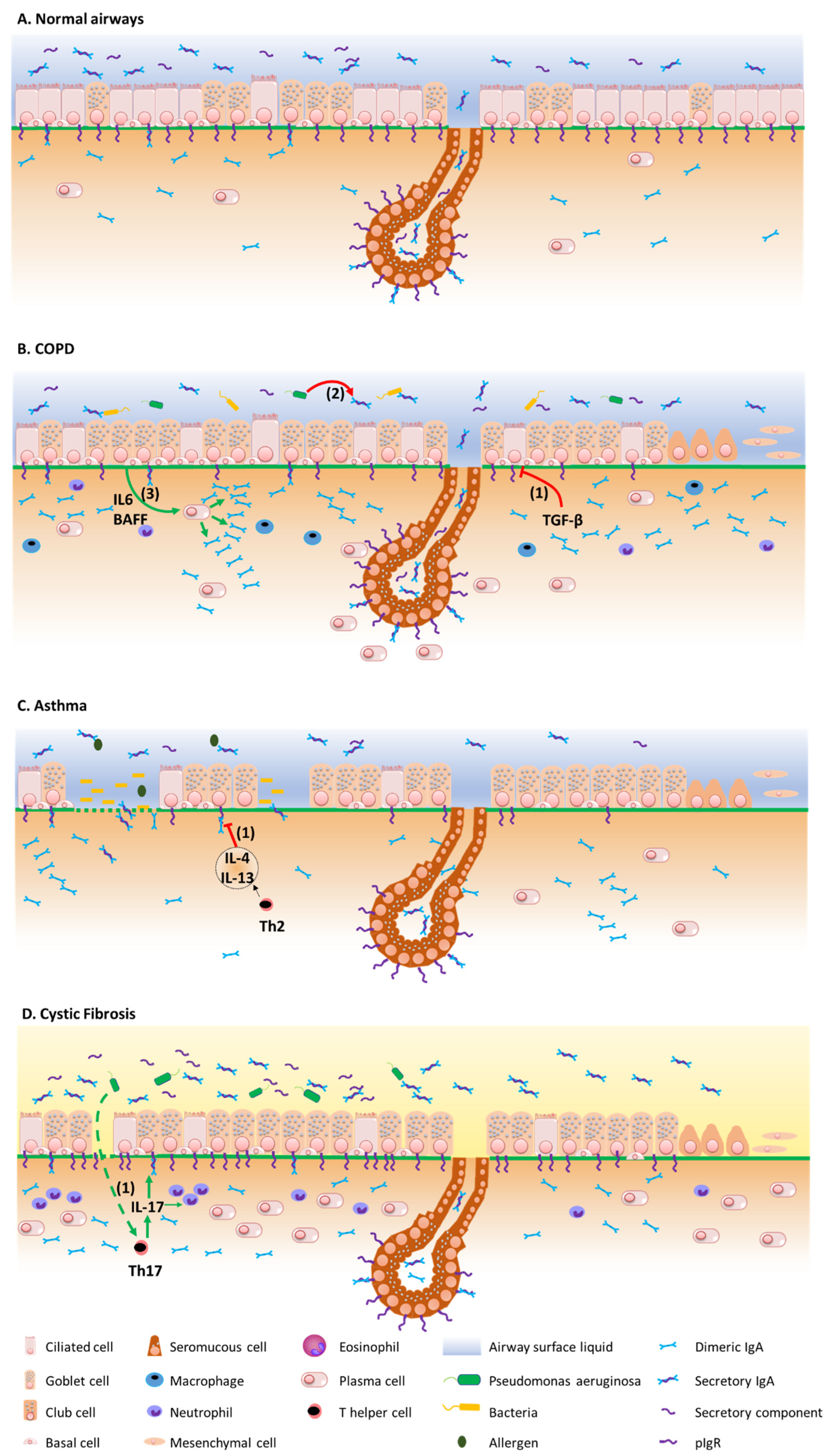
3. The Mucosal S-IgA System in Airway Disease

Figure 2 summarizes the mechanisms by which the IgA/pIgR system is altered in chronic respiratory diseases. Although resulting from complex physiopathological mechanisms in these diseases, respiratory tract colonization by pathogens and trespassing of the mucosal barrier have been shown to trigger these diseases, demonstrating their contribution to the development of such diseases [75,76].

3.1. COPD

The functionality of the IgA/pIgR system in respiratory diseases was first explored in COPD, where the abundant literature now clearly demonstrates its multifaceted alteration [77]. Chronic obstructive pulmonary disease (COPD), currently representing the third leading cause of death worldwide [3], is mainly due to CS with potential additional contributions of other toxics (biomass, occupational, air pollution) and genetic predisposition [78]. It is characterized by a progressive and mostly irreversible airway obstruction related to small airway pathology and destruction of the alveolar walls, referred to as emphysema [79]. Its clinical course can be dotted with acute worsening of respiratory symptoms, called exacerbations, a substantial proportion of which are driven by respiratory pathogens [5,8,80].
In 2001, our team first showed that pIgR/SC expression is decreased in the epithelium of large and small airways from COPD patients, as compared with both non-smokers and non-COPD smokers. In addition, SC expression inversely correlates with COPD severity and related functional parameters such as FEV1, FVC and MEF50 [81]. These observations were later corroborated in a larger cohort demonstrating that pIgR epithelial expression is mainly decreased in the airway epithelium from severe COPD patients, as compared with non-smoker controls, non-COPD smokers and less severe COPD patients, and associated with a persistence of the defect in primary cultures of bronchial epithelial cells from such patients where the mechanism could be shown as involving TGF-β signalling [68]. Recently, a genome-wide association study showed that PIGR gene expression was reduced in airway intermediate and ciliated cells from smokers without COPD [82]. Interestingly, pIgR downregulation in COPD is more obvious in zones of bronchial epithelial remodelling, such as goblet cell hyperplasia, squamous metaplasia, or incompletely differentiated areas. Accordingly, these specific zones also display S-IgA deficiency [83], contributing to localized reduced mucosal immunity, with those areas also exhibiting increased bacterial invasion, macrophage and neutrophil infiltration, as well as NF-κB activation [84].
Data regarding S-IgA concentrations in lung-lining fluids in COPD are more scattered. While a study reported no difference in S-IgA BALF concentrations in COPD patients versus controls [85], another showed decreased S-IgA levels in BALF from COPD patients [83], while a third found increased sputum SC concentrations in COPD [80]. Aside from showing COPD-related decreased S-IgA concentrations at the surface airway epithelium level, Du et al. showed that the S-IgA production was nevertheless quite preserved in COPD at the level of submucosal glands, although probably not compensating for surface epithelium S-IgA deficiency [86]. Interestingly, according to a very recent and as yet not fully published study, submucosal glands could represent a survival niche for IgA-secreting plasma cells [87]. In addition, increased levels of subepithelial IgA1 (but not IgA2) have been observed in COPD following upregulated IgA1 production by lung B cells, including in lymphoid follicles [88]. Globally, these studies depict a situation with increased local IgA production and reduced d-IgA transcytosis at the surface airway epithelium level, with secondary IgA1 subepithelial accumulation in the airways. In addition to impaired secretion, proteolytic degradation by IgA1 proteinases released by pathogens such as Pa that may infect COPD airways [89,90,91], particularly in patients with severe disease and/or bronchiectasis, may also contribute to the decrease in airway S-IgA in this disease.
Apart from pIgR downregulation in situ, in COPD airway tissues, we also showed that the airway epithelium redifferentiated in vitro in air/liquid-interface displays decreased pIgR expression and SC apical release, as well as impaired IgA transepithelial routing of d-IgA [68]. More recently, we demonstrated that the dysregulation of the pIgR/SC system in COPD is durably imprinted in the COPD airway epithelium, as long-term cultures (up to 10 weeks) of ALI-redifferentiated COPD airway epithelial cells exhibit persistent decreases in pIgR expression and SC release, along with impaired pIgR mRNA levels [74], corroborating previous findings obtained at 4 weeks of ALI culture [83]. Interestingly, increased IL-6 and BAFF/APRIL-TACI signalling have been observed in COPD at the epithelium level, driving class switch recombination towards IgA [88]; a feature that was also observed in smokers without COPD [92]. This B-cell immune response, also underlying IgA upregulation in peribronchiolar lymphoid follicles [93], probably relates to toxic and microbial exposures occurring in the COPD lung and could be considered as an attempt to compensate for local IgA deficiency, which is partly inhibited by CS [88].
To further explore the role of pIgR in COPD, pIgR−/− mice, which display no S-IgA (or S-IgM) in mucosal secretions [94], have been used. Interestingly, pIgR−/− mice exhibit increased susceptibility to mycobacterial infections [95] and spontaneously develop COPD-like small airway and parenchymal remodelling upon aging (86). When compared with wild-type mice, they show increased airway wall thickness and emphysema. They also display increased numbers of alveolar neutrophils and macrophages, increased concentrations of neutrophil elastase and matrix metalloproteinase-12, increased NF-κB p65, and increased microbial invasion. Moreover, their inflammatory response to non-typeable Haemophilus influenzae lysates is upregulated, and this is modulated by S-IgA. In addition, CS-exposed pIgR−/− mice were more prone to developing emphysema and airway remodelling upon CS exposure than wild-type mice. Strikingly, the authors finally showed that knockout mice bred in germ-free conditions were protected from developing age-related COPD-like features, demonstrating the role of the lung microbiome in COPD pathogenesis [96].
To the best of our knowledge, no study has yet directly explored whether local bronchial IgA deficiency correlates to the exacerbation rate in COPD. Nevertheless, data obtained from the observational SPIROMICS cohort study showed that COPD patients with severely reduced serum IgA levels (≤70 mg/dL) experienced more frequent and more severe exacerbations [11].

3.2. Asthma

As with COPD, asthma is a broad clinical entity that manifests with symptoms of wheezing, shortness of breath, cough, and chest tightness. Asthma is characterized by reversible airflow obstruction, in contrast with COPD where the airflow obstruction is (mostly) irreversible [97]. Behind this clinical definition, asthma is a heterogeneous disease that relates to diverse pathophysiological mechanisms. Two main immune phenotypes are described, namely type 2 (T2-) high asthma, which includes several subsets according to the age of onset, allergic background, and comorbidities such as aspirin sensitivity and/or nasal polyposis; T2-low asthma, which includes obesity-related asthma [97,98,99,100]. Both early-onset allergic (or extrinsic) asthma and some forms of late-onset non-allergic asthma are characterized by a T2-mediated eosinophilic inflammation, whose underlying upstream mechanisms include epithelial activation by viruses, allergens and/or air pollution. The subepithelial penetration of these irritants is further enhanced by epithelial barrier dysfunction that is due to the decreased tightness of apical junctional complexes, as well as zones of epithelial shedding [77]. These airborne stimuli induce epithelial cells necrosis leading to the release of alarmins such as TSLP, IL-33, IL-25 and GM-CSF, the latter attracting and/or activating mast cells, basophils and eosinophils [51]. In T2-low asthma, macrophages recruitment is also driven by the epithelial release of CCL2 and CCL3 [101], while in obesity-related asthma, leptin activates airway epithelial cells and induces the production of cytokines such as IL-8, further contributing to neutrophil recruitment [101]. In turn, these inflammatory cells mediate airway remodelling, including goblet cells metaplasia, epithelial-to-mesenchymal transition and reticular basement membrane thickening, along with bronchial hyperresponsiveness and smooth muscle hypertrophy [51,98], as well as IgE class switch recombination in B cells [51]. In addition the increased levels of allergen specific IgE, higher IgA levels have been observed in the airway mucosa of allergic asthma patients [102,103,104]. In addition, class switch recombination of mucosal activated B cells towards IgA can be induced by different stimuli involved in an allergic response, such as double stranded RNA viruses in a TLR3-dependent pathway [105], or cytokines such as IL-4, which is increased in an asthmatic response [106] or by microbial stimuli through TGF-β production by dendritic cells [107].
Although the exact role(s) of S-IgA in the development of allergic disorders remains controversial, several lines of evidence suggest that the S-IgA system could play a protective role in asthma and allergic diseases [2]. Thus, higher levels of S-IgA in breast milk were associated to a lower risk of atopic dermatitis up to the age of two [108], and subjects with selective IgA deficiency display higher risks of developing allergic diseases [109]. In addition, populations with higher serum IgA levels display lower rates of house dust mite sensitization and severe airway hyperresponsiveness [110]. Conversely, a positive correlation exists between serum IgA specifically directed against cow’s milk β-lactoglobulin and wheat gliadin at the age of one, and IgE sensitization at the age of 6 [111].
Several mechanisms could explain these clinical observations. Firstly, S-IgA possibly mediates the immune exclusion of allergens, limiting the exposure of the immune system to these triggers [12,109]. It also interacts with several microbiota components, therefore modifying the composition of the microbiome, and influencing the maintenance of immune homeostasis in the gut and the lungs [112]. The alteration in the microbiota could increase the susceptibility to allergic disorders, as early-life antibiotic-treated mice displaying microbiota dysregulation showed a higher risk of developing allergic asthma [113,114]. Secondly, S-IgA seems able to drive tolerogenic immune mechanisms in dendritic cells through its binding to the DC-SIGN receptor [115]. Thus, the ligation of S-IgA to dendritic cells inhibits CD4+ T cell responses, in a DLL4-Notch pathway-dependent manner [116], while inducing a secondary expansion of Tregs [2]. This S-IgA-driven Treg population expansion could play a role in maintaining mucosal tolerance by regulating the excessive function of other cells and hampering aberrant T helper 2 responses observed in asthma [117]. However, this T helper 2-mediated inflammation is also enhanced by TSLP [118], which has also been described as able to inhibit both the generation of inducible Tregs [119,120] and the production of IgA [121].
In contrast, S-IgA can also trigger eosinophil degranulation [2,122] through its binding to FcαRI or to a putative C-lectin type SC receptor [123], leading to the release of IL-4 and IL-5, as well as eosinophil peroxidase and eosinophil cationic protein, thereby promoting Th2 inflammation and asthma pathogenesis and exacerbations [124]. Soluble S-IgA also enhances eosinophil survival in vitro [125]. FcαRI is also expressed by neutrophils, but conflicting data have been obtained regarding the pro- or anti-inflammatory effects of S-IgA binding to neutrophils. Indeed, myeloma-derived IgA alters neutrophil chemotaxis in vitro [126], while FcαRI binding has also been reported to increase the release of several cytokines by neutrophils [127].
Finally, previous findings described decreased S-IgA levels in BAL from symptomatic asthma patients. We also observed a dysregulation of the S-IgA/pIgR system in the airways in allergic asthma, that was reproduced in vitro by exposing the air-liquid interface cultures of human bronchial epithelial cells to exogenous IL-4 and IL-13, underlining the role of T2 inflammation in this impairment [70].

3.3. Cystic Fibrosis

In contrast with asthma and COPD [128], data concerning IgA in cystic fibrosis (CF) are scarcer. CF is a multisystemic disease mainly affecting the respiratory, digestive and reproductive tracts [129]. Chronic pulmonary infections, notably due to Pa, play a crucial role in the prognosis of the disease and constitute a major immune challenge for the airway epithelium [130]. Recently, we undertook one study assessing the pIgR/IgA system in a multimodal project, showing increased epithelial IgA+ B cells, IgA production and pIgR expression in the airway tissues, sputum, and serum from CF patients [72,73]. In addition, a mice model mimicking chronic lung infection by using Pa-coated microbeads instilled in the mice airways showed that infection could upregulate pIgR expression and IgA production in the lungs of F508del mice, partly in an IL-17-dependant manner [72]. This was observed in vivo although in vitro, CF-derived airway epithelial cells displayed reduced pIgR expression, that appeared to be related to the activation unfolded protein response. In parallel, pIgR transcytosis is enhanced in CF, as evidenced by increased SC concentration in CF sputum, although SC seems to be dysfunctional in CF, notably preventing its neutralization capacity of IL-8/CXCL-8 due to an altered glycosylation pattern [131]. Increased IgA concentration has been found in the serum from CF patients, as compared with controls [132], in particular in chronically Pa infected patients [133] or with high clinical severity according to Shwachman Kulczycki scores [134]. Similarly, increased IgA concentrations were found in BALF from CF patients as compared with controls [135]. In addition, increased specific anti-Pa IgA levels were observed in the serum of infected CF patients [136,137,138] and in patients with poor lung function, witnessed by low vital capacity and forced expiratory volume in one second [139], but also in sputum [140] and nasal lavage [141]. Finally, data regarding IgA secretion in the digestive and respiratory systems in CF diverge from results in the respiratory tract, as decreased IgA secretion in CF saliva [136] and gastric luminal perfusate have been reported [142], while an older study reports decreased free SC concentration in the sputum and saliva from CF patients [143]. It appears likely that in CF the IgA system is affected via multiple components and interplays between the host epithelium, immune cells, and microbiome that ultimately differently regulate IgA immunity at mucosal sites.

3.4. Bronchiolitis Obliterans Syndrome

Chronic lung allograft dysfunction (CLAD) represents a major hurdle in lung transplantation (LT), that burdens long-term survival [137]. Depending on pulmonary function tests and computed tomography, CLAD is currently separated into obstructive CLAD (first described as bronchiolitis obliterans syndrome (BOS), restrictive CLAD (also known as restrictive allograft syndrome (RAS), mixed CLAD (combining BOS and RAS features) and undefined CLAD [138]. BOS is the most frequent form of CLAD and is histologically characterized by epithelial injury, bronchocentric mononuclear inflammation, and fibrosis of small airways [144].
As repetitive and/or chronic infections have widely been associated with CLAD risk [145,146,147], it can be questioned whether impaired airway mucosal immunity (and thus reduced microbial eviction) could favor CLAD/BOS. However, the literature in the field is scarce, with no study so far assessing the airway expression of pIgR after LT. One previous study assessed BALF S-IgA levels in LT recipients, showing reduced S-IgA levels during so-called “rejection episodes” [148]. Of note is that, CLAD phenotypes had not been described at the time this study was published. More recently, Vandermeulen and colleagues showed increased total BALF IgA levels in LT recipients with RAS and BOS as compared with control LT recipients [149]. Finally, low serum IgA levels prior to LT represent a risk factor for developing post-LT infections [150,151], while lower post-LT IgA serum levels have been observed in patients with BOS as compared with control LT recipients [151].

4. Conclusions and Perspectives

Recent research demonstrates that the S-IgA system is altered during the course of chronic airway diseases, primarily in relation to a generic mechanism of the downregulation of pIgR expression that is observed upon airway epithelial dedifferentiation. Some mechanisms might prevail in certain disease phenotypes, such as the aberrant reactivation of developmental pathways (e.g., TGF-β and WNT/β-catenin, in COPD), immune activation through the IL-4R (e.g., in asthma) or unfolded protein response (e.g., in CF). In contrast, the production of IgA by B cells is upregulated, showing that the epithelial transport of IgA also represents the limiting factor for IgA immunity in the diseased lung. This epithelial pIgR defect may also lead to increased susceptibility to COPD development, through complex mechanisms involving aging, inflammation and the microbiome as shown in pIgR KO mice, as well as increased susceptibility to lung infections.
A better understanding of IgA mucosal immunity in the airways could deliver therapeutic perspectives. First, passive immunotherapy could be studied by using either specific IgA, as shown in a preclinical model of an airway allergy to ragweed pollen [152], or human polyclonal IgA as achieved with IgG for IgG deficiency. This later case is however not supported by a specific indication as it is known that selective IgA deficiency which represents the most frequent immunodeficiency, very rarely leads to opportunistic and/or frequent infections when present alone (as opposed to combined IgA and IgG deficiency) [153,154]. In contrast, supplementation with secretory IgA could be considered in diseases with evidence of the impaired transport of IgA into mucosa secretions such as asthma or COPD. Evidence of a benefit of such an approach is however lacking at both preclinical and clinical levels. For instance, in asthma, the putative benefit against infections might be at least theoretically counterbalanced by the detrimental activation of eosinophils. In this case of mucosal IgA deficiency, the approach could alternatively involve tackling the underlying mechanism within the epithelium, by interfering with the activation of IL-4/IL-13 (e.g., in asthma) or the TGF-β or WNT pathway (e.g., in COPD). Such an approach, however, implies considering targeted application (referring to the frequently opposed effects in the conducting epithelium versus alveolar epithelium) and careful evaluation of the side effects due to the pleiotropic nature of these pathways in lung homeostasis.
Future research should, thus, appreciate whether current therapies affect the S-IgA system and whether supplementing IgA could provide clinical benefits in patients with COPD or other chronic airway diseases.

Author Contributions

C.d.F., F.M.C. and S.G. wrote the manuscript. C.P. wrote and revised the manuscript. All authors have read and agreed to the published version of the manuscript.

Funding

C.d.F. was supported by the Fondation St-Luc, Brussels, Belgium. F.M.C. was supported by the Fonds National de la Recherche Scientifique (FNRS, grants 1.L505.16 and 1.L505.18) and by the Fondation Mont-Godinne (grants FMG-2015-BC01, FMG-2016-BC01, and FMG-2017-BC01), Belgium. S.G. and C.P. are postdoctoral specialists of the FNRS (Grants 1.R60121F and 1.R016.18 respectively), Belgium.

Institutional Review Board Statement

Not applicable.

Data Availability Statement

Not applicable.

Acknowledgments

The authors would like to thank Chiesi for its support via an unrestricted grant.

Conflicts of Interest

The authors declare no conflict of interest.

Abbreviations

BAFFB-cell activating factor
BALFBronchoalveolar lavage fluid
BOSBronchiolitis obliterans syndrome
CCLC-C Motif Chemokine Ligand
CDCluster of differentiation
CFCystic Fibrosis
CLADChronic lung allograft dysfunction
COPDChronic obstructive pulmonary disease
CSCigarette smoke
CXCL-8C-X-C motif chemokine ligand 8
DC-SIGNDendritic cell specific ICAM-3 grabbing nonintegrin
d-IgADimeric IgA
DLL4Delta-like canonical ligand 4
ECPEosinophil cationic protein
EDNEosinophil-derived neurotoxin
EPOEosinophil peroxydase
FabAntigen-binding fragment
FcAntigen crystallizable fragment
FcαRIFc receptor for IgA
FEVForced expiratory volume
FVCForced vital capacity
IFNInterferon
IFN-γInterferon γ
IgImmunoglobulin
IgAImmunoglobulin A
IgGImmunoglobulin G
IgMImmunoglobulin M
IL-1Interleukin 1
IL-4Interleukin 4
IL-8Interleukin 8
IRF-1IFN regulatory factor 1
J chainJoining chain
LTLung transplantation
MALTMucosa-associated lymphoid tissue
MAMPMicrobe-associated molecular pattern
MEFMaximal expiratory flow
m-IgAMonomeric IgA
NF-κBNuclear Factor κB
PaPseudomonas aeruginosa
p-IgAPolymeric IgA
pIgRPolymeric immunoglobulin receptor
RASRestrictive allograft syndrome
RNARibonucleic acid
SCSecretory component
S-IgASecretory IgA
STAT6Signal transducer and activator of transcription 6
TGF-βTransforming growth factor β
TLRToll-like receptor
TNF-αTumour necrosis factor α
TregRegulatory T cell

References

  1. Corthésy, B. Multi-Faceted Functions of Secretory IgA at Mucosal Surfaces. Front. Immunol. 2013, 4, 185. [Google Scholar] [CrossRef] [PubMed] [Green Version]
  2. Carlier, F.M.; Sibille, Y.; Pilette, C. The epithelial barrier and immunoglobulin A system in allergy. Clin. Exp. Allergy 2016, 46, 1372–1388. [Google Scholar] [CrossRef] [PubMed]
  3. Vos, T.; Lim, S.S.; Abbafati, C.; Abbas, K.M.; Abbasi, M.; Abbasifard, M.; Abbasi-Kangevari, M.; Abbastabar, H.; Abd-Allah, F.; Abdelalim, A. Global burden of 369 diseases and injuries in 204 countries and territories, 1990–2019: A systematic analysis for the Global Burden of Disease Study 2019. Lancet 2020, 396, 1204–1222. [Google Scholar] [CrossRef]
  4. Farrell, P.M. The prevalence of cystic fibrosis in the European Union. J. Cyst. Fibros. 2008, 7, 450–453. [Google Scholar] [CrossRef] [PubMed] [Green Version]
  5. Mallia, P.; Contoli, M.; Caramori, G.; Pandit, A.; Johnston, S.; Papi, A. Exacerbations of Asthma and Chronic Obstructive Pulmonary Disease (COPD): Focus on Virus Induced Exacerbations. Curr. Pharm. Des. 2007, 13, 73–97. [Google Scholar] [CrossRef] [PubMed]
  6. Weiss, K.B.; Sullivan, S.D. The health economics of asthma and rhinitis. I. Assessing the economic impact. J. Allergy Clin. Immunol. 2001, 107, 3–8. [Google Scholar] [CrossRef] [Green Version]
  7. FitzGerald, J.M.; Barnes, P.J.; Chipps, B.E.; Jenkins, C.R.; O’Byrne, P.M.; Pavord, I.D.; Reddel, H.K. The burden of exacerbations in mild asthma: A systematic review. ERJ Open Res. 2020, 6, 00359–2019. [Google Scholar] [CrossRef]
  8. Viniol, C.; Vogelmeier, C.F. Exacerbations of COPD. Eur. Respir. Rev. 2018, 27, 170103. [Google Scholar] [CrossRef] [Green Version]
  9. Goss, C.H.; Burns, J.L. Exacerbations in cystic fibrosis. 1: Epidemiology and pathogenesis. Thorax 2007, 62, 360–367. [Google Scholar] [CrossRef] [Green Version]
  10. Sethi, S.; Murphy, T. Infection in the Pathogenesis and Course of Chronic Obstructive Pulmonary Disease. N. Engl. J. Med. 2008, 359, 2355–2365. [Google Scholar] [CrossRef]
  11. Putcha, N.; Paul, G.G.; Azar, A.; Wise, R.A.; O’Neal, W.K.; Dransfield, M.T.; Woodruff, P.G.; Curtis, J.L.; Comellas, A.P.; Drummond, M.B.; et al. Lower serum IgA is associated with COPD exacerbation risk in SPIROMICS. PLoS ONE 2018, 13, e0194924. [Google Scholar] [CrossRef] [PubMed]
  12. Burrows, P.D.; Cooper, M.D. IgA deficiency. Adv. Immunol. 1997, 65, 245–276. [Google Scholar] [PubMed]
  13. Yang, S.; Yu, M. Role of Goblet Cells in Intestinal Barrier and Mucosal Immunity. J. Inflamm. Res. 2021, 14, 3171–3183. [Google Scholar] [CrossRef] [PubMed]
  14. Boyaka, P.N.; Fujihashi, K. Host Defenses at Mucosal Surfaces. In Clinical Immunology, 5th ed.; Rich, R.R., Fleisher, T.A., Shearer, W.T., Schroeder, H.W., Frew, A.J., Weyand, C.M., Eds.; Elsevier: London, UK, 2019; pp. 285–298.e1. [Google Scholar]
  15. McNabb, P.C.; Tomasi, T.B. Host Defense Mechanisms at Mucosal Surfaces. Annu. Rev. Microbiol. 1981, 35, 477–496. [Google Scholar] [CrossRef] [PubMed]
  16. Davis, J.D.; Wypych, T.P. Cellular and functional heterogeneity of the airway epithelium. Mucosal Immunol. 2021, 14, 978–990. [Google Scholar] [CrossRef] [PubMed]
  17. Gohy, S.; Hupin, C.; Ladjemi, M.Z.; Hox, V.; Pilette, C. Key role of the epithelium in chronic upper airways diseases. Clin. Exp. Allergy 2019, 50, 135–146. [Google Scholar] [CrossRef]
  18. Travaglini, K.J.; Nabhan, A.N.; Penland, L.; Sinha, R.; Gillich, A.; Sit, R.V.; Chang, S.; Conley, S.D.; Mori, Y.; Seita, J.; et al. A molecular cell atlas of the human lung from single-cell RNA sequencing. Nature 2020, 587, 619–625. [Google Scholar] [CrossRef]
  19. Knight, D.A.; Holgate, S.T. The airway epithelium: Structural and functional properties in health and disease. Respirology 2003, 8, 432–446. [Google Scholar] [CrossRef]
  20. Rock, J.R.; Randell, S.H.; Hogan, B.L.M. Airway basal stem cells: A perspective on their roles in epithelial homeostasis and remodeling. Dis. Model. Mech. 2010, 3, 545–556. [Google Scholar] [CrossRef] [Green Version]
  21. Hogan, B.L.; Barkauskas, C.E.; Chapman, H.A.; Epstein, J.A.; Jain, R.; Hsia, C.C.; Niklason, L.; Calle, E.; Le, A.; Randell, S.H. Repair and regeneration of the respiratory system: Complexity, plasticity, and mechanisms of lung stem cell function. Cell Stem Cell 2014, 15, 123–138. [Google Scholar] [CrossRef] [Green Version]
  22. Boers, J.E.; Ambergen, A.W.; Thunnissen, F.B.J.M. Number and Proliferation of Basal and Parabasal Cells in Normal Human Airway Epithelium. Am. J. Respir. Crit. Care Med. 1998, 157, 2000–2006. [Google Scholar] [CrossRef] [PubMed]
  23. Zuo, W.-L.; Shenoy, S.A.; Li, S.; O’Beirne, S.L.; Strulovici-Barel, Y.; Leopold, P.L.; Wang, G.; Staudt, M.R.; Walters, M.S.; Mason, C.; et al. Ontogeny and Biology of Human Small Airway Epithelial Club Cells. Am. J. Respir. Crit. Care Med. 2018, 198, 1375–1388. [Google Scholar] [CrossRef]
  24. Hiemstra, P.S.; Bourdin, A. Club cells, CC10 and self-control at the epithelial surface. Eur. Respir. J. 2014, 44, 831–832. [Google Scholar] [CrossRef] [PubMed] [Green Version]
  25. Boers, J.E.; Brok, J.L.D.; Koudstaal, J.; Arends, J.W.; Thunnissen, F.B. Number and proliferation of neuroendocrine cells in normal human airway epithelium. Am. J. Respir. Crit. Care Med. 1996, 154, 758–763. [Google Scholar] [CrossRef] [PubMed]
  26. Linnoila, R.I. Functional facets of the pulmonary neuroendocrine system. Lab. Investig. 2006, 86, 425–444. [Google Scholar] [CrossRef] [PubMed] [Green Version]
  27. Branchfield, K.; Nantie, L.; Verheyden, J.M.; Sui, P.; Wienhold, M.D.; Sun, X. Pulmonary neuroendocrine cells function as airway sensors to control lung immune response. Science 2016, 351, 707–710. [Google Scholar] [CrossRef] [PubMed] [Green Version]
  28. Montoro, D.T.; Haber, A.L.; Biton, M.; Vinarsky, V.; Lin, B.; Birket, S.E.; Yuan, F.; Chen, S.; Leung, H.M.; Villoria, J.; et al. A revised airway epithelial hierarchy includes CFTR-expressing ionocytes. Nature 2018, 560, 319–324. [Google Scholar] [CrossRef]
  29. He, W.-H.; Zhang, W.-D.; Cheng, C.-C.; Lu, J.; Liu, L.; Chen, Z.-H.; Wang, W.-H. Expression characteristics of polymeric immunoglobulin receptor in Bactrian camel (Camelus bactrianus) lungs. PLoS ONE 2022, 17, e0264815. [Google Scholar] [CrossRef]
  30. Blackburn, J.B.; Schaff, J.A.; Gutor, S.; Du, R.-H.; Nichols, D.; Sherrill, T.; Gutierrez, A.J.; Xin, M.K.; Wickersham, N.; Zhang, Y. Secretory cells are the primary source of pIgR in small airways. bioRxiv 2021. [Google Scholar] [CrossRef]
  31. Schiller, H.B.; Montoro, D.T.; Simon, L.M.; Rawlins, E.L.; Meyer, K.B.; Strunz, M.; Braga, F.A.V.; Timens, W.; Koppelman, G.H.; Budinger, G.R.S.; et al. The Human Lung Cell Atlas: A High-Resolution Reference Map of the Human Lung in Health and Disease. Am. J. Respir. Cell Mol. Biol. 2019, 61, 31–41. [Google Scholar] [CrossRef]
  32. Woof, J.M.; Russell, M.W. Structure and function relationships in IgA. Mucosal Immunol. 2011, 4, 590–597. [Google Scholar] [CrossRef] [Green Version]
  33. Bakema, J.E.; van Egmond, M. Immunoglobulin A: A next generation of therapeutic antibodies? mAbs 2011, 3, 352–361. [Google Scholar] [CrossRef] [Green Version]
  34. Monteiro, R.C. The Role of IgA and IgA Fc Receptors as Anti-Inflammatory Agents. J. Clin. Immunol. 2010, 30, 61–64. [Google Scholar] [CrossRef] [PubMed]
  35. Underdown, B.J.; Schiff, J.M. Immunoglobulin A: Strategic Defense Initiative at the Mucosal Surface. Annu. Rev. Immunol. 1986, 4, 389–417. [Google Scholar] [CrossRef]
  36. Li, Y.; Jin, L.; Chen, T. The Effects of Secretory IgA in the Mucosal Immune System. BioMed Res. Int. 2020, 2020, 2032057. [Google Scholar] [CrossRef] [PubMed]
  37. Pilette, C.; Ouadrhiri, Y.; Godding, V.; Vaerman, J.-P.; Sibille, Y. Lung mucosal immunity: Immunoglobulin-A revisited. Eur. Respir. J. 2001, 18, 571–588. [Google Scholar] [CrossRef] [PubMed] [Green Version]
  38. Conley, M.E.; Delacroix, D.L. Intravascular and Mucosal Immunoglobulin A: Two Separate but Related Systems of Immune Defense? Ann. Intern. Med. 1987, 106, 892. [Google Scholar] [CrossRef] [PubMed]
  39. Leong, K.W.; Ding, J.L. The Unexplored Roles of Human Serum IgA. DNA Cell Biol. 2014, 33, 823–829. [Google Scholar] [CrossRef] [PubMed]
  40. Burnett, D. Immunoglobulins in the lung. Thorax 1986, 41, 337–344. [Google Scholar] [CrossRef] [PubMed] [Green Version]
  41. Delacroix, D.L.; Dive, C.; Rambaud, J.C.; Vaerman, J.P. IgA subclasses in various secretions and in serum. Immunology 1982, 47, 383–385. [Google Scholar]
  42. Reboldi, A.; Arnon, T.I.; Rodda, L.B.; Atakilit, A.; Sheppard, D.; Cyster, J.G. IgA production requires B cell interaction with subepithelial dendritic cells in Peyer’s patches. Science 2016, 352, aaf4822. [Google Scholar] [CrossRef] [PubMed] [Green Version]
  43. Stumbles, P.A.; Upham, J.W.; Holt, P.G. Airway dendritic cells: Co-ordinators of immunological homeostasis and immunity in the respiratory tract. APMIS 2003, 111, 741–755. [Google Scholar] [CrossRef] [PubMed] [Green Version]
  44. Gloudemans, A.K.; Plantinga, M.; Guilliams, M.; Willart, M.A.; Ozir-Fazalalikhan, A.; Van Der Ham, A.; Boon, L.; Harris, N.L.; Hammad, H.; Hoogsteden, H.C.; et al. The Mucosal Adjuvant Cholera Toxin B Instructs Non-Mucosal Dendritic Cells to Promote IgA Production via Retinoic Acid and TGF-β. PLoS ONE 2013, 8, e59822. [Google Scholar] [CrossRef] [PubMed] [Green Version]
  45. Rothenberg, E.V. Multiple Curricula for B Cell Developmental Programming. Immunity 2016, 45, 457–458. [Google Scholar] [CrossRef] [PubMed] [Green Version]
  46. Sanz, I.; Wei, C.; Jenks, S.A.; Cashman, K.S.; Tipton, C.; Woodruff, M.C.; Hom, J.; Lee, F.E.-H. Challenges and Opportunities for Consistent Classification of Human B Cell and Plasma Cell Populations. Front. Immunol. 2019, 10, 2458. [Google Scholar] [CrossRef] [Green Version]
  47. Takhar, P.; Corrigan, C.; Smurthwaite, L.; O’Connor, B.J.; Durham, S.R.; Lee, T.; Gould, H.J. Class switch recombination to IgE in the bronchial mucosa of atopic and nonatopic patients with asthma. J. Allergy Clin. Immunol. 2007, 119, 213–218. [Google Scholar] [CrossRef]
  48. Xu, Z.; Zan, H.; Pone, E.J.; Mai, T.; Casali, P. Immunoglobulin class-switch DNA recombination: Induction, targeting and beyond. Nat. Rev. Immunol. 2012, 12, 517–531. [Google Scholar] [CrossRef] [Green Version]
  49. Pilette, C.; Nouri-Aria, K.T.; Jacobson, M.R.; Wilcock, L.K.; Detry, B.; Walker, S.M.; Francis, J.N.; Durham, S.R. Grass pollen immunotherapy induces an allergen-specific IgA2 antibody response associated with mucosal TGF-beta expression. J. Immunol. 2007, 178, 4658–4666. [Google Scholar] [CrossRef] [Green Version]
  50. Asano, M.; Komiyama, K. Polymeric immunoglobulin receptor. J. Oral Sci. 2011, 53, 147–156. [Google Scholar] [CrossRef] [Green Version]
  51. Turula, H.; Wobus, C.E. The Role of the Polymeric Immunoglobulin Receptor and Secretory Immunoglobulins during Mucosal Infection and Immunity. Viruses 2018, 10, 237. [Google Scholar] [CrossRef] [Green Version]
  52. Johansen, F.-E.; Kaetzel, C. Regulation of the polymeric immunoglobulin receptor and IgA transport: New advances in environmental factors that stimulate pIgR expression and its role in mucosal immunity. Mucosal Immunol. 2011, 4, 598–602. [Google Scholar] [CrossRef] [PubMed] [Green Version]
  53. Mantis, N.J.; Rol, N.; Corthésy, B. Secretory IgA’s complex roles in immunity and mucosal homeostasis in the gut. Mucosal Immunol. 2011, 4, 603–611. [Google Scholar] [CrossRef] [PubMed]
  54. Bustamante-Marin, X.M.; Ostrowski, L.E. Cilia and Mucociliary Clearance. Cold Spring Harb. Perspect. Biol. 2017, 9, a028241. [Google Scholar] [CrossRef] [PubMed]
  55. de Sousa-Pereira, P.; Woof, J.M. IgA: Structure, Function, and Developability. Antibodies 2019, 8, 57. [Google Scholar] [CrossRef] [Green Version]
  56. Yan, H.; Lamm, M.E.; Björling, E.; Huang, Y.T. Multiple Functions of Immunoglobulin A in Mucosal Defense against Viruses: An In Vitro Measles Virus Model. J. Virol. 2002, 76, 10972–10979. [Google Scholar] [CrossRef] [Green Version]
  57. Mazanec, M.B.; Kaetzel, C.S.; Lamm, M.E.; Fletcher, D.; Nedrud, J.G. Intracellular neutralization of virus by immuno-globulin A antibodies. Proc. Natl. Acad. Sci. USA 1992, 89, 6901–6905. [Google Scholar] [CrossRef] [Green Version]
  58. Wright, A.; Yan, H.; Lamm, M.E.; Huang, Y.T. Immunoglobulin A antibodies against internal HIV-1 proteins neutralize HIV-1 replication inside epithelial cells. Virology 2006, 356, 165–170. [Google Scholar] [CrossRef] [Green Version]
  59. Chu, P.G.; Arber, D.A. CD79: A review. Applied immunohistochemistry & molecular morphology. AIMM 2001, 9, 97–106. [Google Scholar]
  60. Diana, J.; Moura, I.; Vaugier, C.; Gestin, A.; Tissandie, E.; Beaudoin, L.; Corthésy, B.; Hocini, H.; Lehuen, A.; Monteiro, R. Secretory IgA Induces Tolerogenic Dendritic Cells through SIGNR1 Dampening Autoimmunity in Mice. J. Immunol. 2013, 191, 2335–2343. [Google Scholar] [CrossRef] [Green Version]
  61. Lamkhioued, B.; Gounni, A.S.; Gruart, V.; Pierce, A.; Capron, A.; Capron, M. Human eosinophils express a receptor for secretory component. Role in secretory IgA-dependent activation. Eur. J. Immunol. 1995, 25, 117–125. [Google Scholar] [CrossRef]
  62. Roos, A.; Bouwman, L.H.; Van Gijlswijk-Janssen, D.J.; Faber-Krol, M.C.; Stahl, G.; Daha, M.R. Human IgA Activates the Complement System Via the Mannan-Binding Lectin Pathway. J. Immunol. 2001, 167, 2861–2868. [Google Scholar] [CrossRef] [PubMed] [Green Version]
  63. Monteiro, R.C.; van de Winkel, J.G.J. IgA Fc Receptors. Annu. Rev. Immunol. 2003, 21, 177–204. [Google Scholar] [CrossRef] [PubMed]
  64. Pilette, C.; Ouadrhiri, Y.; Dimanche, F.; Vaerman, J.P.; Sibille, Y. Secretory component is cleaved by neutrophil serine proteinases but its epithelial production is increased by neutrophils through NF-kappa B- and p38 mitogen-activated protein kinase-dependent mechanisms. Am. J. Respir Cell Mol. Biol. 2003, 28, 485–498. [Google Scholar] [CrossRef] [PubMed]
  65. Hammerschmidt, S.; Talay, S.R.; Brandtzaeg, P.; Chhatwal, G.S. SpsA, a novel pneumococcal surface protein with specific binding to secretory Immunoglobulin A and secretory component. Mol. Microbiol. 1997, 25, 1113–1124. [Google Scholar] [CrossRef]
  66. Mathias, A.; Corthésy, B. Recognition of Gram-positive Intestinal Bacteria by Hybridoma- and Colostrum-derived Secretory Immunoglobulin A Is Mediated by Carbohydrates. J. Biol. Chem. 2011, 286, 17239–17247. [Google Scholar] [CrossRef] [Green Version]
  67. Perrier, C.; Sprenger, N.; Corthésy, B. Glycans on Secretory Component Participate in Innate Protection against Mucosal Pathogens. J. Biol. Chem. 2006, 281, 14280–14287. [Google Scholar] [CrossRef] [Green Version]
  68. Gohy, S.T. Polymeric immunoglobulin receptor down-regulation in chronic obstructive pulmonary disease. Persistence in the cultured epithelium and role of transforming growth factor-beta. Am. J. Respir Crit. Care Med. 2014, 190, 509–521. [Google Scholar] [CrossRef]
  69. Loman, S.; Jansen, H.M.; Out, T.A.; Lutter, R. Interleukin-4 and interferon-gamma synergistically increase secretory component gene expression, but are additive in stimulating secretory immunoglobulin A release by Calu-3 airway epithelial cells. Immunology 1999, 96, 537. [Google Scholar] [CrossRef]
  70. Ladjemi, M.Z.; Gras, D.; Dupasquier, S.; Detry, B.; Lecocq, M.; Garulli, C.; Fregimilicka, C.; Bouzin, C.; Gohy, S.; Chanez, P. Bronchial Epithelial IgA Secretion Is Impaired in Asthma. Role of IL-4/IL-13. Am. J. Res-Piratory Crit. Care Med. 2018, 197, 1396–1409. [Google Scholar] [CrossRef]
  71. Ratajczak, C.; Guisset, A.; Detry, B.; Sibille, Y.; Pilette, C. Dual effect of neutrophils on pIgR/secretory component in human bronchial epithelial cells: Role of TGF-beta. J. Biomed. Biotechnol. 2010, 2010, 428618. [Google Scholar] [CrossRef] [Green Version]
  72. Collin, A.M.; Lecocq, M.; Noel, S.; Detry, B.; Carlier, F.M.; Nana, F.A.; Bouzin, C.; Leal, T.; Vermeersch, M.; De Rose, V.; et al. Lung immunoglobulin A immunity dysregulation in cystic fibrosis. eBioMedicine 2020, 60, 102947. [Google Scholar] [CrossRef] [PubMed]
  73. Gohy, S.; Moeremans, A.; Pilette, C.; Collin, A. Immunoglobulin A Mucosal Immunity and Altered Respiratory Epithelium in Cystic Fibrosis. Cells 2021, 10, 3603. [Google Scholar] [CrossRef]
  74. Carlier, F.M.; Detry, B.; Lecocq, M.; Collin, A.M.; Verleden, S.E.; Stanciu-Pop, C.M.; Janssens, W.; Ambroise, J.; Vanaudenaerde, B.M.; Gohy, S.T. The memory of airway epithelium damage in smokers and COPD patients. bioRxiv 2021. [Google Scholar] [CrossRef]
  75. Guo, M.-Y.; Chen, H.-K.; Ying, H.-Z.; Qiu, F.-S.; Wu, J.-Q. The Role of Respiratory Flora in the Pathogenesis of Chronic Respiratory Diseases. BioMed Res. Int. 2021, 2021, 6431862. [Google Scholar] [CrossRef]
  76. Sato, S.; Kiyono, H. The mucosal immune system of the respiratory tract. Curr. Opin. Virol. 2012, 2, 225–232. [Google Scholar] [CrossRef] [PubMed]
  77. Carlier, F.M.; de Fays, C.; Pilette, C. Epithelial Barrier Dysfunction in Chronic Respiratory Diseases. Front. Physiol. 2021, 12, 691227. [Google Scholar] [CrossRef]
  78. Salvi, S.S.; Barnes, P.J. Chronic obstructive pulmonary disease in non-smokers. Lancet 2009, 374, 733–743. [Google Scholar] [CrossRef]
  79. Vogelmeier, C.F.; Criner, G.J.; Martinez, F.J.; Anzueto, A.; Barnes, P.J.; Bourbeau, J.; Celli, B.R.; Chen, R.; Decramer, M.; Fabbri, L.M.; et al. Global Strategy for the Diagnosis, Management, and Prevention of Chronic Obstructive Lung Disease 2017 Report. GOLD Executive Summary. Am. J. Respir. Crit. Care Med. 2017, 195, 557–582. [Google Scholar] [CrossRef]
  80. Ohlmeier, S.; Mazur, W.; Linja-Aho, A.; Louhelainen, N.; Rönty, M.; Toljamo, T.; Bergmann, U.; Kinnula, V.L. Sputum Proteomics Identifies Elevated PIGR levels in Smokers and Mild-to-Moderate COPD. J. Proteome Res. 2011, 11, 599–608. [Google Scholar] [CrossRef]
  81. Pilette, C.; Godding, V.; Kiss, R.; Delos, M.; Verbeken, E.; Decaestecker, C.; De Paepe, K.; Vaerman, J.-P.; Decramer, M.; Sibille, Y. Reduced Epithelial Expression of Secretory Component in Small Airways Correlates with Airflow Obstruction in Chronic Obstructive Pulmonary Disease. Am. J. Respir. Crit. Care Med. 2001, 163, 185–194. [Google Scholar] [CrossRef]
  82. Zuo, W.-L.; Rostami, M.R.; Shenoy, S.A.; LeBlanc, M.G.; Salit, J.; Strulovici-Barel, Y.; O’Beirne, S.L.; Kaner, R.J.; Leopold, P.L.; Mezey, J.G.; et al. Cell-specific expression of lung disease risk-related genes in the human small airway epithelium. Respir. Res. 2020, 21, 200. [Google Scholar] [CrossRef] [PubMed]
  83. Polosukhin, V.V.; Cates, J.M.; Lawson, W.E.; Zaynagetdinov, R.; Milstone, A.P.; Massion, P.P.; Ocak, S.; Ware, L.B.; Lee, J.W.; Bowler, R.P. Bronchial secretory immunoglobulin a deficiency correlates with airway inflammation and progression of chronic obstructive pulmonary disease. Am. J. Respir Crit. Care Med. 2011, 184, 317–327. [Google Scholar] [CrossRef] [PubMed] [Green Version]
  84. Polosukhin, V.V.; Richmond, B.W.; Du, R.-H.; Cates, J.; Wu, P.; Nian, H.; Massion, P.P.; Ware, L.B.; Lee, J.W.; Kononov, A.; et al. Secretory IgA Deficiency in Individual Small Airways Is Associated with Persistent Inflammation and Remodeling. Am. J. Respir. Crit. Care Med. 2017, 195, 1010–1021. [Google Scholar] [CrossRef] [PubMed]
  85. Atiş, S.; Tutluoğlu, B.; Salepçi, B.; Ocal, Z. Serum IgA and secretory IgA levels in bronchial lavages from patients with a variety of respiratory diseases. J. Investig. Allergy Clin. Immunol. 2001, 11, 112–117. [Google Scholar]
  86. Du, R.-H.; Richmond, B.W.; Blackwell, T.S.; Cates, J.; Massion, P.P.; Ware, L.B.; Lee, J.W.; Kononov, A.; Lawson, W.E.; Polosukhin, V.V. Secretory IgA from submucosal glands does not compensate for its airway surface deficiency in chronic obstructive pulmonary disease. Virchows Arch. 2015, 467, 657–665. [Google Scholar] [CrossRef] [Green Version]
  87. Madissoon, E.; Oliver, A.J.; Kleshchevnikov, V.; Wilbrey-Clark, A.; Polanski, K.; Orsi, A.R.; Mamanova, L.; Bolt, L.; Richoz, N.; Elmentaite, R. A spatial multi-omics atlas of the human lung reveals a novel immune cell survival niche. bioRxiv 2021. [Google Scholar] [CrossRef]
  88. Ladjemi, M.Z.; Lecocq, M.; Weynand, B.; Bowen, H.; Gould, H.J.; Van Snick, J.; Detry, B.; Pilette, C. Increased IgA production by B-cells in COPD via lung epithelial interleukin-6 and TACI pathways. Eur. Respir. J. 2015, 45, 980–993. [Google Scholar] [CrossRef] [Green Version]
  89. Murphy, T.F.; Kirkham, C.; Jones, M.M.; Sethi, S.; Kong, Y.; Pettigrew, M.M. Expression of IgA Proteases by Haemophilus influenzae in the Respiratory Tract of Adults With Chronic Obstructive Pulmonary Disease. J. Infect. Dis. 2015, 212, 1798–1805. [Google Scholar] [CrossRef] [Green Version]
  90. Murphy, T.F.; Kirkham, C.; Gallo, M.C.; Yang, Y.; Wilding, G.E.; Pettigrew, M.M. Immunoglobulin A Protease Variants Facilitate Intracellular Survival in Epithelial Cells By Nontypeable Haemophilus influenzae That Persist in the Human Respiratory Tract in Chronic Obstructive Pulmonary Disease. J. Infect. Dis. 2017, 216, 1295–1302. [Google Scholar] [CrossRef]
  91. Gallo, M.C.; Kirkham, C.; Eng, S.; Bebawee, R.S.; Kong, Y.; Pettigrew, M.M.; Tettelin, H.; Murphy, T.F. Changes in IgA Protease Expression Are Conferred by Changes in Genomes during Persistent Infection by Nontypeable Haemophilus influenzae in Chronic Obstructive Pulmonary Disease. Infect. Immun. 2018, 86, e00313–e00318. [Google Scholar] [CrossRef] [Green Version]
  92. Brandsma, C.-A.; Kerstjens, H.A.; Van Geffen, W.H.; Geerlings, M.; Postma, D.S.; Hylkema, M.N.; Timens, W. Differential switching to IgG and IgA in active smoking COPD patients and healthy controls. Eur. Respir. J. 2012, 40, 313–321. [Google Scholar] [CrossRef] [Green Version]
  93. Ladjemi, M.Z.; Martin, C.; Lecocq, M.; Detry, B.; Nana, F.A.; Moulin, C.; Weynand, B.; Fregimilicka, C.; Bouzin, C.; Thurion, P.; et al. Increased IgA Expression in Lung Lymphoid Follicles in Severe Chronic Obstructive Pulmonary Disease. Am. J. Respir. Crit. Care Med. 2019, 199, 592–602. [Google Scholar] [CrossRef] [PubMed]
  94. Shimada, S.; Kawaguchi-Miyashita, M.; Kushiro, A.; Sato, T.; Nanno, M.; Sako, T.; Matsuoka, Y.; Sudo, K.; Tagawa, Y.-I.; Iwakura, Y.; et al. Generation of polymeric immunoglobulin receptor-deficient mouse with marked reduction of secretory IgA. J. Immunol. 1999, 163, 5367–5373. [Google Scholar] [PubMed]
  95. Tjärnlund, A.; Rodríguez, A.; Cardona, P.-J.; Guirado, E.; Ivanyi, J.; Singh, M.; Troye-Blomberg, M.; Fernández, C.; Duan, X.; Hisaeda, H.; et al. Polymeric IgR knockout mice are more susceptible to mycobacterial infections in the respiratory tract than wild-type mice. Int. Immunol. 2006, 18, 807–816. [Google Scholar] [CrossRef] [PubMed] [Green Version]
  96. Richmond, B.W.; Brucker, R.M.; Han, W.; Du, R.-H.; Zhang, Y.; Cheng, D.-S.; Gleaves, L.; Abdolrasulnia, R.; Polosukhina, D.; Clark, P.E.; et al. Airway bacteria drive a progressive COPD-like phenotype in mice with polymeric immunoglobulin receptor deficiency. Nat. Commun. 2016, 7, 11240. [Google Scholar] [CrossRef]
  97. Kuruvilla, M.E.; Lee, F.E.-H.; Lee, G.B. Understanding Asthma Phenotypes, Endotypes, and Mechanisms of Disease. Clin. Rev. Allergy Immunol. 2019, 56, 219–233. [Google Scholar] [CrossRef]
  98. Wenzel, S.E. Asthma phenotypes: The evolution from clinical to molecular approaches. Nat. Med. 2012, 18, 716–725. [Google Scholar] [CrossRef]
  99. Alhamwe, B.A.; Miethe, S.; Von Strandmann, E.P.; Potaczek, D.P.; Garn, H. Epigenetic Regulation of Airway Epithelium Immune Functions in Asthma. Front. Immunol. 2020, 11, 1747. [Google Scholar] [CrossRef]
  100. Peters, U.; Dixon, A.E.; Forno, E. Obesity and asthma. J. Allergy Clin. Immunol. 2018, 141, 1169–1179. [Google Scholar] [CrossRef] [Green Version]
  101. Potaczek, D.P.; Miethe, S.; Schindler, V.; Alhamdan, F.; Garn, H. Role of airway epithelial cells in the development of different asthma phenotypes. Cell. Signal. 2020, 69, 109523. [Google Scholar] [CrossRef]
  102. Frey, A.; Lunding, L.P.; Ehlers, J.C.; Weckmann, M.; Zissler, U.M.; Wegmann, M. More than Just a Barrier: The Immune Functions of the Airway Epithelium in Asthma Pathogenesis. Front. Immunol. 2020, 11, 761. [Google Scholar] [CrossRef] [PubMed]
  103. Nahm, D.-H.; Kim, H.-Y.; Park, H.-S. Elevation of specific immunoglobulin A antibodies to both allergen and bacterial antigen in induced sputum from asthmatics. Eur. Respir. J. 1998, 12, 540–545. [Google Scholar] [CrossRef] [PubMed] [Green Version]
  104. Peebles, R.S.; Hamilton, R.G.; Lichtenstein, L.M.; Schlosberg, M.; Liu, M.C.; Proud, D.; Togias, A. Antigen-specific IgE and IgA antibodies in bronchoalveolar lavage fluid are associated with stronger antigen-induced late phase reactions. Clin. Exp. Allergy 2001, 31, 239–248. [Google Scholar] [CrossRef]
  105. Xu, W.; Santini, P.A.; Matthews, A.J.; Chiu, A.; Plebani, A.; He, B.; Chen, K.; Cerutti, A. Viral Double-Stranded RNA Triggers Ig Class Switching by Activating Upper Respiratory Mucosa B Cells through an Innate TLR3 Pathway Involving BAFF. J. Immunol. 2008, 181, 276–287. [Google Scholar] [CrossRef] [PubMed] [Green Version]
  106. Stavnezer, J.; Guikema, J.E.; Schrader, C.E. Mechanism and Regulation of Class Switch Recombination. Annu. Rev. Immunol. 2008, 26, 261–292. [Google Scholar] [CrossRef] [PubMed] [Green Version]
  107. Ruane, D.; Chorny, A.; Lee, H.; Faith, J.J.; Pandey, G.; Shan, M.; Simchoni, N.; Rahman, A.; Garg, A.; Weinstein, E.G.; et al. Microbiota regulate the ability of lung dendritic cells to induce IgA class-switch recombination and generate protective gastrointestinal immune responses. J. Exp. Med. 2015, 213, 53–73. [Google Scholar] [CrossRef] [PubMed]
  108. Orivuori, L.; Loss, G.; Roduit, C.; Dalphin, J.C.; Depner, M.; Genuneit, J.; Lauener, R.; Pekkanen, J.; Pfefferle, P.; Riedler, J. Soluble immunoglobulin A in breast milk is inversely associated with atopic dermatitis at early age: The PASTURE cohort study. Clin. Exp. Allergy 2014, 44, 102–112. [Google Scholar] [CrossRef] [PubMed]
  109. Schaffer, F.M.; Monteiro, R.C.; Volanakis, J.E.; Cooper, M.D. IgA deficiency. Immunodefic. Rev. 1991, 3, 15–44. [Google Scholar]
  110. Kim, W.-J.; Choi, I.S.; Kim, C.S.; Lee, J.-H.; Kang, H.-W. Relationship between serum IgA level and allergy/asthma. Korean J. Intern. Med. 2017, 32, 137–145. [Google Scholar] [CrossRef] [Green Version]
  111. Orivuori, L.; Mustonen, K.; Roduit, C.; Braun-Fahrländer, C.; Dalphin, J.-C.; Genuneit, J.; Lauener, R.; Pfefferle, P.; Riedler, J.; Weber, J.; et al. Immunoglobulin A and immunoglobulin G antibodies against β-lactoglobulin and gliadin at age 1 associate with immunoglobulin E sensitization at age 6. Pediatric Allergy Immunol. 2014, 25, 329–337. [Google Scholar] [CrossRef]
  112. Suzuki, K.; Ha, S.-A.; Tsuji, M.; Fagarasan, S. Intestinal IgA synthesis: A primitive form of adaptive immunity that regulates microbial communities in the gut. Semin. Immunol. 2007, 19, 127–135. [Google Scholar] [CrossRef] [PubMed]
  113. Russell, S.L.; Gold, M.J.; Hartmann, M.; Willing, B.P.; Thorson, L.; Wlodarska, M.; Gill, N.; Blanchet, M.-R.; Mohn, W.W.; McNagny, K.M.; et al. Early life antibiotic-driven changes in microbiota enhance susceptibility to allergic asthma. EMBO Rep. 2012, 13, 440–447. [Google Scholar] [CrossRef] [PubMed]
  114. Noverr, M.C.; Falkowski, N.R.; McDonald, R.A.; McKenzie, A.N.; Huffnagle, G.B. Development of allergic airway disease in mice following antibiotic therapy and fungal microbiota increase: Role of host genetics, antigen, and interleukin-13. Infect. Immun. 2005, 73, 30–38. [Google Scholar] [CrossRef] [PubMed] [Green Version]
  115. Mkaddem, S.B.; Christou, I.; Rossato, E.; Berthelot, L.; Lehuen, A.; Monteiro, R.C. IgA, IgA Receptors, and Their Anti-inflammatory Properties. In Fc Receptors; Daeron, M., Nimmerjahn, F., Eds.; Springer International Publishing: Cham, Switzerland, 2014; pp. 221–235. [Google Scholar]
  116. Shen, C.; Detry, B.; Lecocq, M.; Pilette, C. A novel IgA/Delta-like 4/Notch axis induces immunosuppressive activity in human dendritic cells. Clin. Immunol. 2016, 168, 37–46. [Google Scholar] [CrossRef]
  117. Wang, W.; Wei, C.; Cheng, Z.; Yang, J. Aberrant Th2 Immune Responses Are Associated With a Reduced Frequency of IL-35-Induced Regulatory T Cells After Allergen Exposure in Patients With Allergic Asthma. Allergy Asthma Immunol. Res. 2020, 12, 1029–1045. [Google Scholar] [CrossRef]
  118. Lee, H.-C.; Headley, M.; Loo, Y.-M.; Berlin, A.; Gale, M.; Debley, J.S.; Lukacs, N.W.; Ziegler, S.F. Thymic stromal lymphopoietin is induced by respiratory syncytial virus–infected airway epithelial cells and promotes a type 2 response to infection. J. Allergy Clin. Immunol. 2012, 130, 1187–1196.e5. [Google Scholar] [CrossRef] [Green Version]
  119. Lei, L.; Zhang, Y.; Yao, W.; Kaplan, M.H.; Zhou, B. Thymic Stromal Lymphopoietin Interferes with Airway Tolerance by Suppressing the Generation of Antigen-Specific Regulatory T Cells. J. Immunol. 2011, 186, 2254–2261. [Google Scholar] [CrossRef] [Green Version]
  120. Nguyen, K.D.; Vanichsarn, C.; Nadeau, K.C. TSLP directly impairs pulmonary Treg function: Association with aberrant tolerogenic immunity in asthmatic airway. Allergy, Asthma Clin. Immunol. 2010, 6, 4. [Google Scholar] [CrossRef] [Green Version]
  121. van Heerden, D.; van Binnendijk, R.S.; Tromp, S.A.M.; Savelkoul, H.F.J.; van Neerven, R.J.J.; den Hartog, G. Asth-ma-Associated Long TSLP Inhibits the Production of IgA. Int. J. Mol. Sci. 2021, 22, 3592. [Google Scholar] [CrossRef]
  122. McBrien, C.N.; Menzies-Gow, A. The Biology of Eosinophils and Their Role in Asthma. Front. Med. 2017, 4, 93. [Google Scholar] [CrossRef]
  123. Motegi, Y.; Kita, H.; Kato, M.; Morikawa, A. Role of secretory IgA, secretory component, and eosinophils in mucosal inflammation. Int. Arch. Allergy Immunol. 2000, 122, 25–27. [Google Scholar] [CrossRef] [PubMed]
  124. Nakagome, K.; Nagata, M. Involvement and Possible Role of Eosinophils in Asthma Exacerbation. Front. Immunol. 2018, 9, 2220. [Google Scholar] [CrossRef] [PubMed]
  125. Bartemes, K.R.; Cooper, K.M.; Drain, K.L.; Kita, H. Secretory IgA induces antigen-independent eosinophil survival and cytokine production without inducing effector functions. J. Allergy Clin. Immunol. 2005, 116, 827–835. [Google Scholar] [CrossRef] [PubMed]
  126. Van Epps, D.E.; Williams, R.C. Suppression of leukocyte chemotaxis by human IgA myeloma components. J. Exp. Med. 1976, 144, 1227–1242. [Google Scholar] [CrossRef] [PubMed]
  127. van Egmond, M.; Damen, C.A.; van Spriel, A.B.; Vidarsson, G.; van Garderen, E.; van de Winkel, J.G.J. IgA and the IgA Fc receptor. Trends Immunol. 2001, 22, 205–211. [Google Scholar] [CrossRef]
  128. Hupin, C.; Rombaux, P.; Bowen, H.; Gould, H.; Lecocq, M.; Pilette, C. Downregulation of polymeric immunoglobulin receptor and secretory IgA antibodies in eosinophilic upper airway diseases. Allergy 2013, 68, 1589–1597. [Google Scholar] [CrossRef] [PubMed]
  129. Ratjen, F.; Bell, S.C.; Rowe, S.M.; Goss, C.H.; Quittner, A.L.; Bush, A. Cystic fibrosis. Nat. Rev. Dis. Prim. 2015, 1, 15010. [Google Scholar] [CrossRef]
  130. Collin, A.M.; Lecocq, M.; Detry, B.; Carlier, F.M.; Bouzin, C.; de Sany, P.; Hoton, D.; Verleden, S.; Froidure, A.; Pilette, C.; et al. Loss of ciliated cells and altered airway epithelial integrity in cystic fibrosis. J. Cyst. Fibros. 2021, 20, e129–e139. [Google Scholar] [CrossRef]
  131. Marshall, L.J.; Perks, B.; Bodey, K.; Suri, R.; Bush, A.; Shute, J.K. Free Secretory Component from Cystic Fibrosis Sputa Displays the Cystic Fibrosis Glycosylation Phenotype. Am. J. Respir. Crit. Care Med. 2004, 169, 399–406. [Google Scholar] [CrossRef]
  132. Hodson, M.E.; Morris, L.; Batten, J.C. Serum immunoglobulins and immunoglobulin G subclasses in cystic fibrosis related to the clinical state of the patient. Eur. Respir. J. 1988, 1, 701–705. [Google Scholar]
  133. Hassan, J.; Feighery, C.; Bresnihan, B.; Keogan, M.; Fitzgerald, M.X.; Whelan, A. Serum IgA and IGg Subclasses during Treatment for Acute Respiratory Exacerbation in Cystic Fibrosis: Analysis of Patients Colonised with Mucoid or Non-Mucoid Strains of Pseudomonas Aeruginosa. Immunol. Investig. 1994, 23, 1–13. [Google Scholar] [CrossRef]
  134. Van Bever, H.P.; Gigase, P.L.; De Clerck, L.S.; Bridts, C.H.; Franckx, H.; Stevens, W.J. Immune complexes and Pseudomonas aeruginosa antibodies in cystic fibrosis. Arch. Dis. Child. 1988, 63, 1222–1228. [Google Scholar] [CrossRef] [PubMed] [Green Version]
  135. Konstan, M.W.; Hilliard, K.A.; Norvell, T.M.; Berger, M. Bronchoalveolar lavage findings in cystic fibrosis patients with stable, clinically mild lung disease suggest ongoing infection and inflammation. Am. J. Respir Crit. Care Med. 1994, 150, 448–454. [Google Scholar] [CrossRef] [PubMed]
  136. Oh, J.; McGarry, D.P.; Joseph, N.; Peppers, B.; Hostoffer, R. Salivary IgA deficiency in a patient with cystic fibrosis (genotype M470V/V520F). Ann. Allergy, Asthma Immunol. 2018, 121, 619–620. [Google Scholar] [CrossRef]
  137. Verleden, G.M.; Raghu, G.; Meyer, K.C.; Glanville, A.R.; Corris, P. A new classification system for chronic lung allograft dysfunction. J. Hear. Lung Transplant 2014, 33, 127–133. [Google Scholar] [CrossRef] [PubMed]
  138. Verleden, G.M.; Glanville, A.R.; Lease, E.D.; Fisher, A.J.; Calabrese, F.; Corris, P.A.; Ensor, C.R.; Gottlieb, J.; Hachem, R.R.; Lama, V. Chronic lung allograft dysfunction: Definition, diagnostic criteria, and approaches to treatment-A consensus report from the Pulmonary Council of the ISHLT. J. Heart Lung Transplant 2019, 38, 493–503. [Google Scholar] [CrossRef] [Green Version]
  139. Pedersen, S.S.; Espersen, F.; Hoiby, N.; Jensen, T. Immunoglobulin A and immunoglobulin G antibody responses to alginates from Pseudomonas aeruginosa in patients with cystic fibrosis. J. Clin. Microbiol. 1990, 28, 747–755. [Google Scholar] [CrossRef] [Green Version]
  140. Kronborg, G.; Fomsgaard, A.; Galanos, C.; Freudenberg, M.A.; Hoiby, N. Antibody responses to lipid A, core, and O sugars of the Pseudomonas aeruginosa lipopolysaccharide in chronically infected cystic fibrosis patients. J. Clin. Microbiol. 1992, 30, 1848–1855. [Google Scholar] [CrossRef] [Green Version]
  141. Aanaes, K.; Johansen, H.K.; Poulsen, S.S.; Pressler, T.; Buchwald, C.; Høiby, N. Secretory IgA as a diagnostic tool for Pseudomonas aeruginosa respiratory colonization. J. Cyst. Fibros. 2012, 12, 81–87. [Google Scholar] [CrossRef] [Green Version]
  142. Hallberg, K.; Mattsson-Rydberg, A.; Fändriks, L.; Strandvik, B. Gastric IgA in Cystic Fibrosis in Relation to the Migrating Motor Complex. Scand. J. Gastroenterol. 2001, 36, 843–848. [Google Scholar] [CrossRef]
  143. Wallwork, J.C.; McFARLANE, H. The SIgA system and hypersensitivity in patients with cystic fibrosis. Clin. Exp. Allergy 1976, 6, 349–358. [Google Scholar] [CrossRef] [PubMed]
  144. Martinu, T.; Howell, D.N.; Davis, R.D.; Steele, M.P.; Palmer, S.M. Pathologic correlates of bronchiolitis obliterans syn-drome in pulmonary retransplant recipients. Chest 2006, 129, 1016–1023. [Google Scholar] [CrossRef] [PubMed]
  145. Le Pavec, J.; Pradère, P.; Gigandon, A.; Dauriat, G.; Dureault, A.; Aguilar, C.; Henry, B.; Lanternier, F.; Savale, L.; Dolidon, S.; et al. Risk of Lung Allograft Dysfunction Associated With Aspergillus Infection. Transplant. Direct 2021, 7, e675. [Google Scholar] [CrossRef] [PubMed]
  146. Fisher, C.E.; Preiksaitis, C.M.; Lease, E.D.; Edelman, J.; Kirby, K.A.; Leisenring, W.M.; Raghu, G.; Boeckh, M.; Limaye, A.P. Symptomatic Respiratory Virus Infection and Chronic Lung Allograft Dysfunction. Clin. Infect. Dis. 2015, 62, 313–319. [Google Scholar] [CrossRef]
  147. Gregson, A.L. Infectious Triggers of Chronic Lung Allograft Dysfunction. Curr. Infect. Dis. Rep. 2016, 18, 21. [Google Scholar] [CrossRef]
  148. Bastian, A.; Tunkel, C.; Lins, M.; Bottcher, H.; Hirt, S.W.; Cremer, J.; Bewig, B. Immunoglobulin A and secretory immunoglobulin A in the bronchoalveolar lavage from patients after lung transplantation. Clin. Transplant. 2000, 14, 580–585. [Google Scholar] [CrossRef]
  149. Vandermeulen, E.; Verleden, S.E.; Bellon, H.; Ruttens, D.; Lammertyn, E.; Claes, S.; Vandooren, J.; Ugarte-Berzal, E.; Schols, D.; Emonds, M.-P.; et al. Humoral immunity in phenotypes of chronic lung allograft dysfunction: A broncho-alveolar lavage fluid analysis. Transpl. Immunol. 2016, 38, 27–32. [Google Scholar] [CrossRef]
  150. Murthy, S.C.; Avery, R.K.; Budev, M.; Gupta, S.; Pettersson, G.B.; Nowicki, E.R.; Mehta, A.; Chapman, J.T.; Rajeswaran, J.; Blackstone, E.H. Low pretransplant IgA level is associated with early post–lung transplant seromucous infection. J. Thorac. Cardiovasc. Surg. 2018, 156, 882–891.e8. [Google Scholar] [CrossRef]
  151. Chambers, D.C.; Davies, B.; Mathews, A.; Yerkovich, S.T.; Hopkins, P.M. Bronchiolitis obliterans syndrome, hypogammaglobulinemia, and infectious complications of lung transplantation. J. Hear. Lung Transplant. 2013, 32, 36–43. [Google Scholar] [CrossRef]
  152. Sun, L.K.; Fung, M.S.; Sun, W.N.; Sun, C.R.; Chang, W.-I.; Chang, T.W. Human IgA Monoclonal Antibodies Specific for a Major Ragweed Pollen Antigen. Bio/Technology 1995, 13, 779–786. [Google Scholar] [CrossRef]
  153. Kamenov, B.; Kamenov, S.; Kamenov, A.; Stamenkovic, H.; Stankovic, T. Clinical manifestations of selective IgA immu-nodeficiency in childhood. Eur. Respir. J. 2013, 42 (Suppl. 57), P1220. [Google Scholar]
  154. Singh, K.; Chang, C.; Gershwin, M.E. IgA deficiency and autoimmunity. Autoimmun. Rev. 2013, 13, 163–177. [Google Scholar] [CrossRef] [PubMed]
Figure 1. Schematic description of the multiple functions of IgA and the pIgR/S-IgA system at mucosal surfaces. (A) pIgR-mediated endosomal transcytosis of d-IgA, produced by submucosal B cells. (B) S-IgA-driven immune exclusion, including agglutination (1), entrapment of immune complexes (2) and clearance of trapped pathogens (3). (C) Inhibition of bacterial adherence to the mucosal epithelium. (D) Intraepithelial neutralization of penetrating viral antigens. (E) pIgR-mediated elimination of subepithelial immune complexes, after immune exclusion of pathogens by subepithelial d-IgA. (F) (1) S-IgA-induced degranulation of eosinophils resulting from binding of S-IgA to its eosinophil receptor (potentially FcαRI or another receptor such as C-type lectin), occurring possibly after epithelial barrier disruption and releasing eosinophil granule proteins. (2) IgA-induced engagement of phagocytes, enhancing clearance mechanisms.
Cells 11 01324 g001
Figure 2. Overview of the pIgR/IgA system dysregulation mechanisms in chronic respiratory diseases. (A) pIgR/IgA system in airway homeostasis, at the epithelial surface and in submucosal glands. (B) In COPD, TGF-β induces pIgR downregulation at the epithelial surface (1), while pIgR expression is preserved in the submucosal glands. The S-IgA local deficiency relates to the subsequently decreased IgA transcytosis, as well as to S-IgA proteolysis by pathogen-derived proteinases (2), favouring bacterial invasion and innate immune cell infiltration. Subepithelial IgA may accumulate as a result of decreased transepithelial transport and IL-6- and BAFF-driven IgA synthesis (3). Enhanced survival of IgA+ plasma cells around the submucosal glands could contribute to a preserved S-IgA production at this level. (C) In asthma, IL-4/IL-13 may induce pIgR downregulation, also leading to luminal S-IgA deficiency (1). (D) In CF, pIgR is conversely upregulated, along with increased production of IgA and S-IgA in airway tissues and lumen, possibly through chronic infection by Pseudomonas aeruginosa that drives pIgR upregulation through IL-17 (1).
Cells 11 01324 g002
Publisher’s Note: MDPI stays neutral with regard to jurisdictional claims in published maps and institutional affiliations.

