Review

Breast Cancer Stem Cell Membrane Biomarkers: Therapy Targeting and Clinical Implications

1
i3S, Institute of Investigation and Innovation in Health, 4200-135 Porto, Portugal
2
Ipatimup, Institute of Molecular Pathology and Immunology, University of Porto, 4200-135 Porto, Portugal
3
Faculty of Medicine, University of Porto, 4200-319 Porto, Portugal
*
Author to whom correspondence should be addressed.
Cells 2022, 11(6), 934; https://doi.org/10.3390/cells11060934
Submission received: 12 January 2022 / Revised: 28 February 2022 / Accepted: 3 March 2022 / Published: 9 March 2022

Abstract

:
Breast cancer is the most common malignancy affecting women worldwide. Importantly, there have been significant improvements in prevention, early diagnosis, and treatment options, which resulted in a significant decrease in breast cancer mortality rates. Nevertheless, the high rates of incidence combined with therapy resistance result in cancer relapse and metastasis, which still contributes to unacceptably high mortality of breast cancer patients. In this context, a small subpopulation of highly tumourigenic cancer cells within the tumour bulk, commonly designated as breast cancer stem cells (BCSCs), have been suggested as key elements in therapy resistance, which are responsible for breast cancer relapses and distant metastasis. Thus, improvements in BCSC-targeting therapies are crucial to tackling the metastatic progression and might allow therapy resistance to be overcome. However, the design of effective and specific BCSC-targeting therapies has been challenging since there is a lack of specific biomarkers for BCSCs, and the most common clinical approaches are designed for commonly altered BCSCs signalling pathways. Therefore, the search for a new class of BCSC biomarkers, such as the expression of membrane proteins with cancer stem cell potential, is an area of clinical relevance, once membrane proteins are accessible on the cell surface and easily recognized by specific antibodies. Here, we discuss the significance of BCSC membrane biomarkers as potential prognostic and therapeutic targets, reviewing the CSC-targeting therapies under clinical trials for breast cancer.

1. Introduction

Upon cancer stem cells (CSCs) first identification in leukaemia, in 1994 [1], many studies intended to unravel their key traits and mechanisms in cancer biology. Importantly, over the years, increasing evidence has revealed a role for these highly tumourigenic cancer cells in cancer progression and metastasis [2].

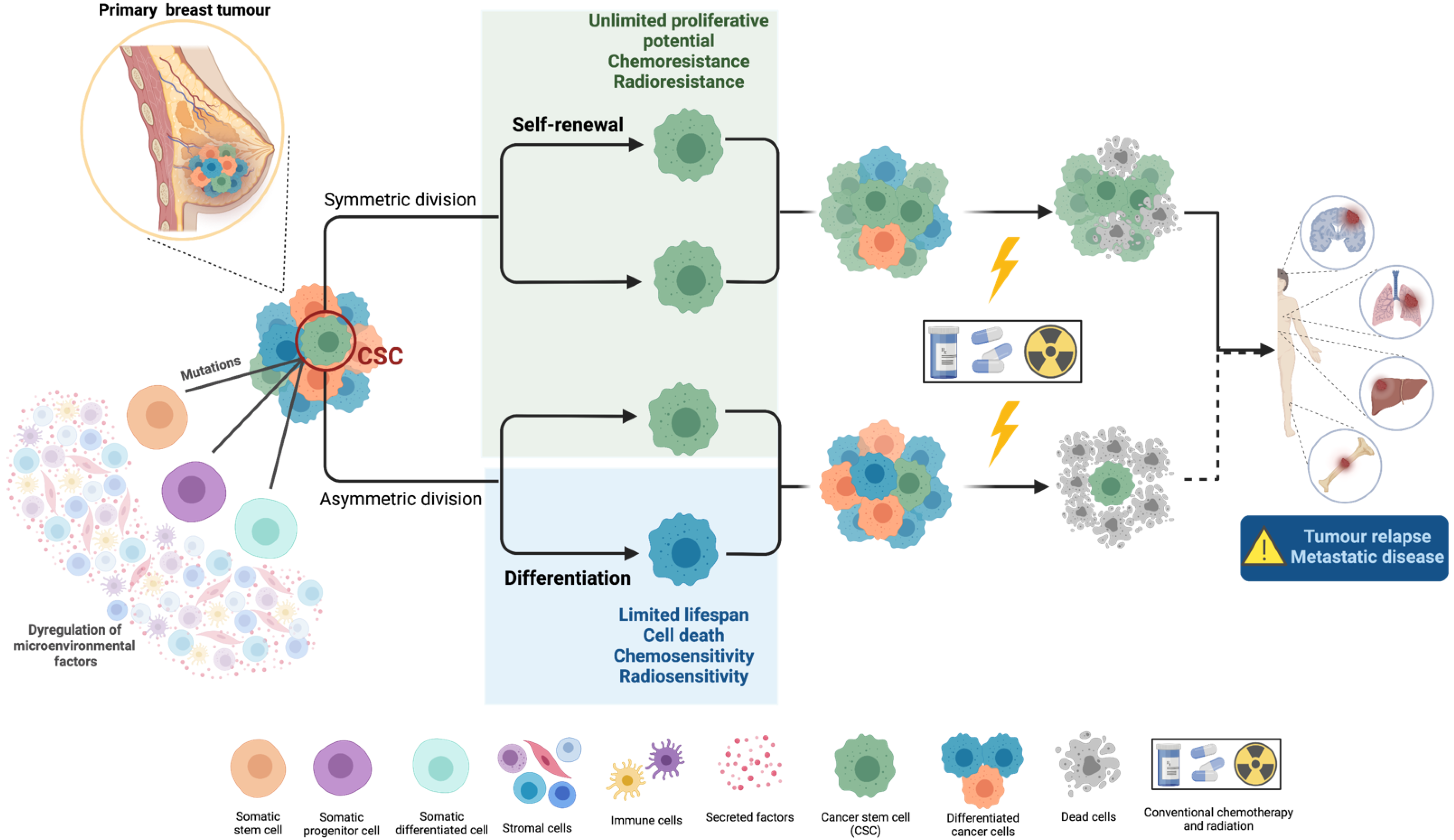
1.1. Cancer Stem Cells’ Biology and Their Role in Metastasis

The CSC theory postulates that tumour heterogeneity has a hierarchical cellular organization, driven by a small subpopulation of cancer cells showing stem-like properties and high tumourigenic potential, which is responsible for tumour formation and progression. The so-called CSCs or tumour-initiating cells (TICs) are characterized by their great plasticity and strong self-renewal ability, as well as by their multilineage differentiation potential [3,4]. Although these key properties are shared by CSCs and normal stem cells, the regulatory mechanisms that maintain the stem/differentiation balance are abolished; therefore, uncontrolled CSC proliferation ultimately leads to tumourigenesis [5].
The cell of origin for CSCs is still a matter of debate. However, it is known that it can vary among individual tumours. There are studies demonstrating that CSCs arise from somatic alterations in normal tissue stem/progenitor cells [3]; however, others suggest that CSCs derive from a pool of intermediary “transient amplifying” cells that are mitotically active and accumulate mutations to dedifferentiate and sustain the CSC pool [6,7]. In addition, CSCs may also arise due to the accumulation of genetic or epigenetic alterations, occurring in somatic differentiated cells. There are still studies suggesting a putative role of inflammation in the induction of a CSC state. For instance, Get et al. demonstrated that microenvironmental stress signals promoted the activation of stress-induced regulatory elements and the silencing of homeostatic signals, triggering the switch of stem cell fate in skin cancer [6,8]. Moreover, despite the cell of origin, it is accepted that there are several CSC pools with distinct properties within tumours, reflecting the increased levels of plasticity that are characteristic of these cells [6].
Similar to normal stem cells, CSCs are also commonly regulated by pluripotent transcription factors, including Oct4, Sox2, Nanog, KLF4, and MYC [5,6], as well as by intracellular pathways, such as Wnt, Notch, and Hedgehog (Hh) signalling [9]. Moreover, CSCs are also regulated by extracellular and microenvironmental factors, including cancer-associated fibroblasts (CAFs), extracellular matrix (ECM), exosomes, vascular niches, tumour-associated macrophages (TAMs), and hypoxia [5].
CSCs’ ability to self-renew and differentiate into multiple cell types confer them the capacity to initiate/drive tumourigenesis or to form/repopulate heterogeneous cancer cell populations, which are crucial for cancer progression. Generally, CSCs undergo asymmetric division, meaning that one CSC gives rise to another CSC with unlimited proliferative potential through self-renewal, and to a phenotypically distinct cancer cell with a limited lifespan which integrates with the tumour bulk through differentiation mechanisms [10]. However, CSCs can also divide symmetrically and self-renew, with two CSCs resulting from cell division, keeping the stem cell progeny. Symmetric division usually occurs to excessively increase tumour growth and repopulation as a response to stress conditions, such as cell loss during treatment [11] (Figure 1).
In solid tumours, the fraction of CSCs was accepted to represent a very small percentage of all cancer cells [12]. However, the identification of CSCs is of clinical relevance since it has significant prognostic and therapeutic implications. In fact, Samanta et al. reported that breast cancer cells displaying a BCSC phenotype are not responsive to chemotherapy and that paclitaxel treatment led to an increase in this BCSC phenotype [13]. Additionally, several lines of evidence demonstrate that BCSCs from different cancer cell line models are resistant to radiotherapy [14]. Thus, over the years, research efforts have been made to find new strategies to identify CSCs. Although the gold standard method to define CSCs consists of serial in vivo transplantation in animal models, CSCs can be also identified through the expression of several markers, mainly cell-surface proteins such as CD44, prominin-1 and EpCAM, for instance, whose expression is generally deregulated in CSCs, compared with their non-CSC counterparts [15]. In addition, in vitro tumoursphere formation assay is also used to identify CSC populations, since it evaluates the self-renewal and anoikis resistance capacity of cells and, therefore, is widely used in preclinical studies [16].

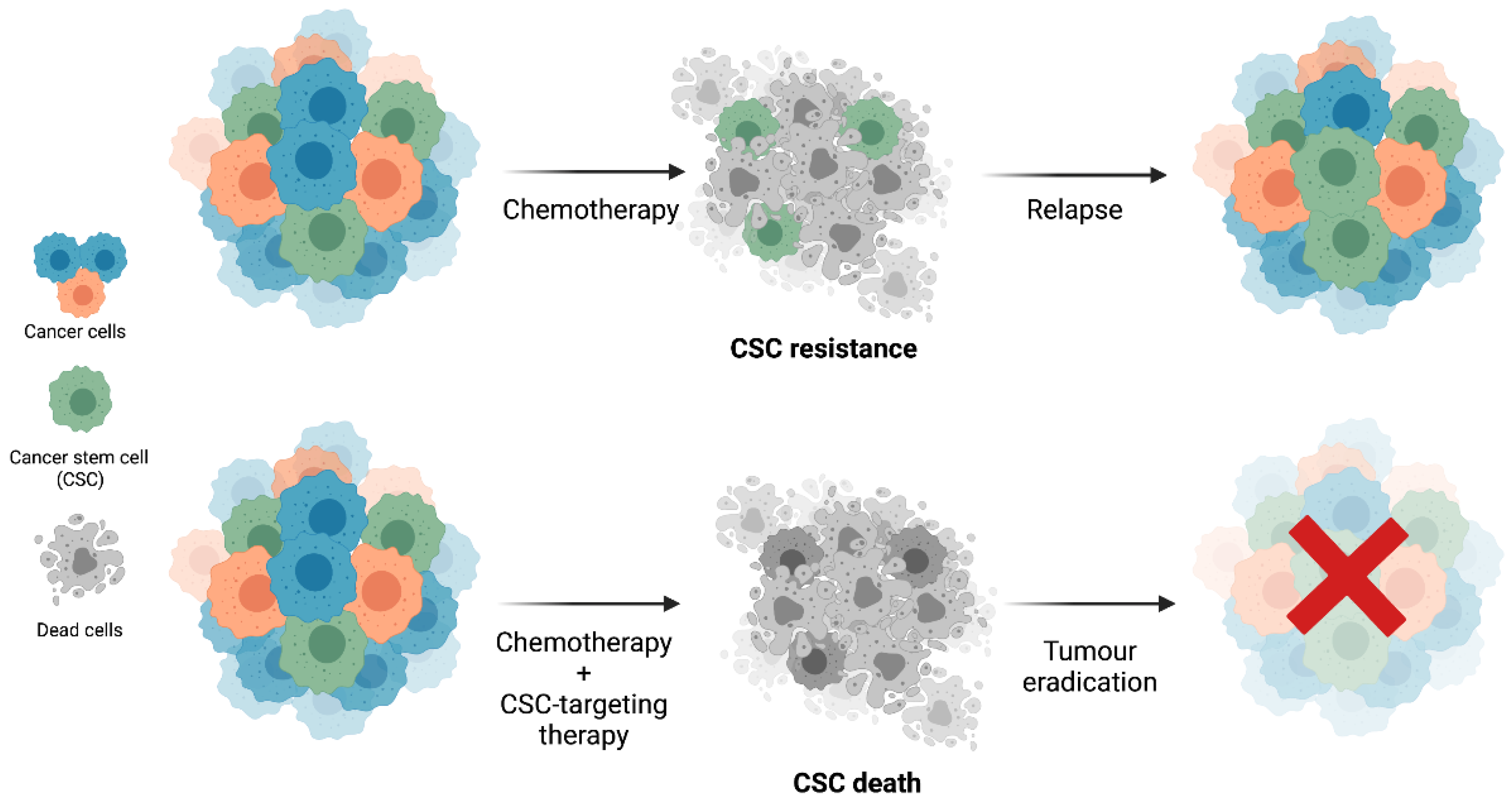
1.2. Cancer Stem Cells’ Resistance to Therapy

CSCs are highly resistant to traditional cancer therapies, including chemotherapy and radiation. This resistance is due to functional or molecular properties that are particularly relevant in CSCs. The mechanisms of CSCs’ resistance to therapy can be intrinsic, explained by genetic alterations leading to the aberrant expression of proteins, or extrinsic, resultant from microenvironmental cues, which largely contribute to tumour relapse and metastasis. Cellular quiescence, increased expression of ATP-binding cassette (ABC) transporters, detoxifying agents and antiapoptotic proteins, evasion to the immune system, deregulation of pathways associated with self-renewal, differentiation, apoptosis and survival, epigenetic reprogramming, enrichment in DNA damage response, and reactive oxygen species (ROS) scavenging and altered metabolism are examples of mechanisms contributing for CSCs’ resistance to conventional anticancer therapies [3,17].
The prime mechanisms by which conventional chemotherapies act are through the induction of DNA damage and apoptosis that target and eliminate highly proliferative cells [18]. CSCs are slow cycling and show the ability to be dormant for long periods of time until there are favourable conditions to their growth. Dormancy is an important feature for CSCs’ therapy resistance, since it is considered to be a quiescent state, where CSCs remain arrested in the G0 phase of the cell cycle, which allows them to be resistant to conventional chemotherapy [15]. Moreover, CSCs show increased expression of antiapoptotic proteins, as well as the ability to enhance their DNA damage response mechanisms. For instance, CSCs present high levels of phosphorylated DNA damage response proteins, increased activation of G2/M checkpoint, and more efficient double-strand break mechanisms in order to repair DNA damage induced by chemotherapeutic agents and radiation, thus avoiding apoptosis [19].
CSCs show aberrant increased expression of ABC transporters, which are multidrug resistance proteins that promote cellular efflux of the chemotherapeutic agents using ATP hydrolysis, thus protecting cancer cells from drug damage and inducing drug resistance [15]. CSCs show increased aldehyde dehydrogenase (ALDH) activity, which is a main responsible for resistance to chemotherapeutic drugs and radiation. ALDH promotes the metabolization of chemotherapeutic agents and eliminates oxidative stress, reducing chemotherapy toxic effects. Additionally, ALDH eliminates free radicals induced for ionizing radiation, stimulating radiation resistance [20]. In addition, CSCs possess more efficient ROS-scavenging mechanisms, which allow the prevention of cell damage induced by ROS [21]. For instance, it has been demonstrated that CD44+CD24 BCSCs have the ability to reduce intracellular ROS levels (induced by radiation) through the overexpression of a radical scavenger by an enhancement of reduced glutathione, as well as increased NADH and FADH2 [22,23,24,25].
Another major factor involved in CSCs’ resistance is the deregulation of intracellular signalling pathways associated with self-renewal, differentiation, apoptosis, and survival, in particular, Notch, Hh, and Wnt signalling. CSCs frequently present abnormal deregulated Notch signalling, which ultimately leads to uncontrolled cell proliferation and decreased differentiation and apoptosis, contributing to CSC maintenance [5]. Increased activation of Wnt signalling has been shown to be very important to the maintenance of CSCs since it plays a key role in inducing the transformation of dormant CSCs into active CSCs to promote cell cycle progression. It also regulates dedifferentiation and inhibition of apoptosis in CSCs and mediates its role in CSC-driven metastasis [5]. It is believed that the activation of Hh signalling is also critical for self-renewal, growth, and metastasis of CSCs. In addition, activation of the Hh signalling pathway is associated with Wnt signalling dysfunction, further contributing to the support of the self-renewal ability of CSCs [17].
Although the metabolic traits, as well as epigenetic reprogramming systems of CSCs, are yet not fully understood, it seems that they might represent important mechanisms contributing to CSCs’ therapy resistance [26,27]. In fact, Magnani et al. demonstrated a connection between methylation events and resistance to endocrine therapies in oestrogen receptor-positive breast cancer [27]. Additionally, Chaffer et al. observed that basal-like breast non-CSCs keep Zeb1 (an epithelial-to-mesenchymal transition transcription factor involved in the generation of BCSCs) promoter in a poised configuration to easily respond to microenvironmental stimuli, maintaining cellular plasticity and promoting the transition of non-BCSCs into BCSCs [28]. Moreover, CSCs show several escape mechanisms, such as loss of expression of key molecules for tumour immunosurveillance (e.g., IFN gamma, IL12, TRAIL). Therefore, they are able to elude the immune system, hence considered immune-privileged cells, which significantly contribute to therapy resistance [29].

2. Breast Cancer Stem Cells (BCSCs)

In breast cancer, CSCs are referred to as breast cancer stem cells (BCSCs). In recent years, there have been significant improvements in prevention, early diagnosis, and treatment options, which resulted in a significant decrease in breast cancer mortality rates. However, despite these improvements, the high rates of incidence combined with therapy resistance result in cancer relapse and metastasis, contributing to the still unacceptably high mortality of breast cancer patients. In fact, there are studies describing BCSCs as key elements in therapy resistance, breast cancer relapse, and metastasis [2,5,15,25,30,31,32]. Accordingly, a high proportion of BCSCs within breast tumours have been demonstrated as being correlated with a poor outcome [33]. Therefore, BCSCs constitute an area of major concern in oncology, highlighting the need for the development of new therapeutic strategies that allow the overcome of therapy resistance for the benefit of breast cancer patients.
BCSCs were firstly described by All-Hajj et al. [34] in 2003. The authors observed that as few as a hundred CD44+CD24 cells, which were further proved to be BCSCs, were able to form tumours when injected in the mammary fat pad of non-obese diabetic severe combined immunodeficient (NOD/SCID) mice, whereas a greater number of breast cancer cells with other phenotypes failed to sustain tumour growth. Moreover, they demonstrated that CD44+CD24 single-cell suspensions showed self-renewal, clonal mammosphere formation, and chemotherapy resistance abilities, as well as extensive proliferation [34].
The great plasticity of BCSCs contributes to intra-tumoural heterogeneity, as well as to the existence of several BCSC pools within tumours, which represents a major challenge for tumour eradication. In fact, Liu et al. observed that phenotypic interconversion between epithelial and mesenchymal states can occur in BCSCs and that each state is associated with different localisation within the tumours, as well as with distinct proliferative and invasive capacities [35]. The transition between these two states is a dynamic and reversible process [36], whose regulation by epigenetic alterations or tumour microenvironment signalling occurring during tumour progression plays a critical role in promoting invasion and metastatic capabilities. Interestingly, epithelial-like BCSCs expressing the BCSC marker ALDH are more proliferative and are located at central areas of the tumour, whereas CD44+CD24 mesenchymal-like BCSCs are more quiescent and located at the invasive front of the tumour. During the metastatic cascade, the mesenchymal CD44+CD24 phenotype is crucial for the initial steps, as it is important for cells to be able to enter the circulation, as well as to seed and form micrometastasis; however, when the invasive edge becomes the interior of the tumour, the switch to the ALDH+ epithelial state is important for establishing macrometastasis. In addition to these two BCSCs populations, the authors also described the existence of a third CD44+CD24ALDH+ population, which was associated with the greatest tumourigenic potential and invasion capability among the three BCSCs’ populations, and it was believed that this phenotype represents the most purified BCSC state [35].
Accumulating evidence suggests that BCSCs play key roles in metastasis. In fact, primary breast tumour cells with a CD44+CD24 profile were associated with increased metastatic capacity and poor survival probability [37]. Moreover, the most direct evidence that CSCs can establish metastasis was demonstrated by Liu et al. using CD44+CD24 BCSCs from breast tumour patient-derived cells show the ability to form tumours, as well as to spontaneously metastasize to the lungs in orthotopic mouse models [38]. In addition, Balic et al. demonstrated that most early disseminated cancer cells detected in the bone marrow of breast cancer patients have a putative breast cancer stem cell phenotype [39]. In further studies, using a xenograft model, Charafe-Jauffret et al. showed that ALDH+ cancer cells display increased metastatic abilities when compared with the ALDH cancer cells [40]. Notably, our group has recently demonstrated that breast cancer cells that have in vivo metastatic capacity in mice models [41] have increased CSC activity, but most importantly, we showed that human metastatic lesions [42], specifically brain metastasis, are enriched for a BCSC phenotype (determined by CD44, integrin subunit α6, EpCAM, cadherin-3, and ALDH1 expression). Importantly, we were able to validate a new in vivo tool based on the chick chorioallantoic membrane (CAM) model that can be used as an alternative, appealing, and novel approach in the CSC research field to study CSC activity. Importantly, this novel tool can also be applied to test the efficacy of anti-CSC drugs, with the potential to be used in a personalized medicine context [41].

2.1. BCSC Biomarkers

Although there are no universal markers that allow the specific recognition of BCSCs, this population is generally identified by the expression of the surface markers CD44, CD24, and ALDH1, which are the most accepted biomarkers associated with a BCSC phenotype. These markers have initially been defined by All-Hajj et al. [34] and further validated by several authors [43,44,45]. However, additional markers or different combinations of their expression are described to identify BCSC populations, including transcription factors commonly associated with normal stem cell functions (e.g., Sox2, Oct4, and Nanog) and cell surface markers such as integrin subunit α6, CD29, CD61, EpCAM, cadherin-3, HER2, prominin-1, and C-X-C motif chemokine receptor 1.

2.1.1. Transcription Factors (TFs)

Although the majority of BCSC biomarkers are intracellular or membrane proteins, there are some pluripotency TFs, such as Sox2, Oct4, and Nanog, as well as epithelial-to-mesenchymal transition (EMT) TFs, such as Zeb1, that have been correlated with BCSCs’ origin. In particular, Oct4 is an embryonic stem cell marker, whose expression has been reported as being an independent prognostic factor in hormone-receptor-positive breast cancer associated with ALDH1 positivity, high proliferative ki67 index, and high histological tumour grade [46]. Additionally, Bhatt et al. demonstrated a possible correlation between Oct4 expression and breast cancer cells’ resistance to tamoxifen treatment [47]. Nanog is also an embryonic stem cell marker that has been described as upregulated in invasive breast cancer. Moreover, it has been directly correlated with tumour size, grade, and stage, as well as with lymph node metastasis and poor overall survival [48]. Sox2 is a pluripotency TF that has been reported as being highly expressed in breast cancer tissues [49,50]. Interestingly, Leis et al. observed that Sox2 is expressed and is necessary for in vitro mammosphere formation. Moreover, the authors observed that silencing Sox2 in breast cancer cells led to a delay in tumour formation in mice xenografts [51].
Increasing lines of evidence suggest a correlation between EMT and its associated TFs as ZEB1 and BCSCs formation. For instance, Jiang et al. demonstrated that ZEB1 knockout mouse-derived breast cancer cells present a decrease in the percentage of CD44+CD24, as well as in the ALDH1+ population, accompanied by a reduction in the mammosphere formation capacity [52]. Moreover, increased ZEB1 expression in triple-negative breast cancer patients, the breast cancer molecular subtype with worse prognosis and with an enrichment in the CSC population, is predictive of radiotherapy relapse [53].

2.1.2. Cell Surface Proteins

Membrane proteins are an area of special interest since they are accessible on the cell surface and are one of the first classes of proteins to be impacted under pathological conditions [54]. Importantly, membrane proteins constitute promising prognostic and therapeutic markers, because they are accessible targets for monoclonal antibodies and other drugs [55].

CD44 Molecule

CD44 is a transmembrane glycoprotein, whose important ligand is hyaluronic acid. It is involved in several biological processes that are important during tumour progression and metastasis—namely, cellular survival, proliferation, differentiation, and adhesion, and is a key mediator in the transendothelial migration of breast cancer cells during the metastatic cascade [56]. BCSCs are characterized by high CD44 expression levels, which are associated with the maintenance of a multipotent phenotype [57]. Regarding this, CD44 has been widely used to identify and isolate BCSCs, in addition to being considered a potential target for BCSCs-targeting therapies [24,58,59].

CD24 Molecule

CD24 or heat-stable antigen (HAS) is a sialoprotein involved in multiple biological processes such as cell adhesion, migration, proliferation, invasion, and metastasis [60]. Although CD24 is an accepted CSC biomarker, whether their expression (overexpression or lack of expression) is associated with stem-like properties and tumourigenic potential seems to be context-dependent, varying across tumour types [61]. However, in BCSCs, CD24 expression is usually very low or even absent. Further corroborating this CD24 BCSC phenotype, evidence from in vitro studies demonstrated that the overexpression of CD24 was associated with the inhibition of stem-like properties in breast cancer cells [62]. In addition, another study suggested that CD24 phenotype was associated with therapy resistance in breast cancer cell lines [63]. Generally, CD24 is used in combination with CD44, making the CD44+CD24 phenotype a classical BCSC biomarker [60,64].

Integrin Subunit α6

Integrin subunit α6, also known as CD49f, is a type I transmembrane glycoprotein, belonging to the α-subfamily. Importantly, CD49f might assume two distinct cytoplasmic domains generated by mRNA alternative splicing variants, α6A and α6B, that show different stem-like functions. Specifically, it has been shown that the α6B variant is the isoform that closely associates with a CSC phenotype [65,66,67]. Usually, CD49f forms heterodimers with integrins β1 (CD29) and β4 (CD104), functioning as a receptor for several laminins and playing an important role in cell adhesion to the extracellular matrix (ECM) [68]. Furthermore, studies demonstrate that CD49f heterodimers cooperate with signalling pathways to induce stem-like and invasive properties to breast cancer cells. In breast cancer, high expression of CD49f is a poor prognostic marker associated with reduced survival [69].

Epithelial Cell Adhesion Molecule

Epithelial cell adhesion molecule (EpCAM) is a transmembrane glycoprotein expressed in the majority of epithelial cells and it mediates homophilic Ca2+ independent cell–cell adhesion [15], contrary to what is seen for epithelial cadherins. It is involved in several biological processes, including cell signalling [70], migration [71], proliferation, differentiation, tumourigenesis [72], and metastasis, thereby being recognized as a well-established CSC marker globally overexpressed in BCSCs [71]. In fact, several lines of evidence show that EpCAM might promote BCSCs survival through the activation of the Wnt signalling pathway [9,73,74]. Importantly enough, EpCAM has been widely recognised as being expressed in circulating tumour cells (CTCs) [75], which are highly suspected to be enriched in a CSC function and activity, allowing the survival of cancer cells in anoikis-independent conditions [76,77].

Cadherin 3

Cadherin 3, also called placental cadherin (P-cadherin), belongs to the classical cadherin family and plays a critical role in mediating cell–cell adhesion and structural integrity of epithelia, through the formation of cadherin–catenin complexes [78]. Its function as a mediator of stem cell properties during embryonic development is also recognised, as it is considered a biomarker for the isolation of stem cells. Due to its important biological role, it is expectable that alterations in P-cadherin are associated with disease, in particular in cancer [79]. Interestingly, P-cadherin expression correlates with poor patient’ prognosis both in primary breast tumours and in lymph node metastasis [80,81]. Moreover, studies from our group have shown that P-cadherin overexpression is associated with cell invasion, through the disruption of E-cadherin mediated cell–cell adhesion and through the modulation of α6β4 integrin-mediated cell-matrix adhesion [82,83,84]. Importantly, P-cadherin expression is also involved in stem-like properties and is associated with the expression of other CSC biomarkers, including CD44, CD49f, and ALDH1 [85]. In fact, our group has demonstrated that downregulation of P-cadherin was associated with reduced in vitro self-renewal ability and decreased in vivo tumourigenic potential [83]. Thus, P-cadherin is a valuable BCSC marker with relevant prognostic implications.

Erb-b2 Receptor Tyrosine Kinase 2

Erb-b2 receptor tyrosine kinase 2 (Erbb2/HER2) belongs to the human epidermal growth factor receptor family and is involved in the regulation of several pathways associated with cell division, proliferation, cell motility, invasion, differentiation, and apoptosis. In breast cancer, HER2 overexpression occurs in 25–30% of patients, composing a HER2-amplified breast cancer subtype, which is associated with a poor survival rate, as well a high risk of metastasis [86]. Emerging studies demonstrate that HER2 plays a role in the regulation of BCSC activity, in particular in self-renewal and radioresistance [87,88,89,90]. In fact, Duru et al. observed that HER2+ BCSCs isolated from HER2- breast cancer cells showed to have enhanced ALDH1 activity, aggressiveness, and radioresistance [88].

Prominin 1

Prominin-1, also known as CD133, is a transmembrane glycoprotein commonly used as a stem cell marker due to its role in suppressing differentiation. In breast cancer, CD133 expression has been associated with poor overall survival probability, high tumour grade, lymph node metastasis, hormone receptor negativity, and HER2 positivity, as well as advanced tumours, nodes, and metastasis (TNM) stage [91]. Moreover, Joseph et al. demonstrated that the overexpression of CD133 was associated with a poor prognosis in invasive breast cancer [92]. The role of CD133 as a potential BCSCs’ biomarker was firstly described in a study by Wright et al., in which an increase in colony-forming efficiency, proliferative rate, and tumourigenic potential was observed in CD133+ cells derived from BRCA1 murine breast tumours [93]. Moreover, Croker et al. demonstrated that the ALDH+CD44+CD133+ population isolated from invasive breast cancer cell lines showed enhanced growth, colony formation, migration, and invasion capabilities, as well as greater in vitro and in vivo malignant and metastatic behaviour [94]. Additionally, Brugnoli et al. reported that CD133 silencing reduced the invasive capacities in triple-negative breast cancer cells [95].

C–X–C Motif Chemokine Receptor 1

C–X–C motif chemokine receptor 1 (CXCR1) is one of the receptors for CXC ligand 8 (CXCL8). In fact, the CXCL8–CXCR1 axis has been described as playing a crucial role in breast cancer stemness [96]. Interestingly, in a gene expression profile study, CXCR1 was found to be upregulated in ALDH+ BCSCs of various breast cancer cell lines [97]. In addition, treatment with recombinant IL-8 led to an enrichment in the BCSC population, since it resulted in an increased ALDH expression and mammosphere forming efficacy, as well as a greater invasive capacity [98]. Ginestier et al. reported that the inhibition of CXCR1 promoted the selective depletion of ALDH+ BCSCs through the FAK–AKT–FOXO3A pathway [97].

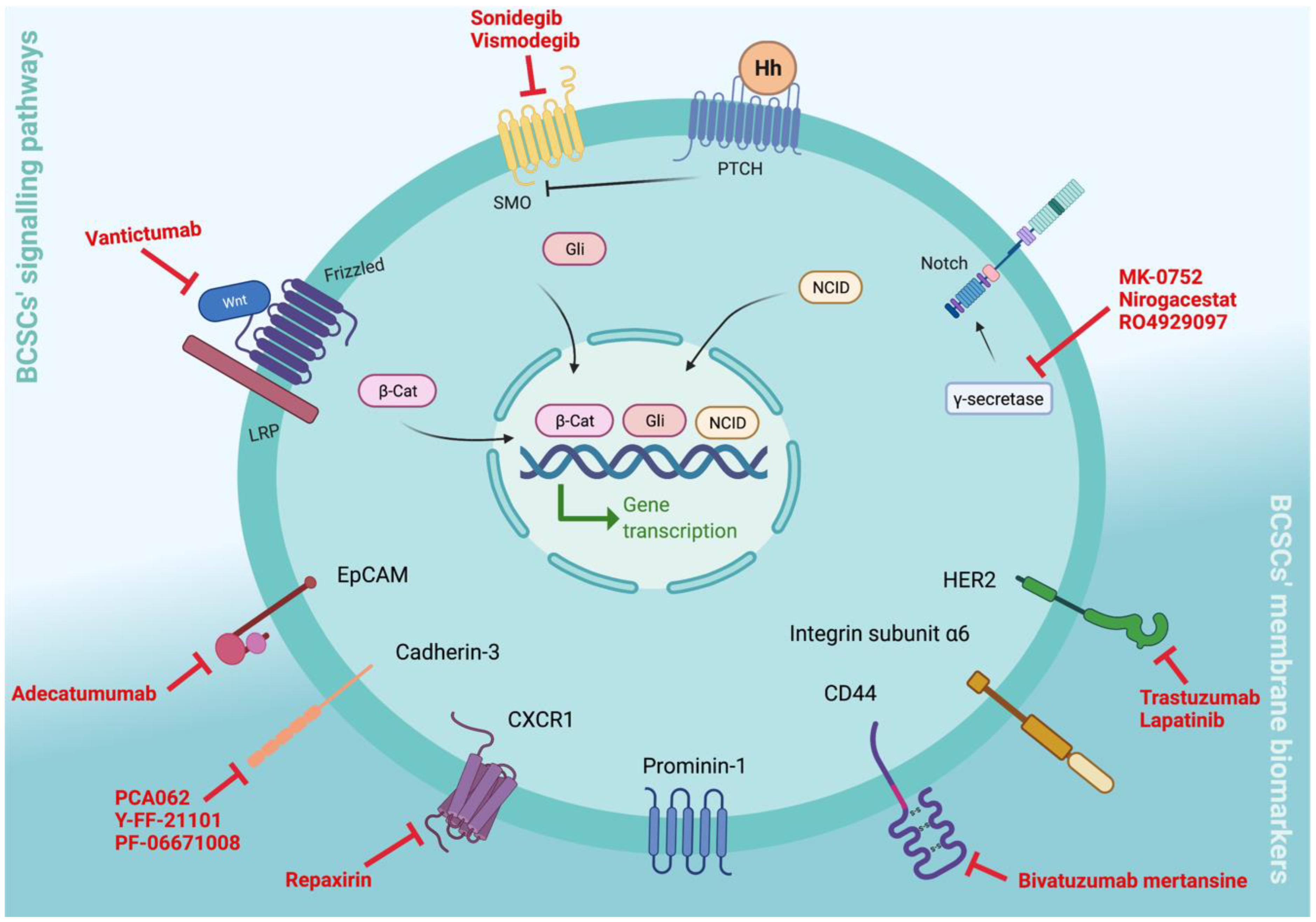
3. Clinical Relevance of BCSC Biomarkers

The high mortality associated with breast cancer is closely related to tumour recurrence and the development of metastatic disease. Importantly, BCSCs play crucial roles in these biological processes, since they are highly tumourigenic cells with self-renewal and differentiation capacities that are able to form or repopulate heterogeneous cancer cell populations. Moreover, BCSCs are highly resistant to conventional chemotherapies, and there may even be an enrichment in BCSCs population in post-treatment settings [6], thereby being mainly responsible for tumour recurrence [2]. In fact, in their clinical studies, Lee et al. demonstrated that there is an enrichment in CD44+CD24 population in primary breast tumours following chemotherapy [99]. Accordingly, Creighton et al. reported an enrichment in the CD44+CD24 population in residual tumour tissue after endocrine therapy, compared with pretreated samples, in a cohort of 52 breast cancer patients in phase II clinical trials [100]. Importantly, besides their role in chemoresistance and tumour recurrence, there are several clinical lines of evidence that suggest a relationship between BCSCs and metastasis. For instance, primary tumours from breast cancer patients with high CD44+CD24 correlated with the presence of distant metastasis, including bone [101] and pleural metastasis [102], as well as show early bone marrow disseminated cancer cells [39]. In addition, ALDH1 expression, in particular the ALDH1A3 isoform, significantly correlated with aggressive clinical–pathological features [103] and distant metastases in inflammatory breast cancer [40], showing that CSC prevalence is directly associated with disease progression. More recently, we have studied the expression of five different BCSC markers (CD44, integrin subunit α6, cadherin-3, EpCAM, and ALDH1) and found significant enrichment for each biomarker in human breast cancer brain metastasis when compared with unmatched primary tumours. More importantly, our study revealed an enriched stem cell signature defined by the simultaneous expression of three to five of these BCSC markers. While the primary breast tumours only show this BCSC signature in 9.3% of the cases, this phenotype was clearly enriched in breast cancer brain metastasis, increasing the percentage to 55.6%. Even though a small proportion of the primary tumours display the BCSC signature, these were found to be significantly associated with poor clinical–pathological features and with decreased survival for breast cancer patients [42]. Thus, BCSCs have clinically relevant prognostic and therapeutic potential.
In this context, BCSC research in the metastatic setting is an urgent topic in oncology, which includes the identification of new BCSC biomarkers and models to study this population of clinical relevance. In fact, genomic approaches could be interesting to more accurately identify BCSC, as previously demonstrated [37]. Nonetheless, these methodologies are not as useful from a drug-targeting perspective, since they recognise a large number of genes that are not necessarily targetable as the cell surface biomarkers.
In this way, the development of BCSC-based tools that allow the prediction of prognosis and that can be translated into the clinic can constitute powerful approaches to ameliorate risk assessment and therapeutic selection of cancer patients. Notably, since there are difficulties in collecting serial biopsies from metastatic sites in patients, alternative biomarker-based methods should be considered. For instance, the search for BCSC biomarkers in circulating tumour cells could serve as surrogate evidence of anti-CSC activity, as already demonstrated by some authors [104,105].
In resume, the design of new therapeutic targeting-CSCs strategies could be a more effective anticancer therapy and might even allow therapy resistance to be overcome (Figure 2).
Importantly, the therapeutic targeting of BCSCs might be performed using different approaches, including cell surface, cytoplasmic and nuclear BCSC markers, stem cell-specific signalling pathways, or even BCSC microenvironmental factors. Immuno- and differentiation therapies might also be promising strategies to overcome BCSCs’ resistance to conventional therapies [32]. To date, there are already several BCSC-targeting therapies in clinical trials for different types of cancer, as previously reviewed by Saygin et al. [6], as well as by Yang et al. [5].
In the following sections, we will describe the BCSC-targeting therapies currently in clinical trials (Figure 3).

3.1. Targeting BCSC Signalling Pathways

3.1.1. Notch Signalling

Notch signalling has been associated with BCSCs as playing a key role in promoting EMT, thereby contributing to CSC-mediated metastasis [17,106]. Evidence showed that inhibition of Notch signalling sensitises BCSCs to chemotherapy and radiation [107]. Importantly, Notch activation occurs after gamma-secretase cleavage of its intracellular domain. Thus, Notch-signalling-targeting studies have employed two different approaches: (1) gamma-secretase inhibition, in order to inhibit its role on Notch cleavage and consequent activation, and (2) the use of antibodies against Notch ligands or receptors. MK-0752 is a gamma-secretase inhibitor, already with a phase II clinical trial completed, that showed promising results targeting BCSCs, since a decrease in CD44+CD24- population, as well as decreased mammosphere forming efficiency, was observed in samples from breast tumour serial biopsies [32,108]. Additionally, RO4929097 (RG-4733) and PF-03084014 (nirogacestat) are other gamma-secretase inhibitors already in clinical trials [31].

3.1.2. Hh Signalling

Activation of Hh signalling has been associated with breast cancer since this pathway is crucial for tissue homeostasis and self-renewal. Importantly, the Hh pathway is abnormally upregulated in BCSCs, thereby promoting BCSCs proliferation [106]. Several Hh signalling pathway inhibitors have been developed for cancer treatment, targeting specifically the smoothened (SMO). Importantly, SMO is a transmembrane protein and a Hh ligand that mediates Hh downstream signalling pathway. Evidence showed that inhibition of Hh signalling, using cyclopamine, sensitizes BCSCs to paclitaxel [73]. In addition, sonidegib (LDE225) and vismodegib (GDC-0449), another SMO antagonist, are already in phase I clinical trials [31].

3.1.3. Wnt Signalling

Increased activation of Wnt–β-catenin signalling significantly contributes to BCSCs’ self-renewal and therapy resistance [15]. Various studies showed that inhibition of the Wnt–β-catenin pathway suppressed BCSC proliferation and self-renewal [109]. Vantictumab is an antibody in phase I clinical trials, which inhibits Wnt–β-catenin signalling by blocking the Wnt receptor. Importantly, vantictumab has been showing interesting results, since it seems to reduce BCSC frequency and promote differentiation in patient-derived xenografts [110].

3.2. Targeting BCSCs’ Cell Surface Membrane Biomarkers

Membrane proteins are an area of special interest and account for more than 60% of current drug targets [55] and, therefore, are the major focus of this section. Importantly, most drugs achieve the therapeutic effect via interacting with membrane proteins, and thus, they represent major disease biomarker candidates and constitute promising therapeutic targets for monoclonal antibodies and other drugs, allowing the modulation of cell signalling through the inhibition of receptors or enzyme activity, for instance [55].

3.2.1. CD44v6

CD44 exists in its standard form (CD44S), as well as in various isoforms (CD44v) due to post-translational modifications and alternative splicing of the primary transcript. CD44v6 is one of the CD44 isoforms and, in breast cancer, its overexpression in endocrine sensitive breast cancer cells has been connected to the activation of epidermal growth factor receptor (EGFR) signalling, to an increased invasive ability, as well as to a decrease in response to endocrine therapy [111,112,113]. Bivatuzumab mertansine is an immunoconjugate composed of a highly potent antimicrotubule agent and a monoclonal antibody against CD44v6. It has been tested in phase I clinical trials (NCT02254005, NCT02254031) in anthracycline and taxane-pretreated patients with metastatic breast cancer who were shown to have CD44v6 expression. In fact, bivatuzumab mertansine produced some interesting results, since it was able to target CD44v6 and to promote disease stabilisation in 50% of patients independently of dose level. However, due to a fatal toxic epidermal necrolysis that occurred in a parallel study, the clinical trials using this immunoconjugate have been discontinued [114].

3.2.2. Epithelial Cell Adhesion Molecule (EpCAM)

Adecatumumab is a novel monoclonal antibody targeting EpCAM, which is already in phase II clinical trials. Unfortunately, adecatumumab treatment did not show an impact on objective tumour regression in EpCAM overexpressing metastatic breast cancer patients [115], highlighting the need for further investigations of this agent in patients with EpCAM-overexpressing tumours.

3.2.3. Cadherin-3 (P-Cadherin)

P-cadherin constitutes an attractive target with therapeutic potential to treat tumours that highly express this protein, with several studies addressing its target using different approaches [116,117,118,119]. In fact, for breast cancer, PCA062, a P-cadherin-targeting antibody–drug conjugate may have clinical potential, since it has been shown to have potent antitumour activity with acceptable efficacy and tolerability in preclinical studies in primates [120]. However, PCA062 administration in phase I clinical trial (NCT02375958) has been discontinued, since the efficacy signals were limited during dose escalation [120], suggesting that further studies using this molecule need to be performed in order to successfully target P-cadherin. Moreover, further phase I clinical trials (NCT02454010) studies suggest the use of P-cadherin-targeted radioimmunotherapy with Y-FF-21101 monoclonal antibody in the treatment of solid tumours as showing promising results [119]; however, this treatment has not been tested in breast cancer patients, which might be an interesting approach for the future since P-cadherin overexpression is a common feature of breast cancer patients. PF-06671008 is a CD3-bispecific molecule-targeting P-cadherin that was able to promote T-cell-mediated regression of established tumours in mice models. Importantly, PF-06671008 is already in phase I clinical trials (NCT02659631) for triple-negative breast cancer patients [118,121].

3.2.4. Erb-b2 Receptor Tyrosine Kinase 2 (Erbb2/HER2)

Increasing evidence demonstrates a connection between HER2 and BCSCs. Importantly, there are already well-established HER2-targeted therapies, such as trastuzumab, that clearly impact tumour recurrence and aggressiveness. Thus, it is plausible to assume that these HER2 inhibitors are also targeting HER2+ BCSCs, as demonstrated in recent and emerging studies. In particular, it has been demonstrated by Li et al. that neoadjuvant lapatinib treatment led to a decrease in the CD44+CD24 population, as well as a reduction in mammosphere formation of samples from breast cancer patient biopsies [122]. Additionally, Korkaya et al. reported that treatment with trastuzumab reduced the BCSC population in HER2-overexpressing breast cancer cell lines [123].

3.2.5. C–X–C Motif Chemokine Receptor 1 (CXCR1)

Repaxirin is an investigational inhibitor of CXCR1 that has been shown to reduce the CSC content of human breast cancer in mice xenografts [97], which is already in phase II clinical trials (NCT02370238, NCT01861054). The agent has shown promising results, promoting a decrease in CXCR1+ cells in serial biopsies following treatment. Additionally, a decrease in the BCSCs’ CD44+CD24 and ALDH1+ populations was observed, with minimal adverse reactions registered in breast cancer patients [124].

4. Conclusions

There are several aspects of BCSC biology that need to be solved in order to successfully develop therapies that achieve BCSC elimination. Currently, a major limitation of the anti-BCSC therapies consists of their target, which is, mainly, stemness-associated transcriptional and/or intracellular signalling factors that are shared with normal stem cells and are also sometimes difficult to target. Moreover, the existing BCSC-signalling targeted therapies are not very specific, highlighting the need for the design of new inhibitors. Thus, it is of major importance to search for new potential biomarkers that could be easily translated to the clinical. The search for putative new CCSC biomarkers and the identification of novel and unique pathways that are active in this subset of cells is of clinical relevance. In particular, cell surface CSC markers seem to be among promising strategies since they constitute accessible targets for monoclonal antibodies or drugs, which allows easy modulation of CSC signalling.

Author Contributions

Conceptualisation, J.P.; writing—original draft preparation, I.C.; writing—review and editing, A.S.R. and J.P. All authors have read and agreed to the published version of the manuscript.

Funding

This research was funded by FEDER (Fundo Europeu de Desenvolvimento Regional) through the COMPETE 2020—Operacional Programme for Competitiveness and Internationalisation (POCI), Portugal 2020, and by FCT (Fundação para a Ciência e a Tecnologia/ Ministério da Ciência, Tecnologia e Ensino Superior) under the project FCT/02/SAICT/2017/030625.

Acknowledgments

We would like to thank to Núcleo Regional do Norte of Liga Portuguesa Contra o Cancro for the attribution of the grant “Bolsas de Investigação LPCC-NRN” to Inês Conde.

Conflicts of Interest

The authors declare no conflict of interest.

References

  1. Lapidot, T.; Sirard, C.; Vormoor, J.; Murdoch, B.; Hoang, T.; Caceres-Cortes, J.; Minden, M.; Paterson, B.; Caligiuri, M.A.; Dick, J.E. A cell initiating human acute myeloid leukaemia after transplantation into SCID mice. Nature 1994, 367, 645–648. [Google Scholar] [CrossRef] [PubMed]
  2. Shiozawa, Y.; Nie, B.; Pienta, K.J.; Morgan, T.M.; Taichman, R.S. Cancer stem cells and their role in metastasis. Pharm. 2013, 138, 285–293. [Google Scholar] [CrossRef] [PubMed] [Green Version]
  3. Yu, Z.; Pestell, T.G.; Lisanti, M.P.; Pestell, R.G. Cancer stem cells. Int. J. Biochem. Cell Biol. 2012, 44, 2144–2151. [Google Scholar] [CrossRef] [PubMed] [Green Version]
  4. Lee, G.; Hall, R.R., 3rd; Ahmed, A.U. Cancer Stem Cells: Cellular Plasticity, Niche, and its Clinical Relevance. J. Stem Cell Res. 2016, 6, 363. [Google Scholar] [CrossRef]
  5. Yang, L.; Shi, P.; Zhao, G.; Xu, J.; Peng, W.; Zhang, J.; Zhang, G.; Wang, X.; Dong, Z.; Chen, F.; et al. Targeting cancer stem cell pathways for cancer therapy. Signal Transduct Target 2020, 5, 8. [Google Scholar] [CrossRef] [Green Version]
  6. Saygin, C.; Matei, D.; Majeti, R.; Reizes, O.; Lathia, J.D. Targeting Cancer Stemness in the Clinic: From Hype to Hope. Cell Stem Cell 2019, 24, 25–40. [Google Scholar] [CrossRef] [PubMed] [Green Version]
  7. Chaffer, C.L.; Weinberg, R.A. How does multistep tumorigenesis really proceed? Cancer Discov. 2015, 5, 22–24. [Google Scholar] [CrossRef] [Green Version]
  8. Ge, Y.; Fuchs, E. Abstract B24: Stem cell lineage infidelity drives wound repair and cancer. Cancer Res. 2018, 78, 24. [Google Scholar] [CrossRef]
  9. Song, K.; Farzaneh, M. Signaling pathways governing breast cancer stem cells behavior. Stem Cell Res. Ther. 2021, 12, 245. [Google Scholar] [CrossRef]
  10. Frank, N.Y.; Schatton, T.; Frank, M.H. The therapeutic promise of the cancer stem cell concept. J. Clin. Invest. 2010, 120, 41–50. [Google Scholar] [CrossRef] [Green Version]
  11. Hill, R.P. Identifying cancer stem cells in solid tumors: Case not proven. Cancer Res. 2006, 66, 1891–1895, discussion 1890. [Google Scholar] [CrossRef] [PubMed] [Green Version]
  12. Ishizawa, K.; Rasheed, Z.A.; Karisch, R.; Wang, Q.; Kowalski, J.; Susky, E.; Pereira, K.; Karamboulas, C.; Moghal, N.; Rajeshkumar, N.V.; et al. Tumor-initiating cells are rare in many human tumors. Cell Stem Cell 2010, 7, 279–282. [Google Scholar] [CrossRef] [PubMed] [Green Version]
  13. Samanta, D.; Gilkes, D.M.; Chaturvedi, P.; Xiang, L.; Semenza, G.L. Hypoxia-inducible factors are required for chemotherapy resistance of breast cancer stem cells. Proc. Natl. Acad. Sci. USA 2014, 111, 5429–5438. [Google Scholar] [CrossRef] [PubMed] [Green Version]
  14. Qi, X.S.; Pajonk, F.; McCloskey, S.; Low, D.A.; Kupelian, P.; Steinberg, M.; Sheng, K. Radioresistance of the breast tumor is highly correlated to its level of cancer stem cell and its clinical implication for breast irradiation. Radiother. Oncol. 2017, 124, 455–461. [Google Scholar] [CrossRef] [PubMed]
  15. Agliano, A.; Calvo, A.; Box, C. The challenge of targeting cancer stem cells to halt metastasis. Semin. Cancer Biol. 2017, 44, 25–42. [Google Scholar] [CrossRef] [PubMed]
  16. Pastrana, E.; Silva-Vargas, V.; Doetsch, F. Eyes wide open: A critical review of sphere-formation as an assay for stem cells. Cell Stem Cell 2011, 8, 486–498. [Google Scholar] [CrossRef] [PubMed] [Green Version]
  17. Saeg, F.; Anbalagan, M. Breast cancer stem cells and the challenges of eradication: A review of novel therapies. Stem Cell Investig. 2018, 5, 39. [Google Scholar] [CrossRef]
  18. Masood, I.; Kiani, M.H.; Ahmad, M.; Masood, M.I.; Sadaquat, H. Major contributions towards finding a cure for cancer through chemotherapy: A historical review. Tumori J. 2016, 102, 6–17. [Google Scholar] [CrossRef]
  19. Peitzsch, C.; Kurth, I.; Kunz-Schughart, L.; Baumann, M.; Dubrovska, A. Discovery of the cancer stem cell related determinants of radioresistance. Radiother. Oncol. 2013, 108, 378–387. [Google Scholar] [CrossRef] [Green Version]
  20. Singh, S.; Brocker, C.; Koppaka, V.; Chen, Y.; Jackson, B.C.; Matsumoto, A.; Thompson, D.C.; Vasiliou, V. Aldehyde dehydrogenases in cellular responses to oxidative/electrophilic stress. Free Radic. Biol. Med. 2013, 56, 89–101. [Google Scholar] [CrossRef] [Green Version]
  21. Diehn, M.; Cho, R.W.; Lobo, N.A.; Kalisky, T.; Dorie, M.J.; Kulp, A.N.; Qian, D.; Lam, J.S.; Ailles, L.E.; Wong, M.; et al. Association of reactive oxygen species levels and radioresistance in cancer stem cells. Nature 2009, 458, 780–783. [Google Scholar] [CrossRef] [PubMed]
  22. Wang, T.; Fahrmann, J.F.; Lee, H.; Li, Y.J.; Tripathi, S.C.; Yue, C.; Zhang, C.; Lifshitz, V.; Song, J.; Yuan, Y.; et al. JAK/STAT3-Regulated Fatty Acid β-Oxidation Is Critical for Breast Cancer Stem Cell Self-Renewal and Chemoresistance. Cell Metab. 2018, 27, 136–150.e135. [Google Scholar] [CrossRef] [PubMed] [Green Version]
  23. Ishimoto, T.; Nagano, O.; Yae, T.; Tamada, M.; Motohara, T.; Oshima, H.; Oshima, M.; Ikeda, T.; Asaba, R.; Yagi, H.; et al. CD44 variant regulates redox status in cancer cells by stabilizing the xCT subunit of system xc(-) and thereby promotes tumor growth. Cancer Cell 2011, 19, 387–400. [Google Scholar] [CrossRef] [PubMed] [Green Version]
  24. Phillips, T.; McBride, W.; Pajonk, F. The Response of CD24−/low/CD44+ Breast Cancer–Initiating Cells to Radiation. J. Natl. Cancer Inst. 2007, 98, 1777–1785. [Google Scholar] [CrossRef] [PubMed] [Green Version]
  25. Palomeras, S.; Ruiz-Martínez, S.; Puig, T. Targeting Breast Cancer Stem Cells to Overcome Treatment Resistance. Molecules 2018, 23, 2193. [Google Scholar] [CrossRef] [PubMed] [Green Version]
  26. Ito, K.; Suda, T. Metabolic requirements for the maintenance of self-renewing stem cells. Nat. Rev. Mol. Cell Biol. 2014, 15, 243–256. [Google Scholar] [CrossRef] [Green Version]
  27. Magnani, L.; Stoeck, A.; Zhang, X.; Lánczky, A.; Mirabella, A.C.; Wang, T.L.; Gyorffy, B.; Lupien, M. Genome-wide reprogramming of the chromatin landscape underlies endocrine therapy resistance in breast cancer. Proc. Natl. Acad. Sci. USA 2013, 110, 1490–1499. [Google Scholar] [CrossRef] [Green Version]
  28. Chaffer, C.L.; Marjanovic, N.D.; Lee, T.; Bell, G.; Kleer, C.G.; Reinhardt, F.; D’Alessio, A.C.; Young, R.A.; Weinberg, R.A. Poised Chromatin at the ZEB1 Promoter Enables Breast Cancer Cell Plasticity and Enhances Tumorigenicity. Cell 2013, 154, 61–74. [Google Scholar] [CrossRef] [Green Version]
  29. Bruttel, V.S.; Wischhusen, J. Cancer stem cell immunology: Key to understanding tumorigenesis and tumor immune escape? Front. Immunol. 2014, 5, 360. [Google Scholar] [CrossRef] [Green Version]
  30. Sousa, B.; Ribeiro, A.S.; Paredes, J. Heterogeneity and Plasticity of Breast Cancer Stem Cells. Adv. Exp. Med. Biol. 2019, 1139, 83–103. [Google Scholar] [CrossRef]
  31. Yang, F.; Xu, J.; Tang, L.; Guan, X. Breast cancer stem cell: The roles and therapeutic implications. Cell Mol. Life Sci. 2017, 74, 951–966. [Google Scholar] [CrossRef] [PubMed]
  32. Zeng, X.; Liu, C.; Yao, J.; Wan, H.; Wan, G.; Li, Y.; Chen, N. Breast cancer stem cells, heterogeneity, targeting therapies and therapeutic implications. Pharm. Res. 2021, 163, 105320. [Google Scholar] [CrossRef] [PubMed]
  33. Zhang, X.; Powell, K.; Li, L. Breast Cancer Stem Cells: Biomarkers, Identification and Isolation Methods, Regulating Mechanisms, Cellular Origin, and Beyond. Cancers 2020, 12, 3765. [Google Scholar] [CrossRef] [PubMed]
  34. Al-Hajj, M.; Wicha, M.S.; Benito-Hernandez, A.; Morrison, S.J.; Clarke, M.F. Prospective identification of tumorigenic breast cancer cells. Proc. Natl. Acad. Sci. USA 2003, 100, 3983–3988. [Google Scholar] [CrossRef] [Green Version]
  35. Liu, M.; Liu, Y.; Deng, L.; Wang, D.; He, X.; Zhou, L.; Wicha, M.S.; Bai, F.; Liu, S. Transcriptional profiles of different states of cancer stem cells in triple-negative breast cancer. Mol. Cancer 2018, 17, 65. [Google Scholar] [CrossRef]
  36. Garber, K. Cancer stem cell pipeline flounders. Nat. Rev. Drug Discov. 2018, 17, 771–773. [Google Scholar] [CrossRef]
  37. Liu, R.; Wang, X.; Chen, G.Y.; Dalerba, P.; Gurney, A.; Hoey, T.; Sherlock, G.; Lewicki, J.; Shedden, K.; Clarke, M.F. The prognostic role of a gene signature from tumorigenic breast-cancer cells. N. Engl. J. Med. 2007, 356, 217–226. [Google Scholar] [CrossRef] [Green Version]
  38. Liu, H.; Patel, M.R.; Prescher, J.A.; Patsialou, A.; Qian, D.; Lin, J.; Wen, S.; Chang, Y.F.; Bachmann, M.H.; Shimono, Y.; et al. Cancer stem cells from human breast tumors are involved in spontaneous metastases in orthotopic mouse models. Proc. Natl. Acad. Sci. USA 2010, 107, 18115–18120. [Google Scholar] [CrossRef] [Green Version]
  39. Balic, M.; Lin, H.; Young, L.; Hawes, D.; Giuliano, A.; McNamara, G.; Datar, R.H.; Cote, R.J. Most early disseminated cancer cells detected in bone marrow of breast cancer patients have a putative breast cancer stem cell phenotype. Clin. Cancer Res. 2006, 12, 5615–5621. [Google Scholar] [CrossRef] [Green Version]
  40. Charafe-Jauffret, E.; Ginestier, C.; Iovino, F.; Tarpin, C.; Diebel, M.; Esterni, B.; Houvenaeghel, G.; Extra, J.M.; Bertucci, F.; Jacquemier, J.; et al. Aldehyde dehydrogenase 1-positive cancer stem cells mediate metastasis and poor clinical outcome in inflammatory breast cancer. Clin. Cancer Res. 2010, 16, 45–55. [Google Scholar] [CrossRef] [Green Version]
  41. Pinto, M.T.; Ribeiro, A.S.; Conde, I.; Carvalho, R.; Paredes, J. The Chick Chorioallantoic Membrane Model: A New In Vivo Tool to Evaluate Breast Cancer Stem Cell Activity. Int. J. Mol. Sci. 2020, 22, 334. [Google Scholar] [CrossRef] [PubMed]
  42. Dionísio, M.R.; Vieira, A.F.; Carvalho, R.; Conde, I.; Oliveira, M.; Gomes, M.; Pinto, M.T.; Pereira, P.; Pimentel, J.; Souza, C.; et al. BR-BCSC Signature: The Cancer Stem Cell Profile Enriched in Brain Metastases that Predicts a Worse Prognosis in Lymph Node-Positive Breast Cancer. Cells 2020, 9, 2442. [Google Scholar] [CrossRef]
  43. Ponti, D.; Costa, A.; Zaffaroni, N.; Pratesi, G.; Petrangolini, G.; Coradini, D.; Pilotti, S.; Pierotti, M.A.; Daidone, M.G. Isolation and in vitro propagation of tumorigenic breast cancer cells with stem/progenitor cell properties. Cancer Res. 2005, 65, 5506–5511. [Google Scholar] [CrossRef] [Green Version]
  44. Fillmore, C.M.; Kuperwasser, C. Human breast cancer cell lines contain stem-like cells that self-renew, give rise to phenotypically diverse progeny and survive chemotherapy. Breast Cancer Res. 2008, 10, R25. [Google Scholar] [CrossRef] [Green Version]
  45. de Beça, F.F.; Caetano, P.; Gerhard, R.; Alvarenga, C.A.; Gomes, M.; Paredes, J.; Schmitt, F. Cancer stem cells markers CD44, CD24 and ALDH1 in breast cancer special histological types. J. Clin. Pathol. 2013, 66, 187–191. [Google Scholar] [CrossRef] [PubMed]
  46. Gwak, J.M.; Kim, M.; Kim, H.J.; Jang, M.H.; Park, S.Y. Expression of embryonal stem cell transcription factors in breast cancer: Oct4 as an indicator for poor clinical outcome and tamoxifen resistance. Oncotarget 2017, 8, 36305–36318. [Google Scholar] [CrossRef] [PubMed] [Green Version]
  47. Bhatt, S.; Stender, J.D.; Joshi, S.; Wu, G.; Katzenellenbogen, B.S. OCT-4: A novel estrogen receptor-α collaborator that promotes tamoxifen resistance in breast cancer cells. Oncogene 2016, 35, 5722–5734. [Google Scholar] [CrossRef] [PubMed]
  48. Zhai, Y.; Han, Y.; Han, Z. Aberrant expression of WWOX and its association with cancer stem cell biomarker expression. Int. J. Clin. Exp. Pathol. 2020, 13, 1176–1184. [Google Scholar] [PubMed]
  49. Zheng, Y.; Qin, B.; Li, F.; Xu, S.; Wang, S.; Li, L. Clinicopathological significance of Sox2 expression in patients with breast cancer: A meta-analysis. Int. J. Clin. Exp. Med. 2015, 8, 22382–22392. [Google Scholar]
  50. Liu, P.; Tang, H.; Song, C.; Wang, J.; Chen, B.; Huang, X.; Pei, X.; Liu, L. SOX2 Promotes Cell Proliferation and Metastasis in Triple Negative Breast Cancer. Front. Pharm. 2018, 9, 942. [Google Scholar] [CrossRef] [Green Version]
  51. Leis, O.; Eguiara, A.; Lopez-Arribillaga, E.; Alberdi, M.J.; Hernandez-Garcia, S.; Elorriaga, K.; Pandiella, A.; Rezola, R.; Martin, A.G. Sox2 expression in breast tumours and activation in breast cancer stem cells. Oncogene 2012, 31, 1354–1365. [Google Scholar] [CrossRef] [PubMed] [Green Version]
  52. Jiang, H.; Zhou, C.; Zhang, Z.; Wang, Q.; Wei, H.; Shi, W.; Li, J.; Wang, Z.; Ou, Y.; Wang, W.; et al. Jagged1-Notch1-deployed tumor perivascular niche promotes breast cancer stem cell phenotype through Zeb1. Nat. Commun. 2020, 11, 5129. [Google Scholar] [CrossRef] [PubMed]
  53. Zhang, P.; Wei, Y.; Wang, L.; Debeb, B.G.; Yuan, Y.; Zhang, J.; Yuan, J.; Wang, M.; Chen, D.; Sun, Y.; et al. ATM-mediated stabilization of ZEB1 promotes DNA damage response and radioresistance through CHK1. Nat. Cell Biol. 2014, 16, 864–875. [Google Scholar] [CrossRef] [PubMed]
  54. Rucevic, M.; Hixson, D.; Josic, D. Mammalian plasma membrane proteins as potential biomarkers and drug targets. Electrophoresis 2011, 32, 1549–1564. [Google Scholar] [CrossRef]
  55. Yin, H.; Flynn, A.D. Drugging Membrane Protein Interactions. Annu. Rev. Biomed. Eng. 2016, 18, 51–76. [Google Scholar] [CrossRef] [Green Version]
  56. Senbanjo, L.T.; Chellaiah, M.A. CD44: A Multifunctional Cell Surface Adhesion Receptor Is a Regulator of Progression and Metastasis of Cancer Cells. Front Cell Dev. Biol. 2017, 5, 18. [Google Scholar] [CrossRef] [Green Version]
  57. Pham, P.V.; Phan, N.L.; Nguyen, N.T.; Truong, N.H.; Duong, T.T.; Le, D.V.; Truong, K.D.; Phan, N.K. Differentiation of breast cancer stem cells by knockdown of CD44: Promising differentiation therapy. J. Transl. Med. 2011, 9, 209. [Google Scholar] [CrossRef] [Green Version]
  58. Goodarzi, N.; Ghahremani, M.H.; Amini, M.; Atyabi, F.; Ostad, S.N.; Shabani Ravari, N.; Nateghian, N.; Dinarvand, R. CD44-targeted docetaxel conjugate for cancer cells and cancer stem-like cells: A novel hyaluronic acid-based drug delivery system. Chem. Biol. Drug Des. 2014, 83, 741–752. [Google Scholar] [CrossRef]
  59. Muntimadugu, E.; Kumar, R.; Saladi, S.; Rafeeqi, T.A.; Khan, W. CD44 targeted chemotherapy for co-eradication of breast cancer stem cells and cancer cells using polymeric nanoparticles of salinomycin and paclitaxel. Colloids. Surf. B Biointerfaces 2016, 143, 532–546. [Google Scholar] [CrossRef]
  60. Kristiansen, G.; Sammar, M.; Altevogt, P. Tumour biological aspects of CD24, a mucin-like adhesion molecule. J. Mol. Histol. 2004, 35, 255–262. [Google Scholar] [CrossRef]
  61. Ortiz-Montero, P.; Liu-Bordes, W.Y.; Londoño-Vallejo, A.; Vernot, J.P. CD24 expression and stem-associated features define tumor cell heterogeneity and tumorigenic capacities in a model of carcinogenesis. Cancer Manag. Res. 2018, 10, 5767–5784. [Google Scholar] [CrossRef] [PubMed] [Green Version]
  62. Schabath, H.; Runz, S.; Joumaa, S.; Altevogt, P. CD24 affects CXCR4 function in pre-B lymphocytes and breast carcinoma cells. J. Cell Sci. 2006, 119, 314–325. [Google Scholar] [CrossRef] [PubMed] [Green Version]
  63. Bensimon, J.; Altmeyer-Morel, S.; Benjelloun, H.; Chevillard, S.; Lebeau, J. CD24−/low stem-like breast cancer marker defines the radiation-resistant cells involved in memorization and transmission of radiation-induced genomic instability. Oncogene 2013, 32, 251–258. [Google Scholar] [CrossRef] [PubMed] [Green Version]
  64. Lim, S.C. CD24 and human carcinoma: Tumor biological aspects. Biomed. Pharm. 2005, 59 (Suppl. 2), S351–S354. [Google Scholar] [CrossRef]
  65. Goel, H.L.; Gritsko, T.; Pursell, B.; Chang, C.; Shultz, L.D.; Greiner, D.L.; Norum, J.H.; Toftgard, R.; Shaw, L.M.; Mercurio, A.M. Regulated splicing of the alpha6 integrin cytoplasmic domain determines the fate of breast cancer stem cells. Cell Rep. 2014, 7, 747–761. [Google Scholar] [CrossRef] [Green Version]
  66. Krebsbach, P.H.; Villa-Diaz, L.G. The Role of Integrin alpha6 (CD49f) in Stem Cells: More than a Conserved Biomarker. Stem Cells Dev. 2017, 26, 1090–1099. [Google Scholar] [CrossRef]
  67. Zhou, Z.; Qu, J.; He, L.; Peng, H.; Chen, P.; Zhou, Y. alpha6-Integrin alternative splicing: Distinct cytoplasmic variants in stem cell fate specification and niche interaction. Stem Cell Res. Ther. 2018, 9, 122. [Google Scholar] [CrossRef]
  68. Radisky, D.; Muschler, J.; Bissell, M.J. Order and disorder: The role of extracellular matrix in epithelial cancer. Cancer Investig. 2002, 20, 139–153. [Google Scholar] [CrossRef] [Green Version]
  69. Friedrichs, K.; Ruiz, P.; Franke, F.; Gille, I.; Terpe, H.J.; Imhof, B.A. High expression level of alpha 6 integrin in human breast carcinoma is correlated with reduced survival. Cancer Res. 1995, 55, 901–906. [Google Scholar]
  70. Maetzel, D.; Denzel, S.; Mack, B.; Canis, M.; Went, P.; Benk, M.; Kieu, C.; Papior, P.; Baeuerle, P.A.; Munz, M.; et al. Nuclear signaling by tumour-associated antigen EpCAM. Nat. Cell Biol. 2009, 11, 162–171. [Google Scholar] [CrossRef]
  71. Osta, W.A.; Chen, Y.; Mikhitarian, K.; Mitas, M.; Salem, M.; Hannun, Y.A.; Cole, D.J.; Gillanders, W.E. EpCAM is overexpressed in breast cancer and is a potential target for breast cancer gene therapy. Cancer Res. 2004, 64, 5818–5824. [Google Scholar] [CrossRef] [PubMed] [Green Version]
  72. Münz, M.; Kieu, C.; Mack, B.; Schmitt, B.; Zeidler, R.; Gires, O. The carcinoma-associated antigen EpCAM upregulates c-myc and induces cell proliferation. Oncogene 2004, 23, 5748–5758. [Google Scholar] [CrossRef] [PubMed]
  73. Zheng, Q.; Zhang, M.; Zhou, F.; Zhang, L.; Meng, X. The Breast Cancer Stem Cells Traits and Drug Resistance. Front. Pharm. 2020, 11, 599965. [Google Scholar] [CrossRef] [PubMed]
  74. Liu, X.; Gao, J.; Sun, Y.; Zhang, D.; Liu, T.; Yan, Q.; Yang, X. Mutation of N-linked glycosylation in EpCAM affected cell adhesion in breast cancer cells. Biol. Chem. 2017, 398, 1119–1126. [Google Scholar] [CrossRef]
  75. Eslami, S.Z.; Cortés-Hernández, L.E.; Alix-Panabières, C. Epithelial Cell Adhesion Molecule: An Anchor to Isolate Clinically Relevant Circulating Tumor Cells. Cells 2020, 9, 1836. [Google Scholar] [CrossRef]
  76. Agnoletto, C.; Corrà, F.; Minotti, L.; Baldassari, F.; Crudele, F.; Cook, W.J.J.; Di Leva, G.; d’Adamo, A.P.; Gasparini, P.; Volinia, S. Heterogeneity in Circulating Tumor Cells: The Relevance of the Stem-Cell Subset. Cancers 2019, 11, 483. [Google Scholar] [CrossRef] [Green Version]
  77. Kasimir-Bauer, S.; Hoffmann, O.; Wallwiener, D.; Kimmig, R.; Fehm, T. Expression of stem cell and epithelial-mesenchymal transition markers in primary breast cancer patients with circulating tumor cells. Breast Cancer Res. 2012, 14, R15. [Google Scholar] [CrossRef] [Green Version]
  78. Paredes, J.; Figueiredo, J.; Albergaria, A.; Oliveira, P.; Carvalho, J.; Ribeiro, A.S.; Caldeira, J.; Costa, Â.M.; Simões-Correia, J.; Oliveira, M.J.; et al. Epithelial E- and P-cadherins: Role and clinical significance in cancer. Biochim. Et Biophys. Acta (BBA)—Rev. Cancer 2012, 1826, 297–311. [Google Scholar] [CrossRef]
  79. Vieira, A.F.; Paredes, J. P-cadherin and the journey to cancer metastasis. Mol. Cancer 2015, 14, 178. [Google Scholar] [CrossRef] [Green Version]
  80. Paredes, J.; Albergaria, A.; Oliveira, J.T.; Jerónimo, C.; Milanezi, F.; Schmitt, F.C. P-cadherin overexpression is an indicator of clinical outcome in invasive breast carcinomas and is associated with CDH3 promoter hypomethylation. Clin. Cancer Res. 2005, 11, 5869–5877. [Google Scholar] [CrossRef] [Green Version]
  81. Vieira, A.F.; Dionísio, M.R.; Gomes, M.; Cameselle-Teijeiro, J.F.; Lacerda, M.; Amendoeira, I.; Schmitt, F.; Paredes, J. P-cadherin: A useful biomarker for axillary-based breast cancer decisions in the clinical practice. Mod. Pathol. 2017, 30, 698–709. [Google Scholar] [CrossRef] [PubMed]
  82. Ribeiro, A.S.; Albergaria, A.; Sousa, B.; Correia, A.L.; Bracke, M.; Seruca, R.; Schmitt, F.C.; Paredes, J. Extracellular cleavage and shedding of P-cadherin: A mechanism underlying the invasive behaviour of breast cancer cells. Oncogene 2010, 29, 392–402. [Google Scholar] [CrossRef] [PubMed] [Green Version]
  83. Vieira, A.F.; Ribeiro, A.S.; Dionísio, M.R.; Sousa, B.; Nobre, A.R.; Albergaria, A.; Santiago-Gómez, A.; Mendes, N.; Gerhard, R.; Schmitt, F.; et al. P-cadherin signals through the laminin receptor α6β4 integrin to induce stem cell and invasive properties in basal-like breast cancer cells. Oncotarget 2014, 5, 679–692. [Google Scholar] [CrossRef] [PubMed] [Green Version]
  84. Ribeiro, A.S.; Sousa, B.; Carreto, L.; Mendes, N.; Nobre, A.R.; Ricardo, S.; Albergaria, A.; Cameselle-Teijeiro, J.F.; Gerhard, R.; Söderberg, O.; et al. P-cadherin functional role is dependent on E-cadherin cellular context: A proof of concept using the breast cancer model. J. Pathol. 2013, 229, 705–718. [Google Scholar] [CrossRef] [PubMed]
  85. Vieira, A.F.; Ricardo, S.; Ablett, M.P.; Dionísio, M.R.; Mendes, N.; Albergaria, A.; Farnie, G.; Gerhard, R.; Cameselle-Teijeiro, J.F.; Seruca, R.; et al. P-cadherin is coexpressed with CD44 and CD49f and mediates stem cell properties in basal-like breast cancer. Stem Cells 2012, 30, 854–864. [Google Scholar] [CrossRef] [PubMed] [Green Version]
  86. Holbro, T.; Beerli, R.R.; Maurer, F.; Koziczak, M.; Barbas, C.F., 3rd; Hynes, N.E. The ErbB2/ErbB3 heterodimer functions as an oncogenic unit: ErbB2 requires ErbB3 to drive breast tumor cell proliferation. Proc. Natl. Acad. Sci. USA 2003, 100, 8933–8938. [Google Scholar] [CrossRef] [Green Version]
  87. Ithimakin, S.; Day, K.C.; Malik, F.; Zen, Q.; Dawsey, S.J.; Bersano-Begey, T.F.; Quraishi, A.A.; Ignatoski, K.W.; Daignault, S.; Davis, A.; et al. HER2 drives luminal breast cancer stem cells in the absence of HER2 amplification: Implications for efficacy of adjuvant trastuzumab. Cancer Res. 2013, 73, 1635–1646. [Google Scholar] [CrossRef] [Green Version]
  88. Duru, N.; Candas, D.; Jiang, G.; Li, J.J. Breast cancer adaptive resistance: HER2 and cancer stem cell repopulation in a heterogeneous tumor society. J. Cancer Res. Clin. Oncol 2014, 140, 1–14. [Google Scholar] [CrossRef] [Green Version]
  89. Duru, N.; Fan, M.; Candas, D.; Menaa, C.; Liu, H.C.; Nantajit, D.; Wen, Y.; Xiao, K.; Eldridge, A.; Chromy, B.A.; et al. HER2-associated radioresistance of breast cancer stem cells isolated from HER2-negative breast cancer cells. Clin. Cancer Res. 2012, 18, 6634–6647. [Google Scholar] [CrossRef] [Green Version]
  90. Diessner, J.; Bruttel, V.; Stein, R.G.; Horn, E.; Häusler, S.F.M.; Dietl, J.; Hönig, A.; Wischhusen, J. Targeting of preexisting and induced breast cancer stem cells with trastuzumab and trastuzumab emtansine (T-DM1). Cell Death Dis. 2014, 5, 1149. [Google Scholar] [CrossRef] [Green Version]
  91. Brugnoli, F.; Grassilli, S.; Al-Qassab, Y.; Capitani, S.; Bertagnolo, V. CD133 in Breast Cancer Cells: More than a Stem Cell Marker. J. Oncol. 2019, 7512632. [Google Scholar] [CrossRef] [PubMed] [Green Version]
  92. Joseph, C.; Arshad, M.; Kurozomi, S.; Althobiti, M.; Miligy, I.M.; Al-Izzi, S.; Toss, M.S.; Goh, F.Q.; Johnston, S.J.; Martin, S.G.; et al. Overexpression of the cancer stem cell marker CD133 confers a poor prognosis in invasive breast cancer. Breast Cancer Res. Treat. 2019, 174, 387–399. [Google Scholar] [CrossRef] [PubMed]
  93. Wright, M.H.; Calcagno, A.M.; Salcido, C.D.; Carlson, M.D.; Ambudkar, S.V.; Varticovski, L. Brca1 breast tumors contain distinct CD44+/CD24- and CD133+ cells with cancer stem cell characteristics. Breast Cancer Res. 2008, 10, R10. [Google Scholar] [CrossRef] [Green Version]
  94. Croker, A.K.; Goodale, D.; Chu, J.; Postenka, C.; Hedley, B.D.; Hess, D.A.; Allan, A.L. High aldehyde dehydrogenase and expression of cancer stem cell markers selects for breast cancer cells with enhanced malignant and metastatic ability. J. Cell Mol. Med. 2009, 13, 2236–2252. [Google Scholar] [CrossRef] [PubMed] [Green Version]
  95. Brugnoli, F.; Grassilli, S.; Piazzi, M.; Palomba, M.; Nika, E.; Bavelloni, A.; Capitani, S.; Bertagnolo, V. In triple negative breast tumor cells, PLC-β2 promotes the conversion of CD133high to CD133low phenotype and reduces the CD133-related invasiveness. Mol. Cancer 2013, 12, 165. [Google Scholar] [CrossRef] [PubMed] [Green Version]
  96. Hwang-Verslues, W.W.; Lee, W.H.; Lee, E.Y. Biomarkers to Target Heterogeneous Breast Cancer Stem Cells. J. Mol. Biomark Diagn. 2012, 6 (Suppl. 8). [Google Scholar] [CrossRef] [PubMed] [Green Version]
  97. Ginestier, C.; Liu, S.; Diebel, M.E.; Korkaya, H.; Luo, M.; Brown, M.; Wicinski, J.; Cabaud, O.; Charafe-Jauffret, E.; Birnbaum, D.; et al. CXCR1 blockade selectively targets human breast cancer stem cells in vitro and in xenografts. J. Clin. Investig. 2010, 120, 485–497. [Google Scholar] [CrossRef]
  98. Singh, J.K.; Farnie, G.; Bundred, N.J.; Simões, B.M.; Shergill, A.; Landberg, G.; Howell, S.J.; Clarke, R.B. Targeting CXCR1/2 significantly reduces breast cancer stem cell activity and increases the efficacy of inhibiting HER2 via HER2-dependent and -independent mechanisms. Clin. Cancer Res. 2013, 19, 643–656. [Google Scholar] [CrossRef] [Green Version]
  99. Lee, H.E.; Kim, J.H.; Kim, Y.J.; Choi, S.Y.; Kim, S.W.; Kang, E.; Chung, I.Y.; Kim, I.A.; Kim, E.J.; Choi, Y.; et al. An increase in cancer stem cell population after primary systemic therapy is a poor prognostic factor in breast cancer. Br. J. Cancer 2011, 104, 1730–1738. [Google Scholar] [CrossRef]
  100. Creighton, C.J.; Li, X.; Landis, M.; Dixon, J.M.; Neumeister, V.M.; Sjolund, A.; Rimm, D.L.; Wong, H.; Rodriguez, A.; Herschkowitz, J.I.; et al. Residual breast cancers after conventional therapy display mesenchymal as well as tumor-initiating features. Proc. Natl. Acad. Sci. USA 2009, 106, 13820–13825. [Google Scholar] [CrossRef] [Green Version]
  101. Abraham, B.K.; Fritz, P.; McClellan, M.; Hauptvogel, P.; Athelogou, M.; Brauch, H. Prevalence of CD44+/CD24-/low cells in breast cancer may not be associated with clinical outcome but may favor distant metastasis. Clin. Cancer Res. 2005, 11, 1154–1159. [Google Scholar] [PubMed]
  102. Yu, F.; Yao, H.; Zhu, P.; Zhang, X.; Pan, Q.; Gong, C.; Huang, Y.; Hu, X.; Su, F.; Lieberman, J.; et al. let-7 regulates self renewal and tumorigenicity of breast cancer cells. Cell 2007, 131, 1109–1123. [Google Scholar] [CrossRef] [PubMed] [Green Version]
  103. Marcato, P.; Dean, C.A.; Pan, D.; Araslanova, R.; Gillis, M.; Joshi, M.; Helyer, L.; Pan, L.; Leidal, A.; Gujar, S.; et al. Aldehyde dehydrogenase activity of breast cancer stem cells is primarily due to isoform ALDH1A3 and its expression is predictive of metastasis. Stem Cells 2011, 29, 32–45. [Google Scholar] [CrossRef] [PubMed]
  104. Aktas, B.; Tewes, M.; Fehm, T.; Hauch, S.; Kimmig, R.; Kasimir-Bauer, S. Stem cell and epithelial-mesenchymal transition markers are frequently overexpressed in circulating tumor cells of metastatic breast cancer patients. Breast Cancer Res. 2009, 11, R46. [Google Scholar] [CrossRef] [Green Version]
  105. Theodoropoulos, P.A.; Polioudaki, H.; Agelaki, S.; Kallergi, G.; Saridaki, Z.; Mavroudis, D.; Georgoulias, V. Circulating tumor cells with a putative stem cell phenotype in peripheral blood of patients with breast cancer. Cancer Lett. 2010, 288, 99–106. [Google Scholar] [CrossRef]
  106. Fultang, N.; Chakraborty, M.; Peethambaran, B. Regulation of cancer stem cells in triple negative breast cancer. Cancer Drug Resist. 2021, 4, 321–342. [Google Scholar] [CrossRef]
  107. Zang, S.; Chen, F.; Dai, J.; Guo, D.; Tse, W.; Qu, X.; Ma, D.; Ji, C. RNAi-mediated knockdown of Notch-1 leads to cell growth inhibition and enhanced chemosensitivity in human breast cancer. Oncol Rep. 2010, 23, 893–899. [Google Scholar] [CrossRef] [Green Version]
  108. Schott, A.F.; Landis, M.D.; Dontu, G.; Griffith, K.A.; Layman, R.M.; Krop, I.; Paskett, L.A.; Wong, H.; Dobrolecki, L.E.; Lewis, M.T.; et al. Preclinical and clinical studies of gamma secretase inhibitors with docetaxel on human breast tumors. Clin. Cancer Res. 2013, 19, 1512–1524. [Google Scholar] [CrossRef] [Green Version]
  109. Jang, G.-B.; Kim, J.-Y.; Cho, S.-D.; Park, K.-S.; Jung, J.-Y.; Lee, H.-Y.; Hong, I.-S.; Nam, J.-S. Blockade of Wnt/β-catenin signaling suppresses breast cancer metastasis by inhibiting CSC-like phenotype. Sci. Rep. 2015, 5, 12465. [Google Scholar] [CrossRef] [Green Version]
  110. Diamond, J.R.; Becerra, C.; Richards, D.; Mita, A.; Osborne, C.; O’Shaughnessy, J.; Zhang, C.; Henner, R.; Kapoun, A.M.; Xu, L.; et al. Phase Ib clinical trial of the anti-frizzled antibody vantictumab (OMP-18R5) plus paclitaxel in patients with locally advanced or metastatic HER2-negative breast cancer. Breast Cancer Res. Treat. 2020, 184, 53–62. [Google Scholar] [CrossRef]
  111. Qiao, G.L.; Song, L.N.; Deng, Z.F.; Chen, Y.; Ma, L.J. Prognostic value of CD44v6 expression in breast cancer: A meta-analysis. Onco Targets 2018, 11, 5451–5457. [Google Scholar] [CrossRef] [PubMed] [Green Version]
  112. Wang, Z.; Zhao, K.; Hackert, T.; Zöller, M. CD44/CD44v6 a Reliable Companion in Cancer-Initiating Cell Maintenance and Tumor Progression. Front. Cell Dev. Biol. 2018, 6. [Google Scholar] [CrossRef] [PubMed] [Green Version]
  113. Chen, C.; Zhao, S.; Karnad, A.; Freeman, J.W. The biology and role of CD44 in cancer progression: Therapeutic implications. J. Hematol. Oncol. 2018, 11, 64. [Google Scholar] [CrossRef] [PubMed] [Green Version]
  114. Rupp, U.; Schoendorf-Holland, E.; Eichbaum, M.; Schuetz, F.; Lauschner, I.; Schmidt, P.; Staab, A.; Hanft, G.; Huober, J.; Sinn, H.P.; et al. Safety and pharmacokinetics of bivatuzumab mertansine in patients with CD44v6-positive metastatic breast cancer: Final results of a phase I study. Anticancer Drugs 2007, 18, 477–485. [Google Scholar] [CrossRef]
  115. Schmidt, M.; Scheulen, M.E.; Dittrich, C.; Obrist, P.; Marschner, N.; Dirix, L.; Schmidt, M.; Rüttinger, D.; Schuler, M.; Reinhardt, C.; et al. An open-label, randomized phase II study of adecatumumab, a fully human anti-EpCAM antibody, as monotherapy in patients with metastatic breast cancer. Ann. Oncol. 2010, 21, 275–282. [Google Scholar] [CrossRef]
  116. Zhang, C.C.; Yan, Z.; Zhang, Q.; Kuszpit, K.; Zasadny, K.; Qiu, M.; Painter, C.L.; Wong, A.; Kraynov, E.; Arango, M.E.; et al. PF-03732010: A fully human monoclonal antibody against P-cadherin with antitumor and antimetastatic activity. Clin. Cancer Res. 2010, 16, 5177–5188. [Google Scholar] [CrossRef] [Green Version]
  117. Root, A.R.; Cao, W.; Li, B.; LaPan, P.; Meade, C.; Sanford, J.; Jin, M.; O’Sullivan, C.; Cummins, E.; Lambert, M.; et al. Development of PF-06671008, a Highly Potent Anti-P-cadherin/Anti-CD3 Bispecific DART Molecule with Extended Half-Life for the Treatment of Cancer. Antibodies 2016, 5, 6. [Google Scholar] [CrossRef] [Green Version]
  118. Gupta, V.R.; Root, A.; Fisher, T.; Norberg, R.; David, J.; Clark, T.; Cohen, J.; May, C.; Giddabasappa, A. Molecular imaging reveals biodistribution of P-cadherin LP-DART bispecific and trafficking of adoptively transferred T cells in mouse xenograft model. Oncotarget 2020, 11, 1344–1357. [Google Scholar] [CrossRef] [Green Version]
  119. Subbiah, V.; Erwin, W.; Mawlawi, O.; McCoy, A.; Wages, D.; Wheeler, C.; Gonzalez-Lepera, C.; Liu, H.; Macapinlac, H.; Meric-Bernstam, F.; et al. Phase I Study of P-cadherin-targeted Radioimmunotherapy with (90)Y-FF-21101 Monoclonal Antibody in Solid Tumors. Clin. Cancer Res. 2020, 26, 5830–5842. [Google Scholar] [CrossRef]
  120. Sheng, Q.; D’Alessio, J.A.; Menezes, D.L.; Karim, C.; Tang, Y.; Tam, A.; Clark, S.; Ying, C.; Connor, A.; Mansfield, K.G.; et al. PCA062, a P-cadherin Targeting Antibody-Drug Conjugate, Displays Potent Antitumor Activity Against P-cadherin-expressing Malignancies. Mol. Cancer 2021, 20, 1270–1282. [Google Scholar] [CrossRef]
  121. Fisher, T.S.; Hooper, A.T.; Lucas, J.; Clark, T.H.; Rohner, A.K.; Peano, B.; Elliott, M.W.; Tsaparikos, K.; Wang, H.; Golas, J.; et al. A CD3-bispecific molecule targeting P-cadherin demonstrates T cell-mediated regression of established solid tumors in mice. Cancer Immunol. Immunother. 2018, 67, 247–259. [Google Scholar] [CrossRef] [PubMed]
  122. Li, X.; Lewis, M.T.; Huang, J.; Gutierrez, C.; Osborne, C.K.; Wu, M.F.; Hilsenbeck, S.G.; Pavlick, A.; Zhang, X.; Chamness, G.C.; et al. Intrinsic resistance of tumorigenic breast cancer cells to chemotherapy. J. Natl. Cancer Inst. 2008, 100, 672–679. [Google Scholar] [CrossRef] [PubMed]
  123. Korkaya, H.; Paulson, A.; Iovino, F.; Wicha, M.S. HER2 regulates the mammary stem/progenitor cell population driving tumorigenesis and invasion. Oncogene 2008, 27, 6120–6130. [Google Scholar] [CrossRef] [PubMed] [Green Version]
  124. Schott, A.F.; Goldstein, L.J.; Cristofanilli, M.; Ruffini, P.A.; McCanna, S.; Reuben, J.M.; Perez, R.P.; Kato, G.; Wicha, M. Phase Ib Pilot Study to Evaluate Reparixin in Combination with Weekly Paclitaxel in Patients with HER-2-Negative Metastatic Breast Cancer. Clin. Cancer Res. 2017, 23, 5358–5365. [Google Scholar] [CrossRef] [Green Version]
Figure 1. Main features of CSC biology: cell of origin, plasticity, and their role in metastasis. CSCs are characterized by their self-renewal and differentiation capacities, which confers them the ability to form/repopulate heterogeneous cancer cell populations. Moreover, due to their enhanced mechanisms of chemo- and radioresistance, CSCs are considered as the main responsible for tumour recurrence and metastases. Generally, CSCs divide asymmetrically, giving rise to a CSC and a differentiated cell that integrates with tumour bulk. However, under stress conditions, as cell loss during anticancer treatment, they can also divide symmetrically, originating two CSCs, which allows the excessive promotion of tumour growth and repopulation. Biorender: https://biorender.com (accessed on 12 January 2022).
Cells 11 00934 g001
Figure 2. CSC resistance to conventional chemotherapy and the potential of CSC-targeting therapies for tumour eradication. The conventional chemotherapy that is currently in clinical practice targets highly proliferative cells and is ineffective against CSCs, which will ultimately be responsible for tumour relapses. The combination of standard chemotherapy with CSC-targeting therapies might be a promising strategy for tumour eradication. Biorender: https://biorender.com (accessed on 4 September 2021).
Cells 11 00934 g002
Figure 3. Breast cancer stem cell membrane biomarkers, signalling pathways, and targeting therapies in clinical trials. CD44, integrin alpha 6, EpCAM, Cadherin 3, HER2, prominin-1, and CXCR1 are commonly recognised as CSC membrane biomarkers, whose expression has been associated with breast cancer. Bivatuzumab mertansine is a CD44v6-targeted therapy that has been tested in phase I clinical trials in anthracycline and taxane pretreated patients with metastatic breast cancer who were shown to have CD44v6 expression. Adecatumumab is an EpCAM-targeting antibody already in phase II clinical trials; however, it does not have a significant impact on EpCAM overexpressing breast cancer patients. PCA062, Y-FF-21101, and PF-06671008 are molecules that specifically target cadherin-3 that are currently in clinical trials. Trastuzumab and lapatinib are drugs already in clinical practice as HER2-targeted therapies, which emerging studies describe as contributing to BCSC elimination. Repaxirin is in phase II clinical trials for CXCR1 inhibition, showing interesting results in the reduction in BCSC content. Since Wnt–β-catenin, Hh, and Notch signalling are associated with self-renewal, differentiation, apoptosis, and survival, they are usually upregulated in cancer and are, therefore, seen as promising for CSC-targeting therapies. In particular, for breast cancer, the CSC-targeting therapies in clinical trials have CSC signalling pathways as their main targets. Specifically, vantictumab is a Wnt inhibitor, sonidegib and vismodegib are SMO inhibitors, and MK-0752, nirogacestat, and RO4929097 are γ-secretase inhibitors. Biorender: https://biorender.com (accessed on 9 November 2021).
Cells 11 00934 g003
Publisher’s Note: MDPI stays neutral with regard to jurisdictional claims in published maps and institutional affiliations.

