Article

Predation Stress Causes Excessive Aggression in Female Mice with Partial Genetic Inactivation of Tryptophan Hydroxylase-2: Evidence for Altered Myelination-Related Processes

by
Evgeniy Svirin
1,2,3,†,
Ekaterina Veniaminova
4,†,
João Pedro Costa-Nunes
4,5,
Anna Gorlova
4,
Aleksei Umriukhin
4,
Allan V. Kalueff
6,7,8,
Andrey Proshin
9,
Daniel C. Anthony
4,10,
Andrey Nedorubov
11,
Anna Chung Kwan Tse
12,
Susanne Walitza
13,
Lee Wei Lim
12,*,‡,
Klaus-Peter Lesch
1,2,4,‡ and
Tatyana Strekalova
1,3,4,*,‡
1
Department of Psychiatry and Neuropsychology, School for Mental Health and Neuroscience, Maastricht University, 6200 MD Maastricht, The Netherlands
2
Division of Molecular Psychiatry, Center of Mental Health, University of Würzburg, 97080 Würzburg, Germany
3
Institute of General Pathology and Pathophysiology, Russian Academy of Medical Sciences, 125315 Moscow, Russia
4
Laboratory of Psychiatric Neurobiology, Institute of Molecular Medicine and Department of Normal Physiology, Sechenov University, 119991 Moscow, Russia
5
Institute of Molecular Medicine, New University of Lisbon, 1649-028 Lisbon, Portugal
6
Neuroscience Program, Sirius University, 354340 Sochi, Russia
7
Moscow Institute of Physics and Technology, School of Biological and Medical Physics, 141701 Dolgoprudny, Russia
8
Institute of Natural Sciences, Ural Federal University, 620002 Yekaterinburg, Russia
9
P.K. Anokhin Research Institute of Normal Physiology, 125315 Moscow, Russia
10
Department of Pharmacology, Oxford University, Oxford OX1 3QT, UK
11
Institute of Translational Medicine and Biotechnology, Sechenov University, 119991 Moscow, Russia
12
Li Ka Shing Faculty of Medicine, School of Biomedical Sciences, The University of Hong Kong, Pokfulam, Hong Kong SAR, China
13
Department for Child and Adolescent Psychiatry and Psychotherapy, University Hospital of Psychiatry Zurich, University of Zurich, 8032 Zurich, Switzerland
*
Authors to whom correspondence should be addressed.
These authors contributed equally to this work.
Cells 2022, 11(6), 1036; https://doi.org/10.3390/cells11061036
Submission received: 11 February 2022 / Revised: 11 March 2022 / Accepted: 15 March 2022 / Published: 18 March 2022

Abstract

:
The interaction between brain serotonin (5-HT) deficiency and environmental adversity may predispose females to excessive aggression. Specifically, complete inactivation of the gene encoding tryptophan hydroxylase-2 (Tph2) results in the absence of neuronal 5-HT synthesis and excessive aggressiveness in both male and female null mutant (Tph2−/−) mice. In heterozygous male mice (Tph2+/−), there is a moderate reduction in brain 5-HT levels, and when they are exposed to stress, they exhibit increased aggression. Here, we exposed female Tph2+/− mice to a five-day rat predation stress paradigm and assessed their emotionality and social interaction/aggression-like behaviors. Tph2+/− females exhibited excessive aggression and increased dominant behavior. Stressed mutants displayed altered gene expression of the 5-HT receptors Htr1a and Htr2a, glycogen synthase kinase-3 β (GSK-3β), and c-fos as well as myelination-related transcripts in the prefrontal cortex: myelin basic protein (Mbp), proteolipid protein 1 (Plp1), myelin-associated glycoprotein (Mag), and myelin oligodendrocyte glycoprotein (Mog). The expression of the plasticity markers synaptophysin (Syp) and cAMP response element binding protein (Creb), but not AMPA receptor subunit A2 (GluA2), were affected by genotype. Moreover, in a separate experiment, naïve female Tph2+/− mice showed signs of enhanced stress resilience in the modified swim test with repeated swimming sessions. Taken together, the combination of a moderate reduction in brain 5-HT with environmental challenges results in behavioral changes in female mice that resemble the aggression-related behavior and resilience seen in stressed male mutants; additionally, the combination is comparable to the phenotype of null mutants lacking neuronal 5-HT. Changes in myelination-associated processes are suspected to underpin the molecular mechanisms leading to aggressive behavior.

1. Introduction

Aggression is a behavior that is frequently accompanied by violence, and, as such, results in numerous social problems and adverse health events. The World Health Organization categorizes violent behavior, the incidence of which continues to increase, among the top 20 causes of disability worldwide [1]. Although women are less aggressive than men, female aggression is often expressed in more indirect forms [2]. Recently, an increased incidence of female aggressive behavior in individuals with neuropsychiatric disorders [3] and more frequent crime statistics involving women have been reported [4]. This rise demands a better understanding of the molecular mechanisms that underpin female aggression, but the neurobiology of female aggression is largely unstudied. The use of experimental animal models to investigate the neurobiology of female aggression is limited, as this type of behavior is usually excluded from the normal repertoire of mouse and rat behavioral assessments, and, when it is evaluated, more commonly focuses on male aggression [5,6].
Female aggression can result from a decreased synthesis of neuronal serotonin (5-HT); studies employing complete inactivation of the gene encoding tryptophan hydroxylase-2 (Tph2), a key enzyme of 5-HT synthesis in the brain, have revealed that there are higher levels of aggression in female Tph2−/− mice [7,8,9,10]. In humans, the Tph2 gene polymorphism G703T was found to contribute to anger-related traits and the expression of anger in women [11]. Other variants of the Tph2 gene were also associated with a higher incidence of anxiety disorder in women and with peripartum major depression [12,13].
Accumulating evidence highlights the importance of gene × environment interaction in neuropsychiatric conditions [2,14,15,16,17] and suggests that genetic factors and, for example, a stressful experience, may interact or synergize at a molecular level in the neurobiology of aggression. Mechanistic studies addressing this interaction in the context of female aggression are scarce. Nevertheless, female aggression has been shown to be influenced by environmental adversity, including stress, both in animal experiments [2,6,18] and in clinical studies where verbal and physical aggression was associated with a traumatic stress experience [19].
The relevance of gene × environment interaction in the manifestation of pathological aggression is supported by studies in male mice heterozygous for Tph2 gene inactivation which exhibits a moderate reduction in brain 5-HT levels of 15–20% [7,8]. Tph2+/− mice showed unaltered social behavior at baseline, but, after sub-chronic rat exposure stress, demonstrated markedly increased levels of aggression and dominancy and reduced sociability compared to wild type controls [20,21]. These changes were accompanied by profound alterations in the brain metabolism of 5-HT, dopamine, and norepinephrine. Together, the phenotype of stressed Tph2+/− male mice is, therefore, very reminiscent of naïve Tph2 null mutants.
The effects of environmental challenges and stress on aggression are known to be gender-specific [6]. In rodents, a decrease in aggressive and dominant behaviors has been reported in females subjected to a maternal separation paradigm in C57BL6 mice [22] and in Wistar rats following social isolation stress [23]. Males, by contrast, exhibited increased aggression in these studies. Here, we sought to clarify how gene × environment interactions affect aggressive behavior in female Tph2+/− mice and whether aggression in stressed female Tph2+/− mice would display similarities to male mutants. Owing to sex differences in the neurobiology of aggression under stressful conditions, we hypothesized that female Tph2+/− mice would not demonstrate the abnormally elevated aggressive behavior found in male mutants. We adopted a previously validated five-day rat exposure paradigm, including an element of restraint by virtue of limiting the space available for the free movement of the Tph2+/− female mice which has been shown to induce changes in monoamine transmitters, neurogenesis, oxidative stress, as well as aggressive behavior in male Tph2+/− mice. This exposure paradigm has been shown to generate similar behavioral changes to those found in another stress protocol variant where animals were placed in larger containers [24]. There is, however, no doubt that immobilizing the mice in the plexiglass tubes will add to the stress experienced, but the approach we adopted reduces the overall number of animals required. The rat exposure procedure applied here has been shown to result in increases in blood levels of CORT in C57BL/6 mice at 6 and 24 h post-stress [24].
In the current study, social interaction/aggression-like behaviors of stressed female mice were scored using measures of home cage social interaction and food competition [25,26,27]. Based on previous findings in Tph2−/− males [7,28,29], we studied the gene expression of 5-HT receptors Htr1a and Htr2a. We also examined the gene expression of glycogen synthase kinase-3β (GSK-3β), a marker of distress and degeneration, where changes in expression are known to accompany aberrant serotoninergic processes [30] and regulate aggression and stress responses [31]. Expression of plasticity markers AMPA receptor subunit GluA2, synaptophysin (Syp), brain-derived neuronal factor (Bdnf), its receptor Trkb, cAMP response element binding protein (Creb), post-synaptic density 95 protein (PSD95), and a marker of neuronal activation c-fos were also measured [32,33,34]. Gene expression relating to brain myelination was also examined based on our previous findings in stressed male Tph2+/− mice [35] where established relationships between myelination and the 5-HT system [36] and stress [37] are recognized. The gene expression of myelin basic protein (Mbp), proteolipid protein 1 (Plp1), myelin-associated glycoprotein (Mag), and myelin oligodendrocyte glycoprotein (Mog) was also measured as clinical studies have suggested that elevated aggression is associated with altered myelination in the cortical brain areas [38,39,40,41]. Finally, we sought to determine whether female Tph2+/− mice resemble features of Tph2+/− males in the broader context of emotional resilience to environmental challenges found in the modified swim test (modFST) and in tests for anxiety-like behavior [20,42] in naïve and stressed female Tph2+/− mutants. Potential molecular changes were investigated in the prefrontal cortex, a region of the brain implicated in the mechanisms of both aggression and the response to stress [43,44,45,46,47]. In addition, in the modified swim test, individual predisposition to an enhanced response to adversity learning has been shown to be correlated with molecular changes in the prefrontal cortex which were not observed in the hippocampus [42,48].

2. Materials and Methods

2.1. The Animals and Housing Conditions

We used 12-week-old Tph2+/− female mice, and their wild type littermates, which were bred and genotyped in the facilities at the Institute of Molecular Medicine, New University of Lisbon, Portugal as previously described as controls [8]. Mice of the same genotype were housed in standard cages in groups of five under controlled laboratory conditions (22 ± 1 °C, 55% humidity) and maintained on a reversed 12-h light/dark cycle (lights on at 19:00), with food and water provided ad libitum. All mice were tested during the dark phase of the light/dark cycle. Laboratory housing conditions and experimental procedures were set up and maintained in accordance with Directive 2010/63/EU of 22 September 2010 and had been approved by the Ethics Committee of the New University of Lisbon (No. 0421/000/000/2013). Given that the emotionality and aggression in rodent females are dependent on the estrous cycle, we co-housed the female experimental mice for 4-weeks prior to the start of the experiments with male littermates, which has been previously shown to result in synchronization of the estrous cycle in C57BL6 mice (Veniaminova and Bonapartes, unpublished data). All efforts were undertaken to minimize the potential discomfort of the experimental animals. Experimental protocols conformed to directive 2010/63/EU and were compliant with ARRIVE guidelines (https://arriveguidelines.org accessed on 14 March 2022).

2.2. Study Design

Female Tph2+/− mice and their wild type littermates (Tph2+/+ controls) were studied for baseline behavior in novel cage and dark-light box paradigms (Figure 1, Experiment 1). Mice from four cages per genotype were studied: two cages per genotype per stress condition. Thereafter, they were subjected to a five-day rat exposure predation stress model and social behavior was evaluated in their home cages, in food competition tests, and on the elevated O-maze. The sequence of the behavioral tests was designed in a manner to minimize any potential effects of the testing procedure on the experimental animals and the outcome of the subsequent tests [49,50]. In total, mice from four cages per genotype were studied: two cages per genotype per stress condition. Mice were sacrificed 24 h after the last behavioral test and their brains were dissected for qRT-PCR assay. During this study, daily food intake was monitored (see below). A separate cohort of mice was studied in the modFST in which the animals were exposed to three 6-min swim sessions on days 1, 2, and 5. The learning of adverse context is defined by an increase in floating behavior from day 2 to day 5 (Figure 1, Experiment 2) [42]. On average, 7–10 animals per group were used for behavioral and molecular assays, group sizes are indicated in figure legends.

2.3. Novel Cage

The vertical exploratory activity of mice was studied in the novel cage test under a red light as previously described [34,50,51]. Briefly, mice were placed into a plastic cage and the number of exploratory rears was counted during a five-minute period under red light.

2.4. Dark-Light Box

The dark-light box (Open Science, Moscow, Russia) consisted of two plexiglass compartments, a dark box (15 × 20 × 25 cm) and a light box (30 × 20 × 25 cm), connected by a tunnel. Mice were placed into the dark compartment, from where they could visit the light compartment, illuminated by bright light (300 lx intensity). The total duration of time spent in the light compartment was scored over 5 min [52].

2.5. Rat Exposure Stress

Mice were introduced into a transparent glass cylinder (15 cm high × 8 cm diameter) and placed into the rat cage between 18:00 and 9:00 for five consecutive nights as described elsewhere [20,24]. Mice had free access to food and water in their home cages between the stress sessions. The timing of the rat exposure model was designed to minimize the impact of food and water deprivation, as the predation period overlaps with the light (inactive) phase of activity of the mice when food and water consumption is minimal [53,54]. As the analysis of aggressive behavior in Tph2+/− male mice that were exposed to a five-day predation stress regimen only exhibited a significant increase of aggressiveness on day 5 [21], we considered the same five-day stress procedure as minimally sufficient for the induction of aggression in the current study.

2.6. Home Cage Interaction

In all experimental groups, dominant, aggressive, and other social behaviors in a home cage were assessed during a ten-minute period under low lighting (5 lx) after 16 hours of food deprivation. In this study, daily food intake was measured three days prior to and one day after the behavioral test. The top of a home cage was replaced by a transparent cover and mice were scored for the latency, total duration and number of episodes of crawl-over, following and agonistic (attacking) behaviors, and the number of mice expressing these behaviors [25,26]. The social interaction behavioral parameters recorded and evaluated here have been validated in previous studies on female mice [26].
The crawl-over behavior, considered as a manifestation of hierarchical dominance [55,56,57], was defined as the movement of a mouse over the body of the partner; predominantly headfirst crossing transversely from one side to the other [56,58]. Following behavior, another sign of hierarchical dominance in female mice [59], was defined as the aggressive and rapid chasing of a fleeing counter-partner where the maximum distance between the animals was one body length (adapted from [57]). Agonistic (attacking) behavior was defined by the occurrence of a physical attack of one mouse against another which involved kicking, wrestling, biting, or rolling over the body of the counter-partner (adapted from [60,61]).

2.7. Food Competition Test

The food competition test was carried out immediately after the recording of the home cage behavior (see Section 2.6). Pairs of 16 h food-deprived mice from different cages and the same experimental group were placed in a plastic observation cage (21 ×  27 × 14 cm) and allowed to compete for a piece of beef meat (2 g) for 10 min under low lighting (5 lx). The number and duration of attacks were scored [25,26]. The same definitions of social behavior as in the home cage interaction situation were used; these parameters were validated in previous studies on female mice [25].

2.8. Elevated O-maze

The apparatus (Open Science, Moscow, Russia) consisted of a circular path (runway width 5.5 cm, diameter 46 cm) that was placed 45 cm above the floor. Two opposing arms were protected by walls (closed area, height 10 cm). The apparatus was placed on a dark surface to maintain control over lighting conditions during testing, which was kept constant at 25 lux. Mice were placed in one of the closed-arm areas of the apparatus. Behavior was assessed using previously validated parameters during a 5-min observation period. The latency to the first exit into the open arms of the maze, the number of exits into the open arms, and time spent in the open arms were all recorded [62].

2.9. Modified Forced Swim Test

The modified forced swim test (modFST) was used here as a model that seeks to mimic the neurobiological changes that involve the enhanced learning of adversities and result in helplessness in a particular context [42]. Mice were subjected to two swimming sessions with an interval of 24 h. After the first two swim sessions, a third swim session was carried out on day 5 as previously described [42,63,64]. All sessions were 6-min long and were performed by placing a mouse in a transparent cylinder (⌀ 17 cm) filled with water (23°C, water height 13 cm, the height of cylinder 20 cm). The floating behavior was defined as the absence of any directed movements of the head or body and was scored by an observer unaware of the identity of the animal with Noldus EthoVision XT 8.5 (Noldus Information Technology, Wageningen, The Netherlands) as described elsewhere [65]. The duration of floating behavior was assessed in 2-min intervals; the latency to float was measured. It is of note that in this model, the increase in floating behavior, which is observed on day 5 compared to day 2, is reversible by pre-treatment with antidepressant compounds [48,64,66]. For this reason, the increase in day 5 floating is regarded as a measure of excessive conditional learning and helplessness in an adverse context [63,64]. The increase in floating behavior during the first observation interval from day 2 to day 5 was expressed as a percentage and interpreted as a measure of learning in an adverse context and helplessness [48,63,64].

2.10. Brain Dissection and Tissue Collection

Mice were terminally anesthetized with an intraperitoneal injection of sodium pentobarbitone (Merck, Darmstadt, Germany); the left ventricle was perfused with 10 mL of ice-cold saline [51]. The brains were removed and the prefrontal cortex was isolated and stored at −80 °C as described elsewhere [21,67].

2.11. Quantitative Real-Time PCR (qRT-PCR)

RNA extraction and cDNA synthesis were performed as described elsewhere [68]. Total mRNA was isolated from each sample with TRI Reagent (Invitrogen, Carlsbad, CA, USA). During first-strand cDNA synthesis, 1 μg total RNA was converted into cDNA using random primers and Superscript III transcriptase (Invitrogen, Carlsbad, CA, USA). qRT-PCR was performed using the SYBR Green master mix (Bio-Rad Laboratories, Philadelphia, PA, USA). qRT-PCR was performed in a 10 μL reaction volume containing a SYBR Green master mix (5 μL), RNase-free water (3 μL), specific forward and reverse primers used at the concentration 20 pmol/μL (1 μL), and cDNA (1 μL). The initial denaturation step for qRT-PCR was at 95 °C for 5 min followed by 40 cycles of denaturation at 95 °C for 30 s and annealing at 60 °C for 30 s. The sequences of primers used are listed in Appendix A Table A1; all primers were purchased from Life Technologies (Carlsbad, CA, USA). All samples were run in triplicate. Relative gene expression was calculated using the ΔΔCt method and normalized to the expression of glyceraldehyde 3-phosphate dehydrogenase (GAPDH) as the housekeeping gene and the expression of the control sample as described elsewhere [34,69]. For technical reasons, i.e., owing to the limited amount of cDNA that was available for the PCR assays, the numbers of samples used in the RT-PCR assays are variable, but the sample allocation was performed before any analysis was performed.

2.12. Statistical Analysis

Data analysis was performed using GraphPad Prism software version 8.3 (San Diego CA, USA). Normally distributed data were analyzed using an unpaired Student’s t-test or a two-way ANOVA test followed by the Tukey’s correction for the pairwise comparisons of the group means of behavioral and molecular data. Specifically, the Tukey’s test was used for the post-hoc analysis of gene expression results, as each RT PCR assay in this study was carried out separately for each transcript and because the confidence intervals obtained for the values of the mRNA concentrations and the fold changes of all investigated transcripts do not include zero values. Nonparametric data were analyzed by Kruskal-Wallis test and Dunn’s post-hoc test. Fisher’s exact test was performed for analysis of contingency tables. Statistical significance was set at p < 0.05. Data are shown as mean ± SEM.

3. Results

3.1. The Predation Stress Procedure Induces Aggressive and Dominant Behavior in Tph2+/− Females

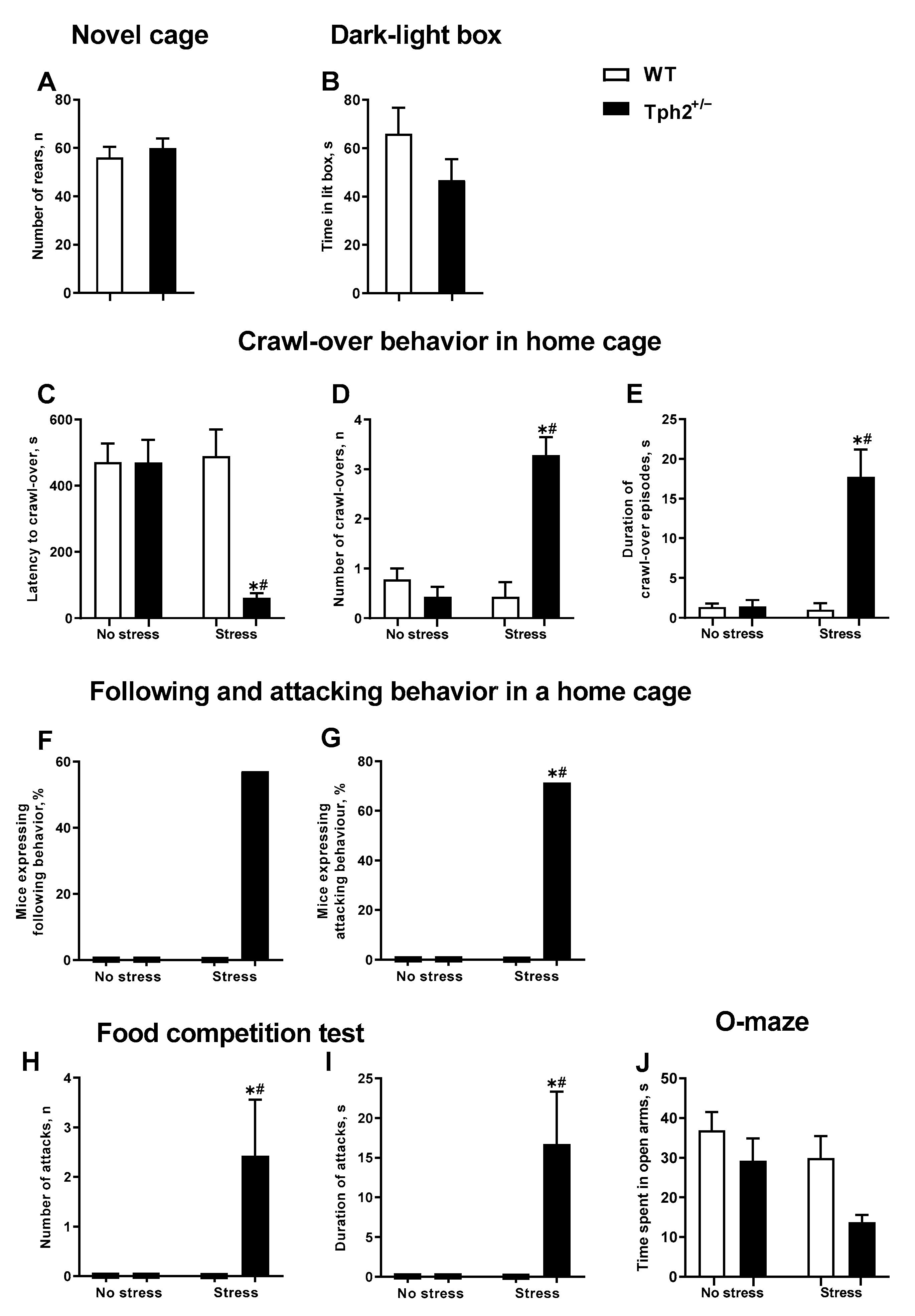
In the novel cage test, the number of exploratory rears did not differ significantly between the Tph2+/+ and Tph2+/− mice (t = 0.6140, df = 22, p = 0.55, unpaired t-test. Figure 2A). The time spent in the lit box of the dark-light box test was also not significantly different between these groups (t = 1.378, df = 18, p = 0.19, unpaired t-test. Figure 2B).
The latency to crawl-over, number of crawl-overs, and total duration of this behavior, as a measure of home cage dominance, were significantly different between the groups as studied in the home cage (H = 15.14, p < 0.01, H = 17.73, p < 0.01 and H = 17.39, p < 0.01, respectively; Kruskal-Wallis test. Figure 2C–E). The latency to crawl-over in the stressed Tph2+/− group was significantly shorter in comparison to both non-stressed Tph2+/− and stressed Tph2+/+ (wild type) animals (both p < 0.01, Dunn’s test). The number of episodes and the duration of crawl-over behavior were significantly higher in the stressed mutant mice in comparison to non-stressed Tph2+/− animals and stressed controls (all p < 0.01). While there was no significant group difference in the number of animals displaying the following behavior (all p = 0.07, Fisher’s exact test. Figure 2F), in comparison to both non-stressed Tph2+/− and stressed Tph2+/+ mice, the number of animals displaying agonistic (attacking) behavior was significantly higher in the stressed Tph2+/− group (both p = 0.02, Figure 2G). None of the non-stressed mice exhibited following or attacking behaviors, regardless of the genotype (Figure 2F,G).
In the food competition test, significant differences were found between the groups in both the number and the duration of attacks (H = 14.57, p < 0.01, and H = 14.57, p < 0.01, respectively. Figure 2H,I). Post-hoc analysis revealed that, in comparison to both non-stressed Tph2+/− group and stressed Tph2+/+ mice, the number and duration of attacks were significantly elevated in the stressed Tph2+/− group (both p = 0.01, Dunn’s test). In a similar manner to the home cage assay, none of the non-stressed mice exhibited following or attacking behaviors in the food competition test, regardless of the genotype (Figure 2H,I). In the O-maze, Kruskal-Wallis testing showed a significant group difference in the time spent in the open arms (H = 14.19, p < 0.01. Figure 2J). The only significant difference was found between the non-stressed wild type mice and stressed mutants (p < 0.01); post-hoc analysis did not show significant differences between genotype-matched or stress-matched groups. The Kruskal-Wallis test did not demonstrate any significant group differences in the food intake (H = 0.17, p = 0.99, Kruskal-Wallis test. Figure A1).

3.2. Altered Gene Expression of Selected Molecular Markers in the Prefrontal Cortex of Stressed Tph2+/− Mice

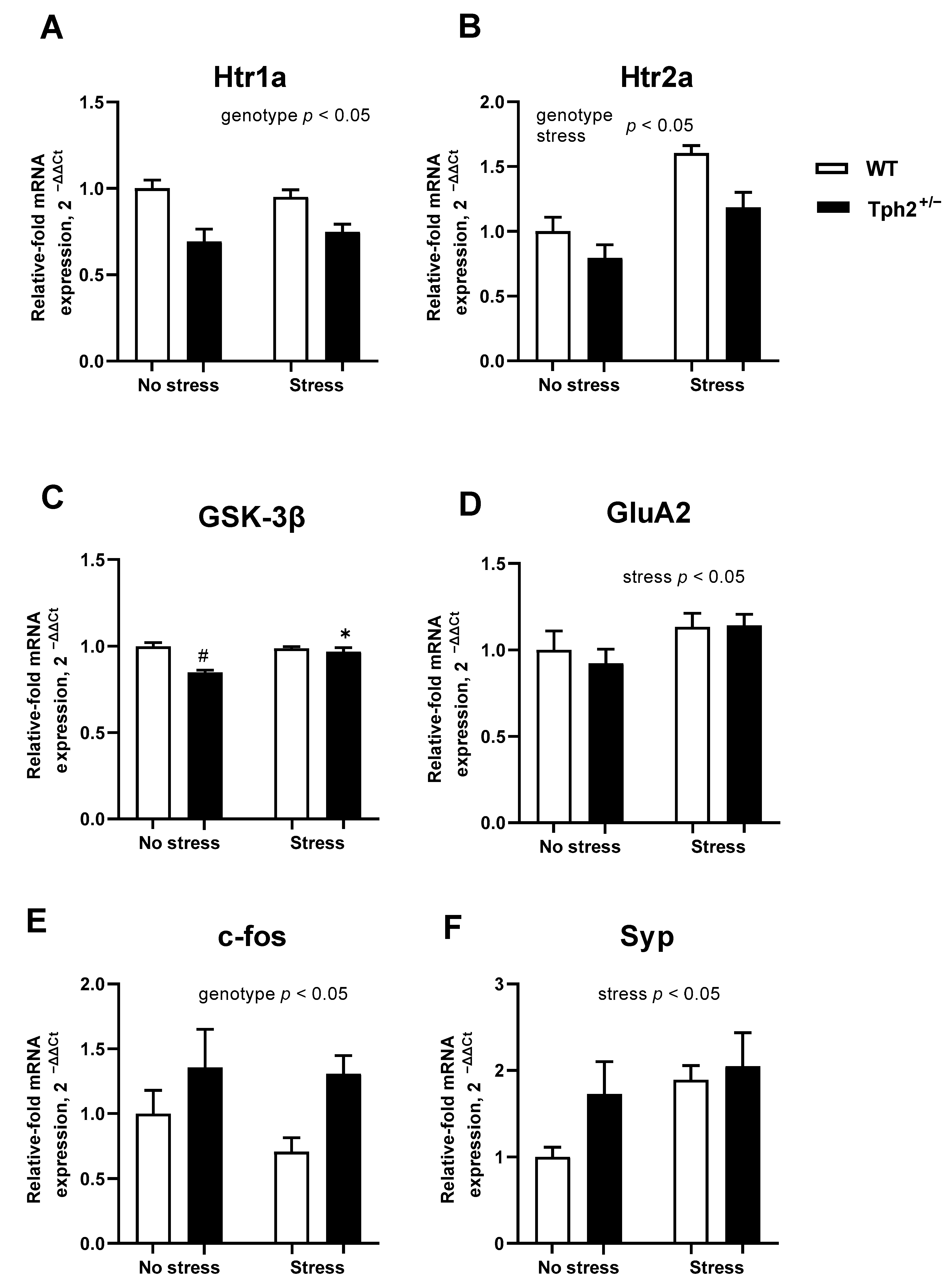
Two-way ANOVA revealed a significant main effect of genotype (F1,21 = 21.40, p < 0.01) and no significant stress × genotype interaction (F1,21 = 0.93, p = 0.35) in Htr1a expression. Independent of stress, a significant decrease in the expression of the Htr1a was found in Tph2+/− animals (Figure 3A). No significant stress × genotype interaction was shown in the expression of Htr2a (F1,20 = 1.240, p = 0.28, two-way ANOVA. Figure 3B), though both main effects of stress and genotype significant altered expression (F1,20 = 26.58, p < 0.01, and F1,20 = 10.59, p < 0.01, respectively, two-way ANOVA). Htr2a expression was significantly higher in the stressed animals, which was independent of genotype, but lower in the mutant groups, independent of stress. These data suggest differential regulation of expression of 5-HT receptor subtypes by stress and partial Tph2 inactivation.
For GSK-3β expression, a significant stress × genotype interaction was observed (F1,16 = 16.47, p < 0.01, two-way ANOVA. Figure 3C). In comparison to non-stressed Tph2+/− animals, post-hoc analysis revealed significantly higher GSK-3β expression in both the stressed Tph2+/− group and the non-stressed Tph2+/+ mice (both p < 0.01, Tukey’s test). GluA2 expression was not significantly affected by stress × genotype interaction (F1,22 = 0.248, p = 0.62. Figure 3D) and only a significant main effect of stress was observed (F1,22 = 4.331, p = 0.05). Specifically, stress elevated GluA2 expression compared to non-stressed animals irrespective of their genotype.
No stress × genotype interaction was found for either c-fos or Syp expression (F1,22 = 0.437, p = 0.52, and F1,22 = 1.149, p = 0.30, respectively, two-way ANOVA), though the main effects of genotype or stress on gene expression were observed. The expression of c-fos was significantly higher in the Tph2+/− mice in comparison with control animals, independent of stress (F1,22 = 6.63, p = 0.02, two-way ANOVA. Figure 3E). The expression of Syp was significantly higher in stressed animals than in controls (F1,22 = 5.24, p = 0.03. Figure 3F), independent of genotype.
Two-way ANOVA revealed significant main effects for genotype and stress (F1,23 = 4.87, p = 0.04 and F1,23 = 10.38, p < 0.01, respectively, two-way ANOVA), but there was no stress × genotype interaction (F1,23 = 1.46, p = 0.24) for Creb expression. This measure was significantly higher in the stressed animals and was independent of the genotype; in the mutant groups, it was independent of the stress (Figure A2A). These data suggest the differential regulation of expression of Creb by stress and partial Tph2 inactivation.
There was no significant stress × genotype interaction and no significant main effects of genotype or stress on Bdnf expression (F1,24 = 0.0047, p = 0.95; F1,24 = 0.28, p = 0.60 and F1,24 = 2.29, p = 0.14, respectively; Figure A2B), Trkb expression (F1,24 = 0.868, p = 0.36; F1,24 = 0.039, p = 0.85 and F1,24 = 0.76, p = 0.39, respectively; Figure A2C), or for the expression of PSD95 (F1,24 = 0.106, p = 0.95; F1,24 = 0.018, p = 0.89 and F1,24 = 1.025, p = 0.32, respectively; Figure A2D).
A stress × genotype interaction exists for Plp1 expression (F1,19 = 4.949, p = 0.04, two-way ANOVA). Post-hoc analysis revealed significantly lower expression of Plp1 in stressed Tph2+/− mice in comparison to non-stressed Tph2+/− mice (p = 0.02, Tukey’s test, Figure 4A). No significant differences were observed between Tph2+/+ stressed and naïve mice (p = 0.07). For Mbp and Mag expression, ANOVA revealed significant stress × genotype interaction (F1,16 = 16.68, p < 0.01 and F1,18 = 7.610, p = 0.01 respectively, Figure 4B,C). Compared to the non-stressed Tph2+/− group, the expression of Mbp and Mag was significantly lower in both stressed Tph2+/− (p = 0.01 and p = 0.02, respectively, Tukey’s test) and non-stressed Tph2+/+ mice (p < 0.01 and p = 0.03, respectively). ANOVA revealed no significant interaction for Mog expression (F1,19 = 4.098, p = 0.06, two-way ANOVA. Figure 4D), though a significant main effect of stress was observed (F1,19 = 10.08, p < 0.01). In comparison to non-stressed mice, stressed animals had a significantly lower expression level of Mog, irrespective of their genotype.

3.3. Naïve Female Tph2+/− Mice Show Signs of Decreased Learning of Adverse Memories and Helplessness as a Manifestation of Stress Resilience

In the modFST, in comparison to wild type mice, Tph2+/− mice demonstrated a significantly smaller increase in floating duration in the first two minutes of the test session between days 2 and 5 (U = 15, p < 0.01, Mann-Whitney test; Figure A3A). In the latency to float and the duration of floating, there was no significant interaction between day and genotype, though a main effect of the test day was found (F1,14 = 91.79 and F1,12 = 89.22, respectively, both p < 0.01, repeated measures two-way ANOVA; Figure A3B,C). No significant group differences in the latency and duration of floating were found on either day of the test.

4. Discussion

Our study has revealed that aggressive and dominant behaviors are induced in female Tph2+/− mice subjected to predation stress, resembling a behavioral profile reported for stressed male Tph2+/− mutants and mice with complete inactivation of Tph2. Wild type stressed controls did not show any of these changes. We also found a decrease in gene expression of Plp1, Mbp, and Mag in the prefrontal cortex of stressed mutants, which may reflect aberrant myelination processes which likely to contribute to stress-induced aggression and dominance behavior. Baseline expression of GSK-3β was lower in the non-stressed Tph2+/− mice than in the wild type animals. Unlike wild type mice, mutants showed relatively increased GSK-3β expression under stress conditions. The lowered basal expression of GSK-3β in female Tph2+/− mutants may also explain a diminished increase in behavioral despair during repeated swimming in the modFST, a sign of stress resilience.
The increased aggression and dominance in stressed mutants were accompanied by genotype effects on the prefrontal cortex expression of Htr1a and Htr2a. Both receptors are known to modulate aggressive behavior [70,71,72]. The expression of Htr1a and Htr2a were decreased in Tph2+/− females regardless of stress, which is also a feature of Tph2−/− mutants; it might be explained by a higher sensitivity of this receptor, at a protein level, to diminished levels of central 5-HT [73]. However, in the Tph2+/− males subjected to predation stress there was no effect on Htr1a or Htr2a expression. For Htr1a, the sex-dependent behavioral effects, which have been reported after the pharmacological targeting of 5-HT1A receptor in rodents [74], suggest that there is likely to be a differential role for this receptor in abnormal aggression in males and females.
The predation stress paradigm used in this work was previously shown to increase 5-HT turnover in the amygdala of male Tph2+/− mice [21]. Furthermore, significantly elevated 5-HT turnover in the prefrontal cortex of stressed male Tph2+/− mice correlated with measures of aggressiveness (Bazhenova and Lesch, unpublished results). Surprisingly, stressed Tph2+/− males exhibited unaltered 5-HT levels in the prefrontal cortex, while wild type mice showed significant increases in 5-HT levels under these conditions. These abnormalities might arise from the compromised 5-HT metabolism in the prefrontal cortex of stressed mutants that results in disrupted cortical top-down control of limbic structures regulating aggression, including the amygdala, and thus, these changes could underpin the social abnormalities observed in the stressed female Tph2+/− mice.
As compromised serotonin metabolism in the Tph2+/− mutants can independently result in the altered regulation of appetite, satiety, and metabolic processes, in which changes in monoamine levels and changes in the expression of their receptors can play a major role [75], the excessive aggression in stressed mutants in our study might be food deprivation-state-dependent. Preliminary studies on Tph2+/− stressed mice, housed under normal conditions, did not reveal any changes in social behavior in the food competition test (Strekalova and Costa-Nunes, unpublished results). In the present study, we used a food deprivation challenge, a well-established inducer of aggression in male mice [76,77], and hierarchical dominance behaviors in female mice [59]. Further studies are warranted to address the issue as to how the changes in serotonin receptor expression and the effects of food deprivation and aggression in stressed Tph2+/− mice are related.
Genetic deficits in 5-HT function are well-established to result in developmental abnormalities of brain connectivity [36,78,79,80]. Compromised frontostriatal white matter integrity and connectivity are believed to underlie increased impulsivity and aggression [41,81,82]. Here, for the first time, we report the increased expression of genes encoding myelination-related proteins in the prefrontal cortex of naïve Tph2+/− female mice and its significant decrease following predation stress. Previous work has shown that there is decreased expression of Mbp and Mag in naïve Tph2+/− males [35]. Thus, the present findings in naïve Tph2+/− females may mirror compensatory effects such as the elevated expression of myelin genes that is neutralized by stress, leading to impaired connectivity and maladaptive aggression in these animals. The stress-induced decrease of myelination-related marker expression was previously reported in other rodent models of stress, such as chronic unpredictable stress, social defeat and social isolation, immobilization stress, and early-life stress [83,84].
Moreover, others have previously demonstrated a relationship between myelination in the prefrontal cortex and aggression and emotional dysregulation. Reduced thickness of the myelin sheath in the prefrontal cortex was reported to correlate with increased aggression caused by juvenile isolation [85]. Group housing was shown to ameliorate both aggressive behaviors and the myelination deficit in another study of social isolation in mice [37,86]. In rats, the overexpression of the myelin transcription factor 1 (MyT1) promotes differentiation of oligodendrocytes, which is also regulated by Plp1 and Mbp [87], and ameliorates anxiety-like and compulsive behaviors [88]. Aberrant myelination is believed to underlie impaired brain connectivity and be associated with impulsive and aggressive behaviors, contributing to neurodevelopmental disorders such as attention deficit hyperactivity disorder (ADHD), autism spectrum disorders (ASD), and schizophrenia [89,90]. We may speculate that the changes observed in the expression of myelin associated transcripts in stressed Tph2+/− mice may reflect developmental abnormalities of white matter and brain connectivity and, though unlikely to be the sole cause of the excessive aggression observed in these mice, may contribute to behavior. This view is further supported by clinical evidence. For example, in women with ADHD and borderline personality disorder, there are correlations between anger-hostility measures and impairments of inferior frontal white matter connectivity [38]. Reduced white matter volume in the frontostriatal tracts, particularly in medial prefrontal regions, was associated with increased impulsivity in healthy subjects maturing from their adolescence to adulthood [41]. Aggression scores correlated with fronto-accumbal white matter integrity and cortical thickness of the orbitofrontal cortex in children with ADHD [39]. In patients recovering from mild traumatic brain injury, reduced fiber integrity in the white matter also correlates with higher measures of aggression [40].
Other molecular processes may potentially contribute to the abnormal social behavior of stressed Tph2+/− mice. Genotype differences in the expression of brain c-fos argue for a role of this factor in the aggressive behavior of stressed female Tph2+/−mice. In males, by comparison, c-fos expression was increased in the amygdala and prefrontal cortex of stressed mice of both genotypes [21]. Over-expression of c-fos in the hippocampus of Tph2−/− mice is accompanied by increased freezing in the fear conditioning paradigm; a trend towards both molecular and behavioral changes was reported in the Tph2+/− mutants [8,91]. It can be speculated that the increased expression of this immediate early gene, as found in the stressed Tph2+/− groups 24 h after the last manipulation, might be related to increased conditioning after the handling procedure. While chronic stress has been reported to suppress the expression of Syp, a marker of neuronal plasticity [92,93], here, Creb expression was elevated in female Tph2+/− mice regardless of stress exposure. This may indicate compensatory plasticity processes related to the up-regulation of myelination in naïve mutants and may further contribute to their stress resilience as shown in the modFST. Indeed, increased CREB activity was previously associated with elevated aggression in female mice [94,95]. While the expression of Creb was shown to be related to levels of BDNF and its receptor [96,97,98], mRNA levels of Bdnf and Trkb were unaltered in this study, as well as gene expression of PSD95, which have been correlated with increased aggression in female rodents in other studies [99]. These results suggest that more complex regulatory interactions underpin emotional control than those described by these plasticity markers alone in the prefrontal cortex.
Upregulated myelination markers may also relate to the decreased baseline expression of GSK-3β, a key indicator of helplessness behavior in naïve mutants [42]. Previous studies point to a reciprocal relationship between GSK-3β and myelination-related factors, e.g., Mbp [100,101], that is in keeping with our findings of increased gene expression of the latter molecules found in naïve mutants. It is of note that decreased basal expression of GSK-3β in the female Tph2+/− mutants may also contribute to the smaller increase in behavioral despair during repeated swimming in the modFST. Previous studies have revealed an important role of increased brain GSK-3β activities in subgroups of mice that display susceptible, but not resilient, responses in this model [42]. In effect, mice that display a prolongation of the floating behavior from day 2 to day 5 above mean values for the group exhibit increased mRNA concentration for GSK-3β, decreased levels of phosphorylated GSK-3β at 9-serine, and a reduced ratio of phosphorylated GSK-3β to overall GSK-3β content, i.e., increased GSK-3β activity, in the prefrontal cortex [42,48]. These behavioral and molecular changes were reduced by pre-treatment with low doses of imipramine or anti-oxidant compounds [48,63,64,68]. Therefore, the lowered baseline expression of GSK-3β in the pre-frontal cortex of Tph2+/− mutants might explain the smaller increase in behavioral despair observed during repeated swimming in the modified swim test. Notably, a functional interaction was previously reported between decreased Tph2 enzymatic activity and GSK-3β in male mice with knock-in of the human R439H mutation [102].
Concerning potential mechanisms for a lower stress/despair response of female Tph2+/− mutants in the modified swim test, we hypothesize that this might also be due to the suppression of the expression of 5-HT1A and 5-HT2A receptors in the brain, whose roles in stress response, major depressive disorder, and consolidation of aversive memories are well established [70,103,104,105]. Furthermore, it can be speculated that in a similar fashion to male Tph2+/− mutants that exhibit ‘stress resilience’ in the modFST [20], female Tph2+/− mice exhibit altered dopamine metabolism; turnover of dopamine in major mesocorticolimbic regions can govern individual susceptibility to stress [106,107] and was particularly marked in female mice [108].
In the present study, stress-induced increases of expression of GSK-3β and GluA2 were not affected by the mutation. Similar results were found in the brain of stressed Tph2+/− males for GSK-3β, but GluA2 was upregulated selectively in the male mutants [21]. This challenges the view that these transcripts play a pivotal role in the aggression elicited in stressed Tph2+/− females [24,33] and further suggests that sex differences result in the differential regulation of aggression ein Tph2+/− mice. For GSK-3β, given that the level of the phosphorylated form of this kinase is the principal determinant of its activity, activity has been shown to correlate with GSK-3β gene expression changes [109]. However, further assessment of the level of GSK-3β phosphorylation might be useful to confirm this association and its role in the behavioral abnormalities of the Tph2+/− females reported here.

5. Conclusions

Taken together: our findings show that an interaction between partial genetic inactivation of neuronal Tph2 expression and environmental adversity results in aggressive and dominant behaviors in female Tph2+/− mice. Naïve female Tph2+/− mice show decreased learning of adverse memories and helplessness, a sign of stress resilience. These behaviors are reminiscent of changes in Tph2+/− males and null mutants of both sexes lacking Tph2. For the first time, we report the altered expression of myelination markers in naïve and stressed female Tph2+/− mice. These data encourage speculation regarding impaired brain connectivity in these mice, which likely contributes to the increased aggression and dominance observed in the stressed Tph2+/− mice. Further studies are required to shed light on the detailed mechanisms of the relationships between serotonin deficiency, stress, and myelination in the context of gene × environment interaction and female aggression.

Author Contributions

Conceptualization: T.S., K.-P.L., S.W. and L.W.L.; Data curation: A.U., J.P.C.-N., A.V.K. and D.C.A.; Formal analysis: A.N., A.V.K. and D.C.A.; Funding acquisition: L.W.L., K.-P.L. and T.S.; Investigation: E.S., A.N., E.V., A.G., J.P.C.-N. and A.C.K.T.; Methodology: E.V., A.P., A.C.K.T., L.W.L. and T.S.; Project administration: A.P., S.W. and T.S.; Resources: A.N., L.W.L., K.-P.L., S.W. and T.S.; Software: A.U. and A.N.; Supervision: A.P., A.U. and T.S.; Visualization: E.S., E.V. and A.G.; Roles/Writing—original draft E.S., A.G., K.-P.L. and T.S.; Writing—review and editing: A.V.K., E.V., A.P., L.W.L., K.-P.L., D.C.A. and S.W. All authors have read and agreed to the published version of the manuscript.

Funding

The authors’ animal work reported here was supported by Deutsche Forschungsgemeinschaft (DFG:CRC TRR58A1/A5), the European Union’s Seventh Framework Programme (FP7/2007–2013) under Grant No. 602805 (Aggressotype), the Horizon 2020 Research and Innovation Programme under Grant No. 728018 (Eat2beNice) (to K.-P.L. and T.S.) and Grant No. 101007642 (PhytoAPP) (to D.C.A. and T.S.), and Swiss-Russian Cooperation grant RPG Russia 2020 (to S.W. and K.-P.L.). Molecular data analysis was supported by RAS N0520-2019-0031 (to E.S. and T.S.). The sponsors had no role in study design, in the collection, analysis, and interpretation of data; in the writing of the report, and in the decision to submit the article for publication.

Institutional Review Board Statement

The study was conducted according to the guidelines of the Directive 2010/63/EU of 22 September 2010 and had been approved by the Ethics Committee of the New University of Lisbon (No. 0421/000/000/2013). Experimental protocols conformed to directive 2010/63/EU and were compliant with ARRIVE guidelines (https://arriveguidelines.org, accessed on 14 March 2022).

Informed Consent Statement

Not applicable.

Data Availability Statement

Data supporting reported results can be obtained on a request. Alternatively, they can be obtained via the links to publicly archived datasets analyzed and generated during the study (https://www.sechenov.ru/univers/structure/nauchno-tekhnologicheskiy-park-biomeditsiny/instituty/institut-molekulyarnoy-meditsiny/laboratorii/psikhneiro, accessed on 14 March 2022).

Acknowledgments

We appreciate the valuable help of Dolores Bonopartos with this project and the kind help of Daniel Radford-Smith with editing the language. We also thank Alexander Silchenko from the Institute of Neuroscience and Medicine (INM-7: Brain and Behavior), Jülich Research Center, Jülich, Germany for their kind help with the statistical analysis of the data.

Conflicts of Interest

The authors declare no conflict of interest. The funders had no role in the design of the study; in the collection, analyses, or interpretation of data; in the writing of the manuscript, or in the decision to publish the results.

Appendix A

Appendix A.1. Supplementary Methods

Table A1. Primer sequences for mRNA expression analysis.
GenePrimerSequence
Htr1aForward5′-GACAGGCGGCAACGATACT-3′
Reverse5′-CCAAGGAGCCGATGAGATAGTT-3′
Htr2aForward5′-TAATGCAATTAGGTGACGACTCG-3′
Reverse5′-GCAGGAGAGGTTGGTTCTGTTT-3′
GSK-3βForward5′-GCACTCTTCAACTTTACCACTCA-3′
Reverse5′-CGAGCATGTGGAGGGATAAG-3′
GluA2Forward5′-GCGTGGAAATAGAAAGGGCC-3′
Reverse5′-ACTCCAGTACCCAATCTTCCG-3′
c-fosForward5′-CGGGTTTCAACGCCGACTA-3′
Reverse5′-TTGGCACTAGAGACGGACAGA-3′
SypForward5′-TGTGTTTGCCTTCCTCTACTC-3′
Reverse5′-TCAGTGGCCATCTTCACATC-3′
Plp1Forward5′-CCAGAATGTATGGTGTTCTCCC-3′
Reverse5′-GGCCCATGAGTTTAAGGACG-3′
MbpForward5′-TCACAGCGATCCAAGTACCTG-3′
Reverse5′-CCCCTGTCACCGCTAAAGAA-3′
MagForward5′-GGTACATGGCGTCTGGTATTTC-3′
Reverse5′-ACTTGTGTGCGGGACTTGAAG-3′
MogForward5′-TCATGCAGCTATGCAGGACAA-3′
Reverse5′-TTTCGGTAGAGGTGAACCACT-3′
CrebForward5′-CAGGGGTCGCAAGGATTGAAG-3′
Reverse5′-ATCGCCTGAGGCAGTGTACT-3′
BdnfForward5′-TGGCTGACACTTTTGAGCAC-3′
Reverse5′-AAGTGTACAAGTCCGCGTCC-3′
TrkbForward5′-CCTCCACGGATGTTGCTGAC-3′
Reverse5′-GCAACATCACCAGCAGGCA-3′
PSD-95Forward5′-GACGCCAGCGACGAAGAG-3′
Reverse5′-CTCGACCCGCCGTTTG-3′
GAPDHForward5′-ATGACCACAGTCCATGCCATC -3′
Reverse5′-GAGCTTCCCGTTCAGCTCTG-3′

Appendix A.2. Supplementary Results

Daily Food Intake of Tph2+/− Mice

The Kruskal-Wallis test did not reveal significant differences in the average daily food intake measured during the observation period (H = 0.17, p = 0.99, Kruskal-Wallis test. Figure A1).
Figure A1. Daily food intake of Tph2+/− mice. No significant group difference in average daily food intake during the observation period was observed. WT—wild type.
Cells 11 01036 g0a1

Appendix A.3. Expression of Neurotrophic Factors in the Prefrontal Cortex of Stressed Tph2+/−

The two-way ANOVA and post-hoc comparisons revealed group differences in the expression of neurotrophic molecules in the brains of the experimental groups (see ms main text; Figure A2).
Figure A2. Expression of Creb, Bdnf, Trkb and PSD95 in the prefrontal cortex of Tph2+/− mice. (A) Creb expression was significantly higher in the stressed animals, independent of genotype (WT NS n = 6, WT S n = 9, Tph2+/− NS n = 6, Tph2+/− S n = 6). (B) No significant differences were found for Bdnf expression (WT NS n = 6, WT S n = 9, Tph2+/− NS n = 6, Tph2+/− S n = 7), (C) Trkb expression (WT NS n = 6, WT S n = 9, Tph2+/− NS n = 6, Tph2+/− S n = 7), or for (D) PSD95 expression (WT NS n = 6, WT S n = 9, Tph2+/− NS n = 6, Tph2+/− S n = 7). WT, wild type.
Cells 11 01036 g0a2

Appendix A.4. Tph2+/− Mice Display Reduced Potentiation of Floating in the modFST Paradigm

The change in floating duration in the first two minutes of the test session between days 2 and 5 in Tph2+/− animals was significantly smaller than in wild type mice (see ms text, Figure A3A). Concerning the latency to float and the duration of floating, only the main effect of the test day was found (see ms text, Figure A3B,C). Post-hoc analysis revealed a significant decrease in latency to float and a significant increase in the duration of floating on days 2 and 5 compared to day 1, irrespective of the genotype (both p < 0.01, Šídák’s multiple comparisons test).
Figure A3. Floating behavior in the modified swim test. (A) A smaller increase in floating duration from day 2 to day 5 was observed in the Tph2+/− mice compared to WT. (B) A significant decrease in latency to float on days 2 and 5 compared to day 1 was observed and was independent of genotype. (C) There was a significant increase in the duration of floating on days 2 and 5 compared to day 1, independent of the genotype. WT—wild type, * p < 0.01 vs. wild type, # p < 0.01 vs. same genotype on day 1. WT no stress n = 13, WT stress n = 13, Tph2+/− NS n = 11, Tph2+/− S n = 12.
Cells 11 01036 g0a3

References

  1. Vakili, V.; Ziaee, M.; Zarifian, A. Aggression: Is That an Issue for Worrying? Iran. J. Public Health 2015, 44, 1561–1562. [Google Scholar] [PubMed]
  2. Xiang, C.; Liu, S.; Fan, Y.; Wang, X.; Jia, Y.; Li, L.; Cong, S.; Han, F. Single Nucleotide Polymorphisms, Variable Number Tandem Repeats and Allele Influence on Serotonergic Enzyme Modulators for Aggressive and Suicidal Behaviors: A Review. Pharmacol. Biochem. Behav. 2019, 180, 74–82. [Google Scholar] [CrossRef] [PubMed]
  3. Freitag, C.M.; Konrad, K.; Stadler, C.; De Brito, S.A.; Popma, A.; Herpertz, S.C.; Herpertz-Dahlmann, B.; Neumann, I.; Kieser, M.; Chiocchetti, A.G.; et al. Conduct Disorder in Adolescent Females: Current State of Research and Study Design of the FemNAT-CD Consortium. Eur. Child Adolesc. Psychiatry 2018, 27, 1077–1093. [Google Scholar] [CrossRef] [PubMed]
  4. Denson, T.F.; O’Dean, S.M.; Blake, K.R.; Beames, J.R. Aggression in Women: Behavior, Brain and Hormones. Front. Behav. Neurosci. 2018, 12, 81. [Google Scholar] [CrossRef] [PubMed] [Green Version]
  5. Neumann Aggression and Anxiety: Social Context and Neurobiological Links. Front. Behav. Neurosci. 2010, 4, 12. [CrossRef] [Green Version]
  6. Takahashi, A.; Miczek, K.A. Neurogenetics of Aggressive Behavior: Studies in Rodents. Curr. Top. Behav. Neurosci. 2014, 17, 3–44. [Google Scholar] [CrossRef] [PubMed] [Green Version]
  7. Gutknecht, L.; Araragi, N.; Merker, S.; Waider, J.; Sommerlandt, F.M.J.; Mlinar, B.; Baccini, G.; Mayer, U.; Proft, F.; Hamon, M.; et al. Impacts of Brain Serotonin Deficiency Following Tph2 Inactivation on Development and Raphe Neuron Serotonergic Specification. PLoS ONE 2012, 7, e43157. [Google Scholar] [CrossRef] [PubMed] [Green Version]
  8. Gutknecht, L.; Popp, S.; Waider, J.; Sommerlandt, F.M.J.; Göppner, C.; Post, A.; Reif, A.; Van Den Hove, D.; Strekalova, T.; Schmitt, A.; et al. Interaction of Brain 5-HT Synthesis Deficiency, Chronic Stress and Sex Differentially Impact Emotional Behavior in Tph2 Knockout Mice. Psychopharmacology 2015, 232, 2429–2441. [Google Scholar] [CrossRef] [PubMed] [Green Version]
  9. Angoa-Pérez, M.; Kane, M.J.; Briggs, D.I.; Sykes, C.E.; Shah, M.M.; Francescutti, D.M.; Rosenberg, D.R.; Thomas, D.M.; Kuhn, D.M. Genetic Depletion of Brain 5HT Reveals a Common Molecular Pathway Mediating Compulsivity and Impulsivity. J. Neurochem. 2012, 121, 974–984. [Google Scholar] [CrossRef] [PubMed] [Green Version]
  10. Weidner, M.T.; Lardenoije, R.; Eijssen, L.; Mogavero, F.; De Groodt, L.P.M.T.; Popp, S.; Palme, R.; Förstner, K.U.; Strekalova, T.; Steinbusch, H.W.M.; et al. Identification of Cholecystokinin by Genome-Wide Profiling as Potential Mediator of Serotonin-Dependent Behavioral Effects of Maternal Separation in the Amygdala. Front. Neurosci. 2019, 13, 460. [Google Scholar] [CrossRef]
  11. Yang, J.; Lee, M.S.; Lee, S.H.; Lee, B.C.; Kim, S.H.; Joe, S.H.; Jung, I.K.; Choi, I.G.; Ham, B.J. Association between Tryptophan Hydroxylase 2 Polymorphism and Anger-Related Personality Traits among Young Korean Women. Neuropsychobiology 2010, 62, 158–163. [Google Scholar] [CrossRef] [PubMed]
  12. Lin, Y.M.J.; Ko, H.C.; Chang, F.M.; Yeh, T.L.; Sun, H.S. Population-Specific Functional Variant of the Tph2 Gene 2755C>A Polymorphism Contributes Risk Association to Major Depression and Anxiety in Chinese Peripartum Women. Arch. Women’s Ment. Health 2009, 12, 401–408. [Google Scholar] [CrossRef] [PubMed]
  13. Fasching, P.A.; Faschingbauer, F.; Goecke, T.W.; Engel, A.; Häberle, L.; Seifert, A.; Voigt, F.; Amann, M.; Rebhan, D.; Burger, P.; et al. Genetic Variants in the Tryptophan Hydroxylase 2 Gene (Tph2) and Depression during and after Pregnancy. J. Psychiatr. Res. 2012, 46, 1109–1117. [Google Scholar] [CrossRef] [PubMed]
  14. Anstrom, K.K.; Miczek, K.A.; Budygin, E.A. Increased Phasic Dopamine Signaling in the Mesolimbic Pathway during Social Defeat in Rats. Neuroscience 2009, 161, 3–12. [Google Scholar] [CrossRef] [PubMed] [Green Version]
  15. Deal, A.L.; Park, J.; Weiner, J.L.; Budygin, E.A. Stress Alters the Effect of Alcohol on Catecholamine Dynamics in the Basolateral Amygdala. Front. Behav. Neurosci. 2021, 15, 640651. [Google Scholar] [CrossRef] [PubMed]
  16. Lee, Y.C.; Chao, Y.L.; Chang, C.E.; Hsieh, M.H.; Liu, K.T.; Chen, H.C.; Lu, M.L.; Chen, W.Y.; Chen, C.H.; Tsai, M.H.; et al. Transcriptome Changes in Relation to Manic Episode. Front. Psychiatry 2019, 10, 280. [Google Scholar] [CrossRef] [PubMed] [Green Version]
  17. Lesch, K.P. Alcohol Dependence and Gene x Environment Interaction in Emotion Regulation: Is Serotonin the Link? Eur. J. Pharmacol. 2005, 526, 113–124. [Google Scholar] [CrossRef] [PubMed]
  18. Haller, J. The Role of Central and Medial Amygdala in Normal and Abnormal Aggression: A Review of Classical Approaches. Neurosci. Biobehav. Rev. 2017, 85, 34–43. [Google Scholar] [CrossRef] [PubMed]
  19. Augsburger, M.; Maercker, A. Associations between Trauma Exposure, Posttraumatic Stress Disorder, and Aggression Perpetrated by Women. A Meta-Analysis. Clin. Psychol. Sci. Pract. 2020, 27, e12322. [Google Scholar] [CrossRef] [Green Version]
  20. Strekalova, T.; Svirin, E.; Waider, J.; Gorlova, A.; Cespuglio, R.; Kalueff, A.; Pomytkin, I.; Schmitt-Boehrer, A.G.; Lesch, K.P.; Anthony, D.C. Altered Behaviour, Dopamine and Norepinephrine Regulation in Stressed Mice Heterozygous in Tph2 Gene. Prog. Neuro-Psychopharmacol. Biol. Psychiatry 2020, 108, 110155. [Google Scholar] [CrossRef] [PubMed]
  21. Gorlova, A.; Ortega, G.; Waider, J.; Bazhenova, N.; Veniaminova, E.; Proshin, A.; Kalueff, A.V.; Anthony, D.C.; Lesch, K.P.; Strekalova, T. Stress-Induced Aggression in Heterozygous Tph2 Mutant Mice Is Associated with Alterations in Serotonin Turnover and Expression of 5-HT6 and AMPA Subunit 2A Receptors. J. Affect. Disord. 2020, 272, 440–451. [Google Scholar] [CrossRef]
  22. Veenema, A.H.; Bredewold, R.; Neumann, I.D. Opposite Effects of Maternal Separation on Intermale and Maternal Aggression in C57BL/6 Mice: Link to Hypothalamic Vasopressin and Oxytocin Immunoreactivity. Psychoneuroendocrinology 2007, 32, 437–450. [Google Scholar] [CrossRef] [PubMed]
  23. de Oliveira, V.E.M.; Neumann, I.D.; de Jong, T.R. Post-Weaning Social Isolation Exacerbates Aggression in Both Sexes and Affects the Vasopressin and Oxytocin System in a Sex-Specific Manner. Neuropharmacology 2019, 156, 107504. [Google Scholar] [CrossRef] [PubMed]
  24. Vignisse, J.; Sambon, M.; Gorlova, A.; Pavlov, D.; Caron, N.; Malgrange, B.; Shevtsova, E.; Svistunov, A.; Anthony, D.C.; Markova, N.; et al. Thiamine and Benfotiamine Prevent Stress-Induced Suppression of Hippocampal Neurogenesis in Mice Exposed to Predation without Affecting Brain Thiamine Diphosphate Levels. Mol. Cell. Neurosci. 2017, 82, 126–136. [Google Scholar] [CrossRef] [PubMed]
  25. Veniaminova, E.; Cespuglio, R.; Markova, N.; Mortimer, N.; Wai Cheung, C.; Steinbusch, H.W.; Lesch, K.-P.; Strekalova, T. Behavioral Features of Mice Fed with a Cholesterol-Enriched Diet:Deficient Novelty Exploration and Unaltered Aggressive Behavior. Transl. Neurosci. Clin. 2016, 2, 87. [Google Scholar] [CrossRef] [Green Version]
  26. Veniaminova, E.; Cespuglio, R.; Cheung, C.W.; Umriukhin, A.; Markova, N.; Shevtsova, E.; Lesch, K.-P.; Anthony, D.C.; Strekalova, T. Autism-Like Behaviours and Memory Deficits Result from a Western Diet in Mice. Neural Plast. 2017, 2017, 9498247. [Google Scholar] [CrossRef] [Green Version]
  27. Veniaminova, E.; Cespuglio, R.; Chernukha, I.; Schmitt-Boehrer, A.G.; Morozov, S.; Kalueff, A.V.; Kuznetsova, O.; Anthony, D.C.; Lesch, K.P.; Strekalova, T. Metabolic, Molecular, and Behavioral Effects of Western Diet in Serotonin Transporter-Deficient Mice: Rescue by Heterozygosity? Front. Neurosci. 2020, 14, 24. [Google Scholar] [CrossRef] [PubMed] [Green Version]
  28. Kim, J.Y.; Kim, A.; Zhao, Z.Q.; Liu, X.Y.; Chen, Z.F. Postnatal Maintenance of the 5-Ht1a-Pet1 Autoregulatory Loop by Serotonin in the Raphe Nuclei of the Brainstem. Mol. Brain 2014, 7, 48. [Google Scholar] [CrossRef] [Green Version]
  29. Mlinar, B.; Montalbano, A.; Waider, J.; Lesch, K.P.; Corradetti, R. Increased Functional Coupling of 5-HT1A Autoreceptors to GIRK Channels in Tph2−/− Mice. Eur. Neuropsychopharmacol. 2017, 27, 1258–1267. [Google Scholar] [CrossRef]
  30. Wang, L.R.; Kim, S.H.; Baek, S.S. Effects of Treadmill Exercise on the Anxiety-like Behavior through Modulation of GSK3β/β-Catenin Signaling in the Maternal Separation Rat Pup. J. Exerc. Rehabil. 2019, 15, 206–212. [Google Scholar] [CrossRef]
  31. Pavlov, D.; Bettendorff, L.; Gorlova, A.; Olkhovik, A.; Kalueff, A.V.; Ponomarev, E.D.; Inozemtsev, A.; Chekhonin, V.; Lesch, K.P.; Anthony, D.C.; et al. Neuroinflammation and Aberrant Hippocampal Plasticity in a Mouse Model of Emotional Stress Evoked by Exposure to Ultrasound of Alternating Frequencies. Prog. Neuro-Psychopharmacol. Biol. Psychiatry 2019, 90, 104–116. [Google Scholar] [CrossRef] [PubMed]
  32. Costa-Nunes, J.; Zubareva, O.; Araújo-Correia, M.; Valença, A.; Schroeter, C.A.; Pawluski, J.L.; Vignisse, J.; Steinbusch, H.; Hermes, D.; Phillipines, M.; et al. Altered Emotionality, Hippocampus-Dependent Performance and Expression of NMDA Receptor Subunit MRNAs in Chronically Stressed Mice. Stress 2014, 17, 108–116. [Google Scholar] [CrossRef] [PubMed]
  33. Costa-Nunes, J.P.; Gorlova, A.; Pavlov, D.; Cespuglio, R.; Gorovaya, A.; Proshin, A.; Umriukhin, A.; Ponomarev, E.D.; Kalueff, A.V.; Strekalova, T.; et al. Ultrasound Stress Compromises the Correlates of Emotional-like States and Brain AMPAR Expression in Mice: Effects of Antioxidant and Anti-Inflammatory Herbal Treatment. Stress 2020, 23, 481–495. [Google Scholar] [CrossRef] [PubMed]
  34. Gorlova, A.; Pavlov, D.; Anthony, D.C.; Ponomarev, E.D.; Sambon, M.; Proshin, A.; Shafarevich, I.; Babaevskaya, D.; Lesch, K.P.; Bettendorff, L.; et al. Thiamine and Benfotiamine Counteract Ultrasound-Induced Aggression, Normalize AMPA Receptor Expression and Plasticity Markers, and Reduce Oxidative Stress in Mice. Neuropharmacology 2019, 156, 107543. [Google Scholar] [CrossRef] [PubMed]
  35. Svirin, E.; Gorlova, A.; Lim, L.W.; Veniaminova, E.; Costa-Nunes, J.; Anthony, D.; Lesch, K.-P.; Strekalova, T. Sexual Bias in the Altered Expression of Myelination Factors in Mice with Partial Genetic Deficiency of Tryptophan Hydroxylase 2 and Pro-Aggressive Effects of Predation Stress. In Proceedings of the IBNS 30th Annual Meeting, Puerto Vallarta, Mexico, 1–5 June 2021. [Google Scholar]
  36. Jha, S.C.; Meltzer-Brody, S.; Steiner, R.J.; Cornea, E.; Woolson, S.; Ahn, M.; Verde, A.R.; Hamer, R.M.; Zhu, H.; Styner, M.; et al. Antenatal Depression, Treatment with Selective Serotonin Reuptake Inhibitors, and Neonatal Brain Structure: A Propensity-Matched Cohort Study. Psychiatry Res.-Neuroimaging 2016, 253, 43–53. [Google Scholar] [CrossRef] [PubMed] [Green Version]
  37. Makinodan, M.; Ikawa, D.; Miyamoto, Y.; Yamauchi, J.; Yamamuro, K.; Yamashita, Y.; Toritsuka, M.; Kimoto, S.; Okumura, K.; Yamauchi, T.; et al. Social Isolation Impairs Remyelination in Mice through Modulation of IL-6. FASEB J. 2016, 30, 4267–4274. [Google Scholar] [CrossRef] [PubMed] [Green Version]
  38. Rüsch, N.; Weber, M.; Il’yasov, K.A.; Lieb, K.; Ebert, D.; Hennig, J.; van Elst, L.T. Inferior Frontal White Matter Microstructure and Patterns of Psychopathology in Women with Borderline Personality Disorder and Comorbid Attention-Deficit Hyperactivity Disorder. Neuroimage 2007, 35, 738–747. [Google Scholar] [CrossRef] [PubMed]
  39. Cha, J.; Fekete, T.; Siciliano, F.; Biezonski, D.; Greenhill, L.; Pliszka, S.R.; Blader, J.C.; Krain Roy, A.; Leibenluft, E.; Posner, J. Neural Correlates of Aggression in Medication-Naive Children with ADHD: Multivariate Analysis of Morphometry and Tractography. Neuropsychopharmacology 2015, 40, 1717–1725. [Google Scholar] [CrossRef] [PubMed] [Green Version]
  40. Dailey, N.S.; Smith, R.; Bajaj, S.; Alkozei, A.; Gottschlich, M.K.; Raikes, A.C.; Satterfield, B.C.; Killgore, W.D.S. Elevated Aggression and Reduced White Matter Integrity in Mild Traumatic Brain Injury: A DTI Study. Front. Behav. Neurosci. 2018, 12, 118. [Google Scholar] [CrossRef] [PubMed]
  41. Ziegler, G.; Hauser, T.U.; Moutoussis, M.; Bullmore, E.T.; Goodyer, I.M.; Fonagy, P.; Jones, P.B.; Lindenberger, U.; Dolan, R.J. Compulsivity and Impulsivity Traits Linked to Attenuated Developmental Frontostriatal Myelination Trajectories. Nat. Neurosci. 2019, 22, 992–999. [Google Scholar] [CrossRef] [PubMed]
  42. Strekalova, T.; Markova, N.; Shevtsova, E.; Zubareva, O.; Bakhmet, A.; Steinbusch, H.M.; Bachurin, S.; Lesch, K.-P. Individual Differences in Behavioural Despair Predict Brain GSK-3beta Expression in Mice: The Power of a Modified Swim Test. Neural Plast. 2016, 2016, 5098591. [Google Scholar] [CrossRef] [PubMed] [Green Version]
  43. Grafman, J.; Schwab, K.; Warden, D.; Pridgen, A.; Brown, H.R.; Salazar, A.M. Frontal Lobe Injuries, Violence, and Aggression: A Report of the Vietnam Head Injury Study. Neurology 1996, 46, 1231–1238. [Google Scholar] [CrossRef] [PubMed]
  44. Arnsten, A.F.T. Stress Signalling Pathways That Impair Prefrontal Cortex Structure and Function. Nat. Rev. Neurosci. 2009, 10, 410–422. [Google Scholar] [CrossRef] [PubMed]
  45. Wall, V.L.; Fischer, E.K.; Bland, S.T. Isolation Rearing Attenuates Social Interaction-Induced Expression of Immediate Early Gene Protein Products in the Medial Prefrontal Cortex of Male and Female Rats. Physiol. Behav. 2012, 107, 440–450. [Google Scholar] [CrossRef] [PubMed] [Green Version]
  46. Takahashi, A.; Nagayasu, K.; Nishitani, N.; Kaneko, S.; Koide, T. Control of Intermale Aggression by Medial Prefrontal Cortex Activation in the Mouse. PLoS ONE 2014, 9, e94657. [Google Scholar] [CrossRef] [Green Version]
  47. Achterberg, M.; van Duijvenvoorde, A.C.K.; Bakermans-Kranenburg, M.J.; Crone, E.A. Control Your Anger! The Neural Basis of Aggression Regulation in Response to Negative Social Feedback. Soc. Cogn. Affect. Neurosci. 2016, 11, 712–720. [Google Scholar] [CrossRef] [PubMed] [Green Version]
  48. Markova, N.; Bazhenova, N.; Anthony, D.C.; Vignisse, J.; Svistunov, A.; Lesch, K.P.; Bettendorff, L.; Strekalova, T. Thiamine and Benfotiamine Improve Cognition and Ameliorate GSK-3β-Associated Stress-Induced Behaviours in Mice. Prog. Neuro-Psychopharmacol. Biol. Psychiatry 2017, 75, 148–156. [Google Scholar] [CrossRef] [PubMed]
  49. Strekalova, T. Optimization of the Chronic Stress Depression Model in C57 BL/6 Mice: Evidences for Improved Validity. In Behavioral Models in Stress Research; LaPorte, J.L., Kalueff, A.V., Eds.; Nova Science Publishers: Hauppauge, NY, USA, 2008; Volume I, pp. 95–139. [Google Scholar]
  50. Strekalova, T.; Steinbusch, H.W.M. Measuring Behavior in Mice with Chronic Stress Depression Paradigm. Prog. Neuropsychopharmacol. Biol. Psychiatry 2010, 34, 348–361. [Google Scholar] [CrossRef] [PubMed]
  51. Couch, Y.; Anthony, D.C.; Dolgov, O.; Revischin, A.; Festoff, B.; Santos, A.I.; Steinbusch, H.W.; Strekalova, T. Microglial Activation, Increased TNF and SERT Expression in the Prefrontal Cortex Define Stress-Altered Behaviour in Mice Susceptible to Anhedonia. Brain. Behav. Immun. 2013, 29, 136–146. [Google Scholar] [CrossRef]
  52. Costa-Nunes, J.P.; Cline, B.H.; Araújo-Correia, M.; Valença, A.; Markova, N.; Dolgov, O.; Kubatiev, A.; Yeritsyan, N.; Steinbusch, H.W.M.; Strekalova, T. Animal Models of Depression and Drug Delivery with Food as an Effective Dosing Method: Evidences from Studies with Celecoxib and Dicholine Succinate. BioMed Res. Int. 2015, 2015, 596126. [Google Scholar] [CrossRef]
  53. Strekalova, T.; Spanagel, R.; Bartsch, D.; Henn, F.A.; Gass, P. Stress-Induced Anhedonia in Mice Is Associated with Deficits in Forced Swimming and Exploration. Neuropsychopharmacology 2004, 29, 2007–2017. [Google Scholar] [CrossRef] [PubMed]
  54. Strekalova, T.; Gorenkova, N.; Schunk, E.; Dolgov, O.; Bartsch, D. Selective Effects of Citalopram in a Mouse Model of Stress-Induced Anhedonia with a Control for Chronic Stress. Behav. Pharmacol. 2006, 17, 271–287. [Google Scholar] [CrossRef] [PubMed]
  55. Clipperton Allen, A.E.; Cragg, C.L.; Wood, A.J.; Pfaff, D.W.; Choleris, E. Agonistic Behavior in Males and Females: Effects of an Estrogen Receptor Beta Agonist in Gonadectomized and Gonadally Intact Mice. Psychoneuroendocrinology 2010, 35, 1008–1022. [Google Scholar] [CrossRef] [PubMed] [Green Version]
  56. Terranova, M.L.; Laviola, G.; Alleva, E. Ontogeny of Amicable Social Behavior in the Mouse: Gender Differences and Ongoing Isolation Outcomes. Dev. Psychobiol. 1993, 26, 467–481. [Google Scholar] [CrossRef] [PubMed]
  57. Williamson, C.M.; Lee, W.; DeCasien, A.R.; Lanham, A.; Romeo, R.D.; Curley, J.P. Social Hierarchy Position in Female Mice Is Associated with Plasma Corticosterone Levels and Hypothalamic Gene Expression. Sci. Rep. 2019, 9, 7324. [Google Scholar] [CrossRef] [Green Version]
  58. Mackintosh, J.H.; Grant, E.C. A Comparison of the Social Postures of Some Common Laboratory Rodents. Behaviour 1963, 21, 246–259. [Google Scholar] [CrossRef]
  59. Alleva, E. Assessment of Aggressive Behavior in Rodents. In Methods in Neurosciences; Conn, P.M., Ed.; Academic Press: Cambridge, MA, USA, 1993; Volume 14, pp. 111–137. [Google Scholar]
  60. Kästner, N.; Richter, S.H.; Urbanik, S.; Kunert, J.; Waider, J.; Lesch, K.P.; Kaiser, S.; Sachser, N. Brain Serotonin Deficiency Affects Female Aggression. Sci. Rep. 2019, 9, 1366. [Google Scholar] [CrossRef] [Green Version]
  61. Kloke, V.; Jansen, F.; Heiming, R.S.; Palme, R.; Lesch, K.P.; Sachser, N. The Winner and Loser Effect, Serotonin Transporter Genotype, and the Display of Offensive Aggression. Physiol. Behav. 2011, 103, 565–574. [Google Scholar] [CrossRef] [PubMed]
  62. Couch, Y.; Trofimov, A.; Markova, N.; Nikolenko, V.; Steinbusch, H.W.; Chekhonin, V.; Schroeter, C.; Lesch, K.P.; Anthony, D.C.; Strekalova, T. Low-Dose Lipopolysaccharide (LPS) Inhibits Aggressive and Augments Depressive Behaviours in a Chronic Mild Stress Model in Mice. J. Neuroinflamm. 2016, 13, 108. [Google Scholar] [CrossRef] [Green Version]
  63. Pavlov, D.; Markova, N.; Bettendorff, L.; Chekhonin, V.; Pomytkin, I.; Lioudyno, V.; Svistunov, A.; Ponomarev, E.; Lesch, K.P.; Strekalova, T. Elucidating the Functions of Brain GSK3α: Possible Synergy with GSK3β Upregulation and Reversal by Antidepressant Treatment in a Mouse Model of Depressive-like Behaviour. Behav. Brain Res. 2017, 335, 122–127. [Google Scholar] [CrossRef]
  64. Pavlov, D.; Gorlova, A.; Bettendorff, L.; Kalueff, A.A.; Umriukhin, A.; Proshin, A.; Lysko, A.; Landgraf, R.; Anthony, D.C.; Strekalova, T. Enhanced Conditioning of Adverse Memories in the Mouse Modified Swim Test Is Associated with Neuroinflammatory Changes—Effects That Are Susceptible to Antidepressants. Neurobiol. Learn. Mem. 2020, 172, 107227. [Google Scholar] [CrossRef] [PubMed]
  65. Malatynska, E.; Steinbusch, H.W.M.; Redkozubova, O.; Bolkunov, A.; Kubatiev, A.; Yeritsyan, N.B.; Vignisse, J.; Bachurin, S.; Strekalova, T. Anhedonic-like Traits and Lack of Affective Deficits in 18-Month-Old C57BL/6 Mice: Implications for Modeling Elderly Depression. Exp. Gerontol. 2012, 47, 552–564. [Google Scholar] [CrossRef] [PubMed]
  66. Strekalova, T.; Bahzenova, N.; Trofimov, A.; Schmitt-Böhrer, A.G.; Markova, N.; Grigoriev, V.; Zamoyski, V.; Serkova, T.; Redkozubova, O.; Vinogradova, D.; et al. Pro-Neurogenic, Memory-Enhancing and Anti-Stress Effects of DF302, a Novel Fluorine Gamma-Carboline Derivative with Multi-Target Mechanism of Action. Mol. Neurobiol. 2018, 55, 335–349. [Google Scholar] [CrossRef] [PubMed]
  67. de Munter, J.; Shafarevich, I.; Liundup, A.; Pavlov, D.; Wolters, E.C.; Gorlova, A.; Veniaminova, E.; Umriukhin, A.; Kalueff, A.; Svistunov, A.; et al. Neuro-Cells Therapy Improves Motor Outcomes and Suppresses Inflammation during Experimental Syndrome of Amyotrophic Lateral Sclerosis in Mice. CNS Neurosci. Ther. 2020, 26, 504–517. [Google Scholar] [CrossRef] [PubMed]
  68. de Munter, J.; Pavlov, D.; Gorlova, A.; Sicker, M.; Proshin, A.; Kalueff, A.V.; Svistunov, A.; Kiselev, D.; Nedorubov, A.; Morozov, S.; et al. Increased Oxidative Stress in the Prefrontal Cortex as a Shared Feature of Depressive- and PTSD-Like Syndromes: Effects of a Standardized Herbal Antioxidant. Front. Nutr. 2021, 8, 661455. [Google Scholar] [CrossRef] [PubMed]
  69. Veniaminova, E.; Oplatchikova, M.; Bettendorff, L.; Kotenkova, E.; Lysko, A.; Vasilevskaya, E.; Kalueff, A.V.; Fedulova, L.; Umriukhin, A.; Lesch, K.-P.; et al. Prefrontal Cortex Inflammation and Liver Pathologies Accompany Cognitive and Motor Deficits Following Western Diet Consumption in Non-Obese Female Mice. Life Sci. 2020, 241, 117163. [Google Scholar] [CrossRef] [PubMed]
  70. Audero, E.; Mlinar, B.; Baccini, G.; Skachokova, Z.K.; Corradetti, R.; Gross, C. Suppression of Serotonin Neuron Firing Increases Aggression in Mice. J. Neurosci. 2013, 33, 8678–8688. [Google Scholar] [CrossRef] [PubMed]
  71. Juárez, P.; Valdovinos, M.G.; May, M.E.; Lloyd, B.P.; Couppis, M.H.; Kennedy, C.H. Serotonin2A/C Receptors Mediate the Aggressive Phenotype of TLX Gene Knockout Mice. Behav. Brain Res. 2013, 256, 354–361. [Google Scholar] [CrossRef] [PubMed]
  72. Godar, S.C.; Mosher, L.J.; Scheggi, S.; Devoto, P.; Moench, K.M.; Strathman, H.J.; Jones, C.M.; Frau, R.; Melis, M.; Gambarana, C.; et al. Gene-Environment Interactions in Antisocial Behavior Are Mediated by Early-Life 5-HT2A Receptor Activation. Neuropharmacology 2019, 159, 107513. [Google Scholar] [CrossRef] [PubMed]
  73. Araragi, N.; Mlinar, B.; Baccini, G.; Gutknecht, L.; Lesch, K.-P.; Corradetti, R. Conservation of 5-HT1A Receptor-Mediated Autoinhibition of Serotonin (5-HT) Neurons in Mice with Altered 5-HT Homeostasis. Front. Pharmacol. 2013, 4, 97. [Google Scholar] [CrossRef] [PubMed] [Green Version]
  74. Terranova, J.I.; Song, Z.; Larkin, T.E.; Hardcastle, N.; Norvelle, A.; Riaz, A.; Albers, H.E. Serotonin and Arginine-Vasopressin Mediate Sex Differences in the Regulation of Dominance and Aggression by the Social Brain. Proc. Natl. Acad. Sci. USA 2016, 113, 13233–13238. [Google Scholar] [CrossRef] [PubMed] [Green Version]
  75. Voigt, J.P.; Fink, H. Serotonin Controlling Feeding and Satiety. Behav. Brain Res. 2015, 277, 14–31. [Google Scholar] [CrossRef] [PubMed]
  76. Ragnauth, A.K.; Devidze, N.; Moy, V.; Finley, K.; Goodwill, A.; Kow, L.M.; Muglia, L.J.; Pfaff, D.W. Female Oxytocin Gene-Knockout Mice, in a Seminatural Environment, Display Exaggerated Aggressive Behavior. Genes Brain Behav. 2005, 4, 229–239. [Google Scholar] [CrossRef] [PubMed]
  77. Wersinger, S.R.; Caldwell, H.K.; Christiansen, M.; Young, W.S. Disruption of the Vasopressin 1b Receptor Gene Impairs the Attack Component of Aggressive Behavior in Mice. Genes Brain Behav. 2007, 6, 653–660. [Google Scholar] [CrossRef] [PubMed] [Green Version]
  78. Misri, S.; Reebye, P.; Kendrick, K.; Carter, D.; Ryan, D.; Grunau, R.E.; Oberlander, T.F. Internalizing Behaviors in 4-Year-Old Children Exposed in Utero to Psychotropic Medications. Am. J. Psychiatry 2006, 163, 1026–1032. [Google Scholar] [CrossRef] [PubMed]
  79. Oberlander, T.F.; Papsdorf, M.; Brain, U.M.; Misri, S.; Ross, C.; Grunau, R.E. Prenatal Effects of Selective Serotonin Reuptake Inhibitor Antidepressants, Serotonin Transporter Promoter Genotype (SLC6A4), and Maternal Mood on Child Behavior at 3 Years of Age. Arch. Pediatr. Adolesc. Med. 2010, 164, 444–451. [Google Scholar] [CrossRef] [PubMed] [Green Version]
  80. Oberlander, T.F.; Reebye, P.; Misri, S.; Papsdorf, M.; Kim, J.; Grunau, R.E. Externalizing and Attentional Behaviors in Children of Depressed Mothers Treated with a Selective Serotonin Reuptake Inhibitor Antidepressant during Pregnancy. Arch. Pediatr. Adolesc. Med. 2007, 161, 22–29. [Google Scholar] [CrossRef] [PubMed] [Green Version]
  81. Blair, R.J.R. The Neurobiology of Impulsive Aggression. J. Child Adolesc. Psychopharmacol. 2016, 26, 4–9. [Google Scholar] [CrossRef] [PubMed]
  82. Ikuta, T.; del Arco, A.; Karlsgodt, K.H. White Matter Integrity in the Fronto-Striatal Accumbofrontal Tract Predicts Impulsivity. Brain Imaging Behav. 2018, 12, 1524–1528. [Google Scholar] [CrossRef] [PubMed]
  83. Antontseva, E.; Bondar, N.; Reshetnikov, V.; Merkulova, T. The Effects of Chronic Stress on Brain Myelination in Humans and in Various Rodent Models. Neuroscience 2020, 441, 226–238. [Google Scholar] [CrossRef] [PubMed]
  84. Chu, X.; Zhou, Y.; Hu, Z.; Lou, J.; Song, W.; Li, J.; Liang, X.; Chen, C.; Wang, S.; Yang, B.; et al. 24-Hour-Restraint Stress Induces Long-Term Depressive-like Phenotypes in Mice. Sci. Rep. 2016, 6, 32935. [Google Scholar] [CrossRef] [PubMed] [Green Version]
  85. Ibi, D.; Takuma, K.; Koike, H.; Mizoguchi, H.; Tsuritani, K.; Kuwahara, Y.; Kamei, H.; Nagai, T.; Yoneda, Y.; Nabeshima, T.; et al. Social Isolation Rearing-Induced Impairment of the Hippocampal Neurogenesis Is Associated with Deficits in Spatial Memory and Emotion-Related Behaviors in Juvenile Mice. J. Neurochem. 2008, 105, 921–932. [Google Scholar] [CrossRef] [PubMed]
  86. Makinodan, M.; Ikawa, D.; Yamamuro, K.; Yamashita, Y.; Toritsuka, M.; Kimoto, S.; Yamauchi, T.; Okumura, K.; Komori, T.; Fukami, S.; et al. Effects of the Mode of Re-Socialization after Juvenile Social Isolation on Medial Prefrontal Cortex Myelination and Function. Sci. Rep. 2017, 7, 5481. [Google Scholar] [CrossRef] [PubMed] [Green Version]
  87. Nielsen, J.A.; Berndt, J.A.; Hudson, L.D.; Armstrong, R.C. Myelin Transcription Factor 1 (Myt1) Modulates the Proliferation and Differentiation of Oligodendrocyte Lineage Cells. Mol. Cell. Neurosci. 2004, 25, 111–123. [Google Scholar] [CrossRef] [PubMed]
  88. Bahi, A.; Dreyer, J.L. Viral-Mediated Overexpression of the Myelin Transcription Factor 1 (MyT1) in the Dentate Gyrus Attenuates Anxiety- and Ethanol-Related Behaviors in Rats. Psychopharmacology 2017, 234, 1829–1840. [Google Scholar] [CrossRef] [PubMed] [Green Version]
  89. Takahashi, N.; Sakurai, T.; Davis, K.L.; Buxbaum, J.D. Linking Oligodendrocyte and Myelin Dysfunction to Neurocircuitry Abnormalities in Schizophrenia. Prog. Neurobiol. 2011, 93, 13–24. [Google Scholar] [CrossRef] [PubMed] [Green Version]
  90. Lesch, K.P. Editorial: Can Dysregulated Myelination Be Linked to ADHD Pathogenesis and Persistence? J. Child Psychol. Psychiatry Allied Discip. 2019, 60, 229–231. [Google Scholar] [CrossRef] [Green Version]
  91. Waider, J.; Popp, S.; Mlinar, B.; Montalbano, A.; Bonfiglio, F.; Aboagye, B.; Thuy, E.; Kern, R.; Thiel, C.; Araragi, N.; et al. Serotonin Deficiency Increases Context-Dependent Fear Learning Through Modulation of Hippocampal Activity. Front. Neurosci. 2019, 13, 245. [Google Scholar] [CrossRef] [PubMed]
  92. Thome, J.; Pesold, B.; Baader, M.; Hu, M.; Gewirtz, J.C.; Duman, R.S.; Henn, F.A. Stress Differentially Regulates Synaptophysin and Synaptotagmin Expression in Hippocampus. Biol. Psychiatry 2001, 50, 809–812. [Google Scholar] [CrossRef]
  93. Xu, H.; He, J.; Richardson, J.S.; Li, X.M. The Response of Synaptophysin and Microtubule-Associated Protein 1 to Restraint Stress in Rat Hippocampus and Its Modulation by Venlafaxine. J. Neurochem. 2004, 91, 1380–1388. [Google Scholar] [CrossRef]
  94. Gammie, S.C.; Nelson, R.J. CFOS and PCREB Activation and Maternal Aggression in Mice. Brain Res. 2001, 898, 232–241. [Google Scholar] [CrossRef]
  95. Jasnow, A.M.; Shi, C.; Israel, J.E.; Davis, M.; Huhman, K.L. Memory of Social Defeat Is Facilitated by CAMP Response Element-Binding Protein Overexpression in the Amygdala. Behav. Neurosci. 2005, 119, 1125–1130. [Google Scholar] [CrossRef]
  96. Sen, T.; Gupta, R.; Kaiser, H.; Sen, N. Activation of PERK Elicits Memory Impairment through Inactivation of CREB and Downregulation of PSD95 After Traumatic Brain Injury. J. Neurosci. 2017, 37, 5900–5911. [Google Scholar] [CrossRef]
  97. Esvald, E.E.; Tuvikene, J.; Sirp, A.; Patil, S.; Bramham, C.R.; Timmusk, T. CREB Family Transcription Factors Are Major Mediators of BDNF Transcriptional Autoregulation in Cortical Neurons. J. Neurosci. 2020, 40, 1405–1426. [Google Scholar] [CrossRef] [PubMed]
  98. Ozdamar Unal, G.; Asci, H.; Erzurumlu, Y.; Ilhan, I.; Hasseyid, N.; Ozmen, O. Dexpanthenol May Protect the Brain against Lipopolysaccharide Induced Neuroinflammation via Anti-Oxidant Action and Regulating CREB/BDNF Signaling. Immunopharmacol. Immunotoxicol. 2022, 44, 186–193. [Google Scholar] [CrossRef]
  99. Been, L.E.; Moore, K.M.; Kennedy, B.C.; Meisel, R.L. Metabotropic Glutamate Receptor and Fragile x Signaling in a Female Model of Escalated Aggression. Biol. Psychiatry 2016, 79, 685–692. [Google Scholar] [CrossRef] [PubMed] [Green Version]
  100. Dohare, P.; Cheng, B.; Ahmed, E.; Yadala, V.; Singla, P.; Thomas, S.; Kayton, R.; Ungvari, Z.; Ballabh, P. Glycogen Synthase Kinase-3β Inhibition Enhances Myelination in Preterm Newborns with Intraventricular Hemorrhage, but Not Recombinant Wnt3A. Neurobiol. Dis. 2018, 118, 22–39. [Google Scholar] [CrossRef] [PubMed]
  101. Azim, K.; Butt, A.M. GSK3β Negatively Regulates Oligodendrocyte Differentiation and Myelination in Vivo. Glia 2011, 59, 540–553. [Google Scholar] [CrossRef] [PubMed]
  102. Beaulieu, J.M.; Zhang, X.; Rodriguiz, R.M.; Sotnikova, T.D.; Cools, M.J.; Wetsel, W.C.; Gainetdinov, R.R.; Caron, M.G. Role of GSK3β in Behavioral Abnormalities Induced by Serotonin Deficiency. Proc. Natl. Acad. Sci. USA 2008, 105, 1333–1338. [Google Scholar] [CrossRef] [PubMed] [Green Version]
  103. Graeff, F.G.; Guimarães, F.S.; De Andrade, T.G.C.S.; Deakin, J.F.W. Role of 5-HT in Stress, Anxiety, and Depression. Pharmacol. Biochem. Behav. 1996, 54, 129–141. [Google Scholar] [CrossRef]
  104. Albert, P.R. Transcriptional Regulation of the 5-HT1A Receptor: Implications for Mental Illness. Philos. Trans. R. Soc. B Biol. Sci. 2012, 367, 2402–2415. [Google Scholar] [CrossRef] [PubMed] [Green Version]
  105. Marchisella, F.; Paladini, M.S.; Guidi, A.; Begni, V.; Brivio, P.; Spero, V.; Calabrese, F.; Molteni, R.; Riva, M.A. Chronic Treatment with the Antipsychotic Drug Blonanserin Modulates the Responsiveness to Acute Stress with Anatomical Selectivity. Psychopharmacology 2020, 237, 1783–1793. [Google Scholar] [CrossRef] [PubMed]
  106. Koo, J.H.; Jang, Y.C.; Hwang, D.J.; Um, H.S.; Lee, N.H.; Jung, J.H.; Cho, J.Y. Treadmill Exercise Produces Neuroprotective Effects in a Murine Model of Parkinson’s Disease by Regulating the TLR2/MyD88/NF-ΚB Signaling Pathway. Neuroscience 2017, 356, 102–113. [Google Scholar] [CrossRef] [PubMed]
  107. Parise, E.M.; Parise, L.F.; Sial, O.K.; Cardona-Acosta, A.M.; Gyles, T.M.; Juarez, B.; Chaudhury, D.; Han, M.H.; Nestler, E.J.; Bolaños-Guzmán, C.A. The Resilient Phenotype Induced by Prophylactic Ketamine Exposure During Adolescence Is Mediated by the Ventral Tegmental Area–Nucleus Accumbens Pathway. Biol. Psychiatry 2021, 90, 482–493. [Google Scholar] [CrossRef] [PubMed]
  108. Peña, C.J.; Smith, M.; Ramakrishnan, A.; Cates, H.M.; Bagot, R.C.; Kronman, H.G.; Patel, B.; Chang, A.B.; Purushothaman, I.; Dudley, J.; et al. Early Life Stress Alters Transcriptomic Patterning across Reward Circuitry in Male and Female Mice. Nat. Commun. 2019, 10, 5098. [Google Scholar] [CrossRef] [PubMed] [Green Version]
  109. He, R.; Du, S.; Lei, T.; Xie, X.; Wang, Y. Glycogen Synthase Kinase 3β in Tumorigenesis and Oncotherapy (Review). Oncol. Rep. 2020, 44, 2373–2385. [Google Scholar] [CrossRef] [PubMed]
Figure 1. Experiment design. (Experiment 1) Female Tph2+/− mice and their wild type littermates were studied for baseline behavior. Thereafter, they were subjected to a five-day rat exposure predation stress model. Mice were studied in a battery of behavioral tests for aggression and anxiety-like behavior before their brains were removed and dissected for qRT-PCR (Experiment 2). A separate cohort of mice was used for the modFST. qRT-PCR—quantitative reverse transcription polymerase chain reaction assay.
Cells 11 01036 g001
Figure 2. Behavioral features of naïve and stressed Tph2+/− female mice. (A) No alteration in the exploratory behavior of naïve Tph2+/− mice was found in the novel cage test and (B) in the time spent in the lit box (controls: n = 14, mutants: n = 10). (C) Significantly lower latency to crawl-over, significantly elevated number of crawl-overs (D), and duration of crawl-over behavior (E) in the social interaction in the home cage were present in the stressed Tph2+/− group. (F) There was no significant group difference in the percentage of the animals exhibiting the following behavior in social interactions in the home cage. (G) In social interactions in the home cage, agonistic behavior was displayed by a significantly higher percentage of animals in the stressed Tph2+/− group, in comparison with non-stressed Tph2+/− mice or stressed wild type animals. (H) In the food competition test, a significantly greater number and (I) duration of attacks were observed in the stressed Tph2+/− group. (J) No significant group differences in the time spent in the open arms were found in the O-maze (CJ) (no stress: n = 9; stress, n = 7). WT—Tph2+/+, * p < 0.05 vs. same-genotype non-stressed group, # p < 0.05 vs. stress-matched WT group.
Cells 11 01036 g002
Figure 3. Expression of 5-HT receptors, GSK-3β, GluA2, c-fos and Syp in the brain of stressed Tph2+/− mice. (A) Compared to control groups, Htr1a expression was significantly lowered in Tph2+/− animals. WT no stress (NS) n = 4, WT stress (S) n = 9, Tph2+/− NS n = 6, Tph2+/− S n = 6. (B) In comparison to non-stressed animals, in stressed groups, Htr2a expression was significantly higher. Irrespectively of stress, Htr2a expression was higher in wild type groups. WT NS n = 4, WT S n = 8, Tph2+/− NS n = 6, Tph2+/− S n = 6. (C) Significantly higher GSK-3β expression in both the stressed Tph2+/− group and non-stressed Tph2+/+ mice was observed in comparison to non-stressed Tph2+/− animals. WT NS n = 4, WT S n = 6, Tph2+/− NS n = 6, Tph2+/− S n = 4. (D) A significant main effect of stress was observed for the GluA2 subunit, where expression was elevated independent of the genotype in stressed groups. WT NS n = 5, WT S n = 9, Tph2+/− NS n = 6, Tph2+/− S n = 6. (E) Expression of the c-fos was higher in Tph2+/− mice than in wild-type mice, irrespective of stress. WT NS n = 5, WT S n = 9, Tph2+/− NS n = 6, Tph2+/− S n = 6. (F) In stressed animals, expression of Syp was higher than in non-stressed animals, irrespectively of the genotype. WT NS n = 6, WT S n = 9, Tph2+/− NS n = 5, Tph2+/− S n = 6. WT—wild type, * p < 0.05 vs. same-genotype non-stressed group, # p < 0.05 vs. stress-matched WT group.
Cells 11 01036 g003
Figure 4. Elevated expression of myelination-related genes in the prefrontal cortex of non-stressed Tph2+/− mice. (A) Significantly lower expression of Plp1 was observed in stressed Tph2+/− mice in comparison to the non-stressed Tph2+/− group. WT no stress (NS) n = 5, WT stress (S) n = 9, Tph2+/− NS n = 5, Tph2+/− S n = 4. (B,C) Compared to non-stressed Tph2+/− group, expression of Mbp and Mag was significantly lower in both stressed Tph2+/− and non-stressed Tph2+/+ mice. Mbp: WT NS n = 4, WT S n = 9, Tph2+/− NS n = 3, Tph2+/− S n = 4. Mag: WT NS n = 5, WT S n = 9, Tph2+/− NS n = 4, Tph2+/− S n = 4. (D) In comparison to non-stressed mice, stressed animals had a significantly lower expression level of Mog, irrespective of the genotype. WT NS n = 5, WT S n = 9, Tph2+/− NS n = 4, Tph2+/− S n = 5. WT—wild type, * p < 0.05 vs. same-genotype non-stressed group, # p < 0.05 vs. stress-matched WT group.
Cells 11 01036 g004
Publisher’s Note: MDPI stays neutral with regard to jurisdictional claims in published maps and institutional affiliations.