Review

Atypical Centriolar Composition Correlates with Internal Fertilization in Fish

1
Department of Biological Sciences, University of Toledo, Toledo, OH 43607, USA
2
Department of Urology, College of Medicine and Life Sciences, University of Toledo, Toledo, OH 43607, USA
*
Author to whom correspondence should be addressed.
Cells 2022, 11(5), 758; https://doi.org/10.3390/cells11050758
Submission received: 13 January 2022 / Revised: 11 February 2022 / Accepted: 17 February 2022 / Published: 22 February 2022

Abstract

:
The sperm competition theory, as proposed by Geoff Parker, predicts that sperm evolve through a cascade of changes. As an example, internal fertilization is followed by sperm morphology diversification. However, little is known about the evolution of internal sperm structures. The centriole has an ancient and evolutionarily conserved canonical structure with signature 9-fold, radially symmetric microtubules that form the cell’s centrosomes, cilia, and flagella. Most animal spermatozoa have two centrioles, one of which forms the spermatozoan flagellum. Both are delivered to the egg and constitute the embryo’s first two centrosomes. The spermatozoa of mammals and insects only have one recognizable centriole with a canonical structure. A second sperm centriole with an atypical structure was recently reported in both animal groups and which, prior to this, eluded discovery by standard techniques and criteria. Because the ancestors of both mammals and insects reproduced by internal fertilization, we hypothesized that the transition from two centrioles with canonical composition in ancestral sperm to an atypical centriolar composition characterized by only one canonical centriole evolved preferentially after internal fertilization. We examined fish because of the diversity of species available to test this hypothesis–as some species reproduce via internal and others via external fertilization–and because their spermatozoan ultrastructure has been extensively studied. Our literature search reports on 277 fish species. Species reported with atypical centriolar composition are specifically enriched among internal fertilizers compared to external fertilizers (7/34, 20.6% versus 2/243, 0.80%; p < 0.00001, odds ratio = 32.4) and represent phylogenetically unrelated fish. Atypical centrioles are present in the internal fertilizers of the subfamily Poeciliinae. Therefore, internally fertilizing fish preferentially and independently evolved spermatozoa with atypical centriolar composition multiple times, agreeing with Parker’s cascade theory.

Graphical Abstract

1. Introduction

In his historical 1970 sperm competition paper, Dr. Geoff Parker discussed the sequence of possible events influencing sperm evolution [1]. On p.526, he says: “Thus it seems likely that the reproductive behavior of insects followed a path from external fertilization to internal fertilization with spermatophores, and later to copulation with the spermatophore deposited inside the reproductive tract of the female. A further stage has been reached in some groups with the introduction of free sperm transfer. Natural selection might have had a subordinate role to intrasexual selection during most of the earlier stages of this pathway.” This series of natural selection events is called the sexual cascade [2]. As part of this, internal fertilizers have evolved a large diversity of sperm morphologies [3,4] including longer sperm [5]. Additionally, significant attention has been given to evolutionary adaptions in sperm head and tail morphology [6,7,8,9,10,11]. However, little is known about the structural evolution of the sperm neck and the centrioles within it (Figure 1).
Centrioles have a distinct structure that has been maintained since the last eukaryotic common ancestor (LECA) about 1.5 billion years ago (Hodges et al., 2010). This structure is easily identifiable by the microtubule bundles organized into nine-fold symmetry, as observed by electron microscopy (Figure 1) [12,13,14,15]. While the most common and ancestral form consists of nine triplet microtubules, centrioles with nine doublet or nine singlet microtubules are also known to occur [16,17]. Based on these structural criteria, it was observed that many animal cell types including sperm cells have two centrioles, which is the ancestral centriolar composition of a sperm cell (Figure 1B). However, some exceptions were noted to have atypical centriolar composition. For example, both insect and mammalian sperm cells either have only one or no centrioles as defined by the above structural criteria [18,19,20,21,22,23,24]. Only recently, due to the discovery of the protein composition of the centriole, a second functional though structurally atypical centriole was observed first in insects [24,25,26,27,28] and later in mammals [22,29,30]. Suggesting that animal sperm cells indeed have two centrioles when they were thought to possess only one typical sperm centriole, even if they cannot be identified using classical structural criteria.
The two centrioles in animal sperm maintain distinct functions and locations relative to the nucleus. The centriole near the nucleus is called the proximal centriole, while the more distant centriole, which also nucleates the flagellum, is called the distal centriole [31]. Both centrioles form a centrosome post-fertilization in the zygote [21,32,33,34,35,36].
Centrioles are involved in several functions in somatic cells, particularly accurate cell division, ciliogenesis, and the synthesis of new centrioles [37,38,39,40]. However, in sperm, their main purpose is to form the tail (i.e., axoneme). Additionally, they have been proposed to regulate tail movement [29]. Consequently, it could be predicted that centrioles become more specialized (i.e., atypical) according to the environment that the sperm is moving through to maximize efficiency. For example, in sperm movement through water, which is the condition in external fertilization, centrioles may remain typical because water is a simple and homogenous environment, and sperm must swim for only a short time directly toward the eggs. However, when sperm travel through the female reproductive tract such as in internal fertilization, they may traverse more complex environments, triggering the next step in the sexual cascade [41,42]. Indeed, sperm swimming through water is a simple process relative to the process of internal fertilization [7].
The female reproductive tract is comprised of several environments that create distinct barriers for sperm travel or storage and may present more opportunities for sperm competition. In mammals, these barriers include the cervix, uterotubal junction (UTJ), and oviduct [43]. In insects, they include storage in specialized organs [44]. Additionally, the female reproductive tract has viscous mucus through which the sperm must move to reach the egg [45]. These properties of the female reproductive tract generate evolutionary pressure on sperm movement. We, therefore, hypothesized that internal fertilization in an elaborate female reproductive tract benefits from remodeled centrioles, whereas external fertilization in a simple water environment benefits from canonical centrioles.
During mammalian evolution, internal fertilization existed before the sperm centriole became atypical. Mammals were preceded by Synapsids, and most Synapsid groups (including birds [46], snakes [47], and turtles [48]) have ancestral centriolar composition with two canonical centrioles. Therefore, internal fertilization preceded the appearance of atypical centrioles in mammalian evolution.
Fish are among the most ancient vertebrate animals, are characterized by incredible diversity, and have a range of reproductive modes including internal and external fertilization. Fish sperm is commonly studied in decisions on the phylogenetic relationships of distinct species, and fish have a variety of sperm morphologies and structures [49,50]. It was noted that the sperm centrioles, like other sperm structures, could take different forms in fish. However, the relationship between centriolar structure and reproduction strategy is unclear. Here, we took advantage of this rich literature to determine the association of sperm centriolar composition with internal or external fertilization strategies. We found a strong and statistically significant correlation between internal fertilization and atypical centriolar composition, suggesting that atypical centrioles evolved as part of the evolutionary sexual cascade that occurred after the appearance of internal fertilization.

2. Materials and Methods

2.1. Fish Centriole Literature Search

We searched PubMed and Google Scholar for published electron microscopy studies of fish sperm and their centrioles using the search terms “fish, sperm, centrioles” and “fish, sperm, electron”. In total, we incorporated over 98 published documents into our study. We also searched two books by Jamieson that describe a systematic evaluation of sperm and included papers that may not otherwise be found by our search because they were not written in English or reported unpublished data [49,50].

2.2. Analysis of Sperm Centriole Number

Each paper was searched for claims on the presence and number of centrioles. We then confirmed these claims by examining the relevant paper’s figures using several known ultrastructural characteristics of the distal and proximal centrioles (Table S1). Distal centriole (also referred to as basal body) presence was confirmed either when an electron-dense axoneme extension was present beyond the longitudinal canal in the neck in a longitudinal section; when nine triplet microtubules were seen in a cross-section at the axoneme base; or when we observed a ring of microtubules that lacked the central tubules characteristic of the axoneme. In some cases, the cross-section only showed an electron-dense ring or blurred triplets, presumably due to low image quality or an imperfect cross-section (oblique section). In one case, the cross-section showed nine doublets. Proximal centriole presence was confirmed when an electron-dense material resembling a longitudinal section of a barrel was present, when nine triplet microtubules were identified or when a ring was observed in the cross-section just below the nucleus. In some cases, we did not have access to the original image of the centriole and could not determine the strength of the evidence for the absence of a sperm centriole. In these cases, we marked the species as unconfirmed (Table S1).

2.3. Statistics

We tested for a statistically significant difference using the Z-score calculator for two population proportions found at https://www.socscistatistics.com/tests/ztest/default2.aspx (accessed on 1 January 2022). The site used the equation:
( p ¯ 1 p ¯ 2 ) 0 p ¯ ( 1 p ¯ ) ( 1 n 1 + 1 n 2 )

2.4. Phylogeny

We used NCBI’s “Taxonomy” tools at https://www.ncbi.nlm.nih.gov/taxonomy (accessed on 1 January 2022) to determine the phylogeny of the fish species in this study.

2.5. Reproduction Mode

We determined the reproductive strategy of the various species by searching PubMed and Google Scholar using the species name. In cases where we did not find the specific reproduction strategy of a species, we looked for the reproduction strategy of related species in the same genus or family. The reproduction mode was also confirmed using Fishbase https://www.fishbase.se/search.php (accessed on 1 January 2022) [51].

3. Results

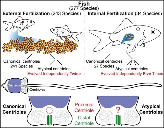
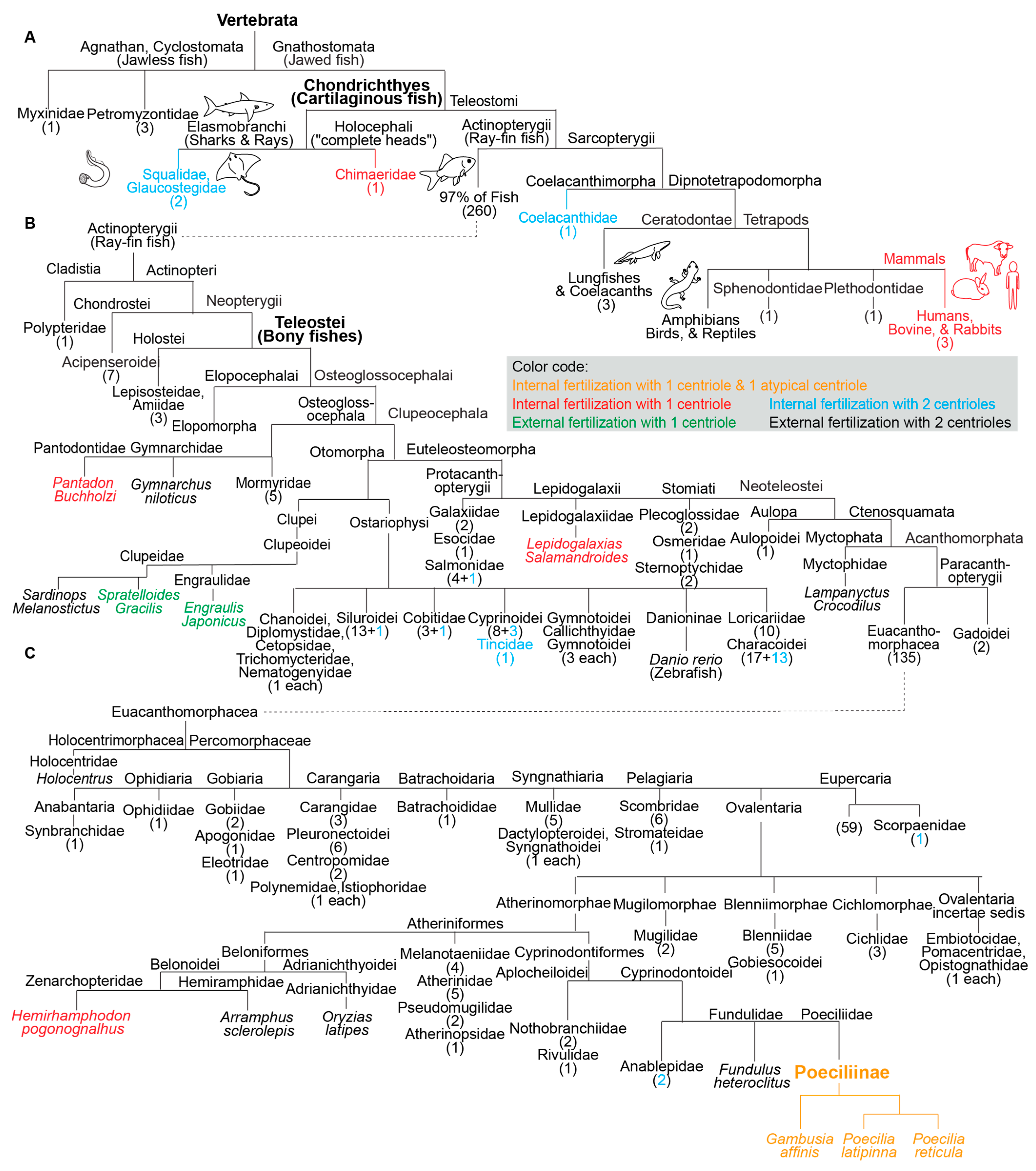
3.1. Most Fish Species Studied Ultrastructurally Are External Fertilizers

By searching the available literature that described electron microscopy studies of sperm, we surveyed reports on the sperm centrioles of 277 fish species (Table S1, Table 1). This is a small fraction of the at least 34,000 extant species. Most species (or their close relatives) were external fertilizers (87.7%, 243/277), and few were internal fertilizers (12.3%, 34/277), a ratio of 7.1 to 1. This ratio is lower than estimated in nature, as it has been reported that more than 95% of fish species (ratio of more than 19:1) are external fertilizers [52], indicating a bias toward ultrastructural studies of internally fertilizing fish.
Of the 277 species studied, 268 (96.8%, 268/277) were reported to have two canonical centrioles, while nine species (3.2%, 9/277) were reported to have only one canonical centriole. This is a ratio of 30 to 1, indicating that most fish studied were characterized by ancestral centriolar composition.

3.2. Two Studied Externally Fertilizing Fish Species have Atypical Centriolar Composition

Of the 243 studied species that reproduce by external fertilization, two were reported to have one canonical centriole, representing 0.8% of the external fertilizers (Table S1). These species reports are as follows:
Gwo et al. reported on p.286 that in Spratelloides gracilis, “No proximal centriole was identified” [53], while it was reported that this species reproduces by external fertilization [54,55].
Fu et al. reported on p.60 that “the proximal centriole of Engraulis japonicus (Engraulididae) is indistinct” [56], and it was also reported to be an external fertilizer [51,57].
Many other externally fertilizing fish have two canonical centrioles. This includes fish from the families Scorpaenidae and Danioninae (which includes the important model species Zebrafish) [58,59]. For example, we studied papers on five species of Salmonidae (Table 1), all of which have two canonical centrioles [60,61,62,63,64].

3.3. A High Rate of Internally Fertilizing Fish Species Studied Have Atypical Centriolar Composition

Of the 34 species that reproduce by internal fertilization, seven (20.6%) were reported to have a single sperm centriole (Table S1, Table 1). We used the Z test for two population proportions and found that internally fertilizing fish had a statistically significant higher proportion of sperm with a single canonical centriole than externally fertilizing fish (p < 0.00001). The relative rate reduction was 19.8% for the reported single canonical centriole. The odds ratio of 32.4 for the group reported with one canonical centriole over the group reported with two canonical centrioles indicates a high difference. A similar conclusion can be drawn if the data are calculated based on the number of genera studied instead of species name (Table 1). This finding suggests that the evolutionary sexual cascade of events resulted first in internal fertilization and later, in some species, in atypical centriolar composition.

3.4. Species of the Internally Fertilizing Fish Subfamily Poeciliinae Have Atypical Centrioles

Of all the fish analyzed, one group appears to have atypical centrioles: the subfamily Poeciliinae (belonging to Teleostei, or bony fishes with protrusible jaws) (Figure 2). Species of the subfamily Poeciliinae and the family Poeciliidae are internal fertilizers [65].
Jamieson reported on p.470 for Poecilia reticulata that “proximal centriole triplets become occluded and is reduced to a remnant by maturity” [49,50,66,67]. This species is an internal fertilizer [68].
Grier et al. reported on p.86 for Poecilia latipinna that “a rather indistinct electron dense structure (Figure 15) was present in the sperm neck” [69]. This species is an internal fertilizer [70].
Emel’yanova and Pavlov reported on p.97 that for Gambusia affinis, the “proximal centriole (pc) is electron dense and no longer contains distinct tubules” [71]. This species is an internal fertilizer [72].
The subfamily Poeciliinae includes many genera in addition to Poecilia and Gambusia that are thought to belong to distinct tribes [73]. Therefore, it is possible that the subfamily Poeciliinae is a monophyletic group that ancestrally had an atypical centriole.

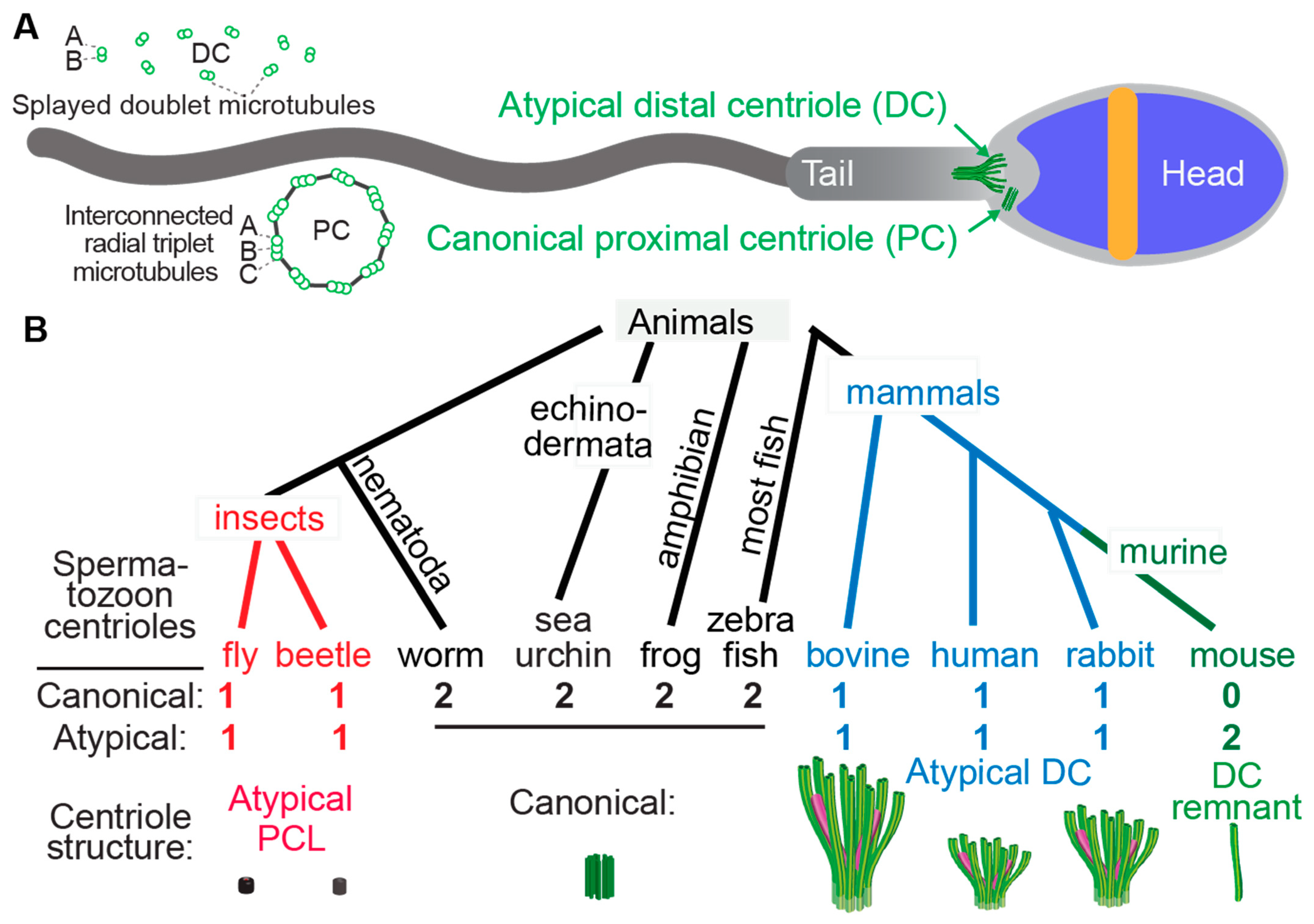
3.5. Species with a Single Canonical Centriole Evolved Independently Multiple Times

In addition to the three studied species of the subfamily Poeciliinae, we identified four additional species with a single canonical spermatozoan centriole that are also internal fertilizers; each of them occupies a distinct phylogenetic clade (Figure 1). The reported internal fertilizers included one Chondrichthyes species (cartilaginous fishes) and three species from distantly related Teleostei (bony fishes with protrusible jaws) groups.
Hydrolagus colliei (Chondrichthyes): It was reported on p.193 that “the proximal centriole has not been seen in mature Hydrolagus sperm” [74], and this species was reported to be an internal fertilizer [75].
Lepidogalaxias salamandroides: It was reported on p.42 by Leung that “No typical 9 × 3 microtubule centrioles have been found in the spermatozoon, but a basal body was located at the anterior of the axoneme” [76], and this species was reported to be an internal fertilizer [77]. Jamieson reported on p.156 for Lepidogalaxias salamandroides that “no typical triplet centrioles have been observed. A basal body…located at the anterior end of the flagellum consists of 9 doublets” [49].
Hemirhamphodon pogonognathus: It was reported on p.251 that “A proximal centriole has not been identified” [78] and this species was reported to be an internal fertilizer [79].
Pantodon buchholzi: It was reported on p.118 that “the centriolar complex consists of a single centriole (basal body), although an additional centriole, presumably the proximal centriole, is sometimes observed parallel to it” [49]. This species was reported to be an internal fertilizer [49,75].
Additionally, the two reported external fertilizers were from two unrelated Teleostei groups. Spratelloides gracilis (silver-stripe round herring) and Sardinops melanostictus both belong to the Clupeoidei suborder, though they are from distinct families (Engraulidae or Anchovies and Clupeidae). The Clupeidae family includes the species Sardinops melanostictus, whose spermatozoa have two canonical centrioles [49]. Therefore, the appearance of atypical centriolar composition in the Clupeoidei suborder occurred twice independently.
The nine species with a single canonical centriole belonged to clades that include many more species with two canonical centrioles, suggesting that ancestrally, they had spermatozoa with two canonical centrioles (Figure 2). Therefore, it appears that a single canonical centriole is a recent innovation in the evolution of these fish species, and that a change from ancestral centriolar composition to atypical centriolar composition took place independently multiple times, indicating convergent evolution due to positive selection.

3.6. The Atypical Centriole Forms during Spermiogenesis

In many animal species, the canonical structures of the proximal and distal centrioles are maintained during spermiogenesis (the differentiation of a round spermatid to a spermatozoon in the testes) [12,80]. Similarly, the canonical structures of the proximal and distal centrioles are maintained during spermatogenesis in fish with ancestral centriolar composition [81]. In mammals and insects, the atypical centriole forms during spermiogenesis via distinct mechanisms [21,23,24,82]. In insects, an atypical proximal centriole forms during spermiogenesis as a new centriole. In mammals, the canonical distal centriole found in the early spermatid is remodeled during spermiogenesis to become atypical. This indicates that insects and mammals use very different mechanisms to create the atypical centrioles during spermiogenesis.
Spermiogenesis in the fish Gambusia affinis, which has atypical centrioles, was studied [69]. In this species, the canonical proximal and distal centrioles are present in the early spermatid differentiation stage, but the proximal centriole becomes occluded in the later spermatid stage, and its microtubules disappear by the end of spermiogenesis. This situation is like that of insects, in that their atypical sperm centriole is the PC, not the DC, as is the case in mammals. However, Gambusia affinis is like mammals in that its atypical sperm centriole is remodeled from a spermatid centriole and not formed initially as an atypical centriole in the spermatid, as it is in insects. It therefore appears that atypical centriolar composition is achieved by distinct subcellular mechanisms in mammals, insects, and fish, which is consistent with the idea that they all evolved independently.

4. Discussion

By reviewing the literature on the sperm centriolar structure of 277 fish species, we made two observations:
  • the presence of only one recognizable centriole is continually (20.6% of internal fertilizers studied) and specifically (p < 0.00001 when comparing internal to external fertilizers) associated with internal fertilization; and
  • the presence of only one recognizable centriole evolved independently at least six times during fish evolution. This is in addition to the two independent incidents of only one canonical centriole evolving in insects and mammals. These data are consistent with Parker’s evolutionary sexual cascade theory.
In insects and mammals that were extensively investigated, it was found that the sperm have, in addition to the one recognizable canonical centriole, a second, atypical centriole, which was recognized only recently due to the use of centriole-specific protein markers [22,24,27,30,83]. Therefore, we speculate that some fish with one recognizable centriole will also have a second centriole that is not readily recognizable. This speculation will need to be investigated further.
We found that two species of external fertilizers only had one recognizable centriole. Possible explanations for this observation are: (i) they may have evolved from a species that was an internal fertilizer in the past and became external fertilizers as a secondary trait; and (ii) the sperm of these species or their ancestors may have been released to a complex external environment that has some similarity to the complex conditions of the female reproductive tract.
One possible explanation for the evolution of atypical sperm centriolar composition is the significantly higher rates of multiple paternity in internally fertilizing fish species [52]. Additionally, most fish that exhibit complex patterns of parental behavior are freshwater forms [84]. Interestingly, we found that six of the seven internal fertilizers with one canonical centriole (Poecilia reticulata, Poecilia latipinna, Gambusia affinis, Lepidogalaxias Salamandroides, Hemirhamphodon pogonognathus, and Pantodon buchholzi) live in freshwater. It is tempting to speculate that the atypical centriole trait is another strategy used, along with complex patterns of parental behavior, to gain an advantage during sperm competition.
Of the above species, Poecilia species are used as a common reproductive model. Guppies and mollies are used to study sexual conflict [85], sperm competition [86], the impact of predation on sperm [87], and many other topics [88]. It would be important to study the precise structure of the atypical centriole in these species using more advanced electron microscopy and to study how the structure evolved in this group. It would also be useful to compare the protein composition of canonical and atypical sperm centrioles in fish and to extend studies of sperm centrioles to other fish groups. For example, the spermatozoa of triploid gynogenetic crucian carp (Carassius auratus) have abnormal expression of centriole-related genes [89], while zebrafish is a widely used fish model with two canonical centrioles that are essential for normal embryo development [90,91].
A key question is why does the sperm centriole become atypical in internal fertilizers? A clue may come from the centriole’s strategic location in the sperm neck, which connects the sperm head and tail. Reproductive success depends on efficient sperm movement driven by axonemal, dynein-mediated microtubule sliding. Some models predict sliding to occur at the base of the tail (i.e., the atypical centriole) [92]. Canonical centrioles are rigid structures that restrict microtubule sliding. We found that, in bovine sperm, the atypical distal centriole, the canonical proximal centriole, and their surrounding atypical pericentriolar matrix form a dynamic basal complex that facilitates a cascade of internal sliding deformations that couple tail beating with asymmetric head kinking [29]. During asymmetric tail beating, the atypical centriole’s right side and the area surrounding it slide ~300 nm rostrally relative to the left side. The resulting deformation throughout the dynamic basal complex is transmitted to the head–tail junction. Thus, when the tail beats to the left, the head tilts to the left, generating a head kinking motion. These findings suggest that the dynamic basal complex evolved as a dynamic linker that couples the sperm head and tail into a single, coordinated system.
In fish and insects, whose atypical centriole is the PC, it is less clear what can be gained from it becoming atypical. One possibility is that the atypical PC occupies less space in a very crowded, narrow location such as the cytoplasmic invagination just below the nucleus of Tribolium castaneum [24]. Another possibility is that the atypical PC provides a different way to couple sperm tail movement to the head, potentially by working with other neck structures that extend to the tail. For example, the Drosophilla melanogaster PCL, which is found above the mitochondria derivative, may interact with it to transmit movement in the sperm tail to the head [23].

5. Conclusions

Atypical centriolar composition has evolved independently at least eight times during animal evolution, suggesting a strong positive selection. In six of the eight instances, centriolar composition changed with internal fertilization clades, suggesting that atypical centrioles tend to evolve in association with internal fertilization as part of the evolutionary sexual cascade. Further research is needed to characterize the fish sperm atypical centriolar structure, composition, and fate post-fertilization in the zygote.

Supplementary Materials

The following supporting information can be downloaded at: https://www.mdpi.com/article/10.3390/cells11050758/s1. Table S1: List of studied fish species included in the current analysis.

Author Contributions

Conceptualization, K.T. and T.A.-R.; Investigation, K.T., N.S. and H.O.S.; Writing—original draft preparation, K.T.; Writing—review and editing, T.A.-R.; Supervision, T.A.-R.; Project administration, T.A.-R.; Funding acquisition, T.A.-R. All authors have read and agreed to the published version of the manuscript.

Funding

This research was funded by NIH, grant number R03 HD098314 and by Agriculture and Food Research Initiative Competitive Grant no. OHOW-2020-02790 from the USDA National Institute of Food and Agriculture.

Institutional Review Board Statement

Not applicable.

Informed Consent Statement

Not applicable.

Data Availability Statement

Not applicable.

Acknowledgments

We would like to thank Professor Eduardo Roland for the introduction suggestions and Luke Achinger and Nahshon A. Puente for editing and help with the figure preparations.

Conflicts of Interest

The authors declare no conflict of interest. The funders had no role in the study’s design, in the collection, analyses, or interpretation of data, in the writing of the manuscript, or in the decision to publish the results.

References

  1. Parker, G.A. Sperm Competition and Its Evolutionary Consequences in the Insects. Biol. Rev. 1970, 45, 525–567. [Google Scholar] [CrossRef]
  2. Parker, G.A. The sexual cascade and the rise of pre-ejaculatory (Darwinian) sexual selection, sex roles, and sexual conflict. Cold Spring Harb. Perspect. Biol. 2014, 6, a017509. [Google Scholar] [CrossRef] [PubMed] [Green Version]
  3. Simmons, L.W.; Garcia-Gonzalez, F. Can Sexual Selection Drive the Evolution of Sperm Cell Structure? Cells 2021, 10, 1227. [Google Scholar] [CrossRef] [PubMed]
  4. Roldan, E.R.S. Sperm competition and the evolution of sperm form and function in mammals. Reprod. Domest. Anim. 2019, 54 (Suppl. S4), 14–21. [Google Scholar] [CrossRef] [PubMed]
  5. Kahrl, A.F.; Snook, R.R.; Fitzpatrick, J.L. Fertilization mode drives sperm length evolution across the animal tree of life. Nat. Ecol. Evol. 2021, 5, 1153–1164. [Google Scholar] [CrossRef] [PubMed]
  6. Lüpold, S.; de Boer, R.A.; Evans, J.P.; Tomkins, J.L.; Fitzpatrick, J.L. How sperm competition shapes the evolution of testes and sperm: A meta-analysis. Philos. Trans. R. Soc. B 2020, 375, 20200064. [Google Scholar] [CrossRef] [PubMed]
  7. Baccetti, B. The evolution of the sperm tail. Symp. Soc. Exp. Biol. 1982, 35, 521–532. [Google Scholar]
  8. Luke, L.; Vicens, A.; Tourmente, M.; Roldan, E.R. Evolution of protamine genes and changes in sperm head phenotype in rodents. Biol. Reprod. 2014, 90, 67. [Google Scholar] [CrossRef]
  9. Tourmente, M.; Gomendio, M.; Roldan, E.R. Sperm competition and the evolution of sperm design in mammals. BMC Evol. Biol. 2011, 11, 12. [Google Scholar] [CrossRef] [Green Version]
  10. Scharer, L.; Littlewood, D.T.; Waeschenbach, A.; Yoshida, W.; Vizoso, D.B. Mating behavior and the evolution of sperm design. Proc. Natl. Acad. Sci. USA 2011, 108, 1490–1495. [Google Scholar] [CrossRef] [Green Version]
  11. Avidor-Reiss, T.; Ha, A.; Basiri, M.L. Transition Zone Migration: A Mechanism for Cytoplasmic Ciliogenesis and Postaxonemal Centriole Elongation. Cold Spring Harb. Perspect. Biol. 2017, 9, a028142. [Google Scholar] [CrossRef] [PubMed]
  12. Avidor-Reiss, T. Rapid Evolution of Sperm Produces Diverse Centriole Structures that Reveal the Most Rudimentary Structure Needed for Function. Cells 2018, 7, 67. [Google Scholar] [CrossRef] [PubMed] [Green Version]
  13. Allen, R.D. The morphogenesis of basal bodies and accessory structures of the cortex of the ciliated protozoan Tetrahymena pyriformis. J. Cell Biol. 1969, 40, 716–733. [Google Scholar] [CrossRef] [PubMed]
  14. Vorobjev, I.A.; Chentsov Yu, S. Centrioles in the cell cycle. I. Epithelial cells. J. Cell Biol. 1982, 93, 938–949. [Google Scholar] [CrossRef] [PubMed]
  15. Fawcett, D.W. A comparative view of sperm ultrastructure. Biol. Reprod. 1970, 2 (Suppl. S2), 90–127. [Google Scholar] [CrossRef] [Green Version]
  16. Callaini, G.; Whitfield, W.G.; Riparbelli, M.G. Centriole and centrosome dynamics during the embryonic cell cycles that follow the formation of the cellular blastoderm in Drosophila. Exp. Cell Res. 1997, 234, 183–190. [Google Scholar] [CrossRef]
  17. Uzbekov, R.E.; Avidor-Reiss, T. Principal Postulates of Centrosomal Biology. Version 2020. Cells 2020, 9, 2156. [Google Scholar] [CrossRef]
  18. Woolley, D.M.; Fawcett, D.W. The degeneration and disappearance of the centrioles during the development of the rat spermatozoon. Anat. Rec. 1973, 177, 289–301. [Google Scholar] [CrossRef]
  19. Chakraborty, J. Neck region of gerbil spermatozoa. Gamete Res. 1979, 2, 25–34. [Google Scholar] [CrossRef]
  20. Manandhar, G.; Sutovsky, P.; Joshi, H.C.; Stearns, T.; Schatten, G. Centrosome reduction during mouse spermiogenesis. Dev. Biol. 1998, 203, 424–434. [Google Scholar] [CrossRef] [Green Version]
  21. Avidor-Reiss, T.; Fishman, E.L. It takes two (centrioles) to tango. Reproduction 2019, 157, 33–51. [Google Scholar] [CrossRef] [PubMed] [Green Version]
  22. Fishman, E.L.; Jo, K.; Nguyen, Q.P.H.; Kong, D.; Royfman, R.; Cekic, A.R.; Khanal, S.; Miller, A.L.; Simerly, C.; Schatten, G.; et al. A novel atypical sperm centriole is functional during human fertilization. Nat. Commun. 2018, 9, 2210. [Google Scholar] [CrossRef] [PubMed]
  23. Khire, A.; Jo, K.H.; Kong, D.; Akhshi, T.; Blachon, S.; Cekic, A.R.; Hynek, S.; Ha, A.; Loncarek, J.; Mennella, V. Centriole remodeling during spermiogenesis in Drosophila. Curr. Biol. 2016, 26, 3183–3189. [Google Scholar] [CrossRef] [PubMed] [Green Version]
  24. Fishman, E.L.; Jo, K.; Ha, A.; Royfman, R.; Zinn, A.; Krishnamurthy, M.; Avidor-Reiss, T. Atypical centrioles are present in Tribolium sperm. Open Biol. 2017, 7, 160334. [Google Scholar] [CrossRef] [Green Version]
  25. Blachon, S.; Khire, A.; Avidor-Reiss, T. The origin of the second centriole in the zygote of Drosophila melanogaster. Genetics 2014, 197, 199–205. [Google Scholar] [CrossRef] [Green Version]
  26. Dallai, R.; Mercati, D.; Lino-Neto, J.; Dias, G.; Lupetti, P. Evidence of a procentriole during spermiogenesis in the coccinellid insect Adalia decempunctata (L): An ultrastructural study. Arthropod. Struct. Dev. 2017, 46, 815–823. [Google Scholar] [CrossRef]
  27. Gottardo, M.; Callaini, G.; Riparbelli, M.G. Structural characterization of procentrioles in Drosophila spermatids. Cytoskeleton 2015, 72, 576–584. [Google Scholar] [CrossRef]
  28. Blachon, S.; Cai, X.; Roberts, K.A.; Yang, K.; Polyanovsky, A.; Church, A.; Avidor-Reiss, T. A proximal centriole-like structure is present in Drosophila spermatids and can serve as a model to study centriole duplication. Genetics 2009, 182, 133–144. [Google Scholar] [CrossRef] [Green Version]
  29. Khanal, S.; Leung, M.R.; Royfman, A.; Fishman, E.L.; Saltzman, B.; Bloomfield-Gadelha, H.; Zeev-Ben-Mordehai, T.; Avidor-Reiss, T. A dynamic basal complex modulates mammalian sperm movement. Nat. Commun. 2021, 12, 3808. [Google Scholar] [CrossRef]
  30. Leung, M.R.; Roelofs, M.C.; Ravi, R.T.; Maitan, P.; Henning, H.; Zhang, M.; Bromfield, E.G.; Howes, S.C.; Gadella, B.M.; Bloomfield-Gadelha, H.; et al. The multi-scale architecture of mammalian sperm flagella and implications for ciliary motility. EMBO J. 2021, 40, 107410. [Google Scholar] [CrossRef]
  31. Manandhar, G.; Simerly, C.; Schatten, G. Centrosome reduction during mammalian spermiogenesis. Curr. Top. Dev. Biol. 2000, 49, 343–363. [Google Scholar] [CrossRef] [PubMed]
  32. Cavazza, T.; Takeda, Y.; Politi, A.Z.; Aushev, M.; Aldag, P.; Baker, C.; Choudhary, M.; Bucevicius, J.; Lukinavicius, G.; Elder, K.; et al. Parental genome unification is highly error-prone in mammalian embryos. Cell 2021, 184, 2860–2877.e22. [Google Scholar] [CrossRef] [PubMed]
  33. Schneider, I.; de Ruijter-Villani, M.; Hossain, M.J.; Stout, T.A.E.; Ellenberg, J. Dual spindles assemble in bovine zygotes despite the presence of paternal centrosomes. J. Cell Biol. 2021, 220, e202010106. [Google Scholar] [CrossRef] [PubMed]
  34. Kai, Y.; Kawano, H.; Yamashita, N. First mitotic spindle formation is led by sperm centrosome-dependent MTOCs in humans. Reproduction 2021, 161, 19–22. [Google Scholar] [CrossRef]
  35. Amargant, F.; Pujol, A.; Ferrer-Vaquer, A.; Durban, M.; Martinez, M.; Vassena, R.; Vernos, I. The human sperm basal body is a complex centrosome important for embryo preimplantation development. Mol. Hum. Reprod. 2021, 27, gaab062. [Google Scholar] [CrossRef]
  36. Avidor-Reiss, T.; Carr, A.; Fishman, E.L. The sperm centrioles. Mol. Cell Endocrinol. 2020, 518, 110987. [Google Scholar] [CrossRef]
  37. Bobinnec, Y.; Khodjakov, A.; Mir, L.M.; Rieder, C.L.; Edde, B.; Bornens, M. Centriole disassembly in vivo and its effect on centrosome structure and function in vertebrate cells. J. Cell Biol. 1998, 143, 1575–1589. [Google Scholar] [CrossRef] [Green Version]
  38. Marshall, W.F. Centrioles take center stage. Curr. Biol. 2001, 11, 487–496. [Google Scholar] [CrossRef] [Green Version]
  39. Rodrigues-Martins, A.; Riparbelli, M.; Callaini, G.; Glover, D.M.; Bettencourt-Dias, M. From centriole biogenesis to cellular function: Centrioles are essential for cell division at critical developmental stages. Cell Cycle 2008, 7, 11–16. [Google Scholar] [CrossRef] [Green Version]
  40. Tung, C.K.; Suarez, S.S. Co-Adaptation of Physical Attributes of the Mammalian Female Reproductive Tract and Sperm to Facilitate Fertilization. Cells 2021, 10, 1297. [Google Scholar] [CrossRef]
  41. Higginson, D.M.; Miller, K.B.; Segraves, K.A.; Pitnick, S. Female reproductive tract form drives the evolution of complex sperm morphology. Proc. Natl. Acad. Sci. USA 2012, 109, 4538–4543. [Google Scholar] [CrossRef] [PubMed] [Green Version]
  42. Druart, X. Sperm interaction with the female reproductive tract. Reprod. Domest. Anim. 2012, 47 (Suppl. S4), 348–352. [Google Scholar] [CrossRef] [PubMed]
  43. Pitnick, S.; Wolfner, M.F.; Dorus, S. Post-ejaculatory modifications to sperm (PEMS). Biol Rev. Camb. Philos. Soc. 2020, 95, 365–392. [Google Scholar] [CrossRef] [PubMed]
  44. Devigili, A.; Fitzpatrick, J.L.; Gasparini, C.; Ramnarine, I.W.; Pilastro, A.; Evans, J.P. Possible glimpses into early speciation: The effect of ovarian fluid on sperm velocity accords with post-copulatory isolation between two guppy populations. J. Evol. Biol. 2018, 31, 66–74. [Google Scholar] [CrossRef] [PubMed] [Green Version]
  45. Jamieson, B.G. Avian spermatozoa: Structure and phylogeny. Reprod. Biol. Phylogeny Birds 2007, 6, 349–511. [Google Scholar]
  46. Hamilton, D.W.; Fawcett, D.W. Unusual features of the neck and middle-piece of snake spermatozoa. J. Ultrastruct. Res. 1968, 23, 81–97. [Google Scholar] [CrossRef]
  47. Hess, R.A.; Thurston, R.J.; Gist, D.H. Ultrastructure of the turtle spermatozoon. Anat. Rec. 1991, 229, 473–481. [Google Scholar] [CrossRef]
  48. Jamieson, B.G. Fish Evolution and Systematics: Evidence from Spermatozoa: With a Survey of Lophophorate, Echinoderm and Protochordate Sperm and an Account of Gamete Cryopreservation; Cambridge University Press: Cambridge, UK, 1991. [Google Scholar]
  49. Jamieson, B.G. Reproductive Biology and Phylogeny of Fishes (Agnathans and Bony Fishes): Phylogeny, Reproductive System, Viviparity, Spermatozoa; CRC Press: Florida, FLA, USA, 2009. [Google Scholar]
  50. Froese, R. FishBase. World Wide Web Electronic Publication. 2022. Available online: http://www.fishbase.org (accessed on 1 January 2022).
  51. Fitzpatrick, J.L. Sperm competition and fertilization mode in fishes. Philos. Trans. R. Soc. London Ser. B Biol. Sci. 2020, 375, 20200074. [Google Scholar] [CrossRef]
  52. Gwo, J.C.; Lin, C.Y.; Yang, W.L.; Chou, Y.C. Ultrastructure of the sperm of blue sprat, Spratelloides gracilis; Teleostei, Clupeiformes, Clupeidae. Tissue Cell 2006, 38, 285–291. [Google Scholar] [CrossRef]
  53. Mogi, K.; Misawa, K.; Utsunomiya, K.; Kawada, Y.; Yamazaki, T.; Takeuchi, S.; Toyoizumi, R. Optic chiasm in the species of order Clupeiformes, family Clupeidae: Optic chiasm of Spratelloides gracilis shows an opposite laterality to that of Etrumeus teres. Laterality 2009, 14, 495–514. [Google Scholar] [CrossRef] [Green Version]
  54. Breder, C.M.; Rosen, D.E. Modes of Reproduction in Fishes, 1st ed.; Natural History Press: New York, NY, USA, 1966. [Google Scholar]
  55. Fu, S.Y.; Jiang, J.H.; Yang, W.X.; Zhu, J.Q. A histological study of testis development and ultrastructural features of spermatogenesis in cultured Acrossocheilus fasciatus. Tissue Cell 2016, 48, 49–62. [Google Scholar] [CrossRef] [PubMed]
  56. LEHODEY, P.; CHAI, F.; HAMPTON, J. FAO species catalogue, Vol. 7. Clupeoid fishes of the world (Suborder Clupeoidei). An annotated and illustrated catalogue of the herrings, sardines, pilchards, sprats, shads, anchovies and wolf-herrings. Part 2-Engraulididae FAO species catalogue, Vol. 7. Clupeoid fishes of the world (Suborder Clupeoidei). An annotated and illustrated catalogue of the herrings, sardines, pilchards, sprats, shads, anchovies and wolf-herrings. Part 2-Engraulididae, 1988. Fish. Oceanogr. 2003, 12, 483–494. [Google Scholar]
  57. Pavlov, D.; Emel’yanova, N. Comparative analysis of spermatozoa morphology in three fish species from the suborder Scorpaenoidei. J. Ichthyol. 2018, 58, 226–238. [Google Scholar] [CrossRef]
  58. Rupik, W.; Huszno, J.; Klag, J. Cellular organisation of the mature testes and stages of spermiogenesis in Danio rerio (Cyprinidae; Teleostei)—Structural and ultrastructural studies. Micron 2011, 42, 833–839. [Google Scholar] [CrossRef] [PubMed]
  59. Gwo, J.-C.; Ohta, H.; Okuzawa, K.; Wu, H.-C. Cryopreservation of sperm from the endangered Formosan landlocked salmon (Oncorhynchus masou formosanus). Theriogenology 1999, 51, 569–582. [Google Scholar] [CrossRef]
  60. Gwo, J.-C.; Lin, X.; Gwo, H.; Wu, H.; Lin, P. The ultrastructure of Formosan landlocked salmon, Oncorbynchus masout formosanus, spermatozoon (Teleostei, Salmoniformes, Salmonidae). J. Submicrosc. Cytol. Pathol. 1996, 28, 33–40. [Google Scholar]
  61. Markova, M.D.; Zhivkova, R.S. Possible cytoskeletal structures of rainbow trout sperm revealed by electron microscopic observation after detergent extraction. Anim. Reprod. Sci. 2003, 79, 127–132. [Google Scholar] [CrossRef]
  62. Guo, W.; Shao, J.; Li, P.; Wu, J.; Wei, Q. Morphology and ultrastructure of Brachymystax lenok tsinlingensis spermatozoa by scanning and transmission electron microscopy. Tissue Cell 2016, 48, 321–327. [Google Scholar] [CrossRef]
  63. Figueroa, E.; Valdebenito, I.; Zepeda, A.B.; Figueroa, C.A.; Dumorné, K.; Castillo, R.L.; Farias, J.G. Effects of cryopreservation on mitochondria of fish spermatozoa. Rev. Aquac. 2017, 9, 76–87. [Google Scholar] [CrossRef]
  64. Thibault, R.E.; Schultz, R.J. Reproductive Adaptations among Viviparous Fishes (Cyprinodontiformes Poeciliidae). Evolution 1978, 32, 320–333. [Google Scholar] [CrossRef]
  65. Billard, R.; Escaffre, A.-M.; Tramasaygues, N. La spermatogenèse de Poecilia reticulata. IV.—La spermiogenèse. Etude ul-trastructurale. Ann. Biol. Anim. Biochim. Biophys. 1970, 10, 493–510. [Google Scholar] [CrossRef]
  66. Mattei, X.; Boisson, C. Le complexe centriolaire du spermatozoïde de Lebistes reticulatus. Comptes Rendus Hebdomadaires des Seances de l Academie des Sciences Serie D 1966, 262, 2620. [Google Scholar]
  67. Balon, E.K. Epigenesis of an epigeneticist: The development of some alternative concepts on the early ontogeny and evolution of fishes. Guelph Ichthyol. Rev. 1990, 1, 1–48. [Google Scholar]
  68. Grier, H.J. Ultrastructure of the testis in the teleost Poecilia latipinna. Spermiogenesis with reference to the intercentriolar lamellated body. J. Ultrastruct. Res. 1973, 45, 82–92. [Google Scholar] [CrossRef]
  69. Wischnath, L. Atlas of Livebearers of the World; TFH publications: Neptune City, NJ, USA, 1993. [Google Scholar]
  70. Emel’yanova, N.G.; Pavlov, D.A. Gamete ultrastructure in some species of the family Mullidae from the South China Sea. J. Ichthyol. 2012, 52, 639–645. [Google Scholar] [CrossRef]
  71. Seale, A. The Mosquito Fish, Gambusia affinis (Baird and Girard), in the Philippine Islands. Philipp. J. Sci. 1917, 12, 177–189. [Google Scholar]
  72. Hrbek, T.; Seckinger, J.; Meyer, A. A phylogenetic and biogeographic perspective on the evolution of poeciliid fishes. Mol. Phylogenet. Evol. 2007, 43, 986–998. [Google Scholar] [CrossRef] [Green Version]
  73. Stanley, H.P. The fine structure of spermatozoa of Hydrolagus colliei (Chondrichthyes, Holocephali). J. Ultrastruct. Res. 1983, 83, 184–194. [Google Scholar] [CrossRef]
  74. Armstrong, R.H. Alaska’s Fish: A Guide to Selected Species; Alaska Northwest Books: Portland, OR, USA, 1996. [Google Scholar]
  75. Leung, L.K. Ultrastructure of the spermatozoon of Lepidogalaxias salamandroides and its phylogenetic significance. Gamete Res. 1988, 19, 41–49. [Google Scholar] [CrossRef]
  76. Allen, G.R.; Midgley, S.H.; Allen, M. Freshwater Fishes of Australia; Western Australian Museum: Perth, Australia, 1989. [Google Scholar]
  77. Jamieson, B.G. Complex spermatozoon of the live-bearing half-beak, Hemirhamphodon pogonognathus (Bleeker): Ultrastructural description (Euteleostei, Atherinomorpha, Beloniformes). Gamete Res. 1989, 24, 247–259. [Google Scholar] [CrossRef]
  78. Baensch, H.; Riehl, R. Aquarien Atlas; Mergus: Melle, Germany, 1987. [Google Scholar]
  79. Longo, F.J.; Anderson, E. The fine structure of pronuclear development and fusion in the sea urchin, Arbacia punctulata. J. Cell Biol. 1968, 39, 339–368. [Google Scholar] [CrossRef] [PubMed] [Green Version]
  80. Grier, H.J. Sperm development in the teleost Oryzias latipes. Cell Tissue Res. 1976, 168, 419–431. [Google Scholar] [CrossRef] [PubMed]
  81. Avidor-Reiss, T.; Turner, K. The Evolution of Centriole Structure: Heterochrony, Neoteny, and Hypermorphosis. Results Probl. Cell Differ. 2019, 67, 3–15. [Google Scholar] [CrossRef] [PubMed]
  82. Dallai, R.; Mercati, D.; Bu, Y.; Yin, Y.W.; Callaini, G.; Riparbelli, M.G. The spermatogenesis and sperm structure of Acerentomon microrhinus (Protura, Hexapoda) with considerations on the phylogenetic position of the taxon. Zoomorphology 2010, 129, 61–80. [Google Scholar] [CrossRef]
  83. Baylis, J.R. The evolution of parental care in fishes, with reference to Darwin’s rule of male sexual selection. Environ. Biol. Fishes 1981, 6, 223–251. [Google Scholar] [CrossRef]
  84. Magurran, A.E. Sexual conflict and evolution in Trinidadian guppies. Genetica 2001, 112–113, 463–474. [Google Scholar] [CrossRef]
  85. Glavaschi, A.; Cattelan, S.; Grapputo, A.; Pilastro, A. Imminent risk of predation reduces the relative strength of postcopulatory sexual selection in the guppy. Philos. Trans. R. Soc. Ser. B Biol. Sci. 2020, 375, 20200076. [Google Scholar] [CrossRef]
  86. Devigili, A.; Evans, J.P.; Fitzpatrick, J.L. Predation shapes sperm performance surfaces in guppies. Proc. Biol. Sci. 2019, 286, 20190869. [Google Scholar] [CrossRef] [Green Version]
  87. Evans, J.P.; Brooks, R.C.; Zajitschek, S.R.; Griffith, S.C. Does genetic relatedness of mates influence competitive fertilization success in guppies? Evolution 2008, 62, 2929–2935. [Google Scholar] [CrossRef]
  88. He, W.; Sun, Y.; Qiang, J.; Luo, X.; Zhang, H.; Yang, C.; Luo, K.; Zhao, R.; Qin, Q.; Zhang, C.; et al. Structural Abnormalities of Spermatozoa in Triploid Gynogenetic Crucian Carp (Carassius auratus). Front. Genet. 2021, 12, 783014. [Google Scholar] [CrossRef]
  89. Yabe, T.; Ge, X.; Pelegri, F. The zebrafish maternal-effect gene cellular atoll encodes the centriolar component sas-6 and defects in its paternal function promote whole genome duplication. Dev. Biol. 2007, 312, 44–60. [Google Scholar] [CrossRef] [PubMed] [Green Version]
  90. Baars, D.L.; Takle, K.A.; Heier, J.; Pelegri, F. Ploidy Manipulation of Zebrafish Embryos with Heat Shock 2 Treatment. J. Vis. Exp. JoVE 2016, 118, e54492. [Google Scholar] [CrossRef] [PubMed] [Green Version]
  91. Vernon, G.G.; Woolley, D.M. Basal sliding and the mechanics of oscillation in a mammalian sperm flagellum. Biophys. J. 2004, 87, 3934–3944. [Google Scholar] [CrossRef] [PubMed] [Green Version]
  92. Woolley, D.M. Flagellar oscillation: A commentary on proposed mechanisms. Biol. Rev. Camb. Philos. Soc. 2010, 85, 453–470. [Google Scholar] [CrossRef] [PubMed]
Figure 1. Atypical centrioles have distinct structures. (A) A mammalian sperm showing the locations of the atypical and canonical centrioles in the sperm neck. To the left, a cross section of the PC and DC depicting microtubule organization. (B) The number and size of spermatozoan centrioles vary throughout evolution. Shown are four animal groups (color-coded) organized based on the number and type of their spermatozoan centrioles. Animals with two centrioles (black). Animals with just one canonical centriole also have an atypical centriole: the proximal centriole-like (PCL, red) structure in insects and the atypical distal centriole (DC, blue) in mammals. Animals with DC remanent (green). Color code in the Atypical DC: green color marks the presence of microtubules, and magenta marks rod proteins.
Cells 11 00758 g001
Figure 2. Fish species with a single canonical centriole evolved independently multiple times from an ancestral state with two canonical centrioles. (AC) A phylogenetic tree depicting fertilization mode (internal or external) and canonical centriole number (1 or 2) in the main branches of vertebrates and fish. (A) Depicts the main fish groups. (B) Depicts ray-fin fish (Actinopterygii). (C) Depicts Euacanthomorphacea. In some cases, we provide the scientific name and common name in parentheses. Vertebrata, Chondrichthyes (Cartilaginous fish), Teleostei (Bony fishes), and Poeciliinae are bolded and enlarged because they are specifically referenced in the text. The number of fish species in a category appear as a numeral in parentheses. The figure contains the names of all fish families analyzed in this survey.
Cells 11 00758 g002
Table 1. Number and percent of species and genera characterized by external or internal fertilization and reported to either have a single canonical centriole or two canonical centrioles.
FertilizationOne CentrioleTwo CentriolesTotal
External2 species (0.8%), 2 genera (1%)
Spratelloides gracilis (Silver-stripe round herring) Engraulis japonicus (Japanese anchovy)
241 species (99.2%)
199 genera (99%)
243 species (87.7%)
201 genera (86.6%)
Internal7 species (20.6%), 6 genera (19.4%)
Hydrolagus colliei (Spotted ratfish)
Lepidogalaxias Salamandroides (Salamanderfish)
Hemirhamphodon pogonognathus
Pantodon buchholzi (Freshwater butterflyfish)
3 Poeciliinae Species: Poecilia reticulata, Poecilia latipinna Guppy, and Gambusia affinis (Western mosquitofish)
27 species (79.4%)
25 genera (80.6%)
34 species (12.3%)
31 genera (13.4)
Total9 species (3.2%)
8 genera (3.4%)
268 species (96.8%)
224 genera (96.6%)
277 species
232 genera
p-valueSpecies: <0.00001
Genera: <0.00001
RatioSpecies: 3.5 (7/2)
Genera: 3 (6/2)
Species: 11.2 (27/241)
Genera: 12.6 (25/199)
RRRSpecies: 20.6%−0.8% = 19.8%
Genera: 19.4%−1% = 18.4%
Odds ratioSpecies: (7/27)/(2/241) = 0.259/0.008 = 32.4
Genera: (6/25)/(2/1.99) = 0.24/0.01 = 24
A Z-score calculator was used to determine the p-value for two population proportions. The species name is provided for those with a single canonical sperm centriole. RRR, Relative rate reduction.
Publisher’s Note: MDPI stays neutral with regard to jurisdictional claims in published maps and institutional affiliations.

