Review

Dying of Stress: Chemotherapy, Radiotherapy, and Small-Molecule Inhibitors in Immunogenic Cell Death and Immunogenic Modulation

Center for Immuno-Oncology, Center for Cancer Research, National Cancer Institute, National Institutes of Health, Bethesda, MD 20892, USA
*
Author to whom correspondence should be addressed.
Cells 2022, 11(23), 3826; https://doi.org/10.3390/cells11233826
Submission received: 2 October 2022 / Revised: 11 November 2022 / Accepted: 26 November 2022 / Published: 29 November 2022
(This article belongs to the Section Cellular Immunology)

Abstract

:
Innovative strategies to re-establish the immune-mediated destruction of malignant cells is paramount to the success of anti-cancer therapy. Accumulating evidence suggests that radiotherapy and select chemotherapeutic drugs and small molecule inhibitors induce immunogenic cell stress on tumors that results in improved immune recognition and targeting of the malignant cells. Through immunogenic cell death, which entails the release of antigens and danger signals, and immunogenic modulation, wherein the phenotype of stressed cells is altered to become more susceptible to immune attack, radiotherapies, chemotherapies, and small-molecule inhibitors exert immune-mediated anti-tumor responses. In this review, we discuss the mechanisms of immunogenic cell death and immunogenic modulation and their relevance in the anti-tumor activity of radiotherapies, chemotherapies, and small-molecule inhibitors. Our aim is to feature the immunological aspects of conventional and targeted cancer therapies and highlight how these therapies may be compatible with emerging immunotherapy approaches.

1. Introduction

Conventional anti-cancer therapies were originally developed to negatively impact rapidly dividing malignant cells while limiting damage to normal tissues. Typically, these therapeutic regimens induce DNA damage or disrupt DNA repair and cell division machineries, resulting in tumor cell death and debulking of cancer lesions [1]. In addition to these cytotoxic and cytostatic effects, some standard-of-care therapies such as radiotherapy, select chemotherapies, and small-molecule inhibitors (SMIs) also trigger cell stress that results in responses spanning from immunogenic cell death to immunogenic modulation. Immunogenic cell death (ICD), wherein dying cells release signals that prompt dendritic cells (DCs) to present tumor antigens to T cells, culminates in the activation and development of immunological memory [2,3]. If the therapy-induced cell stress does not result in cell death, the surviving tumor cells may be sensitized to immune cell targeting via immunogenic modulation, which includes upregulated antigen presentation, pro-apoptotic signaling molecules, and immune-cell-engaging molecules [4]. As immunotherapy emerges as a new pillar among anti-cancer therapies, it is important to recognize the immunogenic properties of conventional anti-cancer therapies and identify how these different treatment modalities can be synthesized together in the clinic. In this review, we discuss preclinical data on ICD and immunogenic modulation mediated by chemotherapy, radiotherapy, and SMIs and clinical data on the immunostimulatory effects of these conventional cancer treatments in the context of combination regimens with immunotherapy.

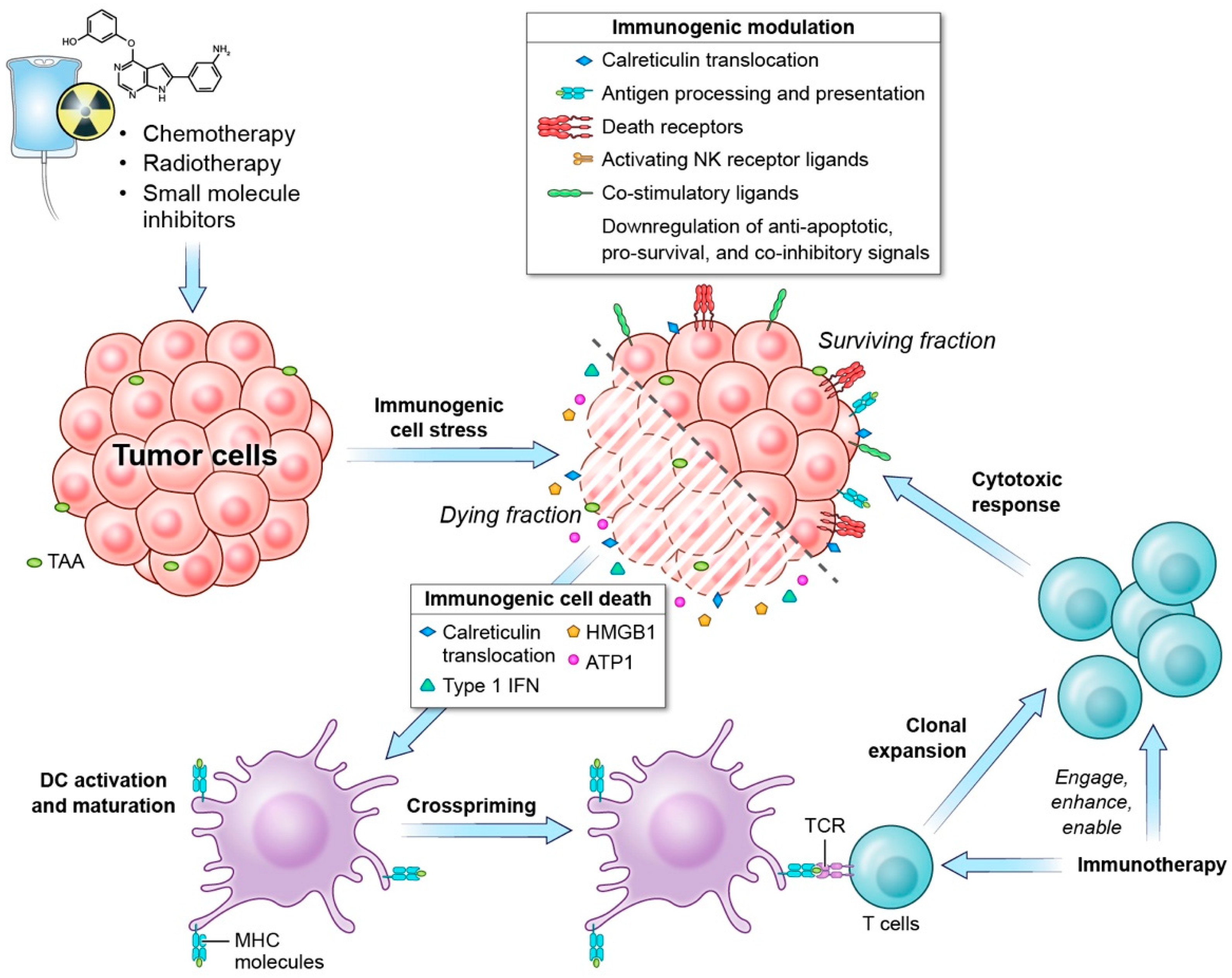
1.1. Immunogenic Cell Death

ICD is defined as “a form of regulated cell death that is sufficient to activate an adaptive immune response in immunocompetent hosts” [5]. ICD is a spatiotemporally defined process that involves the induction of the cell-stress response pathways and entails the release of antigens and danger signals from the dying cell that initiates the immune response (Figure 1). The activation of endoplasmic reticulum (ER) and/or reactive oxygen species (ROS) stress responses is essential for the initiation of ICD [6], and ICD inducers can be classified according to how they elicit ER stress. Most ICD inducers are Type I ICD inducers that instigate cell death by targeting organelles or molecules that are not directly associated with the ER but cause “collateral” ER stress effects [7,8]. Type II ICD inducers, on the other hand, directly target the ER to trigger ER stress and induce cell death [7,8]. Depending on dose and schedule, several anti-cancer therapies have been shown to cause ICD as a consequence of cell stress, and in this review, we concentrate on the immunogenic cell stress mediated by chemotherapy, radiotherapy, and SMIs. However, it should also be noted that other treatment regimens, such as oncolytic viruses [9], oncolytic peptides [10,11], the epithelial growth factor receptor (EGFR)-targeting monoclonal antibody cetuximab [12], and nanopulse stimulation [13], have also been shown to induce immunogenic cell stress.
The anti-cancer adaptive immune response associated with ICD relies on two major factors: (1) the antigenicity of the tumor cells and (2) the release of adjuvant signals from the dying tumor cells [2]. Generally, tumor antigens can be characterized as tumor neoantigens, which are products of non-synonymous mutations that are not covered by central tolerance, or tumor-associated antigens (TAAs), which are non-mutated antigens to which central tolerance is leaky or incomplete [14,15,16]. Epigenetic dysregulation of the cancer genome can additionally trigger the expression of human endogenous retroviruses (HERVs), resulting in tumor-specific or tumor-enriched HERV-derived antigens [17]. Some ICD inducers can cause mutations or introduce post-translational modifications that produce neoantigens or enhance the expression of neoantigens and TAAs [3,18,19,20].
The release of tumor antigens must be accompanied by pro-inflammatory signals in order to elicit an immune response that culminates in the effective immune-mediated killing of cancer cells [21]. Adjuvant signals generated during ICD come in the form of damage-associated molecular patterns (DAMPs) that play key roles in the recruitment and maturation of antigen presenting cells (APCs) [3,22]. Different ICD inducers generate different DAMPs due to variations in the cell-stress response activated by each ICD inducer [3,23]. Examples of DAMPs commonly generated during ICD include calreticulin (CRT), high mobility group box 1 (HMGB1), adenosine triphosphate (ATP), and type 1 interferons (IFN) [3,22,23]. CRT is an ER chaperone protein that translocates to the plasma membrane during ICD and provides an “eat me” signal to APCs [24,25,26,27]. The ligation of CRT to CD91 on APCs promotes phagocytosis of cellular debris and antigen presentation [24]. HMGB1, which is passively released by dying cells during ICD, binds to Toll-like receptor 4 (TLR4) on DCs, resulting in the efficient cross-presentation of antigens from the dying tumor cells [28]. ATP, which is actively released via exocytosis during ICD, provides a “find me” signal to DC precursors and macrophages [29]. Extracellular ATP interacts with purinergic receptor P2Y2 (P2RY2) and directs APC chemotaxis to the site of active ICD [30]. In addition, extracellular ATP can influence DC activation and antigen presentation via IL-1b, which is expressed following activation of the NLRP3 (NOD-, LRR-, and pyrin domain-containing protein (3) inflammasome downstream of ATP signaling through P2RX7 on DCs [31]. Type 1 IFN secretion can be driven by nucleic acid species from dying cells binding to different pattern recognition receptors (PRRs) [32]. Type I IFNs exert immunostimulatory effects, including the enhancement of the cytotoxic functions of cytotoxic T lymphocytes (CTLs) and natural killer (NK) cells [33,34], the promotion of cross-priming capacity of APCs [35], and the production of the T cell chemoattractant, CXCL10, by cancer cells [36]. Other DAMPs, such as annexin A1, heat shock protein (HSP) 70, and HSP90, have also been shown to be secreted or translocated by cells undergoing ICD [22,23]. Emission of these DAMPs according to a specific spatiotemporal order is essential for the recruitment and activation of innate and adaptive immune cells in the tumor lesion to mount an effective anti-cancer immune response [37].

1.2. Immunogenic Modulation

Cell stress induced by certain anti-cancer treatments does not always translate to cell death, but it can sensitize the surviving malignant cells to immune attack. Through the process of immunogenic modulation, therapies such as chemotherapy, radiotherapy, and SMIs alter the tumor phenotype to become more sensitive to CTL and NK cell killing [22,38]. Immunogenic modulation includes, but is not limited to, enhanced antigen presentation, increased pro-apoptotic signals, and changes in surface marker expression of the tumor cells (Figure 1) [4,22,38]. ICD and immunogenic modulation exist on a continuum and represent a spectrum of outcomes resulting from immunogenic cell stress [22].
Similar to ICD, immunogenic modulation requires antigenic determinants on malignant cells to trigger anti-tumor responses. In addition to promoting the expression of neoantigens and TAAs, immunogenic modulators upregulate different components of the antigen processing machinery and promote the expression of MHC class I [18,19,20,39,40]. This increase in antigen processing and presentation has been shown to be associated with enhanced sensitivity of the treated neoplastic cells to TAA-specific CTLs [20]. Since CTL-mediated killing may also be a form of ICD, the resulting anti-tumor effect following ICD or immunogenic modulation can trigger antigen cascade and generate new CTLs against other tumor antigens [41,42,43].
Immunogenic modulators also alter the tumor phenotype by promoting the expression of surface markers that make the tumor cells more susceptible to attack by innate and adaptive immune cells. For example, cell surface translocation of CRT, which acts as an “eat me” signal to APCs during ICD [24,25,26,27], also enables the myeloid cells to deliver stimulatory signals to NK cells [44]. Additionally, immunogenic modulators induce the expression of NK activating ligands on tumor cells such as MICA/B, ULPBs, and B7-H6 [45,46,47]. Furthermore, select immunogenic modulators also upregulate mannose-6-phosphate receptors (M6PR) on the tumor cell surface, which augment cell membrane permeability to granzyme B produced by activated NK cells and CTLs [48,49]. Tumor cells are also sensitized to immune-mediated killing via the upregulation of death receptors, such as Fas and TRAIL receptors, that can interact with death ligands expressed or secreted by CTLs and NK cells [50,51]. Immunogenic modulators also support CTLs through the induction of costimulatory molecules, including CD80, OX40L, and 41BBL [52,53]. Overall, these observations demonstrate that chemotherapy, radiotherapy, and SMIs have capabilities that allow for enhanced anti-tumor immunity that is distinct from but related to ICD [22].

1.3. Role of Immunogenic Cell-Stress Response in Anti-Tumor Response

Mechanistically, it is challenging to exactly elucidate to what extent ICD and immunogenic modulation contribute to improved disease outcomes in cancer patients. However, a body of evidence demonstrates that activation of downstream immune-related components of ICD and immunogenic modulation are correlated to therapeutic benefit in several cancer types. For example, the occurrence of the abscopal effect (see Section 3.1) with radiotherapy is good evidence of radiation-induced anti-tumor immunity that is linked to ICD [54]. Furthermore, high CRT exposure on malignant cells has been linked to enhanced immune activation and improved disease outcomes in patients with acute myeloid leukemia and ovarian cancer [44,55,56]. Esophageal squamous cell carcinoma patients who received pre-operative chemoradiotherapy were found to have elevated HMGB1 compared to patients who did not receive the immunomodulating treatment modality. Furthermore, in this patient cohort, elevated serum HMGB1 was associated with antigen-specific T cell responses and the degree of HMGB1 expression in the tumor microenvironment (TME) was correlated with improved patient survival [57]. On the other hand, loss-of-function mutations in the TLR4 allele have been associated with reduced progression-free survival and overall survival in patients with colorectal cancer receiving oxaliplatin treatment [58], shorter disease-free survival in patients with head and neck cancer receiving adjuvant systemic therapy including cisplatin and 5-fluoruracil [59], and decreased time-to-metastasis in patients with non-metastatic breast cancer undergoing surgery followed by anthracycline-based chemotherapy and local irradiation [28]. These data suggest that ICD and immunogenic modulation play a role in the anti-tumor response and can be exploited via combinatorial approaches, especially with immunotherapy, to improve disease outcomes in cancer patients.

2. Chemotherapy and Immunogenic Cell Stress

2.1. Mechanisms of Chemotherapy-Induced Immunogenic Cell Stress

The use of chemotherapy as a treatment modality for cancer started in the mid-20th century and has remained one of the pillars of cancer therapy [60,61]. Currently, chemotherapy is a component of the first-line treatment option for several cancer types, including breast [62], small-cell lung and non-small-cell lung [63,64], colorectal [65], pancreatic [66], and bladder [67] cancer. As immunotherapy emerges as a new pillar of cancer therapy, identifying how immunotherapy might synthesize with chemotherapy in this new era is critical. Many chemotherapeutic agents have immunosuppressive side effects, such as depletion of effector cells [68]. Theoretically, this characteristic would make chemotherapeutic drugs incompatible with immunotherapy. Paradoxically, depending on dose and schedule, chemotherapy-induced lymphopenia may actually be beneficial for the expansion of tumor antigen-specific effector cells, especially in the context of vaccine treatment and adoptive cell transfer [4,69]. Chemotherapeutic agents also exert anti-cancer immunity by directly interacting with immune cell subsets causing the selective depletion of immunosuppressive cells and the activation of immune effector cells [70]. Lastly, chemotherapy induces immunogenic modulation and ICD, allowing for improved immune targeting of tumor cells [22].
Immunogenic modulation and ICD are consequences of cell stress; however, chemotherapeutic agents do not directly target the ER. Different conventional chemotherapeutic agents have diverse chemical structures and functions, yet their basic mechanisms involve targeting the rudimentary functions of cell division [71]. Chemotherapeutic agents classified as alkylating agents, antimetabolites, anti-tumor antibiotics, and topoisomerase inhibitors primarily target the DNA or DNA replication machinery, whereas mitotic inhibitors disrupt the microtubules [22,68]. These on-target effects also frequently result in inhibition of RNA transcription, protein translation, and cell replication, inducing ER stress response via secondary effects [22,72] that activate the protein kinase RNA (PKR)-like ER kinase (PERK) signaling pathway, which ultimately promotes the cell-surface translocation of CRT, an important DAMP involved in ICD and immunogenic modulation [73,74,75,76]. Notably, abrogating the ER stress response minimizes the immunogenicity of chemotherapy. Mutation or knockdown of PERK and its downstream molecular components such as eIF2a, caspase-8, BAP31, Bax, Bak, or SNAREs inhibits CRT translocation by anthracycline and oxaliplatin. Interestingly, anthracyclines continue to stimulate cell death despite depletion of PERK, caspase-8, or SNAREs but fail to induce ICD [76]. Furthermore, knockdown or blockade of CRT reduces DC-mediated phagocytosis and immune responses to tumor cells treated with anthracyclines [25]. In these studies, immunogenicity was restored via adsorption of recombinant CRT on the tumor cell surface [25,76].
The level of immunogenicity, which can range from immunogenic modulation to ICD, of a particular chemotherapy is associated with the intensity and kinetics of the ER stress response it induces [22,72,77]. Traditional chemotherapeutic agents that are considered bona fide ICD inducers include several anthracyclines such as doxorubicin, epirubicin, idarubicin, and mitoxantrone, as well as some alkylating agents such as cyclophosphamide and oxaliplatin [8]. Other chemotherapeutic agents do not induce ICD but are still immunogenic, albeit to lesser degrees. Non-ICD-inducing chemotherapies including, but not limited to, anti-metabolites (i.e., 5-fluorouracil, 5′-aza-2′deoxycytidine, and gemcitabine), mitotic inhibitors (docetaxel, vinorelbine, and paclitaxel), and alkylating agents (cisplatin, melphalan, and carboplatin) have been reported to promote immunogenic modulation and sensitize tumor cells to immune attack through phenotypic alterations, including increased tumor antigen expression and presentation, increased apoptotic signaling, upregulated expression of activating NK ligands, and augmented cell membrane permeability [4,22,38,78]. For instance, in studies exploring the effects of docetaxel and cisplatin/vinorelbine in diverse human cancer cell lines, it was shown that the chemotherapies enhanced TAA expression and upregulated components of the antigen processing and presentation machinery, including MHC-I, peptide transporters, chaperones, and immunoproteasome subunits [20,79]. Notably, blocking MHC-I was shown to decrease CTL-mediated killing of chemotherapy-treated tumor cells, establishing the role of upregulated MHC-I in the chemotherapy-induced immunogenic response. In addition, docetaxel and cisplatin/vinorelbine promoted pro-apoptotic signals and enhanced sensitivity to CTL-mediated killing, specifically via upregulation of Fas and ICAM-1 [20,79].
The characteristics (structural, chemical, mechanistic, etc.) that enable chemotherapy to induce ICD, immunogenic modulation, or both are currently unknown. Cisplatin, despite its similarity to the ICD-inducer oxaliplatin, is unable to induce the strong ER response needed for the surface exposure of CRT and thus is incapable of promoting bona fide ICD [80,81]. Nevertheless, cisplatin is capable of inducing immunogenic modulation such as the upregulation of MHC class I, M6PR, and Fas expression on tumor cells, thereby sensitizing the tumor cells to immune attack [48,82,83,84]. Meanwhile, some chemotherapies that are capable of promoting CRT translocation are incapable of inducing bona fide ICD [78]. For example, docetaxel treatment increased CTL-mediated killing of tumor cells via CRT translocation and increased antigen processing but did not induce DAMP (ATP or HMGB1) secretion required for ICD [20]. Even with the gaps in knowledge on what exactly dictates ICD versus immunogenic modulation, it appears that both are part of the same spectrum that results from immunogenic cell stress [22].

2.2. Chemotherapy-Induced Immunogenic Cell-Stress Response in Preclinical Models

The immunogenic properties of chemotherapy could be harnessed to potentiate the anti-tumor activity of immunotherapeutic regimens. Immune checkpoint inhibitors (ICIs) targeting CTLA-4 and the PD-1/PD-L1 axis have shown promising clinical activity in multiple cancer types, but so far only a limited population of patients benefits from these therapies [85]. Several preclinical findings suggest that chemotherapy and ICIs are complementary to one another and thus may be rational combination partners to elicit synergistic or additive anti-tumor effects. First, tumors with an inflamed phenotype have higher response rates to ICIs [86,87]. In several murine tumor models, chemotherapy, via ICD and/or direct effects on immune cells, has been shown to promote immune infiltration into the TME, thereby increasing the efficacy of ICIs [78,88,89,90,91]. One study indicated that immunogenic chemotherapy may be capable of converting immunologically cold lesions into hot tumors [88]. Pfirschke, et al. reported that autochthonous tumors lacking T cell infiltrates could be sensitized to anti-tumor immunity when suitable ICD inducers, such as oxaliplatin and cyclophosphamide, were combined with anti-PD-1 and/or anti-CTLA-4. They also found that the immune-mediated tumor rejection was due to the direct action of the chemotherapeutic drugs on the tumor cells, was dependent on innate immune sensing through TLR-4 signaling, and required CD8+ T cell activity [88]. Second, numerous chemotherapies have been reported to upregulate the expression of PD-L1 on tumor or myeloid cells [86,92,93,94,95], which reduces treatment efficacy but paradoxically increases the target for ICIs that can impede this immunosuppressive effect. Grabosch, et al. demonstrated that cisplatin induced PD-L1 expression on murine ovarian cancer partly via the cGAS/STING pathway and the combination of cisplatin with anti-PD-L1 improved the overall survival of tumor-bearing mice [95].
The ability of some chemotherapeutic agents to increase TAA expression and promote antigen processing and presentation makes them a feasible combination partner for cancer vaccines [4,96]. In one study, recombinant poxvirus vaccine in combination with docetaxel was shown to inhibit tumor growth in a murine colorectal cancer model. The tumor growth suppression was associated with the expansion of vaccine antigen-specific CD8+ T cells as well as antigen cascade that generated other T cells specific to vaccine-unrelated antigens [97]. In another study, cisplatin/vinorelbine in combination with a yeast-based vaccine promoted an antigen-specific CD8+ T cell response and increased the tumor surface expression of Fas, which increases sensitivity to CTL-mediated killing, thereby improving the survival of non-small-cell lung-cancer-bearing mice [98]. In these studies, chemotherapy also had a direct effect on the immune cells, including regulatory T cell (Treg) depletion and effector T cell expansion, which additionally contributed to the enhanced anti-tumor response [97,98].

2.3. Chemotherapy-Induced Immunogenic Cell-Stress Response in the Clinic

Chemotherapy in combination with immunotherapy, such as ICIs and other tumor-targeting monoclonal antibodies, has been approved in multiple cancer settings [99,100,101,102]. However, the contribution of chemotherapy-mediated ICD and immunogenic modulation to the immunological response in these combinations is largely unknown. Elucidating the link between immunogenic cell stress response and therapeutic response in the clinic is limited by several factors, including the gap in knowledge with regard to the most effective chemotherapy dose and schedule to induce immunogenic cell stress, lack of systemic biomarkers, and difficulty in obtaining appropriate patient samples for monitoring [103]. In most cases, chemotherapeutic agents are used at or near their maximum tolerated dose, which is effective at inducing tumor cell death but may also result in toxicities [104]. Alternative chemotherapy dosing schedules such as metronomic chemotherapy (i.e., frequent low doses of chemotherapy) and medium-dose intermittent chemotherapy are currently being explored [105,106,107]. Clinical trials identifying ICD-related biomarkers are underway but thus far have given mixed results. For example, in breast cancer, one study showed that chemotherapy was associated with decreased serum HMGB1, which correlated with treatment efficacy [108], while a separate study demonstrated that complete loss of HMGB1 was linked to poor response [109]. In an ongoing study investigating colorectal liver metastases for markers of ICD (NCT01516710), it was observed that patients who had received neoadjuvant chemotherapy (including oxaliplatin) exhibited a gene signature related to toll-like receptor signaling, IFN response, and leukocyte infiltration [110]. However, a follow-up study showed that there was no association between neoadjuvant chemotherapy and intratumoral T cell density within colorectal liver metastases. Investigating further, it appeared that this discrepancy may have been temporal in nature, being associated with a transient increase in T cell density, as there was a significant difference in T cell infiltrate between patients who received neoadjuvant chemotherapy fewer than 9.5 weeks before resection of liver metastases and those who did not receive chemotherapy or those who had a longer interval between treatment and resection [111]. The implications of these findings not only described possible biomarkers but also gave better comprehension that can inform optimal treatment schedules combining chemotherapy and immunotherapy.
Two recent Trial Watch publications identified over 150 ongoing or recently completed clinical trials that included at least one ICD-inducing chemotherapeutic agent [112,113], and we have identified 10 additional trials on the ClinicalTrials.gov database (http://www.clinicaltrials.gov/, accessed on 1 November 2022) examining or utilizing chemotherapy-mediated immunogenic cell-stress responses that were initiated since 2019 (Table 1). In line with the current interest in combining standard-of-care chemotherapy with ICIs, most of these studies employ cyclophosphamide, oxaliplatin, or doxorubicin in combination with anti-PD-1 [78,112,113]. Furthermore, a number of these studies are being applied in the breast cancer setting. The TONIC trial (NCT02499367) aims to identify strategies, including no treatment induction versus radiotherapy, cisplatin, cyclophosphamide, or doxorubicin, that could sensitize metastatic triple negative breast carcinoma (TNBC) to PD-1 blockade [114]. Preliminary reports suggest that the overall objective response rate was 20% and that the majority of responses were observed with doxorubicin, then with cisplatin. Furthermore, RNA analysis revealed an upregulation of gene signatures involved in PD-1/PD-L1 and T cell cytotoxicity pathways after induction with doxorubicin and cisplatin [114]. Furthermore, there was a trend towards improved T cell infiltration and increased T-cell receptor (TCR) diversity in the doxorubicin cohort, which is currently expanded in the TONIC-2 trial (NCT04159818). In two parallel randomized Phase IIb studies, one in metastatic TNBC (ALICE; NCT03164993 [115]) and the other in hormone positive breast cancer (ICON; NCT03409198 [116]), pegylated liposomal doxorubicin and cyclophosphamide are being investigated in combination with atezolizumab and nivolumab plus ipilimumab, respectively. The hypothesis is that the semi-metronomic chemotherapy regimen will induce ICD and counter immunosuppressive cells, thereby sensitizing patients to ICIs. The primary objective of these studies is to investigate the safety and efficacy of adding ICIs to the immunogenic chemotherapeutic regimen. In addition, these studies will include a comprehensive assessment of quality of life, examination of changes in the immunological milieu, analysis of biomarkers, and mechanisms of resistance [115,116]. The results of these trials will inform the development of combinatorial therapeutic interventions involving immunogenic chemotherapies and ICIs.
In the clinic, cancer vaccines have proven safe, although thus far have shown only modest therapeutic efficacy [117]. Several clinical trials have been designed to test the synergistic or additive effect of combining immune-modulating chemotherapies with vaccines. One previously concluded Phase II study assessed docetaxel alone versus docetaxel combined with PANVAC in metastatic breast cancer [118]. PANVAC consists of viral vectors, including a priming dose with vaccinia vector and subsequent boosting doses with fowlfox vector, recombinant for human CEA and MUC-1 genes together with transgenes for a triad of costimulatory molecules B7.1, ICAM-1, and LFA-3 (TRICOM). The docetaxel + PANVAC combination resulted in progression-free survival (PFS) of 7.9 months compared to 3.9 months in the docetaxel arm alone. In the combination arm, 11/16 (69%) evaluable patients developed T cell specific responses to CEA or MUC-1, while in the docetaxel arm alone, 8/15 (53%) patients developed T cell responses to the vaccine-targeted TAAs [118]. This indicated that there was no correlation between generation of T cell specific immune response and time to progression, but rather supported the observation that docetaxel-induced immunogenic modulation was sufficient to mediate the improved outcome in PFS.
An ongoing clinical trial employing the TRICOM vaccine platform targeting the TAA PSA (PROSTVAC) aimed to evaluate the optimal sequence of PROSTVAC and docetaxel in metastatic castration-sensitive prostate cancer (NCT02649855). The study design is composed of three arms with androgen deprivation therapy (ADT) followed by docetaxel plus PROSTVAC versus ADT, followed by sequential docetaxel then PROSTVAC versus ADT followed by PROSTVAC then docetaxel. Preliminary analysis showed that of the patients who received ADT followed by docetaxel and then PROSTVAC, 31%, 50%, and 50% had CD4/CD8 responses to tumor antigens PSA, MUC-1, and brachyury, respectively. Meanwhile, the cohort that received concurrent docetaxel and PROSTVAC had 50%, 58%, and 42% responses, and the cohort that received PROSTVAC and then docetaxel had 72%, 39%, and 71% responses to PSA, MUC-1, and brachyury, respectively. These preliminary data suggest that scheduling the vaccine followed by chemotherapy generated the most robust immune activation [119]. Associations between immune responses and clinical outcomes are yet to be reported.

3. Radiation and Immunogenic Cell Stress

3.1. Mechanisms of Radiation-Induced Immunogenic Cell Stress

While radiation is primarily purposed as a cytotoxic therapy, an immunogenic potential for this therapeutic intervention has also been recognized for more than two decades [120,121]. In 2002, Friedman summarized what was known of this immunogenic potential of radiotherapy, employing Matzinger’s danger model of immunity [122], and made a call-to-action to elucidate the mechanisms of radiation-induced immunogenicity, envisioning that these could be exploited in the field of immuno-oncology [120]. Since then, it has become well-established that radiotherapy-mediated immunogenic cell stress involves the induction of DNA damage and the generation of reactive oxygen species (ROS), effectuating an ER stress response and culminating in the release of DAMPs and/or immunogenic alterations in tumor phenotype [76,123,124,125,126].
The abscopal effect—a rare clinical phenomenon whereby irradiation of a primary lesion leads to regression of a distal lesion outside of the radiation field in metastatic disease—served as early evidence of the immunogenic potential of radiotherapy. In a landmark case report, a patient with metastatic melanoma receiving maintenance ipilimumab experienced surprising and remarkable regression of their metastatic disease following radiotherapy of a paraspinal lesion [127]. Preclinical studies in murine tumor models have been instrumental in providing mechanistic understanding of the contribution of immunogenic cell stress in mediating radiation-induced systemic anti-tumor immunity responsible for the abscopal effect [128]. Utilizing a murine model of mammary carcinoma, Demaria, et al. emphasized that the abscopal effect observed when radiotherapy was combined with a dendritic cell growth factor (Flt3L) was immune-mediated, as it was not recapitulated in nude mice, and antigen-specific, as abscopal regression was not evident in a distal lymphoma lesion of disparate antigenicity [129]. In a study that employed a three-dimensional volume-based lattice radiation modality to deliver high-dose radiation to whole versus partial tumor volumes, it was observed that 20% volume irradiation in two 10% volume lattices mediated equivalent tumor regression as whole volume irradiation in both the primary tumor (i.e., bystander effect) and distal tumor (i.e., abscopal effect) [130]. Furthermore, the tumor regression observed with partial volume irradiation was associated with a robust Th1 immune response, as well as an anti-angiogenic phenotype [130]. Similarly, localized radiotherapy in combination with TAA-targeted vaccine resulted not only in the induction of vaccine-derived TAA-specific T cells but also the activation of T cells against antigens not encoded by the vaccine. This antigen cascade stimulated by radiotherapy and vaccine induced the regression of the primary TAA-expressing tumor, but more remarkably, it also mediated the regression of a distal tumor that was of the same origin as the primary tumor but negative for the TAA targeted by the vaccine [131].
The immunogenic cell stress generated by radiotherapy alters the tumor phenotypic profile, sensitizing the surviving fraction of tumor cells to immune targeting. Radiation, even at sublethal doses, was found to promote surface translocation of CRT, whereas the reduction of surface CRT, either by blockade or siRNA knockdown, abrogated this enhanced effect following radiation exposure [132]. Furthermore, siRNA knockdown of PERK interrupted CRT translocation to the plasma membrane, emphasizing the role of ER stress and CRT translocation in enhancing CTL-mediated lysis of radiation-exposed tumor cells. Radiotherapy is also able to upregulate TAA expression (e.g., CEA and MUC1) and the expression of components of the antigen processing and presentation machinery (MHC-I, peptide transporters, chaperones, and immunoproteasome subunits) [18,132,133,134]. Notably, radiation exposure could diversify the peptide pool being presented by MHC-I on the tumor cell surface, which has profound implications in terms of antigen cascade [134]. Radiotherapy may also shift the balance between pro-apoptotic and anti-apoptotic machinery toward a more precarious state poised for immune-mediated cell death. For example, radiotherapy has been reported to augment Fas and ICAM-1 expression [18,50,133,135]. These molecules may contribute to, but individually are not sole determinants of, radiotherapy-induced immunogenicity, since some radiation-exposed tumor cells that did not upregulate Fas or possessed defective Fas signaling were still susceptible to CTL-mediated killings [18], whereas blocking ICAM-1 did not abrogate CTL-mediated lysis [50]. Nevertheless, in studies employing a murine colorectal cancer model, the sustained upregulation of Fas by radiotherapy in combination with TAA-specific CTL adoptive transfer or vaccine was associated with tumor rejection [50,135]. Furthermore, the anti-tumor efficacy of the radiotherapy and vaccine combination was abrogated in mice defective for Fas signaling, establishing a role for Fas receptor as a mediator of immunogenic cell-stress response following radiotherapy [135]. Lastly, radiotherapy could upregulate costimulatory molecules to facilitate T cell activation. OX40L and 41BBL were reported to increase in three human prostate carcinoma models after radiation exposure, which translated to significantly increased CTL reactivity to target antigen [53].
Overall, these studies confirm that radiotherapy induces cell stress that results in immunogenic modulation and ICD. Similar to what was observed with chemotherapy, these two responses exist on a continuum. A study by Gameiro, et al. observed increasing secretion of ATP and HMGB1 in a dose-dependent manner from sublethal to lethal irradiations. Notably, ATP and HMGB1 were secreted in some tumor models even at sublethal doses of radiation, while CRT surface translocation, classically a cardinal sign of immunogenic cell death, occurred in all tumor models at sublethal doses of radiation [132]. This was corroborated in another study demonstrating significantly enhanced ATP and HMGB1 secretion following sublethal radiation exposure, in conjunction with upregulated immunogenic DAMPs and cytokines [136].

3.2. Different Modalities of Radiotherapy Induce Immunogenic Cell Stress

As the field of radiation oncology evolves with more diverse and advanced modes of radiation delivery [137], the question arises whether each of these different radiation modalities is capable of inducing immunogenic cell stress in the same way as has been described in the above studies, each of which employed photon radiation sources (Figure 2). Radiotherapy involving beta (β) particle emitters has been shown to induce immunogenic cell stress responses. After exposure to β particle emission using Sm-153, a bone-seeking radionuclide used to palliate metastatic bone pain, different human tumor cell lines demonstrated upregulation of at least two of the following markers: Fas, MHC-1, ICAM-1, CEA, and MUC-1 [133]. Sm-153 exposure also resulted in enhanced CTL-mediated lysis specific for MUC-1, CEA, and PSA. In another study employing a β particle emitter, anti-CEA monoclonal antibody radiolabeled with yttrium-90 (Y-90) was combined with CEA-targeted vaccine to treat mice implanted with CEA+ murine carcinoma tumors [138]. This combination resulted in increased survival of tumor-bearing mice when compared to either modality alone. This survival advantage was mediated by the engagement of the Fas/Fas ligand pathway and was also associated with antigen cascade and increased tumor infiltration of CEA-specific T cells. Interestingly, this study further showed that the tumor-infiltrating CD8+ T cells were less radiation-sensitive than naïve CD8+ T cells. This observation was corroborated by an earlier study, which demonstrated that memory CD8+ T cells were more resistant to apoptosis than naïve CD8+ T cells following whole-body irradiation in a murine model [139].
Compared to β particle radiation, alpha particle radiation has a shorter path length and larger linear energy transfer, which translates to less myelosuppression in the adjacent bone marrow compartment [142]. When Ra-223, an alpha particle emitter that complexes to metastatic bone lesions, was used to irradiate different human carcinoma cell lines, it was observed that Ra-223 induced ER stress and upregulated MHC-I and CRT expression [138]. Moreover, exposure to Ra-223 enhanced CTL-mediated lysis, which was dependent upon CRT surface expression, as this effect was abrogated with the addition of calreticulin-blocking peptide.
Proton particle radiation is a new and impressive radiation modality that is capable of delivering high doses of radiation in a precise and accurate manner such that adjacent healthy tissues can be spared, as in cases of spinal cord or skull base tumors [139]. In a study using a diverse set of human cancer cell lines, it was demonstrated that proton particle radiation could upregulate MHC-1, ICAM-1, and TAAs, as well as surface translocation of CRT [137]. Proton particle radiation also mediated enhanced CTL-mediated lysis, which was dependent on CRT. Intriguingly, the study further demonstrated that resident cancer stem cells were more viable following radiotherapy than non-cancer stem cells yet maintained upregulation of calreticulin, thus suggesting that while cancer stem cells are more recalcitrant to the cytotoxic effects of radiotherapy, they remain susceptible to radiation-induced immunogenic cell stress.
Radiofrequency ablation (RFA) is a type of thermal ablation modality [143], as opposed to ionizing radiation (IR), as illustrated by the examples above. When sublethal hyperthermia was applied to a CEA-expressing murine colorectal carcinoma in vitro, it induced Fas, MHC-I, and CEA upregulation, which translated to enhanced CTL-mediated lysis [144]. In vivo, RFA also induced immunogenic modulation, and when combined with CEA-targeting vaccine, promoted tumor regression and abscopal effect, which was associated with CD4 immune responses to CEA and cascade antigens [144].
Another non-ionizing radiotherapy that induces immunogenic cell stress is phototherapy [7,8]. Phototherapy destroys cancer cells by utilizing light to trigger photosensitizers that produce ROS (photodynamic therapy) or photothermal agents that generate heat (photothermal therapy) [145,146]. When appropriate photoagents and light doses are employed, phototherapies can promote the release of TAAs and DAMPs [8,147]. This immunogenic cell stress response can be further exploited to enhance antitumor activity through combination with immunotherapy, including checkpoint blockade, metabolic modulators, targeted antibodies, and CAR T cells (reviewed here: [148,149]). Furthermore, immunotherapy and phototherapy can be integrated such that monoclonal antibodies that recognize tumor antigens are conjugated with photoagents (ex. IR700), allowing for highly targeted photoimmunotherapy (PIT) that is activated by exposure to near infrared light (NIR) [150]. A preclinical study utilizing avelumab, an anti-PD-L1 antibody, that is conjugated with IR700 (avelumab-IR700) showed specific binding and killing of tumor cells after exposure to near-infrared light [151]. Importantly, NIR-PIT using avelumab-IR700 was shown to suppress tumor growth and improve survival in a lung adenocarcinoma model. In the clinic, several trials investigating EGFR-targeting cetuximab-IR700 (RM1929) in head and neck cancer are underway (NCT05265013, NCT05182866, NCT03769506, NCT04305795).
Finally, stereotactic ablative radiotherapy (SABR) represents a technological advancement in the field of radiation oncology that can deliver high doses of photon radiotherapy with high precision and accuracy [152]. Along with these advances come questions regarding optimal scheduling, dosing, and fractionation in order to maximally induce and, in turn, exploit the resulting immunogenic cell stress (reviewed here: [153]).

3.3. Radiotherapy-Induced Immunogenic Cell Stress Response in the Clinic

Observations from several early studies have lent credence to the immunogenic potential of radiotherapy in the clinic. Select ongoing studies on the ClinicalTrials.gov database (http://www.clinicaltrials.gov/, Accessed on 1 November 2022) examining the immunological aspect of radiotherapy are listed in Table 2. In a study by Nesslinger, et al. comparing pre- and post-treatment serum samples in patients with nonmetastatic prostate cancer, it was demonstrated that 4 of 29 (13.8%) patients receiving external beam radiotherapy and 5 of 20 (25%) patients receiving brachytherapy developed antibody responses to TAAs following treatment, as compared to 0 of 14 (0%) patients receiving radical prostatectomy and 2 of 36 (5.6%) patients who chose watchful waiting, serving as control [154]. Meanwhile, Schaue, et al. demonstrated that survivin-specific CD8+ T cells were increased in 9 of 13 (69%) colorectal cancer patients and in 7 of 11 (64%) prostate cancer patients receiving chemoradiotherapy and radiotherapy, respectively [155]. Collectively, these early observations suggested that standard-of-care radiotherapy had the capacity to elicit a TAA-specific immune response. An ongoing observational clinical study in nonmetastatic prostate cancer aims to characterize the immunomodulatory effects of radiotherapy on the T cell, NK cell, B cell, Treg, and Breg compartments (NCT04774133), which will further elucidate the immunogenic potential of radiotherapy.
Clinical trials examining the capability of enhancing TAA-specific immune responses using vaccines in combination with radiotherapy have previously been undertaken. Radiotherapy was combined with PROSTVAC in a randomized Phase II clinical study in patients with localized prostate cancer, and investigation of TAA-specific immune responses demonstrated at least a three-fold increase in PSA-specific T cells in 13 of 17 patients receiving radiotherapy and vaccine, as compared to no detectable increases in 11 patients receiving radiotherapy alone [156]. Furthermore, antigen cascade was observed in six of eight evaluable patients, with TAA-specific T cell responses to PSMA, PAP, PSCA, and/or MUC-1. Another Phase II trial utilizing PROSTVAC, this time in combination with Sm-153, in patients with metastatic prostate cancer with bone lesions having failed prior docetaxel therapy demonstrated a significant increase in progression-free survival to 3.7 months with radiation and vaccine, as compared to 1.7 months with radiation alone (p = 0.041; HR = 0.51, p = 0.046) [157]. Collectively, these studies challenged the traditional dogma that radiotherapy was immunosuppressive and demonstrated that radiotherapy can be combined with vaccines.
A study employing intratumoral injection of poly-ICLC (TLR3 agonist) in combination with an autologous dendritic cell vaccine in heavily pre-treated patients with progressive metastatic solid tumors saw 5 of 10 patients achieve stable disease as their best response [158]. Remarkably, when a second cohort was recruited with the added combination of SABR, five of six patients achieved stable disease as their best response, illustrating a rational partnership between DC vaccine and radiotherapy. There are ongoing clinical trials exploring this rational partnership employing external beam radiotherapy in combination with autologous dendritic cell adoptive cell transfer and pneumococcal vaccine (to induce a Th1 milieu) in patients with unresectable liver cancer (NCT03942328) and another employing subtherapeutic radiotherapy in combination with intratumoral injection of poly-ICLC (TLR3 agonist) and Flt3L (dendritic cell growth factor), in addition to systemic pembrolizumab (anti-PD-1) in patients with non-Hodgkin’s lymphoma, head and neck squamous cell carcinoma, or metastatic breast cancer (NCT03789097).
In a study investigating the combination of radiotherapy and ipilimumab in metastatic melanoma, as best response, 5 of 22 (22.7%) patients achieved partial response and 3 of 22 (13.6%) achieved stable disease [159]. Remarkably, of the patients who progressed and received salvage pembrolizumab, 6 of 12 (50%) responded favorably with long-term survival. In an interim analysis of this clinical trial, Victor, et al. employed the B16-F10 murine melanoma model and demonstrated in the group receiving combination radiotherapy and anti-CTLA4 that distal lesions resistant to abscopal regression had marked upregulation of PD-L1 as a dominant mechanism mediating resistance [160]. Accordingly, they demonstrated that resistance to abscopal regression was surmounted with the addition of PD-1/PD-L1 axis blockade, and that the triple combination was superior to dual checkpoint blockade without radiotherapy. Analysis of tumor-infiltrating lymphocytes (TILs) implicated that anti-CTLA4 predominantly mediated a decrease in the Treg compartment, whereas anti-PD-L1 promoted a strong increase in CD8+ TILs. Moreover, the combination synergistically increased the CD8+/Treg ratio. Finally, while radiation had a small positive impact on CD8+ TILs, the predominant effect brought on by radiation was in diversifying TCR clonotypes, again demonstrating the impact of antigen cascade as a consequence of immunogenic cell stress.
Based on these observations, an ongoing Phase II clinical trial was launched in the setting of metastatic melanoma to investigate the triple combination of radiotherapy plus ipilimumab plus nivolumab (NCT03646617). There is also another ongoing study in the setting of metastatic non-small-cell lung cancer and head-and-neck squamous-cell carcinoma investigating the combination of immune checkpoint inhibition (pembrolizumab or nivolumab or atezolizumab) followed by radiotherapy within 14 days, hypothesizing that a robust effector immune response active at the time of immunogenic cell stress induction with radiotherapy will best exploit this combination (NCT03313804). Another group is investigating the safety and efficacy of different radiotherapy schedules in combination with pembrolizumab in an ongoing clinical trial in the setting of early/operable breast cancer (NCT04454528). All patients will undergo surgery on day 0: arm one will receive radiotherapy on day -14 followed by pembrolizumab on day -7; arm two will receive the reverse schedule; arm three will receive only pembrolizumab on day -14; and arm four will not receive neoadjuvant therapy.

4. Small-Molecule Inhibitors and Immunogenic Cell Stress

Mechanisms of SMI-Induced Immunogenic Cell Stress

Like chemotherapy and radiation, select SMIs have been reported to promote immunogenic cell stress responses [2]. SMIs are drugs, usually less than 500 Da in size, that can target both extracellular and intracellular proteins including receptors, kinases, epigenetic regulatory proteins, DNA damage repair enzymes, and proteasomes [161,162]. As of December 2020, 89 SMIs have been approved in the US and in China for application in different cancer settings [162].
One of the SMIs best described to induce immunogenic cell stress is bortezomib, a proteasome inhibitor approved for the treatment of multiple myeloma (MM) and mantle cell lymphoma (MCL) [163]. As a proteosome inhibitor, bortezomib causes the accumulation of unfolded proteins and increases ER stress [164]. Accordingly, bortezomib has been shown to promote the surface translocation of CRT and HSP90, increasing DC phagocytosis of dying MM cells, which in turn was shown to increase effector memory CD4+ and CD8+ T cell responses in co-culture [165,166]. CRT surface translocation was required for the induction of immune response, as the knockout of the CRT gene in MM cells abolishes the efficacy of the bortezomib-killed cells to protect against tumor rechallenge in immunocompetent mice. Furthermore, bortezomib induced the expression of ICD-related genes, most of which were identified as IFN-stimulated genes, in the CRT-expressing cells but not in CRT-deficient ones. Moreover, the type-1 IFN response induced by bortezomib was demonstrated to be mediated by the cGAS/STING pathway [166]. This study implicates a broader potential for bortezomib in mediating anti-tumor effects, especially in combination with immunotherapy. Currently, this proteasome inhibitor is being evaluated with ICIs (Table 3).
Poly (ADP-ribose) polymerase (PARP) inhibitors prevent the repair of single-strand DNA breaks and have been shown to cause synthetic lethality in homologous recombination repair deficient tumors, such as those with BRCA mutations [167]. As such, PARP inhibitors have been approved for the treatment of BRCA-mutated ovarian, breast, and pancreatic cancer. In addition to inducing synthetic lethality, PARP inhibitors have recently been shown to induce immunogenic modulation. In vitro studies showed that the PARP inhibitor olaparib upregulated surface expression of Fas and TRAIL-R2 [168,169]. In turn, olaparib-treated prostate cancer cells were more susceptible to NK cell-mediated killing, and knocking out TRAIL-R2 was shown to abrogate this enhanced effect [169]. In a preclinical study, the PARP inhibitor veliparib in combination with ionizing radiation significantly increased MHC-I and PD-L1 expression on murine colorectal cancer cells compared to radiation or veliparib alone [170]. However, the addition of veliparib did not improve radiation-induced calreticulin surface translocation, indicating that radiation was the main inducer of ER stress in this combination. Nevertheless, the combination of veliparib, radiation, and PD-1 blockade delayed tumor growth and prolonged survival in tumor-bearing mice compared to any other permutation this triple therapy tested. However, the contribution of veliparib-induced immunogenic modulation to the therapeutic benefit remains to be elucidated. Clinical trials assessing PARP inhibitors with ICIs have recently been reviewed by Wu et al. [171].
Different SMIs that are being applied as endocrine therapy in patients with hormone-sensitive breast and prostate cancer [172] have been shown to promote immunogenic modulation. Androgen-receptor (AR) antagonists, enzalutamide, and abiraterone sensitized murine and human prostate tumor cells to T cell-mediated lysis [173,174]. The immunomodulatory capacity of these antagonists was associated with the downregulated anti-apoptotic molecule neuronal apoptosis inhibitory protein (NAIP) in AR+ cells [173] and upregulated cell surface expression of Fas and MHC-1 [174]. Combinatorial treatment in the spontaneous prostate cancer mouse model TRAMP with enzalutamide and TAA-targeting yeast-based vaccine improved overall survival compared to control mice or mice receiving vaccine or enzalutamide alone [174]. Furthermore, the combination was shown to induce antigen cascade, broadening the immunological response. Although AR antagonists are normally used as a standard of care for prostate cancer, androgen deprivation agents have additionally shown anti-tumor efficacy against breast cancer in preclinical studies as well. Enzalutamide and abiraterone were shown to sensitize breast cancer cells to CTL-mediated lysis independent of detectable AR expression [175]. This effect was linked to increased cell surface expression of TRAIL-R1/R2, as well as decreased expression of osteoprotegerin (OPG). Traditionally, OPG functions to inhibit osteoclastogenesis during bone remodeling, but it was shown in this model that OPG served as a soluble decoy receptor for TRAIL and, accordingly, inhibited TRAIL-mediated apoptosis. Thus, downregulation of OPG in the context of antigen deprivation mitigated this antagonistic effect and enhanced TRAIL-mediated apoptosis [175].
Fulvestrant (selective estrogen degrader (SERD)) and tamoxifen (selective estrogen receptor modulator (SERM)) are two additional endocrine deprivation therapies that mediate estrogen receptor antagonism and have been shown to promote immunogenic modulation in triple-negative breast cancer [176]. Treatment with fulvestrant and the tamoxifen metabolite 4-OHT upregulated Fas and TRAIL-R1/R2 and resulted in increased NK cell-mediated lysis of breast cancer cells regardless of estrogen receptor (ER) status [176]. Furthermore, RNA analysis identified G-protein-coupled receptor for estrogen (GPR30) as a putative player in the immunogenic modulation induced by fulvestrant and 4-OHT. Targeted activation of GPR30 using its specific agonist, G-1, resulted in increased NK cell killing, while the knockdown of this receptor abrogated the NK cell killing mediated by fulvestrant and 4-OHT [176]. Finally, the combination of fulvestrant with the IL-15 superagonist, N803, which has been shown to enhance NK cell activity [177], was found to result in superior anti-tumor activity in a TNBC murine model. Overall, these preclinical studies demonstrate the immunogenic potential of endocrine deprivation therapy using SMIs. Some of the clinical trials on the ClinicalTrials.gov database (http://www.clinicaltrials.gov/, Accessed on 1 November 2022) that opened since January 2020 combining endocrine deprivation therapy with immunotherapy are listed in Table 3.

5. Conclusions

The ability of radiotherapies, chemotherapies, and SMIs to induce ICD and immunogenic modulation serves as an important anti-cancer modality that may be able to enhance or complement the activity of other anti-cancer treatments, specifically immunotherapy. In fact, combination therapy with chemotherapy and immune checkpoint blockade is already approved in several indications [99]. Moreover, some clinical studies combining conventional therapy and immune checkpoint blockade have demonstrated improved overall survival [178,179]. The role of immunogenic cell stress in the clinical response in these combination therapies remains largely unknown and it can be expected that the application of combination therapy with conventional therapeutic agents and immune checkpoint blockade may be expanded and improved as more mechanistic data emerge.
Ongoing clinical studies combining chemotherapy, radiotherapy, and SMIs with immunotherapy involve examining the changes in immune cell milieu and will give important data on the synergistic/additive immunological effects of conventional and/or targeted therapies with immunotherapies. Furthermore, some of these studies will help elucidate the appropriate dosage and sequence of these anti-cancer agents. Promising results have already been obtained; however, a more detailed investigation on the collaboration of immunogenic cell stress with immunotherapy will contribute to improving the formulation of combinatorial therapies that result in superior clinical benefits.

Author Contributions

K.P.F., J.T.K., S.T.R. and J.W.H. wrote and reviewed the manuscript. All authors have read and agreed to the published version of the manuscript.

Funding

This work was supported by the Intramural Research Program of the Center for Cancer Research, National Cancer Institute, National Institutes of Health.

Institutional Review Board Statement

Not applicable.

Informed Consent Statement

Not applicable.

Data Availability Statement

Not applicable.

Acknowledgments

The authors would like to thank Debra Weingarten for her editorial assistance in the preparation of this review.

Conflicts of Interest

The authors declare no conflict of interest.

References

  1. Hosoya, N.; Miyagawa, K. Targeting DNA damage response in cancer therapy. Cancer Sci. 2014, 105, 370–388. [Google Scholar] [CrossRef] [PubMed] [Green Version]
  2. Galluzzi, L.; Vitale, I.; Warren, S.; Adjemian, S.; Agostinis, P.; Martinez, A.B.; Chan, T.A.; Coukos, G.; Demaria, S.; Deutsch, E.; et al. Consensus guidelines for the definition, detection and interpretation of immunogenic cell death. J. Immunother. Cancer 2020, 8, e000337. [Google Scholar] [CrossRef] [PubMed] [Green Version]
  3. Kroemer, G.; Galassi, C.; Zitvogel, L.; Galluzzi, L. Immunogenic cell stress and death. Nat. Immunol. 2022, 23, 487–500. [Google Scholar] [CrossRef] [PubMed]
  4. Hodge, J.W.; Ardiani, A.; Farsaci, B.; Kwilas, A.R.; Gameiro, S.R. The tipping point for combination therapy: Cancer vaccines with radiation, chemotherapy, or targeted small molecule inhibitors. Semin. Oncol. 2012, 39, 323–339. [Google Scholar] [CrossRef] [PubMed] [Green Version]
  5. Galluzzi, L.; Vitale, I.; Aaronson, S.A.; Abrams, J.M.; Adam, D.; Agostinis, P.; Alnemri, E.S.; Altucci, L.; Amelio, I.; Andrews, D.W.; et al. Molecular mechanisms of cell death: Recommendations of the Nomenclature Committee on Cell Death 2018. Cell Death Differ. 2018, 25, 486–541. [Google Scholar] [CrossRef]
  6. Kepp, O.; Menger, L.; Vacchelli, E.; Locher, C.; Adjemian, S.; Yamazaki, T.; Martins, I.; Sukkurwala, A.Q.; Michaud, M.; Senovilla, L.; et al. Crosstalk between ER stress and immunogenic cell death. Cytokine Growth Factor Rev. 2013, 24, 311–318. [Google Scholar] [CrossRef]
  7. Garg, A.D.; Dudek-Peric, A.M.; Romano, E.; Agostinis, P. Immunogenic cell death. Int. J. Dev. Biol. 2015, 59, 131–140. [Google Scholar] [CrossRef] [Green Version]
  8. Dudek, A.M.; Garg, A.D.; Krysko, D.V.; De Ruysscher, D.; Agostinis, P. Inducers of immunogenic cancer cell death. Cytokine Growth Factor Rev. 2013, 24, 319–333. [Google Scholar] [CrossRef]
  9. Workenhe, S.T.; Mossman, K.L. Oncolytic virotherapy and immunogenic cancer cell death: Sharpening the sword for improved cancer treatment strategies. Mol. Ther. 2014, 22, 251–256. [Google Scholar] [CrossRef] [Green Version]
  10. Pasquereau-Kotula, E.; Habault, J.; Kroemer, G.; Poyet, J.-L. The anticancer peptide RT53 induces immunogenic cell death. PLoS ONE 2018, 13, e0201220. [Google Scholar] [CrossRef]
  11. Zhou, H.; Forveille, S.; Sauvat, A.; Yamazaki, T.; Senovilla, L.; Ma, Y.; Liu, P.; Yang, H.; Bezu, L.; Müller, K.; et al. The oncolytic peptide LTX-315 triggers immunogenic cell death. Cell Death Dis. 2016, 7, e2134. [Google Scholar] [CrossRef]
  12. Pozzi, C.; Cuomo, A.; Spadoni, I.; Magni, E.; Silvola, A.; Conte, A.; Sigismund, S.; Ravenda, P.S.; Bonaldi, T.; Zampino, M.G.; et al. The EGFR-specific antibody cetuximab combined with chemotherapy triggers immunogenic cell death. Nat. Med. 2016, 22, 624–631. [Google Scholar] [CrossRef]
  13. Nuccitelli, R.; McDaniel, A.; Anand, S.; Cha, J.; Mallon, Z.; Berridge, J.C.; Uecker, D. Nano-pulse stimulation is a physical modality that can trigger immunogenic tumor cell death. J. Immunother. Cancer 2017, 5, 32. [Google Scholar] [CrossRef] [Green Version]
  14. Heemskerk, B.; Kvistborg, P.; Schumacher, T.N.M. The cancer antigenome. EMBO J. 2013, 32, 194–203. [Google Scholar] [CrossRef]
  15. Schumacher, T.N.; Schreiber, R.D. Neoantigens in cancer immunotherapy. Science 2015, 348, 69–74. [Google Scholar] [CrossRef] [Green Version]
  16. Coulie, P.G.; Van den Eynde, B.J.; van der Bruggen, P.; Boon, T. Tumour antigens recognized by T lymphocytes: At the core of cancer immunotherapy. Nat. Rev. Cancer 2014, 14, 135–146. [Google Scholar] [CrossRef]
  17. Smith, C.C.; Selitsky, S.R.; Chai, S.; Armistead, P.M.; Vincent, B.G.; Serody, J.S. Alternative tumour-specific antigens. Nat. Rev. Cancer 2019, 19, 465–478. [Google Scholar] [CrossRef]
  18. Garnett, C.T.; Palena, C.; Chakarborty, M.; Tsang, K.-Y.; Schlom, J.; Hodge, J.W. Sublethal irradiation of human tumor cells modulates phenotype resulting in enhanced killing by cytotoxic T lymphocytes. Cancer Res. 2004, 64, 7985–7994. [Google Scholar] [CrossRef] [Green Version]
  19. Lhuillier, C.; Rudqvist, N.-P.; Yamazaki, T.; Zhang, T.; Charpentier, M.; Galluzzi, L.; Dephoure, N.; Clement, C.C.; Santambrogio, L.; Zhou, X.K.; et al. Radiotherapy-exposed CD8+ and CD4+ neoantigens enhance tumor control. J. Clin. Investig. 2021, 131, e138740. [Google Scholar] [CrossRef]
  20. Hodge, J.W.; Garnett, C.T.; Farsaci, B.; Palena, C.; Tsang, K.Y.; Ferrone, S.; Gameiro, S.R. Chemotherapy-induced immunogenic modulation of tumor cells enhances killing by cytotoxic T lymphocytes and is distinct from immunogenic cell death. Int. J. Cancer 2013, 133, 624–636. [Google Scholar] [CrossRef]
  21. Chen, D.S.; Mellman, I. Oncology meets immunology: The cancer-immunity cycle. Immunity 2013, 39, 1–10. [Google Scholar] [CrossRef] [PubMed] [Green Version]
  22. Fabian, K.P.; Wolfson, B.; Hodge, J.W. From immunogenic cell death to immunogenic modulation: Select chemotherapy regimens induce a spectrum of immune-enhancing activities in the tumor microenvironment. Front. Oncol. 2021, 11, 728018. [Google Scholar] [CrossRef] [PubMed]
  23. Fucikova, J.; Kepp, O.; Kasikova, L.; Petroni, G.; Yamazaki, T.; Liu, P.; Zhao, L.; Spisek, R.; Kroemer, G.; Galluzzi, L. Detection of immunogenic cell death and its relevance for cancer therapy. Cell Death Dis. 2020, 11, 1013. [Google Scholar] [CrossRef] [PubMed]
  24. Gardai, S.J.; McPhillips, K.A.; Frasch, S.C.; Janssen, W.J.; Starefeldt, A.; Murphy-Ullrich, J.E.; Bratton, D.L.; Oldenborg, P.A.; Michalak, M.; Henson, P.M. Cell-surface calreticulin initiates clearance of viable or apoptotic cells through trans-activation of LRP on the phagocyte. Cell 2005, 123, 321–334. [Google Scholar] [CrossRef] [PubMed] [Green Version]
  25. Obeid, M.; Tesniere, A.; Ghiringhelli, F.; Fimia, G.M.; Apetoh, L.; Perfettini, J.-L.; Castedo, M.; Mignot, G.; Panaretakis, T.; Casares, N.; et al. Calreticulin exposure dictates the immunogenicity of cancer cell death. Nat. Med. 2007, 13, 54–61. [Google Scholar] [CrossRef]
  26. Panaretakis, T.; Joza, N.; Modjtahedi, N.; Tesniere, A.; Vitale, I.; Durchschlag, M.; Fimia, G.M.; Kepp, O.; Piacentini, M.; Froehlich, K.U.; et al. The co-translocation of ERp57 and calreticulin determines the immunogenicity of cell death. Cell Death Differ. 2008, 15, 1499–1509. [Google Scholar] [CrossRef] [Green Version]
  27. Obeid, M. ERP57 membrane translocation dictates the immunogenicity of tumor cell death by controlling the membrane translocation of calreticulin. J. Immunol. 2008, 181, 2533. [Google Scholar] [CrossRef] [Green Version]
  28. Apetoh, L.; Ghiringhelli, F.; Tesniere, A.; Obeid, M.; Ortiz, C.; Criollo, A.; Mignot, G.; Maiuri, M.C.; Ullrich, E.; Saulnier, P.; et al. Toll-like receptor 4–dependent contribution of the immune system to anticancer chemotherapy and radiotherapy. Nat. Med. 2007, 13, 1050–1059. [Google Scholar] [CrossRef]
  29. Martins, I.; Wang, Y.; Michaud, M.; Ma, Y.; Sukkurwala, A.Q.; Shen, S.; Kepp, O.; Métivier, D.; Galluzzi, L.; Perfettini, J.L.; et al. Molecular mechanisms of ATP secretion during immunogenic cell death. Cell Death Differ. 2014, 21, 79–91. [Google Scholar] [CrossRef] [Green Version]
  30. Elliott, M.R.; Chekeni, F.B.; Trampont, P.C.; Lazarowski, E.R.; Kadl, A.; Walk, S.F.; Park, D.; Woodson, R.I.; Ostankovich, M.; Sharma, P.; et al. Nucleotides released by apoptotic cells act as a find-me signal to promote phagocytic clearance. Nature 2009, 461, 282–286. [Google Scholar] [CrossRef]
  31. Ghiringhelli, F.; Apetoh, L.; Tesniere, A.; Aymeric, L.; Ma, Y.; Ortiz, C.; Vermaelen, K.; Panaretakis, T.; Mignot, G.; Ullrich, E.; et al. Activation of the NLRP3 inflammasome in dendritic cells induces IL-1β–dependent adaptive immunity against tumors. Nat. Med. 2009, 15, 1170–1178. [Google Scholar] [CrossRef]
  32. Zitvogel, L.; Galluzzi, L.; Kepp, O.; Smyth, M.J.; Kroemer, G. Type I interferons in anticancer immunity. Nat. Rev. Immunol. 2015, 15, 405–414. [Google Scholar] [CrossRef]
  33. Oh, J.H.; Kim, M.J.; Choi, S.J.; Ban, Y.H.; Lee, H.K.; Shin, E.-C.; Lee, K.-M.; Ha, S.-J. Sustained type I interferon reinforces NK cell–mediated cancer immunosurveillance during chronic virus infection. Cancer Immunol. Res. 2019, 7, 584–599. [Google Scholar] [CrossRef] [Green Version]
  34. Lu, C.; Klement, J.D.; Ibrahim, M.L.; Xiao, W.; Redd, P.S.; Nayak-Kapoor, A.; Zhou, G.; Liu, K. Type I interferon suppresses tumor growth through activating the STAT3-granzyme B pathway in tumor-infiltrating cytotoxic T lymphocytes. J. Immunother. Cancer 2019, 7, 157. [Google Scholar] [CrossRef] [Green Version]
  35. Bek, S.; Stritzke, F.; Wintges, A.; Nedelko, T.; Böhmer, D.F.R.; Fischer, J.C.; Haas, T.; Poeck, H.; Heidegger, S. Targeting intrinsic RIG-I signaling turns melanoma cells into type I interferon-releasing cellular antitumor vaccines. Oncoimmunology 2019, 8, e1570779. [Google Scholar] [CrossRef]
  36. Sistigu, A.; Yamazaki, T.; Vacchelli, E.; Chaba, K.; Enot, D.P.; Adam, J.; Vitale, I.; Goubar, A.; Baracco, E.E.; Remédios, C.; et al. Cancer cell–autonomous contribution of type I interferon signaling to the efficacy of chemotherapy. Nat. Med. 2014, 20, 1301–1309. [Google Scholar] [CrossRef]
  37. Kroemer, G.; Galluzzi, L.; Kepp, O.; Zitvogel, L. Immunogenic cell death in cancer therapy. Annu. Rev. Immunol. 2013, 31, 51–72. [Google Scholar] [CrossRef]
  38. Hodge, J.W.; Kwilas, A.; Ardiani, A.; Gameiro, S.R. Attacking malignant cells that survive therapy: Exploiting immunogenic modulation. Oncoimmunology 2013, 2, e26937. [Google Scholar] [CrossRef] [Green Version]
  39. Liu, W.M.; Fowler, D.W.; Smith, P.; Dalgleish, A.G. Pre-treatment with chemotherapy can enhance the antigenicity and immunogenicity of tumours by promoting adaptive immune responses. Br. J. Cancer 2010, 102, 115–123. [Google Scholar] [CrossRef] [Green Version]
  40. Kaneno, R.; Shurin, G.V.; Kaneno, F.M.; Naiditch, H.; Luo, J.; Shurin, M.R. Chemotherapeutic agents in low noncytotoxic concentrations increase immunogenicity of human colon cancer cells. Cell Oncol. 2011, 34, 97–106. [Google Scholar] [CrossRef]
  41. Jackaman, C.; Majewski, D.; Fox, S.A.; Nowak, A.K.; Nelson, D.J. Chemotherapy broadens the range of tumor antigens seen by cytotoxic CD8+ T cells in vivo. Cancer Immunol. Immunother. 2012, 61, 2343–2356. [Google Scholar] [CrossRef] [PubMed]
  42. Jaime-Sanchez, P.; Uranga-Murillo, I.; Aguilo, N.; Khouili, S.C.; Arias, M.A.; Sancho, D.; Pardo, J. Cell death induced by cytotoxic CD8(+) T cells is immunogenic and primes caspase-3-dependent spread immunity against endogenous tumor antigens. J. Immunother. Cancer 2020, 8, e000528. [Google Scholar] [CrossRef] [PubMed] [Green Version]
  43. Minute, L.; Teijeira, A.; Sanchez-Paulete, A.R.; Ochoa, M.C.; Alvarez, M.; Otano, I.; Etxeberrria, I.; Bolaños, E.; Azpilikueta, A.; Garasa, S.; et al. Cellular cytotoxicity is a form of immunogenic cell death. J. Immunother. Cancer 2020, 8, e000325. [Google Scholar] [CrossRef] [PubMed] [Green Version]
  44. Truxova, I.; Kasikova, L.; Salek, C.; Hensler, M.; Lysak, D.; Holicek, P.; Bilkova, P.; Holubova, M.; Chen, X.; Mikyskova, R.; et al. Calreticulin exposure on malignant blasts correlates with improved natural killer cell-mediated cytotoxicity in acute myeloid leukemia patients. Haematologica 2020, 105, 1868–1878. [Google Scholar] [CrossRef] [PubMed] [Green Version]
  45. Son, C.-H.; Keum, J.-H.; Yang, K.; Nam, J.; Kim, M.-J.; Kim, S.-H.; Kang, C.-D.; Oh, S.-O.; Kim, C.-D.; Park, Y.-S.; et al. Synergistic enhancement of NK cell-mediated cytotoxicity by combination of histone deacetylase inhibitor and ionizing radiation. Radiat. Oncol. 2014, 9, 49. [Google Scholar] [CrossRef] [Green Version]
  46. Cao, G.; Wang, J.; Zheng, X.; Wei, H.; Tian, Z.; Sun, R. Tumor therapeutics work as stress Inducers to enhance tumor sensitivity to natural killer (NK) cell cytolysis by up-regulating NKp30 ligand B7-H6. J. Biol. Chem. 2015, 290, 29964–29973. [Google Scholar] [CrossRef] [Green Version]
  47. Zingoni, A.; Fionda, C.; Borrelli, C.; Cippitelli, M.; Santoni, A.; Soriani, A. Natural killer cell response to chemotherapy-stressed cancer cells: Role in tumor immunosurveillance. Front. Immunol. 2017, 8, 1194. [Google Scholar] [CrossRef] [Green Version]
  48. Ramakrishnan, R.; Assudani, D.; Nagaraj, S.; Hunter, T.; Cho, H.I.; Antonia, S.; Altiok, S.; Celis, E.; Gabrilovich, D.I. Chemotherapy enhances tumor cell susceptibility to CTL-mediated killing during cancer immunotherapy in mice. J. Clin. Investig. 2010, 120, 1111–1124. [Google Scholar] [CrossRef]
  49. Kim, S.; Ramakrishnan, R.; Lavilla-Alonso, S.; Chinnaiyan, P.; Rao, N.; Fowler, E.; Heine, J.; Gabrilovich, D.I. Radiation-induced autophagy potentiates immunotherapy of cancer via up-regulation of mannose 6-phosphate receptor on tumor cells in mice. Cancer Immunol. Immunother. 2014, 63, 1009–1021. [Google Scholar] [CrossRef] [Green Version]
  50. Chakraborty, M.; Abrams, S.I.; Camphausen, K.; Liu, K.; Scott, T.; Coleman, C.N.; Hodge, J.W. Irradiation of tumor cells up-regulates Fas and enhances CTL lytic activity and CTL adoptive immunotherapy. J. Immunol. 2003, 170, 6338–6347. [Google Scholar] [CrossRef]
  51. Elrod, H.A.; Sun, S.-Y. Modulation of death receptors by cancer therapeutic agents. Cancer Biol. Ther. 2008, 7, 163–173. [Google Scholar] [CrossRef] [Green Version]
  52. Sojka, D.K.; Donepudi, M.; Bluestone, J.A.; Mokyr, M.B. Melphalan and other anticancer modalities up-regulate B7-1 gene expression in tumor cells. J. Immunol. 2000, 164, 6230. [Google Scholar] [CrossRef] [Green Version]
  53. Bernstein, M.B.; Garnett, C.T.; Zhang, H.; Velcich, A.; Wattenberg, M.M.; Gameiro, S.R.; Kalnicki, S.; Hodge, J.W.; Guha, C. Radiation-induced modulation of costimulatory and coinhibitory T-cell signaling molecules on human prostate carcinoma cells promotes productive antitumor immune interactions. Cancer Biother. Radiopharm. 2014, 29, 153–161. [Google Scholar] [CrossRef] [Green Version]
  54. Golden, E.B.; Apetoh, L. Radiotherapy and immunogenic cell death. Semin. Radiat. Oncol. 2015, 25, 11–17. [Google Scholar] [CrossRef] [Green Version]
  55. Fucikova, J.; Truxova, I.; Hensler, M.; Becht, E.; Kasikova, L.; Moserova, I.; Vosahlikova, S.; Klouckova, J.; Church, S.E.; Cremer, I.; et al. Calreticulin exposure by malignant blasts correlates with robust anticancer immunity and improved clinical outcome in AML patients. Blood 2016, 128, 3113–3124. [Google Scholar] [CrossRef] [Green Version]
  56. Kasikova, L.; Hensler, M.; Truxova, I.; Skapa, P.; Laco, J.; Belicova, L.; Praznovec, I.; Vosahlikova, S.; Halaska, M.J.; Brtnicky, T.; et al. Calreticulin exposure correlates with robust adaptive antitumor immunity and favorable prognosis in ovarian carcinoma patients. J. Immunother. Cancer 2019, 7, 312. [Google Scholar] [CrossRef]
  57. Suzuki, Y.; Mimura, K.; Yoshimoto, Y.; Watanabe, M.; Ohkubo, Y.; Izawa, S.; Murata, K.; Fujii, H.; Nakano, T.; Kono, K. Immunogenic tumor cell death induced by chemoradiotherapy in patients with esophageal squamous cell carcinoma. Cancer Res. 2012, 72, 3967–3976. [Google Scholar] [CrossRef] [Green Version]
  58. Tesniere, A.; Schlemmer, F.; Boige, V.; Kepp, O.; Martins, I.; Ghiringhelli, F.; Aymeric, L.; Michaud, M.; Apetoh, L.; Barault, L.; et al. Immunogenic death of colon cancer cells treated with oxaliplatin. Oncogene 2010, 29, 482–491. [Google Scholar] [CrossRef] [Green Version]
  59. Bergmann, C.; Bachmann, H.S.; Bankfalvi, A.; Lotfi, R.; Pütter, C.; Wild, C.A.; Schuler, P.J.; Greve, J.; Hoffmann, T.K.; Lang, S.; et al. Toll-like receptor 4 single-nucleotide polymorphisms Asp299Gly and Thr399Ile in head and neck squamous cell carcinomas. J. Transl. Med. 2011, 9, 139. [Google Scholar] [CrossRef] [Green Version]
  60. DeVita, V.T., Jr.; Chu, E. A history of cancer chemotherapy. Cancer Res. 2008, 68, 8643–8653. [Google Scholar] [CrossRef]
  61. Galmarini, D.; Galmarini, C.M.; Galmarini, F.C. Cancer chemotherapy: A critical analysis of its 60 years of history. Crit. Rev. Oncol./Hematol. 2012, 84, 181–199. [Google Scholar] [CrossRef] [PubMed]
  62. Fedele, P.; Ciccarese, M.; Surico, G.; Cinieri, S. An update on first line therapies for metastatic breast cancer. Expert Opin. Pharmacother. 2018, 19, 243–252. [Google Scholar] [CrossRef] [PubMed]
  63. Planchard, D.; Popat, S.; Kerr, K.; Novello, S.; Smit, E.F.; Faivre-Finn, C.; Mok, T.S.; Reck, M.; Van Schil, P.E.; Hellmann, M.D.; et al. Metastatic non-small cell lung cancer: ESMO Clinical Practice Guidelines for diagnosis, treatment and follow-up. Ann. Oncol. 2018, 29, iv192–iv237. [Google Scholar] [CrossRef] [PubMed]
  64. Yang, S.; Zhang, Z.; Wang, Q. Emerging therapies for small cell lung cancer. J. Hematol. Oncol. 2019, 12, 47. [Google Scholar] [CrossRef] [PubMed] [Green Version]
  65. Aparicio, J.; Esposito, F.; Serrano, S.; Falco, E.; Escudero, P.; Ruiz-Casado, A.; Manzano, H.; Fernandez-Montes, A. Metastatic colorectal cancer. First line therapy for unresectable disease. J. Clin. Med. 2020, 9, 3889. [Google Scholar] [CrossRef]
  66. Springfeld, C.; Jäger, D.; Büchler, M.W.; Strobel, O.; Hackert, T.; Palmer, D.H.; Neoptolemos, J.P. Chemotherapy for pancreatic cancer. Presse Med. 2019, 48, e159–e174. [Google Scholar] [CrossRef]
  67. Kamat, A.M.; Hahn, N.M.; Efstathiou, J.A.; Lerner, S.P.; Malmström, P.-U.; Choi, W.; Guo, C.C.; Lotan, Y.; Kassouf, W. Bladder cancer. Lancet 2016, 388, 2796–2810. [Google Scholar] [CrossRef]
  68. Zitvogel, L.; Apetoh, L.; Ghiringhelli, F.; Kroemer, G. Immunological aspects of cancer chemotherapy. Nat. Rev. Immunol. 2008, 8, 59–73. [Google Scholar] [CrossRef]
  69. Sanchez-Perez, L.; Suryadevara, C.M.; Choi, B.D.; Reap, E.A.; Sampson, J.H. Leveraging chemotherapy-induced lymphopenia to potentiate cancer immunotherapy. OncoImmunology 2014, 3, e944054. [Google Scholar] [CrossRef] [Green Version]
  70. Bracci, L.; Schiavoni, G.; Sistigu, A.; Belardelli, F. Immune-based mechanisms of cytotoxic chemotherapy: Implications for the design of novel and rationale-based combined treatments against cancer. Cell Death Differ. 2014, 21, 15–25. [Google Scholar] [CrossRef]
  71. McKnight, J.A. Principles of chemotherapy. Clin. Tech. Small Anim. Pract. 2003, 18, 67–72. [Google Scholar] [CrossRef] [Green Version]
  72. Krysko, D.V.; Garg, A.D.; Kaczmarek, A.; Krysko, O.; Agostinis, P.; Vandenabeele, P. Immunogenic cell death and DAMPs in cancer therapy. Nat. Rev. Cancer 2012, 12, 860–875. [Google Scholar] [CrossRef]
  73. Harding, H.P.; Zhang, Y.; Ron, D. Protein translation and folding are coupled by an endoplasmic-reticulum-resident kinase. Nature 1999, 397, 271–274. [Google Scholar] [CrossRef]
  74. Marciniak, S.J.; Garcia-Bonilla, L.; Hu, J.; Harding, H.P.; Ron, D. Activation-dependent substrate recruitment by the eukaryotic translation initiation factor 2 kinase PERK. J. Cell Biol. 2006, 172, 201–209. [Google Scholar] [CrossRef]
  75. Ron, D.; Walter, P. Signal integration in the endoplasmic reticulum unfolded protein response. Nat. Rev. Mol. Cell Biol. 2007, 8, 519–529. [Google Scholar] [CrossRef]
  76. Panaretakis, T.; Kepp, O.; Brockmeier, U.; Tesniere, A.; Bjorklund, A.-C.; Chapman, D.C.; Durchschlag, M.; Joza, N.; Pierron, G.; van Endert, P.; et al. Mechanisms of pre-apoptotic calreticulin exposure in immunogenic cell death. EMBO J. 2009, 28, 578–590. [Google Scholar] [CrossRef] [Green Version]
  77. Rufo, N.; Garg, A.D.; Agostinis, P. The unfolded protein response in immunogenic cell death and cancer immunotherapy. Trends Cancer 2017, 3, 643–658. [Google Scholar] [CrossRef]
  78. Galluzzi, L.; Humeau, J.; Buqué, A.; Zitvogel, L.; Kroemer, G. Immunostimulation with chemotherapy in the era of immune checkpoint inhibitors. Nat. Rev. Clin. Oncol. 2020, 17, 725–741. [Google Scholar] [CrossRef]
  79. Gameiro, S.R.; Caballero, J.A.; Hodge, J.W. Defining the molecular signature of chemotherapy-mediated lung tumor phenotype modulation and increased susceptibility to T-cell killing. Cancer Biother. Radiopharm. 2012, 27, 23–35. [Google Scholar] [CrossRef] [Green Version]
  80. Martins, I.; Kepp, O.; Schlemmer, F.; Adjemian, S.; Tailler, M.; Shen, S.; Michaud, M.; Menger, L.; Gdoura, A.; Tajeddine, N.; et al. Restoration of the immunogenicity of cisplatin-induced cancer cell death by endoplasmic reticulum stress. Oncogene 2011, 30, 1147–1158. [Google Scholar] [CrossRef]
  81. Terenzi, A.; Pirker, C.; Keppler, B.K.; Berger, W. Anticancer metal drugs and immunogenic cell death. J. Inorg. Biochem. 2016, 165, 71–79. [Google Scholar] [CrossRef] [PubMed]
  82. Matsuzaki, I.; Suzuki, H.; Kitamura, M.; Minamiya, Y.; Kawai, H.; Ogawa, J. Cisplatin induces fas expression in esophageal cancer cell lines and enhanced cytotoxicity in combination with LAK cells. Oncology 2000, 59, 336–343. [Google Scholar] [CrossRef] [PubMed]
  83. Ohtsukasa, S.; Okabe, S.; Yamashita, H.; Iwai, T.; Sugihara, K. Increased expression of CEA and MHC class I in colorectal cancer cell lines exposed to chemotherapy drugs. J. Cancer Res. Clin. Oncol. 2003, 129, 719–726. [Google Scholar] [CrossRef] [PubMed]
  84. Wan, S.; Pestka, S.; Jubin, R.G.; Lyu, Y.L.; Tsai, Y.C.; Liu, L.F. Chemotherapeutics and radiation stimulate MHC class I expression through elevated interferon-beta signaling in breast cancer cells. PLoS ONE 2012, 7, e32542. [Google Scholar] [CrossRef] [PubMed]
  85. Hargadon, K.M.; Johnson, C.E.; Williams, C.J. Immune checkpoint blockade therapy for cancer: An overview of FDA-approved immune checkpoint inhibitors. Int. Immunopharmacol. 2018, 62, 29–39. [Google Scholar] [CrossRef]
  86. Gajewski, T.F.; Corrales, L.; Williams, J.; Horton, B.; Sivan, A.; Spranger, S. Cancer immunotherapy targets based on understanding the T cell-inflamed versus non-T cell-inflamed tumor microenvironment. In Tumor Immune Microenvironment in Cancer Progression and Cancer Therapy; Kalinski, P., Ed.; Springer International Publishing: Cham, Switzerland, 2017; pp. 19–31. [Google Scholar]
  87. Maleki Vareki, S. High and low mutational burden tumors versus immunologically hot and cold tumors and response to immune checkpoint inhibitors. J. Immunother. Cancer 2018, 6, 157. [Google Scholar] [CrossRef]
  88. Pfirschke, C.; Engblom, C.; Rickelt, S.; Cortez-Retamozo, V.; Garris, C.; Pucci, F.; Yamazaki, T.; Poirier-Colame, V.; Newton, A.; Redouane, Y.; et al. Immunogenic chemotherapy sensitizes tumors to checkpoint blockade therapy. Immunity 2016, 44, 343–354. [Google Scholar] [CrossRef] [Green Version]
  89. Rios-Doria, J.; Durham, N.; Wetzel, L.; Rothstein, R.; Chesebrough, J.; Holoweckyj, N.; Zhao, W.; Leow, C.C.; Hollingsworth, R. Doxil synergizes with cancer immunotherapies to enhance antitumor responses in syngeneic mouse models. Neoplasia 2015, 17, 661–670. [Google Scholar] [CrossRef] [Green Version]
  90. Yamazaki, T.; Buqué, A.; Ames, T.D.; Galluzzi, L. PT-112 induces immunogenic cell death and synergizes with immune checkpoint blockers in mouse tumor models. OncoImmunology 2020, 9, 1721810. [Google Scholar] [CrossRef] [Green Version]
  91. Sun, F.; Cui, L.; Li, T.; Chen, S.; Song, J.; Li, D. Oxaliplatin induces immunogenic cells death and enhances therapeutic efficacy of checkpoint inhibitor in a model of murine lung carcinoma. J. Recept. Signal. Transduct. Res. 2019, 39, 208–214. [Google Scholar] [CrossRef]
  92. Peng, J.; Hamanishi, J.; Matsumura, N.; Abiko, K.; Murat, K.; Baba, T.; Yamaguchi, K.; Horikawa, N.; Hosoe, Y.; Murphy, S.K.; et al. Chemotherapy induces programmed cell death-ligand 1 overexpression via the nuclear factor-κB to foster an immunosuppressive tumor microenvironment in ovarian cancer. Cancer Res. 2015, 75, 5034–5045. [Google Scholar] [CrossRef] [PubMed] [Green Version]
  93. Wu, Y.; Deng, Z.; Wang, H.; Ma, W.; Zhou, C.; Zhang, S. Repeated cycles of 5-fluorouracil chemotherapy impaired anti-tumor functions of cytotoxic T cells in a CT26 tumor-bearing mouse model. BMC Immunol. 2016, 17, 29. [Google Scholar] [CrossRef] [PubMed] [Green Version]
  94. Park, S.J.; Ye, W.; Xiao, R.; Silvin, C.; Padget, M.; Hodge, J.W.; Van Waes, C.; Schmitt, N.C. Cisplatin and oxaliplatin induce similar immunogenic changes in preclinical models of head and neck cancer. Oral Oncol. 2019, 95, 127–135. [Google Scholar] [CrossRef] [PubMed]
  95. Grabosch, S.; Bulatovic, M.; Zeng, F.; Ma, T.; Zhang, L.; Ross, M.; Brozick, J.; Fang, Y.; Tseng, G.; Kim, E.; et al. Cisplatin-induced immune modulation in ovarian cancer mouse models with distinct inflammation profiles. Oncogene 2019, 38, 2380–2393. [Google Scholar] [CrossRef] [PubMed]
  96. Wolfson, B.; Hodge, J.W. Next Generation Therapeutic Strateg-Es: Evolving cancer immunotherapy through agents that Engage, Expand and Enable the anti-tumor immune response. ImmunoMedicine 2021, 1, e1020. [Google Scholar] [CrossRef]
  97. Garnett, C.T.; Schlom, J.; Hodge, J.W. Combination of docetaxel and recombinant vaccine enhances T-cell responses and antitumor activity: Effects of docetaxel on immune enhancement. Clin. Cancer Res. 2008, 14, 3536–3544. [Google Scholar] [CrossRef] [Green Version]
  98. Gameiro, S.R.; Caballero, J.A.; Higgins, J.P.; Apelian, D.; Hodge, J.W. Exploitation of differential homeostatic proliferation of T-cell subsets following chemotherapy to enhance the efficacy of vaccine-mediated antitumor responses. Cancer Immunol. Immunother. 2011, 60, 1227–1242. [Google Scholar] [CrossRef] [Green Version]
  99. Vaddepally, R.K.; Kharel, P.; Pandey, R.; Garje, R.; Chandra, A.B. Review of indications of FDA-approved immune checkpoint inhibitors per NCCN guidelines with the level of evidence. Cancers 2020, 12, 738. [Google Scholar] [CrossRef] [Green Version]
  100. Blick, S.K.; Scott, L.J. Cetuximab: A review of its use in squamous cell carcinoma of the head and neck and metastatic colorectal cancer. Drugs 2007, 67, 2585–2607. [Google Scholar] [CrossRef]
  101. Vacchelli, E.; Pol, J.; Bloy, N.; Eggermont, A.; Cremer, I.; Fridman, W.H.; Galon, J.; Marabelle, A.; Kohrt, H.; Zitvogel, L.; et al. Trial watch: Tumor-targeting monoclonal antibodies for oncological indications. OncoImmunology 2015, 4, e985940. [Google Scholar] [CrossRef]
  102. Martínez-Sáez, O.; Prat, A. Current and future management of HER2-positive metastatic breast cancer. JCO Oncol. Pract. 2021, 17, 594–604. [Google Scholar] [CrossRef]
  103. Rapoport, B.L.; Anderson, R. Realizing the clinical potential of immunogenic cell death in cancer chemotherapy and radiotherapy. Int. J. Mol. Sci 2019, 20, 959. [Google Scholar] [CrossRef] [PubMed] [Green Version]
  104. Rasmussen, L.; Arvin, A. Chemotherapy-induced immunosuppression. Environ. Health Perspect. 1982, 43, 21–25. [Google Scholar] [CrossRef]
  105. Wu, J.; Waxman, D.J. Immunogenic chemotherapy: Dose and schedule dependence and combination with immunotherapy. Cancer Lett. 2018, 419, 210–221. [Google Scholar] [CrossRef] [PubMed]
  106. Chen, Y.L.; Chang, M.C.; Cheng, W.F. Metronomic chemotherapy and immunotherapy in cancer treatment. Cancer Lett. 2017, 400, 282–292. [Google Scholar] [CrossRef] [PubMed]
  107. Emens, L.A.; Middleton, G. The interplay of immunotherapy and chemotherapy: Harnessing potential synergies. Cancer Immunol. Res. 2015, 3, 436. [Google Scholar] [CrossRef] [Green Version]
  108. Stoetzer, O.J.; Fersching, D.M.I.; Salat, C.; Steinkohl, O.; Gabka, C.J.; Hamann, U.; Braun, M.; Feller, A.-M.; Heinemann, V.; Siegele, B.; et al. Circulating immunogenic cell death biomarkers HMGB1 and RAGE in breast cancer patients during neoadjuvant chemotherapy. Tumor Biol. 2013, 34, 81–90. [Google Scholar] [CrossRef]
  109. Ladoire, S.; Enot, D.; Andre, F.; Zitvogel, L.; Kroemer, G. Immunogenic cell death-related biomarkers: Impact on the survival of breast cancer patients after adjuvant chemotherapy. Oncoimmunology 2015, 5, e1082706. [Google Scholar] [CrossRef] [Green Version]
  110. Østrup, O.; Dagenborg, V.J.; Rødland, E.A.; Skarpeteig, V.; Silwal-Pandit, L.; Grzyb, K.; Berstad, A.E.; Fretland, Å.A.; Mælandsmo, G.M.; Børresen-Dale, A.L.; et al. Molecular signatures reflecting microenvironmental metabolism and chemotherapy-induced immunogenic cell death in colorectal liver metastases. Oncotarget 2017, 8, 76290–76304. [Google Scholar] [CrossRef] [Green Version]
  111. Dagenborg, V.J.; Marshall, S.E.; Yaqub, S.; Grzyb, K.; Boye, K.; Lund-Iversen, M.; Høye, E.; Berstad, A.E.; Fretland, Å.A.; Edwin, B.; et al. Neoadjuvant chemotherapy is associated with a transient increase of intratumoral T-cell density in microsatellite stable colorectal liver metastases. Cancer Biol. Ther. 2020, 21, 432–440. [Google Scholar] [CrossRef]
  112. Garg, A.D.; More, S.; Rufo, N.; Mece, O.; Sassano, M.L.; Agostinis, P.; Zitvogel, L.; Kroemer, G.; Galluzzi, L. Trial watch: Immunogenic cell death induction by anticancer chemotherapeutics. Oncoimmunology 2017, 6, e1386829. [Google Scholar] [CrossRef] [PubMed] [Green Version]
  113. Vanmeerbeek, I.; Sprooten, J.; De Ruysscher, D.; Tejpar, S.; Vandenberghe, P.; Fucikova, J.; Spisek, R.; Zitvogel, L.; Kroemer, G.; Galluzzi, L.; et al. Trial watch: Chemotherapy-induced immunogenic cell death in immuno-oncology. Oncoimmunology 2020, 9, 1703449. [Google Scholar] [CrossRef] [PubMed] [Green Version]
  114. Voorwerk, L.; Slagter, M.; Horlings, H.M.; Sikorska, K.; van de Vijver, K.K.; de Maaker, M.; Nederlof, I.; Kluin, R.J.C.; Warren, S.; Ong, S.; et al. Immune induction strategies in metastatic triple-negative breast cancer to enhance the sensitivity to PD-1 blockade: The TONIC trial. Nat. Med. 2019, 25, 920–928. [Google Scholar] [CrossRef] [PubMed]
  115. Kyte, J.A.; Røssevold, A.; Falk, R.S.; Naume, B. ALICE: A randomized placebo-controlled phase II study evaluating atezolizumab combined with immunogenic chemotherapy in patients with metastatic triple-negative breast cancer. J. Transl. Med. 2020, 18, 252. [Google Scholar] [CrossRef] [PubMed]
  116. Kyte, J.A.; Andresen, N.K.; Russnes, H.G.; Fretland, S.Ø.; Falk, R.S.; Lingjærde, O.C.; Naume, B. ICON: A randomized phase IIb study evaluating immunogenic chemotherapy combined with ipilimumab and nivolumab in patients with metastatic hormone receptor positive breast cancer. J. Transl. Med. 2020, 18, 269. [Google Scholar] [CrossRef]
  117. Gatti-Mays, M.E.; Redman, J.M.; Collins, J.M.; Bilusic, M. Cancer vaccines: Enhanced immunogenic modulation through therapeutic combinations. Hum. Vacc. Immunother. 2017, 13, 2561–2574. [Google Scholar] [CrossRef] [Green Version]
  118. Heery, C.R.; Ibrahim, N.K.; Arlen, P.M.; Mohebtash, M.; Murray, J.L.; Koenig, K.; Madan, R.A.; McMahon, S.; Marté, J.L.; Steinberg, S.M.; et al. Docetaxel alone or in combination with a therapeutic cancer vaccine (PANVAC) in patients with metastatic breast cancer: A randomized clinical trial. JAMA Oncol 2015, 1, 1087–1095. [Google Scholar] [CrossRef]
  119. Chandran, E.B.A.; Atiq, M.O.; Donahue, R.N.; Karzai, F.; Bilusic, M.; Marte, J.L.; Arlen, P.M.; Cordes, L.M.; Owens, H.; Hankin, A.; et al. Evaluating the optimal sequence of immunotherapy and docetaxel in men with metastatic castration-sensitive prostate cancer. J. Clin. Oncol. 2022, 40, 130. [Google Scholar] [CrossRef]
  120. Friedman, E.J. Immune modulation by ionizing radiation and its implications for cancer immunotherapy. Curr. Pharm. Des. 2002, 8, 1765–1780. [Google Scholar] [CrossRef]
  121. Demaria, S.; Bhardwaj, N.; McBride, W.H.; Formenti, S.C. Combining radiotherapy and immunotherapy: A revived partnership. Int. J. Radiat. Oncol. Biol. Phys. 2005, 63, 655–666. [Google Scholar] [CrossRef]
  122. Matzinger, P. Tolerance, danger, and the extended family. Annu. Rev. Immunol. 1994, 12, 991–1045. [Google Scholar] [CrossRef] [PubMed]
  123. Garg, A.D.; Galluzzi, L.; Apetoh, L.; Baert, T.; Birge, R.B.; Bravo-San Pedro, J.M.; Breckpot, K.; Brough, D.; Chaurio, R.; Cirone, M.; et al. Molecular and translational classifications of DAMPs in immunogenic cell death. Front. Immunol. 2015, 6, 588. [Google Scholar] [CrossRef] [PubMed] [Green Version]
  124. Kim, W.; Lee, S.; Seo, D.; Kim, D.; Kim, K.; Kim, E.; Kang, J.; Seong, K.M.; Youn, H.; Youn, B. Cellular stress responses in radiotherapy. Cells 2019, 8, 1105. [Google Scholar] [CrossRef] [PubMed] [Green Version]
  125. Obeid, M.; Panaretakis, T.; Joza, N.; Tufi, R.; Tesniere, A.; van Endert, P.; Zitvogel, L.; Kroemer, G. Calreticulin exposure is required for the immunogenicity of γ-irradiation and UVC light-induced apoptosis. Cell Death Diff. 2007, 14, 1848–1850. [Google Scholar] [CrossRef] [PubMed]
  126. Wattenberg, M.M.; Fahim, A.; Ahmed, M.M.; Hodge, J.W. Unlocking the combination: Potentiation of radiation-induced antitumor responses with immunotherapy. Radiat. Res. 2014, 182, 126–138. [Google Scholar] [CrossRef] [Green Version]
  127. Postow, M.A.; Callahan, M.K.; Barker, C.A.; Yamada, Y.; Yuan, J.; Kitano, S.; Mu, Z.; Rasalan, T.; Adamow, M.; Ritter, E.; et al. Immunologic correlates of the abscopal effect in a patient with melanoma. N. Engl. J. Med. 2012, 366, 925–931. [Google Scholar] [CrossRef] [Green Version]
  128. Ngwa, W.; Irabor, O.C.; Schoenfeld, J.D.; Hesser, J.; Demaria, S.; Formenti, S.C. Using immunotherapy to boost the abscopal effect. Nat. Rev. Cancer 2018, 18, 313–322. [Google Scholar] [CrossRef]
  129. Demaria, S.; Ng, B.; Devitt, M.L.; Babb, J.S.; Kawashima, N.; Liebes, L.; Formenti, S.C. Ionizing radiation inhibition of distant untreated tumors (abscopal effect) is immune mediated. Int. J. Radiat. Oncol. Biol. Phys. 2004, 58, 862–870. [Google Scholar] [CrossRef]
  130. Kanagavelu, S.; Gupta, S.; Wu, X.; Philip, S.; Wattenberg, M.M.; Hodge, J.W.; Couto, M.D.; Chung, K.D.; Ahmed, M.M. In vivo effects of lattice radiation therapy on local and distant lung cancer: Potential role of immunomodulation. Radiat. Res. 2014, 182, 149–162. [Google Scholar] [CrossRef]
  131. Hodge, J.W.; Sharp, H.J.; Gameiro, S.R. Abscopal regression of antigen disparate tumors by antigen cascade after systemic tumor vaccination in combination with local tumor radiation. Cancer Biother. Radiopharm. 2012, 27, 12–22. [Google Scholar] [CrossRef]
  132. Gameiro, S.R.; Jammeh, M.L.; Wattenberg, M.M.; Tsang, K.Y.; Ferrone, S.; Hodge, J.W. Radiation-induced immunogenic modulation of tumor enhances antigen processing and calreticulin exposure, resulting in enhanced T-cell killing. Oncotarget 2014, 5, 403–416. [Google Scholar] [CrossRef] [PubMed] [Green Version]
  133. Chakraborty, M.; Wansley, E.K.; Carrasquillo, J.A.; Yu, S.; Paik, C.H.; Camphausen, K.; Becker, M.D.; Goeckeler, W.F.; Schlom, J.; Hodge, J.W. The use of chelated radionuclide (samarium-153-ethylenediaminetetramethylenephosphonate) to modulate phenotype of tumor cells and enhance T cell-mediated killing. Clin. Cancer Res. 2008, 14, 4241–4249. [Google Scholar] [CrossRef] [PubMed] [Green Version]
  134. Reits, E.A.; Hodge, J.W.; Herberts, C.A.; Groothuis, T.A.; Chakraborty, M.; Wansley, E.K.; Camphausen, K.; Luiten, R.M.; de Ru, A.H.; Neijssen, J.; et al. Radiation modulates the peptide repertoire, enhances MHC class I expression, and induces successful antitumor immunotherapy. J. Exp. Med. 2006, 203, 1259–1271. [Google Scholar] [CrossRef] [PubMed]
  135. Chakraborty, M.; Abrams, S.I.; Coleman, C.N.; Camphausen, K.; Schlom, J.; Hodge, J.W. External beam radiation of tumors alters phenotype of tumor cells to render them susceptible to vaccine-mediated T-cell killing. Cancer Res. 2004, 64, 4328–4337. [Google Scholar] [CrossRef] [PubMed] [Green Version]
  136. Aryankalayil, M.J.; Makinde, A.Y.; Gameiro, S.R.; Hodge, J.W.; Rivera-Solis, P.P.; Palayoor, S.T.; Ahmed, M.M.; Coleman, C.N. Defining molecular signature of pro-immunogenic radiotherapy targets in human prostate cancer cells. Radiat. Res. 2014, 182, 139–148. [Google Scholar] [CrossRef] [Green Version]
  137. Baskar, R.; Lee, K.A.; Yeo, R.; Yeoh, K.W. Cancer and radiation therapy: Current advances and future directions. Int. J. Med. Sci. 2012, 9, 193–199. [Google Scholar] [CrossRef] [Green Version]
  138. Chakraborty, M.; Gelbard, A.; Carrasquillo, J.A.; Yu, S.; Mamede, M.; Paik, C.H.; Camphausen, K.; Schlom, J.; Hodge, J.W. Use of radiolabeled monoclonal antibody to enhance vaccine-mediated antitumor effects. Cancer Immunol. Immunother. 2008, 57, 1173–1183. [Google Scholar] [CrossRef]
  139. Grayson, J.M.; Harrington, L.E.; Lanier, J.G.; Wherry, E.J.; Ahmed, R. Differential sensitivity of naive and memory CD8+ T cells to apoptosis in vivo. J. Immunol. 2002, 169, 3760–3770. [Google Scholar] [CrossRef] [Green Version]
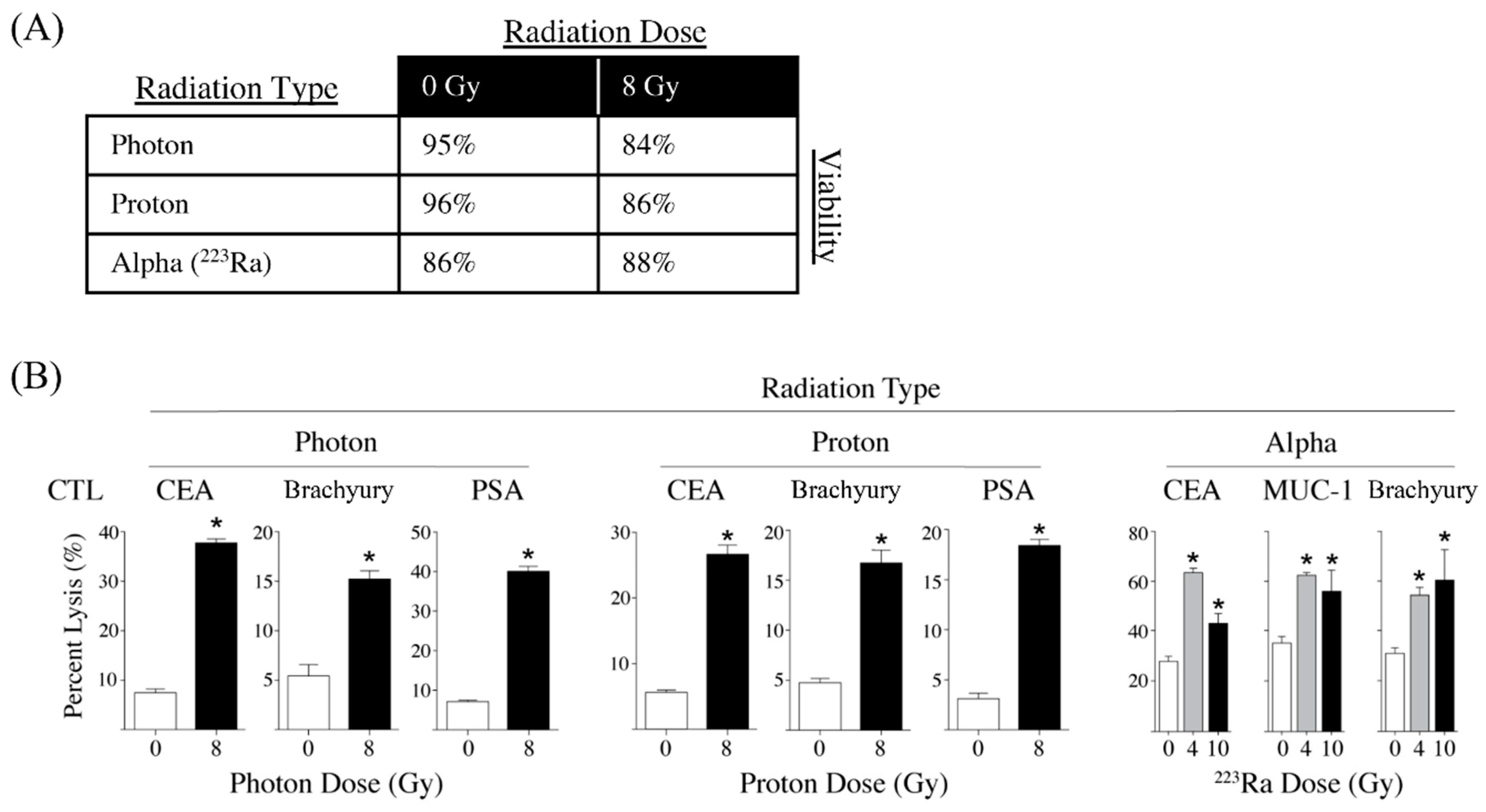
  140. Gameiro, S.R.; Malamas, A.S.; Bernstein, M.B.; Tsang, K.Y.; Vassantachart, A.; Sahoo, N.; Tailor, R.; Pidikiti, R.; Guha, C.P.; Hahn, S.M.; et al. Tumor cells surviving exposure to proton or photon radiation share a common immunogenic modulation signature, rendering them more sensitive to T cell-mediated killing. Int. J. Radiat. Oncol. Biol. Phys. 2016, 95, 120–130. [Google Scholar] [CrossRef] [Green Version]
  141. Malamas, A.S.; Gameiro, S.R.; Knudson, K.M.; Hodge, J.W. Sublethal exposure to alpha radiation (223Ra dichloride) enhances various carcinomas’ sensitivity to lysis by antigen-specific cytotoxic T lymphocytes through calreticulin-mediated immunogenic modulation. Oncotarget 2016, 7, 86937–86947. [Google Scholar] [CrossRef]
  142. Nelson, B.J.B.; Andersson, J.D.; Wuest, F. Targeted alpha therapy: Progress in radionuclide production, radiochemistry, and applications. Pharmaceutics 2021, 13, 49. [Google Scholar] [CrossRef] [PubMed]
  143. Tufano, R.P.; Pace-Asciak, P.; Russell, J.O.; Suárez, C.; Randolph, G.W.; López, F.; Shaha, A.R.; Mäkitie, A.; Rodrigo, J.P.; Kowalski, L.P.; et al. Update of radiofrequency ablation for treating benign and malignant thyroid nodules. The future is now. Front. Endocrinol. (Lausanne) 2021, 12, 698689. [Google Scholar] [CrossRef] [PubMed]
  144. Gameiro, S.R.; Higgins, J.P.; Dreher, M.R.; Woods, D.L.; Reddy, G.; Wood, B.J.; Guha, C.; Hodge, J.W. Combination therapy with local radiofrequency ablation and systemic vaccine enhances antitumor immunity and mediates local and distal tumor regression. PLoS ONE 2013, 8, e70417. [Google Scholar] [CrossRef] [PubMed]
  145. Agostinis, P.; Berg, K.; Cengel, K.A.; Foster, T.H.; Girotti, A.W.; Gollnick, S.O.; Hahn, S.M.; Hamblin, M.R.; Juzeniene, A.; Kessel, D.; et al. Photodynamic therapy of cancer: An update. CA Cancer J. Clin. 2011, 61, 250–281. [Google Scholar] [CrossRef] [PubMed]
  146. Zhao, L.; Zhang, X.; Wang, X.; Guan, X.; Zhang, W.; Ma, J. Recent advances in selective photothermal therapy of tumor. J. Nanobiotechnol. 2021, 19, 335. [Google Scholar] [CrossRef]
  147. Sweeney, E.E.; Cano-Mejia, J.; Fernandes, R. Photothermal Therapy Generates a Thermal Window of Immunogenic Cell Death in Neuroblastoma. Small 2018, 14, e1800678. [Google Scholar] [CrossRef]
  148. Wang, M.; Rao, J.; Wang, M.; Li, X.; Liu, K.; Naylor, M.F.; Nordquist, R.E.; Chen, W.R.; Zhou, F. Cancer photo-immunotherapy: From bench to bedside. Theranostics 2021, 11, 2218–2231. [Google Scholar] [CrossRef]
  149. Zhao, Y.; Liu, X.; Liu, X.; Yu, J.; Bai, X.; Wu, X.; Guo, X.; Liu, Z.; Liu, X. Combination of phototherapy with immune checkpoint blockade: Theory and practice in cancer. Front. Immunol. 2022, 13, 955920. [Google Scholar] [CrossRef]
  150. Kobayashi, H.; Choyke, P.L. Near-Infrared Photoimmunotherapy of Cancer. Acc. Chem. Res. 2019, 52, 2332–2339. [Google Scholar] [CrossRef] [Green Version]
  151. Nagaya, T.; Nakamura, Y.; Sato, K.; Harada, T.; Choyke, P.L.; Hodge, J.W.; Schlom, J.; Kobayashi, H. Near infrared photoimmunotherapy with avelumab, an anti-programmed death-ligand 1 (PD-L1) antibody. Oncotarget 2017, 8, 8807–8817. [Google Scholar] [CrossRef]
  152. Bernstein, M.B.; Krishnan, S.; Hodge, J.W.; Chang, J.Y. Immunotherapy and stereotactic ablative radiotherapy (ISABR): A curative approach? Nat. Rev. Clin. Oncol. 2016, 13, 516–524. [Google Scholar] [CrossRef] [PubMed]
  153. Demaria, S.; Guha, C.; Schoenfeld, J.; Morris, Z.; Monjazeb, A.; Sikora, A.; Crittenden, M.; Shiao, S.; Khleif, S.; Gupta, S.; et al. Radiation dose and fraction in immunotherapy: One-size regimen does not fit all settings, so how does one choose? J. Immunother. Cancer 2021, 9, e002038. [Google Scholar] [CrossRef] [PubMed]
  154. Nesslinger, N.J.; Sahota, R.A.; Stone, B.; Johnson, K.; Chima, N.; King, C.; Rasmussen, D.; Bishop, D.; Rennie, P.S.; Gleave, M.; et al. Standard treatments induce antigen-specific immune responses in prostate cancer. Clin. Cancer Res. 2007, 13, 1493–1502. [Google Scholar] [CrossRef] [PubMed] [Green Version]
  155. Schaue, D.; Comin-Anduix, B.; Ribas, A.; Zhang, L.; Goodglick, L.; Sayre, J.W.; Debucquoy, A.; Haustermans, K.; McBride, W.H. T-cell responses to survivin in cancer patients undergoing radiation therapy. Clin. Cancer Res. 2008, 14, 4883–4890. [Google Scholar] [CrossRef] [PubMed] [Green Version]
  156. Gulley, J.L.; Arlen, P.M.; Bastian, A.; Morin, S.; Marte, J.; Beetham, P.; Tsang, K.Y.; Yokokawa, J.; Hodge, J.W.; Menard, C.; et al. Combining a recombinant cancer vaccine with standard definitive radiotherapy in patients with localized prostate cancer. Clin. Cancer Res. 2005, 11, 3353–3362. [Google Scholar] [CrossRef] [Green Version]
  157. Heery, C.R.; Madan, R.A.; Stein, M.N.; Stadler, W.M.; Di Paola, R.S.; Rauckhorst, M.; Steinberg, S.M.; Marte, J.L.; Chen, C.C.; Grenga, I.; et al. Samarium-153-EDTMP (Quadramet(R)) with or without vaccine in metastatic castration-resistant prostate cancer: A randomized Phase 2 trial. Oncotarget 2016, 7, 69014–69023. [Google Scholar] [CrossRef] [Green Version]
  158. Rodriguez-Ruiz, M.E.; Perez-Gracia, J.L.; Rodriguez, I.; Alfaro, C.; Onate, C.; Perez, G.; Gil-Bazo, I.; Benito, A.; Inoges, S.; Lopez-Diaz de Cerio, A.; et al. Combined immunotherapy encompassing intratumoral poly-ICLC, dendritic-cell vaccination and radiotherapy in advanced cancer patients. Ann. Oncol. 2018, 29, 1312–1319. [Google Scholar] [CrossRef]
  159. Maity, A.; Mick, R.; Rengan, R.; Mitchell, T.C.; Amaravadi, R.K.; Schuchter, L.M.; Pryma, D.A.; Patsch, D.M.; Maity, A.P.; Minn, A.J.; et al. A stratified phase I dose escalation trial of hypofractionated radiotherapy followed by ipilimumab in metastatic melanoma: Long-term follow-up and final outcomes. Oncoimmunology 2021, 10, 1863631. [Google Scholar] [CrossRef]
  160. Twyman-Saint Victor, C.; Rech, A.J.; Maity, A.; Rengan, R.; Pauken, K.E.; Stelekati, E.; Benci, J.L.; Xu, B.; Dada, H.; Odorizzi, P.M.; et al. Radiation and dual checkpoint blockade activate non-redundant immune mechanisms in cancer. Nature 2015, 520, 373–377. [Google Scholar] [CrossRef] [Green Version]
  161. Imai, K.; Takaoka, A. Comparing antibody and small-molecule therapies for cancer. Nat. Rev. Cancer 2006, 6, 714–727. [Google Scholar] [CrossRef]
  162. Zhong, L.; Li, Y.; Xiong, L.; Wang, W.; Wu, M.; Yuan, T.; Yang, W.; Tian, C.; Miao, Z.; Wang, T.; et al. Small molecules in targeted cancer therapy: Advances, challenges, and future perspectives. Signal Transduct. Target. Ther. 2021, 6, 201. [Google Scholar] [CrossRef] [PubMed]
  163. Liu, J.; Zhao, R.; Jiang, X.; Li, Z.; Zhang, B. Progress on the application of bortezomib and bortezomib-based nanoformulations. Biomolecules 2022, 12, 51. [Google Scholar] [CrossRef] [PubMed]
  164. Obeng, E.A.; Carlson, L.M.; Gutman, D.M.; Harrington, W.J., Jr.; Lee, K.P.; Boise, L.H. Proteasome inhibitors induce a terminal unfolded protein response in multiple myeloma cells. Blood 2006, 107, 4907–4916. [Google Scholar] [CrossRef] [PubMed] [Green Version]
  165. Spisek, R.; Charalambous, A.; Mazumder, A.; Vesole, D.H.; Jagannath, S.; Dhodapkar, M.V. Bortezomib enhances dendritic cell (DC)–mediated induction of immunity to human myeloma via exposure of cell surface heat shock protein 90 on dying tumor cells: Therapeutic implications. Blood 2007, 109, 4839–4845. [Google Scholar] [CrossRef] [PubMed] [Green Version]
  166. Gulla, A.; Morelli, E.; Samur, M.K.; Botta, C.; Hideshima, T.; Bianchi, G.; Fulciniti, M.; Malvestiti, S.; Prabhala, R.H.; Talluri, S.; et al. Bortezomib induces anti–multiple myeloma immune response mediated by cGAS/STING pathway activation. Blood Cancer Discov. 2021, 2, 468–483. [Google Scholar] [CrossRef]
  167. Rose, M.; Burgess, J.T.; O’Byrne, K.; Richard, D.J.; Bolderson, E. PARP inhibitors: Clinical relevance, mechanisms of action and tumor resistance. Front. Cell Dev. Biol. 2020, 8, 564601. [Google Scholar] [CrossRef]
  168. Meng, X.W.; Koh, B.D.; Zhang, J.S.; Flatten, K.S.; Schneider, P.A.; Billadeau, D.D.; Hess, A.D.; Smith, B.D.; Karp, J.E.; Kaufmann, S.H. Poly(ADP-ribose) polymerase inhibitors sensitize cancer cells to death receptor-mediated apoptosis by enhancing death receptor expression. J. Biol. Chem. 2014, 289, 20543–20558. [Google Scholar] [CrossRef] [Green Version]
  169. Fenerty, K.E.; Padget, M.; Wolfson, B.; Gameiro, S.R.; Su, Z.; Lee, J.H.; Rabizadeh, S.; Soon-Shiong, P.; Hodge, J.W. Immunotherapy utilizing the combination of natural killer- and antibody dependent cellular cytotoxicity (ADCC)-mediating agents with poly (ADP-ribose) polymerase (PARP) inhibition. J. Immunother. Cancer 2018, 6, 133. [Google Scholar] [CrossRef]
  170. Seyedin, S.N.; Hasibuzzaman, M.M.; Pham, V.; Petronek, M.S.; Callaghan, C.; Kalen, A.L.; Mapuskar, K.A.; Mott, S.L.; Spitz, D.R.; Allen, B.G.; et al. Combination therapy with radiation and PARP inhibition enhances responsiveness to anti-PD-1 therapy in colorectal tumor models. Int. J. Rad. Oncol. Biol. Phys. 2020, 108, 81–92. [Google Scholar] [CrossRef]
  171. Wu, Z.; Cui, P.; Tao, H.; Zhang, S.; Ma, J.; Liu, Z.; Wang, J.; Qian, Y.; Chen, S.; Huang, Z.; et al. The synergistic effect of PARP inhibitors and immune checkpoint inhibitors. Clin. Med. Insights Oncol. 2021, 15, 1179554921996288. [Google Scholar] [CrossRef]
  172. Rachner, T.D.; Coleman, R.; Hadji, P.; Hofbauer, L.C. Bone health during endocrine therapy for cancer. Lancet Diabetes Endocrinol. 2018, 6, 901–910. [Google Scholar] [CrossRef] [PubMed]
  173. Ardiani, A.; Gameiro, S.R.; Kwilas, A.R.; Donahue, R.N.; Hodge, J.W. Androgen deprivation therapy sensitizes prostate cancer cells to T-cell killing through androgen receptor dependent modulation of the apoptotic pathway. Oncotarget 2014, 5, 9335–9348. [Google Scholar] [CrossRef] [PubMed] [Green Version]
  174. Ardiani, A.; Farsaci, B.; Rogers, C.J.; Protter, A.; Guo, Z.; King, T.H.; Apelian, D.; Hodge, J.W. Combination therapy with a second-generation androgen receptor antagonist and a metastasis vaccine improves survival in a spontaneous prostate cancer model. Clin. Cancer Res. 2013, 19, 6205–6218. [Google Scholar] [CrossRef] [PubMed] [Green Version]
  175. Kwilas, A.R.; Ardiani, A.; Gameiro, S.R.; Richards, J.; Hall, A.B.; Hodge, J.W. Androgen deprivation therapy sensitizes triple negative breast cancer cells to immune-mediated lysis through androgen receptor independent modulation of osteoprotegerin. Oncotarget 2016, 7, 23498–23511. [Google Scholar] [CrossRef] [Green Version]
  176. Wolfson, B.; Padget, M.R.; Schlom, J.; Hodge, J.W. Exploiting off-target effects of estrogen deprivation to sensitize estrogen receptor negative breast cancer to immune killing. J. Immunother. Cancer 2021, 9, e002258. [Google Scholar] [CrossRef]
  177. Kim, P.S.; Kwilas, A.R.; Xu, W.; Alter, S.; Jeng, E.K.; Wong, H.C.; Schlom, J.; Hodge, J.W. IL-15 superagonist/IL-15RalphaSushi-Fc fusion complex (IL-15SA/IL-15RalphaSu-Fc; ALT-803) markedly enhances specific subpopulations of NK and memory CD8+ T cells, and mediates potent anti-tumor activity against murine breast and colon carcinomas. Oncotarget 2016, 7, 16130–16145. [Google Scholar] [CrossRef]
  178. Williamson, C.W.; Sherer, M.V.; Zamarin, D.; Sharabi, A.B.; Dyer, B.A.; Mell, L.K.; Mayadev, J.S. Immunotherapy and radiation therapy sequencing: State of the data on timing, efficacy, and safety. Cancer 2021, 127, 1553–1567. [Google Scholar] [CrossRef]
  179. Weinberg, F.; Gadgeel, S. Combination pembrolizumab plus chemotherapy: A new standard of care for patients with advanced non-small-cell lung cancer. Lung Cancer 2019, 10, 47–56. [Google Scholar] [CrossRef]
Figure 1. Radiotherapy, chemotherapy, and small-molecule inhibitors induce immunogenic cell stress resulting in immunological responses spanning from immune modulation to immunogenic cell death. Radiotherapy, select chemotherapy, and small-molecule inhibitors can induce immunogenic cell death, which is characterized by the release of DAMPs that promote antigen presenting cells to phagocytose the dying cells, process tumor antigens, and present tumor antigens to T cells. Tumor cells that were not eradicated by radiotherapy, chemotherapy, or small-molecule inhibitors undergo immunogenic modulation wherein the tumor phenotype is altered such that the malignant cells become more sensitized to T cell targeting. Combining radiotherapy, chemotherapy, and small-molecule inhibitors with immunotherapies that can engage, enhance, and enable effector T cells may result in improved immune-mediated eradication of neoplastic cells. DAMP: damage-associated molecular patterns; TAA: tumor-associated antigen; HMGB1: high-mobility group box 1; ATP: adenosine triphosphate; IFN: interferon; MHC: major histocompatibility complex; TCR: T-cell receptor.
Cells 11 03826 g001
Figure 2. Sublethal exposure to photon, proton, or alpha radiation increases CTL lysis of prostate carcinoma cells in vitro. (A) Sublethal exposure of LNCaP prostate tumor cells in vitro to photon, proton radiation, or alpha radiation (223Ra) has minimal effect on cell viability. Human prostate carcinoma cells were mock-irradiated (0 Gy) or treated with either 8 Gy of photon radiation, 8 Gy proton radiation, or 8 Gy of 223Ra. Radiation-treated cells were cultured for an additional 72–96 h, and viability was determined by AO/PI viability dye. Depicted is % viable cells. (B) Human LNCaP prostate carcinoma cells were mock-irradiated (0 Gy, open bars) or treated with either 8 Gy of photon radiation (closed bars), 8 Gy proton radiation (closed bars), 4 Gy of of 223Ra (gray bars), or 10 Gy of 223Ra (closed bars). Radiation treated cells were cultured for an additional 72–96 h and then used as targets in an overnight CTL lysis assay. CEA-, MUC-1-, and brachyury-specific CD8+ T cells were used as effectors at an E:T ratio of 30:1. Experiments were repeated 1–3 times with similar results. * = p < 0.05. Adapted from [132,140,141].
Cells 11 03826 g002
Table 1. Clinical trials investigating immunogenic cell stress mediated by chemotherapy.
Clinical TrialTrial TitleConditionsTreatmentPhases
NCT03801304Trial to Evaluate Safety and Efficacy of Vinorelbine With Metronomic Administration in Combination With Atezolizumab as Second-line Treatment for Patients With Stage IV Non-small Cell Lung Cancer Non-small-cell lung cancerAtezolizumab
Vinorelbine
Phase II
NCT04159818Immune Induction Strategies to Improve Response to Immune Checkpoint Blockade in Triple Negative Breast Cancer (TNBC) Patients (TONIC-2)Metastatic breast cancerNivolumab
Cisplatin
Low-dose doxorubicin
Phase II
NCT04043195Nivolumab and Ipilimumab in Combination With Immunogenic Chemotherapy for Patients With Advanced NSCLCAdvanced non-small cell lung cancer (NSCLC)Oxaliplatin
Nivolumab
Ipilimumab
Phase I
Phase II
NCT04463368Isolated Hepatic Perfusion in Combination With
Ipilimumab and Nivolumab in Patients With Uveal
Melanoma Metastases (SCANDIUM II)
Uveal melanoma
Liver metastases
Melphalan
Ipilimumab
Nivolumab
Phase I
NCT04072263Adoptive T Cell Therapy in Patients With Recurrent Ovarian Cancer (OVACURE)Recurrent ovarian cancerTumor-infiltrating lymphocytes
Interferon alfa 2A
Carboplatin
Paclitaxel
Phase I
Phase II
NCT04262687Chemotherapy and Immunotherapy as Treatment for MSS Metastatic Colorectal Cancer with High Immune Infiltrate (POCHI)Metastatic colorectal cancer
High immune infiltrate
Microsatellite stable (MSS)
Capecitabine
Oxaliplatin
Bevacizumab
Pembrolizumab
Phase II
NCT05420584Neoadjuvant Arterial Embolization Chemotherapy
Combined PD-1 Inhibitor for Locally Advanced Rectal Cancer (NECI)
Rectal neoplasmsTislelizumab
Capecitabine
Oxaliplatin
Phase II
NCT04989218Durvalumab and Tremelimumab with
Platinum-based Chemo-
therapy in Intrahepatic
Cholangiocarcinoma (ICC)
CholangiocarcinomaGemcitabine
Cisplatin Tremelimumab
Durvalumab
Phase I
Phase II
NCT05144698RAPA-201 Therapy of Solid TumorsBreast cancer
Small cell and non-small cell lung cancer
Triple negative breast cancer
Gastric cancer
Esophageal adenocarcinoma
Gastric junction adeno-
carcinoma
Esophageal squamous cell carcinoma
Head and neck cancer
Squamous cell carcinoma of oral cavity
Squamous cell carcinoma of larynx
Squamous cell carcinoma of nasopharynx
Squamous cell carcinoma of other specified sites of skin
Carcinoma of unknown
primary
Bladder cancer
Malignant melanoma
RAPA-201 cells
Carboplatin
Paclitaxel
Phase II
NCT05307198Rectal Artery Infusion Chemotherapy Combined with Anti-PD1 Antibody for MSS LARC (RAIC)Rectal neoplasmsCapecitabine
Oxaliplatin
Sintilimab
Phase II
NCT02499367Nivolumab After Induction Treatment in Triple-negative Breast Cancer (TNBC) Patients (TONIC)Breast cancerNivolumab
Radiation therapy
Low dose doxorubicin
Cyclophosphamide
Cisplatin
Phase II
NCT03409198Phase IIb Study Evaluating Immunogenic Chemotherapy Combined with Ipilimumab and Nivolumab in Breast Cancer (ICON)Breast cancer
Hormone receptor positive tumor
Metastatic breast cancer
Ipilimumab
Nivolumab
Pegylated liposomal doxorubicin
Cyclophosphamide
Phase II
NCT03164993Atezolizumab Combined with Immunogenic Chemotherapy in Patients with Metastatic Triple-negative Breast Cancer (ALICE)Breast cancer
Triple-negative breast cancer
Atezolizumab
Pegylated liposomal doxorubicin
Cyclophosphamide
Phase II
NCT02649855Docetaxel and PROSTVAC for Metastatic Castration-Sensitive Prostate CancerProstate cancer
Prostate neoplasms
PROSTVAC-V
PROSTVAC-F
Docetaxel
Phase II
PD-1: programmed cell death protein 1, RAPA-201: rapamycin-resistant T cells, PROSTVAC: vaccine targeting prostate-specific antigen.
Table 2. Clinical trials investigating immunogenic cell stress mediated by radiotherapy.
Clinical TrialTrial TitleConditionsTreatmentPhases
NCT04774133The Immunodynamic Effect of Radiotherapy in Prostate Cancer PatientsProstate cancerRadiationN/A
NCT03942328Modified Immune Cells
(Autologous Dendritic Cells) and a Vaccine (Prevnar)
After High-Dose External Beam Radiation Therapy in Treating Patients With
Unresectable Liver Cancer
Hepatocellular carcinoma
Intrahepatic cholangiocarcinoma
External beam radiation, therapeutic autologous dendritic cells, Pneumococcal 13-valent conjugate vaccinePhase I
NCT03789097Vaccination With Flt3L,
Radiation, and Poly-ICLC
Non-Hodgkin’s lymphoma
Metastatic breast cancer
Head-and-neck squamous-cell
carcinoma
Subtherapeutic
radiation
Fl3tL
Poly-ICLC Pembrolizumab
Phase I
Phase II
NCT03646617Ipilimumab and Nivolumab With or Without Hypofractionated Radiotherapy in Patients With Metastatic Melanoma (RadVax)Metastatic melanomaHypofractionated radiation
Ipilimumab
Nivolumab
Phase II
NCT03313804Priming Immunotherapy in Advanced Disease With
Radiation
Non-small-cell lung cancer
Head-and-neck squamous-cell
carcinoma
Stereotactic body radiation or fractionated radiation
Nivolumab or
pembrolizumab or atezolizumab
Phase II
NCT04454528BreastVAX: Radiation Boost to Enhance Immune Checkpoint Blockade Therapy (BreastVAX)Breast cancerHypofractionated radiation
Pembrolizumab
Phase I
Phase II
Flt3L: an immune cell growth factor, Poly-ICLC: immune activating factor.
Table 3. Clinical trials investigating the combination of small molecule inhibitors with immune checkpoint blockade.
Clinical Trial Trial TitleConditionsTreatmentPhases
NCT04265872Bortezomib Followed by Pembrolizumab and Cisplatin in metTNBCBreast cancerBortezomib
Pembrolizumab
Cisplatin
Phase 1
NCT04258683A Study of Pembrolizumab Added to the Standard First-Line Therapy of Cyclophosphamide, Bortezomib, and Dexamethasone (CyBorD) for NDMM NTEMultiple myelomaCyclophosphamide
Bortezomib
Dexamethasone
Pembrolizumab
Phase 2
NCT04191096
NCT04934722
Efficacy and Safety of Pembrolizumab (MK-3475) Plus Enzalutamide Plus Androgen Deprivation Therapy (ADT) Versus Placebo Plus Enzalutamide Plus ADT in Participants With Metastatic Hormone-Sensitive Prostate Cancer (mHSPC) (MK-3475-991/KEYNOTE-991)Metastatic hormone-sensitive prostate cancerEnzalutamide
Pembrolizumab
Phase 3
NCT04471974ZEN-3694, Enzalutamide, and Pembrolizumab for the Treatment of Metastatic
Castration-Resistant Prostate Cancer
Castration-resistant prostate
carcinoma
Metastatic prostate adeno-
carcinoma
Metastatic prostate small cell
carcinoma
Stage IV/IVA/IVB prostate cancer AJCC v8
ZEN-3694
Enzalutamide
Pembrolizumab
Phase 2
NCT04946370Maximizing Responses to Anti-PD1 Immunotherapy With PSMA-targeted Alpha Therapy in mCRPCProstate cancer225Ac-J591
Pembrolizumab
Androgen receptor pathway inhibitor
Phase 1
Phase 2
NCT04262154Study of Abiraterone Acetate, Atezolizumab, GnRH Analog and Radiation
Therapy in Men With Newly Diagnosed Hormone-
sensitive Prostate Cancer
Metastatic prostate cancerAtezolizumab
Abiraterone acetate
Prednisone
Lupron® (leuprolide)
SBRT
Enzalutamide
Phase 2
NCT04190056Pembrolizumab and
Tamoxifen With or Without Vorinostat for the Treatment of Estrogen Receptor Positive Breast Cancer
Breast cancerPembrolizumab
Tamoxifen
Vorinostat
Phase 2
225Ac-J591: alpha-emitter actinium-225 conjugated to the anti-PSMA antibody J591; NDMM: newly diagnosed multiple myeloma; NTE: not transplant eligible; SBRT: stereotactic body radiotherapy; ZEN-3694: BET bromodomain inhibitor.
Publisher’s Note: MDPI stays neutral with regard to jurisdictional claims in published maps and institutional affiliations.

