Review

The XPA Protein—Life under Precise Control

by
Yuliya S. Krasikova
1,
Olga I. Lavrik
1,2 and
Nadejda I. Rechkunova
1,*
1
Institute of Chemical Biology and Fundamental Medicine, Siberian Branch of Russian Academy of Sciences, Novosibirsk 630090, Russia
2
Department of Natural Sciences, Novosibirsk State University, Novosibirsk 630090, Russia
*
Author to whom correspondence should be addressed.
Cells 2022, 11(23), 3723; https://doi.org/10.3390/cells11233723
Submission received: 12 October 2022 / Revised: 15 November 2022 / Accepted: 16 November 2022 / Published: 22 November 2022
(This article belongs to the Special Issue Systems Biology of Apoptotic and Non-apoptotic Signaling)

Abstract

:
Nucleotide excision repair (NER) is a central DNA repair pathway responsible for removing a wide variety of DNA-distorting lesions from the genome. The highly choreographed cascade of core NER reactions requires more than 30 polypeptides. The xeroderma pigmentosum group A (XPA) protein plays an essential role in the NER process. XPA interacts with almost all NER participants and organizes the correct NER repair complex. In the absence of XPA’s scaffolding function, no repair process occurs. In this review, we briefly summarize our current knowledge about the XPA protein structure and analyze the formation of contact with its protein partners during NER complex assembling. We focus on different ways of regulation of the XPA protein’s activity and expression and pay special attention to the network of post-translational modifications. We also discuss the data that is not in line with the currently accepted hypothesis about the functioning of the XPA protein.

1. Introduction

The nucleotide excision repair (NER) pathway is the most universal repair pathway for the removal of a wide range of structurally unrelated DNA lesions, including UV photolesions (e.g., cyclobutane pyrimidine dimers (CPDs) and pyrimidine-pyrimidone (6-4)-photoproducts (6-4PPs)), intrastrand crosslinks, reactive oxygen species-induced base alterations, and bulky adducts of DNA bases with reactive metabolites of some chemical carcinogens or chemotherapeutic agents [1,2,3]. Mutations in NER-related genes are associated with an autosomal recessive disease called xeroderma pigmentosum (XP) [4]. XP is characterized by extreme sensitivity of the skin to sunlight and a dramatically increased risk of skin cancer [5,6]. A subset of XP patients developed a profound neurodegenerative condition known as XP neurological disease [7]. XP patients can be classified into seven complementation groups, XP-A through XP-G, depending on the specific gene that is affected [1]. Patients with known mutations in the XPA gene have the most severe form of XP, indicating a critical role of the XPA protein in the NER process.
Initially, XPA was considered to be the sole damage recognition factor [8]; then, it was proposed that the XPA–RPA complex performs the first recognition [9,10]. Later, enough data was accumulated to suggest that lesions are primarily recognized by the XPC–RAD23B–CEN2 complex [3], and XPA takes part in the damage verification process (together with TFIIH) and overall plays the role of an organizing or scaffold component of NER [11,12,13].
Three-dimensional structures of XPA give us an opportunity to propose a spatial arrangement of XPA inside NER machinery. Today, several spatial structures of human XPA exist: two solution structures of the minimal DNA-binding domain (DBD) determined by NMR spectroscopy (Protein Data Bank (PDB) IDs 1XPA and 1D4U) [14,15], a crystal structure of a redefined XPA DBD (PDB ID: 6J44), and a crystal structure of the XPA–DNA complex (PDB ID: 6LAE) [16,17]. The series of crystal structures of a yeast homolog of XPA (Rad14) in a complex with different lesion-containing DNA substrates illustrates the DNA-binding capacity of the XPA DBD [18,19,20]. Recent advances in cryo-electron microscopy (cryo-EM) gave investigators a unique chance to look into the structure of a TFIIH–XPA–DNA complex (PDB: 6RO4) [21] and confirm the biochemical data about XPA localization inside the DNA repair bubble [22]. Furthermore, cryo-EM data have expanded our knowledge about the modulation of TFIIH activity by XPA and XPG.
This review provides updated information and additional insights into the XPA structural features and the XPA protein’s interaction partners in light of the present knowledge about the contact surface on XPA. We also briefly describe the NER assemble from the standpoint of XPA’s organizing role. Then, we summarize data on the mechanisms of the XPA gene transcription control, and finally, we describe the regulatory mechanisms of XPA post-translational modifications (PTMs) and combine them into a unified network.

2. XPA’s Structure and DNA-Binding and Protein-Binding Abilities

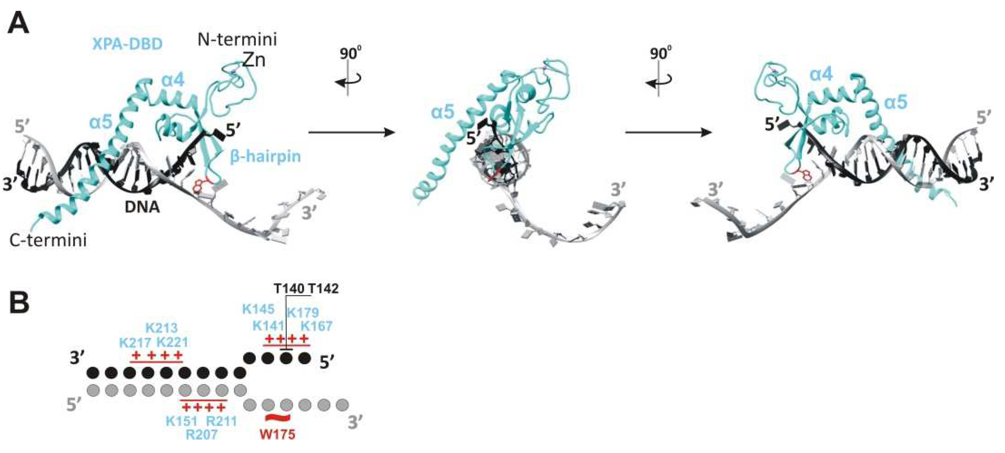
XPA is one of the smallest proteins inside the NER machine: human XPA is 31 kDa and consists of only 273 amino acid residues (aa). XPA is composed of a central globular domain (aa 98–219) that is flanked by dynamically disordered N and C termini (Figure 1A). This kind of structural organization, when structured globular domains are combined with disordered regions, is very common among eukaryotic proteins [23]. Frequently, disordered proteins (entirely disordered or containing disordered sequences, as in XPA) interact with or function as hubs in protein interaction networks or play a central role in an ordered assembly of macromolecular machines [24,25]. Indeed, both low-sequence-complexity parts enable the XPA protein to interact with a variety of protein partners. Future experiments will shed light on disordered XPA part properties: do they do folding upon binding to protein partners (and should be called “intrinsically disordered”), or do these regions not adopt a specific three-dimensional structure during functioning. In addition, the N terminus contains a conserved nuclear localization signal (NLS) that lies in the 13-residue stretch from aa 30 to 42 [26,27] (Figure 1A). The NLS is a tag that ensures that the protein is sorted into the nucleus, but in the case of XPA, things are not so simple, and we discuss this matter below.
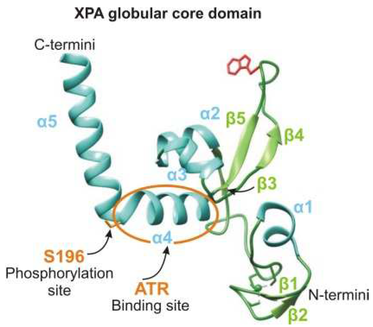
The central domain contains a C4-type Zn-finger (ZnF) motif that has the sequence Cys105-X2-Cys108-X17-Cys126-X2-Cys129 [28] (Figure 1A). Side chains of cysteines Cys105/108 and Cys126/129 coordinate the zinc ion [14]. Although the ZnF of XPA is essential for DNA binding and for NER activity [29], the ZnF core itself is not directly in contact with DNA [16] but rather properly ensures the DBD folding [30]. Moreover, the zinc-containing subdomain is even negatively charged due to many glutamate and aspartate residues [14] (Figure 1D). Notably, the UvrA protein has two units of the same C4 type of the ZnF motif [28].
Originally, the minimal DNA-binding domain (DBD) of XPA was mapped to a central globular core between residues 98 and 219 [14,15,29,31,32]. This region contains a sheet-helix-hairpin motif (residues 138–182) and a helix-turn-helix motif (residues 183–230) that form a shallow clamp-like or right hand-like structure (the sheet-helix-hairpin motif as fingers and helix-turn-helix as a thumb) with a positively charged inner surface (Figure 1D). The internal curvature of the basic cleft fits well to the diameter of a standard B-form dsDNA [14]. Subsequent studies have found that some residues on the C-terminal side beyond the minimal DBD domain are also involved in binding to DNA substrates [33,34,35]. Later, the XPA DBD was redefined and extended by 20 additional C-terminal residues (Asp220–Thr239). The redefined XPA DBD (aa 98–239) can bind to DNA with an affinity nearly identical to that of the full-length XPA protein [34,35]. The crystal structure revealed that the C-terminal extension folds as a long α-helix (α5 in Figure 1A) with a basic residue cluster resulting in the formation of a consecutive positively charged surface [16,17]. Interestingly, structural superposition of the human XPA DBD on a yeast Rad14–DNA complex implies that the α5 extension (Asp217–Thr239) cannot directly come into contact with a DNA substrate.
XPA has an ability to recognize some bulky lesions [22,36] and especially prefers to bind to kinked and branched DNA structures [37,38,39]. All of these DNA structures contain an ss–dsDNA junction. XPA binds to the duplex part of the junction in a non-sequence-specific manner via electrostatic interactions between the positively charged cleft and negatively charged phosphate backbones of the DNA duplex [17] (Figure 1F and Figure 2). This intermediate is further stabilized by hydrogen bonding of the side chain hydroxyl group of Thr142, and a van der Waals contact is formed between the side chain Cβ atom of Thr140 and DNA’s phosphate moieties (Figure 1A and Figure 2). Thereafter, Trp175 from the top of the hairpin between β4 and β5 is stacked with bases of the unpaired ssDNA at the junction, thereby giving rise to a stable conformation of this β-hairpin [17,21]. According to the energy calculations, binding to ssDNA in the 3′→5′ direction is more favorable than that in the 5′→3′ direction (relative to α5), but it is not observed experimentally [29,38].
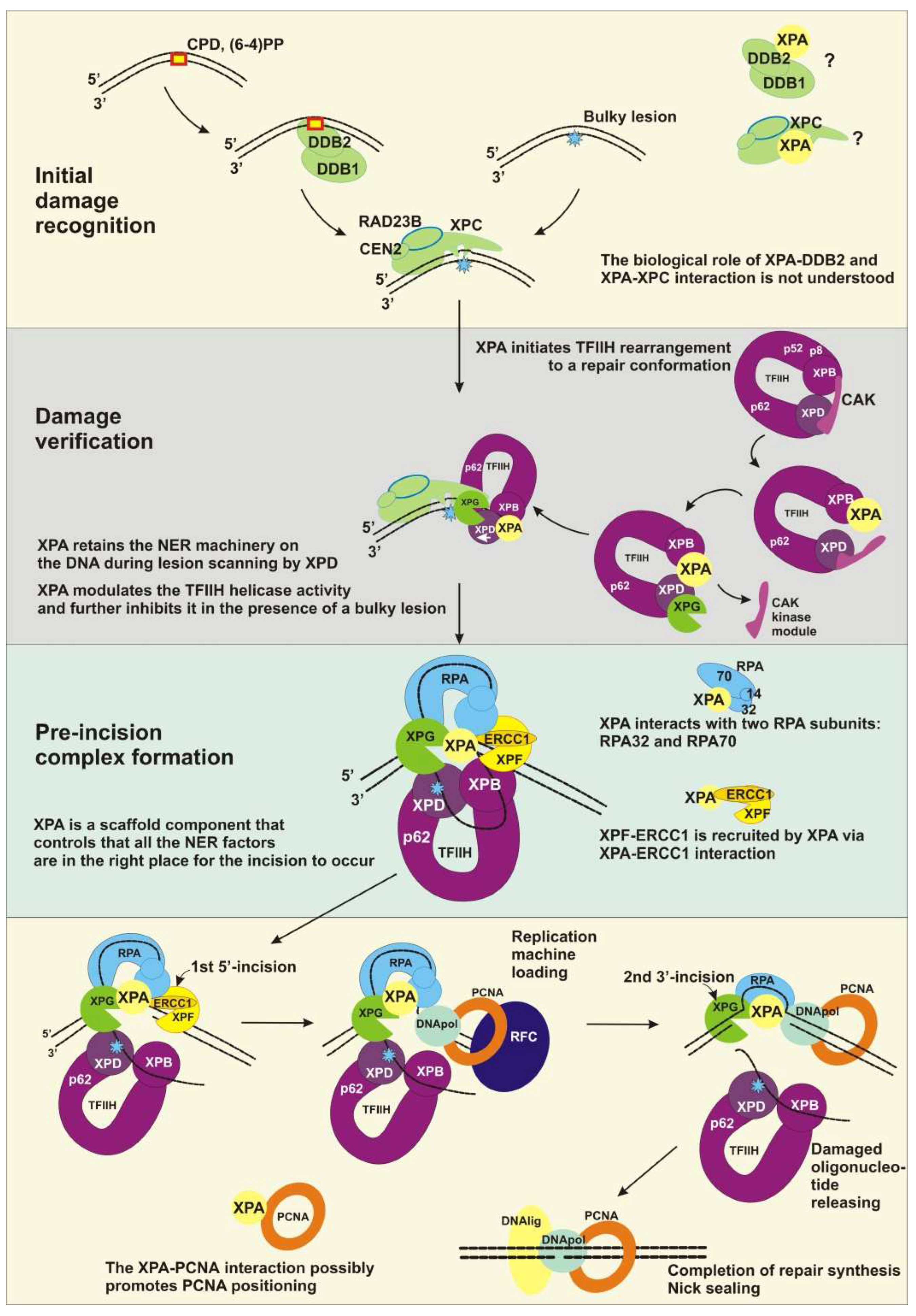
Recently, it was shown with atomic force microscopy, scanning force microscopy, and mathematical modeling that XPA undergoes episodic one-dimensional diffusion to search the DNA for damage [40]. The functional meaning of XPA’s damage recognition ability is not clear, and today, XPA is considered only as a protein scaffold element inside the NER complex. Anyway, XPA interacts with the proteins involved in every step of NER, from damage recognition to DNA synthesis. Yet, it is unknown how many contacts XPA could engage in concurrently, and it is possible that XPA interacts with these proteins not simultaneously but in the order of them joining the repair machinery. Figure 1B lists XPA’s protein interaction partners directly involved in NER according to the following process steps:

2.1. Initial Damage Recognition

XPA interacts physically with DDB2 through aa 185–226, and this interaction can be seen both in vitro and in vivo [41]. UV-damaged DNA-binding protein (DDB1/2) is a heterodimeric protein consisting of subunits DDB1 and DDB2/XPE and has an extraordinarily high binding affinity and specificity for CPD and 6-4PP [2,3]. The biological role of the XPA–DDB2 interaction is unclear, but because the DDB2 interaction site overlaps with the poly(ADP-ribose) [PAR]-binding motif (aa 213–237), we can speculate that the XPA–DDB2 complex is involved in PAR-dependent chromatin remodeling together with PARP1 and XPC [42]. XPC (which functions in the complex with proteins RAD23B and CEN2) is the protein sensor responsible for the detection of a wide variety of DNA lesions that are repaired through the global genome NER (GG-NER) pathway [43]. XPA interacts with XPC and stimulates its binding to the DNA [44,45]. The XPC interaction site in the XPA sequence and the biological meaning of this interaction are unknown.

2.2. Damage Verification

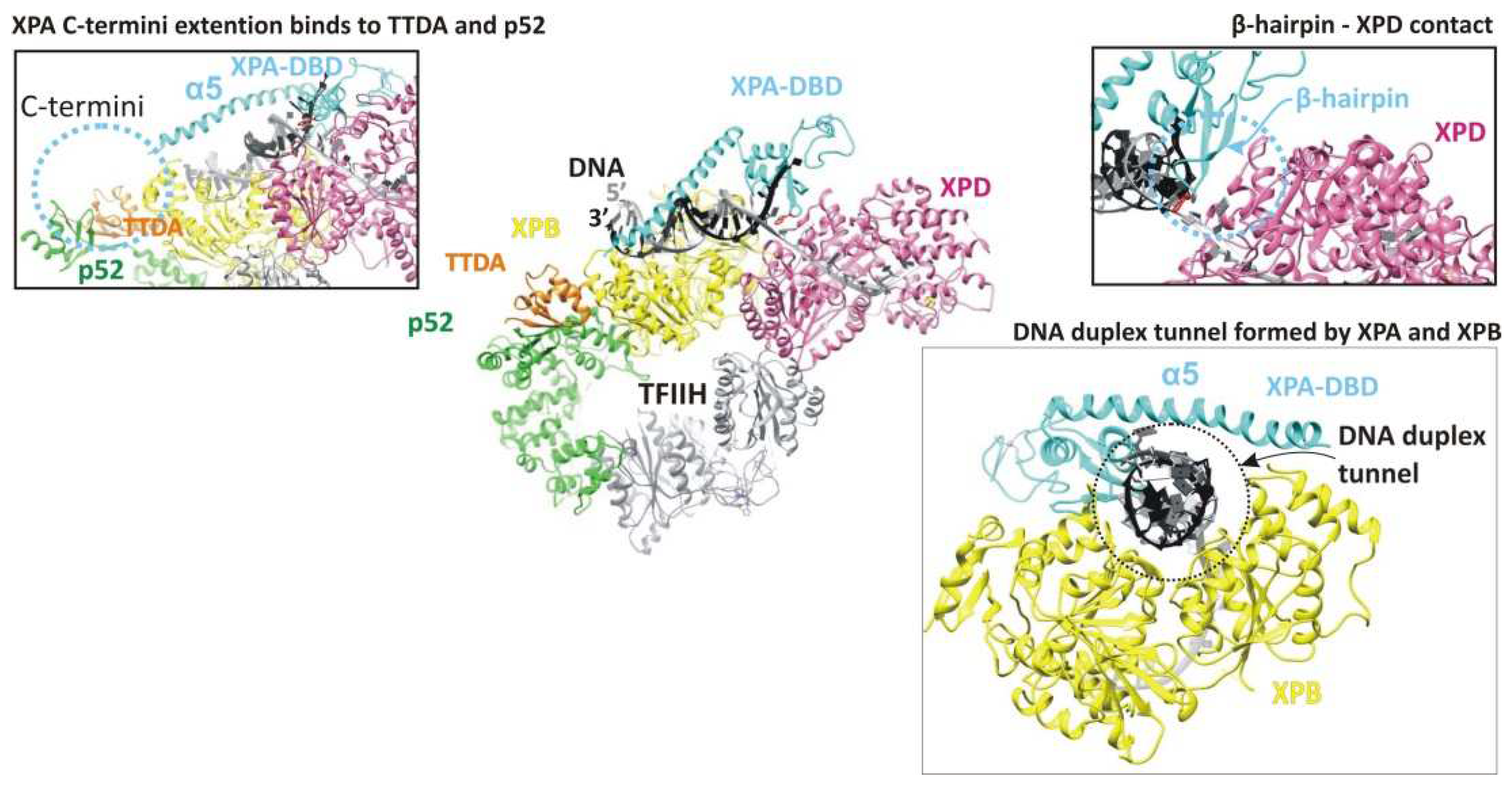
The TFIIH complex is the key protein for the damage verification step. Depending on the context, the TFIIH composition changes from a core of seven subunits, including translocase XPB and helicase XPD, to ten subunits, through the addition of three CAK (Cdk-activating kinase module) kinase subunits [21,46]. XPA’s whole C terminus is involved in an interaction with the TFIIH protein [47]. Recent cryo-EM data expanded our knowledge about the TFIIH–XPA interaction [21]. XPA (together with nuclease XPG) facilitates the CAK kinase module release from the core TFIIH and stabilizes an alternative conformation of TFIIH, where the XPD helicase assumes the open conformation for the functioning. In this complex, XPA forms a bridge between the XPB and the XPD, and moreover, XPA’s extended α5 helix and ATPase XPB form a positively charged tunnel that holds the DNA within. Thus, by trapping the DNA within a duplex tunnel, XPA may keep the NER machinery on the DNA during lesion scanning and processing [21].

2.3. Pre-Incision Complex Formation

Immediately after TFIIH opens the DNA repair bubble, the undamaged ssDNA that is being formed is bound by replication protein A (RPA) [48]. RPA is a heterotrimer consisting of subunits RPA70, RPA32, and RPA14 [49,50]. XPA interacts with two of them: RPA70 (which contains OB-fold domains A, B, and C) and RPA32 (which contains the D OB-fold domain) [51]. It was shown recently that the interaction between XPA (aa 29–46) and RPA32C is important for the initial association of XPA with NER complexes, whereas the interaction between XPA (aa 98–126) and RPA70AB is needed for structural shaping of the complex to enable the dual incision reaction [52]. Pre-incision complex formation is completed by the engagement of nuclease XPF–ERCC1, which is recruited by the XPA through the interaction with ERCC1 [53,54,55,56,57]. XPA aa 59–114 are responsible for this interaction. In particular, the Gly72–Phe75 region (also known as the G motif) and the Glu78–Glu84 region (i.e., the E motif) are the residues necessary for the binding of XPF–ERCC1. Biochemical data have shown that the ZnF motif (aa 102–129) is partially involved in this contact, but the NMR and molecular dynamics simulation revealed that only the 14-amino acid sequence (aa 67–80) mediates this interaction.

2.4. Dual Incision, Resynthesis, and Ligation

After the first incision, proliferating cell nuclear antigen (PCNA) joins the NER complex. PCNA is the processivity factor for DNA polymerases. XPA has been found to interact directly with PCNA via the APIM sequence (the AlkB homolog 2 PCNA-interacting motif), and it has been shown that XPA and PCNA colocalize to the damaged DNA foci in a cell culture [58,59,60]. XPA−/− cells complemented with XPA containing a mutated APIM sequence have high UV sensitivity and a deficient repair of CPDs and 6-4PPs and are consequently more arrested in the S phase as compared to XPA−/− cells complemented with wild-type XPA. Notably, XPA colocalizes with PCNA in the replication foci and is loaded onto a newly synthesized DNA in undamaged cells; thus, it is possible that this interaction is required for DNA-processing pathways other than NER.

2.5. XPA Dimerization

It is well-known that isolated XPA easily forms a homodimer. Moreover, in vitro, it can form the XPA2–RPA complex [61]. Instead, XPA has been widely assumed to be a monomer participating in the mechanism of NER (the first statement about the monomeric functional form was published by [62]. Accordingly, the physiological meaning of the XPA dimerization and the structural mechanism of this process are still unclear. Recently obtained molecular dynamics simulation data indicate that some residues make a contribution to the intermolecular interactions in XPA homodimers, but this needs to be validated by another approach [63]. Notably, XPA is not the sole NER protein that demonstrates easily dimerization characters, but as in the case of XPA, there is no functional explanation found for this ability [64].

2.6. PTM Proteins

Today, it is known that XPA is precisely tuned by several PTMs [13,65,66]. Obviously, XPA interacts with proteins that provide these modifications and removes them, but we know the interaction site only for the ATR kinase: the α4 helix [67] (Figure 1C). Interaction sites with proteins facilitating these modifications—RASSF1A and NDR1—have not been determined either. The only interaction site that has been identified is the one for the Cep164 protein, which functions in ATR-mediated checkpoint activation; it interacts with XPA through aa 10–88 of XPA [68].

2.7. XABs

Using the yeast two-hybrid system, researchers identified a novel set of XPA-interacting proteins that was designated as XABs [69]. XAB1 is a cytoplasmic protein with GTPase activity and binds to the N-terminal region (residues 1–52) of XPA, and the region “aa 30–34” is directly involved in this interaction (Figure 1C). The XAB1-interacting site overlaps with the NLS and raises a question about XAB1′s role in the cytoplasmic sequestering of XPA. Among the five found XABs, only XAB2 has a nuclear function and is intensively investigated. It has been reported that XAB2 interacts with the proteins involved in transcription-coupled NER (TC-NER), for example, CSA, CSB, and RNA polymerase II [70]. XAB2 contains 15 TPR (tetratricopeptide repeat) motifs and appears to have a role in transcription and pre-mRNA splicing [71]. Generally, XAB2 serves as a guardian of POLR2A (the largest catalytic subunit of RNA polymerase II) expression to ensure global gene expression and to antagonize cell senescence [72]. Furthermore, XAB2 promotes homologous recombination and facilitates histone acetylation events linked to homologous recombination [73]. Immunoprecipitation studies have revealed that XAB2 interacts with endonucleases ERCC1–XPF and XPG outside NER under the conditions that favor the formation of R-loops [74]. Altogether, XAB2 is involved in R-loop removal and pre-mRNA splicing; both processes are linked to transcription [75]. Future experiments will shed light on the possible involvement of XPA in processes where XAB2 is active.

3. The Hinge of the NER Complex

DNA damage can be recognized by NER in one of two subpathways, which overlap except for the mode of DNA damage recognition. GG-NER can search for damage anywhere in the genome throughout the cell cycle [76]. TC-NER is responsible for the accelerated repair of lesions in the template DNA strand of actively transcribed genes only. In general, a good NER substrate should be bulky and must destabilize a DNA double helix (i.e., disrupt the base pairing and bend the duplex). To detect both conditions, NER has evolved special bipartite substrate discrimination: firstly, it recognizes a local thermodynamically destabilized site, and then, the latter is probed for the presence of a lesion.
In the case of mammalian GG-NER, lesions are recognized by the XPC–RAD23B–CEN2 complex (hereinafter, XPC) [3] (Figure 3). The XPC complex can detect and bind DNA sites where a regular double-helical structure is perturbed, and as a result, one or more base pairs are disrupted and/or destabilized [7,77,78,79,80]. The major UV-induced photoproducts CPD and 6-4PP are recognized by the DDB1/2 heterodimeric protein, which binds directly to these lesions and introduces a kink and an opening into the duplex, creates a more suitable substrate for XPC, and hands over the damaged substrate to the XPC complex. Next, XPC recruits TFIIH to the lesion site [2,81,82]. The PARP1 protein supports these NER events by chromatin de-condensation and by the poly(ADP-ribosyl)ation (PARylation) of NER participants, thereby facilitating their recruitment to the damage site [66,83]. TC-NER is initiated by the stalling of elongating RNA polymerase II (RNAPII) at DNA lesions [84,85]. Then, after its lesion stalling and assembly of factors CSB, CSA, and UVSSA, the latter promotes TFIIH binding downstream of RNAPII [86].
The TFIIH complex is the key protein for the damage verification step [87]. TFIIH probes the lesion itself and unwinds the DNA duplex around the lesion, thereby making room for the subsequent assembly of repair machinery. Before TFIIH starts acting as a repair factor, it should be restructured from a transcription conformation to a repair one [21]. Both XPB and XPD bind DNA in the NER process, whereas only XPB binds DNA in transcription complexes [88,89]. During transcription, the CAK kinase module inhibits the repair XPD helicase activity via stabilization of the conformation where the XPD arch domain forms a “plug” structure that occupies the DNA-binding pore [21,90,91,92]. XPA (together with XPG) associates with TFIIH, releases the inhibitory CAK kinase module of TFIIH, and displaces the “plug” element from the DNA-binding pore in XPD [21] (Figure 3). This alternative TFIIH conformation results in the binding of XPB and XPD to the duplex and single-stranded 3′-DNA extension, consistently with their translocase and helicase activities, respectively (Figure 4). XPA forms an elongated arch over the DNA that bridges between the two ATPases. XPA binds to XPB and XPD via its extended α5-helix and its intercalating β4–β5 hairpin, respectively. The C-terminal region of XPA extends to p52 and TTDA/p8; this can explain why TTDA/p8 facilitates XPA recruitment in vivo. XPA’s extended α5-helix, together with ATPase XPB, forms a positively charged DNA duplex tunnel holding the DNA duplex within (Figure 4). Thus, XPA may keep NER machinery on the DNA during lesion scanning and processing. XPA chaperons TFIIH–DNA interactions and marks the DNA at the 5′ edge of the repair bubble [21]. Notably, at what moment XPA associates with TFIIH (or what signal promotes this association) is still unknown.
TFIIH’s structural flexibility allows XPD to unwind DNA while tracking along in the 5′→3′ direction [2]. During the tracking process, XPD pulls the DNA through a narrow tunnel that is too small for bulky DNA lesions to pass through. This “damage filtration” process is simple but effective.
Immediately after the formation of an undamaged ssDNA inside the repair bubble, this strand is covered by the RPA protein [48,93]. RPA protects the undamaged strand from a nuclease attack and additionally stabilizes the opened DNA intermediate [22]. The size of the NER-excised fragment coincides with the maximal length of the ssDNA platform for RPA binding (~30 nt), but it is obvious that RPA binds to undamaged ssDNA being formed at the moment when it reaches the size of the minimal RPA-binding platform (8nt) [94]. RPA OB domains bind to ssDNA in a certain order in the 5′→3′ direction: firstly, RPA70A binds to ssDNA closer to the 5′ side, then RPA70B binds from a 3′ side of RPA70A, then RPA70C, and RPA32D binds to the 3′ -side of ssDNA [49,50,95]. XPA tightly interacts with RPA inside the repair bubble, and together, they ensure the correct orientation of XPG and XPF–ERCC1 inside the repair bubble [51]. The interaction site consists of two contacts: one between the XPA N terminus and RPA32D and the other forms between XPA’s ZnF subdomain and two RPA OB domains: RPA70AB. The contact with RPA32D is important for the initial association of XPA with RPA, while the interaction with the large RPA subunit is needed for structural shaping of the NER complex on the opened DNA [52]. The crystal structure of Ustilago maydis RPA revealed that ssDNA in a complex with RPA is also U-shaped; for this reason, the 5′ edge and 3′ edge of the repair bubble are pulled together [96]. Thus, XPA binds the 5′ edge of the repair bubble and could interact with RPA70AB located near the 3′ edge.
After XPD stalls on the lesion (a bulky lesion is “stacked” in the DNA-binding pore), XPG binds to the 3′ edge of the repair bubble. The formation of the NER pre-incision complex is completed by the engagement of XPF–ERCC1, which is recruited by XPA via the XPA–ERCC1 interaction and is properly positioned by RPA via the RPA–XPF interaction [53,54,55,56,57,97].
Accordingly, the interior of the NER pre-incision complex is as follows: TFIIH stalls at the lesion, RPA covers the undamaged opposite strand, XPA marks the 5′ edge of the repair bubble, XPG marks the 3′ edge of the repair bubble, and XPF–ERCC1 binds behind XPA (Figure 3). The latter is a central component in the pre-incision complex space, because it interacts with all its compartments.
The DNA incision is first carried out by XPF–ERCC1 on the 5′ side of the damage site [98]. The resultant DNA intermediate contains a long gap (~30 nt) with a free 3′-OH group, a long flap bearing a lesion, and an intact template. Next, replication machinery loads to start the repair synthesis [99]. The following are possible sets of replication proteins: “DNA polymerase δ/PCNA/RFC1–RFC–p66”, “DNA polymerase ε/CTF18/RFC”, or “DNA polymerase κ/ubiquitinated PCNA/XRCC1” [43]. Which set of replication factors is definitely recruited remains unknown, but generally, this depends on the status of the PCNA ubiquitination, as well as on the use of distinct clamp loader complexes or XRCC1. At this stage, XPA remains in the NER complex via a protein–protein interaction with the RPA that stays bound to the undamaged strand [39]. Moreover, XPA possibly promotes the positioning of the PCNA clamp [59], and RPA recruits clamp loader RFC [100,101,102].
Repair synthesis can proceed halfway through the gap [43]. Then, the XPG-made incision cleaves the flap [64,87] and leaves a 5′ phosphate that is utilized in the nick-sealing reaction carried out by DNA ligase I or by the DNA ligase IIIα–XRCC1 complex [103,104,105]. Now, NER is completed.

4. The Ways to Control XPA

All aspects of DNA damage response (DDR) signaling and DNA repair pathways (NER, base excision repair, mismatch repair, homologous recombination, and nonhomologous end joining) are very important for cell survival; therefore, these processes’ participants are controlled via transcription and are precisely tuned by PTMs. As far as the XPA protein is the keystone for the entire NER process, it is an essential node regulating repair capacity. The XPA amount is controlled at the transcriptional level by a molecular circadian clock (and in several other ways) and at the post-translational level by the ubiquitin–proteasome system. Today, it is known that the XPA activity is modified by several PTMs: phosphorylation, acetylation, and PARylation.

4.1. The XPA Protein Amount Control—A Balance between Production and Degradation

4.1.1. The Amount of XPA Molecules

It has been shown that normal human fibroblasts WI38-VA and HeLa cells have ~150,000–200,000 XPA molecules per cell [106,107]. This amount is nearly equal to that of the most abundant human SSB analog: the RPA protein, which is present at ~200,000–250,000 molecules per cell [107,108,109]. Furthermore, the XPA molecules amount is greater than that of other NER factors: XPC—25,000 per HeLa cell [107], TFIIH—100,000 [107], XPG—80,000 [107], and XPF-ERCC1—100,000 molecules per cell [107]. A possible explanation of this abundance is that XPA takes part in another cellular process. We should mention here that subsequent research in the XPA field has revealed that the XPA amount is not constant and depends on several conditions; therefore, whether this amount (about 200,000 XPA molecules per cell) corresponds to a maximum or minimum protein level is unclear.
The question about the number of XPA molecules is directly connected to another one: whether XPA is a rate-limiting factor in NER. The answer to this question is especially valuable for NER inhibitors screening: Should these compounds be more effective at reducing the XPA protein level or at disrupting the XPA protein function? Some studies indicate that XPA is not a rate-limiting factor of NER [106,110], while others have revealed the opposite: the peak NER activity coincides with the maximum XPA protein levels [111,112,113]. In a normal human fibroblast line immortalized by telomerase overexpression (NHF-1), when XPA is downregulated to 60%, 10%, or 4% of its original value, the rates of repair of both 6-4PPs and CPDs are proportionally reduced [114]. Thus, the UV sensitivity of human cells is a linear function of the XPA concentration [115,116].

4.1.2. The XPA Residence: The Cytoplasm or Nucleus?

Given that XPA contains a NLS signal, it is logical to propose that this sequence ensures that the protein is sorted into the nucleus. Indeed, the first information about the XPA residence indicated that XPA is permanently localized to the cell nucleus [110]. The subsequently obtained information has revealed that XPA nuclear transport is cell cycle-dependent [117] and DNA damage-induced [118,119]. According to these data, in G1-phase cells, most XPA molecules are located in the cytosol, and there is only a slight accumulation of XPA in the nucleus after UV irradiation. In the S phase, most XPA molecules are also located in the cytosol, but they are imported into the nucleus after UV treatment. XPA is predominantly located in the nucleus in the G2-phase cell population with or without UV irradiation. The discrepancy between the reports on XPA localization is possibly due to a difference in the experimental methodology [119,120].
It has been demonstrated that protein adaptors importin-α4 and importin-α7 directly interact with XPA’s NLS for transporting XPA through the nuclear pore complex [119]. The adaptor importin-α7 is involved in XPA nuclear import independently of the DNA damage; hence, it is possible that importin-α7 is mainly responsible for the nuclear import of XPA in the G2 phase and for the basal XPA level required in the nucleus throughout the cell cycle. At the same time, the importin-α4–mediated nuclear import is DNA damage-induced, and DNA damage checkpoint protein kinase ATR is involved in this regulation (we discuss it below). The NLS of XPA cannot be efficiently recognized by importin-α4 in the absence of UV irradiation, perhaps owing to the masking of the XPA NLS by the binding of other cytoplasmic factors.
Tumor suppressor protein p53 is a major downstream effector and a phosphorylation substrate in the ATR-mediated DDR. The p53 status of cells significantly influences the role of ATR in the regulation of DNA repair after UV or cisplatin damage. The XPA nuclear import is dependent on p53 transcriptional activity in p53+/+ cells and is much slower in p53−/− cells, but the import still proceeds. Therefore, the damage-induced ATR activation of p53 appears to be a primary, but not the sole mediator of, XPA nuclear import in p53+/+ as compared to p53−/− cells in the S phase [119,121,122,123].
The reason why, in the G2 phase, XPA stays sequestered in the cytosol, even with DNA damage, is unclear. Nonetheless, the cytoplasmic XPA localization is consistent with the information about XPA interacting with mainly cytoplasmic proteins XAB1 and XAB3–5 [69]. As mentioning above, data about the interactions with these novel proteins have been obtained using the yeast two-hybrid system. The role of these XPA interactions stays unclear. In a database search, XAB3 was identified as metallopeptidase PRSM1 [124], XAB5 is the Golgi reassembly stacking protein GRASP65 [125], and XAB4 has a region homologous to GRASP65.
XPA-binding protein 1 (XAB1) is a GTPase. The XAB1 interaction site (aa 30–34) coincides with the NLS of XPA (aa 30–42), and therefore, it could be suggested that XAB1 is involved in the nuclear localization of XPA or interferes with the system of XPA nuclear transport [69]. Nevertheless, a small-interfering RNA (siRNA) knockdown of XAB1 has no effect on the UV-induced nuclear import of XPA [119]. This contradiction could be due to the different environments within human cells and the yeast model system. A GTPase other than XAB1 must be involved in the nuclear import of XPA in human cells.

4.1.3. The Circadian Rhythm of XPA’s Life

Most of living things on Earth have adapted their life to its 24-h light/dark cycle and have developed an internal oscillating system to sense the light–dark cycles: the “circadian clock system”. In mammals, the circadian clock is “ticking” not only at the organismal level by synchronizing the rhythmicity of sleep/activity cycles and behavioral changes corresponding to light and temperature cycles but also in every single cell to synchronize its biochemical reactions so as to respond to organismal energetic demands [126].
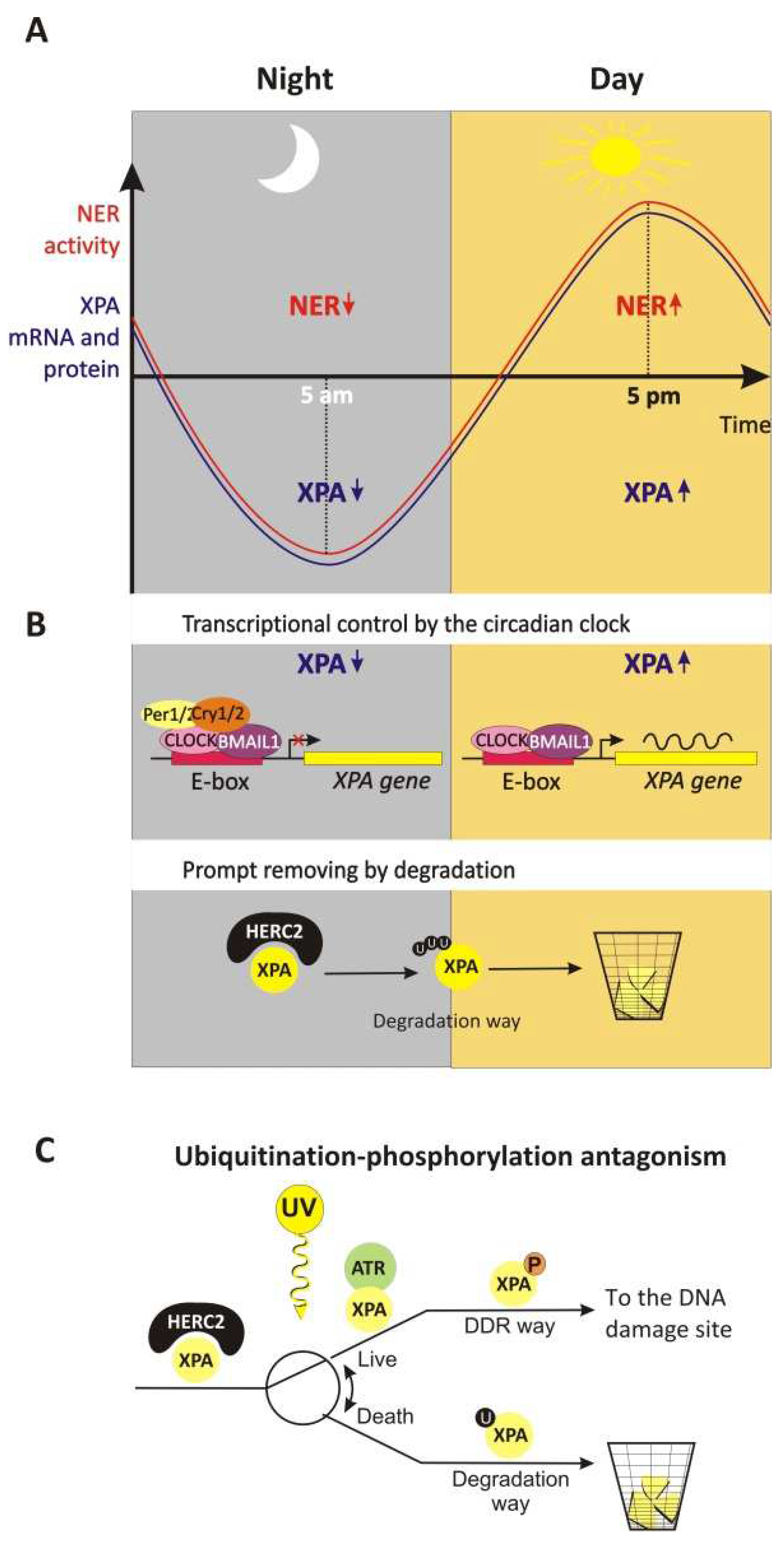
Since NER is the sole DNA repair mechanism capable of removing DNA photolesions in the skin [7], it seems obviously advantageous for organisms to have high levels of NER activity at the time of maximal exposure to sunlight within a day and to reduce NER activity at night, when it apparently is not needed [127]. Indeed, it has been shown that NER activity in mice is regulated by the circadian clock, such that repair activity changes by almost 10-fold over the course of the day, with the zenith in the late afternoon hours (~5 p.m.) and the nadir in the early morning hours (~5 a.m.) (Figure 5A) [111,112]. This regulation, which is accomplished through control over the XPA protein and NER activity oscillation, correlates with the XPA protein level circadian rhythmicity (in the mouse skin [113], in the mouse liver [112], and in the mouse brain [111]).
XPA gene transcription is regulated positively by the core circadian clock factors: circadian locomotor output cycles kaput (CLOCK) and its binding partner, brain and muscle ARNT-like protein 1 (BMAL1) [113]. The CLOCK–BMAL1 transcription activator complex binds DNA directly in regulatory elements (E-boxes) within rhythmicity-related genes to influence their transcription [126]. In our case, the CLOCK–BMAL1 complex binds directly to the promotor region of XPA [128] (Figure 5B). At the same time, major targets of CLOCK–BMAL1 include other core clock genes that encode the mammalian period ortholog (PER1, PER2, and PER3) and cryptochrome (CRY1 and CRY2) repressor proteins [126]. These negative regulators heterodimerize and, then, after a time delay, translocate into the nucleus, where they repress their own gene transcription by inhibiting CLOCK–BMAL1. The time delay between the synthesis of Cry and Per and their actions as repressors generate an oscillatory pattern [111]. Thus, the Cry–Per complex forms a feedback loop ([129] and references therein) and negatively regulates XPA gene transcription [113].
Changing only the XPA gene transcription level may not be sufficient to produce the oscillatory mode of its protein amount profile. Indeed, the XPA protein is subjected to prompt removal by degradation: the XPA half-life is approximately 4 h in the absence of DNA damage [111,112]. A large protein (HECT- and RCC-like domain-containing protein; HERC2) is reported to ubiquitinate XPA, thereby leading to its degradation by the ubiquitin–proteasome system [114,130]. Thus, the relatively short XPA lifetime allows the transcription circadian rhythmicity to form the sufficient amplitude of oscillation of the XPA protein level.
Upon UV-induced DNA damage, XPA is phosphorylated [123]. XPA phosphorylation inhibits HERC2-catalyzed ubiquitination, protecting XPA from subsequent degradation, and induces the accumulation of XPA molecules (Figure 5C). The increase in the XPA protein amount causes a proportional increase in the rate of repair of CPDs and 6-4PPs [114,130]. It is worth noting that UvrA, the key damage recognition protein in bacterial NER, is also tightly regulated by proteolysis [131].

4.1.4. XPA Transcription Control Inside a Solid Tumor

The oxygenation level is generally reduced and heterogeneous within a solid tumor as a consequence of the high tumor cell proliferation rate [132]. Hypoxic regions are thought to be present in about 50% of solid tumors: within most solid tumors, the oxygen level fluctuates between physioxia (~8% O2, i.e., 60 mmHg), hypoxia (~1% O2, i.e., 7.5 mmHg), and anoxia (0% O2) [133]. Unfortunately, hypoxia is associated with malignant progression, treatment resistance, metastasis, and poor prognosis [134]. Tumor cells adapt to low oxygen levels by inducing angiogenesis, increasing glucose consumption, and switching to glycolysis. Master regulators of the transcriptional response to hypoxia are transcription factors HIF (hypoxia-inducible factor), especially HIF-1. The regulation of the HIF-1 levels is very dynamic and adapts a tumor quickly to the oxygen concentration by inducing tumor angiogenesis (for the mechanism of HIFs’ regulation, see [134]).
It has been found that, under hypoxic conditions, subunit HIF1α binds with a strong affinity to a hypoxia response element in the XPA promoter and upregulates XPA expression approximately fivefold [135]. Modulating the expression of HIF1α by siRNA (downregulating HIF1α expression) or cobalt chloride (inducing hypoxia and HIF1α upregulation) markedly reduces or increases the transcription of XPA in lung cancer cell lines, respectively [135]. Consequently, the chemotherapy resistance related to hypoxia could be attributed not only to limited oxygen availability but also to the HIF1α-induced upregulation of XPA and subsequent NER activation.
Genes of several NER factors also contain putative hypoxia response elements in the promoter region (XPB, XPC, XPG, CSA, and CSB), suggesting that HIF-1α is a regulator of DNA repair machinery [136]. It is reported that the expression of XPD is upregulated by HIF-1α immediately after UV light exposure. In the case of XPC, the regulation is more complicated: immediately after UV irradiation, HIF-1α downregulates XPC expression, but after some delay, HIF-1α is phosphorylated and promotes the subsequent XPC expression increasing [136].
Nrf2 (NF-E2-related factor 2) is a transcription factor required for the response to reactive oxygen species or exposure to electrophiles activating the gene transcription of a set of drug-metabolizing enzymes [137]. Recently, it was shown that Nrf2 binds to an enhancer element in the HIF-1α gene, thereby promoting HIF-1α mRNA synthesis under mild hypoxic conditions (5% O2). As a result, Nrf2-dependent transcription counteracts HIF-1α degradation and leads to a preferential cisplatin resistance in hepatocellular carcinoma cells. Therefore, there is probably a Nrf2 influence on XPA expression by means of HIF-1α.
We should mention here that hypoxic conditions promote the formation of cyclopurines (8,5′-cyclopurine-2′-deoxynucleosides) and their accumulation. This specific class of endogenous oxidative DNA lesions is NER substrates and is currently considered the endogenous DNA lesions responsible for neurological problems in XP [7].

4.1.5. Other Ways to Control XPA Transcription

High-mobility group proteins (HMGs) are nonhistone nuclear proteins binding nucleosomes and participating in all aspects of the chromatin structure and function, including DNA repair processes. HMGA family proteins have been demonstrated to preferentially bind to minor grooves of short fragments of A/T-rich DNA by recognizing structures rather than nucleotide sequences [138,139]. The overexpression of HMGA1 in MCF-7 breast cancer cells causes NER deficiency after UV light exposure [140]. Thereafter, it has been revealed that HMGA1 represses XPA transcription via binding to an A/T-rich negative regulatory element in the XPA promoter [141]. The XPA protein levels are also lower in HMGA1-overexpressing MCF-7 cells.
Numerous studies have indicated that C-type lectin domain family 4 member M (CLEC4M) is associated with the progression of various tumor types [142,143,144,145,146]. Moreover, a high CLEC4M expression level is implicated in cisplatin resistance and correlates with a poor prognosis in patients with lung cancer (non–small cell lung cancer) [147]. Further experiments have revealed that CLEC4M improves the NER capacity by raising the XPA and ERCC1 mRNA levels. The mechanism of these regulatory actions is not well-understood, because CLEC4M is normally a transmembrane protein that helps to recognize a range of pathogens and mediates the endocytosis of ligands [148,149]. In addition, CLEC4M is able to promote cell migration with or without cisplatin treatment. Unfortunately, two subsequent simultaneously published reports on a hepatocellular carcinoma model yielded contradictory results: a high CLEC4M expression is associated with a poor prognosis [150] or with a favorable prognosis for the patients [151].

4.2. Fine Tuning of XPA by PTMs

4.2.1. Phosphorylation and Checkpoint/DNA Repair Duties

In response to DNA damage, mammalian cells arrest cell cycle progression and activate the DDR mechanism. The DDR pathway is composed of four principal elements of (i) DNA damage sensors that directly recognize aberrant DNA structures and activate (ii) master transducers—DDR kinases; (iii) signal mediator proteins (which facilitate the phosphorylation events within the DDR network); and (iv) downstream effector molecules that are participants of a wide spectrum of cellular processes important for genomic stability, such as DNA replication, DNA repair, and cell cycle control [152,153].
The checkpoint pathway mediated by the ATR (ataxia–telangiectasia and Rad3-related)–ATRIP (ATR-interacting protein) serine/threonine protein kinase complex plays a central role in the DDR and is mainly triggered by single-stranded breaks and base modifications, including the damage generated by UV irradiation [127]. In particular, the ATR–ATRIP kinase complex may uniquely participate in NER modulation through a direct XPA–ATR interaction [67,118,121]. The ATR interaction site on XPA is the α4-helix located on the N-terminal side of the helix-turn-helix motif of XPA’s DNA-binding site [67] (Figure 1A and Figure 6). The XPA Lys188 residue mediates this interaction but is not directly involved in it. It is possible that ATR is able to remodel its XPA-binding surface, and Lys188 modulates the stability of the α-helix.
The significance of the ATR–XPA interaction has been shown by means of its necessity for XPA nuclear import in response to the UV irradiation of cells [118]. In particular, ATR modulates XPA nuclear transport in a cell cycle-dependent manner. During the S phase, the importin-α4-mediated transport of XPA is activated by UV radiation (in S-phase cells without DNA damage, XPA is mostly cytosolic) and requires functional ATR kinase activity [119,123]. Nonetheless, abrogating the phosphorylation of XPA by ATR has no effect on XPA nuclear import [67]. This finding suggests that the dependence on XPA–importin-α4 binding on ATR may be mediated by ATR’s regulation of other cytoplasmic factors participating in the modulation of XPA–importin-α4 binding [119,122].
Obviously, ATR–XPA complex formation is also required for XPA phosphorylation by ATR. In response to UV in a dose-dependent manner, ATR phosphorylates XPA at serine 196 (S196), which is located in the “turn” element inside the helix-turn-helix motif of the minimal DBD [67] (Figure 1A and Figure 6). As mentioned above, UV-induced ATR–XPA interaction and XPA phosphorylation stimulate NER by facilitating HERC2 dissociation from the XPA–HERC2 complex, leading to the accumulation of XPA proteins [112,114,127,130] (Figure 5). Thereafter, the substitution of Ser196 with aspartic acid for mimicking the phosphorylation of XPA showed delayed degradation kinetics compared with wild-type XPA, owing to impairment of the association with HERC2, resulting in reduced ubiquitination of the S196D mutant and, hence, in an enhanced NER capacity and increased cell resistance to UV irradiation [118,127]. The disruption of this phosphorylation site in XPA by the S196A mutation leads to the persistent association of HERC2 with the XPA complex [130]. XPA-deficient cells complemented with the XPA-S196A mutant manifest significantly reduced the repair efficiency of CPDs but not of 6-4PPs [118]. The NER of 6-4PPs is generally much more efficient than the repair of CPDs [7], and the above finding indicates that XPA phosphorylation may play a role in the removal of persistent DNA lesions, such as CPD [123].
During S-phase progression in unperturbed cells, ATR monitors replication fork progression and is required to protect cells from intrinsic replication stress [152]. XPA phosphorylation occurs in response to replication fork stalling at later stages in lesion removal [118]. Phosphorylation enhances the chromatin retention of XPA and influences XPA’s association with protein partners. In particular, the XPA phosphorylation status affects its binding to RPA, and the S196A substitution decrease resulted in a decreased XPA affinity for RPA70 [130].
ssDNA could arise within different pathways of DNA metabolism, i.e., at DNA damage sites and stalled replication forks, and this intermediate is immediately bound by the RPA protein [49,50,154,155]. The RPA–ssDNA platform also constitutes the key physiological signal that recruits the ATR–ATRIP master kinase complex via a RPA–ATRIP interaction [156]. Thereafter, the sequential recruitment of several protein factors organizes a signaling complex that potentiates ATR–ATRIP kinase activity [152]. According to these complicated protein combinations, we propose the existence of a functional complex that includes RPA, the 9–1–1 complex [154,157], ATR–ATRIP, and XPA (Figure 7) and drives XPA phosphorylation.
Taken together, all the protein factors that positively affect the assembly of the ATR–ATRIP signaling complex and subsequent ATR kinase activation positively influence XPA phosphorylation and NER activation. Even GG-NER protein sensors themselves—XPC and XPE—can activate the ATR pathway in the G1 phase by recruiting ATR kinase to DNA damage sites [104,158,159,160]. These also include centrosomal protein CEP164 and centrosomal duplication participant NDR1. CEP164 was initially identified as a centrosomal protein and then as a chromatin-binding mediator protein functioning in ATR-mediated checkpoint activation upon UV damage [161]. It has been demonstrated that a Cep164 knockdown decreases survival and reduces the efficiency of CPD removal [68]. Upon UV irradiation, CEP164 interacts with XPA through aa 10–88 of XPA. The XPA–CEP164 interaction is essential for the localization of CEP164 to CPDs and for UV-induced cell cycle checkpoint kinase 1 (CHK1) phosphorylation. After DNA damage, ATR initiates a complicated signaling cascade via the phosphorylation of downstream protein substrates such as CHK1, whose activation leads to cell cycle arrest [127]. Thus, the CEP164–XPA interaction denotes a connection between DNA repair and checkpoint activation, especially at the G2–M checkpoint, where CEP164 plays a critical role [161]. A similar story applies to an XPA-interacting kinase called NDR1, which belongs to the nuclear-Dbf2-related (NDR) family of serine/threonine kinases functioning in processes related to cell cycle regulation, including centrosome duplication, apoptosis, and the alignment of mitotic chromosomes [162,163]. In the absence of DNA damage, NDR1 is mainly located in the cytoplasm. After UV irradiation, NDR1 colocalizes with XPA in the nucleus [163]. A siRNA knockdown of NDR1 delays CPD repair but does not affect 6-4PP repair (the result looks similar to the one obtained with XPA S196A mutation, see above). Instead, NDR1-depleted cells display a reduced activity of ATR toward some of its substrates, including CHK1 and p53, suggesting that NDR1 modulates NER indirectly via the ATR pathway.
Of note, another initially centrosomal protein is involved in the NER process: centrin 2 (human centrin 2, HsCen2, CEN2). In the nucleus, HsCen2 forms a stable XPC–RAD23B–CEN2 complex [164], influences the XPA–RPA binding to DNA [165], and even possesses an endonuclease-like activity [166].

4.2.2. Dephosphorylation

Since activation of the DDR is mediated in part by phosphorylation, phosphatases are obvious candidates for the homeostatic regulators of DDRs. XPA dephosphorylation helps to switch off and remove the NER protein complexes after the repair process is completed. XPA is dephosphorylated by wild-type p53-induced phosphatase 1 (WIP1) [167]. This oncogenic phosphatase’s overexpression inactivates XPA and XPC and decreases NER activity; at the same time, WIP1-deficient mice show accelerated repair kinetics for UV lesions and less apoptosis.
Furthermore, WIP1 can inhibit the base excision repair, NER, and the double-stranded break repair proceeding via homologous recombination or nonhomologous end joining. Thus, the overall WIP1 role is to switch off DDR-activated proteins [167].

4.2.3. Acetylation

Meanwhile, it has been reported that XPA is acetylated at Lys63 and Lys67 by an unknown acetyltransferase [168] (Figure 1A). In contrast to the positive phosphorylation effect, XPA acetylation works in a negative way and significantly reduces XPA function in NER. Target lysines Lys63 and Lys67 are located in the flexible disordered N-terminal region in the motif that is necessary for the interaction with ERCC1 and in close proximity to the RPA-binding domain (Figure 1A). Indeed, XPA acetylation significantly reduces its interaction with RPA. In particular, an acetylation-mimicking mutant (K6367Q) significantly reduces the interaction of XPA with RPA32, whereas XPA-deficient cells complemented with XPA-K6367Q have a significantly higher UV sensitivity. Similarly, when cells are treated with deacetylase inhibitors, this increases the wild-type XPA acetylation level, and the interaction with RPA32 is reduced [168].
It has been reported that XPA acetylation decreases NER activity [168], but there are contradictory data about the proportion of acetylated XPA: only 5% of acetylated XPA is observed in the mouse liver [114].

4.2.4. Deacetylation

In mammalian cells, the histone deacetylase SIRT1 (belonging to NAD+-dependent class III of HDACs) is a crucial epigenetic regulator taking part in cell metabolism, genomic stability maintenance, reprograming, aging, and tumorigenesis [169]. In addition, SIRT1 favors NER by deacetylating XPA at residues Lys63 and Lys67 [168] (Figure 1A and Figure 7). UV light exposure promotes SIRT1–XPA complex formation, whereas SIRT1 downregulation significantly sensitizes cells to UV damage. The XPA-K6367R mutant, which mimics hypoacetylated XPA, rescues XPA-deficient cells upon UV treatment to the level seen with wild-type XPA. In contrast, XPA-K6367Q, an acetylation mimetic, results in a less efficient rescue upon UV treatment as compared to wild-type XPA. Furthermore, SIRT1-mediated XPA deacetylation enhances its interaction with RPA [168]. Given that the XPA phosphorylated state is necessary for RPA binding, it is reasonable to propose that this effect is due to the deacetylation presented protein surface for phosphorylation and promotes subsequent XPA–RPA complex formation.
It is known that ATM and ATR kinases often work together to send out a signal about DNA damage and regulate the downstream processes [152]. SIRT1 and ATM have a synergistic relationship, where SIRT1 is recruited to DNA breaks in an ATM-dependent manner, while SIRT1 also deacetylates ATM, and this stimulates its activity by autophosphorylation and stabilizes ATM at double-stranded break sites [170]. Although ATM is primarily activated by double-stranded breaks, ATR responds to a broad spectrum of DNA damages, including UV damage. Consequently, we could suggest, upon UV damage, SIRT1 is phosphorylated by ATR.
A two-hybrid screening has revealed a novel XPA-interacting partner: Ras association domain family member 1A (RASSF1A). This scaffold protein regulates the XPA acetylation–deacetylation cycle by recruiting the deacetylase SIRT1 to its substrate XPA upon UV exposure; thus, the XPA–RASSF1 interaction is essential for XPA repair activity [171,172]. It has been found that the RASSF1A A133S variant associates with several cancers [173], and at the same time, this variant inhibits XPA deacetylation, stabilizes the XPA–RASSF1 complex, and decreases DNA repair [171]. In response to a variety of DNA lesions, RASSF1A acts as a target of ATR/ATM-dependent phosphorylation [152,174]. Furthermore, the tumor suppressor RASSF1A protein is a key member of the RASSF1A–MST1 signaling cascade mediating apoptosis [175]. As mentioned above, XPA interacts with NDR1 kinase, whose kinase activity is activated in this pathway via the RASSF1A–MST1 interaction and triggers apoptosis. Thereafter, we suggest that the XPA protein is a key point between the DDR and apoptosis pathways. Moreover, according to the knowledge that NDR1 is a proximal regulator of the ATR pathway [163], it is possible to outline XPA’s putative network of reciprocal regulatory relations of acetylation/deacetylation and phosphorylation events (Figure 6).
In addition, we should mention that SIRT1 not only plays a NER role in XPA deacetylation but also catalyzes the deacetylation of RPA [176,177]. Additionally, SIRT1 enhances XPC expression [178,179].
Most chemotherapeutics’ anticancer activities are based on the induction of the DDR to promote the apoptotic pathway. Nonetheless, DNA repair pathways counteract this effect by repairing damaged DNA and restoring it to its normal status [180]. SIRT1 overexpression has been identified in several tumors [181]. Therefore, SIRT1-based NER upregulation is the mechanism that may underlie the development of cancer drug resistance [180].
During the manuscript preparation, we found that four articles from Dr. D’Orazio’s laboratory with Jarrett Stuart as the first author were retracted. That is why we did not discuss these articles, which were devoted to the XPA phosphorylation and acetylation.

4.2.5. PARylation

PARylation by PARP1 (PAR polymerase 1) takes part in the PTM of the proteins involved in the various DNA repair pathways; modulates the proteins’ activity, localization, and turnover; and influences protein–DNA and protein–protein binding [83,182,183,184,185]. This modification is covalent and reversible [186]. On the other hand, the PAR polymer itself is widely accepted to be the third nucleic acid, and proteins can bind it noncovalently, just as DNA or RNA, via specific PAR-binding motifs [187,188].
It is reported that endogenous PARP1 is recruited to sites of local UV damage [189,190,191]. On the other hand, XPA has been found to be PARylated rapidly after UV radiation, and this modification facilitates XPA recruitment to a site of DNA damage [192,193] (Figure 1A and Figure 7). Which residues of XPA are PARylated has not yet been determined. According to the finding that XPA PARylation impairs its DNA-binding activity [193], we propose that the modified residues are located in the DBD or in close proximity to it. Therefore, how can we combine the low binding affinity of PARylated XPA with its better recruitment to the DNA damage site? Perhaps in close proximity to damaged DNA, XPA is de-PARylated.
The XPA protein contains a conserved PAR-binding motif within the C-terminal region mapped to residues 213–237 [193,194] (Figure 1A). This region consists of a consensus sequence of basic and hydrophobic residues and, interestingly, overlaps with the DDB2-binding and TFIIH-binding domains [41,47]. Thus, we propose that PAR binding modulates XPA interactions with these proteins and, furthermore, influences (or even manages) the XPA involvement in NER. The XPA affinity to long PAR chains (~55-mers) is much higher than that for short ones (~16-mers) and is in the nanomolar range [195].
Recently, it was reported that PAR molecules initiate the formation of nonmembrane compartments on the damaged DNA. In such compartments, the recruitment of DNA repair proteins is facilitated, while DNA damages and repair proteins are concentrated. Compartments have been shown to form in the presence of RNA-binding protein FUS, and its low-complexity domains are required for the compartmentalization [196]. Since both the N- and C-terminal parts of XPA are disordered, it can be hypothesized that XPA PARylation can initiate the assembly of the compartments that facilitate the NER process.
In addition, XPA physically interacts with PARP1 and further stimulates PARP1 enzymatic activity. The PARylation may strengthen the XPA–PARP1 interaction and promote additional PARylation events. This mutual influence combines with PARylation and is essential for the opening of the chromatin structure and proficient NER [192,193].
Both PARP1 and SIRT1 use NAD+ for their activity and interact physically. Moreover, there is a strong connection between acetylation and PARylation. Under stressful conditions, PARP1 is acetylated, and this modification enhances its enzymatic activity. Nonetheless, after that, SIRT1 may deacetylate PARP1 and inhibit PARP1 enzymatic activity. Under severe stress, PARP1 can become overactivated and may deplete cellular NAD+, thereby leading to repression of the SIRT1 activity [197,198] and suppressing SIRT1 transcription [199]. To prevent this situation, SIRT1 is also capable of negatively regulating the expression of the PARP1 gene [200]. If we add XPA, which is a target of both PARP1-induced and SIRT1-induced modifications, to this sophisticated picture of reciprocal regulatory relations, we will see another link between DNA repair and the stress response [7,201].
Aside from the XPA modification, PARP1 PARylates other NER protein participants: RPA [202], CSB [203], XPC [190], and DDB2 [204]. PARP1 and DDB2 can simultaneously bind to a CPD lesion in vitro [189]. PARP1 interacts with DDB2 and facilitates DNA damage recognition; at the same time, DDB2, just as XPA, stimulates PARP1 activity [183]. PARP1 also forms a stable complex with XPC and rapidly transfers this NER factor to DNA lesions in a DDB2-independent manner and can regulate XPC release [190,205]. PARP1 inhibition leads to the acceleration of DDB2 degradation and reduces XPC recruitment to UV lesions [183,204]. Moreover, the tight interaction between proteins XPC and PARP1 results in XPC-dependent stimulation of the PAR synthesis activity of PARP1 at UV-induced lesions, which facilitates the recruitment of a PAR-dependent chromatin remodeler called ALC1 [42]. XPA interactions with XPC and DDB2 contradict the widely accepted NER sequence of events, because XPA functions downstream of the initial damage recognition. A possible explanation of these interactions is that XPA takes part in chromatin remodeling.
Thus, we propose that (i) PARylation’s modification functional role is to promote XPA recruitment to a DNA damage site; (ii) the XPA–PARP1 interaction stimulates the PARP1 enzymatic activity that is possibly necessary for chromatin remodeling and for recruitment and the PARylation of other NER participants; and (iii) long PAR polymers could sequester XPA and increase its local concentration.
To summarize the described PTM data, we see a sophisticated network of modifications that—as a fine-tuning mechanism—regulate XPA localization, activity, and turnover (Figure 7). All of these modification reactions are regulated by each other in a reciprocal manner, thus raising several questions about how these mutual influences could take place: whether the reason is organized compartments with multiprotein complexes or maybe the modification events occur sequentially, and at each step, the enzymes validate and correct the upstream modifications.

5. Concluding Remarks

Nowadays, the data indicate that NER is a key predictor of the success of cisplatin chemotherapy [206,207]. At the same time, the NER capacity is a function of the XPA protein levels; therefore, knowledge about the ways of XPA control may find practical applications, such as the manipulation of NER repair activity for successful cancer treatment. Furthermore, structural information about the XPA protein–partner’s interaction surface organization is indispensable for targeted inhibitors modeling.
Additionally, the XPA protein level (same as the amounts of proteins that control it) may be a predictor of tumor sensitivity to cisplatin treatment [180]. In ovarian cancer, XPA has been shown to be expressed at a high level in tumors of patients resistant to cisplatin treatment [208]. XPA is expressed at low levels in testicular cancer, which is generally very responsive to cisplatin, thereby providing further correlative evidence for the importance of NER in cisplatin resistance [209,210].
A reduction in the NER capacity may be beneficial for patients with cancer who undergo chemotherapy, because this approach may allow reducing the dose of a DNA damage-inducing drug without compromising its therapeutic efficacy. From another point of view, it will be prominent to know the circadian rhythm in a healthy tissue to administrate the drug during the maximal NER activity to avoid side effects of the drug (cancer tissues are mainly arrhythmic); this approach is called chronotherapy [111,211]. Taking into account the circadian clock in the design of chemotherapeutic regimens will improve cancer chemotherapy by cisplatin and other drugs that produce DNA base damage repairable by NER.

Author Contributions

Conceptualization, Y.S.K. and N.I.R.; literature mining and investigation Y.S.K.; writing—original draft preparation, Y.S.K.; writing—review and editing, Y.S.K. and N.I.R.; visualization, Y.S.K.; supervision, O.I.L.; project administration, N.I.R. and O.I.L.; funding acquisition, O.I.L. All authors have read and agreed to the published version of the manuscript.

Funding

This work was funded by the Ministry of Science and Higher Education of the Russian Federation (Agreement No. 075-15-2022-263). The APC was funded by the Ministry of Science and Higher Education of the Russian Federation (Agreement No. 075-15-2022-263).

Acknowledgments

The authors thank Nikolai A. Shevchuk for the comments and proofreading.

Conflicts of Interest

The authors declare no conflict of interest.

References

  1. Gillet, L.C.; Schärer, O.D. Molecular mechanisms of mammalian global genome nucleotide excision repair. Chem Rev. 2006, 106, 253–276. [Google Scholar] [CrossRef] [PubMed]
  2. Mu, H.; Geacintov, N.E.; Broyde, S.; Yeo, J.E.; Schärer, O.D. Molecular basis for damage recognition and verification by XPC-RAD23B and TFIIH in nucleotide excision repair. DNA Repair 2018, 71, 33–42. [Google Scholar] [CrossRef] [PubMed]
  3. Kusakabe, M.; Onishi, Y.; Tada, H.; Kurihara, F.; Kusao, K.; Furukawa, M.; Iwai, S.; Yokoi, M.; Sakai, W.; Sugasawa, K. Mechanism and regulation of DNA damage recognition in nucleotide excision repair. Genes Environ. 2019, 41, 2. [Google Scholar] [CrossRef] [PubMed] [Green Version]
  4. Rapin, I.; Lindenbaum, Y.; Dickson, D.W.; Kraemer, K.H.; Robbins, J.H. Cockayne syndrome and xeroderma pigmentosum. Neurology 2000, 55, 1442–1449. [Google Scholar] [CrossRef] [Green Version]
  5. DiGiovanna, J.J.; Kraemer, K.H. Shining a light on xeroderma pigmentosum. J. Invest. Dermatol. 2012, 132, 785–796. [Google Scholar] [CrossRef] [Green Version]
  6. Fassihi, H.; Sethi, M.; Fawcett, H.; Wing, J.; Chandler, N.; Mohammed, S.; Craythorne, E.; Morley, A.M.; Lim, R.; Turner, S.; et al. Deep phenotyping of 89 xeroderma pigmentosum patients reveals unexpected heterogeneity dependent on the precise molecular defect. Proc. Natl. Acad. Sci. USA 2016, 113, 1236–1245. [Google Scholar] [CrossRef] [Green Version]
  7. Krasikova, Y.; Rechkunova, N.; Lavrik, O. Nucleotide Excision Repair: From Molecular Defects to Neurological Abnormalities. Int. J. Mol. Sci. 2021, 22, 6220. [Google Scholar] [CrossRef]
  8. Cleaver, J.E.; States, J.C. The DNA damage-recognition problem in human and other eukaryotic cells: The XPA damage binding protein. Biochem. J. 1997, 328, 1–12. [Google Scholar] [CrossRef]
  9. Wakasugi, M.; Sancar, A. Order of assembly of human DNA repair excision nuclease. J. Biol. Chem. 1999, 274, 18759–18768. [Google Scholar] [CrossRef] [Green Version]
  10. Thoma, B.S.; Vasquez, K.M. Critical DNA damage recognition functions of XPC-hHR23B and XPA-RPA in nucleotide excision repair. Mol. Carcinog. 2003, 38, 1–13. [Google Scholar] [CrossRef]
  11. Li, C.L.; Golebiowski, F.M.; Onishi, Y.; Samara, N.L.; Sugasawa, K.; Yang, W. Tripartite DNA Lesion Recognition and Verification by XPC, TFIIH, and XPA in Nucleotide Excision Repair. Mol. Cell 2015, 59, 1025–1034. [Google Scholar] [CrossRef] [PubMed] [Green Version]
  12. Sugitani, N.; Sivley, R.M.; Perry, K.E.; Capra, J.A.; Chazin, W.J. XPA: A key scaffold for human nucleotide excision repair. DNA Repair 2016, 44, 123–135. [Google Scholar] [CrossRef] [PubMed] [Green Version]
  13. Borszéková Pulzová, L.; Ward, T.A.; Chovanec, M. XPA: DNA Repair Protein of Significant Clinical Importance. Int. J. Mol. Sci. 2020, 21, 2182. [Google Scholar] [CrossRef] [PubMed] [Green Version]
  14. Ikegami, T.; Kuraoka, I.; Saijo, M.; Kodo, N.; Kyogoku, Y.; Morikawa, K.; Tanaka, K.; Shirakawa, M. Solution structure of the DNA- and RPA-binding domain of the human repair factor XPA. Nat. Struct. Biol. 1998, 5, 701–706. [Google Scholar] [CrossRef] [PubMed]
  15. Buchko, G.W.; Daughdrill, G.W.; de Lorimier, R.; Rao, B.K.; Isern, N.G.; Lingbeck, J.M.; Taylor, J.S.; Wold, M.S.; Gochin, M.; Spicer, L.D.; et al. Interactions of human nucleotide excision repair protein XPA with DNA and RPA70 Delta C327: Chemical shift mapping and 15N NMR relaxation studies. Biochemistry 1999, 38, 15116–15128. [Google Scholar] [CrossRef]
  16. Lian, F.M.; Yang, X.; Yang, W.; Jiang, Y.L.; Qian, C. Structural characterization of the redefined DNA-binding domain of human XPA. Biochem. Biophys. Res. Commun. 2019, 514, 985–990. [Google Scholar] [CrossRef]
  17. Lian, F.M.; Yang, X.; Jiang, Y.L.; Yang, F.; Li, C.; Yang, W.; Qian, C. New structural insights into the recognition of undamaged splayed-arm DNA with a single pair of non-complementary nucleotides by human nucleotide excision repair protein XPA. Int. J. Biol. Macromol. 2020, 148, 466–474. [Google Scholar] [CrossRef]
  18. Koch, S.C.; Kuper, J.; Gasteiger, K.L.; Simon, N.; Strasser, R.; Eisen, D.; Geiger, S.; Schneider, S.; Kisker, C.; Carell, T. Structural insights into the recognition of cisplatin and AAF-dG lesion by Rad14 (XPA). Proc. Natl. Acad. Sci. USA 2015, 112, 8272–8277. [Google Scholar] [CrossRef] [Green Version]
  19. Simon, N.; Ebert, C.; Schneider, S. Structural basis for bulky-Adduct DNA-lesion recognition by the nucleotide excision repair protein Rad14. Chemistry 2016, 22, 10782–10785. [Google Scholar] [CrossRef]
  20. Ebert, C.; Simon, N.; Schneider, S.; Carell, T. Structural insights into the recognition of N(2) -Aryl- and C8-Aryl DNA lesions by the repair protein XPA/ Rad14. Chembiochem 2017, 18, 1379–1382. [Google Scholar] [CrossRef]
  21. Kokic, G.; Chernev, A.; Tegunov, D.; Dienemann, C.; Urlaub, H.; Cramer, P. Structural basis of TFIIH activation for nucleotide excision repair. Nat. Commun. 2019, 10, 2885. [Google Scholar] [CrossRef] [PubMed] [Green Version]
  22. Krasikova, Y.S.; Rechkunova, N.I.; Maltseva, E.A.; Petruseva, I.O.; Lavrik, O.I. Localization of xeroderma pigmentosum group A protein and replication protein A on damaged DNA in nucleotide excision repair. Nucleic Acids Res. 2010, 38, 8083–8094. [Google Scholar] [CrossRef] [PubMed] [Green Version]
  23. Wright, P.E.; Dyson, H.J. Intrinsically disordered proteins in cellular signalling and regulation. Nat. Rev. Mol. Cell Biol. 2015, 16, 18–29. [Google Scholar] [CrossRef] [PubMed]
  24. Dunker, A.K.; Cortese, M.S.; Romero, P.; Iakoucheva, L.M.; Uversky, V.N. Flexible nets. The roles of intrinsic disorder in protein interaction networks. FEBS J. 2005, 272, 5129–5148. [Google Scholar] [CrossRef] [PubMed]
  25. Kim, P.M.; Sboner, A.; Xia, Y.; Gerstein, M. The role of disorder in interaction networks: A structural analysis. Mol. Syst. Biol. 2008, 4, 179. [Google Scholar] [CrossRef] [PubMed]
  26. Miyamoto, I.; Miura, N.; Niwa, H.; Miyazaki, J.; Tanaka, K. Mutational analysis of the structure and function of the xeroderma pigmentosum group A complementing protein. Identification of essential domains for nuclear localization and DNA excision repair. J. Biol. Chem. 1992, 267, 12182–12187. [Google Scholar] [CrossRef]
  27. Barve, A.; Ghaskadbi, S.; Ghaskadbi, S. Structural and Sequence Similarities of Hydra Xeroderma Pigmentosum A Protein to Human Homolog Suggest Early Evolution and Conservation. BioMed Res. Int. 2013, 2013, 854745. [Google Scholar] [CrossRef] [Green Version]
  28. Tanaka, K.; Miura, N.; Satokata, I.; Miyamoto, I.; Yoshida, M.C.; Satoh, Y.; Kondo, S.; Yasui, A.; Okayama, H.; Okada, Y. Analysis of a human DNA excision repair gene involved in group A xeroderma pigmentosum and containing a zinc-finger domain. Nature 1990, 348, 73–76. [Google Scholar] [CrossRef]
  29. Buchko, G.W.; Tung, C.S.; McAteer, K.; Isern, N.G.; Spicer, L.D.; Kennedy, M.A. DNA-XPA interactions: A 31P NMP and molecular modeling study of dCCAATTAACC association with the minimal DNA-binding domain (M98-F219) of the Nucleotide Excision Repair protein XPA. Nucleic Acids Res. 2001, 29, 2635–2643. [Google Scholar] [CrossRef] [Green Version]
  30. Smirnova, J.; Zhukova, L.; Witkiewicz-Kucharczyk, A.; Kopera, E.; Oledzki, J.; Wysłouch-Cieszyńska, A.; Palumaa, P.; Hartwig, A.; Bal, W. Quantitative electrospray ionization mass spectrometry of zinc finger oxidation: The reaction of XPA zinc finger with H(2)O(2). Anal Biochem 2007, 369, 226–231. [Google Scholar] [CrossRef]
  31. Kuraoka, I.; Morita, E.H.; Saijo, M.; Matsuda, T.; Morikawa, K.; Shirakawa, M.; Tanaka, K. Identification of a damaged-DNA binding domain of the XPA protein. Mutat. Res. 1996, 362, 87–95. [Google Scholar] [CrossRef]
  32. Buchko, G.W.; Ni, S.; Thrall, B.D.; Kennedy, M.A. Structural features of the minimal DNA binding domain (M98-F219) of human nucleotide excision repair protein XPA. Nucleic Acids Res. 1998, 26, 2779–2788. [Google Scholar] [CrossRef] [PubMed] [Green Version]
  33. Hilton, B.; Shkriabai, N.; Musich, P.R.; Kvaratskhelia, M.; Shell, S.; Zou, Y. A new structural insight into XPA-DNA interactions. Biosci. Rep. 2014, 34, e00162. [Google Scholar] [CrossRef] [PubMed]
  34. Sugitani, N.; Shell, S.M.; Soss, S.E.; Chazin, W.J. Redefining the DNA-binding domain of human XPA. J. Am. Chem. Soc. 2014, 136, 10830–10833. [Google Scholar] [CrossRef] [PubMed] [Green Version]
  35. Sugitani, N.; Voehler, M.W.; Roh, M.S.; Topolska-Wos, A.M.; Chazin, W.J. Analysis of DNA binding by human factor xeroderma pigmentosum complementation group A (XPA) provides insight into its interactions with nucleotide excision repair substrates. J. Biol. Chem. 2017, 292, 16847–16857. [Google Scholar] [CrossRef] [Green Version]
  36. Missura, M.; Buterin, T.; Hindges, R.; Hubscher, U.; Kasparkova, J.; Brabec, V.; Naegeli, H. Double-check probing of DNA bending and unwinding by XPA-RPA: An architectural function in DNA repair. EMBO J. 2001, 20, 3554–3564. [Google Scholar] [CrossRef]
  37. Camenisch, U.; Dip, R.; Schumacher, S.B.; Schuler, B.; Naegeli, H. Recognition of helical kinks by xeroderma pigmentosum group A protein triggers DNA excision repair. Nat. Struct. Mol. Biol. 2006, 13, 278–284. [Google Scholar] [CrossRef] [PubMed]
  38. Yang, Z.; Roginskaya, M.; Colis, L.C.; Basu, A.K.; Shell, S.M.; Liu, Y.; Musich, P.R.; Harris, C.M.; Harris, T.M.; Zou, Y. Specific and efficient binding of xeroderma pigmentosum complementation group A to double-strand/single-strand DNA junctions with 3'- and/or 5'-ssDNA branches. Biochemistry 2006, 45, 15921–15930. [Google Scholar] [CrossRef] [Green Version]
  39. Krasikova, Y.S.; Rechkunova, N.I.; Maltseva, E.A.; Lavrik, O.I. RPA and XPA interaction with DNA structures mimicking intermediates of the late stages in nucleotide excision repair. PLoS ONE 2018, 13, e0190782. [Google Scholar] [CrossRef] [Green Version]
  40. Beckwitt, E.C.; Jang, S.; Carnaval Detweiler, I.; Kuper, J.; Sauer, F.; Simon, N.; Bretzler, J.; Watkins, S.C.; Carell, T.; Kisker, C.; et al. Single molecule analysis reveals monomeric XPA bends DNA and undergoes episodic linear diffusion during damage search. Nat. Commun. 2020, 11, 1356. [Google Scholar] [CrossRef]
  41. Wakasugi, M.; Kasashima, H.; Fukase, Y.; Imura, M.; Imai, R.; Yamada, S.; Cleaver, J.E.; Matsunaga, T. Physical and functional interaction between DDB and XPA in nucleotide excision repair. Nucleic Acids Res. 2009, 37, 516–525. [Google Scholar] [CrossRef] [PubMed] [Green Version]
  42. Blessing, C.; Apelt, K.; van den Heuvel, D.; Gonzalez-Leal, C.; Rother, M.B.; van der Woude, M.; González-Prieto, R.; Yifrach, A.; Parnas, A.; Shah, R.G.; et al. XPC-PARP complexes engage the chromatin remodeler ALC1 to catalyze global genome DNA damage repair. Nat. Commun. 2022, 13, 4762. [Google Scholar] [CrossRef] [PubMed]
  43. Schärer, O.D. Nucleotide excision repair in eukaryotes. Cold Spring Harb. Perspect. Biol. 2013, 5, a012609. [Google Scholar] [CrossRef] [PubMed] [Green Version]
  44. Bunick, C.G.; Miller, M.R.; Fuller, B.E.; Fanning, E.; Chazin, W.J. Biochemical and structural domain analysis of xeroderma pigmentosum complementation group C protein. Biochemistry 2006, 45, 14965–14979. [Google Scholar] [CrossRef] [PubMed] [Green Version]
  45. Krasikova, Y.S.; Rechkunova, N.I.; Maltseva, E.A.; Petruseva, I.O.; Silnikov, V.N.; Zatsepin, T.S.; Oretskaya, T.S.; Schärer, O.D.; Lavrik, O.I. Interaction of nucleotide excision repair factors XPC-HR23B, XPA, and RPA with damaged DNA. Biochemistry 2008, 73, 886–896. [Google Scholar] [CrossRef] [PubMed]
  46. Tsutakawa, S.E.; Tsai, C.L.; Yan, C.; Bralić, A.; Chazin, W.J.; Hamdan, S.M.; Schärer, O.D.; Ivanov, I.; Tainer, J.A. Envisioning how the prototypic molecular machine TFIIH functions in transcription initiation and DNA repair. DNA Repair 2020, 96, 102972. [Google Scholar] [CrossRef] [PubMed]
  47. Park, C.H.; Mu, D.; Reardon, J.T.; Sancar, A. The general transcription-repair factor TFIIH is recruited to the excision repair complex by the XPA protein independent of the TFIIE transcription factor. J. Biol. Chem. 1995, 270, 4896–4902. [Google Scholar] [CrossRef] [PubMed] [Green Version]
  48. Chen, R.; Wold, M.S. Replication protein A: Single-stranded DNA's first responder: Dynamic DNA-interactions allow replication protein A to direct single-strand DNA intermediates into different pathways for synthesis or repair. Bioessays 2014, 36, 1156–1161. [Google Scholar] [CrossRef] [PubMed] [Green Version]
  49. Wold, M.S. Replication protein A: A heterotrimeric, single-stranded DNA-binding protein required for eukaryotic DNA metabolism. Annu. Rev. Biochem. 1997, 66, 61–92. [Google Scholar] [CrossRef] [PubMed]
  50. Krasikova, Y.S.; Rechkunova, N.I.; Lavrik, O.I. Replication protein A as a major eukaryotic single-stranded DNA-binding protein and its role in DNA repair. Mol. Biol. 2016, 50, 735–750. [Google Scholar] [CrossRef]
  51. Topolska-Woś, A.M.; Sugitani, N.; Cordoba, J.J.; Le Meur, K.V.; Le Meur, R.A.; Kim, H.S.; Yeo, J.E.; Rosenberg, D.; Hammel, M.; Schärer, O.D.; et al. A key interaction with RPA orients XPA in NER complexes. Nucleic Acids Res. 2020, 48, 2173–2188. [Google Scholar] [CrossRef] [PubMed] [Green Version]
  52. Kim, M.; Kim, H.S.; D'Souza, A.; Gallagher, K.; Jeong, E.; Topolska-Wós, A.; Ogorodnik Le Meur, K.; Tsai, C.L.; Tsai, M.S.; Kee, M.; et al. Two interaction surfaces between XPA and RPA organize the preincision complex in nucleotide excision repair. Proc. Natl. Acad. Sci. USA 2022, 119, e2207408119. [Google Scholar] [CrossRef] [PubMed]
  53. Li, L.; Elledge, S.J.; Peterson, C.A.; Bales, E.S.; Legerski, R.J. Specific association between the human DNA repair proteins XPA and ERCC1. Proc. Natl. Acad. Sci. USA 1994, 91, 5012–5016. [Google Scholar] [CrossRef] [PubMed] [Green Version]
  54. Park, C.H.; Sancar, A. Formation of a ternary complex by human XPA, ERCC1, and ERCC4(XPF) excision repair proteins. Proc. Natl. Acad. Sci. USA 1994, 91, 5017–5021. [Google Scholar] [CrossRef] [PubMed] [Green Version]
  55. Tsodikov, O.V.; Ivanov, D.; Orelli, B.; Staresincic, L.; Shoshani, I.; Oberman, R.; Schärer, O.D.; Wagner, G.; Ellenberger, T. Structural basis for the recruitment of ERCC1-XPF to nucleotide excision repair complexes by XPA. EMBO J. 2007, 26, 4768–4776. [Google Scholar] [CrossRef] [PubMed] [Green Version]
  56. Croteau, D.L.; Peng, Y.; Van Houten, B. DNA repair gets physical: Mapping an XPA-binding site on ERCC1. DNA Repair 2008, 7, 819–826. [Google Scholar] [CrossRef] [PubMed] [Green Version]
  57. Fadda, E. Conformational determinants for the recruitment of ERCC1 by XPA in the nucleotide excision repair (NER) Pathway: Structure and dynamics of the XPA binding motif. Biophys J. 2013, 104, 2503–2511. [Google Scholar] [CrossRef] [PubMed] [Green Version]
  58. Gilljam, K.M.; Feyzi, E.; Aas, P.A.; Sousa, M.M.; Muller, R.; Vagbo, C.B.; Catterall, T.C.; Liabakk, N.B.; Slupphaug, G.; Drablos, F.; et al. Identification of a novel, widespread, and functionally important PCNA-binding motif. J. Cell Biol. 2009, 186, 645–654. [Google Scholar] [CrossRef] [PubMed] [Green Version]
  59. Gilljam, K.M.; Müller, R.; Liabakk, N.B.; Otterlei, M. Nucleotide excision repair is associated with the replisome and its efficiency depends on a direct interaction between XPA and PCNA. PLoS ONE 2012, 7, e49199. [Google Scholar] [CrossRef] [PubMed] [Green Version]
  60. Hara, K.; Uchida, M.; Tagata, R.; Yokoyama, H.; Ishikawa, Y.; Hishiki, A.; Hashimoto, H. Structure of proliferating cell nuclear antigen (PCNA) bound to an APIM peptide reveals the universality of PCNA interaction. Acta Cryst. F Struct. Biol. Commun. 2018, 74, 214–221. [Google Scholar] [CrossRef] [PubMed]
  61. Yang, Z.G.; Liu, Y.; Mao, L.Y.; Zhang, J.T.; Zou, Y. Dimerization of human XPA and formation of XPA(2)-RPA protein complex. Biochemistry 2002, 41, 13012–13020. [Google Scholar] [CrossRef]
  62. Rademakers, S.; Volker, M.; Hoogstraten, D.; Nigg, A.L.; Moné, M.J.; Van Zeeland, A.A.; Hoeijmakers, J.H.; Houtsmuller, A.B.; Vermeulen, W. Xeroderma pigmentosum group A protein loads as a separate factor onto DNA lesions. Mol. Cell. Biol. 2003, 23, 5755–5767. [Google Scholar] [CrossRef] [Green Version]
  63. Pradhan, S.; Sarma, H.; Mattaparthi, V.S.K. Investigation of the probable homo-dimer model of the Xeroderma pigmentosum complementation group A (XPA) protein to represent the DNA-binding core. J. Biomol. Struct. Dyn. 2019, 37, 3322–3336. [Google Scholar] [CrossRef] [PubMed]
  64. Muniesa-Vargas, A.; Theil, A.F.; Ribeiro-Silva, C.; Vermeulen, W.; Lans, H. XPG: A multitasking genome caretaker. Cell Mol. Life Sci. 2022, 79, 166. [Google Scholar] [CrossRef] [PubMed]
  65. Park, J.M.; Kang, T.H. Transcriptional and posttranslational regulation of nucleotide excision repair: The guardian of the genome against ultraviolet radiation. Int. J. Mol. Sci. 2016, 17, 1840. [Google Scholar] [CrossRef] [PubMed]
  66. Rechkunova, N.I.; Maltseva, E.A.; Lavrik, O.I. Post-translational Modifications of Nucleotide Excision Repair Proteins and Their Role in the DNA Repair. Biochemistry 2019, 84, 1008–1020. [Google Scholar] [CrossRef] [PubMed]
  67. Shell, S.M.; Li, Z.; Shkriabai, N.; Kvaratskhelia, M.; Brosey, C.; Serrano, M.A.; Chazin, W.J.; Musich, P.R.; Zou, Y. Checkpoint kinase ATR promotes nucleotide excision repair of UV-induced DNA damage via physical interaction with xeroderma pigmentosum group A. J. Biol. Chem. 2009, 284, 24213–24222. [Google Scholar] [CrossRef] [PubMed] [Green Version]
  68. Pan, Y.R.; Lee, E.Y. UV-dependent interaction between Cep164 and XPA mediates localization of Cep164 at sites of DNA damage and UV sensitivity. Cell Cycle 2009, 8, 655–664. [Google Scholar] [CrossRef] [PubMed] [Green Version]
  69. Nitta, M.; Saijo, M.; Kodo, N.; Matsuda, T.; Nakatsu, Y.; Tamai, H.; Tanaka, K. A novel cytoplasmic GTPase XAB1 interacts with DNA repair protein XPA. Nucleic Acids Res. 2000, 28, 4212–4218. [Google Scholar] [CrossRef] [Green Version]
  70. Nakatsu, Y.; Asahina, H.; Citterio, E.; Rademakers, S.; Vermeulen, W.; Kamiuchi, S.; Yeo, J.P.; Khaw, M.C.; Saijo, M.; Kodo, N.; et al. XAB2, a novel tetratricopeptide repeat protein involved in transcription-coupled DNA repair and transcription. J. Biol. Chem. 2000, 275, 34931–34937. [Google Scholar] [CrossRef] [PubMed]
  71. Kuraoka, I.; Ito, S.; Wada, T.; Hayashida, M.; Lee, L.; Saijo, M.; Nakatsu, Y.; Matsumoto, M.; Matsunaga, T.; Handa, H.; et al. Isolation of XAB2 complex involved in pre-mRNA splicing, transcription, and transcription-coupled repair. J. Biol. Chem. 2008, 283, 940–950. [Google Scholar] [CrossRef] [PubMed] [Green Version]
  72. Hou, S.; Qu, D.; Li, Y.; Zhu, B.; Liang, D.; Wei, X.; Tang, W.; Zhang, Q.; Hao, J.; Guo, W.; et al. XAB2 depletion induces intron retention in POLR2A to impair global transcription and promote cellular senescence. Nucleic Acids Res. 2019, 47, 8239–8254. [Google Scholar] [CrossRef] [PubMed] [Green Version]
  73. Onyango, D.O.; Howard, S.M.; Neherin, K.; Yanez, D.A.; Stark, J.M. Tetratricopeptide repeat factor XAB2 mediates the end resection step of homologous recombination. Nucleic Acids Res. 2016, 44, 5702–5716. [Google Scholar] [CrossRef] [PubMed] [Green Version]
  74. Goulielmaki, E.; Tsekrekou, M.; Batsiotos, N.; Ascensão-Ferreira, M.; Ledaki, E.; Stratigi, K.; Chatzinikolaou, G.; Topalis, P.; Kosteas, T.; Altmüller, J.; et al. The splicing factor XAB2 interacts with ERCC1-XPF and XPG for R-loop processing. Nat. Commun. 2021, 12, 3153. [Google Scholar] [CrossRef] [PubMed]
  75. Donnio, L.M.; Cerutti, E.; Magnani, C.; Neuillet, D.; Mari, P.O.; Giglia-Mari, G. XAB2 dynamics during DNA damage-dependent transcription inhibition. Elife 2022, 11, e77094. [Google Scholar] [CrossRef] [PubMed]
  76. Moné, M.J.; Bernas, T.; Dinant, C.; Goedvree, F.A.; Manders, E.M.; Volker, M.; Houtsmuller, A.B.; Hoeijmakers, J.H.; Vermeulen, W.; van Driel, R. In vivo dynamics of chromatin-associated complex formation in mammalian nucleotide excision repair. Proc. Natl. Acad. Sci. USA 2004, 101, 15933–15937. [Google Scholar] [CrossRef] [PubMed] [Green Version]
  77. Min, J.H.; Pavletich, N.P. Recognition of DNA damage by the Rad4 nucleotide excision repair protein. Nature 2007, 449, 570–575. [Google Scholar] [CrossRef] [PubMed]
  78. Paul, D.; Mu, H.; Zhao, H.; Ouerfelli, O.; Jeffrey, P.D.; Broyde, S.; Min, J.H. Structure and mechanism of pyrimidine-pyrimidone (6-4) photoproduct recognition by the Rad4/XPC nucleotide excision repair complex. Nucleic Acids Res. 2019, 47, 6015–6028. [Google Scholar] [CrossRef] [PubMed] [Green Version]
  79. Cheon, N.Y.; Kim, H.S.; Yeo, J.E.; Schärer, O.D.; Lee, J.Y. Single-molecule visualization reveals the damage search mechanism for the human NER protein XPC-RAD23B. Nucleic Acids Res. 2019, 47, 8337–8347. [Google Scholar] [CrossRef] [PubMed] [Green Version]
  80. Paul, D.; Mu, H.; Tavakoli, A.; Dai, Q.; Chen, X.; Chakraborty, S.; He, C.; Ansari, A.; Broyde, S.; Min, J.H. Tethering- facilitated DNA 'opening' and complementary roles of β-hairpin motifs in the Rad4/XPC DNA damage sensor protein. Nucleic Acids Res. 2020, 48, 12348–12364. [Google Scholar] [CrossRef] [PubMed]
  81. Ribeiro-Silva, C.; Sabatella, M.; Helfricht, A.; Marteijn, J.A.; Theil, A.F.; Vermeulen, W.; Lans, H. Ubiquitin and TFIIH-stimulated DDB2 dissociation drives DNA damage handover in nucleotide excision repair. Nat. Commun. 2020, 11, 4868. [Google Scholar] [CrossRef]
  82. Van Eeuwen, T.; Shim, Y.; Kim, H.J.; Zhao, T.; Basu, S.; Garcia, B.A.; Kaplan, C.D.; Min, J.H.; Murakami, K. Cryo-EM structure of TFIIH/Rad4-Rad23-Rad33 in damaged DNA opening in nucleotide excision repair. Nat. Commun. 2021, 12, 3338. [Google Scholar] [CrossRef] [PubMed]
  83. Alemasova, E.E.; Lavrik, O.I. Poly(ADP-ribosyl)ation by PARP1: Reaction mechanism and regulatory proteins. Nucleic Acids Res. 2019, 47, 3811–3827. [Google Scholar] [CrossRef] [PubMed] [Green Version]
  84. Nakazawa, Y.; Hara, Y.; Oka, Y.; Komine, O.; van den Heuvel, D.; Guo, C.; Daigaku, Y.; Isono, M.; He, Y.; Shimada, M.; et al. Ubiquitination of DNA Damage-Stalled RNAPII Promotes Transcription-Coupled Repair. Cell 2020, 180, 1228–1244. [Google Scholar] [CrossRef] [PubMed]
  85. Son, K.; Schärer, O.D. Repair, Removal, and Shutdown: It All Hinges on RNA Polymerase II Ubiquitylation. Cell 2020, 180, 1039–1041. [Google Scholar] [CrossRef] [PubMed]
  86. Van der Weegen, Y.; Golan-Berman, H.; Mevissen, T.E.T.; Apelt, K.; González-Prieto, R.; Goedhart, J.; Heilbrun, E.E.; Vertegaal, A.C.O.; van den Heuvel, D.; Walter, J.C.; et al. The cooperative action of CSB, CSA, and UVSSA target TFIIH to DNA damage-stalled RNA polymerase II. Nat. Commun. 2020, 11, 2104. [Google Scholar] [CrossRef] [PubMed]
  87. Tsutakawa, S.E.; Sarker, A.H.; Ng, C.; Arvai, A.S.; Shin, D.S.; Shih, B.; Jiang, S.; Thwin, A.C.; Tsai, M.S.; Willcox, A.; et al. Human XPG nuclease structure, assembly, and activities with insights for neurodegeneration and cancer from pathogenic mutations. Proc. Natl. Acad. Sci. USA 2020, 117, 14127–14138. [Google Scholar] [CrossRef] [PubMed]
  88. He, Y.; Yan, C.; Fang, J.; Inouye, C.; Tjian, R.; Ivanov, I.; Nogales, E. Near-atomic resolution visualization of human transcription promoter opening. Nature 2016, 533, 359–365. [Google Scholar] [CrossRef] [Green Version]
  89. Schilbach, S.; Hantsche, M.; Tegunov, D.; Dienemann, C.; Wigge, C.; Urlaub, H.; Cramer, P. Structures of transcription pre-initiation complex with TFIIH and Mediator. Nature 2017, 551, 204–209. [Google Scholar] [CrossRef] [PubMed]
  90. Abdulrahman, W.; Iltis, I.; Radu, L.; Braun, C.; Maglott-Roth, A.; Giraudon, C.; Egly, J.M.; Poterszman, A. ARCH domain of XPD, an anchoring platform for CAK that conditions TFIIH DNA repair and transcription activities. Proc. Natl. Acad. Sci. USA 2013, 110, 633–642. [Google Scholar] [CrossRef] [PubMed]
  91. Greber, B.J.; Nguyen, T.H.D.; Fang, J.; Afonine, P.V.; Adams, P.D.; Nogales, E. The cryo-electron microscopy structure of human transcription factor IIH. Nature 2017, 549, 414–417. [Google Scholar] [CrossRef] [Green Version]
  92. Greber, B.J.; Toso, D.B.; Fang, J.; Nogales, E. The complete structure of the human TFIIH core complex. Elife 2019, 8, e44771. [Google Scholar] [CrossRef]
  93. Dueva, R.; Iliakis, G. Replication protein A: A multifunctional protein with roles in DNA replication, repair and beyond. NAR Cancer 2020, 2, zcaa022. [Google Scholar] [CrossRef]
  94. Hass, C.S.; Lam, K.; Wold, M.S. Repair-specific functions of replication protein A. J. Biol. Chem. 2012, 287, 3908–3918. [Google Scholar] [CrossRef] [Green Version]
  95. Kolpashchikov, D.M.; Khodyreva, S.N.; Khlimankov, D.Y.; Wold, M.S.; Favre, A.; Lavrik, O.I. Polarity of human replication protein A binding to DNA. Nucleic Acids Res. 2001, 29, 373–379. [Google Scholar] [CrossRef]
  96. Fan, J.; Pavletich, N.P. Structure and conformational change of a replication protein A heterotrimer bound to ssDNA. Genes Dev. 2012, 26, 2337–2347. [Google Scholar] [CrossRef] [PubMed] [Green Version]
  97. Fisher, L.A.; Bessho, M.; Wakasugi, M.; Matsunaga, T.; Bessho, T. Role of interaction of XPF with RPA in nucleotide excision repair. J. Mol. Biol. 2011, 413, 337–346. [Google Scholar] [CrossRef] [PubMed] [Green Version]
  98. Fagbemi, A.F.; Orelli, B.; Schärer, O.D. Regulation of endonuclease activity in human nucleotide excision repair. DNA Repair 2011, 10, 722–729. [Google Scholar] [CrossRef] [Green Version]
  99. Staresincic, L.; Fagbemi, A.F.; Enzlin, J.H.; Gourdin, A.M.; Wijgers, N.; Dunand-Sauthier, I.; Giglia-Mari, G.; Clarkson, S.G.; Vermeulen, W.; Schärer, O.D. Coordination of dual incision and repair synthesis in human nucleotide excision repair. EMBO J. 2009, 28, 1111–1120. [Google Scholar] [CrossRef] [PubMed] [Green Version]
  100. Riedl, T.; Hanaoka, F.; Egly, J.M. The comings and goings of nucleotide excision repair factors on damaged DNA. EMBO J. 2003, 22, 5293–5303. [Google Scholar] [CrossRef] [PubMed]
  101. Mocquet, V.; Laine, J.P.; Riedl, T.; Yajin, Z.; Lee, M.Y.; Egly, J.M. Sequential recruitment of the repair factors during NER: The role of XPG in initiating the resynthesis step. EMBO J. 2008, 27, 155–167. [Google Scholar] [CrossRef] [PubMed] [Green Version]
  102. Overmeer, R.M.; Moser, J.; Volker, M.; Kool, H.; Tomkinson, A.E.; van Zeeland, A.A.; Mullenders, L.H.; Fousteri, M. Replication protein A safeguards genome integrity by controlling NER incision events. J. Cell Biol. 2011, 192, 401–415. [Google Scholar] [CrossRef] [Green Version]
  103. Moser, J.; Kool, H.; Giakzidis, I.; Caldecott, K.; Mullenders, L.H.; Fousteri, M. Sealing of chromosomal DNA nicks during nucleotide excision repair requires XRCC1 and DNA ligase III alpha in a cell-cycle-specific manner. Mol. Cell 2007, 27, 311–323. [Google Scholar] [CrossRef] [Green Version]
  104. Kemp, M.G.; Gaddameedhi, S.; Choi, J.H.; Hu, J.; Sancar, A. DNA repair synthesis and ligation affect the processing of excised oligonucleotides generated by human nucleotide excision repair. J. Biol. Chem. 2014, 289, 26574–26583. [Google Scholar] [CrossRef] [PubMed] [Green Version]
  105. Kemp, M.G.; Reardon, J.T.; Lindsey-Boltz, L.A.; Sancar, A. Mechanism of release and fate of excised oligonucleotides during nucleotide excision repair. J. Biol. Chem. 2012, 287, 22889–22899. [Google Scholar] [CrossRef] [Green Version]
  106. Köberle, B.; Roginskaya, V.; Wood, R.D. XPA protein as a limiting factor for nucleotide excision repair and UV sensitivity in human cells. DNA Repair 2006, 5, 641–648. [Google Scholar] [CrossRef] [PubMed]
  107. Araújo, S.J.; Nigg, E.A.; Wood, R.D. Strong functional interactions of TFIIH with XPC and XPG in human DNA nucleotide excision repair, without a preassembled repairosome. Mol. Cell. Biol. 2001, 21, 2281–2291. [Google Scholar] [CrossRef] [PubMed] [Green Version]
  108. Luijsterburg, M.S.; von Bornstaedt, G.; Gourdin, A.M.; Politi, A.Z.; Moné, M.J.; Warmerdam, D.O.; Goedhart, J.; Vermeulen, W.; van Driel, R.; Höfer, T. Stochastic and reversible assembly of a multiprotein DNA repair complex ensures accurate target site recognition and efficient repair. J. Cell Biol. 2010, 189, 445–463. [Google Scholar] [CrossRef] [PubMed] [Green Version]
  109. Seroussi, E.; Lavi, S. Replication protein A is the major single-stranded DNA binding protein detected in mammalian cell extracts by gel retardation assays and UV cross-linking of long and short single-stranded DNA molecules. J. Biol. Chem. 1993, 268, 7147–7154. [Google Scholar] [CrossRef]
  110. Köberle, B.; Roginskaya, V.; Zima, K.S.; Masters, J.R.; Wood, R.D. Elevation of XPA protein level in testis tumor cells without increasing resistance to cisplatin or UV radiation. Mol. Carcinog. 2008, 47, 580–586. [Google Scholar] [CrossRef]
  111. Kang, T.-H.; Reardon, J.T.; Kemp, M.; Sancar, A. Circadian oscillation of nucleotide excision repair in mammalian brain. Proc. Natl. Acad. Sci. USA 2009, 106, 2864–2867. [Google Scholar] [CrossRef] [PubMed] [Green Version]
  112. Kang, T.-H.; Lindsey-Boltz, L.A.; Reardon, J.T.; Sancar, A. Circadian control of XPA and excision repair of cisplatin-DNA damage by cryptochrome and HERC2 ubiquitin ligase. Proc. Natl. Acad. Sci. USA 2010, 107, 4890–4895. [Google Scholar] [CrossRef] [PubMed] [Green Version]
  113. Gaddameedhi, S.; Selby, C.P.; Kaufmann, W.K.; Smart, R.C.; Sancar, A. Control of skin cancer by the circadian rhythm. Proc. Natl. Acad. Sci. USA 2011, 108, 18790–18795. [Google Scholar] [CrossRef] [PubMed] [Green Version]
  114. Kang, T.-H.; Reardon, J.T.; Sancar, A. Regulation of nucleotide excision repair activity by transcriptional and post-transcriptional control of the XPA protein. Nucleic Acids Res. 2010, 39, 3176–3187. [Google Scholar] [CrossRef] [PubMed] [Green Version]
  115. Cleaver, J.E.; Charles, W.C.; McDowell, M.L.; Sadinski, W.J.; Mitchell, D.L. Overexpression of the XPA repair gene increases resistance to ultraviolet radiation in human cells by selective repair of DNA damage. Cancer Res. 1995, 55, 6152–6160. [Google Scholar] [PubMed]
  116. Muotri, A.R.; Marchetto, M.C.; Suzuki, M.F.; Okazaki, K.; Lotfi, C.F.; Brumatti, G.; Amarante-Mendes, G.P.; Menck, C.F. Low amounts of the DNA repair XPA protein are sufficient to recover UV-resistance. Carcinogenesis 2002, 23, 1039–1046. [Google Scholar] [CrossRef] [PubMed] [Green Version]
  117. Musich, P.R.; Li, Z.; Shell, S.M.; Zou, Y. XPA is primarily cytoplasmic but is transported into the nucleus upon UV damage in a cell cycle dependent manner. DNA Repair 2017, 60, 50–51. [Google Scholar] [CrossRef] [PubMed]
  118. Wu, X.; Shell, S.M.; Liu, Y.; Zou, Y. ATR-dependent checkpoint modulates XPA nuclear import in response to UV irradiation. Oncogene 2007, 26, 757–764. [Google Scholar] [CrossRef] [Green Version]
  119. Li, Z.; Musich, P.R.; Cartwright, B.M.; Wang, H.; Zou, Y. UV-induced nuclear import of XPA is mediated by importin-α4 in an ATR-dependent manner. PLoS ONE 2013, 8, e68297. [Google Scholar] [CrossRef] [Green Version]
  120. Wood, R.D.; Manandhar, M.; Lowery, M.; Boulware, K.S.; Lin, K.; Lu, Y. Response to “XPA is primarily cytoplasmic but is transported into the nucleus upon UV damage”. DNA Repair 2018, 62, 30–31. [Google Scholar] [CrossRef] [PubMed]
  121. Auclair, Y.; Rouget, R.; Affar, E.B.; Drobetsky, E.A. ATR kinase is required for global genomic nucleotide excision repair exclusively during S phase in human cells. Proc. Natl. Acad. Sci. USA 2008, 105, 17896–17901. [Google Scholar] [CrossRef] [PubMed] [Green Version]
  122. Li, Z.; Musich, P.R.; Serrano, M.A.; Dong, Z.; Zou, Y. XPA-mediated regulation of global nucleotide excision repair by ATR Is p53-dependent and occurs primarily in S-phase. PLoS ONE 2011, 6, e28326. [Google Scholar] [CrossRef] [Green Version]
  123. Musich, P.R.; Li, Z.; Zou, Y. Xeroderma PigmentosaGroup A (XPA), Nucleotide Excision Repair and Regulation by ATR in Response to Ultraviolet Irradiation. Adv. Exp. Med. Biol. 2017, 996, 41–54. [Google Scholar]
  124. Scott, I.C.; Halila, R.; Jenkins, J.M.; Mehan, S.; Apostolou, S.; Winqvist, R.; Callen, D.F.; Prockop, D.J.; Peltonen, L.; Kadler, K.E. Molecular cloning, expression and chromosomal localization of a human gene encoding a 33 kDa putative metallopeptidase (PRSM1). Gene 1996, 174, 135–143. [Google Scholar] [CrossRef]
  125. Barr, F.A.; Puype, M.; Vandekerckhove, J.; Warren, G. GRASP65, a protein involved in the stacking of Golgi cisternae. Cell 1997, 91, 253–262. [Google Scholar] [CrossRef] [Green Version]
  126. Cox, K.H.; Takahashi, J.S. Circadian clock genes and the transcriptional architecture of the clock mechanism. J. Mol. Endocrinol. 2019, 63, R93–R102. [Google Scholar] [CrossRef] [PubMed] [Green Version]
  127. Kang, T.-H. Circadian Rhythm of NER and ATR Pathways. Biomolecules 2021, 11, 715. [Google Scholar] [CrossRef] [PubMed]
  128. Dakup, P.P.; Porter, K.I.; Little, A.A.; Gajula, R.P.; Zhang, H.; Skornyakov, E.; Kemp, M.G.; Van Dongen, H.P.A.; Gaddameedhi, S. The circadian clock regulates cisplatin-induced toxicity and tumor regression in melanoma mouse and human models. Oncotarget 2018, 9, 14524–14538. [Google Scholar] [CrossRef] [Green Version]
  129. Dakup, P.; Gaddameedhi, S. Impact of the Circadian Clock on UV-Induced DNA Damage Response and Photocarcinogenesis. Photochem Photobiol. 2017, 93, 296–303. [Google Scholar] [CrossRef] [Green Version]
  130. Lee, T.H.; Park, J.M.; Leem, S.H.; Kang, T.H. Coordinated regulation of XPA stability by ATR and HERC2 during nucleotide excision repair. Oncogene 2014, 33, 19–25. [Google Scholar] [CrossRef] [Green Version]
  131. Pruteanu, M.; Baker, T.A. Controlled degradation by ClpXP protease tunes the levels of the excision repair protein UvrA to the extent of DNA damage. Mol. Microbiol. 2009, 71, 912–924. [Google Scholar] [CrossRef]
  132. Rey, S.; Schito, L.; Koritzinsky, M.; Wouters, B.G. Molecular Targeting of Hypoxia in Radiotherapy. Adv. Drug Deliv. 2017, 109, 45–62. [Google Scholar] [CrossRef]
  133. Barker, H.E.; Paget, J.T.E.; Khan, A.A.; Harrington, K.J. The Tumour Microenvironment After Radiotherapy: Mechanisms of Resistance and Recurrence. Nat. Rev. Cancer 2015, 15, 409–425. [Google Scholar] [CrossRef] [Green Version]
  134. Rakotomalala, A.; Escande, A.; Furlan, A.; Meignan, S.; Lartigau, E. Hypoxia in Solid Tumors: How Low Oxygenation Impacts the "Six Rs" of Radiotherapy. Front. Endocrinol. 2021, 12, 742215. [Google Scholar] [CrossRef] [PubMed]
  135. Liu, Y.; Bernauer, A.M.; Yingling, C.M.; Belinsky, S.A. HIF1α regulated expression of XPA contributes to cisplatin resistance in lung cancer. Carcinogenesis 2012, 33, 1187–1192. [Google Scholar] [CrossRef]
  136. Rezvani, H.R.; Mahfouf, W.; Ali, N.; Chemin, C.; Ged, C.; Kim, A.L.; de Verneuil, H.; Taïeb, A.; Bickers, D.R.; Mazurier, F. Hypoxia-inducible factor-1alpha regulates the expression of nucleotide excision repair proteins in keratinocytes. Nucleic Acids Res. 2010, 38, 797–809. [Google Scholar] [CrossRef] [Green Version]
  137. Jin, X.; Gong, L.; Peng, Y.; Li, L.; Liu, G. Enhancer-bound Nrf2 licenses HIF-1α transcription under hypoxia to promote cisplatin resistance in hepatocellular carcinoma cells. Aging 2020, 13, 364–375. [Google Scholar] [CrossRef] [PubMed]
  138. Reeves, R. High mobility group (HMG) proteins: Modulators of chromatin structure and DNA repair in mammalian cells. DNA Repair 2015, 36, 122–136. [Google Scholar] [CrossRef]
  139. Liang, G.; He, Z. High Mobility Group Proteins in Sepsis. Front. Immunol. 2022, 13, 911152. [Google Scholar] [CrossRef]
  140. Adair, J.E.; Kwon, Y.; Dement, G.A.; Smerdon, M.J.; Reeves, R. Inhibition of nucleotide excision repair by high mobility group protein HMGA1. J. Biol. Chem. 2005, 280, 32184–32192. [Google Scholar] [CrossRef] [Green Version]
  141. Adair, J.E.; Maloney, S.C.; Dement, G.A.; Wertzler, K.J.; Smerdon, M.J.; Reeves, R. High-mobility group A1 proteins inhibit expression of nucleotide excision repair factor xeroderma pigmentosum group A. Cancer Res. 2007, 67, 6044–6052. [Google Scholar] [CrossRef]
  142. Jiang, Y.; Zhang, C.; Chen, K.; Chen, Z.; Sun, Z.; Zhang, Z.; Ding, D.; Ren, S.; Zuo, Y. The clinical significance of DC-SIGN and DC-SIGNR, which are novel markers expressed in human colon cancer. PLoS ONE 2014, 9, e114748. [Google Scholar] [CrossRef] [Green Version]
  143. Liu, X.; Zhang, H.; Su, L.; Yang, P.; Xin, Z.; Zou, J.; Ren, S.; Zuo, Y. Low expression of dendritic cell-specific intercellular adhesion molecule-grabbing nonintegrin-related protein in lung cancer and significant correlations with brain metastasis and natural killer cells. Mol. Cell Biochem. 2015, 407, 151–160. [Google Scholar] [CrossRef]
  144. Na, H.; Liu, X.; Li, X.; Zhang, X.; Wang, Y.; Wang, Z.; Yuan, M.; Zhang, Y.; Ren, S.; Zuo, Y. Novel roles of DC-SIGNR in colon cancer cell adhesion, migration, invasion, and liver metastasis. J. Hematol. Oncol. 2017, 10, 28. [Google Scholar] [CrossRef] [Green Version]
  145. Zhang, Y.; Zhang, Q.; Zhang, M.; Yuan, M.; Wang, Z.; Zhang, J.; Zhou, X.; Zhang, Y.; Lin, F.; Na, H.; et al. DC—SIGNR by influencing the lncRNA HNRNPKP2 upregulates the expression of CXCR4 in gastric cancer liver metastasis. Mol. Cancer 2017, 16, 78. [Google Scholar] [CrossRef] [Green Version]
  146. Zhang, Y.; Wei, H.; Fan, L.; Fang, M.; He, X.; Lu, B.; Pang, Z. CLEC4s as Potential Therapeutic Targets in Hepatocellular Carcinoma Microenvironment. Front. Cell Dev. Biol. 2021, 9, 681372. [Google Scholar] [CrossRef]
  147. Tan, L.M.; Li, X.; Qiu, C.F.; Zhu, T.; Hu, C.P.; Yin, J.Y.; Zhang, W.; Zhou, H.H.; Liu, Z.Q. CLEC4M is associated with poor prognosis and promotes cisplatin resistance in NSCLC patients. J. Cancer 2019, 10, 6374–6383. [Google Scholar] [CrossRef] [Green Version]
  148. Zhang, F.; Ren, S.; Zuo, Y. DC-SIGN, DC-SIGNR and LSECtin: C-type lectins for infection. Int. Rev. Immunol. 2014, 33, 54–66. [Google Scholar] [CrossRef]
  149. Khoo, U.S.; Chan, K.Y.; Chan, V.S.; Lin, C.L. DC-SIGN and L-SIGN: The SIGNs for infection. J. Mol. Med. 2008, 86, 861–874. [Google Scholar] [CrossRef]
  150. Luo, L.; Chen, L.; Ke, K.; Zhao, B.; Wang, L.; Zhang, C.; Wang, F.; Liao, N.; Zheng, X.; Liu, X.; et al. High expression levels of CLEC4M indicate poor prognosis in patients with hepatocellular carcinoma. Oncol. Lett. 2020, 19, 1711–1720. [Google Scholar] [CrossRef]
  151. Yu, Q.; Gao, K. CLEC4M overexpression inhibits progression and is associated with a favorable prognosis in hepatocellular carcinoma. Mol. Med. Rep. 2020, 22, 2245–2252. [Google Scholar] [CrossRef] [PubMed]
  152. Maréchal, A.; Zou, L. DNA damage sensing by the ATM and ATR kinases. Cold Spring Harb. Perspect. Biol. 2013, 5, a012716. [Google Scholar] [CrossRef] [PubMed]
  153. Lindsey-Boltz, L.A. Bringing It All Together: Coupling Excision Repair to the DNA Damage Checkpoint. Photochem. Photobiol. 2016, 93, 238–244. [Google Scholar] [CrossRef] [PubMed] [Green Version]
  154. Maréchal, A.; Zou, L. RPA-coated single-stranded DNA as a platform for post-translational modifications in the DNA damage response. Cell Res. 2015, 25, 9–23. [Google Scholar] [CrossRef] [PubMed] [Green Version]
  155. Pokhrel, N.; Caldwell, C.C.; Corless, E.I.; Tillison, E.A.; Tibbs, J.; Jocic, N.; Tabei, S.M.A.; Wold, M.S.; Spies, M.; Antony, E. Dynamics and selective remodeling of the DNA-binding domains of RPA. Nat. Struct. Mol. Biol. 2019, 26, 129–136. [Google Scholar] [CrossRef]
  156. Yang, X.H.; Zou, L. Recruitment of ATR-ATRIP, Rad17, and 9-1-1 complexes to DNA damage. Methods Enzymol. 2006, 409, 118–131. [Google Scholar] [CrossRef]
  157. Wu, X.; Shell, S.M.; Zou, Y. Interaction and colocalization of Rad9/Rad1/Hus1 checkpoint complex with replication protein A in human cells. Oncogene 2005, 24, 4728–4735. [Google Scholar] [CrossRef] [Green Version]
  158. Ray, A.; Milum, K.; Battu, A.; Wani, G.; Wani, A.A. NER initiation factors, DDB2 and XPC, regulate UV radiation response by recruiting ATR and ATM kinases to DNA damage sites. DNA Repair 2013, 12, 273–283. [Google Scholar] [CrossRef] [Green Version]
  159. Lindsey-Boltz, L.A.; Kemp, M.G.; Reardon, J.T.; DeRocco, V.; Iyer, R.R.; Modrich, P.; Sancar, A. Coupling of human DNA excision repair and the DNA damage checkpoint in a defined in vitro system. J. Biol. Chem. 2014, 289, 5074–5082. [Google Scholar] [CrossRef] [Green Version]
  160. Ray, A.; Blevins, C.; Wani, G.; Wani, A.A. ATR- and ATM-Mediated DNA damage response is dependent on excision repair assembly during G1 but bot in S phase of cell cycle. PLoS ONE 2016, 11, e0159344. [Google Scholar] [CrossRef] [Green Version]
  161. Sivasubramaniam, S.; Sun, X.M.; Pan, Y.R.; Wang, S.H.; Lee, E.Y.H.P. Cep164 is a mediator protein required for the maintenance of genomic stability through modulation of MDC1, RPA and CHK1. Genes Dev. 2008, 22, 587–600. [Google Scholar] [CrossRef]
  162. Hergovich, A.; Cornils, H.; Hemmings, B.A. Mammalian NDR protein kinases: From regulation to a role in centrosome duplication. Biochim. Biophys. Acta 2008, 1784, 3–15. [Google Scholar] [CrossRef]
  163. Park, J.M.; Choi, J.Y.; Yi, J.M.; Chung, J.W.; Leem, S.H.; Koh, S.S.; Kang, T.H. NDR1 modulates the UV-induced DNA-damage checkpoint and nucleotide excision repair. Biochem. Biophys. Res. Commun. 2015, 461, 543–548. [Google Scholar] [CrossRef]
  164. Nishi, R.; Okuda, Y.; Watanabe, E.; Mori, T.; Iwai, S.; Masutani, C.; Sugasawa, K.; Hanaoka, F. Centrin 2 stimulates nucleotide excision repair by interacting with xeroderma pigmentosum group C protein. Mol. Cell. Biol. 2005, 25, 5664–5674. [Google Scholar] [CrossRef] [PubMed] [Green Version]
  165. Krasikova, Y.S.; Rechkunova, N.I.; Maltseva, E.A.; Craescu, C.T.; Petruseva, I.O.; Lavrik, O.I. Influence of centrin 2 on the interaction of nucleotide excision repair factors with damaged DNA. Biochemistry 2012, 77, 346–353. [Google Scholar] [CrossRef]
  166. Yang, J.; Zhao, Y.; Yang, B. Phosphorylation promotes the endonuclease-like activity of human centrin 2. RSC Adv. 2022, 12, 21892–21903. [Google Scholar] [CrossRef] [PubMed]
  167. Nguyen, T.A.; Slattery, S.D.; Moon, S.H.; Darlington, Y.F.; Lu, X.B.; Donehower, L.A. The oncogenic phosphatase WIP1 negatively regulates nucleotide excision repair. DNA Repair 2010, 9, 813–823. [Google Scholar] [CrossRef] [Green Version]
  168. Fan, W.; Luo, J. SIRT1 regulates UV-induced DNA repair through deacetylating XPA. Mol. Cell 2010, 39, 247–258. [Google Scholar] [CrossRef]
  169. Alves-Fernandes, D.K.; Jasiulionis, M.G. The Role of SIRT1 on DNA Damage Response and Epigenetic Alterations in Cancer. Int. J. Mol. Sci. 2019, 20, 3153. [Google Scholar] [CrossRef] [Green Version]
  170. Dobbin, M.M.; Madabhushi, R.; Pan, L.; Chen, Y.; Kim, D.; Gao, J.; Ahanonu, B.; Pao, P.-C.; Qiu, Y.; Zhao, Y.; et al. SIRT1 collaborates with ATM and HDAC1 to maintain genomic stability in neurons. Nat. Neurosci. 2013, 16, 1008–1015. [Google Scholar] [CrossRef] [Green Version]
  171. Donninger, H.; Clark, J.; Rinaldo, F.; Nelson, N.; Barnoud, T.; Schmidt, M.L.; Hobbing, K.R.; Vos, M.D.; Sils, B.; Clark, G.J. The RASSF1A tumor suppressor regulates XPA-mediated DNA repair. Mol. Cell. Biol. 2015, 35, 277–287. [Google Scholar] [CrossRef] [PubMed]
  172. García-Gutiérrez, L.; McKenna, S.; Kolch, W.; Matallanas, D. RASSF1A Tumour Suppressor: Target the Network for Effective Cancer Therapy. Cancers 2020, 12, 229. [Google Scholar] [CrossRef] [PubMed] [Green Version]
  173. Gordon, M.; El-Kalla, M.; Baksh, S. RASSF1 Polymorphisms in Cancer. Mol. Biol. Int. 2012, 2012, 365213. [Google Scholar] [CrossRef] [PubMed]
  174. Pefani, D.E.; Latusek, R.; Pires, I.; Grawenda, A.M.; Yee, K.S.; Hamilton, G.; van der Weyden, L.; Esashi, F.; Hammond, E.M.; O'Neill, E. RASSF1A-LATS1 signalling stabilizes replication forks by restricting CDK2-mediated phosphorylation of BRCA2. Nat. Cell Biol. 2014, 16, 962–971. [Google Scholar] [CrossRef] [Green Version]
  175. Vichalkovski, A.; Gresko, E.; Cornils, H.; Hergovich, A.; Schmitz, D.; Hemmings, B.A. NDR kinase is activated by RASSF1A/MST1 in response to Fas receptor stimulation and promotes apoptosis. Curr. Biol. 2008, 18, 1889–1895. [Google Scholar] [CrossRef] [PubMed] [Green Version]
  176. He, H.; Wang, J.; Liu, T. UV-Induced RPA1 Acetylation Promotes Nucleotide Excision Repair. Cell Rep. 2017, 20, 2010–2025. [Google Scholar] [CrossRef] [Green Version]
  177. Zhao, M.; Geng, R.; Guo, X.; Yuan, R.; Zhou, X.; Zhong, Y.; Huo, Y.; Zhou, M.; Shen, Q.; Li, Y.; et al. PCAF/GCN5-mediated acetylation of RPA1 promotes nucleotide excision repair. Cell Rep. 2017, 20, 1997–2009. [Google Scholar] [CrossRef] [Green Version]
  178. Ming, M.; Shea, C.R.; Guo, X.; Li, X.; Soltani, K.; Han, W.; He, Y.Y. Regulation of global genome nucleotide excision repair by SIRT1 through xeroderma pigmentosum C. Proc. Natl. Acad. Sci USA 2010, 107, 22623–22628. [Google Scholar] [CrossRef] [PubMed] [Green Version]
  179. Ming, M.; Soltani, K.; Shea, C.R.; Li, X.; He, Y.Y. Dual role of SIRT1 in UVB-induced skin tumorigenesis. Oncogene 2015, 34, 357–363. [Google Scholar] [CrossRef] [Green Version]
  180. Choi, J.Y.; Park, J.M.; Yi, J.M.; Leem, S.H.; Kang, T.H. Enhanced nucleotide excision repair capacity in lung cancer cells by preconditioning with DNA-damaging agents. Oncotarget 2015, 6, 22575–22586. [Google Scholar] [CrossRef] [Green Version]
  181. Yousafzai, N.A.; Jin, H.; Ullah, M.; Wang, X. Recent advances of SIRT1 and implications in chemotherapeutics resistance in cancer. Am. J. Cancer Res. 2021, 11, 5233–5248. [Google Scholar]
  182. Mangerich, A.; Burkle, A. Pleiotropic cellular functions of PARP1 in longevity and aging: Genome maintenance meets inflammation. Oxidative Med. Cell. Longev. 2012, 2012, 321653. [Google Scholar] [CrossRef] [PubMed]
  183. Robu, M.; Shah, R.G.; Petitclerc, N.; Brind’Amour, J.; Kandan-Kulangara, F.; Shah, G.M. Role of poly(ADP-ribose) polymerase-1 in the removal of UV-induced DNA lesions by nucleotide excision repair. Proc. Natl. Acad. Sci. USA 2013, 110, 1658–1663. [Google Scholar] [CrossRef] [PubMed] [Green Version]
  184. Khodyreva, S.N.; Lavrik, O.I. Poly(ADP-ribose) polymerase 1 as a key regulator of DNA repair. Mol. Biol. 2016, 50, 580–595. [Google Scholar] [CrossRef]
  185. Liu, C.; Vyas, A.; Kassab, M.A.; Singh, A.K.; Yu, X. The role of poly ADP-ribosylation in the first wave of DNA damage response. Nucleic Acids Res. 2017, 45, 8129–8141. [Google Scholar] [CrossRef]
  186. Rechkunova, N.I.; Krasikova, Y.S.; Lavrik, O.I. Interactome of Base and Nucleotide Excision DNA Repair Systems. Mol. Biol. 2021, 55, 181–193. [Google Scholar] [CrossRef]
  187. Krietsch, J.; Rouleau, M.; Pic, E.; Ethier, C.; Dawson, T.M.; Dawson, V.L.; Masson, J.Y.; Poirier, G.G.; Gagne, J.P. Reprogramming cellular events by poly(ADP-ribose)-binding proteins. Mol. Asp. Med. 2013, 34, 1066–1087. [Google Scholar] [CrossRef] [PubMed]
  188. Alemasova, E.E.; Lavrik, O.I. At the Interface of Three Nucleic Acids: The Role of RNA-Binding Proteins and Poly(ADP-ribose) in DNA Repair. Acta Nat. 2017, 9, 4–16. [Google Scholar] [CrossRef]
  189. Purohit, N.K.; Robu, M.; Shah, R.G.; Geacintov, N.E.; Shah, G.M. Characterization of the interactions of PARP-1 with UV-damaged DNA in vivo and in vitro. Sci. Rep. 2016, 6, 19020. [Google Scholar] [CrossRef]
  190. Robu, M.; Shah, R.G.; Purohit, N.K.; Zhou, P.; Naegeli, H.; Shah, G.M. Poly(ADP-ribose) polymerase 1 escorts XPC to UV-induced DNA lesions during nucleotide excision repair. Proc. Natl. Acad. Sci. USA 2017, 114, E6847–E6856. [Google Scholar] [CrossRef] [Green Version]
  191. Robu, M.; Shah, R.G.; Shah, G.M. Methods to Study Intracellular Movement and Localization of the Nucleotide Excision Repair Proteins at the DNA Lesions in Mammalian Cells. Front. Cell Dev. Biol. 2020, 8, 590242. [Google Scholar] [CrossRef]
  192. King, B.S.; Cooper, K.L.; Liu, K.J.; Hudson, L.G. Poly(ADP-ribose) contributes to an association between poly(ADP-ribose) polymerase-1 and xeroderma pigmentosum complementation group A in nucleotide excision repair. J. Biol. Chem. 2012, 287, 39824–39833. [Google Scholar] [CrossRef]
  193. Fischer, J.M.; Popp, O.; Gebhard, D.; Veith, S.; Fischbach, A.; Beneke, S.; Leitenstorfer, A.; Bergemann, J.; Scheffner, M.; Ferrando-May, E.; et al. Poly(ADP-ribose)-mediated interplay of XPA and PARP1 leads to reciprocal regulation of protein function. FEBS J. 2014, 281, 3625–3641. [Google Scholar] [CrossRef] [PubMed] [Green Version]
  194. Pleschke, J.M.; Kleczkowska, H.E.; Strohm, M.; Althaus, F.R. Poly(ADP-ribose) binds to specific domains in DNA damage checkpoint proteins. J. Biol. Chem. 2000, 275, 40974–40980. [Google Scholar] [CrossRef] [PubMed] [Green Version]
  195. Fahrer, J.; Kranaster, R.; Altmeyer, M.; Marx, A.; Burkle, A. Quantitative analysis of the binding affinity of poly(ADP-ribose) to specific binding proteins as a function of chain length. Nucleic Acids Res. 2007, 35, e143. [Google Scholar] [CrossRef] [PubMed]
  196. Singatulina, A.S.; Hamon, L.; Sukhanova, M.V.; Desforges, B.; Joshi, V.; Bouhss, A.; Lavrik, O.I.; Pastré, D. PARP-1 Activation Directs FUS to DNA Damage Sites to Form PARG-Reversible Compartments Enriched in Damaged DNA. Cell Rep. 2019, 27, 1809–1821. [Google Scholar] [CrossRef] [PubMed] [Green Version]
  197. Kolthur-Seetharam, U.; Dantzer, F.; McBurney, M.W.; de Murcia, G.; Sassone-Corsi, P. Control of AIF-mediated cell death by the functional interplay of SIRT1 and PARP-1 in response to DNA damage. Cell Cycle 2006, 5, 873–877. [Google Scholar] [CrossRef] [Green Version]
  198. Pillai, J.B.; Isbatan, A.; Imai, S.; Gupta, M.P. Poly(ADP-ribose) polymerase-1-dependent cardiac myocyte cell death during heart failure is mediated by NAD+ depletion and reduced Sir2alpha deacetylase activity. J. Biol. Chem. 2005, 280, 43121–43130. [Google Scholar] [CrossRef] [Green Version]
  199. Zhang, Q.; Wang, S.Y.; Fleuriel, C.; Leprince, D.; Rocheleau, J.V.; Piston, D.W.; Goodman, R.H. Metabolic regulation of SIRT1 transcription via a HIC1:CtBP corepressor complex. Proc. Natl. Acad. Sci. USA 2007, 104, 829–833. [Google Scholar] [CrossRef] [PubMed] [Green Version]
  200. Rajamohan, S.B.; Pillai, V.B.; Gupta, M.; Sundaresan, N.R.; Birukov, K.G.; Samant, S.; Hottinger, M.O.; Gupta, M.P. SIRT1 promotes cell survival under stress by deacetylation-dependent deactivation of poly(ADP-ribose) polymerase 1. Mol. Cell. Biol. 2009, 29, 4116–4129. [Google Scholar] [CrossRef] [Green Version]
  201. Fang, E.F.; Scheibye-Knudsen, M.; Brace, L.E.; Kassahun, H.; SenGupta, T.; Nilsen, H.; Mitchell, J.R.; Croteau, D.L.; Bohr, V.A. Defective mitophagy in XPA via PARP-1 hyperactivation and NAD(+)/SIRT1 reduction. Cell 2014, 157, 882–896. [Google Scholar] [CrossRef] [Green Version]
  202. Maltseva, E.A.; Krasikova, Y.S.; Sukhanova, M.V.; Rechkunova, N.I.; Lavrik, O.I. Replication protein A as a modulator of the poly(ADP-ribose)polymerase 1 activity. DNA Repair 2018, 72, 28–38. [Google Scholar] [CrossRef] [PubMed]
  203. Thorslund, T.; von Kobbe, C.; Harrigan, J.A.; Indig, F.E.; Christiansen, M.; Stevnsner, T.; Bohr, V.A. Cooperation of the Cockayne syndrome group B protein and poly(ADP-ribose) polymerase 1 in the response to oxidative stress. Mol. Cell. Biol. 2005, 25, 7625–7636. [Google Scholar] [CrossRef] [Green Version]
  204. Pines, A.; Vrouwe, M.G.; Marteijn, J.A.; Typas, D.; Luijsterburg, M.S.; Cansoy, M.; Hensbergen, P.; Deelder, A.; de Groot, A.; Matsumoto, S.; et al. PARP1 promotes nucleotide excision repair through DDB2 stabilization and recruitment of ALC1. J. Cell Biol. 2012, 199, 235–249. [Google Scholar] [CrossRef] [PubMed] [Green Version]
  205. Apelt, K.; Lans, H.; Schärer, O.D.; Luijsterburg, M.S. Nucleotide excision repair leaves a mark on chromatin: DNA damage detection in nucleosomes. Cell Mol. Life Sci. 2021, 78, 7925–7942. [Google Scholar] [CrossRef] [PubMed]
  206. Wang, D.; Lippard, S.J. Cellular processing of platinum anticancer drugs. Nat. Rev. Drug Discov. 2005, 4, 307–320. [Google Scholar] [CrossRef]
  207. Bowden, N.A. Nucleotide excision repair: Why is it not used to predict response to platinum-based chemotherapy? Cancer Lett. 2014, 346, 163–171. [Google Scholar] [CrossRef]
  208. Stevens, E.V.; Raffeld, M.; Espina, V.; Kristensen, G.B.; Trope, C.G.; Kohn, E.C.; Davidson, B. Expression of xeroderma pigmentosum A protein predicts improved outcome in metastatic ovarian carcinoma. Cancer 2005, 103, 2313–2319. [Google Scholar] [CrossRef]
  209. Welsh, C.; Day, R.; McGurk, C.; Masters, J.R.; Wood, R.D.; Koberle, B. Reduced levels of XPA, ERCC1 and XPF DNA repair proteins in testis tumor cell lines. Int. J. Cancer 2004, 110, 352–361. [Google Scholar] [CrossRef] [PubMed]
  210. Mendoza, J.; Martinez, J.; Hernandez, C.; Perez-Montiel, D.; Castro, C.; Fabian-Morales, E.; Santibanez, M.; Gonzalez-Barrios, R.; Diaz-Chavez, J.; Andonegui, M.A.; et al. Association between ERCC1 and XPA expression and polymorphisms and the response to cisplatin in testicular germ cell tumours. Br. J. Cancer 2013, 109, 68–75. [Google Scholar] [CrossRef] [PubMed]
  211. Ballesta, A.; Innominato, P.F.; Dallmann, R.; Rand, D.A.; Lévi, F.A. Systems Chronotherapeutics. Pharmacol. Rev. 2017, 69, 161–199. [Google Scholar] [CrossRef] [PubMed]
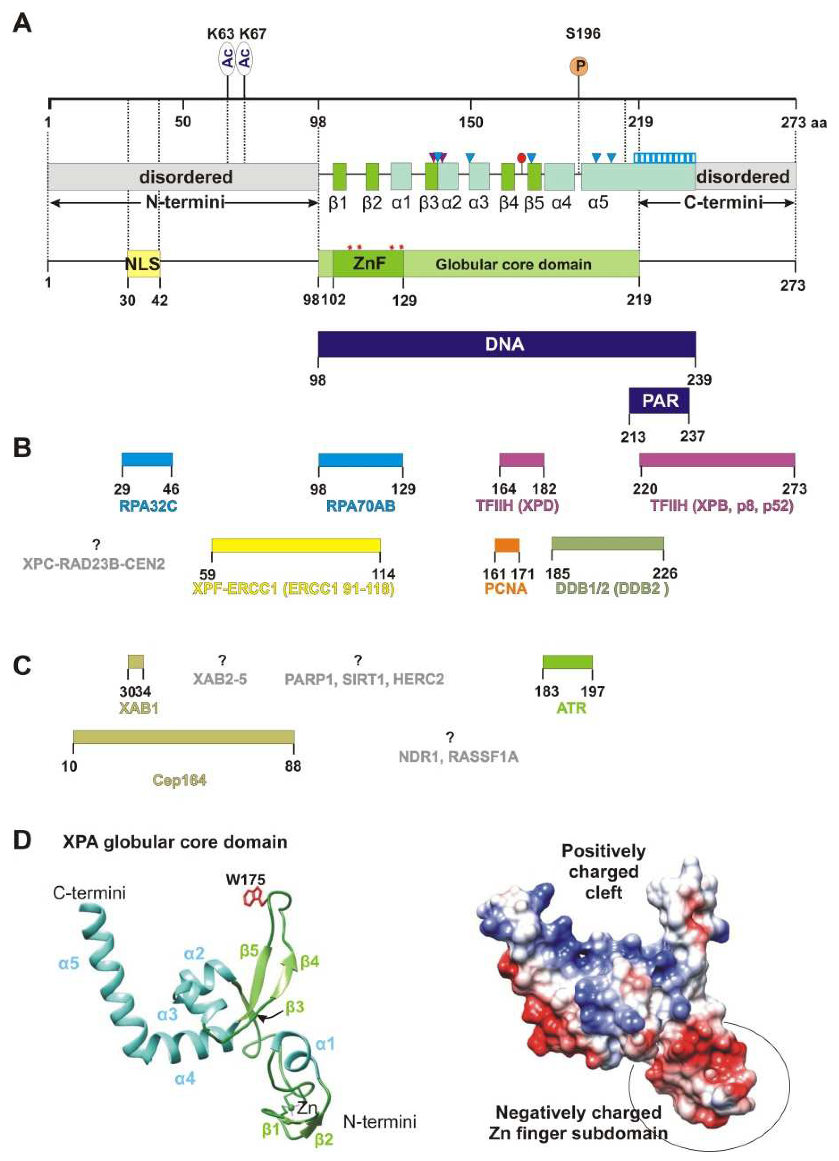
Figure 1. XPA’s structure and interaction partners. (A) The map of the XPA domain structure and known points of PTMs: phosphorylation at S196 and acetylation at K63 and K67. Secondary-structure elements are shown according to crystal structures PDB 6LAE and 6J44: β-strands are green (β1: aa 103–104, β2: aa 111–112, β3: aa 138–140, β4: aa 164–167, and β5: aa 178–172), and α-helices are light blue (α1: aa 116–121, α2: aa 141–148, α3: aa 151–157, α4: aa 183–194, and α5: aa 197–239). Positively charged residues K141, K151, K179, R207, and R211, which are directly involved in interactions with backbones of a DNA duplex, are shown as blue triangles. Two residues (Thr140 and Thr142, indicated as purple triangles) interact with the DNA backbone through a van der Waals contact and a hydrogen bond, respectively. Extended helix α5 contains several positively charged residues (Lys217/218/221/222/224/236 and Arg227/228/231/237) that are possibly involved in DNA binding, which are shown as a blue striped box. Conserved residue Trp175 intercalates into unpaired bases of single-stranded DNA (ssDNA) at the ss–dsDNA junction and is displayed as a red circle. Unstructured N- and C-terminal regions are gray. Zinc-coordinated conserved cysteine residues (C105, C108, C126, and C129) are presented as red asterisks and a Zn-finger motif (ZnF, aa 102–129) colored green. The N terminus accommodates a nuclear localization signal (NLS, aa 30–42), which is yellow. DNA-binding (aa 98–239) and poly(ADP-ribose) (PAR)-binding (aa 213–237) motifs are mapped to the overall XPA structure and are highlighted in dark blue. (B) Interaction sites for NER protein partners on XPA, which are aligned with the XPA residues involved in each interaction. Proteins whose interaction sites are unknown are gray. (C) XPA interaction partners outside NER. (D) A structural model of the XPA globular core domain (PDB ID: 6LAE). A ribbon diagram with color codes according to (A). The Trp175 residue is shown in red. A distribution of the electrostatic potential on the surface for the same structure: a positive charge is shown in blue, and a negative charge is red. The structures were generated using UCSF Chimera software (version 1.16).
Cells 11 03723 g001
Figure 2. XPA interaction with the ss–dsDNA junction. (A) Cryo-EM structure PDB: 6RO4 provides details of the XPA–DBD interaction with the ss–dsDNA junction. XPA demarcates the 5′ edge of the DNA repair bubble. XPA inserts its intercalating β-hairpin between DNA single strands at the junction. Red colored Trp175 from the tip of the β-hairpin stacks against the base of the DNA 3′-extension at the junction. The structures were generated using UCSF Chimera software (version 1.16). (B) Schematic representation of the interactions between side chains of the XPA and DNA junction, according to cryo-EM structure PDB: 6RO4 [21] and crystal structure PDB: 6LAE. DNA nucleotides are indicated as circles. Patches of positively charged residues in proximity to the DNA backbone are indicated by red pluses. Hydrogen bonding of T142 and a van der Waals contact of T140 are indicated as black lines.
Cells 11 03723 g002
Figure 3. XPA organizes GG-NER machinery functioning. (Initial damage recognition) XPA interacts physically with DDB2 and XPC, but the biological role of these interactions is not understood. (Damage verification) XPC recruits TFIIH through XPC–p62 interaction. When XPA joins this complex through interaction with p8 and p52, it induces TFIIH rearrangement through binding XPB. During XPB binding, XPA engages the Cen2-binding site on XPB and possibly promotes XPC complex displacing. Next, XPG joins the complex by occupying the CAK-binding site on XPD. Thus, XPA and XPG promote CAK releasing and XPA “turning on” TFIIH repair conformation. During the damage verification, XPA retains XPB on DNA (see also Figure 4), modulates XPD helicase activity, and further inhibits it in the presence of a bulky lesion. (Pre-incision complex) XPA drives RPA binding to the undamaged strand through XPA–RPA32 interaction and stabilizes the PIC interior by additional interaction with RPA70. PIC becomes completed after XPF–ERCC1 engagement, which is recruited by XPA. XPA is a central component in the PIC, which makes sure that all the NER factors are in the right place for the incision to occur. At the later stages, XPA remains in the NER complex via a protein–protein interaction with the RPA that stays bound to the undamaged strand. Additionally, XPA possibly promotes the positioning of the PCNA clamp.
Cells 11 03723 g003
Figure 4. Cryo-EM structure of the human core TFIIH–XPA–DNA complex (PDB ID: 6RO4). XPA is colored light blue. The TFIIH subunits that are directly involved in the interaction with XPA: XPB, XPD, p52, and TTDA/p8 are colored yellow, magenta, green, and orange, respectively. In this complex, XPA forms a bridge between XPB and XPD, and XPA’s extended α5 helix and XPB form a positively charged tunnel that holds the DNA duplex within. XPA binds to XPD via its intercalating β-hairpin. The C-terminal region of XPA should be extended to p52 and TTDA/p8 and bind to these subunits, but unfortunately, it was observed at a lower resolution and was not used for the final model building (for details, please, see original research article [21]). The structures were generated using UCSF Chimera software (version 1.16).
Cells 11 03723 g004
Figure 5. The circadian rhythm of XPA’s life. (A) The circadian rhythm of NER activity is due to the circadian oscillation of XPA mRNA and the protein levels. In a mouse model, it was shown for the brain, skin, and liver that the elimination of cisplatin–DNA adducts by the NER system exhibits a robust circadian rhythm, with the zenith in the late afternoon hours (~5 p.m.) and the nadir in the early morning hours (~5 a.m.). This oscillation is caused by the circadian rhythmicity of Xpa transcription and translation. (B) The balance between production and degradation. The mammalian circadian clock is generated by a transcriptional–translational feedback loop: core clock proteins CLOCK and BMAL1 activate the transcription of many clock-controlled genes, including repressor genes period (PER1/2) and cryptochrome (CRY1/2), by binding to E-box elements in their promoters and activating their transcription. After a time delay, the CRY and PER proteins accumulate in the cytoplasm, then form the CRY–PER complex, and are translocated back into the nucleus to inhibit their own transcription, as well as the transcription of clock-controlled output genes, through the inhibition of CLOCK–BMAL1 activity. The amount of repressor proteins CRY and PER is also regulated by secondary feedback loops at the transcriptional level, as well as by proteolytic degradation [126]. Accordingly, the XPA mRNA and protein levels are regulated positively by the CLOCK–BMAL1 complex and negatively by the CRY–PER complex. To attain a sufficient amplitude of the XPA protein oscillation level, circadian transcriptional regulation is coupled with the permanent prompt removal of the XPA protein by ubiquitin-dependent degradation. (C) UV light-induced damage and protective phosphorylation. In response to UV in a dose-dependent manner, ATR binds to and phosphorylates XPA. The XPA–ATR interaction facilitates HERC2 dissociation from the XPA complex, resulting in the accumulation of XPA molecules.
Cells 11 03723 g005
Figure 6. The ATR interaction site on XPA is the α4-helix located on the N-terminal side of the α4-helix-turn-α5-helix motif. ATR phosphorylates XPA at serine 196 (S196), which is located in the “turn” element inside the helix-turn-helix motif. The color codes correspond to Figure 1A. The structure was generated using UCSF Chimera software (version 1.16).
Cells 11 03723 g006
Figure 7. The network of reciprocal regulatory mechanisms of XPA’s PTMs. (A) The putative phosphorylation complex. ATR phosphorylates XPA at S196. (B) Acetylation–phosphorylation regulation. XPA is acetylated at Lys63 and Lys67. SIRT1 promotes NER by deacetylating XPA. The existence of an ATR–SIRT1 interaction is unclear and is indicated by the question mark. (C) PARylation–acetylation interplay. XPA is PARylated by PARP1. Both PARP1 and SIRT1 use NAD+ for their activity and interact physically. There is a strong connection between acetylation and PARylation. SIRT1 may deacetylase PARP1 and inhibit the PARP1 enzymatic activity. Under severe stress, PARP1 can become overactivated and may deplete cellular NAD+, thereby leading to the repression of SIRT1 activity and suppressing SIRT1 transcription. To prevent this situation, SIRT1 is also capable of negatively regulating the expression of the PARP1 gene. Moreover, XPA has a high affinity for long PAR polymers. Whether the PARylation events involve phosphorylated XPA is unclear and is denoted by the question mark.
Cells 11 03723 g007
Publisher’s Note: MDPI stays neutral with regard to jurisdictional claims in published maps and institutional affiliations.