Article

Mitochondria-Targeted Antioxidant SkQ1 Prevents the Development of Experimental Colitis in Mice and Impairment of the Barrier Function of the Intestinal Epithelium

by
Artem V. Fedorov
1,*,
Maria A. Chelombitko
2,3,
Daniil A. Chernyavskij
2,
Ivan I. Galkin
2,
Olga Yu. Pletjushkina
2,
Tamara V. Vasilieva
1,
Roman A. Zinovkin
2,4 and
Boris V. Chernyak
2,*
1
Department of Cell Biology and Histology, Biology Faculty, Lomonosov Moscow State University, 119991 Moscow, Russia
2
Belozersky Institute of Physico-Chemical Biology, Lomonosov Moscow State University, 119991 Moscow, Russia
3
Russian Clinical Research Center for Gerontology of the Ministry of Healthcare of the Russian Federation, Pirogov Russian National Research Medical University, 129226 Moscow, Russia
4
HSE University, 101000 Moscow, Russia
*
Authors to whom correspondence should be addressed.
Cells 2022, 11(21), 3441; https://doi.org/10.3390/cells11213441
Submission received: 24 September 2022 / Revised: 22 October 2022 / Accepted: 28 October 2022 / Published: 31 October 2022
(This article belongs to the Section Tissues and Organs)

Abstract

:
Mitochondria-targeted antioxidants have become promising candidates for the therapy of various pathologies. The mitochondria-targeted antioxidant SkQ1, which is a derivative of plastoquinone, has been successfully used in preclinical studies for the treatment of cardiovascular and renal diseases, and has demonstrated anti-inflammatory activity in a number of inflammatory disease models. The present work aimed to investigate the therapeutic potential of SkQ1 and C12TPP, the analog of SkQ1 lacking the antioxidant quinone moiety, in the prevention of sodium dextran sulfate (DSS) experimental colitis and impairment of the barrier function of the intestinal epithelium in mice. DSS-treated animals exhibited weight loss, bloody stool, dysfunction of the intestinal epithelium barrier (which was observed using FITC-dextran permeability), reduced colon length, and histopathological changes in the colon mucosa. SkQ1 prevented the development of clinical and histological changes in DSS-treated mice. SkQ1 also reduced mRNA expression of pro-inflammatory molecules TNF, IL-6, IL-1β, and ICAM-1 in the proximal colon compared with DSS-treated animals. SkQ1 prevented DSS-induced tight junction disassembly in Caco-2 cells. Pretreatment of mice by C12TPP did not protect against DSS-induced colitis. Furthermore, C12TPP did not prevent DSS-induced tight junction disassembly in Caco-2 cells. Our results suggest that SkQ1 may be a promising therapeutic agent for the treatment of inflammatory bowel diseases, in particular ulcerative colitis.

1. Introduction

Ulcerative colitis (UC) is a disease of unknown etiology characterized by inflammation of the mucosa and submucosa of the colon and rectum, leading to the development of ulcers [1]. UC is considered a global health problem with an accelerating incidence [2]. The impaired barrier function of the intestinal epithelium and dysregulated immune response in the intestinal mucosa play a key role in the pathogenesis of UC [3]. The integrity of the intestinal barrier depends on antibacterial peptides, immunoglobulin A, mucous layer, and tight junctions (TJs) between intestinal epithelial cells (IEC). TJs are mainly composed of the transmembrane proteins occludin and claudins and the peripheral protein Zonula Ocludence-1 (ZO-1). The disruption of TJs results in the increase of paracellular permeability and penetration of bacteria through the barrier, leading to inflammation [4,5]. Dysfunction of the intestinal epithelium presumably precedes inflammation in the pathogenesis of inflammatory bowel disease (IBD) and colitis. The only anti-inflammatory therapies available include inhibitors of immune cell activation, monoclonal antibodies against inflammatory cytokines, and interferons α/β, which stimulate the production of anti-inflammatory cytokines [3].
Increased oxidative stress has long been recognized as an important pathogenic factor in IBD and colitis, which can impair the intestinal barrier [6]. Signs of oxidative stress are detected both in various experimental models and in patients with IBD. Activated immune cells such as neutrophils produce a predominant amount of reactive oxygen species (ROS) during intestinal inflammation. However, at the initial stages of the disease, IEC-generated ROS may play a key role. Thus, patients with Crohn’s disease and colitis have higher levels of cytoskeletal protein carbonylation and NO production in their intestinal mucosa [7].
In a mouse model of UC induced by sodium dextran sulfate (DSS), oxidative stress was detected using an in vivo electron spin resonance (ESR), or a nitroxyl probe method [8]. It has been shown that intracellular ROS are produced in the initial stage of colitis, and then, in the advanced stage of colitis, ROS are produced predominantly extracellularly. Later, using Overhauser-enhanced magnetic resonance imaging, the same researchers demonstrated ROS production in the epithelium of the distal and proximal colon in the initial stages of DSS-induced colitis before the development of inflammatory changes in the colon mucosa [9]. Ex vivo analysis of colon tissue from DSS-treated mice revealed elevated levels of malondialdehyde and 3-nitrotyrosine, reliable markers of oxidative stress [10].
Mitochondria in the intestine play important roles in immune cell activation, in maintaining barrier integrity, as well as in stemness and differentiation of stem cells [11]. Dysfunction of mitochondria in IEC contributes to the pathogenesis of IBD, including Crohn’s disease and UC. Detailed analysis of mucosal transcriptomes across very large cohorts of pediatric and adult UC patients revealed a significant reduction of mitochondrial metabolic-associated gene expression [12]. Importantly, expression of all 13 genes encoded in the mitochondrial genome was markedly suppressed in UC. In addition, the expression of PGC-1α, the master regulator of mitochondrial biogenesis, was strongly reduced across UC patients. The paralleled analysis of the mitochondrial functions in colonic from UC patients also revealed a significant decline. Mitochondrial unfolded protein response, a universal cell response to severe mitochondrial dysfunction, has been observed in IBD patients’ IEC and UC mouse models [11].
Mitochondrial ROS (mtROS) overproduction is one of the most important consequences of mitochondrial dysfunction. MtROS play a key role in the induction of apoptosis of IEC, leading to the barrier disruption in UC [13]. MtROS are an important component in signaling leading to the activation of nuclear factor-κB (NF-κB) transcription factor [14,15]. NF-κB activation in macrophages and IEC from biopsy samples from patients with IBD, as well as in mouse models of colitis, correlated with inflammation severity [16]. Activation of NF-κB is associated with the production of the pro-inflammatory cytokines in IEC and immune cells, as well as with the disassembly of tight junctions in the intestinal epithelium [17].
Mitochondria-targeted antioxidants (MTA) become a very important tool for studies of mtROS-dependent signaling and are promising candidates for the therapy of various pathologies [18,19]. At the same time, the results of MTA application in models of IBD appear quite controversial (see Section 4. Discussion). Thus, the mitochondria-targeted derivative of ubiquinone (MitoQ) was found protective in the DSS-induced mouse model of UC in the early study [10], but later the opposite effect of MitoQ was reported in the same model [20]. Despite this, the clinical trial of oral MitoQ in moderate ulcerative colitis began in 2021 [21].
The mitochondria-targeted derivative of plastoquinone, SkQ1 (10-(6′-plastoquinonyl) decyltriphenylphosphonium bromide) was shown to be an effective antioxidant in vitro [22] and in vivo [23,24]. It was successfully used in preclinical studies for the treatment of cardiovascular and renal diseases [25,26], and also demonstrated anti-inflammatory activity in acute bacterial infection [27] and the systemic inflammatory response syndrome (SIRS) model [15]. SkQ1 efficiently stimulates compromised dermal wound healing in old and diabetic mice [24,28]. In human neutrophils, SkQ1 prevents oxidative burst, degranulation, and neutrophil extracellular trap (NET) formation caused by various stimuli [29,30]. In endothelial cell culture, SkQ1 has been shown to prevent the disassembly of intercellular contacts [14] and apoptosis [31] induced by TNF. It was shown that SkQ1 prevented TNF-induced activation of NF-κB and expression of adhesion molecules necessary for the adhesion of neutrophil to endothelium during inflammation [14,15].
In the present work, we have shown that SkQ1 at very low doses prevents the development of clinical and histological changes, impairment of the intestinal epithelial barrier, and production of inflammatory cytokines in the DSS-induced mouse model of colitis. In the Caco-2 cell model of the intestinal epithelium, SkQ1 prevented DSS-induced tight junction disassembly.

2. Materials and Methods

2.1. Mice

Male C57Bl/6 (12 to 15 weeks of age) mice were purchased from the Stolbovaya Research Center for Biomedical Technologies of the Federal Medical and Biological Agency of Russia. Four to five animals were housed per cage. Mice had free access to drinking water and briquetted feed (“Laboratorkorm”, Russia). The experimental protocol was approved by the institutional animal ethics committee (protocol No.1, 6 February 2020).

2.2. Induction of Colitis

Mice were divided into four groups: the control group (n = 5), DSS-induced colitis group (n = 9), DSS-induced colitis + C12TPP (decyltriphenylphosphonium bromide) group (n = 8), and DSS-induced colitis + SkQ1 (10-(6′-plastoquinonyl) decyltriphenylphosphonium bromide) group (n = 8). Mice received 250 nmol/kg of body weight per day of SkQ1 or C12TPP (synthesized at the Institute of Mitoengineering, Lomonosov Moscow State University) with drinking water for 18 days before induction of colitis and peroral using an automatic pipette during induction of colitis (Supplementary Figure S1).
For 6 days, mice were given 1% DSS (Dextran sulfate sodium salt, Mr ~ 40,000, Sigma-Aldrich, St. Louis, MO, USA) in their drinking water to induce colitis. Control mice were given tap water. At day 8, all animals received 440 mg/kg of body weight of FITC-dextran (4 kDa, Sigma-Aldrich, St. Louis, MO, USA) and fluorescence was measured in the plasma 4 hours later to evaluate the barrier function of IEC. After isoflurane anesthesia, blood from jugular veins was taken into heparinized tubes. Blood was centrifuged at 2000× g for 5 min to collect plasma. The fluorescence of FITC-dextran in plasma was measured on a Fluoroskan Ascent plate fluorometer (Thermo Scientific, Hercules, CA, USA) at a wavelength of 485 nm (extinction wavelength 530 nm).
Clinical scores of colitis were evaluated by the measurement of weight change and colorectal bleeding. Colorectal bleeding was assessed on the following scale: 0—normal stool, 1—soft stools, 2—soft stools with traces of blood, and 3—watery stools with visible rectal bleeding.
The lengths of removed colons were measured in sacrificed mice. The colon was washed and divided into three sections – proximal, medial, and distal. Proximal colon samples were frozen in liquid nitrogen for further mRNA isolation and gene expression analysis. Samples of the distal colon were fixed in 10% neutral buffered formalin for further histological analysis.

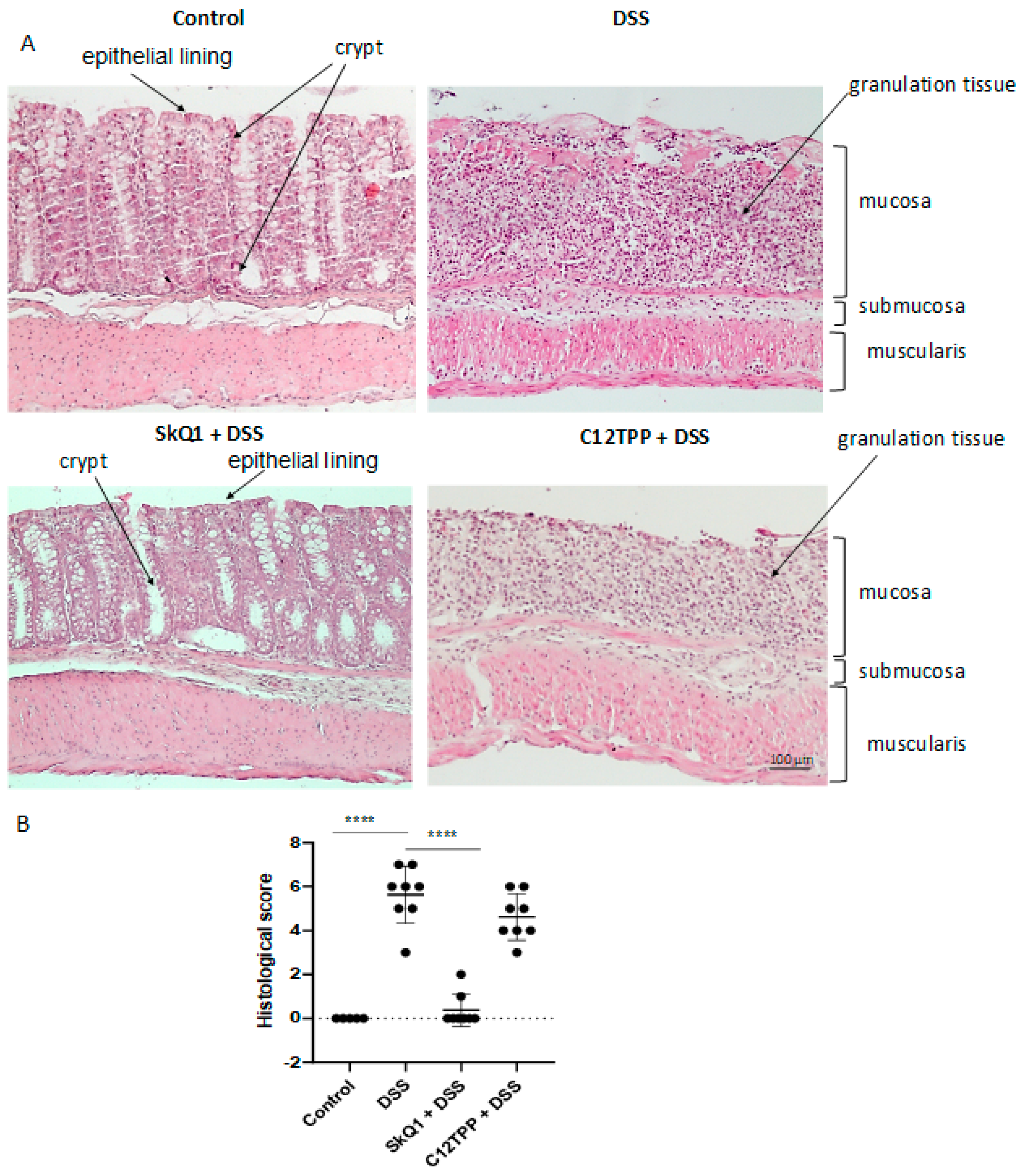
2.3. Histological Analysis

After fixation, the colon samples were processed for paraffin embedding, sectioned at 5 µm thickness, and stained with hematoxylin and eosin. Histological scoring was performed as previously described [32]. The histological grading of colitis is described in Supplementary Table S1.

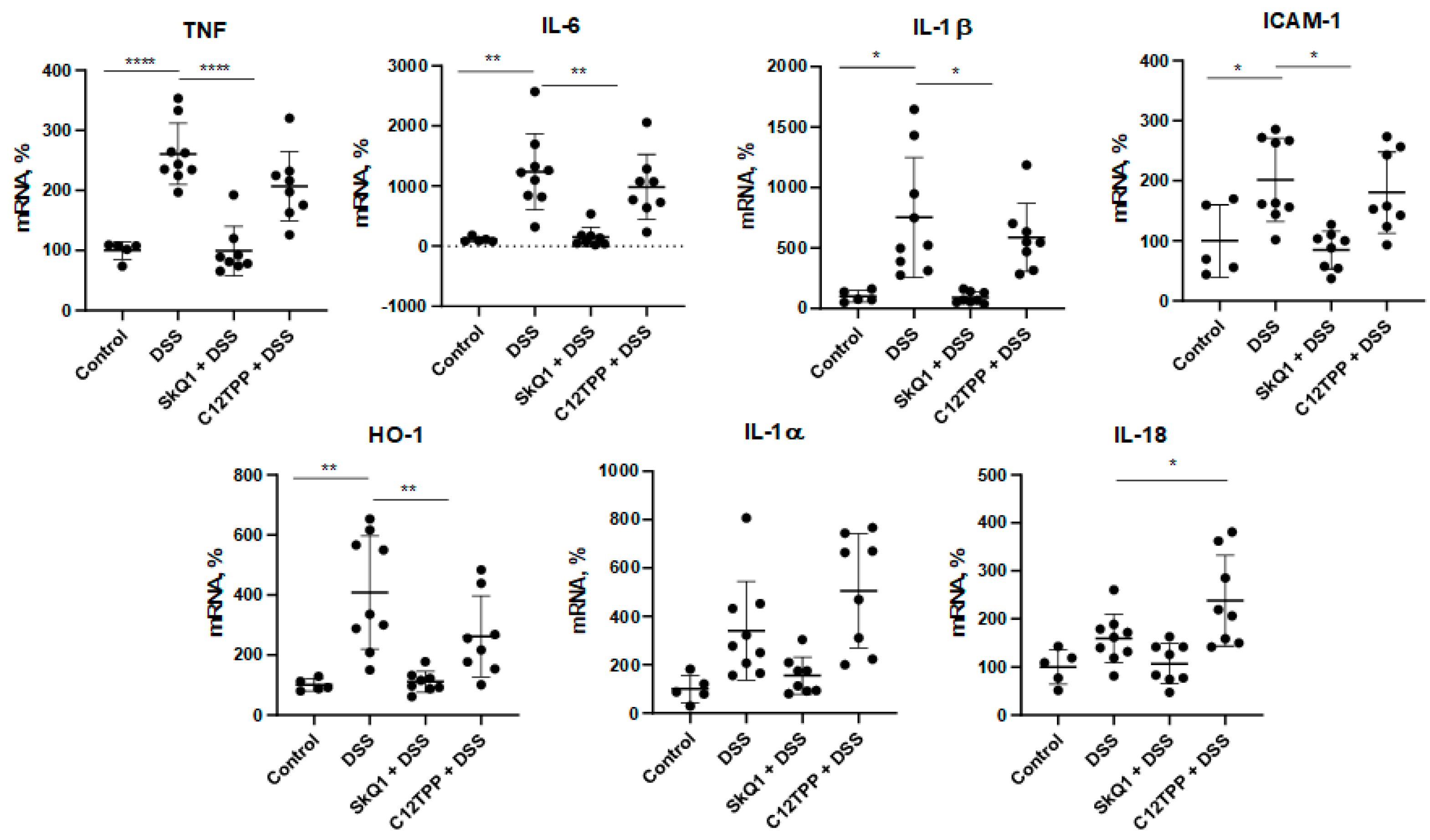
2.4. Reverse Transcription-PCR

RNA from the proximal colonic section was isolated using a QIAGEN RNeasy Blood and Tissue kit according to the manufacturer’s instructions. The integrity and purity of RNA were verified by agarose gel electrophoresis and spectrophotometry using a NanoDrop ND-1000. RNA was reverse transcribed by SuperScript III reverse transcriptase (Thermo Fisher Scientific, Waltham, MA, USA). Quantitative PCR (qPCR) was carried out with EvaGreen intercalating dye (Syntol, Moscow, Russia) on the CFX96 Touch™ Real-Time PCR Detection System (BioRad, Hercules, CA, USA). PCR conditions included a preheating step at 95 °C for 3 min and 40 cycles at 95 °C for 15 s, 57 °C for 15 s, and 72 °C for 15 s coupled with fluorescence measurement. The amplification specificity was confirmed by melt curves analysis. Each sample was run in triplicate, and a non-template control was added to each run. The choice of reference genes TATA-box-binding protein (Tbp) and eukaryotic translation elongation factor 2 (Eef2) was based on the results of a stability study of the reference genes in a mouse model of DSS-induced colitis [33]. The expression levels of the target genes were determined by the ΔΔCt method with the calculated primer efficiencies and normalized on the geometric mean of selected reference genes. Primer sequences are listed in Supplementary Table S2.

2.5. Cell Culture

Caco-2 cells (ATCC HTB-37) were cultivated in the DMEM medium containing 10% fetal bovine serum (FBS) at 37 °C and 5% CO2. The cells were seeded on glass coverslips in a 6-well plate (100,000 cells/well). After cell adhesion, SkQ1 and C12TPP were added to the final concentration of 2 nM. This concentration was found to be the most effective in our previous work [15]. Two days after incubation, the cells were treated with 2% DSS for two days.

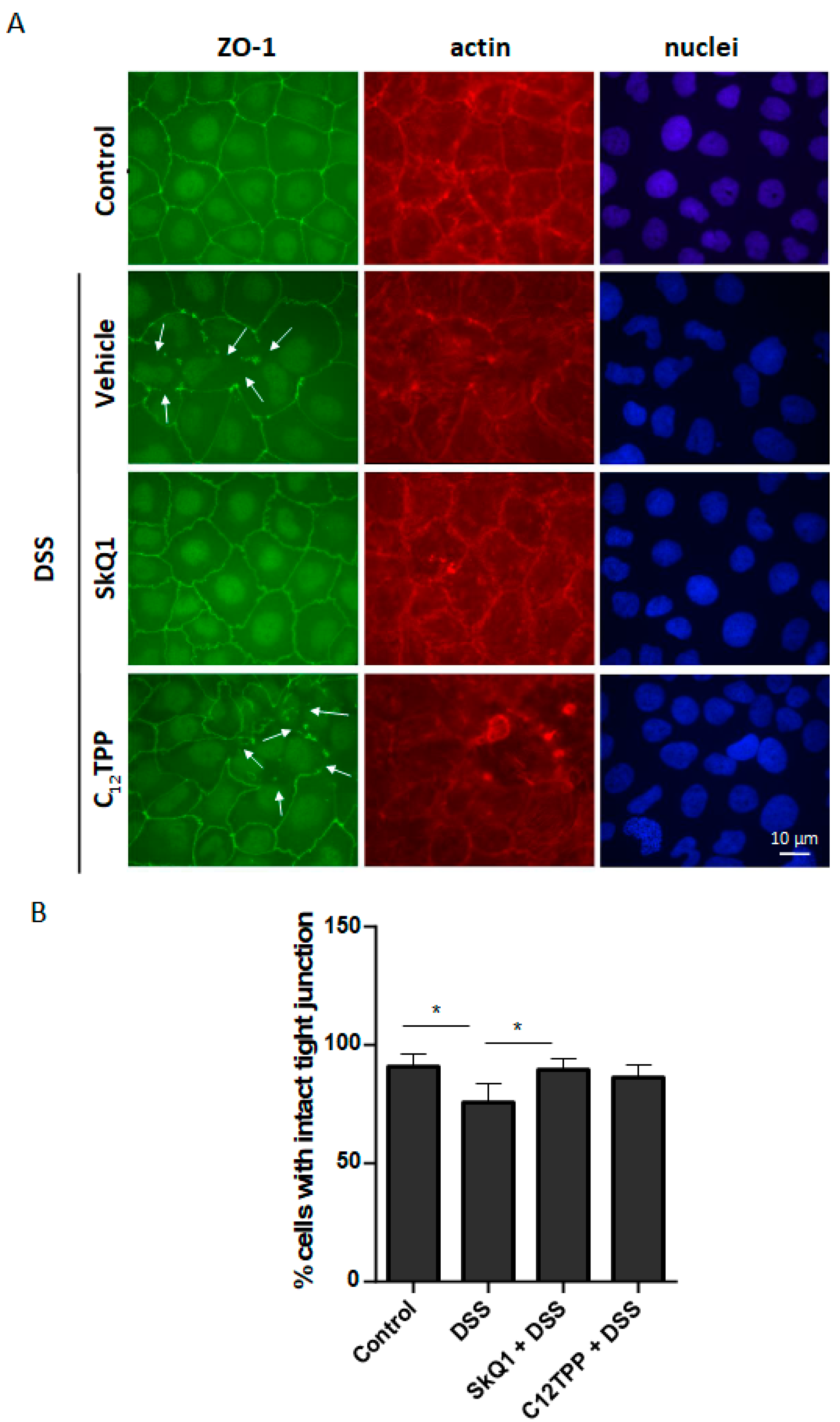
2.6. Fluorescence Microscopy

Cells were washed with DMEM, fixed with a 2% solution of paraformaldehyde in DMEM for 10 min at 37 °C, and permeabilized with 0.2% Triton X-100 in PBS for 5 min at RT. Cells were stained with anti-ZO-1 polyclonal antibodies (AB2272, Merck Millipore, Billerica, MA, USA) overnight at +4 °C. The cells were stained for 1 h, at RT with secondary cross-adsorbed Goat anti-Rabbit IgG (H+L) antibodies, conjugated with Alexa Fluor 488 (A11034 Thermo Fisher Scientific, Waltham, MA, USA), phalloidin-TRITC (Sigma-Aldrich, St. Louis, MO,), and Hoechst 33342 (Biotium, Fremont, CA, USA). After washing with PBS, the cells were embedded using Aqua-Poly/Mount (Polysciences, Warrington, PA, USA). Microscopy was performed using a fluorescent microscope Axiovert 200M (Zeiss, Oberkochen, Germany) with objective EC Plan-NEOFLUAR 63x/0,75 Ph2 (Zeiss, Oberkochen, Germany). Analysis of morphological data was performed in ImageJ. The percentage of cells with intact tight junctions was estimated. In four separate experiments, 500 to 600 cells were counted. Cells with a continuous line of ZO-1 around their perimeter were identified as cells with intact tight junctions.

2.7. Statistical Analysis

ANOVA data analysis with post-hoc Dunnett’s multiple comparison test was performed using the GraphPad Prism 8.0 software (GraphPad Software, San Diego, CA, USA). The DSS-treated group was set as a control group for Dunnett’s post-hoc tests. Data are presented as mean ± SD. p-values less than 0.05 (*), 0.01 (**), 0.001 (***), and 0.0001 (****) were considered significant.

3. Results

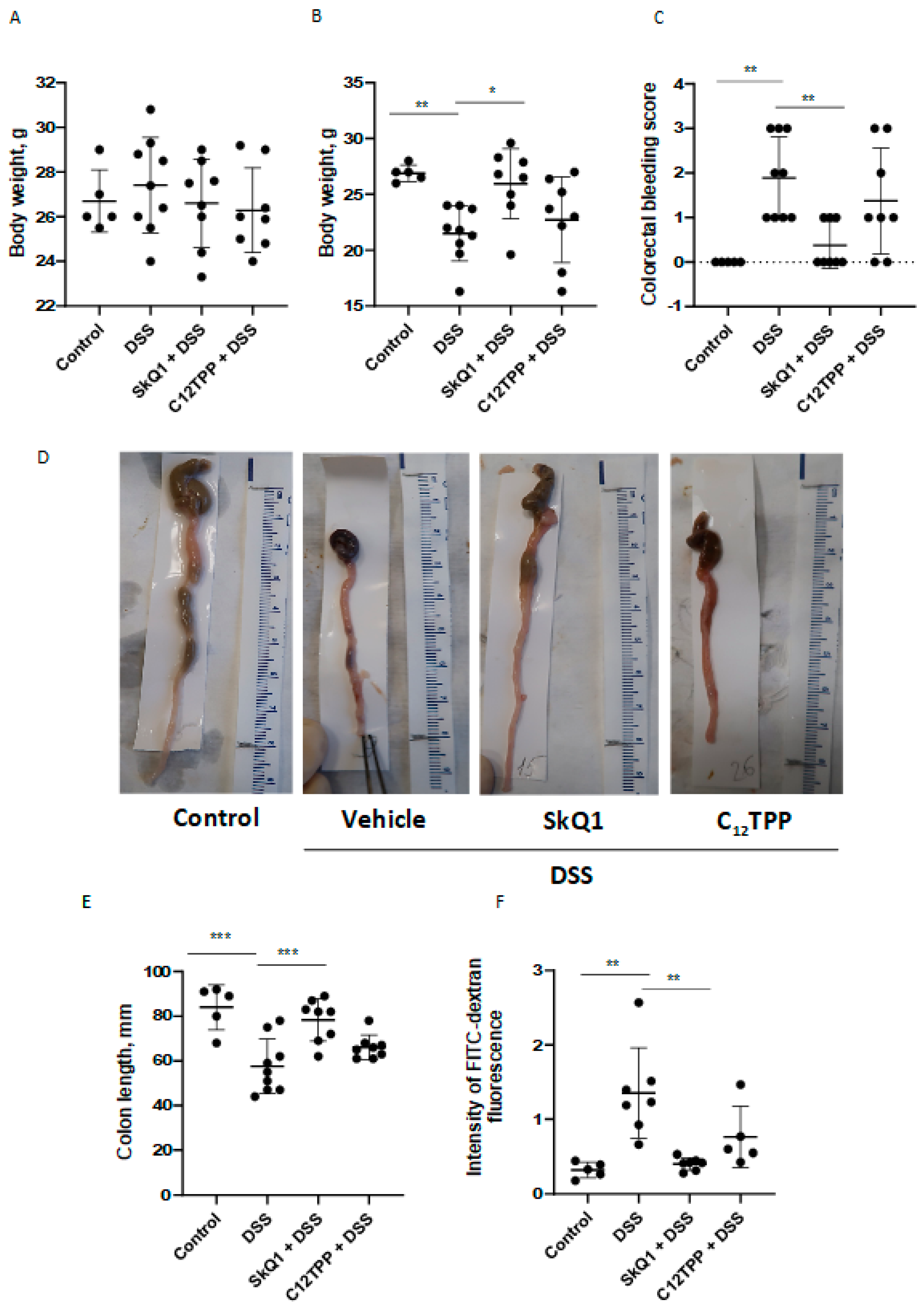
3.1. SkQ1 Prevents the Development of Dextran Sulfate Sodium-Induced Colitis in Mice

In mice treated with DSS, clinical signs of colitis such as weight loss and bloody stool were observed. The development of DSS-induced colitis was accompanied by a significant decrease in body weight (Figure 1A,B) and colorectal bleeding (Figure 1C), which were prevented by SkQ1. Treatment of mice by C12TPP, the analog of SkQ1 lacking the antioxidant quinone moiety, using the same scheme did not protect against colitis (Figure 1).
An important sign of the development of UC is a decrease in the length of the colon. The severity of UC is determined by the reduction of colon length. In agreement with this, DSS-treated mice had significantly reduced colon length compared with control mice (Figure 1D,E). SkQ1 prevented the development of this trait, while C12TPP had no protective effect (Figure 1D,E).
An increase in intestinal epithelium permeability is another critical parameter in UC. A significant increase in mouse intestinal permeability for FITC-dextran was observed in DSS-treated mice compared with control mice (Figure 1F). In mice treated with SkQ1, there was a significant decrease in intestinal permeability compared with a group with colitis, while C12TPP had no noticeable effect (Figure 1F).
The results of histological examination of the distal colon in mice treated with DDS confirmed the development of colitis. An almost complete absence of crypts and epithelial lining of the mucosa (mucous membrane) was observed. In the lamina propria of the mucous membrane, instead of crypts, there was granulation tissue with dilated blood vessels and a pronounced inflammatory infiltrate. Edema and inflammatory infiltration were also observed in the submucosal layer (submucosa). In mice treated with SkQ1, the microscopic structure of the intestine did not significantly differ from that of intact control animals, while C12TPP had no protective effect (Figure 2).
The development of the inflammatory process in the colon in mice with DSS-induced colitis was confirmed by the results of the analysis of pro-inflammatory cytokine gene expression. Thus, a significant increase in mRNA of TNF, IL-6, IL-1β, and ICAM-1 genes in the proximal colon in mice with DSS-induced colitis compared with control mice (Figure 3). In full agreement with its protective effect, SkQ1 prevented an increase in the expression of all these genes, while C12TPP had no effect. No significant difference was found in the expression of IL-1б and IL-18. An increase in heme oxygenase-1 (HO-1) gene expression was observed, and that SkQ1, but not C12TPP, prevented this increase (Figure 3). HO-1 expression is a reliable marker of Nrf2 activation, so these data suggest that DSS induces NRf2 activation due to the action of mtROS.

3.2. SkQ1 Prevents the DSS-Induced Disassembly of Intercellular Contacts in the Caco-2 Cells

To test the hypothesis that the therapeutic effect of SkQ1 may be explained by its direct effect on the barrier function of the intestinal epithelium, we evaluated the SkQ1 effect on DSS-induced disassembly of intercellular contacts in the Caco-2 intestinal epithelial cells. Disassembly of the contact structure was assessed by the relocation of TJ ZO-1 protein from the contact area to the cytoplasm. Pretreatment of cells with SkQ1 suppressed the release of ZO-1 from the contacts, while C12TPP had no effect (Figure 4). In addition, we observed an increase in the number of actin filaments resembling stress fibers in cells treated with DSS. Such fibers are usually formed in response to mechanical and other stresses. SkQ1, but not C12TPP, prevented the formation of these fibers (Figure 4).

4. Discussion

Oral administration of SkQ1 prevented the development of morphological and clinical signs of UC pathology in DSS-induced mice (Figure 1). SkQ1 also prevented an increase in intestinal epithelial barrier permeability (Figure 1D) and the expression of inflammatory mediators in the proximal colon of DSS-treated mice (Figure 2). Histological changes confirmed the prevention of pathological changes in the colon by SkQ1 (Figure 3). The control compound C12TPP did not affect the development of pathologies, thus it can be assumed that the scavenging of mtROS mediates the therapeutic action of SkQ1. Previous studies have shown that SkQ1 is an effective antioxidant in vitro [22] and in vivo [23,24].
DSS-induced colitis is a widely used mouse model of UC. In fact, MitoQ was the first MTA found to be protective in this model [10]. Oral administration of MitoQ for 7 days after UC induction reduced clinical and histological changes in the colon and suppressed activation of pro-inflammatory cytokines IL-1β and IL-18. The production of these cytokines is predominantly dependent on the activation of NLRP3 inflammasome in macrophages, so it was assumed that these cells are the main target of MitoQ [10]. However, the opposite effect of MitoQ on macrophage activation was later reported using the same model [20]. MitoQ has been shown to reduce oxidative stress and NF-κB activation but exacerbated colitis, presumably by inhibiting macrophage polarization to the anti-inflammatory M2 phenotype. In both studies, the integrity of the intestinal epithelial barrier was not analyzed. As shown in Figure 3, SkQ1 inhibited the expression of immature IL-1β, as well as the inflammatory cytokines TNF and IL-6, and adhesion molecule ICAM-1. Since NF-κB controls the expression of all these genes, activation of NF-κB by mtROS is proposed in the DSS-induced UC model. This assumption is in good agreement with our earlier studies, which showed the prevention of TNF-induced NF-κB activation by SkQ1 was shown in the endothelium [14,15].
More recently, two independent studies have shown that the effect of MitoQ in ischemic [34] and lipopolysaccharide-induced [35] mouse models of UC was mediated by activation of nuclear factor erythroid 2-related factor 2 (Nrf2). Nrf2 is a transcription factor up-regulated by oxidative and electrophilic stresses to activate the expression of antioxidant and other protective genes. The protective effect of Nrf2 in IBD is well known [36]. Activation of Nrf2 by MitoQ can be explained by the pro-oxidant activity of this MTA inherent in quinone-based antioxidants [37]. Antioxidant enzymes controlled by Nrf2 can protect mitochondria as well as other cellular structures from cytosolic ROS, so the role of mtROS scavenging by MitoQ in protection against UC remains unclear. As shown in Figure 3, DSS stimulated and SkQ1 prevented the expression of hemoxygenase-1 (HO-1), a strong marker of Nrf2 activation [38]. These data assume that Nrf2 activation in the DSS-induced model of UC is mtROS dependent. Importantly, SkQ1 was used at a significantly lower daily dose (0.25 µmol/kg) in our study than MitoQ was (6–80 µmol /kg) in the cited studies. Furthermore, SkQ1’s pro-oxidant activity is approximately 100 times lower than that of MitoQ [39]. The low pro-oxidant activity of SkQ1 may explain the lack of additional Nrf2 activation in the DSS-induced UC model.
Another MTA, MitoTEMPO, has been shown to reduce intestinal barrier dysfunction as assessed by Escherichia coli transepithelial flux in the DSS-induced mouse model of UC [40]. The possible activation of Nrf2 was not analyzed in this study, so the detailed mechanism of action of MitoTEMPO remains unclear. More recently, MitoTEMPO has been shown to protect against IBD in mice with an inducible deletion of prohibitin 1 in intestinal epithelial cells [41]. Prohibitin 1 is a chaperone for mitochondrial DNA, which expression is downregulated during active human IBD and DSS-induced colitis in mice [42], so its possible role in the pathogenesis of IBD may be mediated by excessive production of mtROS. MitoTEMPO does not rapidly regenerate in mitochondria, so unlike SkQ1 and MitoQ, it cannot act as a “rechargeable” antioxidant. This is one of the reasons why MitoTEMPO is used at relatively high doses in cellular or animal models, and no clinical trials have been reported with it.
As shown in Figure 4, SkQ1 prevents tight junction disassembly in Caco-2 cells, suggesting that mtROS regulates this type of cell junction. We measured the total level of ROS in the cells using DCFH2-DA but found no significant changes after DSS treatment (Figure S2). Moreover, we did not register any changes induced by SkQ1 in either control or DSS-treated cells. These data suggest that the Caco-2 model of DSS treatment may not be completely relevant to the animal colitis model. The latter involves a complex interaction of many cell types and supposedly has prominent ROS induction. Thus, our suggestion is based only on previous studies that clearly demonstrate that SkQ1 effectively removes mtROS in a variety of animal models and cell types [22,23,24]. We hypothesize that the increase in mtROS induced by DSS may be either small or transient, so our methods were unable to detect this response. Loss of ZO-1 from TJ complexes and increased permeability of the intestinal epithelium have been shown to precede the development of significant intestinal inflammation in DSS-induced colitis [43]. Disruption of the TJ complex probably underlies the development of the inflammatory infiltrate observed in colitis. It has been previously shown that oxidative stress induced by moderate hypoxia followed by reoxygenation destroys TJ complexes in the blood-brain barrier [44]. Exogenous superoxide-producing xanthine oxidase [45] as well as acetylsalicylic acid induce ROS-dependent TJ disassembly in Caco-2 cells [46]. In Caco-2 cells treated with inflammatory cytokines, nitrosative stress associated with ROS overproduction can also contribute to TJ disruption [47]. The possible role of mtROS in these models has not been studied. MitoQ and MitoTEMPO have been shown to protect against TJ degradation induced by osmotic stress and DSS in Caco2, but important experiments with control TPP-based compounds were not presented [48].
Disassembly of the TJ complex may be dependent on actin cytoskeleton reorganization [49], particularly the formation of contractile actin stress fibers [50]. On the other hand, activation of actomyosin contractility of stress fibers improves TJ assembly and epithelial barrier function [51,52]. The formation of stress-like fibers was induced by DSS in Caco-2 cells and was prevented by SkQ1 (Figure 4), indicating that mtROS play a key role in the reorganization of the actin cytoskeleton. The possible role of these actin fibers in TJ disassembly and disruption of the integrity of the intestinal epithelial barrier requires further study.
Clinical trials with traditional dietary antioxidants in IBD patients gave controversial results. In one of the randomized controlled trials, it was shown that oral supplementation of the antioxidants resulted in significant clinical improvement in patients with colitis and a decreased requirement for corticosteroids, indicating that oxidative stress may have a causative role in the disease [53]. By contrast, other randomized controlled trials did not reveal an effect of antioxidant vitamins C and E on the disease progression while the indices of oxidative stress significantly decreased [54]. The reasons for this discrepancy remain unknown, but probably reflects the differences in dosage and potency of antioxidants.
Mitochondria-targeted antioxidants, such as SkQ1, MitoQ, and MitoTEMPO demonstrated very high efficiency in preclinical studies of various inflammatory pathologies [19]. The high efficiency of eye drops containing SkQ1 has been demonstrated not only in various models of inflammatory eye diseases in animals [55], but also in a clinical study of dry eye syndrome [56]. The clinical trial of oral MitoQ in moderate ulcerative colitis was started in 2021 [21]. The findings of this investigation point to SkQ1 as a potentially effective therapeutic agent for the management of inflammatory bowel diseases, particularly ulcerative colitis.

5. Conclusions

This work confirms the potential of mitochondria-targeted antioxidants for the treatment of inflammatory diseases, including IBD and colitis. SkQ1 consumption had a profound protective effect on the colon of DSS-treated mice. The underlying mechanism of protective SkQ1 action includes the preservation of cell-to-cell contact integrity in vitro. Our study provides additional proof for the investigation of the mitochondria-targeted antioxidants in future clinical trials. Thus, mitochondrial ROS inhibition is a potential approach for the prevention and treatment of IBD.

Supplementary Materials

The following supporting information can be downloaded at: https://www.mdpi.com/article/10.3390/cells11213441/s1, Table S1: histological grading of colitis; Table S2: primer sequences used in this study; Figure S1: experimental design for DSS-induced colitis in mice. Two groups of mice received SkQ1 and C12TPP for 18 days with drinking water and per os from day 19 to 26. To induce colitis, mice were given 1% DSS in drinking water from day 19 to 24, then DSS was changed to tap water for 1 day. On day 8, the clinical assessment of colitis was examined. Mice received 4 kDa FITC-dextran per os. After 4 h, mice were anesthetized, and blood was taken from the jugular veins for further measurement of fluorescence in plasma. The colons were removed, measured, and used for further histological and gene expression analysis; Figure S2: the effect of SkQ1 on intracellular ROS level in Caco-2 intestinal cells after DSS treatment was shown. Intracellular ROS were measured as DCFH2-DA oxidation with flow cytometry. After treatment with 2 nM of SkQ1 for 48 h, cells were treated with 2% DSS for the indicated time. Thereafter, they were loaded with 5 μM DCFH2-DA for 20 min at 37 °C. Flow cytometric analysis was performed using a flow cytometer Beckman Coulter FC500. Results are expressed as mean ± SD (n = 3).

Author Contributions

Conceptualization, A.V.F., I.I.G. and B.V.C.; methodology, A.V.F., M.A.C., I.I.G. and R.A.Z.; investigation, A.V.F., M.A.C., I.I.G., D.A.C., T.V.V. and O.Y.P.; writing—original draft preparation, A.V.F., M.A.C., R.A.Z. and B.V.C.; writing—review and editing, A.V.F., M.A.C., R.A.Z. and B.V.C. All authors have read and agreed to the published version of the manuscript.

Funding

This research received no external funding.

Institutional Review Board Statement

The animal study protocol was approved by the Institutional Ethics Committee) of Belozersky Institute of Physico-Chemical Biology, Lomonosov Moscow State University (protocol No.1, 6 February 2020).

Informed Consent Statement

Not applicable.

Data Availability Statement

Not applicable.

Acknowledgments

This research has been supported by the Interdisciplinary Scientific and Educational School of Moscow University “Molecular Technologies of the Living Systems and Synthetic Biology”.

Conflicts of Interest

The authors declare no conflict of interest.

References

  1. Collins, P.; Rhodes, J. Ulcerative Colitis: Diagnosis and Management. BMJ 2006, 333, 340–343. [Google Scholar] [CrossRef] [PubMed] [Green Version]
  2. Ray, K. IBD: The Changing Epidemiology of IBD. Nat. Rev. Gastroenterol. Hepatol. 2017, 14, 690. [Google Scholar] [PubMed]
  3. Kaur, A.; Goggolidou, P. Ulcerative Colitis: Understanding Its Cellular Pathology Could Provide Insights into Novel Therapies. J. Inflamm. 2020, 17, 15. [Google Scholar] [CrossRef] [PubMed] [Green Version]
  4. Suzuki, T. Regulation of Intestinal Epithelial Permeability by Tight Junctions. Cell. Mol. Life Sci. 2013, 70, 631–659. [Google Scholar] [CrossRef]
  5. Zihni, C.; Mills, C.; Matter, K.; Balda, M.S. Tight Junctions: From Simple Barriers to Multifunctional Molecular Gates. Nat. Rev. Mol. Cell Biol. 2016, 17, 564–580. [Google Scholar] [CrossRef]
  6. John, L.J.; Fromm, M.; Schulzke, J.-D. Epithelial Barriers in Intestinal Inflammation. Antioxid. Redox Signal. 2011, 15, 1255–1270. [Google Scholar] [CrossRef]
  7. Keshavarzian, A.; Banan, A.; Farhadi, A.; Komanduri, S.; Mutlu, E.; Zhang, Y.; Fields, J.Z. Increases in Free Radicals and Cytoskeletal Protein Oxidation and Nitration in the Colon of Patients with Inflammatory Bowel Disease. Gut 2003, 52, 720–728. [Google Scholar] [CrossRef] [Green Version]
  8. Yasukawa, K.; Miyakawa, R.; Yao, T.; Tsuneyoshi, M.; Utsumi, H. Non-Invasive Monitoring of Redox Status in Mice with Dextran Sodium Sulphate-Induced Colitis. Free Radic. Res. 2009, 43, 505–513. [Google Scholar] [CrossRef]
  9. Yasukawa, K.; Hirago, A.; Yamada, K.; Tun, X.; Ohkuma, K.; Utsumi, H. In Vivo Redox Imaging of Dextran Sodium Sulfate-Induced Colitis in Mice Using Overhauser-Enhanced Magnetic Resonance Imaging. Free Radic. Biol. Med. 2019, 136, 1–11. [Google Scholar] [CrossRef]
  10. Dashdorj, A.; Jyothi, K.R.; Lim, S.; Jo, A.; Nguyen, M.N.; Ha, J.; Yoon, K.-S.; Kim, H.J.; Park, J.-H.; Murphy, M.P.; et al. Mitochondria-Targeted Antioxidant MitoQ Ameliorates Experimental Mouse Colitis by Suppressing NLRP3 Inflammasome-Mediated Inflammatory Cytokines. BMC Med. 2013, 11, 178. [Google Scholar] [CrossRef]
  11. Rath, E.; Berger, E.; Messlik, A.; Nunes, T.; Liu, B.; Kim, S.C.; Hoogenraad, N.; Sans, M.; Sartor, R.B.; Haller, D. Induction of dsRNA-Activated Protein Kinase Links Mitochondrial Unfolded Protein Response to the Pathogenesis of Intestinal Inflammation. Gut 2012, 61, 1269–1278. [Google Scholar] [CrossRef] [PubMed]
  12. Haberman, Y.; Karns, R.; Dexheimer, P.J.; Schirmer, M.; Somekh, J.; Jurickova, I.; Braun, T.; Novak, E.; Bauman, L.; Collins, M.H.; et al. Ulcerative Colitis Mucosal Transcriptomes Reveal Mitochondriopathy and Personalized Mechanisms Underlying Disease Severity and Treatment Response. Nat. Commun. 2019, 10, 38. [Google Scholar] [CrossRef] [PubMed] [Green Version]
  13. Wan, Y.; Yang, L.; Jiang, S.; Qian, D.; Duan, J. Excessive Apoptosis in Ulcerative Colitis: Crosstalk Between Apoptosis, ROS, ER Stress, and Intestinal Homeostasis. Inflamm. Bowel Dis. 2022, 28, 639–648. [Google Scholar] [CrossRef]
  14. Zinovkin, R.A.; Romaschenko, V.P.; Galkin, I.I.; Zakharova, V.V.; Pletjushkina, O.Y.; Chernyak, B.V.; Popova, E.N. Role of Mitochondrial Reactive Oxygen Species in Age-Related Inflammatory Activation of Endothelium. Aging 2014, 6, 661–674. [Google Scholar] [CrossRef] [PubMed] [Green Version]
  15. Zakharova, V.V.; Pletjushkina, O.Y.; Galkin, I.I.; Zinovkin, R.A.; Chernyak, B.V.; Krysko, D.V.; Bachert, C.; Krysko, O.; Skulachev, V.P.; Popova, E.N. Low Concentration of Uncouplers of Oxidative Phosphorylation Decreases the TNF-Induced Endothelial Permeability and Lethality in Mice. Biochim. Biophys. Acta Mol. Basis Dis. 2017, 1863, 968–977. [Google Scholar] [CrossRef] [PubMed]
  16. Rogler, G.; Brand, K.; Vogl, D.; Page, S.; Hofmeister, R.; Andus, T.; Knuechel, R.; Baeuerle, P.A.; Schölmerich, J.; Gross, V. Nuclear Factor kappaB Is Activated in Macrophages and Epithelial Cells of Inflamed Intestinal Mucosa. Gastroenterology 1998, 115, 357–369. [Google Scholar] [CrossRef]
  17. Kaminsky, L.W.; Al-Sadi, R.; Ma, T.Y. IL-1β and the Intestinal Epithelial Tight Junction Barrier. Front. Immunol. 2021, 12, 767456. [Google Scholar] [CrossRef]
  18. Zielonka, J.; Joseph, J.; Sikora, A.; Hardy, M.; Ouari, O.; Vasquez-Vivar, J.; Cheng, G.; Lopez, M.; Kalyanaraman, B. Mitochondria-Targeted Triphenylphosphonium-Based Compounds: Syntheses, Mechanisms of Action, and Therapeutic and Diagnostic Applications. Chem. Rev. 2017, 117, 10043–10120. [Google Scholar] [CrossRef]
  19. Zinovkin, R.A.; Zamyatnin, A.A. Mitochondria-Targeted Drugs. Curr. Mol. Pharmacol. 2019, 12, 202–214. [Google Scholar] [CrossRef]
  20. Formentini, L.; Santacatterina, F.; de Arenas, C.N.; Stamatakis, K.; López-Martínez, D.; Logan, A.; Fresno, M.; Smits, R.; Murphy, M.P.; Cuezva, J.M. Mitochondrial ROS Production Protects the Intestine from Inflammation through Functional M2 Macrophage Polarization. Cell Rep. 2017, 19, 1202–1213. [Google Scholar] [CrossRef]
  21. Gwyer Findlay, E.; Sutton, G.; Ho, G.-T. The MARVEL Trial: A Phase 2b Randomised Placebo-Controlled Trial of Oral MitoQ in Moderate Ulcerative Colitis. Immunoth. Adv. 2020, 1, ltaa002. [Google Scholar] [CrossRef] [PubMed]
  22. Antonenko, Y.N.; Roginsky, V.A.; Pashkovskaya, A.A.; Rokitskaya, T.I.; Kotova, E.A.; Zaspa, A.A.; Chernyak, B.V.; Skulachev, V.P. Protective Effects of Mitochondria-Targeted Antioxidant SkQ in Aqueous and Lipid Membrane Environments. J. Membr. Biol. 2008, 222, 141–149. [Google Scholar] [CrossRef] [PubMed]
  23. Shabalina, I.G.; Vyssokikh, M.Y.; Gibanova, N.; Csikasz, R.I.; Edgar, D.; Hallden-Waldemarson, A.; Rozhdestvenskaya, Z.; Bakeeva, L.E.; Vays, V.B.; Pustovidko, A.V.; et al. Improved Health-Span and Lifespan in mtDNA Mutator Mice Treated with the Mitochondrially Targeted Antioxidant SkQ1. Aging 2017, 9, 315–339. [Google Scholar] [CrossRef] [Green Version]
  24. Demyanenko, I.A.; Zakharova, V.V.; Ilyinskaya, O.P.; Vasilieva, T.V.; Fedorov, A.V.; Manskikh, V.N.; Zinovkin, R.A.; Pletjushkina, O.Y.; Chernyak, B.V.; Skulachev, V.P.; et al. Mitochondria-Targeted Antioxidant SkQ1 Improves Dermal Wound Healing in Genetically Diabetic Mice. Oxid. Med. Cell. Longev. 2017, 2017, 6408278. [Google Scholar] [CrossRef] [PubMed] [Green Version]
  25. Bakeeva, L.E.; Barskov, I.V.; Egorov, M.V.; Isaev, N.K.; Kapelko, V.I.; Kazachenko, A.V.; Kirpatovsky, V.I.; Kozlovsky, S.V.; Lakomkin, V.L.; Levina, S.B.; et al. Mitochondria-Targeted Plastoquinone Derivatives as Tools to Interrupt Execution of the Aging Program. 2. Treatment of Some ROS- and Age-Related Diseases (heart Arrhythmia, Heart Infarctions, Kidney Ischemia, and Stroke). Biochemistry 2008, 73, 1288–1299. [Google Scholar] [CrossRef] [PubMed]
  26. Plotnikov, E.Y.; Pevzner, I.B.; Zorova, L.D.; Chernikov, V.P.; Prusov, A.N.; Kireev, I.I.; Silachev, D.N.; Skulachev, V.P.; Zorov, D.B. Mitochondrial Damage and Mitochondria-Targeted Antioxidant Protection in LPS-Induced Acute Kidney Injury. Antioxidants 2019, 8, 176. [Google Scholar] [CrossRef] [Green Version]
  27. Plotnikov, E.Y.; Morosanova, M.A.; Pevzner, I.B.; Zorova, L.D.; Manskikh, V.N.; Pulkova, N.V.; Galkina, S.I.; Skulachev, V.P.; Zorov, D.B. Protective Effect of Mitochondria-Targeted Antioxidants in an Acute Bacterial Infection. Proc. Natl. Acad. Sci. USA 2013, 110, E3100–E3108. [Google Scholar] [CrossRef] [Green Version]
  28. Demyanenko, I.A.; Popova, E.N.; Zakharova, V.V.; Ilyinskaya, O.P.; Vasilieva, T.V.; Romashchenko, V.P.; Fedorov, A.V.; Manskikh, V.N.; Skulachev, M.V.; Zinovkin, R.A.; et al. Mitochondria-Targeted Antioxidant SkQ1 Improves Impaired Dermal Wound Healing in Old Mice. Aging 2015, 7, 475–485. [Google Scholar] [CrossRef] [Green Version]
  29. Vorobjeva, N.; Prikhodko, A.; Galkin, I.; Pletjushkina, O.; Zinovkin, R.; Sud’ina, G.; Chernyak, B.; Pinegin, B. Mitochondrial Reactive Oxygen Species Are Involved in Chemoattractant-Induced Oxidative Burst and Degranulation of Human Neutrophils in Vitro. Eur. J. Cell Biol. 2017, 96, 254–265. [Google Scholar] [CrossRef]
  30. Vorobjeva, N.; Galkin, I.; Pletjushkina, O.; Golyshev, S.; Zinovkin, R.; Prikhodko, A.; Pinegin, V.; Kondratenko, I.; Pinegin, B.; Chernyak, B. Mitochondrial Permeability Transition Pore Is Involved in Oxidative Burst and NETosis of Human Neutrophils. Biochim. Biophys. Acta Mol. Basis Dis. 2020, 1866, 165664. [Google Scholar] [CrossRef]
  31. Galkin, I.I.; Pletjushkina, O.Y.; Zinovkin, R.A.; Zakharova, V.V.; Birjukov, I.S.; Chernyak, B.V.; Popova, E.N. Mitochondria-Targeted Antioxidants Prevent TNFα-Induced Endothelial Cell Damage. Biochemistry 2014, 79, 124–130. [Google Scholar] [CrossRef] [PubMed]
  32. Kitajima, S.; Takuma, S.; Morimoto, M. Histological Analysis of Murine Colitis Induced by Dextran Sulfate Sodium of Different Molecular Weights. Exp. Anim. 2000, 49, 9–15. [Google Scholar] [CrossRef] [PubMed] [Green Version]
  33. Eissa, N.; Hussein, H.; Wang, H.; Rabbi, M.F.; Bernstein, C.N.; Ghia, J.-E. Stability of Reference Genes for Messenger RNA Quantification by Real-Time PCR in Mouse Dextran Sodium Sulfate Experimental Colitis. PLoS ONE 2016, 11, e0156289. [Google Scholar] [CrossRef] [Green Version]
  34. Hu, Q.; Ren, J.; Li, G.; Wu, J.; Wu, X.; Wang, G.; Gu, G.; Ren, H.; Hong, Z.; Li, J. The Mitochondrially Targeted Antioxidant MitoQ Protects the Intestinal Barrier by Ameliorating Mitochondrial DNA Damage via the Nrf2/ARE Signaling Pathway. Cell Death Dis. 2018, 9, 403. [Google Scholar] [CrossRef] [PubMed] [Green Version]
  35. Zhang, S.; Zhou, Q.; Li, Y.; Zhang, Y.; Wu, Y. MitoQ Modulates Lipopolysaccharide-Induced Intestinal Barrier Dysfunction via Regulating Nrf2 Signaling. Mediators Inflamm. 2020, 2020, 3276148. [Google Scholar] [CrossRef] [Green Version]
  36. Piotrowska, M.; Swierczynski, M.; Fichna, J.; Piechota-Polanczyk, A. The Nrf2 in the Pathophysiology of the Intestine: Molecular Mechanisms and Therapeutic Implications for Inflammatory Bowel Diseases. Pharmacol. Res. 2021, 163, 105243. [Google Scholar] [CrossRef]
  37. Wang, Y.; Hekimi, S. Understanding Ubiquinone. Trends Cell Biol. 2016, 26, 367–378. [Google Scholar] [CrossRef]
  38. Furfaro, A.L.; Traverso, N.; Domenicotti, C.; Piras, S.; Moretta, L.; Marinari, U.M.; Pronzato, M.A.; Nitti, M. The Nrf2/HO-1 Axis in Cancer Cell Growth and Chemoresistance. Oxid. Med. Cell Longev. 2016, 2016, 1958174. [Google Scholar] [CrossRef] [Green Version]
  39. Antonenko, Y.N.; Avetisyan, A.V.; Bakeeva, L.E.; Chernyak, B.V.; Chertkov, V.A.; Domnina, L.V.; Ivanova, O.Y.; Izyumov, D.S.; Khailova, L.S.; Klishin, S.S.; et al. Mitochondria-Targeted Plastoquinone Derivatives as Tools to Interrupt Execution of the Aging Program. 1. Cationic Plastoquinone Derivatives: Synthesis and in Vitro Studies. Biochemistry 2008, 73, 1273–1287. [Google Scholar] [CrossRef]
  40. Wang, A.; Keita, Å.V.; Phan, V.; McKay, C.M.; Schoultz, I.; Lee, J.; Murphy, M.P.; Fernando, M.; Ronaghan, N.; Balce, D.; et al. Targeting Mitochondria-Derived Reactive Oxygen Species to Reduce Epithelial Barrier Dysfunction and Colitis. Am. J. Pathol. 2014, 184, 2516–2527. [Google Scholar] [CrossRef]
  41. Jackson, D.N.; Panopoulos, M.; Neumann, W.L.; Turner, K.; Cantarel, B.L.; Thompson-Snipes, L.; Dassopoulos, T.; Feagins, L.A.; Souza, R.F.; Mills, J.C.; et al. Mitochondrial Dysfunction during Loss of Prohibitin 1 Triggers Paneth Cell Defects and Ileitis. Gut 2020, 69, 1928–1938. [Google Scholar] [CrossRef] [PubMed] [Green Version]
  42. Theiss, A.L.; Idell, R.D.; Srinivasan, S.; Klapproth, J.-M.; Jones, D.P.; Merlin, D.; Sitaraman, S.V. Prohibitin Protects against Oxidative Stress in Intestinal Epithelial Cells. FASEB J. 2007, 21, 197–206. [Google Scholar] [CrossRef] [PubMed]
  43. Poritz, L.S.; Garver, K.I.; Green, C.; Fitzpatrick, L.; Ruggiero, F.; Koltun, W.A. Loss of the Tight Junction Protein ZO-1 in Dextran Sulfate Sodium Induced Colitis. J. Surg. Res. 2007, 140, 12–19. [Google Scholar] [CrossRef] [PubMed]
  44. Zehendner, C.M.; Librizzi, L.; Hedrich, J.; Bauer, N.M.; Angamo, E.A.; de Curtis, M.; Luhmann, H.J. Moderate Hypoxia Followed by Reoxygenation Results in Blood-Brain Barrier Breakdown via Oxidative Stress-Dependent Tight-Junction Protein Disruption. PLoS ONE 2013, 8, e82823. [Google Scholar] [CrossRef] [PubMed]
  45. Rao, R.K.; Basuroy, S.; Rao, V.U.; Karnaky, K.J., Jr.; Gupta, A. Tyrosine Phosphorylation and Dissociation of Occludin-ZO-1 and E-Cadherin-Beta-Catenin Complexes from the Cytoskeleton by Oxidative Stress. Biochem. J. 2002, 368, 471–481. [Google Scholar] [CrossRef]
  46. Fukui, A.; Naito, Y.; Handa, O.; Kugai, M.; Tsuji, T.; Yoriki, H.; Qin, Y.; Adachi, S.; Higashimura, Y.; Mizushima, K.; et al. Acetyl Salicylic Acid Induces Damage to Intestinal Epithelial Cells by Oxidation-Related Modifications of ZO-1. Am. J. Physiol. Gastrointest. Liver Physiol. 2012, 303, G927–G936. [Google Scholar] [CrossRef] [PubMed]
  47. Han, X.; Fink, M.P.; Delude, R.L. Proinflammatory Cytokines Cause NO*-Dependent and -Independent Changes in Expression and Localization of Tight Junction Proteins in Intestinal Epithelial Cells. Shock 2003, 19, 229–237. [Google Scholar] [CrossRef]
  48. Gangwar, R.; Meena, A.S.; Shukla, P.K.; Nagaraja, A.S.; Dorniak, P.L.; Pallikuth, S.; Waters, C.M.; Sood, A.; Rao, R. Calcium-Mediated Oxidative Stress: A Common Mechanism in Tight Junction Disruption by Different Types of Cellular Stress. Biochem. J. 2017, 474, 731–749. [Google Scholar] [CrossRef] [Green Version]
  49. Ivanov, A.I.; Parkos, C.A.; Nusrat, A. Cytoskeletal Regulation of Epithelial Barrier Function during Inflammation. Am. J. Pathol. 2010, 177, 512–524. [Google Scholar] [CrossRef]
  50. Tojkander, S.; Gateva, G.; Lappalainen, P. Actin Stress Fibers—Assembly, Dynamics and Biological Roles. J. Cell Sci. 2012, 125, 1855–1864. [Google Scholar] [CrossRef]
  51. Yu, D.; Marchiando, A.M.; Weber, C.R.; Raleigh, D.R.; Wang, Y.; Shen, L.; Turner, J.R. MLCK-Dependent Exchange and Actin Binding Region-Dependent Anchoring of ZO-1 Regulate Tight Junction Barrier Function. Proc. Natl. Acad. Sci. USA 2010, 107, 8237–8241. [Google Scholar] [CrossRef] [PubMed] [Green Version]
  52. Graham, W.V.; He, W.; Marchiando, A.M.; Zha, J.; Singh, G.; Li, H.-S.; Biswas, A.; Ong, M.L.D.M.; Jiang, Z.-H.; Choi, W.; et al. Intracellular MLCK1 Diversion Reverses Barrier Loss to Restore Mucosal Homeostasis. Nat. Med. 2019, 25, 690–700. [Google Scholar] [CrossRef] [PubMed]
  53. Seidner, D.L.; Lashner, B.A.; Brzezinski, A.; Banks, P.L.C.; Goldblum, J.; Fiocchi, C.; Katz, J.; Lichtenstein, G.R.; Anton, P.A.; Kam, L.Y.; et al. An Oral Supplement Enriched with Fish Oil, Soluble Fiber, and Antioxidants for Corticosteroid Sparing in Ulcerative Colitis: A Randomized, Controlled Trial. Clin. Gastroenterol. Hepatol. 2005, 3, 358–369. [Google Scholar] [CrossRef]
  54. Aghdassi, E.; Wendland, B.E.; Steinhart, A.H.; Wolman, S.L.; Jeejeebhoy, K.; Allard, J.P. Antioxidant Vitamin Supplementation in Crohn’s Disease Decreases Oxidative Stress. a Randomized Controlled Trial. Am. J. Gastroenterol. 2003, 98, 348–353. [Google Scholar]
  55. Neroev, V.V.; Archipova, M.M.; Bakeeva, L.E.; Fursova, A.Z.; Grigorian, E.N.; Grishanova, A.Y.; Iomdina, E.N.; Ivashchenko, Z.N.; Katargina, L.A.; Khoroshilova-Maslova, I.P.; et al. Mitochondria-Targeted Plastoquinone Derivatives as Tools to Interrupt Execution of the Aging Program. 4. Age-Related Eye Disease. SkQ1 Returns Vision to Blind Animals. Biochemistry 2008, 73, 1317–1328. [Google Scholar] [CrossRef]
  56. Brzheskiy, V.V.; Efimova, E.L.; Vorontsova, T.N.; Alekseev, V.N.; Gusarevich, O.G.; Shaidurova, K.N.; Ryabtseva, A.A.; Andryukhina, O.M.; Kamenskikh, T.G.; Sumarokova, E.S.; et al. Results of a Multicenter, Randomized, Double-Masked, Placebo-Controlled Clinical Study of the Efficacy and Safety of Visomitin Eye Drops in Patients with Dry Eye Syndrome. Adv. Ther. 2015, 32, 1263–1279. [Google Scholar] [CrossRef]
Figure 1. Effect of SkQ1 and C12TPP on the development of DSS-induced colitis in mice. (A) Body weight of mice on day 18 before colitis induction. (B) Body weight of mice on day 24 after colitis development. (C) Results of assessment of colorectal bleeding score on day 26. (D) Representative images of freshly removed colons. (E) The results of measurement of colon length. The units are mm. (F) Permeability of the intestinal epithelium, measured by the fluorescence of FITC-dextran in blood plasma. Animal groups (n = 5–9) were designed as follows: “Control”—untreated animals; “DSS”—DSS-treated mice; “SkQ1 + DSS”—SkQ1 treated DSS mice; “C12TPP + DSS”—C12TPP-treated DSS mice. Results are expressed as mean ± SD (n = 5–9). * p < 0.05, ** p < 0.01, *** p < 0.001 as compared with the DSS-treated mice.
Cells 11 03441 g001
Figure 2. Effect of SkQ1 and C12TPP on the microscopic structure of the distal colon in mice with DSS-induced colitis. Animal groups are indicated in Figure 1. (A) Representative sections of the distal colon stained with hematoxylin and eosin. (B) Results of histological assessment of the extent of damage to the colon. Results are expressed as mean ± SD (n = 5–9). **** p < 0.0001 as compared with the DSS-treated mice.
Cells 11 03441 g002
Figure 3. Effect of SkQ1 and C12TPP on gene expression of pro-inflammatory molecules in the proximal colon in mice with DSS-induced colitis. Animal groups are indicated in Figure 1. Results are expressed as mean ± SD (n = 5–9). * p < 0.05, ** p < 0.01, **** p < 0.0001 as compared with the DSS-treated mice.
Cells 11 03441 g003
Figure 4. Effect of SkQ1 and C12TPP on tight junction disruption in Caco-2 cells after DSS exposure. (A) Immunofluorescence of the tight junction (green), actin cytoskeleton (red), and nuclei (blue) in cells. After treatment with 2 nM of SkQ1 or C12TPP for 48 h, cells were treated with 2% DSS for 2 days. The cells were fixed with 2% paraformaldehyde and stained with anti-ZO-1 antibody (ZO-1) with phalloidin-TRITC (actin), and Hoechst 33342 (nuclei). (B) The percentage of cells with intact tight junctions. The arrows indicate the disassembly of tight junctions (lack of ZO-1 between cells). Between 500 to 600 cells were analyzed in four independent experiments. Results are expressed as mean ± SD. * p < 0.05 as compared with the DSS-treated cells.
Cells 11 03441 g004
Publisher’s Note: MDPI stays neutral with regard to jurisdictional claims in published maps and institutional affiliations.

