Review

Effects of Noonan Syndrome-Germline Mutations on Mitochondria and Energy Metabolism

1
Department of Pediatric Oncology, Hematology and Transplantology, Poznan University of Medical Sciences, Ul. Fredry 10, 61701 Poznan, Poland
2
Department of Diagnostics and Public Health, Section of Pharmacology, University of Verona, Piazzale L. A. Scuro 10, 37134 Verona, Italy
*
Authors to whom correspondence should be addressed.
Cells 2022, 11(19), 3099; https://doi.org/10.3390/cells11193099
Submission received: 6 August 2022 / Revised: 21 September 2022 / Accepted: 28 September 2022 / Published: 1 October 2022
(This article belongs to the Special Issue Recent Advances in Metabolism and Oxidative Stress in Human Diseases)

Abstract

:
Noonan syndrome (NS) and related Noonan syndrome with multiple lentigines (NSML) contribute to the pathogenesis of human diseases in the RASopathy family. This family of genetic disorders constitute one of the largest groups of developmental disorders with variable penetrance and severity, associated with distinctive congenital disabilities, including facial features, cardiopathies, growth and skeletal abnormalities, developmental delay/mental retardation, and tumor predisposition. NS was first clinically described decades ago, and several genes have since been identified, providing a molecular foundation to understand their physiopathology and identify targets for therapeutic strategies. These genes encode proteins that participate in, or regulate, RAS/MAPK signalling. The RAS pathway regulates cellular metabolism by controlling mitochondrial homeostasis, dynamics, and energy production; however, little is known about the role of mitochondrial metabolism in NS and NSML. This manuscript comprehensively reviews the most frequently mutated genes responsible for NS and NSML, covering their role in the current knowledge of cellular signalling pathways, and focuses on the pathophysiological outcomes on mitochondria and energy metabolism.

1. Introduction

RASopathies are one of the most prominent groups of developmental disorders in humans with mutations in genes encoding proteins involved in the RAS/MAPK cascade. Over 20 genes have been associated with RASopathies so far [1]. Since the RAS pathway regulates cell cycle, cell growth, proliferation, differentiation, and metabolism, and it plays a crucial role in developing and maintaining homeostasis in different tissues, it is not surprising that its genetic dysregulations lead to severe clinical complications. The RASopathies include Noonan Syndrome [NS; Mendelian Inheritance in Men (MIM) #163950] and Noonan Syndrome with Multiple Lentigines [NSML, also termed LEOPARD syndrome; MIM #151100], and other related syndromes, which share overlapping clinical phenotypes spanning from developmental delay and reduced cognitive skills to heart defects and early-onset cancer [2]. Pre-diagnosis is based on recognising clinical phenotypes and is confirmed through molecular genetic testing. An accurate diagnosis improves the management of patients’ symptoms and aids in designing clinical trials to develop potential treatments for the disease [3]. Mitochondria are the major source of adenosine triphosphate (ATP), synthesized by the mitochondrial respiratory chain through the process of oxidative phosphorylation (OXPHOS). ATP is the primary energy substrate required for all active processes within the cells, and ATP deficiency leads to cellular dysfunction and, ultimately, cell death. Though not all encoded proteins of NS/NSML genes have been reported directly localized to the mitochondria, research efforts suggest they might still play a role in mitochondrial function since they are important regulators of RAS signalling [1,4,5,6,7]. RAS proteins and associated pathways have been extensively studied for their role in disease development, revealing their regulation and significance in cellular proliferation, differentiation, and survival [8]. Considering studies have tried to link RAS signalling to energy metabolism and human diseases [1]. From that perspective, we focus on understanding how NS germline mutations affect mitochondrial metabolism by looking at the most commonly mutated genes responsible for the pathogenesis of associated human diseases. Noteworthy that NS/NSML are considered a model disorder of the RASopathy family given its clinical peculiarity and challenges among the other RASopathies [9,10].
This review describes the pathophysiological impact of NS and NSML-related germline mutations on cell metabolism. We revise the current knowledge of RAS/MAPK pathway dysregulation and its pathophysiological consequences on mitochondria and energy metabolism. Finally, we summarise the overall remarks, including therapeutic perspectives and future directions.

2. The RAS/MAPK Pathway

The RAS-MAPK pathway plays a profound role in developmental, endocrinal and metabolic processes, which comprises the cascade of downstream signals involving a coordinated action of proteins stimulated by extracellular factors. This signalling cascade incorporates complex and multiple levels of regulation, including transcriptional, post-translational, protein-lipid interactions and crosstalk with other signalling pathways. Here, we focus on the main elements of NS and NSML involved with this pathway while referring our readers to detailed information [8,11] for full knowledge of other components of the pathway.
The activation of the signal cascade starts with the binding of ligands (mitogens) such as growth factor (GF) and epidermal growth factors (EGFS) to cell surface receptors allowing a small Ras protein (GTPase) to swap GDP molecule for a GTP molecule, flipping the “on/off switch” of the pathway. The ligand-binding causes autophosphorylation of tyrosine residues, resulting in the formation of a binding site for GRB2 adaptor protein and recruitment of the guanine nucleotide exchange factor SOS. Activated SOS promotes the removal of GDP from a member of the Ras subfamily (particularly H and K Ras), allowing RAS binding to GTP to become active. Apart from SOS, another key player, the tyrosine phosphatase SHP2, can activate Ras by dephosphorylating a series of inhibitory kinase proteins on docking proteins (src regulatory proteins) or RAS itself [12,13]. Activated Ras then activates RAF, the first protein kinase of the so-called MAPK pathway [14]. Dephosphorylating PP1C (protein phosphatase 1 catalytic subunit gamma), an inhibitory kinase activated by SHOC protein, is required for full RAF activation. RAF kinase then phosphorylates and activates MAPK/ERK Kinase (MEK1 or MEK2). ERK then phosphorylates and activates cytoplasmic and nuclear components such as translation regulators and transcription factors, initiating a sequence of cellular responses.

3. Noonan Syndrome (NS) and Noonan Syndrome with Multiple Lentigines (NSML)

NS is a relatively common autosomal-dominant genetic disorder with an incidence of 1 per 2000–2500 live births that was first described by pediatric cardiologist Jacqueline Noonan 53 years ago [15]. NS is characterised by distinctive phenotypic traits such as abnormal facial features (i.e., hypertelorism, ptosis, down slanting palpebral fissures, low-set posteriorly rotated ears, and short/webbed neck), cardiopathies, growth retardation including learning disabilities, cryptorchidism in males, predisposition to myeloproliferative disorders, and endocrine/metabolic imbalance. Due to the high mutation rates and heterogeneity in NS from other RASopathies, diagnosis among NS patients has been challenging [9,10]. Distinctive facial features are prominent from birth until middle childhood and decrease in adulthood [16]. Germline mutations described in NS and NSML patients, their locations, and mutational rate are presented in Table 1, with a high mutation prevalence observed on the PTPN11 gene. Variations in mutated genes of NS consequently lead to overlapping phenotypes with characteristic features resembling that of other RASopathies. As observed in NSML, the mutations PTPN11, BRAF and RAF1 account for approximately 95% of mutations in NS; thus, the phenotypic resemblance though NSML has its own distinctive features characterised by multiple lentigines dispersed throughout the body, café-au-lait spots, and a higher prevalence of electrocardiographic conduction abnormalities, obstructive cardiomyopathy and sensorineural hearing deficits [17,18].

4. Energy Metabolism and the Oxidative Phosphorylation (OXPHOS) Machinery

Metabolism is generally defined as the collection of biochemical reactions that either produce (catabolism) or consume (anabolism) energy. Understanding the physiological mechanisms that regulate cell metabolism, and the pathophysiological consequences of its dysregulation have been among the most fruitful pursuits in disease-oriented research [36]. The amount of ATP, AMP, ADP, and phosphocreatine/creatine in a cell, as well as the availability of glucose, amino acids, or fatty acids determines the state of intracellular energy [37].
Nutrients required by cells to generate energy in the form of nucleotides (ATP and GTP) [38] are metabolised and shuttled into the tricarboxylic acid (TCA) cycle, where electrons are stored in the reducing equivalents NADH and FADH2 via oxidative processes [38]. The electron carriers NADH and FADH2 move electrons into the electron transport chain (ETC) of the inner mitochondrial membrane which uses electron flow to pump protons into the intermembranous space (IMS) [39]. The OXPHOS machinery consists of an ETC and the F0-F1 ATP synthase, also known as complex V. The ETC is a group of multiprotein-complexes that include NADH dehydrogenase (complex I), succinate dehydrogenase (complex II), coenzyme Q: cytochrome c–oxidoreductase (complex III), and cytochrome c oxidase (COX, complex IV) plus two electron-shuttle molecules, Coenzyme Q and Cytochrome C. Electron transport is coupled to proton pumping via complexes I, III and IV, resulting in the generation of a mitochondrial membrane potential (ΔΨm) across the inner mitochondrial membrane. This membrane potential is utilized by ATP synthase to synthesize ATP from ADP and phosphate via the counterflow of protons from the mitochondrial IMS to the matrix. Though OXPHOS accounts for most of the energy produced in the cell, it is accompanied by physiological production of reactive oxygen species [40].
Cellular metabolic changes are likely due to the numerous cellular processes and signalling pathways impacted by RAS. Dard et al. described the involvement of RAF-MEK-ERK pathway in activating transcription factors which induce several genes responsible for cell cycle progression and migration, cell survival, cell growth, endocytosis and calcium signalling, thus contributing profoundly to the cells’ mitochondrial dynamics and homoestastis [1]. A study by Feramisco et al., showed that RAS is sufficient to stimulate the proliferation of dormant cells [41]. Thus, the RAS proteins that regulate cell activity also regulate energy metabolism via the RAF-MEK-ERK and PI3K-AKT-mTOR pathways [42]. One of the mechanisms for modulating cellular energy and cell growth involves S6 kinases 1 and 2 (S6Ks) and the AMP-activated protein kinases (AMPKs) signal transduction. Phosphorylation of AMPK suppresses the sensitivity of cell growth to nutrient availability and mTOR/S6K, thus activating catabolic processes to release ATP. Likewise, inhibition of AMPK turns on the anabolic pathways utilising ATP [43].
Many diseases such as mitochondrial diseases [44,45], metabolic syndrome [46], neurodegenerative diseases [47,48,49,50] and cancer [48,51,52] arise when the mitochondria are perturbed by genetic or environmental toxin-mediated cues causing defects in the structural composition of the mitochondrial membrane, defects in ETC protein complexes, defects of mitochondrial protein import or defects in mitochondrial motility [44,45,49,50,53] among others. However, the impacts of mutations in genes involved in the RAS signalling pathway on mitochondria and energy metabolism have emerged only recently. Thus, in the next section, we discuss the pathophysiological consequences of NS mutations in signalling pathways on mitochondria metabolism.

5. Functional and Pathophysiological Effects of NS and NSML Mutations on Mitochondria and Energy Metabolism

5.1. PTPN11/SHP2

Ptpn11, the gene found most frequently mutated in NS and NSML [19,20,34], encodes for SHP2 (Src-homology 2 domain-containing phosphatase 2), a non-receptor tyrosine phosphatase (PTP). SHP2 structure comprises two N-terminal Src homology 2 (SH2) domains and a central tyrosine phosphatase domain. SHP-2 is found in various tissues and cell types, where it targets FGF (fibroblast growth factor), cytokines such as IL-3, GM-CSF, and EPO, as well as insulin and interferon receptors. The central tyrosine phosphatase domain docks several signalling intermediate proteins in the cytoplasm, including Grb2, FRS-2, Jak2, the p85 subunit of PI3 kinase, IRS-1, and Gab1 and 2 [54]. SHP2 has also enzymatic activity on specific membrane proteins such as leukaemia inhibitory factor (LIF), in which it binds and reduces the level of this LIF, thereby mediating the JAK/STAT3 pathway [55]. SHP2 bound to LIF receptor (LIFR) dephosphorylates LIF. The dephohosphorylation of LIF inactivates JAK/STAT3 signalling resulting in the inhibition of cell proliferation in mouse embryonic stem cells [56,57]. The C-terminus of SHP2 contains two regulatory phosphorylation sites required for downstream MAPK activation via fibroblast growth factor and platelet-derived growth factor, but not epidermal growth factor signalling [58]. Shp2 is a unique player in signalling pathways such as Janus-tyrosine kinase 2 (Jak2)/signal transducer and activator of transcription (STAT3) and mitogen-activated protein kinase (MAPK) [59], thus suggesting its role in cell development, growth, and survival. According to Saxton and colleagues, homozygous mice with dysfunctional SHP2 lacking the N-terminal SH2 domain-required for phosphotyrosine recognition-die before E.11.5 of gestation [60]. Likewise, Ptpn11-null mice die much earlier during the peri-implantation period [61]. Notably, mutated Ptpn11 is not the only cause of NS or NSML but also other haematological malignancies such as leukaemia [62,63,64,65]. Mostly, NS mutations lead to constitutive active SHP2, and in particular, the D61G substitution (PTPN11D61G) results in a 10-fold increase of basal SHP2 activity, while the leukaemia-causing D61Y change (PTPN11D61Y) induces 20-fold SHP2 basal activity [66]. Interestingly, SHP2 was found located in the intercristae/intermembrane space (IMS) of mitochondria from rat brain [5,6,67,68].
A recent study in vivo illustrated the role of SHP2 as a critical regulator of MAPK and PI3K signalling pathways in development and metabolism homeostasis. Male mice with a gain-of-function (GOF) T468M heterozygous mutation (Ptpn11T468M/+) causing a complex NSML phenotype showed reduced adipocytes in their epididymal adipose tissue resulting from increased metabolic activity. In particular, an enhanced lipolytic effect was due to the overexpression of HSL and ATGL, key lipases in the adipose tissue [69], which was paralleled by a higher rate of glycerol release from the adipose tissue, thus enhancing lipolysis. It should be noted that enhanced lipolysis contributes to defects in adipose tissue maintenance. The consequences of reduced lipogenesis on whole-body metabolism were assessed in Ptpn11T468M/+ and Ptpn11Y279C/+ mice by oral glucose tolerance test (OGTT). Increased glucose tolerance was observed due to enhanced insulin sensitivity, thus suggesting NSML mice display a lean phenotype [69]. This phenotype corresponded with defective adipogenesis, better insulin signalling, and enhanced energy expenditure and was reverted with MEK inhibitors and Rapamycin, suggesting activating Ptptn11 mutation alters energy balance through hormonal regulation [69]. However, in another study, dominant-active mutant SHP2D61A transgenic mice showed resistance to high-fat diet (HFD)-induced obesity, followed by improved insulin resistance and glucose homeostasis. This phenotype was accompanied by an enhanced energy expenditure and reduced food intake was observed only in females due to the synergistic action of leptin and estrogen [70]. This synergistic effect was observed by enhanced Erk phosphorylation when combined hormones were injected into the SHP2D61A mouse. Since SHP2 is essential for controlling specific physiological processes and metabolism, genetic ablation in the central nervous system results in gross obesity associated with leptin resistance and decreased p-Erk [71,72]. Despite the positive role of an active SHP2 in managing obesity by activating leptin receptors (LepRb) in the hypothalamus, threatening consequences of active mutant Shp2 exist, such as the development of tumors [73].
Kleefstra et al. conducted a study on 18 patients diagnosed with RAS/MAPK defects, with most of them exhibiting clinical manifestations of NS and NSML. Molecular analysis identified heterozygous de novo pathogenic mutations in BRAF (p.Thr241Pro), PTPN11 (p.Thr42Ala, p.Tyr279Ser), KRAS, and HRAS. Metabolic screening of five patients revealed lactic acidemia in serum, 3-methylglutaconic aciduria-known markers for mitochondrial dysfunction [74] and increased urinary ethylmalonic acid excretion. All patients showed a mitochondrial disease criterion (MDC) score greater than 6, indicating mitochondrial dysfunction. Biochemical analysis of muscular mitochondrial function in four patients indicated an altered OXPHOS, i.e., decrease in complex I and multiple complex deficiencies (complexes II, III) and a significant drop in ATP production. [75,76].
Xu et al., demonstrated that the activating heterozygous Ptpn11D61G/+ mutation in mice increased ROS levels with a decreased buffer capacity of H2O2 in fresh bone marrow cells. The higher levels of ROS did not originate from hematopoietic stem cells (HSCs) but rather from Myeloid progenitors, including common myeloid progenitors (CMPs), granulocyte-macrophage progenitors (GMPs), and megakaryocyte erythroid progenitors (MEPs). This was confirmed also in Ptpn11D61G/+/Ptpn11E76K/+ myeloid progenitors. Further, bone marrow Ptpn11D61G/+ cells cultured with IL-3 showed a tremendous increase in ROS compared to Ptpn11+/+ control cells [77]. Interestingly, several findings of Ptpn11 GOF mutations associated with Juvenile myelomonocytic leukaemia (JMML), a common clinical feature of NS and NSML are characterised by cytokine hypersensitivity in myeloid progenitors [62,64,78]. Thus, cytokine signalling could be responsible for controlling ROS-induced cellular responses. Since SHP2 also localises in the mitochondria [5,6], Xu et al., hypothesised that there might be substrates or proteins interacting with SHP2 to provide a mechanistic insight of GOF Ptpn11 mutation on mitochondria metabolism; they identified an135 kDa tyrosine-phosphorylated protein (p135) trapped by Shp2 D425A in murine pro B-cells (BAF3), suggesting that the protein might be responsible for the pathogenicity of GOF mutant Ptpn11 on mitochondrial metabolism and related malignancies seen in NS patients [77]. Arachiche and coworkers identified SHP2 specifically localised in brain mitochondria, and showed that ATP addition enhanced SHP2 in brain mitochondria. Further enhancement was observed in the presence of the tyrosine phosphatase inhibitor orthovanadate, suggesting that SHP2 may play a vital role in brain mitochondrial respiration through tyrosine phosphorylation of complexes mediated by tyrosine-protein kinase Src autoactivation [6].
Studies of constitutively active SHP2 (D61G) on mouse embryonic fibroblasts (MEF) showed deregulation of cytochrome c oxidase subunits and activity associated with a decrease in mitochondrial membrane potential (MMP) [7]. Therefore, SHP2 (D61G) might affect proton pumping across the complexes of the ETC due to the down-regulated cytochrome c oxidase subunits. Kadenbach proposed that dephosphorylation of COX causes a “slide” in proton pumping activity, i.e., a decrease in H+/e− stoichiometry, thus leading to decreased mitochondrial membrane potential and ATP production [79]. Lee and colleagues suggested that decreasing MMP and ATP production is associated with the D61G cells’ high energy demands since an increased growth rate was observed in these cells compared to the controls [7]. In summary, alterations of SHP2 could induce changes in MMP critical for mitochondrial ATP and ROS production. Lee et al. speculated that these changes in MMP are caused by the exchange of a vast number of ions, elements, and molecules with the cytoplasm due to prolonged opening of mitochondria membrane pores [7].
Ptpn11 is a critical gene for mitochondrial function [80]. The expression level of SHP2 on mitochondria was regulated by a growth arrest-specific gene 6 (Gas6) [80]. This was observed through a qPCR evaluation to identify transcripts associated with increased Ptpn11 in oocytes culture of female mice subjected to Gas6 suppression. Previously, the authors identified that oocytes in which Gas6 was deleted showed insufficient oocyte maturation due to excessive mitochondrial activation. Intriguingly, mitochondrial over-activation was accompanied by the inhibition of mitophagy leading to the cumulation of dysfunctional mitochondria [81]. Thus, suggesting increased levels of Ptpn11 due to the downregulation of Gas6 might be associated with changes in mitochondrial respiratory function [80]. Though, unfortunately, the authors failed to assess the mitochondrial respiratory function i.e., MMP, ETC complexes and ATP production.
In a study by Zheng et al., inducible Ptpn11E76K Knock-in MEFs showed elevated levels of intracellular ROS, and of mitochondrial respiration through an unknown mechanism [82]. These data were confirmed in mitochondria from liver-specific Ptpn11E76K/+ knock-in mice which had higher respiration but decreased coupling efficiency [82]. Additionally, Ptpn11 KO MEF had reduced ATP and ROS levels [82]. Together, these findings suggest that this particular GOF SHP2 phenotype enhances mitochondrial oxygen consumption because of reduced coupling efficiency, which is a general indicator of mitochondrial dysfunction [83] while its absence reduces mitochondrial metabolism.
SHP2 also regulates mitochondrial Cyt C release and apoptosis. In rat cardiac myocytes, 48 h of serum deprivation caused Cyt C release, and this effect was greatly enhanced by concomitant administration of NSC87877, an inhibitor of SHP2. These findings suggested that inhibiting SHP2 might promote apoptosis by favouring the release of Cyt C from the mitochondria [84,85].
The metabolic effects of hepatic SHP2 deletion in mice fed with a high-fat diet (HFD) have been studied to investigate further its role in lipid metabolism and energy balance [85]. Liver-specific SHP2 knockout (LSHKO) mice fed with HFD gained significantly less body weight than controls, suggesting hepatic SHP2 deficiency affects systemic energy balance. Notably, the energy expenditure was significantly higher in LSHKO mice than in controls. Thus, these findings suggest hepatic SHP2 knockout increased whole-body energy expenditure [85]. Taken together, these data indicate that GOF Ptnp11 mutations affect mitochondria function and metabolic homeostasis via phosphorylation and hyperactivation of Erk, and P13K/Akt signalling resulting in mitochondrial complex deficiencies, decrease coupling efficiency, Cyt C release, mitochondrial hyperactivation and abnormal mitochondrial accumulation, excessive ROS production, as well as altered lipogenesis and energy balance. These defects together result in the phenotypes observed in NS models and patients, (Figure 1) and connect the mechanistic insights and outcomes of Ptpn11 mutation on mitochondria metabolism.
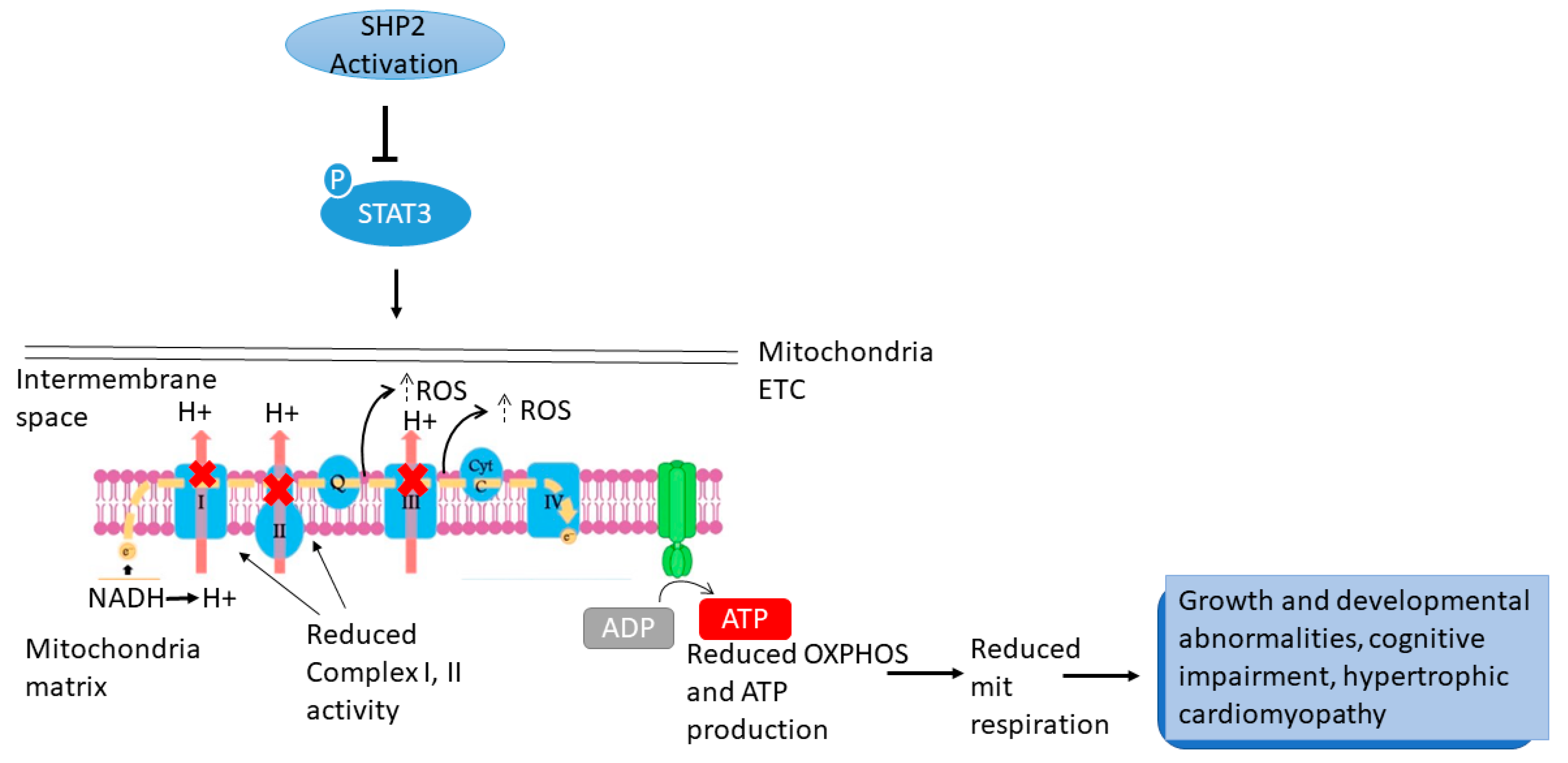
STAT3, a known substrate of SHP2 phosphatase [86,87], plays a vital role in the mitochondria [88]. The phosphorylation levels of STAT3 at Tyr705 and Ser727 were significantly reduced in Ptpn11E76K/+ cells and isolated mitochondria [84]. STAT3 localised in the mitochondria of cultured cells and primary tissues such as liver and heart. Furthermore, complex I and II-driven respirations were reduced in STAT3-/- mitochondria, confirming defects in mitochondrial respiration in the absence of STAT3 [88]. Furthermore, STAT3 deficiency in splenocytes increased mitochondrial oxygen consumption and ROS levels not related to ATP synthesis [89], similar to the mitochondrial phenotypes in Ptpn11E76K/+ cells described above [82].
Nagata and coworkers examined STAT3 phosphorylation to determine the molecular basis for increased energy expenditure in LSHKO mice. SHP2 has been linked to the regulation of STAT3 phosphorylation on Tyr705 in mouse heart [90] and liver [91]. Notably, this phosphorylation activates STAT3, and phosphorylation at Ser727 is required for maximal STAT3 transcriptional activity [88,92] for its mitochondrial actions and regulation of OXPHOS [88,93]. STAT3 (Tyr705) phosphorylation was increased in LSHKO liver lysates and isolated mitochondrial fractions compared to control mice. Together, findings show that total and mitochondrial STAT3 phosphorylation increased in HFD-LSHKO mice, implying that SHP2 deficiency may contribute to increased energy expenditure [85].
Finally, to check whether increased STAT3 (Ser727) phosphorylation may affect mitochondrial respiration, the oxygen consumption rate (OCR) of LSHKO and control mitochondria was measured. ADP-stimulated respiration rate in LSHKO mitochondria was significantly higher than in control mice. Furthermore, LSHKO mice had a significantly higher complex III-linked reactive oxygen species production than control mice. These findings show that hepatic SHP2 modulates mitochondrial function, including respiration and electron transport, in the liver of HFD-fed mice, which may contribute, at least in part, to the increased energy expenditure in LSHKO mice [85]. However, additional studies are needed to understand better the SHP2 regulation of mitochondrial function.
Together, these data indicated that, the inhibition (dephosphorylation) of STAT3 accounts for at least part of the changes in mitochondria function associated with SHP2 mutations. These changes include reduction in CI and II activity accompanied by decreased OXPHOS and ATP production, and increased ROS production in cellular models (Figure 2).

5.2. KRAS

GOF KRAS mutations (KRASG12V, KRASY71H) cause Erk activation and cell proliferation [94]. Heart and craniofacial abnormalities were observed in KRAS-targeted morpholino knockdown zebrafish embryos, while the expression of mutant KRAS caused heart maldevelopment [95]. A recent study on an NS patient’s DNA with a heterozygous de novo mutation c.211T>G, p.Tyr71Asp identified in exon 2 of KRAS described clinical features of intellectual impairment and nerve root hypertrophy, though the onset mechanism remains unknown [21]. However, only a few studies have investigated the consequences of KRAS mutations on mitochondria and cellular metabolism to this extent.
A tetracycline-inducible human embryonic kidney (HEK293) cell model was used to demonstrate that activation of mutated K-RASG12V causes mitochondrial dysfunction [96]. Structural mitochondrial changes such as swollen, pale matrix, disorganised cristae, disruption of the mitochondrial network and aberrant shapes were observed after induction of K-RASG12V expression. [96]. These cells notably reduced mitochondrial respiratory chain activity [97]. More specifically, complex I was the site of the respiratory defect in the cells expressing K-RASG12V, whereas the function of the other mitochondrial respiratory chain complexes appeared normal or slightly upregulated. Immunoblot analysis revealed that the level of the nuclear-encoded proteins, NDUFA 6,7 (20-kD component of complex I) was significantly reduced indicating complex I impairment in K-RASG12V-expressing cells [97]. Because the mitochondrial respiratory chain produces ROS [98,99], and K-RASG12V causes changes in the respiratory complex, changes in ROS generation were investigated. Data showed that activating KRASG12V caused a rapid increase in ROS generation with lower protein levels of two major antioxidant enzymes, superoxide dismutase 2 (SOD2) and catalase.
After prolonged K-RASG12V expression, the cells initiated a metabolic adaptation to mitochondrial dysfunction by increasing glycolytic activity. The connection between early mitochondrial dysfunction and the relatively late increase in glycolytic activity suggests that glycolysis upregulation was most likely a cellular response to metabolic stress caused by a decrease in mitochondrial respiration caused by K-RASG12V [96].
Even though mutant KRASG12D activity increases glucose uptake and reroutes glucose metabolism into the hexosamine biosynthesis and pentose phosphate pathways [100], the metabolic impact of KRAS mut copy gain is unknown. The glycolytic gene expression profiles of KRASG12D/WT and KRASWT/WT MEFs were similar except for Slc2a1 (coding GLUT1) and Slc2a3 (coding GLUT3). In contrast, glycolytic gene expression was significantly upregulated in KRASG12D/G12D MEFs, in agreement with increased glucose uptake, lactate secretion, and glycolytic capacity. As a result, KRASG12D copy gain induces a glycolytic switch, whereas a heterozygous KRAS mutation is insufficient to upregulate glycolysis [101].
Despite having comparable proliferative rates, cell volume, diameter, protein, and RNA content, KRASG12D/G12D MEFs exhibited a glycolytic switch compared to KRASG12D/WT cells. Furthermore, despite a KRASG12D-associated decrease in mitochondrial membrane potential observed under homozygosity, mitochondrial morphology and function were consistent across genotypes [96,97]. Therefore, the glycolytic switch may reflect alternative glucose utilisation by KRASG12D/G12D cells [101]. Metabolomics analysis confirmed KRASG12D/G12D cells enhanced glycolytic phenotype and, unexpectedly, revealed a significant increase in glucose-derived TCA cycle metabolites in KRAS mut homozygous MEFs, confirming their intact mitochondrial function. More importantly, these findings revealed a KRAS mutant copy gain-specific metabolic rewiring as well as a glucose metabolism signature [101]. Despite their differences in glucose utilisation, KRASG12D/WT and KRASG12D/G12D MEFs had similar levels of OXPHOS, implying other TCA cycle differences. Because KRAS mut cells preferentially use glutamine over glucose to fuel the TCA cycle [102,103], glutamine metabolism was investigated. Glutamine-derived TCA cycle metabolites and glutamine-derived oxygen consumption increased in KRASG12D/WT cells, but not KRASG12D/G12D MEFs [101].
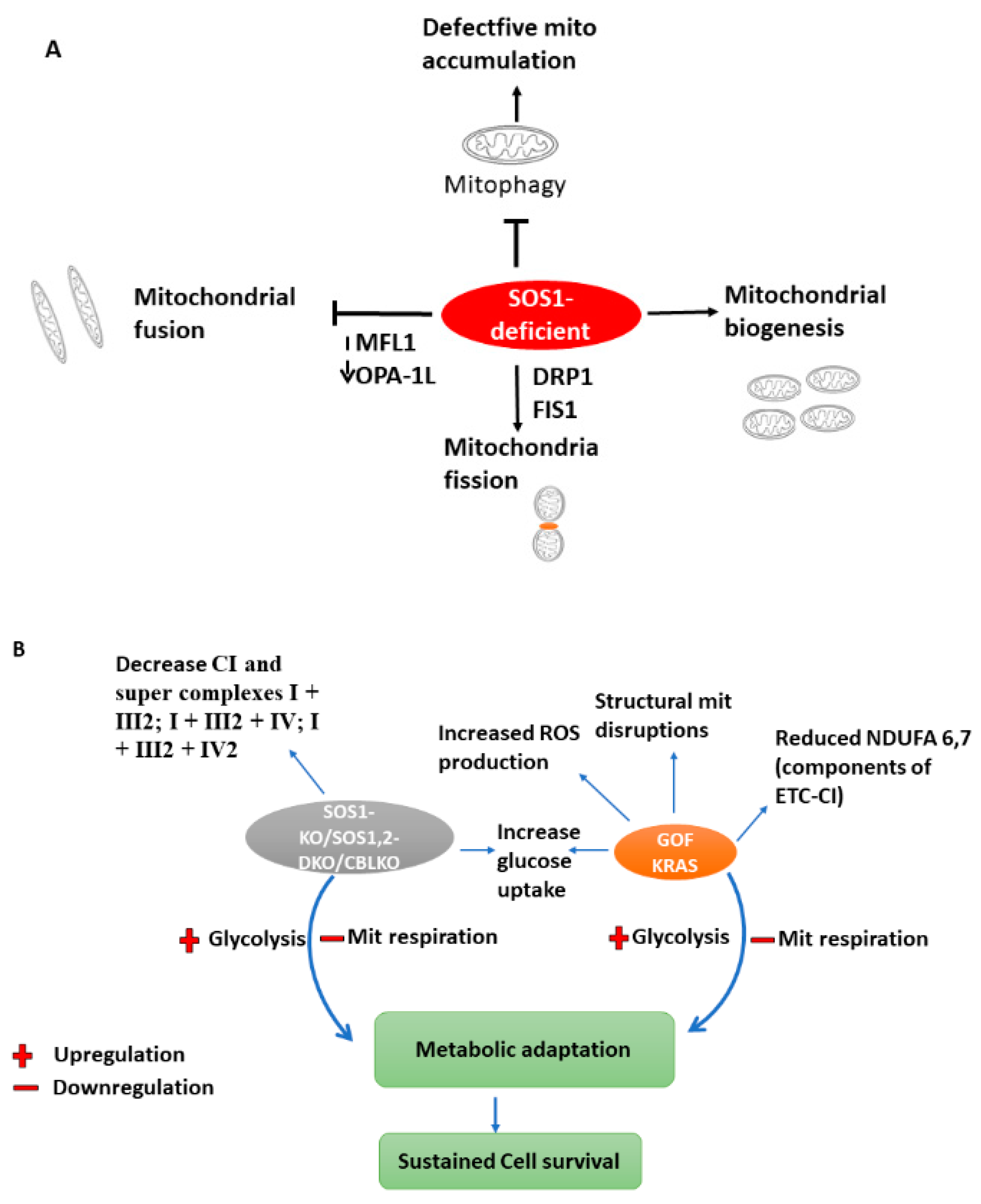
Enhanced pyruvate dehydrogenase (Pdh) activity [104] could explain the reprogramming of homozygous cells’ glucose metabolism. However, Pdh E1 component subunit alpha (Pdhe1a) expression and Pdh activity were comparable in KRASG12D/G12D and KRASG12D/WT MEFs [101]. The authors then hypothesised that genotype-specific metabolic requirements instead drive this metabolic switch. Surprisingly, glutathione (GSH) and its precursors were significantly enriched in glucose-derived carbons in KRASmut homozygous cells, implying that the KRASmut copy gain rewired glucose metabolism towards glutathione biosynthesis, with glutamine even more efficiently metabolised towards GSH biosynthesis in KRASG12D/G12D cells MEFs. Accordingly, KRASG12D/WT MEFs had lower ROS levels and a higher NADPH/NADP+ ratio than KRASWT/WT, consistently with previous findings [103,105]. Nonetheless, KRASG12D/G12D MEFs had a distinct antioxidant signature, as evidenced by significantly increased NADPH and GSH synthesis, NADPH/NADP+, and GSH/GSSG ratios, as well as lower ROS levels and resistance to ROS-inducing agents such as H2O2 [101]. Because the metabolic heterogeneity of KRASmut cells may limit the efficacy of generalised targeting approaches [106], potential KRASmut copy number-dependent susceptibilities were investigated. KRASG12D/G12D MEFs, unlike heterozygotes, were extremely sensitive to low glucose and to the hexokinase 2 (HK2) inhibitor 2-Deoxy-D-glucose (2DG), which triggered a considerable apoptotic response. Meanwhile, KRASG12D/WT MEFs were more sensitive to low glutamine levels. KRASG12D/G12D cells (but not KRASWT/WT or KRASG12D/WT) showed high ROS levels after treatment with 2DG, confirming a reliance on glucose for efficient ROS management [101]. In summary, a prolong effect of RAS mutations on primary cells could initiate a metabolic adaptation to mitochondrial dysfunction via glycolysis upregulation accompanied by decrease mitochondria respiration (Figure 3B).

5.3. SOS1/SOS2 (Sons of the Sevenless)

SOS1 and SOS2, the two highly homologous mammalian orthologues of SOS, are the most abundant RAS-GEFs (guanine nucleotide exchange factors) in metazoan cells [107,108,109] which induce GTP/GDP exchange on cellular RAS proteins in the context of a multitude of distinct cellular surface Receptor tyrosine Kinase signalling pathways (RTKs) [107,110]. Several genetic alterations or mutations in different SOS1 (rarely SOS2) domains have been identified in inherited RASopathies such as NS [14,111]. Despite the homology, data on SOS1/2-KO mice have shown that SOS1 is more functional than SOS2 in cellular processes such as proliferation, migration, inflammation, and maintenance of intracellular redox homeostasis; however, certain functional redundancy cannot be ruled out, especially at the organismal level [112,113].
García-Navas et al. recently investigated the relationship between SOS and mitochondrial function using primary MEFs derived from four SOS genotypes, wild-type (WT), SOS1 single Knockout (SOS1-KO), SOS2 single knockout (SOS2-KO) and SOS1/2 double knockout (SOS1/2 DKO) mice [114]. The authors evaluated various structural mitochondrial subtypes in MEFS from all four SOS genotypes [115], revealing a distinct increase in spherical-shaped mitochondrial subtypes and a decrease in tube-shaped mitochondrial subtypes in MEFs lacking SOS1 and SOS1/2 compared to WT and SOS2 deficient cells [116]. Quantification of signals from mitochondrial immunofluorescence [115] showed apparent increase in the number of individual mitochondrial structures in the cytoplasm of MEFs; however, their average size was consistently smaller in SOS1-deficient MEFs compared to WT and SOS2-KO MEFs. SOS1 and SOS1/2-DKO -deficient MEFs had a significant increase in mitochondrial mass compared to WT or SOS-KO cells [116]. The average mitochondrial morphology results from a dynamic equilibrium between fusion and fission [117]. In SOS1-deficient MEFs, analysis of mitochondrial fusion and fission regulators [117,118,119] revealed reduction of fusion-promoting proteins such as Mitofusin 1 (MFN1) [120] and the long-form of mitochondrial dynamin-like GTPase (OPA-1L) [121] as well as stable levels of fission promoters such as GTPase Dynamin-Related protein 1 (DRP1) and Mitochondrial fission 1 (FIS1) [122]. DRP1 expression was also higher in pure mitochondrial preparations. These findings point to a shift in the equilibrium toward mitochondrial fission in SOS1-deficient MEFs, implying that SOS1 deletion results in particular changes in mitochondrial morphology, mass, and dynamics [116].
Moreover, SOS1-defective cells showed an increased population of defective mitochondria, and a significantly lower rate of superoxide production in SOS1-KO and SOS1/2-DKO mitochondria, confirming a severe loss of mitochondrial functionality connected to SOS1 absence in these cell models [116,123,124].
The expression of representative OXPHOS respiratory complex subunits [125] in MEFs of the SOS genotypes was next assessed via western blotting and revealed a specific increased NDUSF3 subunit of complex I [126] and UQCRC2 subunit of complex III [127] in mitochondria lacking SOS1, particularly SOS1/2-DKO [116]. SOS1-KO and SOS1/2-DKO samples exhibited significantly lower formation of super complexes, including complex I (CI and super complexes I + III2; I + III2 + IV; I + III2 + IV2) as compared to the control and SOS2 deleted genotype. Furthermore, SOS1-deficient samples showed a distinct form of a complex III2 + IV released from CI that was not seen in WT or SOS2-KO samples and much lower amounts of free complex IV [116,128]. These findings imply that SOS1 is required for the correct assembly of mitochondrial supercomplexes and that SOS1 deficiency is associated with specific alterations in mitochondrial respiratory complex components and supercomplex assembly.
The respiratory and metabolic profiles [129] of SOS1/2-deficient MEFs revealed a much lower basal respiration and spare respiratory capacity compared to controls and single SOS1 deficient cells, confirming prior findings [113]. Moreover, the loss of SOS1 in MEFs resulted in significantly lower rates of ATP synthesis from glycolysis (glycoATP) and oxidative phosphorylation (mitoATP). MitoATP and glycoATP synthesis distinctly distinguished WT and SOS2 from the other phenotypes, which showed much less energetic and more quiescent phenotypic profiles [116,130]. In summary, these data demonstrated that mitochondrial respiratory and metabolic defects are linked to SOS1 ablation.
Because different cell types use different nutrient substrates to support oxidative energy metabolism [131], the Garca-Navas team evaluated whether the lack of SOS1 in MEFs would affect the type of preferred substrates or their oxidation mechanisms. All SOS-deficient genotypes examined, revealed meagre glutamine oxidation rates, with the rates in SOS1-deficient MEFs even lower or nearly negligible when compared to controls and SOS2-KO cells. According to the authors, SOS deficient phenotypes showed different patterns of oxidation rates in the presence of endogenous or externally administered fatty acid [116].
Finally, the OCR tracings for glucose consumption showed that glucose was the preferred oxidation substrate for MEFs, and also revealed that SOS1-deficient cells utilize glucose as the only oxidative substrate, but they did so much less efficiently (50%) than WT or SOS2-KO counterparts. SOS1/2-DKO MEFs showed cleaved caspase-3 (CC3) in the absence of glucose, implying that the GEF activity provided by either SOS isoform is required to avoid cell death and sustain MEF survival following glucose deprivation. Addition of glucose increased hexokinase-I HK1 (but not HK2) [132] and decreased phospho-AMP-activated protein kinase (pAMPK) [133] and lactate dehydrogenase A (LDHA) in SOS1 and SOS1/2 deficient phenotypes when compared to the other genotypes [116]. Together, SOS1-deficient cells downregulate pAMPK thereby evoking specific alterations in substrate oxidation accompanied by alterations in mitochondrial respiration to sustain survival. SOS1 deficiency blocks mitophagy, hence causing accumulation of defective mitochondria. In addition, changes within the mitochondria include: reduction in fusion-promoting proteins such as Mitofusin 1 (MFN1) and the mitochondrial dynamin-like GTPase (OPA-1L) resulting to inhibition of mitochondrial fusion. Meanwhile the presence of GTPase Dynamin-Related protein 1 (DRP1) and Mitochondrial fission 1 (FIS1) favours mitochondria fission, hence the changes in mitochondria morphology, mass and dynamics summarises the mechanistic insight of SOS mutation on energy metabolism. (Figure 3A,B).

5.4. CBL

Casitas B-lineage Lymphoma (CBL) is a proto-oncogene found mutated in NS patients [25,134,135]. CBL encodes an E3-ubiquitin ligase from the CBL family that mediates protein ubiquitination and, once heterozygously mutated, causes abnormalities clinically similar to NS patients [25,26,136]. This protein has a phosphotyrosine-binding domain at its N-terminus that allows it to engage with a variety of tyrosine-phosphorylated substrates and target them for proteasomal or enzymatic destruction. As a result, CBL proteins act as a negative regulator in various signal transduction pathways, including P13K/AKT pathway [137].
Previously, studies performed on CBL-knockout and knockdown monocyte cells and mice revealed significant enlargement in mitochondria size and high energy expenditure [137,138]. CBL-KO and wild-type human monocytic cell line (THP-1)-derived macrophages had higher glycolytic capacity, glycolytic reserve rates and elevated OXPHOS than WT cells [139]. Since glycolysis is required for NLRP3 inflammasome activation, Lin et al. further investigated the mechanisms involved in Cbl-mediated glycolytic control [139]. In a previous study, Cbl-deficient mice showed a noticeable increase in glucose tolerance [138], suggesting that Cbl inhibition may enhance the transportation of glucose inside the cells. The uptake of the glucose analogue 2-NBDG was significantly higher in CBL-KO cells or in WT cells treated with the Cbl inhibitor hydrocotarnine compared with the untreated control. Thus, these findings imply that Cbl negatively contrlos cellular glucose uptake in THP-1-derived macrophages [139].
Cellular glucose uptake occurs through glucose transporters (GLUTs) [140,141,142,143], with GLUT1 being the most abundant in macrophages [144]. In fact, the surface expression of GLUT1 protein in CBL deficient cells was higher than in WT cells [139]. In summary, Lin’s research demonstrated that inhibiting Cbl enhanced cellular glucose uptake, glycolytic capability, and mitochondrial OXPHOS capacity [139].
Together, these data suggest that CBL mutations not only affect mitochondria size and energy expenditure but also mitochondrial respiration via glucose upregulation.

5.5. RRAS

Because of the large number of genes mutated in NS, approximately 10–20% of diagnosed patients do not have mutations in previously known NS genes, implying that additional unidentified genes may be present and contribute to the disease onset. In a recent study, Flex et al. described a new disease-associated gene called RRAS carrying a GOF mutation and encoding a small monomeric GTPase that controls cell adhesion, spreading, and migration, underlying a rare and variable phenotype with features that partially overlap NS [31]. Later, Capri et al. also identified activating RRAS2 mutation in six unrelated subjects/families with NS [30].
Despite identifying this gene in NS patients, there is still little known about the effects of hyperactivated RRAS on mitochondria and energy metabolism. Alcover and coworkers recently studied RRAS2 mutation in hypomyelinated disease-mouse models. The authors hypothesised that the cause of hypomyelination in vertebrates could be associated with metabolic alterations since axonal synaptic transmission depends on myelin insulation and metabolic support. Understanding the mitochondria adaptations in knockout RRAS2 phenotype gave insights into the degree of axonal myelination. In R-Ras2/(R-Ras2KO) mice, where fewer axons were myelinated, the number of mitochondria in nonmyelinated axons increased, suggesting dysfunction in these mitochondria structures [145]. However, the defects or alterations associated with mitochondrial dysfunction in nonmyelinated axons are still unknown. According to the authors, the physiological outcome of RRAS loss-of-function in human disorders could promote axonal degeneration and hypomyelination [145].

5.6. RAF

Among the RAF protein family isoforms (B-RAF, C-RAF) encoded by the RAF gene and found mutated in NS patients, C-RAF (also called RAF1) is the only one with a direct target on the mitochondria [146]. The amino-terminal domain of active C-RAF binds the mitochondrial surface with high affinity and regulates mitochondrial shape and cellular distribution through its coupling with MEK [146] though the mechanism remains unclear. Such morphologic alterations of the mitochondrial network are involved in cell migration and Ca2+ metabolism in endothelial cells [147,148]. Wang discovered that proteins encoded by the active C-RAF gene regulate mitochondrial function by phosphorylating outer mitochondrial membrane proteins such as BAD (a pro-apoptotic BCl-2 homolog), subsequently phosphorylating ERK-1 and 2 thus inhibiting apoptosis [149]. A recent study of patients clinically diagnosed with NS and NSML revealed the presence of RAF1 and BRAF pathogenic variants, with these patients also displaying systemic disorders such as congenital heart disease [35,150]. C-RAF is an essential effector of Ras-mediated signalling and a critical regulator of the ERK/MAPK pathway [151]. The cooperation of C-RAF with intracellular kinases such as PAK5 (a serine/threonine-protein kinase that targets C-RAF to mitochondria in a serine 338 independent manner) regulates C-RAF activity and controls the C-RAF-dependent Raf/MEK signalling at the mitochondria [152]. An Inhibitory kinase of C-RAF, termed RAF kinase inhibitory protein (RKIP), block its translocation to the mitochondrial membrane [153]. However, fibroblast growth factor (FGF) induces C-RAF mitochondrial translocation in vascular endothelial cells by phosphorylating its Ser338 (S338) [154]. Overexpression of specific intracellular proteins such as Bcl-2 (regulator of apoptosis) recruits C-RAF into the mitochondria conferring cell survival upon dimerisation and phosphorylation of the stress-activated pro-apoptotic kinase MST2 [155]. Furthermore, C-RAF and B-RAF, independent of the usual RAF signalling pathway, can induce some metabolic features (such as fusion, fission, biogenesis and mitophagy) mediated by mitochondria [155,156]. The distinct role of C-RAF in the mitochondria provides compelling evidence for targeting mitochondrial RAF proteins to maintain mitochondrial homeostasis [149,157].
Tsai et al., examined mitochondria function and ROS production of wild-type and C-RAF knockout MEF treated with RAF inhibitors sorafenib and GW5074. There was a 6-fold increase in ROS production and a decrease in mitochondrial membrane potential in C-RAF-WT but not in C-RAF-/-. Furthermore, the therapy induced mitochondria fragmentation with donut and blob-shaped formation in C-RAF-WT but not C-RAF-/- MEFs. Thus, these findings suggest that inhibiting C-RAF causes mitochondrial dysfunction, which may have contributed to cell death [158]. Moreover, C-RAF interacts with Ser308 of phosphorylated DAPK (Death-associated protein kinase), directing its colocalisation to the mitochondria for regulating mitochondrial modelling [158].
The inhibition of RAF1 expression resulted in low conductance gating of the mitochondrial permeability transition pore (mPTP) (Figure 4), with a decrease in the inner mitochondrial membrane potential in rats or cells lines treated with MEDICA (MEthyl-substituted DICarboxylic Acids) analogues, known inhibitors of RAF1 [159]. Previously, MEDICA analogues were demonstrated to suppress basal P-Raf1 (Ser-338), indicating inhibition of Raf1 kinase activity [160]. In this light, the inhibition of RAF1 by MEDICA analogues could be attributed to the suppression of RAF1 expression and its kinase activity. The role of RAF1 as an upstream target for MEDICA analogues was confirmed further by measuring cAMP levels and P-Bad (Ser-155) in COS-1 cells that overexpress RAF1. RAF1 overexpression prevented the decrease in cAMP and P-Bad (Ser-155) (Figure 4), indicating that RAF1 suppression may account for low conductance-MPTP gating by MEDICA analogues. Over the years, low conductance gating has been characterised by the continuous passage/flow of ions across the mitochondria membrane [161]. Thus, Samovski’s group suggested that this low conductance gating following RAF1 mutation might have resulted from the prolonged opening of mPTP [159].
The role of these mutations in Ras signaling and their impact on mitochondrial metabolism are summarised in Figure 5.

6. Conclusions

PTPN11 accounts for the majority of germline mutations responsible for NS and NSML, followed by SOS1, KRAS isoform (except HRAS), RAF1 and SHOC2. These genes play an essential role in the pathogenesis of mitochondrial/metabolic-related disorders. Exploring the bioenergetics of Noonan syndrome in mouse models and human cell lines is required to fully understand the effect of RAS hyperactivation on mitochondrial function and tissue development processes. This analysis will provide insights into energy metabolism and the onset of various clinical complications.
In most cases, research has revealed that the whole action of any one of the germline mutations requires interaction and cooperation with intracellular proteins, kinases, transporters, or transcription factors (such as GLUT1, Gas6, STAT3, GEF, ROCK2, cAMP, p-AMPK, p- Bad) involved in the MAPK/ERK signalling. This has been seen most prominently in RAF, KRAS, PTPN11, and SOS1 mutations [80,81,82,85,101,116,139,159,162].
Findings have shown that different mutations (homozygous or heterozygous, GOF or non-functional) on the same gene could cause heterogeneous pathological effects, which may depend on the different impacts on the mitochondria and energy metabolism [69,70]. Furthermore, not only has a single gene mutation been linked to a defect in energy metabolism but copy variants and double mutations have also been identified as having functional implications [101,116].
The longer time an active RAS (KRAS) mutant is expressed on a cell, it leaves the cell with persistent dysfunctional mitochondria resulting in a metabolic adaptation [96]. A profound influence of GOF PTPN11 (SHP2) mutation on mitochondrial cytochrome oxidase and cytochrome C led to a decrease in mitochondrial membrane permeability (MMP), increased ROS production, and decreased ATP production [7]. These changes signal bioenergetic stress and may result in the release of apoptotic factors, culminating in cell death and accumulated ROS, which may cause oxidative damage in tissues, potentiating tissue failure [84,101,163,164]. Interestingly, it was also found that SHP2 alone is not responsible for the changes in energy metabolism, but it does require hormonal support to mediate specific cellular processes, including glucose homeostasis [70]. As a result, targeting hormonal pathways in SHP2 mutations may be critical for regulating energy metabolism.
According to other observations, the major causes of alterations in respiration and ATP production rates during OXPHOS or changes in glycolytic rate can be attributed to mitochondrial defects, i.e., changes in mitochondrial shape, mass, and dynamics, lower or extreme high activity rates of the ETC, changes in respiratory complexes and their assembly into mitochondrial supercomplexes, abnormal levels of mitochondrial proteins (VDAC, TOM20, and TIM13), and dysfunctional MPTP, as well as the distinct patterns of substrate preference for oxidative energy metabolism and cells’ reliance on glucose for survival [113,116,159,165].
Although cognitive impairment has been reported in NS/NSML patients [166,167], there is still a lack of evidence on how energy metabolism is affected in this condition, particularly when looking at the most frequently mutated genes in NS individuals.
Because heterozygous expression of an NS/NSML (LEOPARD syndrome) mutant of SHP2 enhances energy metabolism and protects against obesity and diabetes [69], one might wonder if equivalent pharmacological modulation of SHP2, that functionally mimics NSML mutations, could be a powerful strategy to alleviate obesity and associated pathologies. Indeed, it has been shown that SHP2 expression is upregulated in metabolic tissues of obese animals, suggesting that increased SHP2 expression/activity contributes to the development of obesity [69,70,85].
NS/NSML mutations alter metabolism via enhanced phosphorylation or decreased phosphorylation of target proteins in the MAPK/ERK pathway [70,82,85]. In addition, RAS mutation in primary human cells revealed inhibited mitophagy by activating LC3B (autophagy-related protein) in a Jun N-terminal kinase (JNK) dependent manner. Because mitophagy is only one of the mechanisms that sheds light on mitochondrial impairment in cells, inhibiting mitophagy through altered RAS allowed cells to accumulate defective mitochondria, resulting in decreased mitochondrial respiration due to insufficient glucose uptake by the cells [168]. The molecular mechanism linking RAS activation and mitophagy inhibition in primary cells should be investigated further. Whether the observed effects on mitochondrial or metabolic functions are secondary events due to main changes in other relevant processes linked to Ras signalling, this cannot be ruled out. To support the findings of Kim et al. [80] a similar study involving Gas6 and active Ptpn11 mutation should be carried out but in this case, assessing the mitochondrial respiratory function. Some authors have demonstrated mechanistic insights into the indirect regulation of other processes controlling mitochondrial homeostasis by altered Ras signalling [169], In such study, HRAS was shown to cooperate by inhibiting several modulators linked to AMPK to alter mitochondrial function in Rasopathies [169].
Although several genes are mutated in NS children, i.e., NRAS, RIT1, A2ML1, SHOC2, RASA2, and LZTR1 experimental evidence of their action on mitochondrial function is not available to date. Further investigations are needed to understand the mechanisms linking NS to mitochondrial dysfunction and altered energy metabolism. In addition, we still lack insights into the outcomes of Cbl and SOS1/2 activation on mitochondria metabolism since the available data are limited to the effects of Cbl and SOS1/2 deletion or deficiency. Determining the role of Cbl and SOS1/SOS2 GOF mutation on mitochondrial function is therefore critical. Because of the location of SHP2 on the mitochondria and its numerous disruptive effects, as discussed here, future research should focus on identifying potent pharmacological regulators that compete with SHP2 ligands/substrates, as this may be critical for alleviating complications in NS/NSML patients. Furthermore, the discussed mechanisms and pathways may serve as future targets for therapeutic solutions.
To sum up, the knowledge of mitochondrial dysfunction in syndromic, Ras/MAPK pathway-related gene mutation carriers is beneficial for accurate diagnosis and applying the appropriate treatment to a pathological condition. Research into the underlying signalling pathways and mutations in NS/NSML children that cause mitochondrial dysfunction and abnormal energy metabolism is critical for future prognosis assessment, genetic counselling, and plausible therapy.

Author Contributions

Conceptualization, original draft preparation and revision: D.B. and E.B. Data collection and preparation of tables and figures: D.B. and E.B. Supervision and manuscript editing: E.B. and K.D. Intellectual input and manuscript review: D.B., E.B. and K.D. All authors have read and agreed to the published version of the manuscript.

Funding

This research received no external funding.

Institutional Review Board Statement

Not applicable.

Informed Consent Statement

Not applicable.

Data Availability Statement

Not applicable.

Acknowledgments

The authors acknowledge the Poznan university of medical sciences and the Polish National Agency for academic exchange for their support in promoting research and internationalisation. DB is a participant of STER Internationalisation of Doctoral Schools Programme from NAWA Polish National Agency for Academic Exchange No. PPI/STE/2020/1/00014/DEC/02.

Conflicts of Interest

The authors declare no conflict of interest.

Abbreviations

2DG2-Deoxy-D-glucose
2-NBDG 2-(N-(7-Nitrobenz-2-oxa-1,3-diazol-4-yl)Amino)-2-Deoxyglucose
3T3-L1 3-day transfer, inoculum 3 x 10⁵ cells- fibroblast-like cell line
8GM-CSF Granulocyte-macrophage colony-stimulating factor
ADP Adenosine diphosphate
AMPAdenosine monophosphate
AMPK AMP-activated protein kinase
ATGLAdipose triglyceride lipase
ATPAdenosine triphosphate
BADBCL2 associated agonist of cell death
BAF3IL-3 dependent murine pro B cell
BakBcl-2 homologous antagonist/killer
BaxBcl-2-like protein 4
BCL-2B-cell lymphoma 2
Beta-MHC Beta-Myosin heavy chain
BIMBcl-2-like protein 11
CAAX C-Cysteine, AA-2 Aliphatic residues, X-any C-terminal amino acid
CBL-KO THPHuman CBL knockout HEK-293T cell line
CC3Cleaved caspase-3
CcOCytochrome C oxidase
CI,II,III,IV Complex I-IV
CMPscommon myeloid progenitors
COIICytochrome C oxidase subunit 2
COS-1 kidney fibroblast-like cell line /CV-1 (simian) in Origin, and carrying the SV40 genetic mate
Cyt cCytochrome c
DAPK Death-Associated Protein Kinase
DRP1Dynamin-related protein 1
ECARExtracellular acidification rate
EGFEpidermal growth factor
EGFR Epidermal growth factor receptor
EPOErythropoietin
EREndoplamic reticulum
ETCElectron transport chain
FADH2 flavin adenine dinucleotide
FCCPCarbonyl cyanide-p-trifluoromethoxyphenylhydrazone
FGFFibroblast growth factor
FIS1Mitochondrial fission 1 protein
FRS-2FGF receptor substrate 2
Fru1,6bisP Fructose-1-bisPhosphate
Gab1GRB2-associated-binding protein 1
GDP Guanosine diphosphate
GF Growth factor
GLUT1Glucose transporter 1
GLUT3 Glucose transporter 3
glycoATP Glycolytic ATP
GM-CSF Granulocyte macrophage-colony stimulating factor
GMPsGranulocyte macrophage progenitors
GOFGain-of-function
GRB2Growth factor receptor-bound protein 2
GSHGlutathione
GSH/GSSG Glutathione/Glutathione disulfide (reduced glutathione/Oxidised glutathione) ratio
GTP Guanosine-5′-triphosphate
GTPase GTP hydrolase
GW5074 3-(3,5-Dibromo-4-hydroxybenzyliden)-5-iodo-1,3-dihydroindol-2-one (a C-RAF inhibit)
HEK293 Human embryonic kidney 293 cells
HFDHight fat diet
HGPhepatic glucose production
HK1/HK2 Hexokinase 1 and 2
HSCsHematopoietic stem cells
HSLHormone-sensitive lipase
IL-3Interleukin 3
IMSintermembranous space
IRS-1Insulin receptor substrate 1
Jak2 Janus kinase 2
JMML Juvenile myelomonocytic leukemia
JNK c-Jun N-terminal kinases
KO Knock-out
LC3BLight chain 3B (subunit of Microtubule-associated proteins 1A/1B)
LDHA Lactate Dehydrogenase A
LIF Leukemia inhibitory factor
LSHKO Liver-specific Shp2 Knock-out
MAPK/ERK Mitogen-activated protein kinase/Extracellular signal-regulated kinase 1/2
MDC Mitochondria disease criteria
MEDICA MEthyl-substituted DICarboxylic Acids
MEFMouse embryonic fibroblast
MEK1 or MAP2K1 Dual specificity mitogen-activated protein kinase kinase 1
MEPsMegakaryocyte erythroid progenitors
MFN1 Mitofusin-1 protein
Mit Mitochondria
mitoATP/mitATP mitochondria ATP
MitoSOXTM Red Mitochondrial Superoxide Indicator
MitoTrackerTM Red CMXRos MitoTracker Red and Chloromethyl-X-rosamine
MMPMitochondrial membrane potential
mPTPMitochondrial permeability transition pores
MST2mammalian Sterile 20-like kinase 2
mtDNA Mitochondrial DNA
mTOR/S6K mammalian target of rapamycin (mTOR)/ribosomal protein S6 kinase
mutmutation
MYBPC3 Cardiac myosin binding protein C3
NADH Nicotinamide adenine dinucleotide
NADP+ Oxidised form of Nicotinamide adenine dinucleotide phosphate (NADPH)
NDNADH Dehydrogenase
NDUSF3 NADH dehydrogenase [ubiquinone] iron-sulfur protein 3
NLRP3 NLR family pyrin domain containing 3
NSC87877 8-Hydroxy-7-[(6-sulfo-2-naphthyl)azo]-5-quinolinesulfonic acid (Inhibitor of SHP2/SHP1)
OCROxygen consumption rate
OGTT Oral glucose tolerance test
OPA-1L Dominant optic atrophy (long form of mitochondrial dynamin-like GTPase)
OXPHOS Oxidative phosphorylation
pAMPKphospho-AMP-activated protein kinase
Pdh Pyruvate dehydrogenase
Pdhe1a Pyruvate dehydrogenase E1 component subunit alpha
p-ERKPhosphorylated ERK
PI3K-AKT-mTOR The phosphatidylinositol-3-kinase (PI3K)/Akt and the mammalian target of rapamycin (mTOR)
PP1CProtein phosphatase 1 catalytic subunit
PPPPentose phosphate pathway
RAS/MAPK Rat sarcoma virus/Mitogen-activated protein kinase
RB2 Retinoblastoma-like protein 2
ROSReactive oxygen species
RTKReceptor tyrosin kinase
SOD2Superoxide dismutase 2
SOS1/2-DKODouble knock-out SOS1/2
SOS1-KO Single Knock-out SOS1
SOS2-KO Single Knock-out SOS2
SRCSpare respiratory capacity
STAT3 Signal transducer and activator of transcription 3
TCATricarboxylic acid cycle
THP-1Tamm-Horsfall Protein 1 (Spontaneously immortalized monocyte-like cell line)
TIM13Translocase of inner membrane 13
TOM20 Translocase of outer membrane 20
TOMM40 Translocase of outer mitochondria membrane 40
UQCRC2 Cytochrome b-c1 complex subunit 2
VDACVoltage-dependent anion channels (mitochondrial porins)
WT Wildtype

References

  1. Dard, L.; Bellance, N.; Lacombe, D.; Rossignol, R. RAS signalling in energy metabolism and rare human diseases. Biochim. Biophys. Acta 2018, 1859, 845–867. [Google Scholar] [CrossRef] [PubMed]
  2. Tajan, M.; Paccoud, R.; Branka, S.; Edouard, T.; Yart, A. The RASopathy Family: Consequences of Germline Activation of the RAS/MAPK Pathway. Endocr. Rev. 2018, 39, 676–700. [Google Scholar] [CrossRef]
  3. Rauen, K.A. The RASopathies. Annu. Rev. Genom. Hum. Genet. 2013, 14, 355–369. [Google Scholar] [CrossRef] [PubMed] [Green Version]
  4. Rossmann, M.P.; Dubois, S.M.; Agarwal, S.; Zon, L.I. Mitochondrial function in development and disease. Dis. Model. Mech. 2021, 14, dmm048912. [Google Scholar] [CrossRef]
  5. Salvi, M.; Stringaro, A.; Brunati, A.M.; Agostinelli, E.; Arancia, G.; Clari, G.; Toninello, A. Tyrosine phosphatase activity in mitochondria: Presence of Shp-2 phosphatase in mitochondria. Experientia 2004, 61, 2393–2404. [Google Scholar] [CrossRef] [PubMed]
  6. Arachiche, A.; Augereau, O.; Decossas, M.; Pertuiset, C.; Gontier, E.; Letellier, T.; Dachary-Prigent, J. Localization of PTP-1B, SHP-2, and Src Exclusively in Rat Brain Mitochondria and Functional Consequences. J. Biol. Chem. 2008, 283, 24406–24411. [Google Scholar] [CrossRef] [Green Version]
  7. Lee, I.; Pecinova, A.; Pecina, P.; Neel, B.G.; Araki, T.; Kucherlapati, R.; Roberts, A.E.; Hüttemann, M. A suggested role for mitochondria in Noonan syndrome. Biochim. Biophys. Acta BBA Mol. Basis Dis. 2010, 1802, 275–283. [Google Scholar] [CrossRef] [Green Version]
  8. Simanshu, D.K.; Nissley, D.V.; McCormick, F. RAS Proteins and Their Regulators in Human Disease. Cell 2017, 170, 17–33. [Google Scholar] [CrossRef] [PubMed] [Green Version]
  9. Tartaglia, M.; Gelb, B.D.; Zenker, M. Noonan syndrome and clinically related disorders. Best Pract. Res. Clin. Endocrinol. Metab. 2011, 25, 161–179. [Google Scholar] [CrossRef] [Green Version]
  10. Tartaglia, M.; Zampino, G.; Gelb, B.D. Noonan Syndrome: Clinical Aspects and Molecular Pathogenesis. Mol. Syndromol. 2010, 1, 2–26. [Google Scholar] [CrossRef] [Green Version]
  11. Atay, O.; Skotheim, J.M. Spatial and temporal signal processing and decision making by MAPK pathways. J. Cell Biol. 2017, 216, 317–330. [Google Scholar] [CrossRef] [PubMed] [Green Version]
  12. Dance, M.; Montagner, A.; Salles, J.-P.; Yart, A.; Raynal, P. The molecular functions of Shp2 in the Ras/Mitogen-activated protein kinase (ERK1/2) pathway. Cell. Signal. 2008, 20, 453–459. [Google Scholar] [CrossRef] [PubMed]
  13. Bunda, S.; Burrell, K.; Heir, P.; Zeng, L.; Alamsahebpour, A.; Kano, Y.; Raught, B.; Zhang, Z.Y.; Zadeh, G.; Ohh, M. Inhibition of SHP2-mediated dephosphorylation of Ras suppresses oncogenesis. Nat. Commun. 2015, 6, 8859. [Google Scholar] [CrossRef] [PubMed] [Green Version]
  14. Avruch, J. Ras Activation of the Raf Kinase: Tyrosine Kinase Recruitment of the MAP Kinase Cascade. Recent Prog. Horm. Res. 2001, 56, 127–156. [Google Scholar] [CrossRef] [PubMed]
  15. Noonan, J.A. Hypertelorism With Turner Phenotype: A New Syndrome With Associated Congenital Heart Disease. Am. J. Dis. Child. 1968, 116, 373. [Google Scholar] [CrossRef] [PubMed]
  16. Allanson, J.E.; Bohring, A.; Dörr, H.-G.; Dufke, A.; Gillessen-Kaesbach, G.; Horn, D.; König, R.; Kratz, C.P.; Kutsche, K.; Pauli, S.; et al. The face of Noonan syndrome: Does phenotype predict genotype. Am. J. Med. Genet. Part A 2010, 152A, 1960–1966. [Google Scholar] [CrossRef] [PubMed] [Green Version]
  17. Digilio, M.C.; Conti, E.; Sarkozy, A.; Mingarelli, R.; Dottorini, T.; Marino, B.; Pizzuti, A.; Dallapiccola, B. Grouping of Multiple-Lentigines/LEOPARD and Noonan Syndromes on the PTPN11 Gene. Am. J. Hum. Genet. 2002, 71, 389–394. [Google Scholar] [CrossRef] [PubMed] [Green Version]
  18. Digilio, M.C.; Sarkozy, A.; de Zorzi, A.; Pacileo, G.; Limongelli, G.; Mingarelli, R.; Calabrò, R.; Marino, B.; Dallapiccola, B. LEOPARD syndrome: Clinical diagnosis in the first year of life. Am. J. Med. Genet. Part A 2006, 140A, 740–746. [Google Scholar] [CrossRef] [PubMed]
  19. Tartaglia, M.; Mehler, E.L.; Goldberg, R.; Zampino, G.; Brunner, H.G.; Kremer, H.; Van Der Burgt, I.; Crosby, A.H.; Ion, A.; Jeffery, S.; et al. Mutations in PTPN11, encoding the protein tyrosine phosphatase SHP-2, cause Noonan syndrome. Nat. Genet. 2001, 29, 465–468. [Google Scholar] [CrossRef]
  20. Li, X.; Yao, R.; Tan, X.; Li, N.; Ding, Y.; Li, J.; Chang, G.; Chen, Y.; Ma, L.; Wang, J.; et al. Molecular and phenotypic spectrum of Noonan syndrome in Chinese patients. Clin. Genet. 2019, 96, 290–299. [Google Scholar] [CrossRef] [PubMed]
  21. Ando, Y.; Sawada, M.; Kawakami, T.; Morita, M.; Aoki, Y. A Patient with Noonan Syndrome with a KRAS Mutation Who Presented Severe Nerve Root Hypertrophy. Case Rep. Neurol. 2021, 13, 108–118. [Google Scholar] [CrossRef] [PubMed]
  22. Kim, J.; Cho, S.Y.; Yang, A.; Jang, J.-H.; Choi, Y.; Lee, J.-E.; Jin, D.-K. An atypical case of Noonan syndrome with KRAS mutation diagnosed by targeted exome sequencing. Ann. Pediatr. Endocrinol. Metab. 2017, 22, 203–207. [Google Scholar] [CrossRef] [PubMed] [Green Version]
  23. Cirstea, I.C.; Kutsche, K.; Dvorsky, R.; Gremer, L.; Carta, C.; Horn, D.; Roberts, A.E.; Lepri, F.R.; Merbitz-Zahradnik, T.; König, R.; et al. A restricted spectrum of NRAS mutations causes Noonan syndrome. Nat. Genet. 2009, 42, 27–29. [Google Scholar] [CrossRef]
  24. Xu, S.; Fan, Y.; Sun, Y.; Wang, L.; Gu, X.; Yu, Y. Targeted/exome sequencing identified mutations in ten Chinese patients diagnosed with Noonan syndrome and related disorders. BMC Med. Genom. 2017, 10, 62. [Google Scholar] [CrossRef] [PubMed] [Green Version]
  25. Martinelli, S.; Stellacci, E.; Pannone, L.; D’Agostino, D.; Consoli, F.; Lissewski, C.; Silvano, M.; Cencelli, G.; Lepri, F.; Maitz, S.; et al. Molecular Diversity and Associated Phenotypic Spectrum of Germline CBL Mutations. Hum. Mutat. 2015, 36, 787–796. [Google Scholar] [CrossRef] [PubMed]
  26. Coe, R.R.; McKinnon, M.; Tarailo-Graovac, M.; Ross, C.J.; Wasserman, W.; Friedman, J.M.; Rogers, P.C.; van Karnebeek, C.D. A case of splenomegaly in CBL syndrome. Eur. J. Med. Genet. 2017, 60, 374–379. [Google Scholar] [CrossRef] [PubMed]
  27. Vissers, L.; Bonetti, M.; Overman, J.P.; Nillesen, W.M.; Frints, S.G.; de Ligt, J.; Zampino, G.; Justino, A.; Machado, J.C.; Schepens, M.; et al. Heterozygous germline mutations in A2ML1 are associated with a disorder clinically related to Noonan syndrome. Eur. J. Hum. Genet. 2014, 23, 317–324. [Google Scholar] [CrossRef]
  28. Yamamoto, G.L.; Aguena, M.; Gos, M.; Hung, C.; Pilch, J.; Fahiminiya, S.; Abramowicz, A.; Cristian, I.; Buscarilli, M.; Naslavsky, M.S.; et al. Rare variants in SOS2 and LZTR1 are associated with Noonan syndrome. J. Med. Genet. 2015, 52, 413–421. [Google Scholar] [CrossRef] [Green Version]
  29. Roberts, A.E. Noonan Syndrome. In GeneReviews®; Adam, M.P., Ardinger, H.H., Pagon, R.A., Wallace, S.E., Bean, L.J., Gripp, K.W., Mirzaa, G.M., Amemiya, A., Eds.; University of Washington: Seattle, WA, USA, 1993. Available online: http://www.ncbi.nlm.nih.gov/books/NBK1124/ (accessed on 5 April 2022).
  30. Capri, Y.; Flex, E.; Krumbach, O.H.; Carpentieri, G.; Cecchetti, S.; Lißewski, C.; Adariani, S.R.; Schanze, D.; Brinkmann, J.; Piard, J.; et al. Activating Mutations of RRAS2 Are a Rare Cause of Noonan Syndrome. Am. J. Hum. Genet. 2019, 104, 1223–1232. [Google Scholar] [CrossRef] [Green Version]
  31. Flex, E.; Jaiswal, M.; Pantaleoni, F.; Martinelli, S.; Strullu, M.; Fansa, E.K.; Caye, A.; De Luca, A.; Lepri, F.; Dvorsky, R.; et al. Activating mutations in RRAS underlie a phenotype within the RASopathy spectrum and contribute to leukaemogenesis. Hum. Mol. Genet. 2014, 23, 4315–4327. [Google Scholar] [CrossRef] [Green Version]
  32. Aoki, Y.; Niihori, T.; Banjo, T.; Okamoto, N.; Mizuno, S.; Kurosawa, K.; Ogata, T.; Takada, F.; Yano, M.; Ando, T.; et al. Gain-of-Function Mutations in RIT1 Cause Noonan Syndrome, a RAS/MAPK Pathway Syndrome. Am. J. Hum. Genet. 2013, 93, 173–180. [Google Scholar] [CrossRef] [PubMed] [Green Version]
  33. Lauriol, J.; Cabrera, J.R.; Roy, A.; Keith, K.; Hough, S.M.; Damilano, F.; Wang, B.; Segarra, G.C.; Flessa, M.E.; Miller, L.E.; et al. Developmental SHP2 dysfunction underlies cardiac hypertrophy in Noonan syndrome with multiple lentigines. J. Clin. Investig. 2016, 126, 2989–3005. [Google Scholar] [CrossRef] [PubMed] [Green Version]
  34. Conboy, E.; Dhamija, R.; Wang, M.; Xie, J.; Dyck, P.J.; Bridges, A.G.; Spinner, R.J.; Clayton, A.C.; Watson, R.E.; Messiaen, L.; et al. Paraspinal neurofibromas and hypertrophic neuropathy in Noonan syndrome with multiple lentigines. J. Med. Genet. 2015, 53, 123–126. [Google Scholar] [CrossRef] [PubMed]
  35. Rodríguez, F.; Ponce, D.; Berward, F.J.; Lopetegui, B.; Cassorla, F.; Aracena, M. RAF1 variant in a patient with Noonan syndrome with multiple lentigines and craniosynostosis. Am. J. Med. Genet. Part A 2019, 179, 1598–1602. [Google Scholar] [CrossRef]
  36. DeBerardinis, R.J.; Thompson, C.B. Cellular Metabolism and Disease: What Do Metabolic Outliers Teach Us? Cell 2012, 148, 1132–1144. [Google Scholar] [CrossRef] [Green Version]
  37. Benard, G.; Bellance, N.; Jose, C.; Melser, S.; Nouette-Gaulain, K.; Rossignol, R. Multi-site control and regulation of mitochondrial energy production. Biochim. Biophys. Acta 2010, 1797, 698–709. [Google Scholar] [CrossRef] [Green Version]
  38. Walsh, C.T.; Tu, B.P.; Tang, Y. Eight Kinetically Stable but Thermodynamically Activated Molecules that Power Cell Metabolism. Chem. Rev. 2017, 118, 1460–1494. [Google Scholar] [CrossRef]
  39. Sazanov, L. A giant molecular proton pump: Structure and mechanism of respiratory complex I. Nat. Rev. Mol. Cell Biol. 2015, 16, 375–388. [Google Scholar] [CrossRef]
  40. Chen, Q.; Vazquez, E.J.; Moghaddas, S.; Hoppel, C.L.; Lesnefsky, E.J. Production of Reactive Oxygen Species by Mitochondria. J. Biol. Chem. 2003, 278, 36027–36031. [Google Scholar] [CrossRef] [Green Version]
  41. Feramisco, J.R.; Gross, M.; Kamata, T.; Rosenberg, M.; Sweet, R.W. Microinjection of the oncogene form of the human H-ras (t-24) protein results in rapid proliferation of quiescent cells. Cell 1984, 38, 109–117. [Google Scholar] [CrossRef]
  42. Posada, I.M.D.; Lectez, B.; Siddiqui, F.A.; Oetken-Lindholm, C.; Sharma, M.; Abankwa, D. Opposite feedback from mTORC1 to H-ras and K-ras4B downstream of SREBP1. Sci. Rep. 2017, 7, 8944. [Google Scholar] [CrossRef] [PubMed]
  43. Aguilar, V.; Alliouachene, S.; Sotiropoulos, A.; Sobering, A.; Athea, Y.; Djouadi, F.; Miraux, S.; Thiaudière, E.; Foretz, M.; Viollet, B.; et al. S6 Kinase Deletion Suppresses Muscle Growth Adaptations to Nutrient Availability by Activating AMP Kinase. Cell Metab. 2007, 5, 476–487. [Google Scholar] [CrossRef] [PubMed] [Green Version]
  44. DiMauro, S.; Schon, E.A. Mitochondrial Respiratory-Chain Diseases. New Engl. J. Med. 2003, 348, 2656–2668. [Google Scholar] [CrossRef] [PubMed]
  45. Bugiardini, E.; Bottani, E.; Marchet, S.; Poole, O.V.; Beninca, C.; Horga, A.; Woodward, C.; Lam, A.; Hargreaves, I.; Chalasani, A.; et al. Expanding the Molecular and Phenotypic Spectrum of Truncating MT-ATP6 Mutations. Neurol. Genet. 2020, 6, e381. [Google Scholar] [CrossRef] [PubMed] [Green Version]
  46. Inak, G.; Rybak-Wolf, A.; Lisowski, P.; Pentimalli, T.M.; Jüttner, R.; Glažar, P.; Uppal, K.; Bottani, E.; Brunetti, D.; Secker, C.; et al. Defective metabolic programming impairs early neuronal morphogenesis in neural cultures and an organoid model of Leigh syndrome. Nat. Commun. 2021, 12, 1929. [Google Scholar] [CrossRef]
  47. Burbulla, L.F.; Song, P.; Mazzulli, J.R.; Zampese, E.; Wong, Y.C.; Jeon, S.; Santos, D.P.; Blanz, J.; Obermaier, C.D.; Strojny, C.; et al. Dopamine oxidation mediates mitochondrial and lysosomal dysfunction in Parkinson’s disease. Science 2017, 357, 1255–1261. [Google Scholar] [CrossRef] [Green Version]
  48. Dolci, S.; Mannino, L.; Bottani, E.; Campanelli, A.; Di Chio, M.; Zorzin, S.; D’Arrigo, G.; Amenta, A.; Segala, A.; Paglia, G.; et al. Therapeutic induction of energy metabolism reduces neural tissue damage and increases microglia activation in severe spinal cord injury. Pharmacol. Res. 2022, 178, 106149. [Google Scholar] [CrossRef]
  49. Brunetti, D.; Bottani, E.; Segala, A.; Marchet, S.; Rossi, F.; Orlando, F.; Malavolta, M.; Carruba, M.O.; Lamperti, C.; Provinciali, M.; et al. Targeting Multiple Mitochondrial Processes by a Metabolic Modulator Prevents Sarcopenia and Cognitive Decline in SAMP8 Mice. Front. Pharmacol. 2020, 11, 1171. [Google Scholar] [CrossRef]
  50. Bottani, E.; Cerutti, R.; Harbour, M.E.; Ravaglia, S.; Dogan, S.A.; Giordano, C.; Fearnley, I.M.; D’Amati, G.; Viscomi, C.; Fernandez-Vizarra, E.; et al. TTC19 Plays a Husbandry Role on UQCRFS1 Turnover in the Biogenesis of Mitochondrial Respiratory Complex III. Mol. Cell 2017, 67, 96–105.e4. [Google Scholar] [CrossRef] [Green Version]
  51. Luo, Y.; Ma, J.; Lu, W. The Significance of Mitochondrial Dysfunction in Cancer. Int. J. Mol. Sci. 2020, 21, 5598. [Google Scholar] [CrossRef]
  52. Carmona-Carmona, C.A.; Pozza, E.D.; Ambrosini, G.; Cisterna, B.; Palmieri, M.; Decimo, I.; Cuezva, J.M.; Bottani, E.; Dando, I. Mitochondrial Elongation and OPA1 Play Crucial Roles during the Stemness Acquisition Process in Pancreatic Ductal Adenocarcinoma. Cancers 2022, 14, 3432. [Google Scholar] [CrossRef] [PubMed]
  53. Viscomi, C.; Bottani, E.; Civiletto, G.; Cerutti, R.; Moggio, M.; Fagiolari, G.; Schon, E.A.; Lamperti, C.; Zeviani, M. In Vivo Correction of COX Deficiency by Activation of the AMPK/PGC-1α Axis. Cell Metab. 2011, 14, 80–90. [Google Scholar] [CrossRef] [PubMed] [Green Version]
  54. Qu, C.K. The SHP-2 tyrosine phosphatase: Signaling mechanisms and biological functions. Cell Res. 2000, 10, 279–288. [Google Scholar] [CrossRef] [PubMed] [Green Version]
  55. Cha, Y.; Park, K.-S. SHP2 is a downstream target of ZAP70 to regulate JAK1/STAT3 and ERK signaling pathways in mouse embryonic stem cells. FEBS Lett. 2010, 584, 4241–4246. [Google Scholar] [CrossRef] [PubMed] [Green Version]
  56. Annerén, C.; Cowan, C.A.; Melton, D.A. The Src Family of Tyrosine Kinases Is Important for Embryonic Stem Cell Self-renewal. J. Biol. Chem. 2004, 279, 31590–31598. [Google Scholar] [CrossRef] [PubMed] [Green Version]
  57. Xu, D. Protein tyrosine phosphatases in the JAK/STAT pathway. Front. Biosci. 2008, 13, 4925–4932. [Google Scholar] [CrossRef] [PubMed] [Green Version]
  58. Araki, T.; Nawa, H.; Neel, B.G. Tyrosyl Phosphorylation of Shp2 Is Required for Normal ERK Activation in Response to Some, but Not All, Growth Factors. J. Biol. Chem. 2003, 278, 41677–41684. [Google Scholar] [CrossRef] [Green Version]
  59. Chong, Z.Z.; Maiese, K. The Src homology 2 domain tyrosine phosphatases SHP-1 and SHP-2: Diversified control of cell growth, inflammation, and injury. Histol. Histopathol. 2007, 22, 1251–1267. [Google Scholar] [CrossRef]
  60. Saxton, T.M.; Henkemeyer, M.; Gasca, S.; Shen, R.; Rossi, D.J.; Shalaby, F.; Feng, G.-S.; Pawson, T. Abnormal mesoderm patterning in mouse embryos mutant for the SH2 tyrosine phosphatase Shp-2. EMBO J. 1997, 16, 2352–2364. [Google Scholar] [CrossRef] [PubMed]
  61. Yang, W.; Klaman, L.D.; Chen, B.; Araki, T.; Harada, H.; Thomas, S.M.; George, E.L.; Neel, B.G. An Shp2/SFK/Ras/Erk Signaling Pathway Controls Trophoblast Stem Cell Survival. Dev. Cell 2006, 10, 317–327. [Google Scholar] [CrossRef] [PubMed] [Green Version]
  62. Araki, T.; Mohi, M.G.; Ismat, F.A.; Bronson, R.T.; Williams, I.R.; Kutok, J.L.; Yang, W.; Pao, L.I.; Gilliland, D.G.; Epstein, J.A.; et al. Mouse model of Noonan syndrome reveals cell type—And gene dosage—Dependent effects of Ptpn11 mutation. Nat. Med. 2004, 10, 849–857. [Google Scholar] [CrossRef]
  63. Mohi, M.G.; Williams, I.R.; Dearolf, C.R.; Chan, G.; Kutok, J.L.; Cohen, S.; Morgan, K.; Boulton, C.; Shigematsu, H.; Keilhack, H.; et al. Prognostic, therapeutic, and mechanistic implications of a mouse model of leukemia evoked by Shp2 (PTPN11) mutations. Cancer Cell 2005, 7, 179–191. [Google Scholar] [CrossRef] [Green Version]
  64. Chan, R.J.; Leedy, M.B.; Munugalavadla, V.; Voorhorst, C.S.; Li, Y.; Yu, M.; Kapur, R. Human somatic PTPN11 mutations induce hematopoietic-cell hypersensitivity to granulocyte-macrophage colony-stimulating factor. Blood 2005, 105, 3737–3742. [Google Scholar] [CrossRef] [PubMed] [Green Version]
  65. Yu, W.-M.; Daino, H.; Chen, J.; Bunting, K.D.; Qu, C.-K. Effects of a Leukemia-associated Gain-of-Function Mutation of SHP-2 Phosphatase on Interleukin-3 Signaling. J. Biol. Chem. 2006, 281, 5426–5434. [Google Scholar] [CrossRef] [PubMed] [Green Version]
  66. Keilhack, H.; David, F.S.; McGregor, M.; Cantley, L.; Neel, B.G. Diverse Biochemical Properties of Shp2 Mutants. J. Biol. Chem. 2005, 280, 30984–30993. [Google Scholar] [CrossRef] [PubMed] [Green Version]
  67. Salvi, M.; Brunati, A.M.; Bordin, L.; La Rocca, N.; Clari, G.; Toninello, A. Characterization and location of Src-dependent tyrosine phosphorylation in rat brain mitochondria. Biochim. Biophys. Acta 2002, 1589, 181–195. [Google Scholar] [CrossRef]
  68. Augereau, O.; Claverol, S.; Boudes, N.; Basurko, M.-J.; Bonneu, M.; Rossignol, R.; Mazat, J.-P.; Letellier, T.; Dachary-Prigent, J. Identification of tyrosine-phosphorylated proteins of the mitochondrial oxidative phosphorylation machinery. Experientia 2005, 62, 1478–1488. [Google Scholar] [CrossRef] [PubMed]
  69. Tajan, M.; Batut, A.; Cadoudal, T.; Deleruyelle, S.; Le Gonidec, S.; Laurent, C.S.; Vomscheid, M.; Wanecq, E.; Tréguer, K.; Serra-Nédélec, A.D.R.; et al. LEOPARD syndrome-associated SHP2 mutation confers leanness and protection from diet-induced obesity. Proc. Natl. Acad. Sci. USA 2014, 111, E4494–E4503. [Google Scholar] [CrossRef] [PubMed] [Green Version]
  70. He, Z.; Zhang, S.S.; Meng, Q.; Li, S.; Zhu, H.H.; Raquil, M.-A.; Alderson, N.; Zhang, H.; Wu, J.; Rui, L.; et al. Shp2 Controls Female Body Weight and Energy Balance by Integrating Leptin and Estrogen Signals. Mol. Cell. Biol. 2012, 32, 1867–1878. [Google Scholar] [CrossRef] [PubMed] [Green Version]
  71. Krajewska, M.; Banares, S.; Zhang, E.E.; Huang, X.; Scadeng, M.; Jhala, U.S.; Feng, G.-S.; Krajewski, S. Development of Diabesity in Mice with Neuronal Deletion of Shp2 Tyrosine Phosphatase. Am. J. Pathol. 2008, 172, 1312–1324. [Google Scholar] [CrossRef] [Green Version]
  72. Zhang, E.E.; Chapeau, E.; Hagihara, K.; Feng, G.-S. Neuronal Shp2 tyrosine phosphatase controls energy balance and metabolism. Proc. Natl. Acad. Sci. USA 2004, 101, 16064–16069. [Google Scholar] [CrossRef] [PubMed] [Green Version]
  73. Dong, L.; Han, D.; Meng, X.; Xu, M.; Zheng, C.; Xia, Q. Activating Mutation of SHP2 Establishes a Tumorigenic Phonotype Through Cell-Autonomous and Non-Cell-Autonomous Mechanisms. Front. Cell Dev. Biol. 2021, 9, 630712. [Google Scholar] [CrossRef] [PubMed]
  74. Wortmann, S.B.; Kluijtmans, L.A.; Engelke, U.F.H.; Wevers, R.A.; Morava, E. The 3-methylglutaconic acidurias: What’s new? J. Inherit. Metab. Dis. 2010, 35, 13–22. [Google Scholar] [CrossRef] [Green Version]
  75. Kleefstra, T.; Wortmann, S.B.; Rodenburg, R.J.T.; Bongers, E.M.H.F.; Hadzsiev, K.; Noordam, C.; Heuvel, L.P.V.D.; Nillesen, W.M.; Hollódy, K.; Gillessen-Kaesbach, G.; et al. Mitochondrial dysfunction and organic aciduria in five patients carrying mutations in the Ras-MAPK pathway. Eur. J. Hum. Genet. 2010, 19, 138–144. [Google Scholar] [CrossRef] [PubMed]
  76. Aeby, A.; Sznajer, Y.; Cavé, H.; Rebuffat, E.; Van Coster, R.; Rigal, O.; Van Bogaert, P. Cardiofaciocutaneous (CFC) syndrome associated with muscular coenzyme Q10 deficiency. J. Inherit. Metab. Dis. 2007, 30, 827. [Google Scholar] [CrossRef] [PubMed]
  77. Xu, D.; Zheng, H.; Yu, W.-M.; Qu, C.-K. Activating Mutations in Protein Tyrosine Phosphatase Ptpn11 (Shp2) Enhance Reactive Oxygen Species Production That Contributes to Myeloproliferative Disorder. PLoS ONE 2013, 8, e63152. [Google Scholar] [CrossRef] [PubMed] [Green Version]
  78. Birnbaum, R.A.; O’Marcaigh, A.; Wardak, Z.; Zhang, Y.-Y.; Dranoff, G.; Jacks, T.; Clapp, D.; Shannon, K.M. Nf1 and Gmcsf Interact in Myeloid Leukemogenesis. Mol. Cell 2000, 5, 189–195. [Google Scholar] [CrossRef]
  79. Kadenbach, B. Intrinsic and extrinsic uncoupling of oxidative phosphorylation. Biochim. Biophys. Acta 2003, 1604, 77–94. [Google Scholar] [CrossRef] [Green Version]
  80. Kim, K.-H.; Kim, E.-Y.; Ko, J.-J.; Lee, K.-A. Gas6 is a reciprocal regulator of mitophagy during mammalian oocyte maturation. Sci. Rep. 2019, 9, 10343. [Google Scholar] [CrossRef] [Green Version]
  81. Kim, K.-H.; Kim, E.-Y.; Lee, S.-Y.; Ko, J.-J.; Lee, K.-A. Oocyte Cytoplasmic Gas6 and Heparan Sulfate (HS) are Required to Establish the Open Chromatin State in Nuclei During Remodeling and Reprogramming. Cell. Physiol. Biochem. 2017, 45, 37–53. [Google Scholar] [CrossRef] [PubMed] [Green Version]
  82. Zheng, H.; Li, S.; Hsu, P.; Qu, C.-K. Induction of a Tumor-associated Activating Mutation in Protein Tyrosine Phosphatase Ptpn11 (Shp2) Enhances Mitochondrial Metabolism, Leading to Oxidative Stress and Senescence. J. Biol. Chem. 2013, 288, 25727–25738. [Google Scholar] [CrossRef] [PubMed] [Green Version]
  83. Brand, M.D.; Nicholls, D.G. Assessing mitochondrial dysfunction in cells. Biochem. J. 2011, 435, 297–312. [Google Scholar] [CrossRef] [PubMed] [Green Version]
  84. Wang, G.; Jin, C.; Hou, Y.; Zhang, L.; Li, S.; Zhang, L.; Wu, B.; Li, Q.; Xu, C.; Tian, Y.; et al. Overexpression of Shp-2 attenuates apoptosis in neonatal rat cardiac myocytes through the ERK pathway. Exp. Mol. Pathol. 2012, 93, 50–55. [Google Scholar] [CrossRef] [PubMed]
  85. Nagata, N.; Matsuo, K.; Bettaieb, A.; Bakke, J.; Matsuo, I.; Graham, J.; Xi, Y.; Liu, S.; Tomilov, A.; Tomilova, N.; et al. Hepatic Src Homology Phosphatase 2 Regulates Energy Balance in Mice. Endocrinology 2012, 153, 3158–3169. [Google Scholar] [CrossRef] [Green Version]
  86. Zhang, W.; Chan, R.J.; Chen, H.; Yang, Z.; He, Y.; Zhang, X.; Luo, Y.; Yin, F.; Moh, A.; Miller, L.C.; et al. Negative Regulation of Stat3 by Activating PTPN11 Mutants Contributes to the Pathogenesis of Noonan Syndrome and Juvenile Myelomonocytic Leukemia. J. Biol. Chem. 2009, 284, 22353–22363. [Google Scholar] [CrossRef] [Green Version]
  87. Bard-Chapeau, E.A.; Li, S.; Ding, J.; Zhang, S.S.; Zhu, H.H.; Princen, F.; Fang, D.D.; Han, T.; Bailly-Maitre, B.; Poli, V.; et al. Ptpn11/Shp2 Acts as a Tumor Suppressor in Hepatocellular Carcinogenesis. Cancer Cell 2011, 19, 629–639. [Google Scholar] [CrossRef] [Green Version]
  88. Wegrzyn, J.; Potla, R.; Chwae, Y.-J.; Sepuri, N.B.V.; Zhang, Q.; Koeck, T.; Derecka, M.; Szczepanek, K.; Szelag, M.; Gornicka, A.; et al. Function of Mitochondrial Stat3 in Cellular Respiration. Science 2009, 323, 793–797. [Google Scholar] [CrossRef] [Green Version]
  89. Mantel, C.; Messina-Graham, S.; Moh, A.; Cooper, S.; Hangoc, G.; Fu, X.-Y.; Broxmeyer, H.E. Mouse hematopoietic cell–targeted STAT3 deletion: Stem/progenitor cell defects, mitochondrial dysfunction, ROS overproduction, and a rapid aging–like phenotype. Blood 2012, 120, 2589–2599. [Google Scholar] [CrossRef] [Green Version]
  90. Princen, F.; Bard, E.; Sheikh, F.; Zhang, S.S.; Wang, J.; Zago, W.M.; Wu, D.; Trelles, R.D.; Bailly-Maitre, B.; Kahn, C.R.; et al. Deletion of Shp2 Tyrosine Phosphatase in Muscle Leads to Dilated Cardiomyopathy, Insulin Resistance, and Premature Death. Mol. Cell. Biol. 2009, 29, 378–388. [Google Scholar] [CrossRef] [Green Version]
  91. Bard-Chapeau, E.A.; Yuan, J.; Droin, N.; Long, S.; Zhang, E.; Nguyen, T.V.; Feng, G.-S. Concerted Functions of Gab1 and Shp2 in Liver Regeneration and Hepatoprotection. Mol. Cell. Biol. 2006, 26, 4664–4674. [Google Scholar] [CrossRef] [Green Version]
  92. Schindler, C.; Levy, D.E.; Decker, T. JAK-STAT Signaling: From Interferons to Cytokines. J. Biol. Chem. 2007, 282, 20059–20063. [Google Scholar] [CrossRef] [PubMed] [Green Version]
  93. Myers, M.G. Moonlighting in Mitochondria. Science 2009, 323, 723–724. [Google Scholar] [CrossRef] [PubMed]
  94. Cirstea, I.C.; Gremer, L.; Dvorsky, R.; Zhang, S.-C.; Piekorz, R.P.; Zenker, M.; Ahmadian, M.R. Diverging gain-of-function mechanisms of two novel KRAS mutations associated with Noonan and cardio-facio-cutaneous syndromes. Hum. Mol. Genet. 2012, 22, 262–270. [Google Scholar] [CrossRef] [PubMed] [Green Version]
  95. Razzaque, A.; Komoike, Y.; Nishizawa, T.; Inai, K.; Furutani, M.; Higashinakagawa, T.; Matsuoka, R. Characterization of a novel KRAS mutation identified in Noonan syndrome. Am. J. Med. Genet. Part A 2012, 158A, 524–532. [Google Scholar] [CrossRef]
  96. Hu, Y.; Lu, W.; Chen, G.; Wang, P.; Chen, Z.; Zhou, Y.; Ogasawara, M.; Trachootham, D.; Feng, L.; Pelicano, H.; et al. K-rasG12V transformation leads to mitochondrial dysfunction and a metabolic switch from oxidative phosphorylation to glycolysis. Cell Res. 2011, 22, 399–412. [Google Scholar] [CrossRef] [Green Version]
  97. Baracca, A.; Chiaradonna, F.; Sgarbi, G.; Solaini, G.; Alberghina, L.; Lenaz, G. Mitochondrial Complex I decrease is responsible for bioenergetic dysfunction in K-ras transformed cells. Biochim. Biophys. Acta BBA Bioenerg. 2010, 1797, 314–323. [Google Scholar] [CrossRef] [Green Version]
  98. Mazat, J.-P.; Devin, A.; Ransac, S. Modelling mitochondrial ROS production by the respiratory chain. Cell. Mol. Life Sci. 2019, 77, 455–465. [Google Scholar] [CrossRef]
  99. Nolfi-Donegan, D.; Braganza, A.; Shiva, S. Mitochondrial electron transport chain: Oxidative phosphorylation, oxidant production, and methods of measurement. Redox Biol. 2020, 37, 101674. [Google Scholar] [CrossRef]
  100. Ying, H.; Kimmelman, A.C.; Lyssiotis, C.A.; Hua, S.; Chu, G.C.; Fletcher-Sananikone, E.; Locasale, J.W.; Son, J.; Zhang, H.; Coloff, J.L.; et al. Oncogenic Kras Maintains Pancreatic Tumors through Regulation of Anabolic Glucose Metabolism. Cell 2012, 149, 656–670. [Google Scholar] [CrossRef] [Green Version]
  101. Kerr, E.M.; Gaude, E.; Turrell, F.K.; Frezza, C.; Martins, C.P. Mutant Kras copy number defines metabolic reprogramming and therapeutic susceptibilities. Nat. Cell Biol. 2016, 531, 110–113. [Google Scholar] [CrossRef] [Green Version]
  102. Gaglio, D.; Metallo, C.M.; Gameiro, P.A.; Hiller, K.; Danna, L.S.; Balestrieri, C.; Alberghina, L.; Stephanopoulos, G.; Chiaradonna, F. Oncogenic K-Ras decouples glucose and glutamine metabolism to support cancer cell growth. Mol. Syst. Biol. 2011, 7, 523. [Google Scholar] [CrossRef] [PubMed]
  103. Son, J.; Lyssiotis, C.A.; Ying, H.; Wang, X.; Hua, S.; Ligorio, M.; Perera, R.M.; Ferrone, C.R.; Mullarky, E.; Shyh-Chang, N.; et al. Glutamine supports pancreatic cancer growth through a KRAS-regulated metabolic pathway. Nature 2013, 496, 101–105. [Google Scholar] [CrossRef] [PubMed] [Green Version]
  104. Kaplon, J.; Zheng, L.; Meissl, K.; Chaneton, B.; Selivanov, V.; Mackay, G.; Van Der Burg, S.H.; Verdegaal, E.M.E.; Cascante, M.; Shlomi, T.; et al. A key role for mitochondrial gatekeeper pyruvate dehydrogenase in oncogene-induced senescence. Nature 2013, 498, 109–112. [Google Scholar] [CrossRef] [PubMed]
  105. DeNicola, G.M.; Karreth, F.A.; Humpton, T.J.; Gopinathan, A.; Wei, C.; Frese, K.; Mangal, D.; Yu, K.H.; Yeo, C.J.; Calhoun, E.S.; et al. Oncogene-induced Nrf2 transcription promotes ROS detoxification and tumorigenesis. Nature 2011, 475, 106–109. [Google Scholar] [CrossRef] [Green Version]
  106. Bryant, K.L.; Mancias, J.D.; Kimmelman, A.C.; Der, C.J. KRAS: Feeding pancreatic cancer proliferation. Trends Biochem. Sci. 2014, 39, 91–100. [Google Scholar] [CrossRef] [Green Version]
  107. Pierre, S.; Bats, A.-S.; Coumoul, X. Understanding SOS (Son of Sevenless). Biochem. Pharmacol. 2011, 82, 1049–1056. [Google Scholar] [CrossRef] [Green Version]
  108. Bandaru, P.; Kondo, Y.; Kuriyan, J. The Interdependent Activation of Son-of-Sevenless and Ras. Cold Spring Harb. Perspect. Med. 2018, 9, a031534. [Google Scholar] [CrossRef] [Green Version]
  109. Toma-Fukai, S.; Shimizu, T. Structural Insights into the Regulation Mechanism of Small GTPases by GEFs. Molecules 2019, 24, 3308. [Google Scholar] [CrossRef] [Green Version]
  110. Rojas, J.M.; Oliva, J.L.; Santos, E. Mammalian Son of Sevenless Guanine Nucleotide Exchange Factors: Old Concepts and New Perspectives. Genes Cancer 2011, 2, 298–305. [Google Scholar] [CrossRef] [Green Version]
  111. Cordeddu, V.; Yin, J.C.; Gunnarsson, C.; Virtanen, C.; Drunat, S.; Lepri, F.; De Luca, A.; Rossi, C.; Ciolfi, A.; Pugh, T.J.; et al. Activating Mutations Affecting the Dbl Homology Domain of SOS2 Cause Noonan Syndrome. Hum. Mutat. 2015, 36, 1080–1087. [Google Scholar] [CrossRef] [Green Version]
  112. Baltanás, F.C.; Pérez-Andrés, M.; Ginel-Picardo, A.; Diaz, D.; Jimeno, D.; Liceras-Boillos, P.; Kortum, R.L.; Samelson, L.E.; Orfao, A.; Santos, E. Functional Redundancy of Sos1 and Sos2 for Lymphopoiesis and Organismal Homeostasis and Survival. Mol. Cell. Biol. 2013, 33, 4562–4578. [Google Scholar] [CrossRef] [PubMed] [Green Version]
  113. Liceras-Boillos, P.; García-Navas, R.; Ginel-Picardo, A.; Anta, B.; Pérez-Andrés, M.; Lillo, C.; Gómez, C.; Jimeno, D.; Fernández-Medarde, A.; Baltanás, F.C.; et al. Sos1 disruption impairs cellular proliferation and viability through an increase in mitochondrial oxidative stress in primary MEFs. Oncogene 2016, 35, 6389–6402. [Google Scholar] [CrossRef]
  114. Peng, J.-Y.; Lin, C.-C.; Chen, Y.-J.; Kao, L.-S.; Liu, Y.-C.; Chou, C.-C.; Huang, Y.-H.; Chang, F.-R.; Wu, Y.-C.; Tsai, Y.-S.; et al. Automatic Morphological Subtyping Reveals New Roles of Caspases in Mitochondrial Dynamics. PLOS Comput. Biol. 2011, 7, e1002212. [Google Scholar] [CrossRef] [PubMed] [Green Version]
  115. Iershov, A.; Nemazanyy, I.; Alkhoury, C.; Girard, M.; Barth, E.; Cagnard, N.; Montagner, A.; Chretien, D.; Rugarli, E.I.; Guillou, H.; et al. The class 3 PI3K coordinates autophagy and mitochondrial lipid catabolism by controlling nuclear receptor PPARα. Nat. Commun. 2019, 10, 1566. [Google Scholar] [CrossRef] [PubMed] [Green Version]
  116. García-Navas, R.; Liceras-Boillos, P.; Gómez, C.; Baltanás, F.C.; Calzada, N.; Nuevo-Tapioles, C.; Cuezva, J.M.; Santos, E. Critical requirement of SOS1 RAS-GEF function for mitochondrial dynamics, metabolism, and redox homeostasis. Oncogene 2021, 40, 4538–4551. [Google Scholar] [CrossRef]
  117. Ong, S.-B.; Kalkhoran, S.B.; Cabrera-Fuentes, H.A.; Hausenloy, D.J. Mitochondrial fusion and fission proteins as novel therapeutic targets for treating cardiovascular disease. Eur. J. Pharmacol. 2015, 763, 104–114. [Google Scholar] [CrossRef] [Green Version]
  118. Schrepfer, E.; Scorrano, L. Mitofusins, from Mitochondria to Metabolism. Mol. Cell 2016, 61, 683–694. [Google Scholar] [CrossRef] [Green Version]
  119. Serasinghe, M.N.; Chipuk, J.E. Mitochondrial Fission in Human Diseases. Antibiotics 2016, 240, 159–188. [Google Scholar] [CrossRef]
  120. Zhang, Z.; Li, T.-E.; Chen, M.; Xu, D.; Zhu, Y.; Hu, B.-Y.; Lin, Z.-F.; Pan, J.-J.; Wang, X.; Wu, C.; et al. MFN1-dependent alteration of mitochondrial dynamics drives hepatocellular carcinoma metastasis by glucose metabolic reprogramming. Br. J. Cancer 2019, 122, 209–220. [Google Scholar] [CrossRef]
  121. Buday, L.; Downward, J. Many faces of Ras activation. Biochim. Biophys. Acta 2008, 1786, 178–187. [Google Scholar] [CrossRef]
  122. Seo, B.J.; Choi, J.; La, H.; Habib, O.; Choi, Y.; Hong, K.; Do, J.T. Role of mitochondrial fission-related genes in mitochondrial morphology and energy metabolism in mouse embryonic stem cells. Redox Biol. 2020, 36, 101599. [Google Scholar] [CrossRef]
  123. Babu, D.; Leclercq, G.; Motterlini, R.; Lefebvre, R.A. Differential Effects of CORM-2 and CORM-401 in Murine Intestinal Epithelial MODE-K Cells under Oxidative Stress. Front. Pharmacol. 2017, 8, 31. [Google Scholar] [CrossRef] [Green Version]
  124. Maus, M.; Cuk, M.; Patel, B.; Lian, J.; Ouimet, M.; Kaufmann, U.; Yang, J.; Horvath, R.; Hornig-Do, H.-T.; Chrzanowska-Lightowlers, Z.; et al. Store-Operated Ca2+ Entry Controls Induction of Lipolysis and the Transcriptional Reprogramming to Lipid Metabolism. Cell Metab. 2017, 25, 698–712. [Google Scholar] [CrossRef] [PubMed] [Green Version]
  125. Guo, R.; Gu, J.; Zong, S.; Wu, M.; Yang, M. Structure and mechanism of mitochondrial electron transport chain. Biomed. J. 2018, 41, 9–20. [Google Scholar] [CrossRef] [PubMed]
  126. Vogel, R.O.; Dieteren, C.E.J.; Heuvel, L.P.W.J.V.D.; Willems, P.H.; Smeitink, J.A.M.; Koopman, W.J.H.; Nijtmans, L.G.J. Identification of Mitochondrial Complex I Assembly Intermediates by Tracing Tagged NDUFS3 Demonstrates the Entry Point of Mitochondrial Subunits. J. Biol. Chem. 2007, 282, 7582–7590. [Google Scholar] [CrossRef] [Green Version]
  127. Wang, D.-W.; Su, F.; Zhang, T.; Yang, T.-C.; Wang, H.-Q.; Yang, L.-J.; Zhou, F.-F.; Feng, M.-H. The miR-370/UQCRC2 axis facilitates tumorigenesis by regulating epithelial-mesenchymal transition in Gastric Cancer. J. Cancer 2020, 11, 5042–5055. [Google Scholar] [CrossRef] [PubMed]
  128. Jha, P.; Wang, X.; Auwerx, J. Analysis of Mitochondrial Respiratory Chain Supercomplexes Using Blue Native Polyacrylamide Gel Electrophoresis (BN-PAGE). Curr. Protoc. Mouse Biol. 2016, 6, 1–14. [Google Scholar] [CrossRef] [Green Version]
  129. Baker, N.; Patel, J.; Khacho, M. Linking mitochondrial dynamics, cristae remodeling and supercomplex formation: How mitochondrial structure can regulate bioenergetics. Mitochondrion 2019, 49, 259–268. [Google Scholar] [CrossRef] [PubMed]
  130. Seyfried, T.N.; Arismendi-Morillo, G.; Mukherjee, P.; Chinopoulos, C. On the Origin of ATP Synthesis in Cancer. iScience 2020, 23, 101761. [Google Scholar] [CrossRef] [PubMed]
  131. Martínez-Reyes, I.; Chandel, N.S. Mitochondrial TCA cycle metabolites control physiology and disease. Nat. Commun. 2020, 11, 102. [Google Scholar] [CrossRef] [PubMed] [Green Version]
  132. Nagdas, S.; Kashatus, J.A.; Nascimento, A.; Hussain, S.S.; Trainor, R.E.; Pollock, S.; Adair, S.J.; Michaels, A.D.; Sesaki, H.; Stelow, E.B.; et al. Drp1 Promotes KRas-Driven Metabolic Changes to Drive Pancreatic Tumor Growth. Cell Rep. 2019, 28, 1845–1859.e5. [Google Scholar] [CrossRef] [Green Version]
  133. Ren, Y.; Shen, H.-M. Critical role of AMPK in redox regulation under glucose starvation. Redox Biol. 2019, 25, 101154. [Google Scholar] [CrossRef] [PubMed]
  134. Li, R.; Baskfield, A.; Lin, Y.; Beers, J.; Zou, J.; Liu, C.; Jaffré, F.; Roberts, A.E.; Ottinger, E.A.; Kontaridis, M.I.; et al. Generation of an induced pluripotent stem cell line (TRNDi003-A) from a Noonan syndrome with multiple lentigines (NSML) patient carrying a p.Q510P mutation in the PTPN11 gene. Stem Cell Res. 2018, 34, 101374. [Google Scholar] [CrossRef] [PubMed]
  135. Tekendo-Ngongang, C.; Agenbag, G.; Bope, C.D.; Esterhuizen, A.I.; Wonkam, A. Noonan Syndrome in South Africa: Clinical and Molecular Profiles. Front. Genet. 2019, 10, 333. [Google Scholar] [CrossRef] [Green Version]
  136. Mohapatra, B.; Ahmad, G.; Nadeau, S.; Zutshi, N.; An, W.; Scheffe, S.; Dong, L.; Feng, D.; Goetz, B.; Arya, P.; et al. Protein tyrosine kinase regulation by ubiquitination: Critical roles of Cbl-family ubiquitin ligases. Biochim. Biophys. Acta 2012, 1833, 122–139. [Google Scholar] [CrossRef] [Green Version]
  137. Chung, I.-C.; Yuan, S.-N.; Ouyang, C.-N.; Lin, H.-C.; Huang, K.-Y.; Chen, Y.-J.; Chung, A.-K.; Chu, C.-L.; Ojcius, D.M.; Chang, Y.-S.; et al. Src-family kinase-Cbl axis negatively regulates NLRP3 inflammasome activation. Cell Death Dis. 2018, 9, 1109. [Google Scholar] [CrossRef] [Green Version]
  138. Molero, J.C.; Jensen, T.E.; Withers, P.C.; Couzens, M.; Herzog, H.; Thien, C.B.; Langdon, W.Y.; Walder, K.; Murphy, M.A.; Bowtell, D.D.; et al. c-Cbl–deficient mice have reduced adiposity, higher energy expenditure, and improved peripheral insulin action. J. Clin. Investig. 2004, 114, 1326–1333. [Google Scholar] [CrossRef] [PubMed] [Green Version]
  139. Lin, H.-C.; Chen, Y.-J.; Wei, Y.-H.; Chuang, Y.-T.; Hsieh, S.-H.; Hsieh, J.-Y.; Hsieh, Y.-L.; Ojcius, D.M.; Huang, K.-Y.; Chung, I.-C.; et al. Cbl Negatively Regulates NLRP3 Inflammasome Activation through GLUT1-Dependent Glycolysis Inhibition. Int. J. Mol. Sci. 2020, 21, 5104. [Google Scholar] [CrossRef]
  140. Broz, P.; Dixit, V.M. Inflammasomes: Mechanism of assembly, regulation and signalling. Nat. Rev. Immunol. 2016, 16, 407–420. [Google Scholar] [CrossRef]
  141. Lu, A.; Magupalli, V.G.; Ruan, J.; Yin, Q.; Atianand, M.K.; Vos, M.R.; Schröder, G.F.; Fitzgerald, K.A.; Wu, H.; Egelman, E.H. Unified Polymerization Mechanism for the Assembly of ASC-Dependent Inflammasomes. Cell 2014, 156, 1193–1206. [Google Scholar] [CrossRef] [Green Version]
  142. Muñoz-Planillo, R.; Kuffa, P.; Martínez-Colón, G.; Smith, B.L.; Rajendiran, T.M.; Núñez, G. K+ Efflux Is the Common Trigger of NLRP3 Inflammasome Activation by Bacterial Toxins and Particulate Matter. Immunity 2013, 38, 1142–1153. [Google Scholar] [CrossRef] [Green Version]
  143. Anand, P.K.; Malireddi, R.K.; Kanneganti, T.-D. Role of the Nlrp3 Inflammasome in Microbial Infection. Front. Microbiol. 2011, 2, 12. [Google Scholar] [CrossRef] [PubMed] [Green Version]
  144. Freemerman, A.J.; Johnson, A.R.; Sacks, G.N.; Milner, J.J.; Kirk, E.L.; Troester, M.A.; Macintyre, A.N.; Goraksha-Hicks, P.; Rathmell, J.C.; Makowski, L. Metabolic reprogramming of macrophages: Glucose transporter 1 (GLUT1)-mediated glucose metabolism drives a proinflammatory phenotype. J. Biol. Chem. 2014, 289, 7884–7896. [Google Scholar] [CrossRef] [Green Version]
  145. Alcover-Sanchez, B.; Garcia-Martin, G.; Escudero-Ramirez, J.; Gonzalez-Riano, C.; Lorenzo, P.; Gimenez-Cassina, A.; Formentini, L.; de la Villa-Polo, P.; Pereira, M.P.; Wandosell, F.; et al. Absence of R-Ras1 and R-Ras2 causes mitochondrial alterations that trigger axonal degeneration in a hypomyelinating disease model. Glia 2020, 69, 619–637. [Google Scholar] [CrossRef] [PubMed]
  146. Galmiche, A.; Fueller, J.; Santel, A.; Krohne, G.; Wittig, I.; Doye, A.; Rolando, M.; Flatau, G.; Lemichez, E.; Rapp, U.R. Isoform-specific Interaction of C-RAF with Mitochondria. J. Biol. Chem. 2008, 283, 14857–14866. [Google Scholar] [CrossRef] [PubMed] [Green Version]
  147. Szabadkai, G.; Simoni, A.; Bianchi, K.; De Stefani, D.; Leo, S.; Wieckowski, M.; Rizzuto, R. Mitochondrial dynamics and Ca2+ signaling. Biochim. Biophys. Acta 2006, 1763, 442–449. [Google Scholar] [CrossRef] [PubMed]
  148. Campello, S.; Lacalle, R.A.; Bettella, M.; Manñes, S.; Scorrano, L.; Viola, A. Orchestration of lymphocyte chemotaxis by mitochondrial dynamics. J. Exp. Med. 2006, 203, 2879–2886. [Google Scholar] [CrossRef] [PubMed]
  149. Wang, H.-G.; Rapp, U.R.; Reed, J.C. Bcl-2 Targets the Protein Kinase Raf-1 to Mitochondria. Cell 1996, 87, 629–638. [Google Scholar] [CrossRef] [Green Version]
  150. Chinton, J.; Huckstadt, V.; Moresco, A.; Gravina, L.P.; Obregon, M.G. Clinical and molecular characterization of children with Noonan syndrome and other RASopathies in Argentina. Arch. Argent. de Pediatr. 2019, 117, 330–337. [Google Scholar] [CrossRef]
  151. Roberts, P.J.; Der, C.J. Targeting the Raf-MEK-ERK mitogen-activated protein kinase cascade for the treatment of cancer. Oncogene 2007, 26, 3291–3310. [Google Scholar] [CrossRef] [PubMed] [Green Version]
  152. Wu, X.; Carr, H.S.; Dan, I.; Ruvolo, P.P.; Frost, J.A. p21 activated kinase 5 activates Raf-1 and targets it to mitochondria. J. Cell. Biochem. 2008, 105, 167–175. [Google Scholar] [CrossRef] [Green Version]
  153. Kim, S.Y. RKIP Downregulation Induces the HBx-Mediated Raf-1 Mitochondrial Translocation. J. Microbiol. Biotechnol. 2011, 21, 525–528. [Google Scholar] [CrossRef] [PubMed] [Green Version]
  154. Alavi, A.; Hood, J.D.; Frausto, R.; Stupack, D.G.; Cheresh, D.A. Role of Raf in Vascular Protection from Distinct Apoptotic Stimuli. Science 2003, 301, 94–96. [Google Scholar] [CrossRef]
  155. O’Neill, E.; Rushworth, L.; Baccarini, M.; Kolch, W. Role of the Kinase MST2 in Suppression of Apoptosis by the Proto-Oncogene Product Raf-1. Science 2004, 306, 2267–2270. [Google Scholar] [CrossRef] [PubMed]
  156. Haq, R.; Shoag, J.; Andreu-Perez, P.; Yokoyama, S.; Edelman, H.; Rowe, G.C.; Frederick, D.T.; Hurley, A.D.; Nellore, A.; Kung, A.L.; et al. Oncogenic BRAF regulates oxidative metabolism via PGC1α and MITF. Cancer Cell 2013, 23, 302–315. [Google Scholar] [CrossRef] [PubMed] [Green Version]
  157. Zebisch, A.; Troppmair, J. Back to the roots: The remarkable RAF oncogene story. Experientia 2006, 63, 1314–1330. [Google Scholar] [CrossRef] [PubMed]
  158. Tsai, Y.-T.; Chuang, M.-J.; Tang, S.-H.; Wu, S.-T.; Chen, Y.-C.; Sun, G.-H.; Hsiao, P.-W.; Huang, S.-M.; Lee, H.-J.; Yu, C.-P.; et al. Novel Cancer Therapeutics with Allosteric Modulation of the Mitochondrial C-Raf–DAPK Complex by Raf Inhibitor Combination Therapy. Cancer Res. 2015, 75, 3568–3582. [Google Scholar] [CrossRef] [PubMed] [Green Version]
  159. Samovski, D.; Kalderon, B.; Yehuda-Shnaidman, E.; Bar-Tana, J. Gating of the Mitochondrial Permeability Transition Pore by Long Chain Fatty Acyl Analogs in Vivo. J. Biol. Chem. 2010, 285, 6879–6890. [Google Scholar] [CrossRef] [PubMed] [Green Version]
  160. Wellbrock, C.; Karasarides, M.; Marais, R. The RAF proteins take centre stage. Nat. Rev. Mol. Cell Biol. 2004, 5, 875–885. [Google Scholar] [CrossRef]
  161. Koshkin, V.; Dai, F.F.; Robson-Doucette, C.A.; Chan, C.; Wheeler, M. Limited Mitochondrial Permeabilization Is an Early Manifestation of Palmitate-induced Lipotoxicity in Pancreatic β-Cells. J. Biol. Chem. 2008, 283, 7936–7948. [Google Scholar] [CrossRef] [Green Version]
  162. Grefte, S.; Wagenaars, J.A.; Jansen, R.; Willems, P.H.; Koopman, W.J. Rotenone inhibits primary murine myotube formation via Raf-1 and ROCK2. Biochim. Biophys. Acta 2015, 1853, 1606–1614. [Google Scholar] [CrossRef] [PubMed] [Green Version]
  163. Chávez, M.D.; Tse, H.M. Targeting Mitochondrial-Derived Reactive Oxygen Species in T Cell-Mediated Autoimmune Diseases. Front. Immunol. 2021, 12, 703972. [Google Scholar] [CrossRef] [PubMed]
  164. Lin, Q.; Li, S.; Jiang, N.; Shao, X.; Zhang, M.; Jin, H.; Zhang, Z.; Shen, J.; Zhou, Y.; Zhou, W.; et al. PINK1-parkin pathway of mitophagy protects against contrast-induced acute kidney injury via decreasing mitochondrial ROS and NLRP3 inflammasome activation. Redox Biol. 2019, 26, 101254. [Google Scholar] [CrossRef] [PubMed]
  165. Kim, J.-H.; Kim, H.Y.; Lee, Y.-K.; Yoon, Y.-S.; Xu, W.G.; Yoon, J.-K.; Choi, S.-E.; Ko, Y.-G.; Kim, M.-J.; Lee, S.-J.; et al. Involvement of mitophagy in oncogenic K-Ras-induced transformation. Autophagy 2011, 7, 1187–1198. [Google Scholar] [CrossRef] [PubMed] [Green Version]
  166. Garg, S.; Brooks, A.; Burns, A.; Burkitt-Wright, E.; Kerr, B.; Huson, S.; Emsley, R.; Green, J. Autism spectrum disorder and other neurobehavioural comorbidities in rare disorders of the Ras/MAPK pathway. Dev. Med. Child Neurol. 2017, 59, 544–549. [Google Scholar] [CrossRef] [Green Version]
  167. Levy, A.; Xiao, X.; Shaw, J.; Devi, S.P.S.; Katrancha, S.M.; Bennett, A.M.; Greer, C.A.; Howe, J.R.; Machida, K.; Koleske, A.J. Noonan Syndrome-Associated SHP2 Dephosphorylates GluN2B to Regulate NMDA Receptor Function. Cell Rep. 2018, 24, 1523–1535. [Google Scholar] [CrossRef] [Green Version]
  168. Schofield, J.; Schafer, Z.T. Mitochondrial Reactive Oxygen Species and Mitophagy: A Complex and Nuanced Relationship. Antioxid. Redox Signal. 2021, 34, 517–530. [Google Scholar] [CrossRef]
  169. Kontaridis, M.I.; Chennappan, S. Mitochondria and the future of RASopathies: The emergence of bioenergetics. J. Clin. Investig. 2022, 132, 1–5. [Google Scholar] [CrossRef]
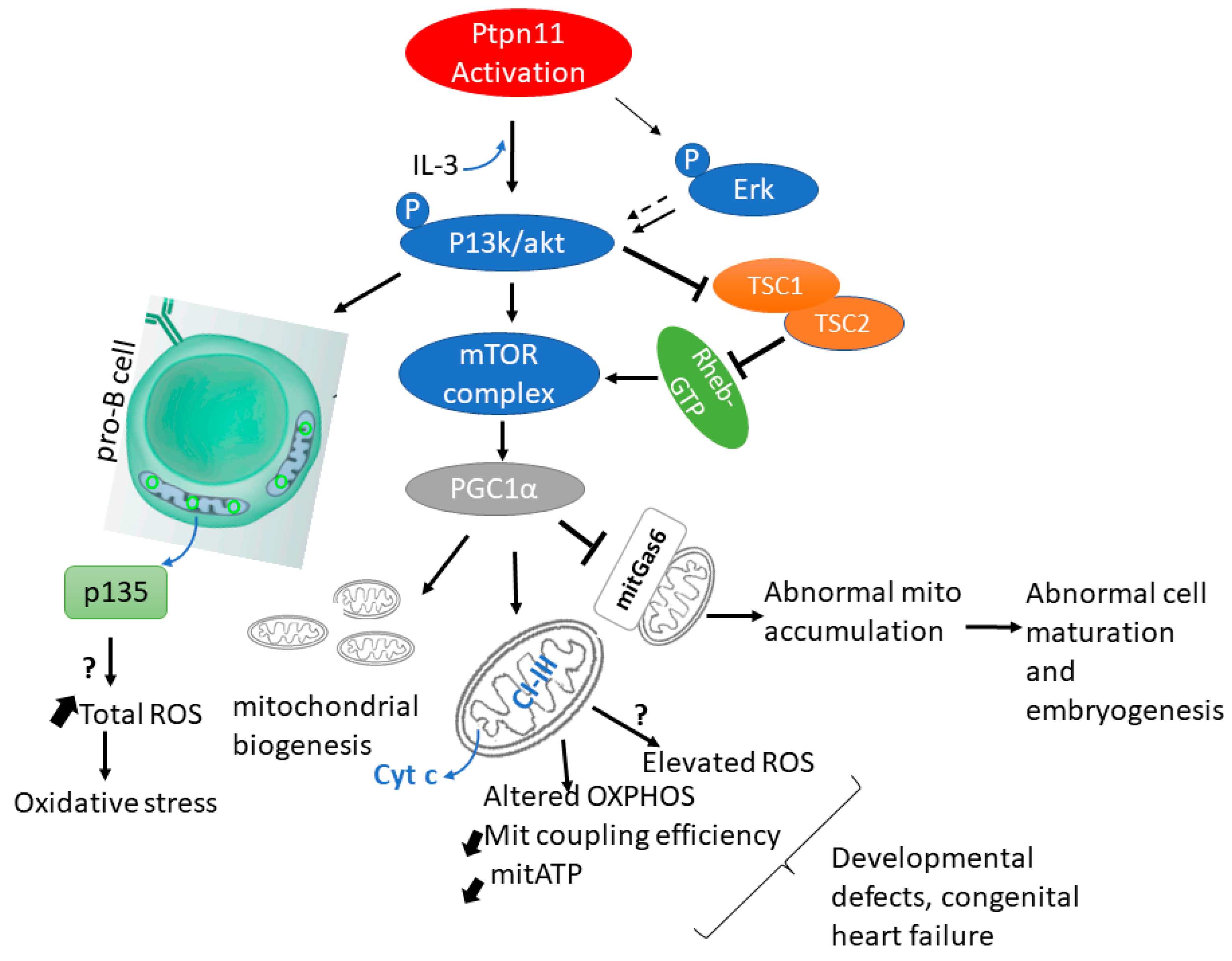
Figure 1. Activating Ptpn11 mutation through Erk/PI3k/Akt/mTOR signalling affects mitochondrial function. The presence of IL-3 (upward-blue arrow), active ptpn11 activates p13k/Akt signalling which inturn triggers the release of a mitochondria protein, p135 through an unknown mechanism(?). p135 release causes an increase (up-slanted arrows) in total ROS responsible for oxidative stress in tissues. Activated PI3K/Akt activates mTOR. Hypeactivated mTOR blocks the mitochondrial Growth arrest protein (mitGas6) leading to abnormal cell maturation. Via hyperactivated mTOR, Ptpn11 alters mitochondria respiration by decreasing (down-slanted arrows) CI-III of the ETC and coupling efficiency, releasing cyt c to the cytoplasm and reducing mitochondrial ATP.
Cells 11 03099 g001
Figure 2. Activating SHP2 and STAT3 signalling in mitochondrial metabolism. Mutated SHP2 blocks STAT3 phosphorylation which inturn initiates a cascade of actions in the mitochondrial ETC. Reduced complex I, II activity results in deficient OXPHOS and ATP generation and is accompanied by increased ROS (upward dotted arrows), thus decreasing mitochondria respiration resulting in phenotypes observed in NS/NNSML.
Cells 11 03099 g002
Figure 3. (A)SOS1 alters mitochondria morphology, mass, and dynamics. SOS1 deficiency blocks mitophagy, hence causes accumulation of defective mitochondria. Reduced (dotted down arrow) fusion-promoting proteins such as Mitofusin 1 (MFN1) and the long-form of mitochondrial dynamin-like GTPase (OPA-1L) leads to inhibition of mitochondrial fusion while the presence of GTPase Dynamin-Related protein 1 (DRP1) and Mitochondrial fission 1 (FIS1) favours mitochondria fission, hence the changes in mitochondria morphology, mass and dynamics. (B)SOS1/2 and KRAS mutation redirects cellular metabolism. The actions of SOS1, SOS1/2-DKO, CBL-KO and GOF KRAS rewires cellular metabolism as a consequence of decrease in respiration chain complexes, change in mitochondria structure, and utilisation of less oxidative substrates (blue arrows).
Cells 11 03099 g003
Figure 4. Effect of PTPN11 and RAF1 mutation on mitochondria permeability transition pore (mPTP). Upon PTPN11 activation or RAF1 inactivation, ERK signalling is phosphorylated, in turn phosphorylating and activating apoptotic regulators such as Bcl-2-like protein 11 (BIM); Bcl-2-like protein 4 (Bax), and Bcl-2 homologous antagonist/killer (Bak) localized on the mitochondrial membrane. This event destabilizes the opening and closure of the mitochondria permeability transition pores (mPTP), causing a blocked or prolonged opening state of the membrane channels. Thus, a huge efflux of Ca2+ (prolong state) is observed, resulting in decreased mitochondrial membrane potential and altered and uncontrolled cellular processes.
Cells 11 03099 g004
Figure 5. An overview of the effects of NS/NSML mutations on mitochondria metabolism via RAS signalling. The sequence of events coordinated by the major RAS/MAPK signalling and other pathways such as P13K/AKT results from the activation or inactivation of heterozygous/homozygous mutations in genes: PTPN11, SOS1/2, RAS, RAF1, BRAF1, NRAS, KRAS, CBL that cause NS and related syndrome. Outcomes observed include decreasing or increasing mitochondria metabolism, increasing ROS, and inhibiting apoptosis.
Cells 11 03099 g005
Table 1. Germline mutations of RAS/MAPK pathway responsible for NS and NSML phenotype.
DiseaseGeneLocationAmino Acid ChangeMutation Rate (%)Protein NameProtein ClassREF
NSPTPN1112q24p.D61G, p.G60A50–60SHP2: Protein tyrosine phosphatase
non-receptor type 11; Src Homology 2
Phosphatase[7,19]
SOS12p22-p21p.A1654G10–13Son of sevenless (SOS) homolog 1RasGEF[20]
RAF13p25.2p.R89L5–10v-Raf-1 murine leukemia viral oncogene
homolog 1
Kinase[20]
KRAS12p12.1p.G12X (X = any amino acid)
c.A458T
p.D153V
p.Y71D
10V-Ki-Ras2 Kirsten rat sarcoma viral
oncogene homolog
GTPase[20,21,22]
NRAS1p13.2p. G12X
p.Q61X
<1Neuroblastoma RAS viral (v-RAS)
oncogene homolog
GTPase[20,23,24]
SHOC210q25.2p.S2G12soc-2 suppressor of clear homologScaffolding[20]
CBL11q23.3p.Y371X
p.C404R
p.W408R
p.G415V
p.L380P
p.C840W
<1Casitas B-lineage lymphomaUbiquitin Ligase[20,25,26]
BRAF7q34p.V600E<1Serine/Threonine-Protein Kinase B-RafKinase[24]
A2ML112p13.31p.R802H, p.R592L, p.R802L<1α-2-macroglobulin-like 1Protease inhibitor[27]
SOS 22p22.1p.T376SL<1Son of sevenless homolog 2RasGEF[28]
LZTR122q11.21p.R284C, p.H287Y, p.Y119C,
p.G248R, p.S247 N
<1Leucine Zipper-Like Transcription
Regulator 1
Adaptor protein[20,28]
RASA23q23p.Y326C, p.Y326N, p.R511C<1Ras P21 Protein Activator 2RasGAP[29]
RRAS19q13.33p.G39dup, p.V55M<1Related RAS Viral (R-Ras) Oncogene
Homolog
GTPase[30,31]
RIT11q22p.S35 T, p.A57G, p.E81G, p.F82V,
p.F82L, p.T83P, p.Y89H, p.M90I,
p.G95A
710 Ras-Like Without CAAX 1GTPase[20,32]
NSMLPTPN1112q24p.Q510P90SHP2: Protein tyrosine phosphatase
non-receptor type 11; Src Homology 2
Phosphatase[20,33,34]
RAF13p25.2p.V263G
p.S257 L
5v-Raf-1 murine leukemia viral oncogene homolog 1Kinase[24,35]
BRAF7q34 <1Serine/Threonine-Protein Kinase B-RafKinase[24]
Publisher’s Note: MDPI stays neutral with regard to jurisdictional claims in published maps and institutional affiliations.