Article

High Expression of a Cancer Stemness-Related Gene, Chromobox 8 (CBX8), in Normal Tissue Adjacent to the Tumor (NAT) Is Associated with Poor Prognosis of Colorectal Cancer Patients

Department of Surgery, School of Clinical Medicine, Li Ka Shing Faculty of Medicine, The University of Hong Kong, Hong Kong SAR, China
*
Authors to whom correspondence should be addressed.
Cells 2022, 11(11), 1852; https://doi.org/10.3390/cells11111852
Submission received: 21 March 2022 / Revised: 27 May 2022 / Accepted: 2 June 2022 / Published: 6 June 2022

Abstract

:
Background: Several studies have demonstrated that the molecular profile of normal tissue adjacent to the tumor (NAT) is prognostic for recurrence in patients with different cancers. This study investigated the clinical significance of CBX8 gene expression, a cancer stemness-related gene, in tumor and NAT tissue of colorectal cancer (CRC) patients. Methods: The gene level of CBX8 in paired CRC and NAT specimens from 95 patients was determined by quantitative PCR. CBX8 protein level in CRC and NAT specimens from 66 patients was determined by immunohistochemistry. CBX8 gene and protein levels were correlated with the patients’ clinicopathological parameters and circulatory immune cell profiles. The association between CBX8 and pluripotency-associated genes was analyzed using the TCGA database. Results: NAT CBX8 gene level positively correlated with TNM stage, tumor invasion, lymph node metastasis and distant metastasis, indicating its association with tumor progression and metastasis. There was no correlation between NAT CBX8 protein level and clinicopathological parameters. Moreover, a high level of CBX8 gene and protein in NAT both correlated with poor DFS and OS. There was an inverse correlation between CBX8 gene level and post-operative platelet counts and platelet to lymphocyte level, suggesting its association with systematic inflammation. Finally, TCGA analysis showed that CBX8 level was correlated with a couple of pluripotency-associated genes, supporting its association with cancer stemness. Conclusions: High NAT CBX8 is a poor prognostic factor for tumor progression and survival in CRC patients.

1. Introduction

Colorectal cancer (CRC) is one of the commonest cancers and a leading cause of cancer deaths worldwide. The 5-year survival rate remains below 65% in developed countries and under 50% in developing countries [1], mainly due to recurrence and metastasis. Recent studies demonstrated that the molecular profile of normal tissue adjacent to the tumor (NAT) is prognostic for recurrence in patients with different cancers including CRC [2,3,4], HCC [5], prostate [6], oral carcinoma [7], breast cancer [8] and head and neck cancer [9]. We believe a better understanding of the molecular profile in NAT is crucial for identifying molecular biomarkers for predicting the prognosis of CRC patients and therapeutic targets to combat recurrence.
Epigenetic gene repression by Chromobox (CBX) proteins is crucial for the maintenance and self-renewal capabilities of pluripotent and multipotent stem cells [10]. Chromobox 8 (CBX8) is one of the members of CBX protein family. Previous studies demonstrated that CBX8 is overexpressed in CRC and it functionally induces CRC cell proliferation and inhibits tumor apoptosis [11,12], suggesting that CBX8 plays an oncogenic role in CRC. More importantly, a recent study performed by Zhang et al. showed that CBX8 contributes to increased cancer stemness and decreased chemosensitivity in CRC [13]. In line with this study, CBX8 is also reported to induce stemness in HCC [14] and breast cancer [15]. Moreover, the prognostic potential of tumor CBX8 expression for the survival of CRC patients has been investigated in three studies [12,16,17], yet the findings were ambiguous. Tang J. et al. showed in their own clinical specimens that CBX8 protein level was overexpressed in CRC when compared to paired adjacent normal tissue, but surprisingly low expression level of CBX8 was associated with poor disease-free survival and overall survival [12]. Song X. et al. on the other hand, by analyzing an independent dataset (GSE38832) reported that a high CBX8 gene level was significantly associated with worse overall survival [16]. In a recent study, Li Q. et al. showed no correlation between CBX8 and disease-free survival or overall survival by using GEPIA and Kaplan–Meier plotter online database analysis tools [17]. These findings suggested that the prognostic significance of tumor CBX8 level remains inconclusive. Moreover, the significance of NAT CBX8 gene expression has not been reported. Hence, the clinical significance of CBX8 expression in tumor and NAT of CRC patients warrants further investigation for a better understanding of its prognostic significance for CRC recurrence.
This study investigated the association of tumor and NAT CBX8 level with clinicopathological parameters and disease-free and overall survival of CRC patients. The correlation between CBX8 level and CRC progression is further explained by its correlation with changes in immune cell profile and co-expression with other pluripotency-associated genes.

2. Materials and Methods

2.1. Patient Specimens

Specimens of the CRC tumor and its normal tissue adjacent to the tumor (NAT) counterpart were obtained from 95 patients who underwent surgical resection of primary CRC between 2008 and 2012 at the Department of Surgery, Queen Mary Hospital, the University of Hong Kong. Based on the guidelines of TCGA which clearly specifies that NAT samples must be collected at a distance greater than 2 cm from the tumor margins and/or the tissue must be histopathologically validated to not contain any tumor cells [18,19,20,21], all NAT specimens were collected at a distance ranging from 2 cm (minimum distance from the tumor margin) to a maximum distance of 10 cm. All samples were immediately frozen in liquid nitrogen and kept at −80 °C until analysis. The study was approved by the Institutional Review Board of the University of Hong Kong/Hospital Authority Hong Kong West Cluster (HKU/HA HKW IRB) and written informed consents were obtained from patients prior to their inclusion. Comprehensive CRC patient demographic and clinical characteristics included the year of diagnosis, age, sex, race/ethnicity, tumor location, tumor size, tumor stage, and receipt of cancer treatment (such as surgery, chemotherapy, and radiation therapy).

2.2. Blood Immune Cell Profile of CRC Patients

Preoperative and postoperative immune cell count data including lymphocyte, neutrophil, neutrophil-to-lymphocyte ratio (NLR), platelet (PLT), platelet-to-lymphocyte ratio (PLR), monocyte, lymphocyte-to-monocyte ratio (LMR), albumin, and prognostic nutritional index (PNI) were obtained from the patient data database. Preoperative data were defined as results from the final blood test performed before surgery (usually one day before operation). Postoperative data were defined as those taken within 21–90 days after surgery but before starting adjuvant chemotherapy to minimize the influence of the systemic inflammatory response due to the operation and the effect of chemotherapy [22,23]. The dynamic changes in their level between post- and pre-operative blood were calculated as delta values (post-operative level minus pre-operative level).

2.3. RNA Extraction and Quantitative PCR (qPCR) Analysis

Total RNA was extracted from tumors and NAT using Trizol reagent and RNA miniprep extraction kit according to the manufacturer’s instruction (Life Technologies) and 500 ng of each RNA sample was used to prepare cDNA using PrimeScript™ RT Master Mix (TaKaRa). For qPCR, 1 μL of cDNA template was added to a master mix that consists of SYBR Premix EX Taq II (TaKaRa), forward and reverse primers (5′ to 3′, CBX8 forward: GGC CTC GGA GAG TAC CTC AA, reverse: TAG GGC TGG TCA CAC CAC AC; GAPDH forward: TTC ACC ACC ATG GAG AAG GC, reverse: GGC ATG GAC TGT GGT CAT GA), and ROX reference dye to form a 15 μL reaction mixture. A duplicate of each sample was made for each run. The reaction was conducted with 7900HT Fast Real-Time PCR System (Applied Biosystems, Waltham, MA, USA). The relative CBX8 expression was expressed as fold to GAPDH, which was calculated with the 2−ΔCt(CBX8-GAPDH) formula.

2.4. Expression Correlation of CBX8 with Other Pluripotency-Associated Genes Using an Online Database

To analyze the association of CBX8 with CSC features, we analyzed its co-expression with other pluripotency gene markers in CRC and NAT from the TCGA database at GEPIA (http://gepia2.cancer-pku.cn/#index (accessed on 20 December 2021)).

2.5. Immunohistochemistry

Human colorectal specimens and mice xenograft tumors were first paraffin-embedded into blocks; the tissue blocks were then cut into 5 μm histosections and prepared on glass slides. The slides were deparaffinized using xylene, followed by gradual rehydration from alcohol to water. Antigen retrieval was done by heating for 10 min in pH 6.0 citrate buffer using a microwave. The slides were then immersed in 3% hydrogen peroxide and 5% horse serum in TBST to block endogenous peroxidase activity and any non-specific binding of immunoglobulin, respectively. The sections were incubated in anti-human 1:30 CBX8 (Novus Biologicals) antibody overnight at 4 °C. After 1 h incubation of biotinylated secondary antibody at room temperature, the slides were further incubated at room temperature with streptavidin-peroxidase complex for 1 h. 3,3′-diaminobenzidine chromogen solution was used to develop the signal, followed by haematoxylin for counterstaining. Dako LSABTM+ kit (Dako, Glostrup, Denmark) was used in this part of the study. All slides were assessed independently by two investigators and the extent of signal was evaluated by the staining intensity and density (score from 0 to 4). The score was the average value of the two investigators.

2.6. Statistical Analysis

Student’s t-test was used to analyze differences between experimental groups of clinical specimens. Survival curves were generated using the Kaplan–Meier test and compared by the log-rank test. The association between CBX8 expression and immune cell profile was analyzed with Pearson and Spearman correlation. The criterion for statistical significance was p < 0.05. All statistical analyses were conducted using SigmaPlot version 10.0 (Systat Software Inc., San Jose, CA, USA).

3. Results

3.1. CBX8 Gene and Protein Expressions in CRC and Normal Tissue Adjacent to Tumor (NAT) Specimens and Their Clinicopathological Significances

The expression of CBX8 in 95 paired human CRC and adjacent non-tumor specimens was determined by quantitative PCR and correlated with the patients’ clinicopathological data. The expression of CBX8 was significantly higher in CRC compared to the NAT specimens (Figure 1A, p < 0.001). On average, the CBX8 level in CRC was ~2.92-fold higher than NAT (Figure 1B). More specifically, 55 out of the 95 patients demonstrated at least 2-fold CBX8 overexpression in CRC. The correlation of CBX8 levels in CRC and NAT speicmens with patients’ clinicopathological data was subsequently analyzed (Table 1). We found that tumor CBX8 level was not associated with any clinical parameters, except a trend of inverse correlation with tumor invasion was seen (p = 0.152). This observation is in line with a previous study showing high CBX8 impaired CRC metastasis [12]. On the other hand, high NAT CBX8 showed a positive correlation with stage (p = 0.026), tumor invasion (p = 0.0035), lymph node metastasis (p = 0.036) and distant metastasis (p = 0.044), indicating its association with tumor progression and metastasis.
Among these 95 patients, formalin-fixed and parafilm-embedded blocks were available from 66 of them, hence we performed immunohistochemistry to determine the protein level of CBX8 in these patients. CBX8 was significantly overexpressed in CRC when compared to paired NAT (p < 0.001, Figure 1B,C). In CBX8-positive NAT specimens, both crypt epithelial cells and stroma cells were able to express CBX8 (Supplementary Figure S1). The CBX protein level was significantly correlated with CBX8 gene level in NAT (R = 0.405, p = 0.00074) but not in CRC (Figure 1D). No significant correlation was found between CRC nor NAT CBX8 protein level and clinicopathological parameters.

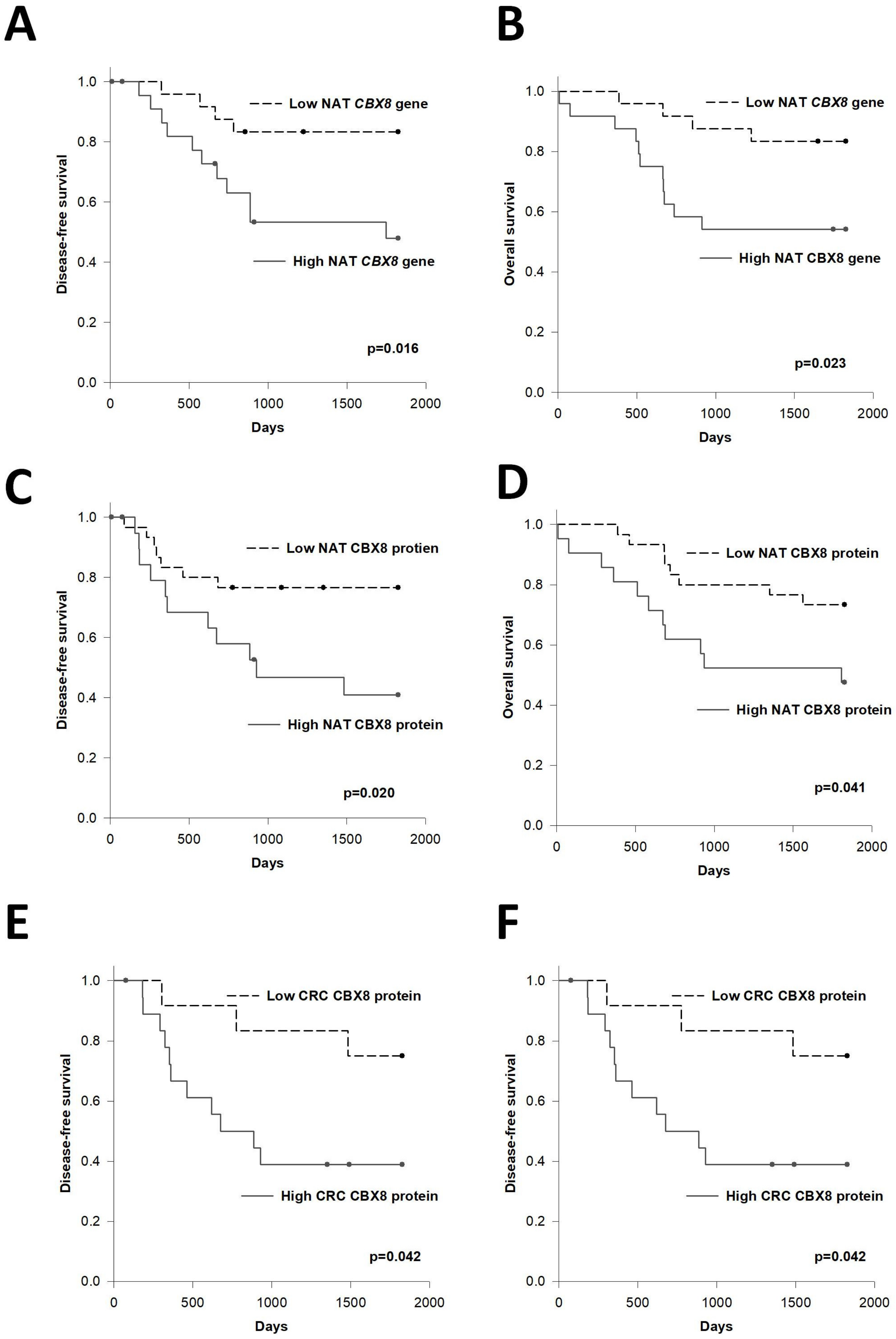
3.2. Prognostic Significance of CBX8

We investigated the association of CBX8 with 5-year disease-free survival (DFS) and overall survival (OS) of CRC patients. Our data showed that the tumor CBX gene level was not associated with DFS nor OS of patients (data not shown). On the other hand, when we stratified the patients into high NAT CBX8 (relative level above 0.000288, i.e., the upper quartile level) or low NAT CBX8 (relative level below 0.000166, i.e., the lower quartile level), patients with high NAT CBX8 demonstrated significantly worse DFS (1224 vs. 1618 days, p = 0.016) and OS (1223 vs. 1651 days, p = 0.023), compared to those with low NAT CBX8 level (Figure 2A,B).
We also investigated the correlation between CBX8 protein levels and prognosis of the patients (Figure 2C–F). Our data showed that high NAT CBX8 protein levels (IHC score ≥ 2.5, i.e., the upper quartile level), when compared to low NAT CBX level group (IHC score ≤ 1.5, i.e., the lower quartile level), showed significantly shorter DFS (1081 vs. 1478 days, p = 0.020) and OS (1195 vs. 1559 days, p = 0.041). Similarly, patients with high CRC CBX8 protein levels (IHC score ≥ 3.5, i.e., the upper quartile level) also demonstrated significantly shorter DFS (1002 vs. 1582 days, p = 0.042) and OS (1150 vs. 1658 days, p = 0.036) when compared to the low CRC CBX group (IHC score ≤ 1.5, i.e., the lower quartile level).
To summarize, our clinical analysis demonstrated that high CBX8 gene and protein levels in NAT or CRC are associated with poor prognosis in CRC patients. Furthermore, the NAT CBX8 gene level is closely associated with clinicopathological parameters related to tumor progression and metastasis.

3.3. Association between CBX8 Expression and Immune Cell Profile of CRC Patients

The immune system plays the most vital role in protection against diseases, including cancer. We previously reported that certain immune cell levels are correlated with the prognosis of post-operative CRC patients [24]. Hence, in this study, we investigated the association between tumor and NAT CBX8 level with immune cell profile of pre-operative and post-operative blood samples, as well as the dynamic changes in their level between post- and pre-operative blood (expressed as a delta value, which is calculated as post-operative level minus pre-operative level).
Our results showed that there was no correlation between tumor nor NAT CBX8 level with any pre-operative blood parameters. On the other hand, NAT CBX8 gene level was inversely associated with post-operative PLT (R = −0.342, p = 0.0008, Figure 3A) and PLR (R = −0.333, p = 0.01, Figure 3B), whereas low PLT and PLR were associated with inferior OS in patients with metastatic CRC [25]. Moreover, NAT CBX8 gene level showed a positive trend with post-operative CEA (R = 0.233, p = 0.075, Figure 3C), which is a risk factor for recurrence [26,27,28,29]. On the other hand, tumor CBX8 gene level was associated with post-operative LMR (R = 0.479, p = 0.0001, Figure 3D), which indicated a better prognosis [23]. In addition, a higher tumor CBX8 gene level was correlated with lower delta-LMR (R = −0.433, p = 0.0007, Figure 3E) and higher delta-eosinophil (R = 0.302, p = 0.0201, Figure 3F). Though the prognostic value of these parameters has not been reported in CRC, low delta-LMR and high delta-eosinophil were associated with better OS or DFS in renal cell carcinoma and head and neck cancer patients [30,31], respectively. These correlations suggested that tumor CBX8 gene level is associated with better prognosis in CRC patients.
Comparing the CBX8 gene levels, less correlation was found between CBX8 protein level in NAT or CRC with immune cell profile of the patients. Surprisingly, CRC CBX8 protein level demonstrated an inverse correlation with delta-eosinophil, which is opposite to the pattern observed for CRC CBX8 gene level (R = −0.361, p = 0.0281, Figure 4A). Moreover, CBX8 protein levels in both NAT and CRC were positively correlated with post-operative CEA levels and delta-CEA levels (Figure 4B–E). These findings indicate that high NAT and CRC CBX8 levels are poor prognostic factors for CRC patients.

3.4. Association of CBX8 with Pluripotency-Associated Genes in CRC and NAT

CBX8 has been shown to induce cancer stemness and chemoresistance in CRC through activation of the transcription of LGR5 in a noncanonical manner [13]. To further demonstrate its association with CSC features, we analyzed its co-expression with other pluripotentcy gene markers in CRC from the TCGA database at GEPIA (http://gepia2.cancer-pku.cn/#index (accessed on 21 February 2022)). In line with the above study, CBX8 was positively correlated with LGR5 in CRC (Table 2). Furthermore, we found that CBX8 level in colon cancer was positively correlated with a couple of pluripotentcy-associated genes including BMI1, OCT4, MYC, USP36, RPTOR, UBE2O, JMJD6, SLCO4A1, UTP18, E2F6 and CD44. These data demonstrated the close association between CBX8 and colorectal cancer stemness.
We are also interested in the correlation between CBX8 and pluripotency-associated genes in NAT of colorectal cancer patients (Table 2). Since the sample number is lower for NAT at GEPIA, we considered pluripotency-associated genes, which showed a correlation with CBX8 (p-value below 0.1), as noteworthy candidates. Similar to the finding in CRC specimens, the significant correlations of CBX8 with MYC and UBE2O were also observed in NAT, and a trend of correlations were also observed for LGR5, UTP18 and ABHD5. Furthermore, a trend of positive correlation with CD44 was also shown in NAT but not in CRC. These results suggested that high CBX8 level in NAT might indicate the presence of CSC-like cells within NAT that lead to recurrence and hence poor DFS and OS.

4. Discussion

Previous studies demonstrated that CBX8 is overexpressed in CRC and it functionally induces CRC cell proliferation and inhibits tumor apoptosis [11,12], and CBX8 is associated with cancer stemness and chemoresistance in CRC [13], yet the prognostic potential of tumor CBX8 expression for the survival of CRC patients was ambiguous among current reports [12,16,17]. Hence this study comprehensively investigated the clinicopathological and prognostic significance of CBX8 gene and protein levels in both NAT and CRC specimens.
This study demonstrated that high NAT CBX8 gene and protein level was associated with higher risk of recurrence and mortality. The significance of NAT tissue has been suggested since Slaughter et al. first described the “field cancerization” theory [32], suggesting a cumulative process of carcinogenesis in which genetic alterations are acquired step-wise, leaving the NAT tissue in an intermediate, pre-neoplastic state composed of morphologically normal but molecularly altered cells. A recent study on histologically normal breast epithelium by Graham K et al. suggested that the microenvironment surrounding the tumor is essential for understanding recurrence [33]. In line with this phenomenon, the NAT molecular profile is reported to show the prognostic significance for recurrence in patients with different cancers including CRC [2,3,4], HCC [5], prostate [6], oral carcinoma [7], breast cancer [8] and head and neck cancer [9]. Further investigation on CBX8, as well as other dysregulated NAT molecular markers, for prognosis of CRC patients, warrants evaluation for their clinical use.
On the other hand, the tumor CBX8 gene and protein level demonstrated a rather opposite pattern in its prognostic significance. Although tumor CBX8 gene level was not associated with DFS and OS of CRC patients in this study, our findings demonstrated that higher CBX8 level in tumor was associated with higher post-operative LMR and lower delta LMR, which have been reported as factors of better prognosis for CRC patients [30,31]. In addition, our data revealed that higher tumor CBX8 showed a trend towards lower tumor invasion. On the other hand, high CRC CBX8 protein level was associated with poor survival and higher CEA. Since our data showed that there was no significant correlation between CBX8 gene and protein level, this might explain the discrepancies observed in our study as well as among the different CBX8 studies within CRC patients.
Chromobox protein homolog 8 (CBX8), together with CBX2, CBX4, CBX6, and CBX7, forms the CBX family, which is the central part of the polycomb repressive complex 1 (PRC1) [34]. CBX8 has recently emerged as a potential oncogenic target in multiple malignancies [35]. Previous studies demonstrated that CBX8 is overexpressed in CRC and it functionally induces CRC cell proliferation and inhibits tumor apoptosis [11,12], suggesting that CBX8 plays an oncogenic role in CRC. More importantly, CBX8 contributes to increased cancer stemness in CRC [13], as well as HCC [14] and breast cancer [15]. This study demonstrated the association between CBX8 level and a couple of pluripotency-associated genes including LGR5, Bmi1, OCT4, MYC, USP36, RPTOR, UBE2O, JMJD6, SLCO4A1, UTP18 and E2F6, and inversely with CD166, ABHD5, PDCD4 and GPA33. Of note, CD166 is associated with a decreased risk of vascular invasion in CRC in a meta-analysis consisting of 3332 cases [36]. In addition, ABDH5 impairs colon cancer stemness [37]; PDCD4 is a well-known tumor suppressor in CRC [38] and it represses stemness of adipose-derived stem cells [39], gastric [40] and cervical cancer cells [41]; GPA33 is a cell surface antigen expressed across a panel of CSC derived from various stages of CRC, in addition to exhibiting 100% penetrance across over 50 primary and metastatic colorectal cancer specimens, and its application as a therapeutic target is in progress [42,43]. These findings indicate the high CBX8 expression in CRC is indeed related to its CSC feature. CBX8 was also correlated with MYC, UBE2O, UTP18, ABHD5, LGR5 and CD44 in NAT, suggesting that a high CBX8 level in NAT might indicate the presence of CSC-like cells within NAT that can lead to recurrence.
In fact, CSCs are known to be responsible for therapeutic resistance and c-MYC plays a pivotal role in the regulation of CSCs [44]. c-MYC is upregulated in several cancers and the TCGA database shows that 28% of all cancers harbour genetic abnormalities in at least one of the Myc family [45]. c-MYC contributes to tumor progression and metastasis and induces cancer stemness properties in multiple neoplasms [46]. It also regulates the transcription of over 15% of human genes, such as CCND, CDK4 and E2F1, which are involved in tumour progression [47]. Furthermore, Strippoli et al. showed that alterations in the transcriptional factor c-MYC are involved in anti-EGFR resistance in metastatic colorectal cancer; patients with higher c-MYC expression showed a significantly lower PFS and OS when compared to those with low c-MYC expression [48]. Moreover, with expression gene profiling, they pointed out the pivotal role of c-MYC in CRC-related cell-cycle, apoptosis, signal transduction and cell-growth pathways. More importantly, our supplementary experiments showed that CBX8 overexpression in the CRC cell line HCT116 induced the protein level of c-MYC (Supplementary Figure S2), as well as repressed the level of let-7, which is reported to downregulate c-MYC in CRC as well as being down-regulated by c-MYC via LIN28 repression [49]. These results suggest that CBX8 not only was associated with the level of CSC-associated genes, but also functionally regulated the CSC features through regulating c-MYC and possibly other CSC-related genes. It is also reported that UBE2C, a gene that encodes an enzyme required for cell cycle progression, is directly involved in cetuximab resistance and could be a valid target to overcome the EGFR-blockage resistance [50]. A gene belonging to the same family (UBE2O) has been shown to promote the proliferation, epithelial–mesenchymal transformation and stemness properties of breast cancer cells through the UBE2O/AMPKα2/mTORC1-MYC positive feedback loop [51]. This same gene has been demonstrated to be correlated with CBX8 levels both in colon cancer and in NAT, emphasizing the intricate interconnection between CBX8 levels, stemness, tumor progression and resistance to therapy.
In light of the functional significance of CBX8 in inducing malignancy in multiple cancer types, it has been suggested as a therapeutic target for cancer treatment and very recently its inhibitors have been developed and are being tested [35,52,53]. Our study showed that patients with high NAT CBX8 were associated with a worse stage, tumor invasion, lymph node metastasis, distant metastasis, and poor DFS and OS, suggesting that CRC patients with high NAT CBX8 are potential candidates for CBX8-targeted treatment. Moreover, we showed that post-operative PLT and PLR levels were associated with NAT CBX8 levels. It is warranted to investigate the potential of PLR and PLT as biomarkers to predict and monitor the response to CBX8 inhibitor treatment.
The limitation of this study is that the functional significance of CBX8 in NAT was not investigated. Nevertheless, CBX8 has been demonstrated to induce tumor growth or progression in CRC and other cancers, and our supplementary experiment indicated that its overexpression induced the protein level of c-MYC. We believe the high expression of CBX8 in NAT is not just a biomarker but is one of the factors contributing to tumor progression, recurrence and metastasis. Therefore, further investigation of the treatment effect on recurrence by repressing its expression in NAT in animal models following primary tumor removal, is warranted to evaluate the potential of treating post-operative CRC patients using this novel approach.

Supplementary Materials

The following supporting information can be downloaded at: https://www.mdpi.com/article/10.3390/cells11111852/s1, Figure S1: Representative IHC stainings of CBX8; Figure S2: Effect of CBX8 overexpression on c-MYC.

Author Contributions

Conceptualization, L.N., R.W.-C.P. and W.-L.L.; methodology, H.-S.L. and A.K.-M.C.; formal analysis, L.N., A.T.-K.M. and H.-S.L.; resources, R.W.-C.P., D.C.-C.F., O.S.-H.L. and W.-L.L.; writing—original draft preparation, L.N. and H.-S.L.; writing—review and editing, L.N. and W.-L.L.; funding acquisition, W.-L.L. All authors have read and agreed to the published version of the manuscript.

Funding

This research was funded by the Health and Medical Research Fund of the Research Fund Secretariat, Food and Health Bureau, HKSAR, grant number 06173406.

Institutional Review Board Statement

The study was conducted according to the guidelines of the Declaration of Helsinki, and approved by the Institutional Review Board (or Ethics Committee) of The University of Hong Kong (protocol code UW 17-416 and date 1 November 2017.

Informed Consent Statement

Patient consent was waived due to this is a retrospective review of medical records and the patient management will not be affected.

Data Availability Statement

The data presented in this study are available on request from the corresponding author. The data are not publicly available due to privacy.

Acknowledgments

The authors would like to thank Ryan Sin, David Cheung and Ivan Au for technical support and Ambrose Chan for English language editing of this manuscript.

Conflicts of Interest

The authors declare no conflict of interest.

References

  1. Allemani, C.; Weir, H.K.; Carreira, H.; Harewood, R.; Spika, D.; Wang, X.S.; Bannon, F.; Ahn, J.V.; Johnson, C.J.; Bonaventure, A.; et al. Global surveillance of cancer survival 1995–2009: Analysis of individual data for 25,676,887 patients from 279 population-based registries in 67 countries (CONCORD-2). Lancet 2015, 385, 977–1010. [Google Scholar] [CrossRef] [Green Version]
  2. Schneider, S.; Park, D.J.; Yang, D.; El-Khoueiry, A.; Sherrod, A.; Groshen, S.; Streeter, O.; Iqbal, S.; Danenberg, K.D.; Lenz, H.J. Gene expression in tumor-adjacent normal tissue is associated with recurrence in patients with rectal cancer treated with adjuvant chemoradiation. Pharm. Genom. 2006, 16, 555–563. [Google Scholar] [CrossRef] [PubMed]
  3. Kuan, J.C.; Wu, C.C.; Sun, C.A.; Chu, C.M.; Lin, F.G.; Hsu, C.H.; Kan, P.C.; Lin, S.C.; Yang, T.; Chou, Y.C. DNA methylation combinations in adjacent normal colon tissue predict cancer recurrence: Evidence from a clinical cohort study. PLoS ONE 2015, 10, e0123396. [Google Scholar] [CrossRef] [PubMed] [Green Version]
  4. Grassi, A.; Perilli, L.; Albertoni, L.; Tessarollo, S.; Mescoli, C.; Urso, E.D.L.; Fassan, M.; Rugge, M.; Zanovello, P. A coordinate deregulation of microRNAs expressed in mucosa adjacent to tumor predicts relapse after resection in localized colon cancer. Mol. Cancer 2018, 17, 17. [Google Scholar] [CrossRef] [PubMed] [Green Version]
  5. Pan, Q.; Qin, F.; Yuan, H.; He, B.; Yang, N.; Zhang, Y.; Ren, H.; Zeng, Y. Normal tissue adjacent to tumor expression profile analysis developed and validated a prognostic model based on Hippo-related genes in hepatocellular carcinoma. Cancer Med. 2021, 10, 3139–3152. [Google Scholar] [CrossRef] [PubMed]
  6. Zhou, R.; Feng, Y.; Ye, J.; Han, Z.; Liang, Y.; Chen, Q.; Xu, X.; Huang, Y.; Jia, Z.; Zhong, W. Prediction of Biochemical Recurrence-Free Survival of Prostate Cancer Patients Leveraging Multiple Gene Expression Profiles in Tumor Microenvironment. Front. Oncol. 2021, 11, 632571. [Google Scholar] [CrossRef]
  7. Reis, P.P.; Waldron, L.; Perez-Ordonez, B.; Pintilie, M.; Galloni, N.N.; Xuan, Y.; Cervigne, N.K.; Warner, G.C.; Makitie, A.A.; Simpson, C.; et al. A gene signature in histologically normal surgical margins is predictive of oral carcinoma recurrence. BMC Cancer 2011, 11, 437. [Google Scholar] [CrossRef] [Green Version]
  8. Dadiani, M.; Bossel Ben-Moshe, N.; Paluch-Shimon, S.; Perry, G.; Balint, N.; Marin, I.; Pavlovski, A.; Morzaev, D.; Kahana-Edwin, S.; Yosepovich, A.; et al. Tumor Evolution Inferred by Patterns of microRNA Expression through the Course of Disease, Therapy, and Recurrence in Breast Cancer. Clin. Cancer Res. 2016, 22, 3651–3662. [Google Scholar] [CrossRef] [Green Version]
  9. Ganci, F.; Sacconi, A.; Manciocco, V.; Covello, R.; Benevolo, M.; Rollo, F.; Strano, S.; Valsoni, S.; Bicciato, S.; Spriano, G.; et al. Altered peritumoral microRNA expression predicts head and neck cancer patients with a high risk of recurrence. Mod. Pathol. 2017, 30, 1387–1401. [Google Scholar] [CrossRef] [Green Version]
  10. Van Wijnen, A.J.; Bagheri, L.; Badreldin, A.A.; Larson, A.N.; Dudakovic, A.; Thaler, R.; Paradise, C.R.; Wu, Z. Biological functions of chromobox (CBX) proteins in stem cell self-renewal, lineage-commitment, cancer and development. Bone 2021, 143, 115659. [Google Scholar] [CrossRef]
  11. Yang, S.; Liu, W.; Li, M.; Wen, J.; Zhu, M.; Xu, S. Insulin-Like Growth Factor-1 Modulates Polycomb Cbx8 Expression and Inhibits Colon Cancer Cell Apoptosis. Cell Biochem. Biophys. 2015, 71, 1503–1507. [Google Scholar] [CrossRef]
  12. Tang, J.; Wang, G.; Zhang, M.; Li, F.Y.; Sang, Y.; Wang, B.; Hu, K.; Wu, Y.; Luo, R.; Liao, D.; et al. Paradoxical role of CBX8 in proliferation and metastasis of colorectal cancer. Oncotarget 2014, 5, 10778–10790. [Google Scholar] [CrossRef] [Green Version]
  13. Zhang, Y.; Kang, M.; Zhang, B.; Meng, F.; Song, J.; Kaneko, H.; Shimamoto, F.; Tang, B. m(6)A modification-mediated CBX8 induction regulates stemness and chemosensitivity of colon cancer via upregulation of LGR5. Mol. Cancer 2019, 18, 185. [Google Scholar] [CrossRef] [Green Version]
  14. Tang, B.; Tian, Y.; Liao, Y.; Li, Z.; Yu, S.; Su, H.; Zhong, F.; Yuan, G.; Wang, Y.; Yu, H.; et al. CBX8 exhibits oncogenic properties and serves as a prognostic factor in hepatocellular carcinoma. Cell Death Dis. 2019, 10, 52. [Google Scholar] [CrossRef] [PubMed] [Green Version]
  15. Chung, C.Y.; Sun, Z.; Mullokandov, G.; Bosch, A.; Qadeer, Z.A.; Cihan, E.; Rapp, Z.; Parsons, R.; Aguirre-Ghiso, J.A.; Farias, E.F.; et al. Cbx8 Acts Non-canonically with Wdr5 to Promote Mammary Tumorigenesis. Cell Rep. 2016, 16, 472–486. [Google Scholar] [CrossRef] [Green Version]
  16. Song, X.; Tang, T.; Li, C.; Liu, X.; Zhou, L. CBX8 and CD96 Are Important Prognostic Biomarkers of Colorectal Cancer. Med. Sci. Monit. Int. Med. J. Exp. Clin. Res. 2018, 24, 7820–7827. [Google Scholar] [CrossRef]
  17. Li, Q.; Pan, Y.; Cao, Z.; Zhao, S. Comprehensive Analysis of Prognostic Value and Immune Infiltration of Chromobox Family Members in Colorectal Cancer. Front. Oncol. 2020, 10, 582667. [Google Scholar] [CrossRef]
  18. Aran, D.; Camarda, R.; Odegaard, J.; Paik, H.; Oskotsky, B.; Krings, G.; Goga, A.; Sirota, M.; Butte, A.J. Comprehensive analysis of normal adjacent to tumor transcriptomes. Nat. Commun. 2017, 8, 1077. [Google Scholar] [CrossRef] [Green Version]
  19. Cancer Genome Atlas Research, N.; Kandoth, C.; Schultz, N.; Cherniack, A.D.; Akbani, R.; Liu, Y.; Shen, H.; Robertson, A.G.; Pashtan, I.; Shen, R.; et al. Integrated genomic characterization of endometrial carcinoma. Nature 2013, 497, 67–73. [Google Scholar] [CrossRef]
  20. The Cancer Genome Atlas (TCGA) Research Network. Comprehensive molecular portraits of human breast tumours. Nature 2012, 490, 61–70. [Google Scholar] [CrossRef] [PubMed] [Green Version]
  21. The Cancer Genome Atlas Research Network. Comprehensive molecular characterization of urothelial bladder carcinoma. Nature 2014, 507, 315–322. [Google Scholar] [CrossRef] [PubMed] [Green Version]
  22. Chan, J.C.Y.; Diakos, C.I.; Chan, D.L.H.; Engel, A.; Pavlakis, N.; Gill, A.; Clarke, S.J. A Longitudinal Investigation of Inflammatory Markers in Colorectal Cancer Patients Perioperatively Demonstrates Benefit in Serial Remeasurement. Ann. Surg. 2018, 267, 1119–1125. [Google Scholar] [CrossRef]
  23. Yasui, K.; Shida, D.; Nakamura, Y.; Ahiko, Y.; Tsukamoto, S.; Kanemitsu, Y. Postoperative, but not preoperative, inflammation-based prognostic markers are prognostic factors in stage III colorectal cancer patients. Br. J. Cancer 2021, 124, 933–941. [Google Scholar] [CrossRef]
  24. Ng, L.; Foo, D.C.; Wong, C.K.; Man, A.T.; Lo, O.S.; Law, W.L. Repurposing DPP-4 Inhibitors for Colorectal Cancer: A Retrospective and Single Center Study. Cancers 2021, 13, 3588. [Google Scholar] [CrossRef] [PubMed]
  25. Chang, J.; Lin, G.; Ye, M.; Tong, D.; Zhao, J.; Zhu, D.; Yu, Q.; Zhang, W.; Li, W. Decreased mean platelet volume predicts poor prognosis in metastatic colorectal cancer patients treated with first-line chemotherapy: Results from mCRC biomarker study. BMC Cancer 2019, 19, 15. [Google Scholar] [CrossRef]
  26. Lin, J.K.; Lin, C.C.; Yang, S.H.; Wang, H.S.; Jiang, J.K.; Lan, Y.T.; Lin, T.C.; Li, A.F.; Chen, W.S.; Chang, S.C. Early postoperative CEA level is a better prognostic indicator than is preoperative CEA level in predicting prognosis of patients with curable colorectal cancer. Int. J. Colorectal Dis. 2011, 26, 1135–1141. [Google Scholar] [CrossRef]
  27. You, W.; Yan, L.; Cai, Z.; Xie, L.; Sheng, N.; Wang, G.; Wu, X.; Wang, Z. Clinical Significances of Positive Postoperative Serum CEA and Post-preoperative CEA Increment in Stage II and III Colorectal Cancer: A Multicenter Retrospective Study. Front. Oncol. 2020, 10, 671. [Google Scholar] [CrossRef]
  28. Bhatti, I.; Patel, M.; Dennison, A.R.; Thomas, M.W.; Garcea, G. Utility of postoperative CEA for surveillance of recurrence after resection of primary colorectal cancer. Int. J. Surg. 2015, 16, 123–128. [Google Scholar] [CrossRef]
  29. Konishi, T.; Shimada, Y.; Hsu, M.; Tufts, L.; Jimenez-Rodriguez, R.; Cercek, A.; Yaeger, R.; Saltz, L.; Smith, J.J.; Nash, G.M.; et al. Association of Preoperative and Postoperative Serum Carcinoembryonic Antigen and Colon Cancer Outcome. JAMA Oncol. 2018, 4, 309–315. [Google Scholar] [CrossRef] [Green Version]
  30. Herrmann, T.; Ginzac, A.; Molnar, I.; Bailly, S.; Durando, X.; Mahammedi, H. Eosinophil counts as a relevant prognostic marker for response to nivolumab in the management of renal cell carcinoma: A retrospective study. Cancer Med. 2021, 10, 6705–6713. [Google Scholar] [CrossRef]
  31. Lin, C.H.; Chou, W.C.; Wu, Y.Y.; Lin, C.Y.; Chang, K.P.; Liao, C.T.; Ho, T.Y.; Yeh, C.M.; Liu, C.J.; Hung, S.P.; et al. Prognostic significance of dynamic changes in lymphocyte-to-monocyte ratio in patients with head and neck cancer treated with radiotherapy: Results from a large cohort study. Radiother. Oncol. 2021, 154, 76–86. [Google Scholar] [CrossRef] [PubMed]
  32. Slaughter, D.P.; Southwick, H.W.; Smejkal, W. Field cancerization in oral stratified squamous epithelium; clinical implications of multicentric origin. Cancer 1953, 6, 963–968. [Google Scholar] [CrossRef]
  33. Graham, K.; Ge, X.; de Las Morenas, A.; Tripathi, A.; Rosenberg, C.L. Gene expression profiles of estrogen receptor-positive and estrogen receptor-negative breast cancers are detectable in histologically normal breast epithelium. Clin. Cancer Res. 2011, 17, 236–246. [Google Scholar] [CrossRef] [Green Version]
  34. Jia, Y.; Wang, Y.; Zhang, C.; Chen, M.Y. Upregulated CBX8 Promotes Cancer Metastasis via the WNK2/MMP2 Pathway. Mol. Ther. Oncolytics 2020, 19, 188–196. [Google Scholar] [CrossRef]
  35. Wang, S.; Denton, K.E.; Hobbs, K.F.; Weaver, T.; McFarlane, J.M.B.; Connelly, K.E.; Gignac, M.C.; Milosevich, N.; Hof, F.; Paci, I.; et al. Optimization of Ligands Using Focused DNA-Encoded Libraries To Develop a Selective, Cell-Permeable CBX8 Chromodomain Inhibitor. ACS Chem. Biol. 2020, 15, 112–131. [Google Scholar] [CrossRef]
  36. Han, S.; Yang, W.; Zong, S.; Li, H.; Liu, S.; Li, W.; Shi, Q.; Hou, F. Clinicopathological, prognostic and predictive value of CD166 expression in colorectal cancer: A meta-analysis. Oncotarget 2017, 8, 64373–64384. [Google Scholar] [CrossRef] [Green Version]
  37. Gu, Y.; Chen, Y.; Wei, L.; Wu, S.; Shen, K.; Liu, C.; Dong, Y.; Zhao, Y.; Zhang, Y.; Zhang, C.; et al. ABHD5 inhibits YAP-induced c-Met overexpression and colon cancer cell stemness via suppressing YAP methylation. Nat. Commun. 2021, 12, 6711. [Google Scholar] [CrossRef]
  38. Long, J.; Yin, Y.; Guo, H.; Li, S.; Sun, Y.; Zeng, C.; Zhu, W. The mechanisms and clinical significance of PDCD4 in colorectal cancer. Gene 2019, 680, 59–64. [Google Scholar] [CrossRef]
  39. Bai, Y.; Shang, Q.; Zhao, H.; Pan, Z.; Guo, C.; Zhang, L.; Wang, Q. Pdcd4 restrains the self-renewal and white-to-beige transdifferentiation of adipose-derived stem cells. Cell Death Dis. 2016, 7, e2169. [Google Scholar] [CrossRef] [Green Version]
  40. Zhu, Y.D.; Ba, H.; Chen, J.; Zhang, M.; Li, P. Celastrus orbiculatus Extract Reduces Stemness of Gastric Cancer Stem Cells by Targeting PDCD4 and EIF3H. Integr. Cancer Ther. 2021, 20, 15347354211058168. [Google Scholar] [CrossRef]
  41. Liu, D.; Ke, J.; Liu, Y.; Rao, H.; Tang, Z.; Liu, Y.; Zhang, Z.; You, L.; Luo, X.; Sun, Z.; et al. The interaction between PDCD4 and YB1 is critical for cervical cancer stemness and cisplatin resistance. Mol. Carcinog. 2021, 60, 813–825. [Google Scholar] [CrossRef]
  42. Moore, P.A.; Shah, K.; Yang, Y.; Alderson, R.; Roberts, P.; Long, V.; Liu, D.; Li, J.C.; Burke, S.; Ciccarone, V.; et al. Development of MGD007, a gpA33 x CD3-Bispecific DART Protein for T-Cell Immunotherapy of Metastatic Colorectal Cancer. Mol. Cancer Ther. 2018, 17, 1761–1772. [Google Scholar] [CrossRef] [PubMed] [Green Version]
  43. Wu, Z.; Guo, H.F.; Xu, H.; Cheung, N.V. Development of a Tetravalent Anti-GPA33/Anti-CD3 Bispecific Antibody for Colorectal Cancers. Mol. Cancer Ther. 2018, 17, 2164–2175. [Google Scholar] [CrossRef] [Green Version]
  44. Elbadawy, M.; Usui, T.; Yamawaki, H.; Sasaki, K. Emerging Roles of C-Myc in Cancer Stem Cell-Related Signaling and Resistance to Cancer Chemotherapy: A Potential Therapeutic Target Against Colorectal Cancer. Int. J. Mol. Sci. 2019, 20, 2340. [Google Scholar] [CrossRef] [Green Version]
  45. Schaub, F.X.; Dhankani, V.; Berger, A.C.; Trivedi, M.; Richardson, A.B.; Shaw, R.; Zhao, W.; Zhang, X.; Ventura, A.; Liu, Y.; et al. Pan-cancer Alterations of the MYC Oncogene and Its Proximal Network across the Cancer Genome Atlas. Cell Syst. 2018, 6, 282–300.e2. [Google Scholar] [CrossRef] [PubMed] [Green Version]
  46. Blancato, J.; Singh, B.; Liu, A.; Liao, D.J.; Dickson, R.B. Correlation of amplification and overexpression of the c-myc oncogene in high-grade breast cancer: FISH, in situ hybridisation and immunohistochemical analyses. Br. J. Cancer 2004, 90, 1612–1619. [Google Scholar] [CrossRef] [PubMed]
  47. Meyer, N.; Penn, L.Z. Reflecting on 25 years with MYC. Nat. Rev. Cancer 2008, 8, 976–990. [Google Scholar] [CrossRef]
  48. Strippoli, A.; Cocomazzi, A.; Basso, M.; Cenci, T.; Ricci, R.; Pierconti, F.; Cassano, A.; Fiorentino, V.; Barone, C.; Bria, E.; et al. c-MYC Expression Is a Possible Keystone in the Colorectal Cancer Resistance to EGFR Inhibitors. Cancers 2020, 12, 638. [Google Scholar] [CrossRef] [PubMed] [Green Version]
  49. Wang, T.; Wang, G.; Hao, D.; Liu, X.; Wang, D.; Ning, N.; Li, X. Aberrant regulation of the LIN28A/LIN28B and let-7 loop in human malignant tumors and its effects on the hallmarks of cancer. Mol. Cancer 2015, 14, 125. [Google Scholar] [CrossRef] [Green Version]
  50. Cacciola, N.A.; Calabrese, C.; Malapelle, U.; Pellino, G.; De Stefano, A.; Sepe, R.; Sgariglia, R.; Quintavalle, C.; Federico, A.; Bianco, A.; et al. UbcH10 expression can predict prognosis and sensitivity to the antineoplastic treatment for colorectal cancer patients. Mol. Carcinog. 2016, 55, 793–807. [Google Scholar] [CrossRef]
  51. Liu, X.; Ma, F.; Liu, C.; Zhu, K.; Li, W.; Xu, Y.; Li, G.; Niu, Z.; Liu, J.; Chen, D.; et al. UBE2O promotes the proliferation, EMT and stemness properties of breast cancer cells through the UBE2O/AMPKalpha2/mTORC1-MYC positive feedback loop. Cell Death Dis. 2020, 11, 10. [Google Scholar] [CrossRef] [PubMed]
  52. Milosevich, N.; Wilson, C.R.; Brown, T.M.; Alpsoy, A.; Wang, S.; Connelly, K.E.; Sinclair, K.A.D.; Ponio, F.R.; Hof, R.; Dykhuizen, E.C.; et al. Polycomb Paralog Chromodomain Inhibitors Active against Both CBX6 and CBX8*. ChemMedChem 2021, 16, 3027–3034. [Google Scholar] [CrossRef] [PubMed]
  53. Suh, J.L.; Bsteh, D.; Hart, B.; Si, Y.; Weaver, T.M.; Pribitzer, C.; Lau, R.; Soni, S.; Ogana, H.; Rectenwald, J.M.; et al. Reprogramming CBX8-PRC1 function with a positive allosteric modulator. Cell Chem. Biol. 2021, 29, 555–571.e11. [Google Scholar] [CrossRef] [PubMed]
Figure 1. Clinical significance of CBX8 expression in CRC patients. (A) The CBX8 gene was significantly overexpressed in CRC when compared to paired NAT tissue (n = 95). (B) The CBX8 protein level was significantly higher in CRC when compared to paired NAT tissue (n = 66). (C) Representative IHC stainings of CBX8 in patient #490 showed higher CBX8 level in CRC compared to paired NAT (Magnification: 40×). (D) A significant correlation was observed between CBX8 gene and protein levels within the NAT tissue (n = 66).
Cells 11 01852 g001
Figure 2. Correlation between CBX8 level and survival of CRC patients. (A,B) CRC patients with high NAT CBX8 gene levels, compared to those with low NAT CBX8 levels, have a significantly poorer (A) disease-free survival and (B) overall survival. (C,D) CRC patients with high NAT CBX8 protein levels, compared to those with low NAT CBX8 levels, have a significantly poorer (C) disease-free survival and (D) overall survival. (E,F) CRC patients with high CRC CBX8 protein levels, compared to those with low CRC CBX8 levels, have a significantly poorer (E) disease-free survival and (F) overall survival.
Cells 11 01852 g002
Figure 3. Correlation between NAT or CRC CBX8 gene expression and blood immune cell profiles in CRC patients. (A,B) NAT CBX8 level was inversely correlated with level of (A) platelets (PLT) and (B) platelet to lymphocyte ratio (PLR) in post-operative blood of CRC patients. (C) NAT CBX8 level showed a positive trend with level of post-operative CEA. (D) Tumor CBX8 level was positively correlated with post-operative LMR level. (E,F) Tumor CBX8 level was inversely and positively correlated with dymamic changes (post-operative minus pre-operative) in (E) LMR level and (F) eosinophil level, respectively.
Cells 11 01852 g003
Figure 4. Correlation between NAT or CRC CBX8 protein level and blood immune cell profiles in CRC patients. (A) The CRC CBX8 level was inversely correlated with dyanmic changes (post-operative minus pre-operative) in eosinophil level. (B,C) CBX8 protein levels in (B) NAT and (C) CRC were positively correlated with post-operative CEA level. (D,E) CBX8 protein levels in (D) NAT and (E) CRC were positively correlated with dynamic changes (post-operative minus pre-operative) in CEA level.
Cells 11 01852 g004
Table 1. Clinicopathological correlation of CBX8 level in Tumor and NAT specimens of 95 CRC patients.
ParameterCategoryNumber of CasesTumor CBX8p-ValueNAT CBX8p-Value
GenderMale520.000592NS0.000251NS
Female430.0005260.000260
Age≤70360.000543NS0.000249NS
>70590.0005640.000260
LocationColon630.000575NS0.0002920.004
Rectum320.0005200.000184
Tumor Size<5 cm540.000546NS0.000237NS
≥5 cm410.0005690.000281
TNM StagingI to II390.000584NS0.0002080.026
III to IV560.0005370.000289
Tumor invasionT1 to T3790.000578NS0.0002390.035
T4160.0004490.000339
Lymph node metastasisAbsent480.000544NS0.0002190.034
Present470.0005690.000294
Distant
Metastasis
Present730.000572NS0.0002360.044
Absent220.0005060.000321
NS: not significant (p > 0.05).
Table 2. Correlation between expression of CBX8 and pluripotency-associated genes in CRC and NAT specimens from the TCGA database.
TumorNAT
Correlation Coefficient (R)p-ValueCorrelation Coefficient (R)p-Value
Positive correlation
LGR50.120.020.240.095 *
BMI10.130.031 NS
OCT40.160.0017 NS
MYC0.23<0.00010.30.0063
USP360.46<0.0001 NS
RPTOR0.41<0.0001 NS
UBE2O0.49<0.00010.220.045
JMJD60.44<0.0001 NS
SLCO4A10.38<0.0001 NS
UTP180.42<0.00010.280.08 *
E2F60.42<0.0001 NS
CD44 NS0.290.062 *
Inverse correlation
CD166−0.130.01 NS
ABHD5−0.210.00034−0.30.053 *
PDCD4−0.24<0.0001 NS
GPA33−0.28<0.0001 NS
NS: not significant. * denotes trend of correlation that the p-value is below 0.1.
Publisher’s Note: MDPI stays neutral with regard to jurisdictional claims in published maps and institutional affiliations.

