Article

A Unique Spectrum of Spontaneous Tumors in Dino Knockout Mice Identifies Tissue-Specific Requirements for Tumor Suppression

1
Division of Translational Oncology, Department of Radiation Oncology, Memorial Sloan Kettering Cancer Center, New York, NY 10065, USA
1
Division of Translational Oncology, Department of Radiation Oncology, Memorial Sloan Kettering Cancer Center, New York, NY 10065, USA
*
Author to whom correspondence should be addressed.
Cells 2022, 11(11), 1818; https://doi.org/10.3390/cells11111818
Submission received: 15 April 2022 / Revised: 27 May 2022 / Accepted: 29 May 2022 / Published: 2 June 2022
(This article belongs to the Special Issue Regulatory Roles of Non-coding RNAs in Cancer)

Abstract

:
Here, we report that Dino, a lncRNA required for p53 signaling, suppresses spontaneous tumorigenesis in mice. Dino−/− mice develop significantly more malignant tumors than Dino+/+ littermate controls, consisting predominantly of sarcomas, B cell lymphomas and additional rare tumors. While the prevalence of lymphomas and sarcomas in Dino−/− mice is similar to that of mice with p53 loss, important distinctions emerged. p53-null mice predominantly develop T cell lymphomas; however, no spontaneous T cell lymphoma was observed in Dino−/− mice. Rather than being a phenocopy of the p53-null tumor spectrum, spontaneous tumors in Dino−/− mice resemble the spectrum of human cancers in which DINO is recurrently silenced by methylation in a manner that is mutually exclusive with TP53 alterations, suggesting that similar tissues in human and mouse require DINO for tumor suppression. Consistent with a tissue-specific role for Dino in tumor suppression, loss of Dino had no impact on the development of radiation-induced T cell lymphoma and oncogene-driven medulloblastoma, tumors that are accelerated by the loss of p53. Taken together, these data indicate that Dino serves as a potent tumor suppressor molecule specific to a select subset of tissues in mice and humans.

1. Introduction

Long non-coding RNAs (lncRNAs) have emerged as important regulators of gene expression and chromatin states in normal cellular differentiation and development, where they can broadly direct transcriptional programs by interacting with proteins, chromatin or messenger RNAs to control cellular behavior [1]. While hundreds of genetic knockout animals exist for protein-coding genes, proportionally few have been generated for lncRNA genes despite their great abundance in the genome [2]. Studies of the lncRNA knockout mice have revealed critical roles in embryonic viability and development, fertility, aging and immune system development, although in several circumstances, the mouse phenotype is only apparent after an exogenous stimulus or perturbation is applied [3]. LncRNA activity has been implicated in multiple human diseases, and a large body of work suggests that lncRNAs contribute important roles in the development of cancer [4,5,6]. While many lncRNAs have been shown with in vitro assays, such as cellular proliferation and survival, to regulate key cellular pathways that define the “hallmarks of cancer”, in vivo evidence of oncogenic or tumor suppressor activity of lncRNAs at the organismal level is limited.
Preserving genomic integrity is critical for both normal cellular health and for suppression of cancer. p53 is the central guardian of the genome that functions as a tumor-suppressive transcription factor onto which multiple stress pathways converge. For example, DNA damage or oncogenic stress signaling converges onto p53 to elicit cellular phenotypes such as cell cycle arrest, senescence or apoptosis [7]. The stabilization of p53 in stress conditions leads to activation and expression of large numbers of p53-responsive protein-coding and non-coding genes, the profile of which can be modified by p53′s interactions with other proteins, post-translational modification of p53 [8] and the activity of a variety of non-coding RNAs, including microRNAs and lncRNAs [9]. While TP53 is commonly mutated in many solid tumors in adults [10], many types of cancer including childhood [11] and hematological cancers [12] are frequently TP53-wildtype at diagnosis, and thus, cancer must have escaped p53-dependent tumor suppression by other mechanisms. The protein-coding target genes responsible for p53-mediated acute cell fate decisions such as cell cycle arrest, senescence and apoptosis are well characterized and include the canonical p53 targets p21, Puma and Noxa. However, these genes are rarely altered in human cancer and are dispensable for tumor suppression in most model systems [13,14], suggesting that other components of the p53 tumor-suppressive pathway are central to the tumor suppressor phenotype and targeted for alteration in tumorigenesis.
Multiple groups have identified roles for p53-regulated lncRNAs and microRNAs in mediating the downstream effects of the p53 pathway [15], but rigorous genetic proof of lncRNAs performing essential functions in tumor suppression has largely been unavailable. Dino is an evolutionarily conserved lncRNA that is required for the p53 response to DNA damage in both humans and mice. It directly binds p53 protein promoting p53 stability, enhancing p53 signaling and regulating cell cycle arrest, apoptosis and senescence [16]. We have recently identified Dino as a haplo-insufficient tumor suppressor lncRNA in oncogene driven mouse models of tumorigenesis, using oncogenes E1A; HRasV12 to transform mouse embryonic fibroblasts into fibrosarcoma, and the well-characterized Eµ-myc model of B cell lymphoma [17]. In both systems, loss of one or two alleles of Dino significantly accelerated tumorigenesis, similar to loss of p53 [18,19].
In this study, we examined the function of Dino in tumor suppression by assessing the susceptibility of Dino-null mice to the formation of spontaneous tumors, revealing a predisposition towards the development of soft tissue and bone sarcoma, B lymphomas and other rare tumors, providing to our knowledge the only rigorous, genetic evidence that loss of lncRNA is sufficient to promote spontaneous tumorigenesis. Furthermore, the spectrum of spontaneous tumors in Dino−/− demonstrated both similarities and differences from the spectrum of tumors in mice with loss of p53. By assessing the role of Dino in suppressing tumorigenesis in other tissue types with well-characterized requirements for p53-mediated tumor suppression, we provide genetic evidence that Dino functions as a tumor suppressor in a subset of mouse tissues in which p53 is an essential tumor suppressor. Finally, we show that the spectrum of spontaneous tumors in Dino−/− reflects the spectrum of human cancers in which a genetic relationship is evidenced between TP53 mutations and DINO silencing by hypermethylation, indicating that DINO functions as a p53 pathway tumor suppressor lncRNA in similar tissues in mice and humans.

2. Materials and Methods

2.1. Animals

C57BL/6 Dino−/− mice (C57BL/6 Dinoegfp/egfp) were previously described [16]. Dino+/+ (10 female and 9 male) and Dino−/− (15 female and 13 male) animals in the spontaneous tumor cohort were littermates from Dino+/− breeding pairs. For acute radiation experiments, wildtype C57BL/6 mice (#027) were purchased from Charles River Laboratories, Wilmington, MA, USA. SmoM2-eYFPloxP/loxP (#005130) and Math1-Cre (#011104) mice were purchased from Jackson Laboratory, Bar Harbor, ME, USA. p53−/− mice were kindly provided by Scott Lowe. All experiments were performed in accordance with the MSKCC Institutional Animal Care and Use Committee (IACUC) protocol.

2.2. Pathology

Moribund mice were humanely euthanized, and recovered tissue was fixed in 10% buffered formalin. The tissue was paraffin-embedded, sectioned and stained with hematoxylin and eosin (H&E). Histopathologic analyses were performed by the American College of Veterinary Pathologists (ACVP) board-certified veterinary anatomic pathologists in the Laboratory of Comparative Pathology at Memorial Sloan Kettering Cancer Center (New York, NY, USA). Lymphoid malignancies were diagnosed using B220 and CD3 immunoreactivity to differentiate B and T cell lymphomas. Leiomyosarcoma was confirmed with immunoreactivity to Vimentin and alpha-SMA. Pheochromocytoma was confirmed via Chromogranin A immunopositivity (data not shown).

2.3. Total Body Irradiation

For acute DNA damage response experiments, 3 six-week-old male C57BL/6 mice were irradiated with 5Gy (thymus), and 3 postnatal day 16 male mice were irradiated with 10Gy (cerebellum). In each instance, 3 age-matched males were used as non-irradiated controls. Tissue was harvested 6 h post irradiation after humane euthanasia (CO2). For irradiation-induced carcinogenesis, 18 Dino+/+ (8 female and 10 male) and 18 Dino−/− (13 female and 5 male) mice were irradiated once a week for 4 weeks with 1.8 Gy, starting on postnatal day 28 as previously described [20]. All animals were irradiated using a 137Cs source Gammacell-40 Animal Irradiator (Best Theratronics Ltd., Ottawa, ON, Canada).

2.4. Survival Analysis

Mice were euthanized when showing evidence of disease including immobility, severe ataxia, rapid breathing, ascites or palpable tumors greater than 1 cm3, or when required due to conditions such as rectal prolapse and limb fracture.

2.5. RNA Isolation and Quantitative Reverse Transcription-PCR

RNA was isolated from mouse tissue by Trizol (Ambion (Austin, TX, USA), 15596026) chloroform (Thermo Fisher Scientific (Waltham, MA, USA), C298-500) extraction, followed by isopropanol precipitation with sodium acetate (Ambion, AM9740) and Glycoblue (Thermo Fisher Scientific, AM9515). Precipitated RNA was resuspended in water and treated with Turbo DNase (Thermo Fisher Scientific, AM2238), then further purified through an RNAeasy column as per the manufacturer protocol (Qiagen (Germantown, MD, USA), 74106). Quantitative reverse-transcription was carried out using Power SYBR Green RNA-to-Ct 1-step kit (Applied biosystems (Waltham, MA, USA), 4389986) on an Applied Biosystems QuantStudio 6 Flex Real-Time PCR system. Expression was normalized to Beta-actin, and fold change was calculated using the delta-delta Ct method. N = 3 for each condition tested, with significance calculated by T-test and error bars showing standard deviation. Primer sequences:
Dino Forward: gcaatggtgtgcctgactat        Dino Reverse: acttctggcttcccagagc
B-Actin Forward: tcctagcaccatgaagatcaagatc       B-actin Reverse: ctgcttgctgatccacatctg

2.6. Statistics

Incidences of tumors in wildtype and Dino−/− animals were analyzed using a 2 × 2 contingency table and a one-tailed Fisher’s exact test since we predicted that Dino−/− animals would have increased spontaneous tumors compared to wildtype animals given our prior results in oncogene-induced tumor models [17]. Quantitative RT-PCR was analyzed using an unpaired, two-tailed t-test. Significance in Kaplan–Meier survival curves was assessed using the Mantel–Cox Log-Rank test. All analyses were carried out in GraphPad Prism 9, and p < 0.05 was considered significant.

3. Results

3.1. Spontaneous Tumorigenesis in Dino−/− Mice Reveals a Predisposition for Sarcoma

A colony of 28 Dino−/− and 19 littermate Dino+/+ mice was followed for 3 years until all animals in the cohort had died (see Table S1 for details). Animals with gross tumors were allowed to continue aging until moribund or the tumor reached 1 cm3; therefore, overall survival was not a suitable measure of tumor-free survival in this cohort (Figure S1a). To determine whether Dino functions as a suppressor of tumorigenesis in vivo, we focused our analysis on the spectrum of tumors identified after death.
In total, 78.6% (22/28) of Dino−/− mice developed a malignant tumor prior to death, significantly more than their Dino+/+ littermates (Table 1, p = 0.029). Furthermore, several Dino−/− mice had multiple distinct cancers. A total of 28 unique malignant tumors were identified in the cohort of 28 mice (an average of 1.0 malignant tumors per mouse). In contrast, 9 mice of 19 in the Dino+/+ littermate cohort developed a total of 10 distinct malignant tumors (an average of 0.53 malignant tumors per mouse). The most frequently observed malignant tumor in both groups was histiocytic sarcoma, a common malignancy of aging C57BL/6 mice [21,22]. Consistent with our previous finding that Dino is a haplo-insufficient tumor suppressor in Eµ-myc driven B cell lymphoma [17], Dino−/− mice had nearly a two-fold increased incidence of B cell lymphoma (Figure S1b), although this trend did not reach statistical significance in this small cohort. Moreover, Dino−/− mice developed a spectrum of spontaneous soft tissue and bone sarcomas not found in littermate animals nor commonly reported in aging C57BL/6 mice [23] (Table 1 (p = 0.035), Figure 1a). This included three instances of hemangiosarcoma, one myxosarcoma, one leiomyosarcoma and one sarcoma arising from the bone, most likely representing osteosarcoma (Figure 1b–e). Dino−/− mice also spontaneously developed one melanoma of the skin and one tubulopapillary adenocarcinoma of the upper gastrointestinal tract, additional tumor types that are rare in the C57BL/6 background (Figure S1c,d). While the loss of p53 has extensively been linked to the accelerated formation of lung adenocarcinoma [24], we observed no increase in the rate of lung adenocarcinoma in Dino−/− mice.
These data demonstrate that genetic loss of the lncRNA Dino is sufficient to facilitate tumorigenesis and that Dino is a bone fide tumor suppressor capable of preventing the formation of tumors in vivo, the first example of a spontaneous tumor phenotype in a lncRNA knockout mouse to our knowledge. Furthermore, the spectrum of tumors observed in Dino−/− shared some common features with the spectrum of spontaneous cancers that develop in p53+/− and p53−/− mice, with sarcoma and lymphomas being predominant cancer types, consistent with Dino’s role in p53-dependent tumor suppression.

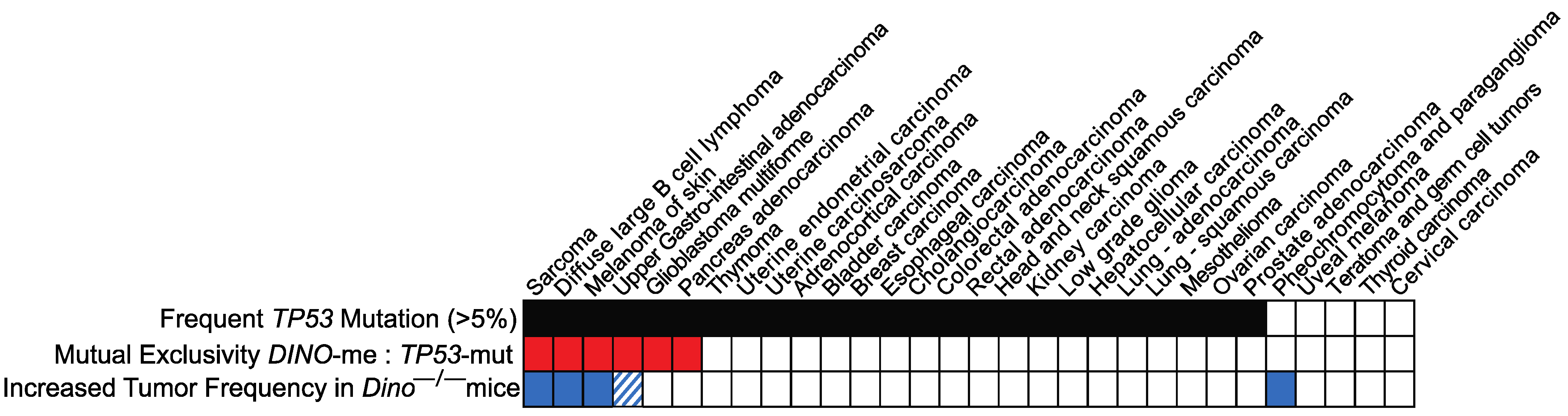
3.2. The Spectrum of Spontaneous Tumors in Dino−/− Mice Is Similar to Types of Human Cancers in Which Epigenetic Silencing of DINO Is Mutually Exclusive with TP53 Mutations

We have previously reported that human DINO expression is regulated via methylation in the shore of a CpG island located at the shared DINO/CDKN1A locus and that hypermethylation of this region had no effect on the expression of CDKN1A. Examining 30 types of human cancers represented in the TCGA dataset, we observed that TP53 mutations and DINO hypermethylation are mutually exclusive cancer alterations in six types of human cancer, including soft tissue sarcoma, skin melanoma, diffuse large B cell lymphoma and stomach adenocarcinoma [17]. Mutual exclusivity of genetic alterations in cancer typically indicates that two genes contribute to the same mechanism and alteration of just one of the genes is sufficient to facilitate tumorigenesis [25], suggesting that loss of DINO was sufficient for escape from p53-dependent tumor suppression despite retention of wildtype TP53. Intriguingly, we observed that DINO hypermethylation is not mutually exclusive with TP53 mutations in several other types of cancer in which TP53 is recurrently mutated, including lung adenocarcinoma. That DINO hypermethylation is not mutually exclusive with TP53 mutations in these cancer types suggested that silencing of DINO was insufficient to impair the p53-dependent tumor suppressor phenotype in these tissues.
The spectrum of spontaneous tumors that developed in Dino−/− mice was similar to the types of human cancers in which DINO silencing is mutually exclusive with TP53 mutations (Figure 2). We observed spontaneous tumors in Dino−/− mice corresponding to three of six of the human tumor subtypes found to have mutual exclusivity between TP53 mutation and DINO hypermethylation: sarcoma, B cell lymphoma and skin melanoma (Figure 1b–e and Figure S1b,c). Another rare spontaneous tumor observed in Dino−/− was an upper gastrointestinal adenocarcinoma arising in the jejunum (Figure S1d). While the TCGA did not examine the genetics of small bowel adenocarcinomas, we did observe that TP53 mutations and DINO hypermethylation were mutually exclusive in stomach adenocarcinoma, another type of adenocarcinoma arising from the upper gastrointestinal tract. The relationship between the spontaneous tumor spectrum of Dino−/− mice and the human tumor types in which TP53 mutation and DINO hypermethylation are mutually exclusive suggests a similarity in the tissue types in which DINO serves as an essential tumor suppressor in humans and in mice.

3.3. Loss of Dino Is Insufficient for Tumorigenesis in Selected Tissues That Depend on p53 for Tumor Suppression

The spectrum of spontaneous tumors in Dino−/− mice reflects human cancers in which DINO hypermethylation and TP53 mutations are mutually exclusive and was consistent with our prior results that loss of Dino enhanced oncogene-induced tumorigenesis in B cell and sarcoma [17]. Therefore, we next set out to examine whether Dino functions as an essential tumor suppressor in tissues in which p53 is required, but for which human genetics and spontaneous tumors in Dino−/− mice did not suggest an essential role for DINO in p53-dependent tumor suppression.
While p53-null mice predominantly develop spontaneous T lymphomas of thymic origin, we were surprised to observe no incidence of T cell lymphoma in Dino−/− mice (Table 1) [18,26]. Given the well-established role of Dino in p53 pathway regulation in multiple cell types, including T cells [16,17,27], the absence of spontaneous T cell lymphoma in Dino-null mice is particularly striking. p53 is critical for the protection of cells against radiation-induced carcinogenesis [28], particularly in the thymus, which undergoes extensive p53-dependent apoptosis after radiation [29,30]. Dino is robustly induced in adult mouse thymus after 5Gy TBI (Figure 3a), and we have previously demonstrated that Dino is required for radiation-induced apoptosis in thymocytes [16]. The involvement of Dino in p53-dependent cell fate decisions in T cells is expected to be highly suggestive of a tumor-suppressive role for Dino in T cell tumorigenesis.
To interrogate the role of Dino in suppressing T cell lymphoma directly, we utilized a model of thymic lymphomagenesis induced by exposure to ionizing radiation at a dose that effectively induces thymic T cell lymphomas in p53+/+ mice [31] and which is significantly accelerated by the loss of p53, demonstrating that p53-dependent tumor suppression is essential in this model [28,32]. Consistent with observations in spontaneous tumors, the loss of Dino had no effect on T cell lymphoma-free survival after irradiation (Figure 3b). This suggests that although Dino has a functional role in the acute cellular response to radiation in the thymus, it does not function as an essential tumor suppressor in this cell type when constitutively inactivated by genetic deletion.
p53 is also an essential suppressor of tumorigenesis in mouse models of Sonic hedgehog (Shh) medulloblastoma. Loss of p53 significantly accelerates medulloblastoma development in mice with mutations that activate Shh signaling, such as loss of the negative regulator Patched [33]. We similarly observed that loss of p53 significantly accelerated medulloblastoma development in mice expressing a constitutively active, oncogenic Smoothened variant, SmoM2c. In this model, mice with a second allele (Math1-cre) selectively express SmoM2c in cerebellar granule neurons after recombination excises a LoxP-STOP-LoxP cassette that otherwise prematurely terminates the expression of the SmoM2c allele [34]. Shh medulloblastoma develop at a significantly more rapid rate in p53−/−; Math1-Cre; SmoM2-eYFPloxP/loxP mice compared to p53+/+; Math1-Cre; SmoM2-eYFPloxP/loxP mice (Figure S2), indicating that the p53 pathway is essential to suppression of medulloblastoma formation in this model. We next examined whether Dino is expressed in the developing cerebellum, a region of the brain that is largely composed of cerebellar granule neurons, the cell of origin of Shh medulloblastoma [35,36]. Similar to our observations in thymus and those previously reported for B cells and fibroblasts [17], Dino is highly induced by genotoxic stress in the developing cerebellum (Figure 3c). However, genetic loss of Dino did not accelerate medulloblastoma development in Math1-Cre; SmoM2-eYFPloxP/loxP mice (Figure 3d), indicating that Dino, unlike p53, does not contribute to an essential tumor suppressor role in cerebellar granule neural precursors. Furthermore, the lack of Dino-mediated tumor suppression in this oncogene-dependent model system underscores that tumor suppression by Dino is independent of the tumor-initiating genotoxic event. Together, these experimental models identify that Dino is dispensable for tumor suppression in T cells and cerebellar granule neural precursors, two tissues in which p53 is an essential tumor suppressor, a stark contrast to observations that Dino is a haplo-insufficient tumor suppressor in B cells and fibroblasts [17].

4. Discussion

This study provides evidence that Dino is a bona fide tumor suppressor lncRNA that inhibits cancer formation in a subset of tissues, including mesenchymal cells and B cells, while also being dispensable to the p53-dependent tumor suppressor program in other tissue types, such as T cells and cerebellar granule neural precursors. This study contrasts markedly with most studies characterizing lncRNAs in cancer phenotypes. Currently, most evidence for functional lncRNA activity is suggested by in vitro cellular assays after exogenous over-expression or acute loss-of-function using RNA interference or antisense oligonucleotides rather than through the study of phenotypes arising on an organismal level as a result of gene deletion. Since lncRNA knockout mice often lack overt phenotypes, skepticism has arisen about the relevance of discoveries made in cellular assays to cancer phenotypes in organisms. However, rigorous and carefully designed studies, often utilizing environmental, genotoxic or oncogenic stressors, have revealed important roles for lncRNAs in many contexts. For example, the lncRNA Malat1 has been implicated in both oncogenesis and tumor suppression when examining gene expression profiles in human tumors and in vitro mechanistic studies [37]. However, Malat1 knockout mice do not appear to have an apparent cancer phenotype [38,39] but instead display defects in the immune response when exposed to multiple pathogens [40,41]. Similarly, genetic deletion of the p53-inducible lncRNA Neat1 promotes the transformation of E1A; HRasV12-expressing MEFs and enhances the formation of preneoplastic lesions in a KrasG12D-driven mouse model of pancreatic ductal carcinoma [42]. However, despite ample additional in vitro evidence for the role of Neat1 in tumor initiation and progression in multiple tumor types [43], Neat1−/− mice are not reported to spontaneously develop tumors [44].
The identification of Dino as a suppressor of spontaneous tumor formation in mice definitively establishes Dino as a functional tumor suppressor in vivo, loss of which predisposes mice towards the development of a subset of p53-associated spontaneous tumors. p53-null mice predominantly and rapidly develop lymphomas of thymic origin, with a high frequency of sarcomas and B cell lymphomas also reported [18,26]. This characteristic tumor spectrum is recapitulated in multiple p53-pathway associated animals such as p19ARF−/− [45], Atm−/− [46] and transgenic Mdm2-amplified animals [47]. However, not all canonical p53-pathway genes studied in knockout animals have revealed a spontaneous tumor phenotype. For example, Cdkn1a/p21 knockout animals are not prone to spontaneous tumor formation when monitored for their entire lifespan, and p21 loss has been found to be protective in radiation-induced thymic lymphoma [20,32], again emphasizing the distinct role of the Dino lncRNA transcribed from the shared Dino/Cdkn1a locus in tumor suppression. Deletion of the pro-apoptotic p53-response gene Puma is not reported to predispose animals to spontaneous tumor formation but does protect against lesions induced by genotoxic stress [48,49]. It should be noted, however, that Puma−/− mice were only followed for 6 months, based on the timeline for tumor development in p53−/− mice. p53 heterozygous mice have a much longer latency to tumor development and death, developing fewer lymphomas and instead of succumbing to a wider array of sarcomas [18]. A similarly delayed onset of tumor formation and shift in tumor spectrum has been observed in various knock-in p53 mutant mice strains, including p53S23A/S23A [50] and p53R172P/R172P [51]. Dino−/− mice did not develop early-onset tumors, and yet, they had a significant increase in tumor incidence at necropsy. This raises the possibility that tumor suppressor phenotypes may be missed in mutant strains of both canonical p53-associated protein-coding genes and p53-associated lncRNAs when experimental design assumes a highly penetrant tumor-prone phenotype similar to that of p53 knockout animals and relies on decreased over-all survival as the only indicator of in vivo spontaneous tumor suppression.
The spectrum of spontaneous tumors identified in Dino−/− mice reveals a predisposition towards sarcoma formation, similar to the spectrum of tumors seen in p53−/− mice which lack T, B and NK cells [52]. This suggests that Dino may be a critical tissue-specific p53-associated tumor suppressor in the development of sarcoma, a finding supported by the mutual exclusivity between DINO silencing through DNA methylation and p53 mutational status in human sarcomas. Indeed, the spectrum of spontaneous tumors developed in Dino−/− mice closely mirrors the spectrum of human tumors in which DINO hypermethylation was mutually exclusive with TP53 mutations. This further strengthens the argument that the DINO lncRNA is an important modulator of human tumorigenesis and establishes the Dino−/− mouse as a model that recapitulates observations in human cancers.
The notable differences in the spontaneous tumor spectrum of p53−/− and Dino−/− mice indicate distinct requirements for tumor suppression between tissue types. Nuanced differences in prior reports on the spectrum of spontaneous tumors in mice with different alterations in the p53 protein and p53 tumor suppressor pathway have similarly suggested this, but this has not been thoroughly examined. Work from several groups that have examined the role of acute p53-activation and radiation-induced T cell lymphoma has illustrated the complexity of p53 pathway functions in tumor suppression. In studies using a mouse in which both copies of the p53 gene had been replaced with one encoding tamoxifen-inducible p53, Christophorou et al. concluded that p53 activation at the time of genotoxic treatment with irradiation did not inhibit eventual lymphomagenesis [53]. Similarly, Hinkal et al. demonstrated that inducible deletion of p53 immediately prior to TBI had no effect on subsequent lymphoma incidence [54]. Both model systems involve a genetic background in which p53 is either completely absent prior to irradiation or permanently deleted after Cre-induced recombination. Using a compound transgenic mouse in which the p53 gene remains intact but rapid temporal silencing can be achieved by doxycycline-induced expression of a p53-targeting shRNA, Lee et al. determined that the acute p53 apoptotic response to fractionated irradiation prevents radiation-induced lymphoma in otherwise p53 wildtype mice [55]. These data contrast markedly with multiple studies in which loss of p53-dependent apoptotic effects significantly accelerates the development of B lymphomas, highlighting the distinct requirements for p53-dependent apoptotic functions in T cells and B cells. That Dino is required for DNA-damage-induced, p53-dependent apoptosis in T cells but loss of Dino did not result in spontaneous or radiation-induced T lymphomas may indicate that Dino contributes to p53 pathway functions that are essential for tumor suppression in certain tissues but not others. This conclusion is further supported by the lack of Dino-mediated tumor suppression in Shh-driven medulloblastoma, a tumor in which p53 inactivation accelerates tumor formation and is associated with the most high-risk human patients [56]. Future studies are needed to understand the mechanism of Dino’s tissue-specific tumor-suppressor phenotype.

Supplementary Materials

The following supporting information can be downloaded at: https://www.mdpi.com/article/10.3390/cells11111818/s1, Figure S1: Overall survival and spontaneous tumors of Dino−/− mice; p53 suppresses medulloblastoma in the Math1-Cre; SmoM2 model. Table S1: Detailed histopathology of spontaneous malignant tumors in Dino−/− and Dino+/+ littermates.

Author Contributions

Investigation: C.B.M., E.S.A., R.B. and A.M.S.; methodology: C.B.M., E.S.A. and A.M.S.; formal analyses: C.B.M., E.S.A. and A.M.S.; data curation: C.B.M., E.S.A. and A.M.S.; funding acquisition: A.M.S.; validation: C.B.M., E.S.A. and A.M.S.; visualization: C.B.M., E.S.A. and A.M.S.; writing: C.B.M. and A.M.S.; conceptualization, project administration, funding acquisition and supervision: A.M.S. All authors have read and agreed to the published version of the manuscript.

Funding

This study was supported by funding from the American Cancer Society RSG-19-158-01-RMC, NIH R35GM124909, NIH SPORE in Soft Tissue Sarcoma, the Geoffrey Beene Cancer Research Center Research Grant, and the NIH Cancer Center Core Grant P30 CA008748, which includes support for the Laboratory of Comparative Pathology which reviewed histopathology for all tumors.

Institutional Review Board Statement

The animal study protocol was approved by the Institutional Review Board of Memorial Sloan Kettering Cancer Center (protocol 15-07-011, initial approval September 2015, most recent approval June 2021).

Informed Consent Statement

Not applicable.

Data Availability Statement

All data are available in the main text or the Supplementary Materials.

Acknowledgments

We thank Azusa Tanaka for valuable discussions and feedback. Thanks to Scott Lowe for kindly providing the p53−/− mice used in this study. Animal husbandry was provided by the MSKCC/Weill Cornell animal facility.

Conflicts of Interest

The authors declare no conflict of interest.

References

  1. Statello, L.; Guo, C.-J.; Chen, L.-L.; Huarte, M. Gene regulation by long non-coding RNAs and its biological functions. Nat. Rev. Mol. Cell Biol. 2021, 22, 96–118. [Google Scholar] [CrossRef] [PubMed]
  2. Moore, J.E.; The ENCODE Project Consortium; Purcaro, M.J.; Pratt, H.E.; Epstein, C.B.; Shoresh, N.; Adrian, J.; Kawli, T.; Davis, C.A.; Dobin, A.; et al. Expanded encyclopaedias of DNA elements in the human and mouse genomes. Nature 2020, 583, 699–710. [Google Scholar] [CrossRef] [PubMed]
  3. Andergassen, D.; Rinn, J.L. From genotype to phenotype: Genetics of mammalian long non-coding RNAs in vivo. Nat. Rev. Genet. 2022, 23, 229–243. [Google Scholar] [CrossRef]
  4. Schmitt, A.M.; Chang, H.Y. Long Noncoding RNAs in Cancer Pathways. Cancer Cell 2016, 29, 452–463. [Google Scholar] [CrossRef] [PubMed] [Green Version]
  5. Schmitt, A.M.; Chang, H.Y. Long Noncoding RNAs: At the Intersection of Cancer and Chromatin Biology. Cold Spring Harb. Perspect. Med. 2017, 7, a026492. [Google Scholar] [CrossRef] [Green Version]
  6. Winkler, L.; Dimitrova, N. A mechanistic view of long noncoding RNAs in cancer. Wiley Interdiscip. Rev. RNA 2021, 13, e1699. [Google Scholar] [CrossRef]
  7. Lane, D.; Levine, A. p53 Research: The Past Thirty Years and the Next Thirty Years. Cold Spring Harb. Perspect. Biol. 2010, 2, a000893. [Google Scholar] [CrossRef] [Green Version]
  8. Liu, Y.; Tavana, O.; Gu, W. p53 modifications: Exquisite decorations of the powerful guardian. J. Mol. Cell Biol. 2019, 11, 564–577. [Google Scholar] [CrossRef] [PubMed] [Green Version]
  9. Chen, S.; Thorne, R.F.; Zhang, X.D.; Wu, M.; Liu, L. Non-coding RNAs, guardians of the p53 galaxy. Semin. Cancer Biol. 2021, 75, 72–83. [Google Scholar] [CrossRef]
  10. Kandoth, C.; McLellan, M.D.; Vandin, F.; Ye, K.; Niu, B.; Lu, C.; Xie, M.; Zhang, Q.; McMichael, J.F.; Wyczalkowski, M.A.; et al. Mutational landscape and significance across 12 major cancer types. Nature 2013, 502, 333–339. [Google Scholar] [CrossRef] [Green Version]
  11. Kusafuka, T.; Fukuzawa, M.; Oue, T.; Komoto, Y.; Yoneda, A.; Okada, A. Mutation analysis of p53 gene in childhood malignant solid tumors. J. Pediatr. Surg. 1997, 32, 1175–1180. [Google Scholar] [CrossRef]
  12. The Cancer Genome Atlas Research Network. Genomic and epigenomic landscapes of adult de novo acute myeloid leukemia. N. Engl. J. Med. 2013, 368, 2059–2074. [Google Scholar] [CrossRef] [PubMed] [Green Version]
  13. Valente, L.J.; Gray, D.H.D.; Michalak, E.M.; Pinon-Hofbauer, J.; Egle, A.; Scott, C.L.; Janic, A.; Strasser, A. p53 Efficiently Suppresses Tumor Development in the Complete Absence of Its Cell-Cycle Inhibitory and Proapoptotic Effectors p21, Puma, and Noxa. Cell Rep. 2013, 3, 1339–1345. [Google Scholar] [CrossRef] [PubMed] [Green Version]
  14. Li, T.; Kon, N.; Jiang, L.; Tan, M.; Ludwig, T.; Zhao, Y.; Baer, R.; Gu, W. Tumor Suppression in the Absence of p53-Mediated Cell-Cycle Arrest, Apoptosis, and Senescence. Cell 2012, 149, 1269–1283. [Google Scholar] [CrossRef] [Green Version]
  15. Grossi, E.; Sánchez, Y.; Huarte, M. Expanding the p53 regulatory network: LncRNAs take up the challenge. Biochim. Biophys. Acta 2016, 1859, 200–208. [Google Scholar] [CrossRef]
  16. Schmitt, A.; Garcia, J.T.; Hung, T.; Flynn, R.; Shen, Y.; Qu, K.; Payumo, A.Y.; Peres-Da-Silva, A.; Broz, D.K.; Baum, R.; et al. An inducible long noncoding RNA amplifies DNA damage signaling. Nat. Genet. 2016, 48, 1370–1376. [Google Scholar] [CrossRef] [Green Version]
  17. Marney, C.B.; Anderson, E.S.; Adnan, M.; Peng, K.-L.; Hu, Y.; Weinhold, N.; Schmitt, A.M. p53-intact cancers escape tumor suppression through loss of long noncoding RNA Dino. Cell Rep. 2021, 35, 109329. [Google Scholar] [CrossRef]
  18. Jacks, T.; Remington, L.; Williams, B.O.; Schmitt, E.M.; Halachmi, S.; Bronson, R.T.; Weinberg, R.A. Tumor spectrum analysis in p53-mutant mice. Curr. Biol. 1994, 4, 1–7. [Google Scholar] [CrossRef]
  19. Lowe, S.W.; Bodis, S.; McClatchey, A.; Remington, L.; Ruley, H.E.; Fisher, D.E.; Housman, D.E.; Jacks, T. p53 Status and the Efficacy of Cancer Therapy in Vivo. Science 1994, 266, 807–810. [Google Scholar] [CrossRef]
  20. Martín-Caballero, J.; Flores, J.M.; García-Palencia, P.; Serrano, M. Tumor Susceptibility of p21Waf1/Cip1-deficient Mice1. Cancer Res. 2001, 61, 6234–6238. [Google Scholar]
  21. Pettan-Brewer, C.; Treuting, P.M.M. Practical pathology of aging mice. Pathobiol. Aging Age-Relat. Dis. 2011, 1, 7202. [Google Scholar] [CrossRef] [PubMed] [Green Version]
  22. Lacroix-Triki, M.; Lacoste-Collin, L.; Jozan, S.; Charlet, J.-P.; Caratero, C.; Courtade, M. Histiocytic Sarcoma in C57BL/6J Female Mice is Associated with Liver Hematopoiesis: Review of 41 Cases. Toxicol. Pathol. 2003, 31, 304–309. [Google Scholar] [CrossRef] [PubMed] [Green Version]
  23. BBlackwell, B.-N.; Bucci, T.J.; Hart, R.W.; Turturro, A. Longevity, Body Weight, and Neoplasia in Ad Libitum-Fed and Diet-Restricted C57BL6 Mice Fed NIH-31 Open Formula Diet. Toxicol. Pathol. 1995, 23, 570–582. [Google Scholar] [CrossRef]
  24. Robles, A.I.; Linke, S.P.; Harris, C.C. The p53 network in lung carcinogenesis. Oncogene 2002, 21, 6898–6907. [Google Scholar] [CrossRef] [PubMed] [Green Version]
  25. Ciriello, G.; Cerami, E.; Sander, C.; Schultz, N. Mutual exclusivity analysis identifies oncogenic network modules. Genome Res. 2012, 22, 398–406. [Google Scholar] [CrossRef] [PubMed] [Green Version]
  26. Donehower, L.A.; Harvey, M.; Slagle, B.L.; McArthur, M.J.; Montgomery, C.A., Jr.; Butel, J.S.; Bradley, A. Mice deficient for p53 are developmentally normal but susceptible to spontaneous tumours. Nature 1992, 356, 215–221. [Google Scholar] [CrossRef]
  27. Liu, Q.; Xiao, Y.; Cai, P.; Li, J.; Li, D. Long noncoding RNA DINO (damage induced noncoding) represses the development of gastric cancer by modulating p21 and Bcl-2 Associated X Protein (Bax) expression. J. Cell. Biochem. 2019, 120, 11190–11195. [Google Scholar] [CrossRef]
  28. Kemp, C.J.; Wheldon, T.; Balmain, A. p53-deficient mice are extremely susceptible to radiation-induced tumorigenesis. Nat. Genet. 1994, 8, 66–69. [Google Scholar] [CrossRef]
  29. Lowe, S.W.; Schmitt, E.M.; Smitht, S.W.; Osbornet, B.A.; Jacks, T. pS3 is required for radiation-induced apoptosis in mouse thymocytes. Nature 1993, 362, 847–849. [Google Scholar] [CrossRef]
  30. Clarke, A.R.; Purdie, C.A.; Harrison, D.J.; Morris, R.G.; Bird, C.C.; Hooper, M.L.; Wyllie, A.H. Thymocyte apoptosis induced by p53-dependent and independent pathways. Nature 1993, 362, 849–852. [Google Scholar] [CrossRef]
  31. Kaplan, H.S.; Brown, M.B. A quantitative dose-response study of lymphoid-tumor development in irradiated C 57 black mice. J. Natl. Cancer Inst. 1952, 13, 185–208. [Google Scholar] [PubMed]
  32. De La Cueva, E.; García-Cao, I.; Herranz, M.; López, P.; García-Palencia, P.; Flores, J.M.; Serrano, M.; Fernández-Piqueras, J.; Martín-Caballero, J. Tumorigenic activity of p21Waf1/Cip1 in thymic lymphoma. Oncogene 2006, 25, 4128–4132. [Google Scholar] [CrossRef] [PubMed] [Green Version]
  33. Jiang, D.; Brady, C.A.; Johnson, T.M.; Lee, E.Y.; Park, E.J.; Scott, M.P.; Attardi, L.D. Full p53 transcriptional activation potential is dispensable for tumor suppression in diverse lineages. Proc. Natl. Acad. Sci. USA 2011, 108, 17123–17128. [Google Scholar] [CrossRef] [Green Version]
  34. Ocasio, J.; Babcock, B.; Malawsky, D.; Weir, S.J.; Loo, L.; Simon, J.M.; Zylka, M.J.; Hwang, D.; Dismuke, T.; Sokolsky, M.; et al. scRNA-seq in medulloblastoma shows cellular heterogeneity and lineage expansion support resistance to SHH inhibitor therapy. Nat. Commun. 2019, 10, 5829. [Google Scholar] [CrossRef] [PubMed] [Green Version]
  35. Schüller, U.; Heine, V.M.; Mao, J.; Kho, A.T.; Dillon, A.K.; Han, Y.-G.; Huillard, E.; Sun, T.; Ligon, A.H.; Qian, Y.; et al. Acquisition of Granule Neuron Precursor Identity Is a Critical Determinant of Progenitor Cell Competence to Form Shh-Induced Medulloblastoma. Cancer Cell 2008, 14, 123–134. [Google Scholar] [CrossRef] [Green Version]
  36. Yang, Z.-J.; Ellis, T.; Markant, S.L.; Read, T.-A.; Kessler, J.D.; Bourboulas, M.; Schüller, U.; Machold, R.; Fishell, G.; Rowitch, D.H.; et al. Medulloblastoma Can Be Initiated by Deletion of Patched in Lineage-Restricted Progenitors or Stem Cells. Cancer Cell 2008, 14, 135–145. [Google Scholar] [CrossRef] [PubMed] [Green Version]
  37. Chen, Q.; Zhu, C.; Jin, Y. The Oncogenic and Tumor Suppressive Functions of the Long Noncoding RNA MALAT1: An Emerging Controversy. Front. Genet. 2020, 11, 93. Available online: https://www.frontiersin.org/article/10.3389/fgene.2020.00093 (accessed on 4 April 2022). [CrossRef] [Green Version]
  38. Eißmann, M.; Gutschner, T.; Hämmerle, M.; Günther, S.; Caudron-Herger, M.; Groß, M.; Schirmacher, P.; Rippe, K.; Braun, T.; Zörnig, M.; et al. Loss of the abundant nuclear non-coding RNA MALAT1 is compatible with life and development. RNA Biol. 2012, 9, 1076–1087. [Google Scholar] [CrossRef] [Green Version]
  39. Zhang, B.; Arun, G.; Mao, Y.S.; Lazar, Z.; Hung, G.; Bhattacharjee, G.; Xiao, X.; Booth, C.J.; Wu, J.; Zhang, C.; et al. The lncRNA Malat1 Is Dispensable for Mouse Development but Its Transcription Plays a cis-Regulatory Role in the Adult. Cell Rep. 2012, 2, 111–123. [Google Scholar] [CrossRef] [Green Version]
  40. Hewitson, J.P.; West, K.A.; James, K.R.; Rani, G.F.; Dey, N.; Romano, A.; Brown, N.; Teichmann, S.A.; Kaye, P.M.; Lagos, D. Malat1 Suppresses Immunity to Infection through Promoting Expression of Maf and IL-10 in Th Cells. J. Immunol. 2020, 204, 2949–2960. [Google Scholar] [CrossRef] [Green Version]
  41. Liu, W.; Wang, Z.; Liu, L.; Yang, Z.; Liu, S.; Ma, Z.; Liu, Y.; Ma, Y.; Zhang, L.; Zhang, X.; et al. LncRNA Malat1 inhibition of TDP43 cleavage suppresses IRF3-initiated antiviral innate immunity. Proc. Natl. Acad. Sci. USA 2020, 117, 23695–23706. [Google Scholar] [CrossRef] [PubMed]
  42. Mello, S.S.; Sinow, C.; Raj, N.; Mazur, P.K.; Bieging-Rolett, K.; Broz, D.K.; Imam, J.F.C.; Vogel, H.; Wood, L.D.; Sae, J.; et al. Neat1 is a p53-inducible lincRNA essential for transformation suppression. Genes Dev. 2017, 31, 1095–1108. [Google Scholar] [CrossRef] [Green Version]
  43. Dong, P.; Xiong, Y.; Yue, J.; Hanley, S.J.B.; Kobayashi, N.; Todo, Y.; Watari, H. Long Non-coding RNA NEAT1: A Novel Target for Diagnosis and Therapy in Human Tumors. Front. Genet. 2018, 9, 471. Available online: https://www.frontiersin.org/article/10.3389/fgene.2018.00471 (accessed on 31 March 2022). [CrossRef] [PubMed] [Green Version]
  44. Nakagawa, S.; Naganuma, T.; Shioi, G.; Hirose, T. Paraspeckles are subpopulation-specific nuclear bodies that are not essential in mice. J. Cell Biol. 2011, 193, 31–39. [Google Scholar] [CrossRef] [PubMed]
  45. Sharpless, N.E.; Ramsey, M.R.; Balasubramanian, P.; Castrillon, D.H.; DePinho, R.A. The differential impact of p16INK4A or p19ARF deficiency on cell growth and tumorigenesis. Oncogene 2004, 23, 379–385. [Google Scholar] [CrossRef] [Green Version]
  46. Genik, P.C.; Bielefeldt-Ohmann, H.; Liu, X.; Story, M.D.; Ding, L.; Bush, J.M.; Fallgren, C.M.; Weil, M.M. Strain Background Determines Lymphoma Incidence in Atm Knockout Mice. Neoplasia 2014, 16, 129–136. [Google Scholar] [CrossRef] [PubMed] [Green Version]
  47. Jones, S.N.; Hancock, A.R.; Vogel, H.; Donehower, L.A.; Bradley, A. Overexpression of Mdm2 in mice reveals a p53-independent role for Mdm2 in tumorigenesis. Proc. Natl. Acad. Sci. USA 1998, 95, 15608–15612. [Google Scholar] [CrossRef] [Green Version]
  48. Jeffers, J.R.; Parganas, E.; Lee, Y.; Yang, C.; Wang, J.; Brennan, J.; MacLean, K.H.; Han, J.; Chittenden, T.; Ihle, J.N.; et al. Puma is an essential mediator of p53-dependent and -independent apoptotic pathways. Cancer Cell 2003, 4, 321–328. [Google Scholar] [CrossRef] [Green Version]
  49. Qiu, W.; Carson-Walter, E.B.; Kuan, S.F.; Zhang, L.; Yu, J. PUMA Suppresses Intestinal Tumorigenesis in Mice. Cancer Res. 2009, 69, 4999–5006. [Google Scholar] [CrossRef] [Green Version]
  50. MacPherson, D.; Kim, J.; Kim, T.; Rhee, B.K.; Van Oostrom, C.T.M.; DiTullio, R.A.; Venere, M.; Halazonetis, T.D.; Bronson, R.; De Vries, A.; et al. Defective apoptosis and B-cell lymphomas in mice with p53 point mutation at Ser 23. EMBO J. 2004, 23, 3689–3699. [Google Scholar] [CrossRef] [Green Version]
  51. Liu, G.; Parant, J.M.; Lang, G.; Chau, P.; Chavez-Reyes, A.; El-Naggar, A.K.; Multani, A.S.; Chang, S.; Lozano, G. Chromosome stability, in the absence of apoptosis, is critical for suppression of tumorigenesis in Trp53 mutant mice. Nat. Genet. 2003, 36, 63–68. [Google Scholar] [CrossRef] [PubMed]
  52. Landuzzi, L.; Ianzano, M.L.; Nicoletti, G.; Palladini, A.; Grosso, V.; Ranieri, D.; Ora, M.D.; Raschi, E.; Laranga, R.; Gambarotti, M.; et al. Genetic prevention of lymphoma in p53 knockout mice allows the early development of p53-related sarcomas. Oncotarget 2014, 5, 11924–11938. [Google Scholar] [CrossRef] [PubMed]
  53. Christophorou, M.A.; Ringshausen, I.; Finch, A.J.; Swigart, L.B.; Evan, G.I. The pathological response to DNA damage does not contribute to p53-mediated tumour suppression. Nature 2006, 443, 214–217. [Google Scholar] [CrossRef] [PubMed]
  54. Hinkal, G.; Parikh, N.; Donehower, L.A. Timed Somatic Deletion of p53 in Mice Reveals Age-Associated Differences in Tumor Progression. PLoS ONE 2009, 4, e6654. [Google Scholar] [CrossRef] [PubMed]
  55. Lee, C.-L.; Castle, K.D.; Moding, E.J.; Blum, J.M.; Williams, N.; Luo, L.; Ma, Y.; Borst, L.; Kim, Y.; Kirsch, D.G. Acute DNA damage activates the tumour suppressor p53 to promote radiation-induced lymphoma. Nat. Commun. 2015, 6, 8477. [Google Scholar] [CrossRef] [PubMed] [Green Version]
  56. Ramaswamy, V.; Nör, C.; Taylor, M.D. p53 and Meduloblastoma. Cold Spring Harb. Perspect. Med. 2016, 6, a026278. [Google Scholar] [CrossRef] [Green Version]
Figure 1. Dino−/− mice spontaneously develop soft-tissue and bone sarcomas. (a) Kaplan–Meier plot of sarcoma free-survival in Dino+/+ and Dino−/− mice; 6/28 Dino−/− mice developed sarcoma over their lifespan compared to 0/19 Dino+/+ mice. (b) Leiomyosarcoma of the hind limb. Representative images of H&E staining and diagnostic immunohistochemistry showing strong positive staining for vimentin and α-SMA (≥90%) and negative staining for S100 (≤5%). (c) Hemangiosarcoma of the liver (H&E). (d) Myxosarcoma arising from the muscle of the abdominal wall (H&E). (e) Sarcoma of the bone most likely represents osteosarcoma (H&E). All images taken 40X, scale bar = 20 µm.
Cells 11 01818 g001
Figure 2. The spectrum of spontaneous tumors in Dino−/− mice reflects the spectrum of human cancers in which TP53 mutations and DINO hypermethylation are mutually exclusive. Analysis of 30 human tumor types in the TCGA dataset identifies a subset of six tumor types in which TP53 mutations are mutually exclusive with DINO hypermethylation (red). Dino−/− mice spontaneously developed tumors in 3 of these tumor classifications including sarcoma, diffuse large B cell lymphoma and melanoma of the skin (blue). DINO hypermethylation and TP53 mutations are mutually exclusive in stomach adenocarcinoma, an upper gastrointestinal malignancy, and another upper gastrointestinal adenocarcinoma arising from the jejunum was observed in a Dino−/− mouse (blue stripes). Fisher’s exact test for an interaction; p = 0.001.
Cells 11 01818 g002
Figure 3. Loss of Dino has no effect on tumor-free survival in mouse models of T cell lymphoma or Shh-driven medulloblastoma. (a) Dino is induced in adult thymus 6 h after 5 Gy TBI. n = 3, *** p < 0.001, t-test. (b) Kaplan–Meier curve indicates no significant difference in survival between WT and Dino mutant mice post fractionated irradiation. Median thymic lymphoma-free survival Dino+/+ 189 days (n = 18), Dino−/− 177 days (n = 18), p = 0.6, Log-Rank test. (c) Dino is induced in postnatal day 16 cerebellum 6 h after 10 Gy irradiation. N = 3, *** p < 0.001, t-test. (d) Kaplan–Meier curve indicates no significant difference in survival between WT and Dino mutant mice in a Math1-Cre; SmoM2- eYFPloxP/loxP background. Median medulloblastoma-free survival Dino+/+ 42 days (n = 8), Dino−/− 38 days (n = 7). p = 0.62, Log-Rank test.
Cells 11 01818 g003
Table 1. The spectrum of spontaneous malignant tumors in Dino+/+ and Dino−/− mice.
TumorDino+/+ (%)Dino−/− (%)
Histiocytic sarcoma4 (21.1)9 (32.1)
Sarcoma (bone and soft tissue)0 (0)6 (21.4) 1
B and non-T cell lymphoma3 (16.7)8 (28.6)
T cell lymphoma0 (0)0 (0)
Lung adenocarcinoma2 (10.5)2 (7.1)
Melanoma0 (0)1 (3.6)
Gastrointestinal adenocarcinoma0 (0)1 (3.6)
Pheochromocytoma0 (0)1 (3.6)
Total Mice with Tumors9 (47.4)22 (78.6) 2
1 p = 0.035; 2 p = 0.029; Fisher’s exact test, one sided.
Publisher’s Note: MDPI stays neutral with regard to jurisdictional claims in published maps and institutional affiliations.

