Review

Role of Cytoskeletal Diaphanous-Related Formins in Hearing Loss

by
Chiara Chiereghin
1,†,
Michela Robusto
2,†,
Valentina Massa
3,
Pierangela Castorina
4,
Umberto Ambrosetti
5,
Rosanna Asselta
1,6 and
Giulia Soldà
1,6,*
1
IRCCS Humanitas Research Hospital, Via Manzoni 56, 20089 Milan, Italy
2
Experimental Therapeutics Program, IFOM ETS—The AIRC Institute of Molecular Oncology, Via Adamello 16, 20139 Milan, Italy
3
Dipartimento di Scienze della Salute, Università degli Studi di Milano, Via Di Rudinì 8, 20146 Milan, Italy
4
Casa di Cura Igea, Via Marcona 69, 20129 Milan, Italy
5
Dipartimento di Scienze Cliniche e di Comunità, Università degli Studi di Milano and Fondazione IRCCS Cà Granda Ospedale Maggiore Policlinico, UO Audiologia, Via F. Sforza 35, 20122 Milan, Italy
6
Department of Biomedical Sciences, Humanitas University, Via Rita Levi Montalcini 4, 20072 Milan, Italy
*
Author to whom correspondence should be addressed.
These authors contributed equally to this work.
Cells 2022, 11(11), 1726; https://doi.org/10.3390/cells11111726
Submission received: 16 March 2022 / Revised: 16 May 2022 / Accepted: 18 May 2022 / Published: 24 May 2022
(This article belongs to the Special Issue The Cytoskeleton: Structural, Functional, and Pathological Aspects)

Abstract

:
Hearing relies on the proper functioning of auditory hair cells and on actin-based cytoskeletal structures. Diaphanous-related formins (DRFs) are evolutionarily conserved cytoskeletal proteins that regulate the nucleation of linear unbranched actin filaments. They play key roles during metazoan development, and they seem particularly pivotal for the correct physiology of the reproductive and auditory systems. Indeed, in Drosophila melanogaster, a single diaphanous (dia) gene is present, and mutants show sterility and impaired response to sound. Vertebrates, instead, have three orthologs of the diaphanous gene: DIAPH1, DIAPH2, and DIAPH3. In humans, defects in DIAPH1 and DIAPH3 have been associated with different types of hearing loss. In particular, heterozygous mutations in DIAPH1 are responsible for autosomal dominant deafness with or without thrombocytopenia (DFNA1, MIM #124900), whereas regulatory mutations inducing the overexpression of DIAPH3 cause autosomal dominant auditory neuropathy 1 (AUNA1, MIM #609129). Here, we provide an overview of the expression and function of DRFs in normal hearing and deafness.

1. Introduction

The process of hearing relies on the proper functioning of auditory hair cells and on the integrity of all the cochlear structures contributing to mechanotransduction. In particular, the ability of hair cells to convert sound into nerve impulse is critically dependent upon actin-based cytoskeletal structures. Therefore, it should come as no surprise that mutations in genes encoding actin (e.g., ACTG1, encoding γ-actin, the predominant actin isoform in hair cells), actin-binding proteins (e.g., TRIOBP, an actin bundler, and ESPN, an actin crosslinker), actin-activated motor proteins such as myosins (e.g., MYO1A, MYO3A, MYO6, MYO7A, MYO15A, MYH14, and MYH9), and actin regulators (such as DIAPH1 and DIAPH3) could cause hereditary deafness in humans [1].
This review summarizes the physiological role of a specific family of actin regulators, the Diaphanous-Related Formins, focusing on their expression and function in the auditory system. Data deriving from human families and genetically modified animal models are discussed. The cellular and molecular functions of DRFs have been extensively studied and are reviewed elsewhere [2,3]. An overview of the involvement of DRFs and other formin subfamilies in human diseases has also recently been published [4].

2. The Diaphanous Family

2.1. Evolution

Diaphanous-Related Formins (DRFs or Dia) are members of a metazoan subfamily of cytoskeletal regulators and constitute a major branch of the formin family, which comprises six other subfamilies in mammals: DAAM (disheveled-associated activator of morphogenesis), FRL (formin-related gene in leukocytes), FHOD (formin homology domain-containing protein), INF (inverted formin), FMN (formin), and delphilin [5,6]. They are involved both in actin nucleation and in the stabilization of microtubules [7].
The diaphanous (dia) gene was initially isolated as a gene involved in cytokinesis in the fruit fly, Drosophila melanogaster, during a screen for mutations causing male sterility [8]. It is located on chromosome 2L and is composed of five exons. It is considered an essential mitotic gene because null mutations cause lethality early on during pupal development.
However, the evolutionary origin of the Dia formin family is probably older, as a protein with sequence similarities to metazoan Dia was identified in the choanoflagellate Monosiga brevicollis [9], and further phylogenetic analyses reinforced the idea that the emergence of Dia anticipated the evolution of animals and multicellularity [2]. In vertebrate genomes, the dia gene underwent duplication to give rise to three homologs called Diaph1, Diaph2, and Diaph3 (Figure 1a). Gene duplicates are assumed to be completely redundant at the time of origin, and then they might get silenced by accumulating mutations (nonfunctionalization) or be maintained by natural selection. The latter might happen either in case of neofunctionalization, which is the acquisition of novel beneficial functions, or subfunctionalization, in which each gene copy retains only a part of the original function [10]. Concerning DRFs, nonredundant roles of mammalian Dia family members have recently been described in ciliogenesis and cilia maintenance, suggesting subfunctionalization. Indeed, the depletion of each of the three DIAPH proteins produced similar defects in ciliation and cilia length, which could be rescued only by the specific isoform. Moreover, the co-depletion of all DRFs did not show additive effects, indicating that the three proteins contributed independently, possibly acting at different steps of the ciliogenesis process [11,12]. Similarly, the nonredundant roles of the DIAPH1-3 proteins were described in cell motility and the capture of cortical microtubules. In this case, functional differences could be attributed to the differential binding of each DIAPH protein to a distinct set of interactors [13].

2.2. Structure

Members of the Dia family are multimodular proteins containing a series of domains and functional motifs (Figure 1b). They interact with numerous partners, including actin regulators, adapters, and signaling proteins. DRFs can be functionally divided into two halves. The C-terminal half is required for actin assembly and includes three structural and functional domains: (i) the profilin-binding FH1 (formin homology 1) domain, (ii) the actin-binding FH2 (formin homology 2) domain, and (iii) the DAD (diaphanous autoregulatory domain), which can interact with the N-terminal portion. The N-terminal half is the regulatory region of DRFs and contains the GTPase-binding domain (GBD) and the diaphanous inhibitory domain (DID) [14]. Furthermore, a dimerization domain (DD) and a coiled coil (CC) domain are present to mediate the dimerization of the N-terminal of DRFs, which are active as dimers [3]. The interaction between the N-terminal DID and the C-terminal DAD determines the transition of the DRF to its inactive conformation (Figure 1b). In addition, the first portion of the DID domain is also involved in the interaction with Rho family GTPases [15,16]. As a result of the partial overlap of the DAD and Rho binding sites, the binding of Rho GTPases induces the release of the DAD domain and stimulates actin nucleation functions (Figure 1b) [17]. The disruption of autoinhibition and/or Rho-dependent activation, by deletion or specific point mutation within the GBD, DID, or DAD domains, leads to a constitutively active protein with an abnormally upregulated actin polymerization [18,19,20].
Although the basic mechanism of Rho-dependent activation is shared among Dia family members, DIAPH1–3 show different specificity of interaction with small GTP-binding proteins: while Diaph1 specifically binds to and is activated by RhoA, B, and C, Diaph2 and 3 are more promiscuous and can also be activated by other Rho homologues such as Rac1, Cdc42, and RhoD. The structural basis of this specificity has been attributed to a small motif consisting of three asparagine residues (‘NNN’) in the mouse Diaph1, which is substituted for a Threonine-Serine-Histidine (‘TSH’ motif) in Diaph2 and 3 [5].
Crystal structures of N-terminal and C-terminal domains of the mouse Diaph1 protein as well as the structure of complexes between the N-terminal and C-terminal fragments have been resolved and have given insight into the mechanisms of actin nucleation, Rho-dependent activation, and autoinhibition [15,16,21,22,23]. No full-length crystals have been produced so far, mainly because the large unstructured FH1 domain is expected to prevent crystallization [22]. However, a prediction of the tertiary structure of the full-length Diaph1 polypeptide (accession number AF-O08808-F1) has become available through the AlphaFold machine learning approach (Figure 1c) [24,25]. Moreover, a three-dimensional reconstruction of the full-length mouse Diaph1 in the autoinhibited conformation was obtained by single particle electron microscopy at a resolution of 24 Å [26]. These and other structural and biochemical studies have shown that under basal conditions, diaphanous proteins form closed autoinhibited dimers in which actin filament binding is sterically obstructed. The efficient and full activation of diaphanous protein probably requires not only the binding of Rho proteins, but also additional factors such as membrane lipids, nucleation-promoting factors, and protein kinases [26].

2.3. Actin and Microtubule-Modulating Activity of Diaphanous Proteins

The role of diaphanous proteins in actin regulation is well-characterized. The FH2 domain alone is sufficient for actin nucleation by binding actin monomers and the barbed-end of filaments, while the elongation of existing actin filaments depends on the interaction of the FH1 domain with the G-actin-binding protein Profilin. Some DRFs, such as Diaph3 (but not Diaph1), also have actin filament side binding affinity, which allows actin bundling activity in addition to nucleation and elongation. Moreover, members of the diaphanous protein family interact with numerous actin regulators, adapters, and signaling components that can modulate their activity, either by promoting or inhibiting actin polymerization, or by controlling their localization within the cell and their association with the plasma membrane [2].
Besides actin polymerization, diaphanous proteins are known to contribute to the regulation of microtubule dynamics. Both Diaph1 and Diaph3 have been shown to bind microtubules directly, although with different stoichiometries. The interaction is mediated mainly by the FH2 domain for Diaph1, whereas for Diaph3, the C-terminal domain is also required for high-affinity binding [7]. All three DRFs mainly have a stabilizing effect on microtubules: the expression of constitutively active Diaph1 or Diaph3 promoted the formation of stable microtubules in several cell types, and Diaph2 was shown to be necessary for stable kinetochore–microtubule interaction [7]. The stabilizing activity of DRFs is partly intrinsic and partly due to their binding with other microtubule-interacting proteins such as EB1 and APC (adenomatous polyposis coli protein), which are both plus-end-tracking proteins involved in microtubule stabilization and positioning [7,27]. Because DRFs bind both actin and microtubules, and the interactions are mediated by the FH2 domain, the possibility that the binding of one cytoskeletal component could affect the binding of the other was also verified. Interestingly, the presence of actin monomers did not affect the binding of either Diaph1 or Diaph3 to microtubules; instead, actin polymerization was inhibited at various degrees in the presence of microtubules. In particular, the actin polymerization activity of Diaph1 was only partially blocked, whereas that of Diaph3 was completely inhibited [7].
Recently, diaphanous proteins were demonstrated to localize at the base of the cilia and to play significant and non-redundant roles in ciliogenesis and cilia maintenance, likely by mediating the trafficking of post-Golgi and recycling endosomal vesicles to the cilia [11,12]. More importantly, at least for DIAPH1, the effects observed are mediated by both its actin-dependent and microtubule-dependent activities [11]. However, the DIAPH3-mediated regulation of ciliogenesis and cilia length relies on microtubule but not actin function [12]. Worthy of note here is that both DRF depletion and basal body targeted overexpression produced cilia defects: the lack of any of DIAPH1-3 genes caused a reduction in ciliation and cilia length, whereas their activation induced elongated cilia or cilia with a bulbous protrusion at their distal tips [11,12]. In addition, when different DIAPH1 mutants were tested—some corresponding to likely gain-of-function mutations (e.g., NM_005219.4: c.3637C>T, p.R1213X),and others to loss-of-function mutations (e.g., NM_005219.4: c.2332C>T, Q778X and NM_005219.4: c.2099T>A, p.I530S)—all showed cilia abnormalities. None of the mutants were able to completely rescue the impairment of ciliogenesis caused by siRNA-mediated DIAPH1 depletion [11].
Collectively, these observations provide new insight into the possible link between DRF function and hearing. First, even though diseases caused by defective cilia (collectively known as ciliopathies) typically show multiple organ disfunctions, some of them, such as the Bardet–Biedl syndrome, Alstrom syndrome, and Usher syndrome, are characterized by hearing dysfunction. Second, auditory hair cells possess specialized primary cilia known as kinocilia. Although kinocilia do not directly mediate auditory mechano-electrical transduction, they mediate hair cell morphogenesis and planar cell polarity, thus influencing the proper arrangement of stereocilia. Indeed, the loss of kinocilia results in abnormal and misoriented stereociliary bundles and auditory impairment. Such defects are seen, among others in mutants overexpressing DIAPH3 [28,29], suggesting a possible link between the microtubule-modulating activity of the DIAPH3 protein, the (kino)cilia function, and hearing loss.

2.4. Expression of Diaphanous Proteins in the Inner Ear

In Drosophila, the dia gene is ubiquitously expressed, with a peak of expression observed within the first day of embryonic development, during late larval stages, and during early pupal stages [30]. Similarly, all Diaphanous genes are widely expressed, being detectable in most human tissues [31]. At the RNA level, DIAPH3 shows lower and more restricted expression compared to DIAPH1 and DIAPH2. At the protein level, DIAPH1 shows moderate to strong cytoplasmic reactivity in the immunohistochemistry analysis of the majority of tissues, with weaker staining in lymphoid tissues. DIAPH2 displays generally weak to moderate cytoplasmic staining, with the highest expression in the reproductive system (endometrium, fallopian tubes, and ovaries in females, testis in males), large intestines, and lung macrophages. Concerning DIAPH3, no immunohistochemistry data are currently available [31].
For a long time, relatively little information was present in the literature concerning the specific expression of diaphanous proteins in the inner ear, and the available data are limited to Diaph1 and Diaph3; the expression profile of Diaph2 in the auditory system is unknown. Diaph1 expression in the cochlea of wild-type adult mice (aged between 2 and 6 months) was first characterized in 2017 by both immunohistochemistry and immunofluorescence. In the Organ of Corti (OC), Diaph1 localizes to the inner pillar cells and at the base of the outer hair cells (likely within Deiters cell extensions). Moreover, Diaph1 is also found in spiral ganglion neurons (SGNs) and in the central part of the cochlear nerve [32]. A more recent study reported the endogenous expression of mouse Diaph1 during and after the differentiation of the OC, between P5 and P14 [33]. Besides confirming expression in various types of supporting cells, including Dieters cells and inner and outer pillar cells, Diaph1 was also detected in hair cells. During cochlear maturation, Diaph1 becomes progressively more expressed, with a basal-to-apical gradient, from a weak and regional distribution to a strong and widespread expression. Interestingly, Diaph1 shows specific enrichment at the apical junctional complexes (AJCs) between hair cells and supporting cells. These actin-based structures, which are composed of tight junctions and adherens junctions, have a fundamental role in maintaining the integrity and function of hair cells [33].
Diaph3 expression in the inner ear was assessed by cell-type-specific transcriptomic analysis in newborn and by qRT-PCR in adult (24 weeks of age) wild-type mice [28,34]. Low levels of expression were reported in all cell types, including sensory and non-sensory epithelium, blood vessels, neurons, and mesenchyme. Higher expression was detected in neurons as compared to sensory and non-sensory cells [34]. Unfortunately, it was not possible to determine the tissue and cellular distribution of the Diaph3 protein by immunohistochemistry because available antibodies showed nonspecific expression [28].

3. DRF Mutations and Hearing Loss

Mutations in DIAPH1 and DIAPH3 have been associated with different types of hearing loss (Table 1, Table S1). Alterations in all the other members of the formin family do not seem to cause an auditory phenotype, with the exception of inverted formin 2 (INF2) [35]. However, other types of genetic conditions, which do not include a hearing phenotype, have also been associated with mutations in the three diaphanous genes in humans (Table S2). For instance, biallelic mutations in DIAPH1 cause seizures, cortical blindness, and microcephaly syndrome (SCBMS, MIM: #616632), while chromosomal rearrangements and copy number variation involving the DIAPH2 gene have been recurrently found in patients with premature ovarian insufficiency (also known as POF2A, MIM: #300511), and DIAPH3 mutations have been associated with autism spectrum disorders.

3.1. DIAPH1

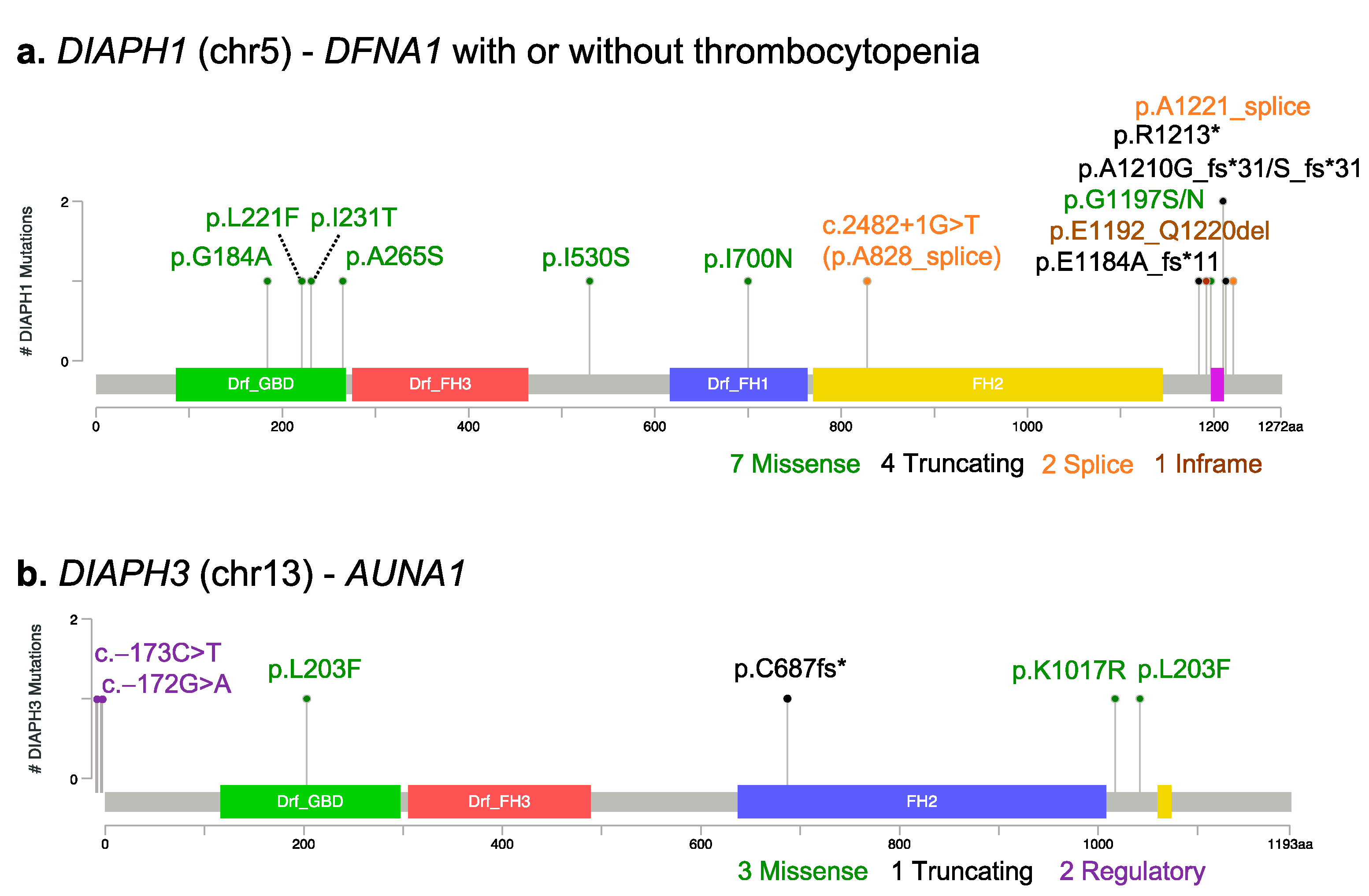
Heterozygous and likely gain-of-function mutations in the DIAPH1 gene, located on chromosome 5, are associated with autosomal dominant deafness 1 with or without thrombocytopenia (DFNA1, MIM #124900) (Figure 2a and Table S1).
DIAPH1 is one of the earliest mapped deafness genes. About 25 years ago, it was identified as the causative gene for DFNA1 in a large Costa Rican family with early-onset (~10 years) low-frequency deafness, progressing to bilateral profound deafness in all frequencies. The causative variant (NM_005219.4: c.3661+1G>T) was a splicing variation that disrupted the donor splice site of intron 17. As a consequence, four bases are inserted into the mRNA, leading to a frameshift and the formation of a premature stop codon [37]. Since then, several families with autosomal dominant deafness have been reported to carry mutations in DIAPH1. The regions of the DIAPH1 protein that are more often mutated are the DAD domain at the C-terminal, and the GTPase-binding domain/DID domain at the N-terminal (Figure 2a), which are critical for the activation and regulation of the protein. Ueyama and colleagues demonstrated that a nonsense mutation (the NM_005219.4: c.3637C>T, p.R1213X), which caused the deletion of a portion of the DAD domain, disrupted the autoinhibitory DID–DAD interaction of DIAPH1, resulting in a mildly constitutively active protein [18]. Missense mutations in the GBD and DID domain of DIAPH1 possibly act via a similar gain-of-function mechanism [20,38,39,40,41,42]. For instance, single-molecule fluorescence microscopy was recently used to confirm the pathogenicity of one of these mutation (p.A265S, located in the DID domain), suggesting that—in analogy with mutations in the DAD—it also disrupts the autoinhibitory interaction between the two domains [43].
Some patients who carry truncating variants (frameshift, nonsense, or in-frame deletion) in the DAD domain present with specific hematological symptoms in addition to deafness, such as thrombocytopenia, enlarged platelets, and increased bleeding tendency [32,39,44,45,46,47,48,49]. Interestingly, a relationship between the extent of the DAD truncation and the hematological phenotype has been suggested [47]. Recently, one heterozygous frameshift variant in the DAD domain has been reported in a Chinese family with auditory neuropathy, a specific type of hearing impairment characterized by the alteration of the auditory pathway but with a normal cochlear outer hair cell function [50].
Biallelic mutations in DIAPH1 do not affect hearing function, but they have been causally linked with seizures, cortical blindness, and microcephaly syndrome (SCBMS, MIM #616632), a rare and severe autosomal recessive neurodevelopmental disorder (Table S2). In this case, the reported mutations are mainly frameshift or nonsense variants located in the FH2 domain, possibly leading to nonsense-mediated mRNA decay and thus resulting in the loss of DIAPH1 function [51,52,53].
Studies in mice have contributed to a better understanding of the pathogenic mechanism and the genotype–phenotype correlation of the different types of DIAPH1 mutations. For example, transgenic mice expressing the human NM_005219.4:c.3637C>T, p.R1213X variant showed progressive deafness caused by cochlear hair cell loss and stereocilia abnormalities. Conversely, Diaph1 knock-out mice showed no hearing phenotype [19]. Altogether, these data further support the fact that DFNA1 pathogenesis is due to the gain-of-function mutations in the DIAPH1 gene. In addition, the observation of a myeloproliferative- and a myelodysplastic-like phenotype in a different Diaph1 knock-out mouse [54] suggests that the DIAPH1 gene plays a critical negative regulatory role in myelopoiesis and is consistent with the thrombocytopenia observed in humans carrying DIAPH1 gain-of-function mutations. Conversely, Diaph1-deficient mice, despite demonstrating the crucial role of the gene in brain development, do not recapitulate the SCBMS phenotype observed in humans and present cerebral ventricular enlargement, but not microcephaly nor blindness [51]. Finally, Diaph1 knock-out mice also show defects in T lymphocyte migration and proliferation [55,56], and this impaired function could explain the additional immunodeficiency observed in some SCBMS patients [53].

3.2. DIAPH2

The first studies associating DIAPH2, located on chromosome X, with a human phenotype reported the presence of chromosomal rearrangements (translocations, deletions, and duplications, listed in Table S2) involving this gene in patients with premature ovarian failure (POF2A, MIM: 300511), a dominant form of ovarian insufficiency characterized by secondary amenhorrea and premature menopause [57,58,59,60,61]. In some cases, ovarian dysgenesis is also present, which is consistent with the reported expression of mouse Diaph2 in the ovaries of developing mice [58] and the known role of the diaphanous protein in follicle cell division in female fruit flies [8]. However, no information on the presence of hearing impairment in POF2A patients with DIAPH2 rearrangements are available. Hence, a possible link between pathogenic variants in the DIAPH2 gene and deafness is still to be determined.

3.3. DIAPH3

Mutations in the human DIAPH3 gene, which is located on chromosome 13, have been involved in nonsyndromic, autosomal dominant auditory neuropathy 1 (AUNA1, MIM #609129) (Figure 2b and Table S1). Auditory neuropathy is an inherited form of progressive human deafness characterized by abnormal or absent auditory brainstem responses but preserved cochlear outer hair cell function, as assessed by otoacustic emissions (OAE). The hearing impairment likely results from defects in the auditory nerve (VIII nerve), the inner hair cells, or the afferent synapses. The first DIAPH3 mutation causing AUNA1 was described in an American family of European descent [36,62]. The reported mutation was an heterozygous transition (NM_001042517.2:c.-172G>A) in a highly conserved element within the 5′ untranslated region of DIAPH3. The variant caused a 2–3-fold overexpression of DIAPH3 mRNA and a 1.5-fold overexpression of DIAPH3 protein levels, consistent with a gain-of-function effect. Subsequently, a second Spanish family was reported to carry a different 5′UTR variant just one nucleotide upstream (NM_001042517.2:c.-173C>T), but no functional characterization was performed to confirm a gain-of-function effect [63]. In addition, missense variants in DIAPH3 have been reported during genetic screening for mutations in individual patients with nonsyndromic sensorineural hearing loss or auditory neuropathy [64,65], but the actual pathogenicity of these variants is not clear. In the ClinVar database, apart from the well-characterized 5′UTR mutation, only one additional variant is classified as pathogenic (ClinVar Accession number VCV000984530): it is a single-base deletion that leads to a frameshift mutation, found at the heterozygous state in a patient affected by AUNA1.
Several animal models support Diaph3 gain-of-function as being the cause of hearing loss in AUNA1. First of all, a drosophila mutant strain constitutively overexpressing the dia gene in the auditory organ showed a significant reduction in sound-evoked potential, mirroring the human AUNA1 phenotype [62]. Similarly, at least two different transgenic mouse models overexpressing Diaph3 have been generated, and both showed specific defects in the inner hair cells and progressive hearing impairment, with the preservation of the outer hair cell function, thus mirroring the human AUNA1 phenotype. The defect seems to depend on the alteration of both the actin and the microtubule dynamics, with a profound alteration of the stereocilia structure and number [28,29]. Instead, loss-of-function DIAPH3 mutants in both mice (knock-out model) and zebrafish (knock-down by antisense morpholino oligonucleotides) show an extremely severe phenotype, which in most cases leads to embryonic lethality. In zebrafish, lethality occurs at very early stages, before the formation of the auditory organ; in mice, although the majority of embryos died between E12.5 and E14.5, some animals survived until birth, showing severe developmental defects such as small brain, reduced body size, and facial deformities [66,67]. To better understand the function of DIAPH3 in the brain, a conditional knock-out mouse was produced, in which the Diaph3 gene was selectively inactivated in the cerebral cortex. These animals showed a profound alteration in neurogenesis, a marked reduction in the number of cortical progenitors and neurons, microcephaly, locomotor impairment, and defects in social interaction, which may indicate an autistic-like behavior [68]. This seems in line with clinical findings in humans, where DIAPH3 was linked to autism in two independent studies (Table S2). The first case identified two different genetic defects in DIAPH3 in the same patient: a missense substitution affecting the FH1 domain on one allele, and a large chromosomal deletion in the second allele [69]. The second case reported a de novo missense mutation within the DIAPH3 FH2 domain together with another de novo mutation in a known autism-associated gene, SET2 [70].

4. Conclusions

Despite extensive studies on the cellular and molecular functions of diaphanous-related formins, their role in the inner ear is still not well-understood, and the mechanisms by which specific genetic defects in the DIAPH1 and DIAPH3 genes primarily lead to a hearing phenotype is yet to be elucidated. For instance, the use of animal models has contributed to the characterization of the physiological expression of diaphanous proteins in the inner ear [32,33], and to the understanding of the gain-of-function effect on actin polymerization associated with a specific truncating mutation at the C-terminal of DIAPH1 [19,33]. However, for other types of mutations, the mechanism by which DRF mutations lead to disease and affect or not the auditory system is less clear. In addition, in some cases, the animal model does not recapitulate the corresponding human phenotype.
Comprehensive genetic analysis and clinical characterization are available for several independent hearing-impaired families carrying mutations in the DIAPH1 and DIAPH3 genes. However, in some cases, a detailed clinical assessment of the affected patients and/or the functional characterization of the candidate pathogenic variants is lacking. The widespread use of whole-exome and whole-genome sequencing in the molecular diagnosis of inherited diseases, including hearing loss, is expected to provide evidence of novel and likely pathogenic variants in DRF genes. In particular, the development of sensitive and efficient molecular tests to verify the actual impact of the newly identified variants on the function of DRFs will help both in confirming pathogenicity and in understanding phenotype–genotype correlations. In this context, a novel and promising approach has recently been developed, which is based on the use of single-molecule fluorescence microscopy to evaluate the actin elongation activity of mutant DIAPH1 in cultured cells [43].
In conclusion, additional in vitro and in vivo studies and genetic analyses of human pedigrees will be needed to shed light on the redundant and non-redundant functions of DRFs in normal hearing and deafness.

Supplementary Materials

The following are available online at https://www.mdpi.com/article/10.3390/cells11111726/s1, Table S1: Pathogenic mutations in DRF genes associated with a hearing phenotype; Table S2: Pathogenic mutations in DRF genes associated with diseases that do not include a hearing phenotype [71,72,73,74,75,76,77].

Author Contributions

Conceptualization, G.S.; Data curation, C.C., M.R.; Writing—original draft preparation, C.C., M.R., and G.S.; Writing—review and editing, all authors; Visualization (figure design and preparation), C.C., M.R., V.M., and G.S.; Resources, P.C. and U.A.; Supervision, G.S. and R.A.; Project administration, G.S.; Funding acquisition, R.A All authors have read and agreed to the published version of the manuscript.

Funding

This research was funded by Fondazione Cariplo, Grant No. 2013-0825.

Institutional Review Board Statement

Not applicable.

Informed Consent Statement

Not applicable.

Data Availability Statement

Not applicable.

Acknowledgments

We thank Morag A. Lewis, Susana Caetano, and Karen P. Steel (Wolfson Centre for Age-Related Diseases, King’s College London, UK) for helpful discussion and critical reading of the manuscript. We are indebted to Stefano Duga (16/09/1967-10/11/2021) for his enthusiasm, inspiration and supervision over the years.

Conflicts of Interest

The authors declare no conflict of interest.

References

  1. Drummond, M.C.; Belyantseva, I.A.; Friderici, K.H.; Friedman, T.B. Actin in hair cells and hearing loss. Hear. Res. 2012, 288, 89–99. [Google Scholar] [CrossRef] [PubMed] [Green Version]
  2. Bogdan, S.; Schultz, J.; Grosshans, J. Formin’ cellular structures: Physiological roles of Diaphanous (Dia) in actin dynamics. Commun. Integr. Biol. 2013, 6, e27634. [Google Scholar] [CrossRef] [PubMed] [Green Version]
  3. Campellone, K.G.; Welch, M.D. A nucleator arms race: Cellular control of actin assembly. Nat. Rev. Mol. Cell Biol. 2010, 11, 237–251. [Google Scholar] [CrossRef] [PubMed] [Green Version]
  4. Labat-De-Hoz, L.; Alonso, M.A. Formins in Human Disease. Cells 2021, 10, 2554. [Google Scholar] [CrossRef] [PubMed]
  5. Lammers, M.; Meyer, S.; Kühlmann, D.; Wittinghofer, A. Specificity of Interactions between mDia Isoforms and Rho Proteins. J. Biol. Chem. 2008, 283, 35236–35246. [Google Scholar] [CrossRef] [PubMed] [Green Version]
  6. Chesarone, M.A.; DuPage, A.G.; Goode, B.L. Unleashing formins to remodel the actin and microtubule cytoskeletons. Nat. Rev. Mol. Cell Biol. 2009, 11, 62–74. [Google Scholar] [CrossRef] [PubMed]
  7. Gaillard, J.; Ramabhadran, V.; Neumanne, E.; Gurel, P.; Blanchoin, L.; Vantard, M.; Higgs, H.N. Differential interactions of the formins INF2, mDia1, and mDia2 with microtubules. Mol. Biol. Cell 2011, 22, 4575–4587. [Google Scholar] [CrossRef]
  8. Castrillon, D.; Wasserman, S. Diaphanous is required for cytokinesis in Drosophila and shares domains of similarity with the products of the limb deformity gene. Development 1994, 120, 3367–3377. [Google Scholar] [CrossRef]
  9. Grunt, M.; Žárský, V.; Cvrčková, F. Roots of angiosperm formins: The evolutionary history of plant FH2 domain-containing proteins. BMC Evol. Biol. 2008, 8, 115. [Google Scholar] [CrossRef] [Green Version]
  10. Lynch, M.; Conery, J.S. The Evolutionary Fate and Consequences of Duplicate Genes. Science 2000, 290, 1151–1155. [Google Scholar] [CrossRef] [Green Version]
  11. Palander, O.; Trimble, W.S. DIAPH1 regulates ciliogenesis and trafficking in primary cilia. FASEB J. 2020, 34, 16516–16535. [Google Scholar] [CrossRef] [PubMed]
  12. Palander, O.; Lam, A.; Collins, R.F.; Moraes, T.J.; Trimble, W.S. Nonredundant roles of DIAPHs in primary ciliogenesis. J. Biol. Chem. 2021, 296, 100680. [Google Scholar] [CrossRef] [PubMed]
  13. Daou, P.; Hasan, S.; Breitsprecher, D.; Baudelet, E.; Camoin, L.; Audebert, S.; Goode, B.L.; Badache, A. Essential and nonredundant roles for Diaphanous formins in cortical microtubule capture and directed cell migration. Mol. Biol. Cell 2014, 25, 658–668. [Google Scholar] [CrossRef] [PubMed]
  14. Goode, B.L.; Eck, M.J. Mechanism and Function of Formins in the Control of Actin Assembly. Annu. Rev. Biochem. 2007, 76, 593–627. [Google Scholar] [CrossRef]
  15. Otomo, T.; Otomo, C.; Tomchick, D.; Machius, M.; Rosen, M.K. Structural Basis of Rho GTPase-Mediated Activation of the Formin mDia1. Mol. Cell 2005, 18, 273–281. [Google Scholar] [CrossRef]
  16. Rose, R.; Weyand, M.; Lammers, M.; Ishizaki, T.; Ahmadian, M.R.; Wittinghofer, A. Structural and mechanistic insights into the interaction between Rho and mammalian Dia. Nature 2005, 435, 513–518. [Google Scholar] [CrossRef]
  17. Young, K.G.; Copeland, J.W. Formins in cell signaling. Biochim. Biophys. Acta 2010, 1803, 183–190. [Google Scholar] [CrossRef] [Green Version]
  18. Watanabe, N.; Kato, T.; Fujita, A.; Ishizaki, T.; Narumiya, S. Cooperation between mDia1 and ROCK in Rho-induced actin reorganization. Nat. Cell Biol. 1999, 1, 136–143. [Google Scholar] [CrossRef]
  19. Ueyama, T.; Ninoyu, Y.; Nishio, S.-Y.; Miyoshi, T.; Torii, H.; Nishimura, K.; Sugahara, K.; Sakata, H.; Thumkeo, D.; Sakaguchi, H.; et al. Constitutive activation of DIA 1 ( DIAPH 1) via C-terminal truncation causes human sensorineural hearing loss. EMBO Mol. Med. 2016, 8, 1310–1324. [Google Scholar] [CrossRef]
  20. Kim, B.J.; Ueyama, T.; Miyoshi, T.; Lee, S.; Han, J.H.; Park, H.-R.; Kim, A.R.; Oh, J.; Kim, M.Y.; Kang, Y.S.; et al. Differential disruption of autoinhibition and defect in assembly of cytoskeleton during cell division decide the fate of human DIAPH1-related cytoskeletopathy. J. Med. Genet. 2019, 56, 818–827. [Google Scholar] [CrossRef]
  21. Nezami, A.G.; Poy, F.; Eck, M.J. Structure of the Autoinhibitory Switch in Formin mDia1. Structure 2006, 14, 257–263. [Google Scholar] [CrossRef] [PubMed] [Green Version]
  22. Nezami, A.; Poy, F.; Toms, A.; Zheng, W.; Eck, M.J. Crystal Structure of a Complex between Amino and Carboxy Terminal Fragments of mDia1: Insights into Autoinhibition of Diaphanous-Related Formins. PLoS ONE 2010, 5, e12992. [Google Scholar] [CrossRef] [PubMed] [Green Version]
  23. Otomo, T.; Tomchick, D.R.; Otomo, C.; Machius, M.; Rosen, M.K. Crystal Structure of the Formin mDia1 in Autoinhibited Conformation. PLoS ONE 2010, 5, e12896. [Google Scholar] [CrossRef] [PubMed]
  24. Jumper, J.; Evans, R.; Pritzel, A.; Green, T.; Figurnov, M.; Ronneberger, O.; Tunyasuvunakool, K.; Bates, R.; Žídek, A.; Potapenko, A.; et al. Highly accurate protein structure prediction with AlphaFold. Nature 2021, 596, 583–589. [Google Scholar] [CrossRef]
  25. Varadi, M.; Anyango, S.; Deshpande, M.; Nair, S.; Natassia, C.; Yordanova, G.; Yuan, D.; Stroe, O.; Wood, G.; Laydon, A.; et al. AlphaFold Protein Structure Database: Massively expanding the structural coverage of protein-sequence space with high-accuracy models. Nucleic Acids Res. 2022, 50, D439–D444. [Google Scholar] [CrossRef]
  26. Maiti, S.; Michelot, A.; Gould, C.; Blanchoin, L.; Sokolova, O.; Goode, B.L. Structure and activity of full-length formin mDia1. Cytoskeleton 2012, 69, 393–405. [Google Scholar] [CrossRef] [Green Version]
  27. Bartolini, F.; Gundersen, G. Formins and microtubules. Biochim. Biophys. Acta 2010, 1803, 164–173. [Google Scholar] [CrossRef] [Green Version]
  28. Schoen, C.J.; Burmeister, M.; Lesperance, M.M. Diaphanous homolog 3 (Diap3) Overexpression Causes Progressive Hearing Loss and Inner Hair Cell Defects in a Transgenic Mouse Model of Human Deafness. PLoS ONE 2013, 8, e56520. [Google Scholar] [CrossRef] [Green Version]
  29. Surel, C.; Guillet, M.; Lenoir, M.; Bourien, J.; Sendin, G.; Joly, W.; Delprat, B.; Lesperance, M.M.; Puel, J.-L.; Nouvian, R. Remodeling of the Inner Hair Cell Microtubule Meshwork in a Mouse Model of Auditory Neuropathy AUNA1. eNeuro 2016, 3, ENEURO.0295-16.2016. [Google Scholar] [CrossRef] [Green Version]
  30. Larkin, A.; Marygold, S.J.; Antonazzo, G.; Attrill, H.; dos Santos, G.; Garapati, P.V.; Goodman, J.L.; Gramates, L.S.; Millburn, G.; Strelets, V.B.; et al. FlyBase: Updates to the Drosophila melanogaster knowledge base. Nucleic Acids Res. 2020, 49, D899–D907. [Google Scholar] [CrossRef]
  31. The Human Protein Atlas. Available online: https://www.proteinatlas.org/ (accessed on 23 February 2022).
  32. Neuhaus, C.; Lang-Roth, R.; Zimmermann, U.; Heller, R.; Eisenberger, T.; Weikert, M.; Markus, S.; Knipper, M.; Bolz, H.J. Extension of the clinical and molecular phenotype of DIAPH1-associated autosomal dominant hearing loss (DFNA1). Clin. Genet. 2016, 91, 892–901. [Google Scholar] [CrossRef] [PubMed]
  33. Ninoyu, Y.; Sakaguchi, H.; Lin, C.; Suzuki, T.; Hirano, S.; Hisa, Y.; Saito, N.; Ueyama, T. The integrity of cochlear hair cells is established and maintained through the localization of Dia1 at apical junctional complexes and stereocilia. Cell Death Dis. 2020, 11, 536. [Google Scholar] [CrossRef] [PubMed]
  34. Hertzano, R.; Elkon, R.; Kurima, K.; Morrisson, A.; Chan, S.-L.; Sallin, M.; Biedlingmaier, A.; Darling, D.S.; Griffith, A.J.; Eisenman, D.J.; et al. Cell Type–Specific Transcriptome Analysis Reveals a Major Role for Zeb1 and miR-200b in Mouse Inner Ear Morphogenesis. PLoS Genet. 2011, 7, e1002309. [Google Scholar] [CrossRef] [PubMed] [Green Version]
  35. Boyer, O.; Nevo, F.; Plaisier, E.; Funalot, B.; Gribouval, O.; Benoit, G.; Cong, E.H.; Arrondel, C.; Tête, M.-J.; Montjean, R.; et al. INF2Mutations in Charcot–Marie–Tooth Disease with Glomerulopathy. N. Engl. J. Med. 2011, 365, 2377–2388. [Google Scholar] [CrossRef] [Green Version]
  36. Kim, T.B.; Isaacson, B.; Sivakumaran, T.A.; Starr, A.; Keats, B.J.B.; Lesperance, M.M. A gene responsible for autosomal dominant auditory neuropathy (AUNA1) maps to 13q14-21. J. Med. Genet. 2004, 41, 872–876. [Google Scholar] [CrossRef] [Green Version]
  37. Lynch, E.D.; Lee, M.K.; Morrow, J.E.; Welcsh, P.L.; León, P.E.; King, M.C. Nonsyndromic deafness DFNA1 associated with mutation of a human homolog of the Drosophila gene diaphanous. Science 1997, 278, 1315–1318. [Google Scholar] [CrossRef] [Green Version]
  38. Shearer, A.E.; Black-Ziegelbein, E.A.; Hildebrand, M.; Eppsteiner, R.W.; Ravi, H.; Joshi, S.; Guiffre, A.C.; Sloan, C.M.; Happe, S.; Howard, S.D.; et al. Advancing genetic testing for deafness with genomic technology. J. Med. Genet. 2013, 50, 627–634. [Google Scholar] [CrossRef]
  39. Iwasa, Y.-I.; Nishio, S.-Y.; Usami, S.-I. Comprehensive Genetic Analysis of Japanese Autosomal Dominant Sensorineural Hearing Loss Patients. PLoS ONE 2016, 11, e0166781. [Google Scholar] [CrossRef]
  40. Sloan-Heggen, C.M.; Bierer, A.O.; Shearer, A.E.; Kolbe, D.L.; Nishimura, C.J.; Frees, K.L.; Ephraim, S.S.; Shibata, S.B.; Booth, K.T.; Campbell, C.A.; et al. Comprehensive genetic testing in the clinical evaluation of 1119 patients with hearing loss. Hum. Genet. 2016, 135, 441–450. [Google Scholar] [CrossRef] [Green Version]
  41. Kang, T.-H.; Baek, J.-I.; Sagong, B.; Park, H.-J.; Park, C.I.; Lee, K.-Y.; Kim, U.-K. A novel missense variant in the DIAPH1 gene in a Korean family with autosomal dominant nonsyndromic hearing loss. Genes Genet. Syst. 2016, 91, 289–292. [Google Scholar] [CrossRef] [Green Version]
  42. Brozkova, D.S.; Marková, S.P.; Mészárosová, A.U.; Jenčík, J.; Čejnová, V.; Čada, Z.; Laštůvková, J.; Rašková, D.; Seeman, P. Spectrum and frequencies of non GJB2 gene mutations in Czech patients with early non-syndromic hearing loss detected by gene panel NGS and whole-exome sequencing. Clin. Genet. 2020, 98, 548–554. [Google Scholar] [CrossRef] [PubMed]
  43. Miyoshi, T.; Belyantseva, I.A.; Kitajiri, S.-I.; Miyajima, H.; Nishio, S.-Y.; Usami, S.-I.; Kim, B.J.; Choi, B.Y.; Omori, K.; Shroff, H.; et al. Human deafness-associated variants alter the dynamics of key molecules in hair cell stereocilia F-actin cores. Qual. Life Res. 2021, 141, 363–382. [Google Scholar] [CrossRef] [PubMed]
  44. Stritt, S.; Nurden, P.; Turro, E.; Greene, D.; Jansen, S.B.; Westbury, S.; Petersen, R.; Astle, W.; Marlin, S.; Bariana, T.K.; et al. A gain-of-function variant in DIAPH1 causes dominant macrothrombocytopenia and hearing loss. Blood 2016, 127, 2903–2914. [Google Scholar] [CrossRef] [PubMed] [Green Version]
  45. Ganaha, A.; Kaname, T.; Shinjou, A.; Chinen, Y.; Yanagi, K.; Higa, T.; Kondo, S.; Suzuki, M. Progressive macrothrombocytopenia and hearing loss in a large family with DIAPH1 related disease. Am. J. Med. Genet. Part A 2017, 173, 2826–2830. [Google Scholar] [CrossRef]
  46. Karki, N.R.; Ajebo, G.; Savage, N.; Kutlar, A. DIAPH1 Mutation as a Novel Cause of Autosomal Dominant Macrothrombocytopenia and Hearing Loss. Acta Haematol. 2020, 144, 91–94. [Google Scholar] [CrossRef]
  47. Westbury, S.K.; Downes, K.; Burney, C.; Lozano, M.L.; Obaji, S.G.; Toh, C.H.; Sevivas, T.; Morgan, N.V.; Erber, W.; Kempster, C.; et al. Phenotype description and response to thrombopoietin receptor agonist in DIAPH1-related disorder. Blood Adv. 2018, 2, 2341–2346. [Google Scholar] [CrossRef]
  48. Turro, E.; Astle, W.J.; Megy, K.; Gräf, S.; Greene, D.; Shamardina, O.; Allen, H.L.; Sanchis-Juan, A.; Frontini, M.; Thys, C.; et al. Whole-genome sequencing of patients with rare diseases in a national health system. Nature 2020, 583, 96–102. [Google Scholar] [CrossRef]
  49. Lakha, R.; Montero, A.M.; Jabeen, T.; Costeas, C.C.; Ma, J.; Vizcarra, C.L. Variable Autoinhibition among Deafness-Associated Variants of Diaphanous 1 (DIAPH1). Biochemistry 2021, 60, 2320–2329. [Google Scholar] [CrossRef]
  50. Wu, K.; Wang, H.; Guan, J.; Lan, L.; Zhao, C.; Zhang, M.; Wang, D.; Wang, Q. A novel variant in diaphanous homolog 1 (DIAPH1) as the cause of auditory neuropathy in a Chinese family. Int. J. Pediatr. Otorhinolaryngol. 2020, 133, 109947. [Google Scholar] [CrossRef]
  51. Ercan-Sencicek, A.G.; Jambi, S.; Franjic, D.; Nishimura, S.; Li, M.; El-Fishawy, P.; Morgan, T.M.; Sanders, S.J.; Bilguvar, K.; Suri, M.; et al. Homozygous loss of DIAPH1 is a novel cause of microcephaly in humans. Eur. J. Hum. Genet. 2014, 23, 165–172. [Google Scholar] [CrossRef] [Green Version]
  52. Al-Maawali, A.; Barry, B.J.; Rajab, A.; El-Quessny, M.; Seman, A.; Coury, S.N.; Barkovich, A.J.; Yang, E.; Walsh, C.A.; Mochida, G.H.; et al. Novel loss-of-function variants in DIAPH1 associated with syndromic microcephaly, blindness, and early onset seizures. Am. J. Med. Genet. Part A 2015, 170, 435–440. [Google Scholar] [CrossRef] [PubMed] [Green Version]
  53. Kaustio, M.; Nayebzadeh, N.; Hinttala, R.; Tapiainen, T.; Åström, P.; Mamia, K.; Pernaa, N.; Lehtonen, J.; Glumoff, V.; Rahikkala, E.; et al. Loss of DIAPH1 causes SCBMS, combined immunodeficiency, and mitochondrial dysfunction. J. Allergy Clin. Immunol. 2021, 148, 599–611. [Google Scholar] [CrossRef] [PubMed]
  54. Peng, J.; Kitchen, S.M.; West, R.A.; Sigler, R.; Eisenmann, K.M.; Alberts, A.S. Myeloproliferative Defects following Targeting of the Drf1 Gene Encoding the Mammalian Diaphanous–Related Formin mDia1. Cancer Res. 2007, 67, 7565–7571. [Google Scholar] [CrossRef] [PubMed] [Green Version]
  55. Eisenmann, K.M.; West, R.A.; Hildebrand, D.; Kitchen, S.M.; Peng, J.; Sigler, R.; Zhang, J.; Siminovitch, K.A.; Alberts, A.S. T Cell Responses in Mammalian Diaphanous-related Formin mDia1 Knock-out Mice. J. Biol. Chem. 2007, 282, 25152–25158. [Google Scholar] [CrossRef] [PubMed] [Green Version]
  56. Sakata, D.; Taniguchi, H.; Yasuda, S.; Adachi-Morishima, A.; Hamazaki, Y.; Nakayama, R.; Miki, T.; Minato, N.; Narumiya, S. Impaired T lymphocyte trafficking in mice deficient in an actin-nucleating protein, mDia1. J. Exp. Med. 2007, 204, 2031–2038. [Google Scholar] [CrossRef] [PubMed] [Green Version]
  57. Salaa, C.; Arrigoa, G.; Torri, G.P.; Martinazzia, F.; Riva, P.; Larizzab, L.; Philippec, C.; Jonveauxc, P.; Sloanc, F.; Labellad, T.; et al. Eleven X Chromosome Breakpoints Associated with Premature Ovarian Failure (POF) Map to a 15-Mb YAC Contig Spanning Xq21. Genomics 1997, 40, 123–131. [Google Scholar] [CrossRef] [PubMed]
  58. Bione, S.; Sala, C.; Manzini, M.C.; Arrigo, G.; Zuffardi, O.; Banfi, S.; Borsani, G.; Jonveaux, P.; Philippe, C.; Zuccotti, M.; et al. A Human Homologue of the Drosophila melanogaster diaphanous Gene Is Disrupted in a Patient with Premature Ovarian Failure: Evidence for Conserved Function in Oogenesis and Implications for Human Sterility. Am. J. Hum. Genet. 1998, 62, 533–541. [Google Scholar] [CrossRef] [Green Version]
  59. Marozzi, A.; Manfredini, E.; Tibiletti, M.; Furlan, D.; Villa, N.; Vegetti, W.; Crosignani, P.; Ginelli, E.; Meneveri, R.; Dalprà, L. Molecular definition of Xq common-deleted region in patients affected by premature ovarian failure. Qual. Life Res. 2000, 107, 304–311. [Google Scholar] [CrossRef]
  60. Genesio, R.; Mormile, A.; Licenziati, M.R.; De Brasi, D.; Leone, G.; Balzano, S.; Izzo, A.; Bonfiglio, F.; Conti, A.; Fioretti, G.; et al. Short stature and primary ovarian insufficiency possibly due to chromosomal position effect in a balanced X;1 translocation. Mol. Cytogenet. 2015, 8, 50. [Google Scholar] [CrossRef] [Green Version]
  61. Bestetti, I.; Castronovo, C.; Sironi, A.; Caslini, C.; Sala, C.; Rossetti, R.; Crippa, M.; Ferrari, I.; Pistocchi, A.; Toniolo, D.; et al. High-resolution array-CGH analysis on 46,XX patients affected by early onset primary ovarian insufficiency discloses new genes involved in ovarian function. Hum. Reprod. 2019, 34, 574–583. [Google Scholar] [CrossRef]
  62. Schoen, C.J.; Emery, S.B.; Thorne, M.C.; Ammana, H.R.; Śliwerska, E.; Arnett, J.; Hortsch, M.; Hannan, F.; Burmeister, M.; Lesperance, M.M. Increased activity of Diaphanous homolog 3 (DIAPH3)/diaphanous causes hearing defects in humans with auditory neuropathy and in Drosophila. Proc. Natl. Acad. Sci. USA 2010, 107, 13396–13401. [Google Scholar] [CrossRef] [PubMed] [Green Version]
  63. Sánchez-Martínez, A.; Benito-Orejas, J.I.; Tellería-Orriols, J.J.; Alonso-Ramos, M.J. Neuropatía auditiva autosómica dominante y variante DIAPH3 (c.-173C>T). Acta Otorrinolaringol. Esp. 2017, 68, 183–185. [Google Scholar] [CrossRef] [PubMed]
  64. Sommen, M.; Schrauwen, I.; Vandeweyer, G.; Boeckx, N.; Corneveaux, J.J.; Ende, J.V.D.; Boudewyns, A.; De Leenheer, E.; Janssens, S.; Claes, K.; et al. DNA Diagnostics of Hereditary Hearing Loss: A Targeted Resequencing Approach Combined with a Mutation Classification System. Hum. Mutat. 2016, 37, 812–819. [Google Scholar] [CrossRef] [PubMed]
  65. Bae, S.-H.; Baek, J.-I.; Lee, J.D.; Song, M.H.; Kwon, T.-J.; Oh, S.-K.; Jeong, J.Y.; Choi, J.Y.; Lee, K.-Y.; Kim, U.-K. Genetic analysis of auditory neuropathy spectrum disorder in the Korean population. Gene 2013, 522, 65–69. [Google Scholar] [CrossRef]
  66. Lai, S.-L.; Chan, T.-H.; Lin, M.-J.; Huang, W.-P.; Lou, S.-W.; Lee, S.-J. Diaphanous-Related Formin 2 and Profilin I Are Required for Gastrulation Cell Movements. PLoS ONE 2008, 3, e3439. [Google Scholar] [CrossRef] [PubMed] [Green Version]
  67. Damiani, D.; Goffinet, A.M.; Alberts, A.; Tissir, F. Lack of Diaph3 relaxes the spindle checkpoint causing the loss of neural progenitors. Nat. Commun. 2016, 7, 13509. [Google Scholar] [CrossRef]
  68. Lau, E.O.-C.; Damiani, D.; Chehade, G.; Ruiz-Reig, N.; Saade, R.; Jossin, Y.; Aittaleb, M.; Schakman, O.; Tajeddine, N.; Gailly, P.; et al. DIAPH3 deficiency links microtubules to mitotic errors, defective neurogenesis, and brain dysfunction. eLife 2021, 10, e61974. [Google Scholar] [CrossRef]
  69. Vorstman, J.A.S.; Van Daalen, E.; Jalali, G.R.; Schmidt, E.R.; Pasterkamp, R.J.; De Jonge, M.; Hennekam, E.A.M.; Janson, E.; Staal, W.G.; Van Der Zwaag, B.; et al. A double hit implicates DIAPH3 as an autism risk gene. Mol. Psychiatry 2010, 16, 442–451. [Google Scholar] [CrossRef]
  70. Xie, J.; Li, H.; Zhu, H.; Huang, L.; Li, H.; Zhang, X.; Zhou, Y.; Zhou, Q.; Xu, W. Analysis of DIAPH3 gene mutation in a boy with autism spectrum disorder. Zhonghua Yi Xue Yi Chuan Xue Za Zhi 2016, 33, 481–484. (In Chinese) [Google Scholar] [CrossRef]
  71. Azaiez, H.; Booth, K.T.; Ephraim, S.S.; Crone, B.; Black-Ziegelbein, E.A.; Marini, R.J.; Shearer, A.E.; Sloan-Heggen, C.M.; Kolbe, D.; Casavant, T.; et al. Genomic Landscape and Mutational Signatures of Deafness-Associated Genes. Am. J. Hum. Genet. 2018, 103, 484–497. [Google Scholar] [CrossRef] [Green Version]
  72. Ji, H.; Lu, J.; Wang, J.; Li, H.; Lin, X. Combined examination of sequence and copy number variations in human deafness genes improves diagnosis for cases of genetic deafness. BMC Ear Nose Throat Disord. 2014, 14, 9. [Google Scholar] [CrossRef] [PubMed] [Green Version]
  73. Pavlenkova, Z.; Varga, L.; Borecka, S.; Karhanek, M.; Huckova, M.; Skopkova, M.; Profant, M.; Gasperikova, D. Comprehensive molecular-genetic analysis of mid-frequency sensorineural hearing loss. Sci. Rep. 2021, 11, 22488. [Google Scholar] [CrossRef] [PubMed]
  74. Misceo, D.; Rødningen, O.; Barøy, T.; Sorte, H.; Mellembakken, J.; Strømme, P.; Fannemel, M.; Frengen, E. A translocation between Xq21.33 and 22q13.33 causes an intragenic SHANK3 deletion in a woman with Phelan-McDermid syndrome and hypergonadotropic hypogonadism. Am. J. Med. Genet. Part A 2011, 155, 403–408. [Google Scholar] [CrossRef] [PubMed]
  75. The Human Gene Mutation Database (HGMD). Available online: http://www.hgmd.cf.ac.uk/ac/index.php (accessed on 14 March 2022).
  76. Richards, S.; Aziz, N.; Bale, S.; Bick, D.; Das, S.; Gastier-Foster, J.; Grody, W.W.; Hegde, M.; Lyon, E.; Spector, E.; et al. Standards and guidelines for the interpretation of sequence variants: A joint consensus recommendation of the American College of Medical Genetics and Genomics and the Association for Molecular Pathology. Genet. Med. 2015, 17, 405–424. [Google Scholar] [CrossRef] [PubMed] [Green Version]
  77. Kearney, H.M.; Thorland, E.C.; Brown, K.K.; Quintero-Rivera, F.; South, S.T. American College of Medical Genetics standards and guidelines for interpretation and reporting of postnatal constitutional copy number variants. Genet. Med. 2011, 13, 680–685. [Google Scholar] [CrossRef] [Green Version]
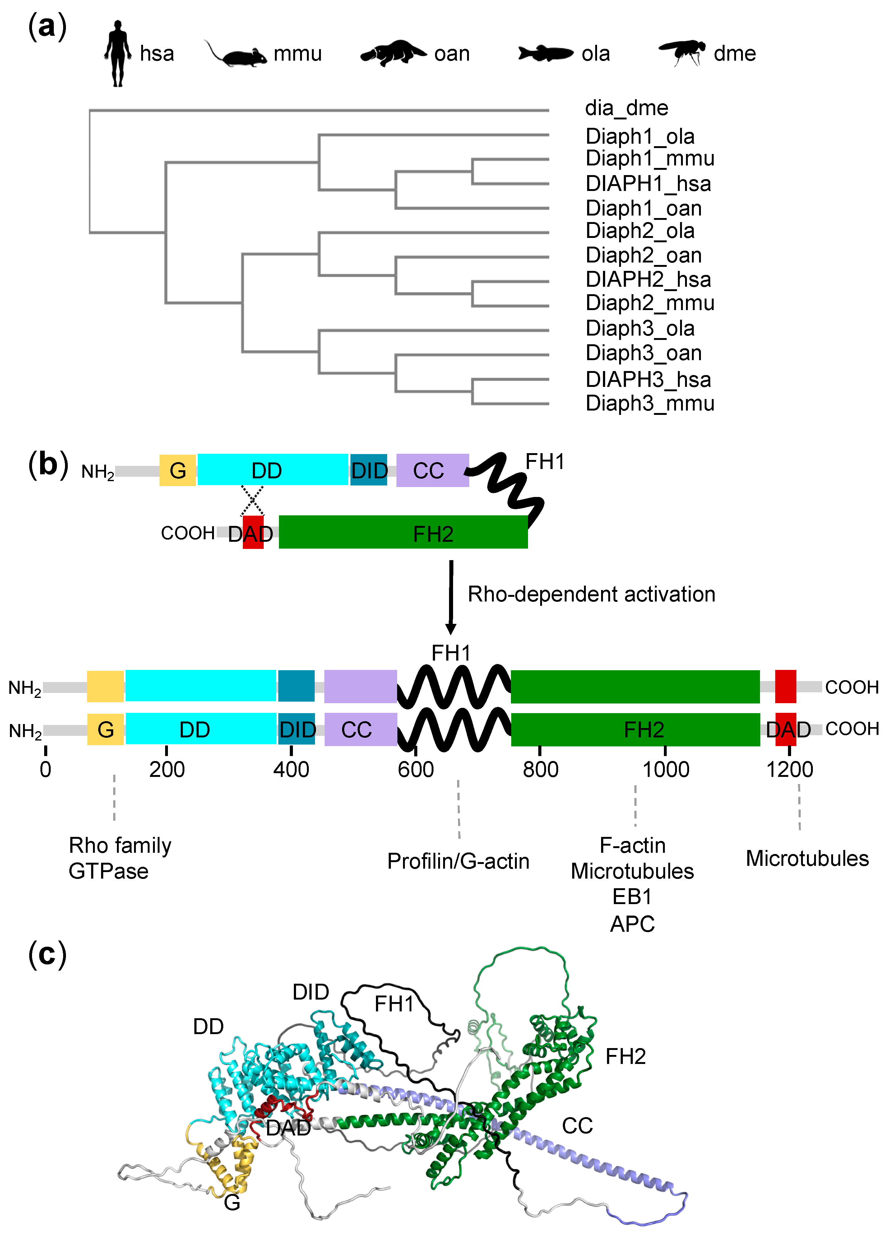
Figure 1. The Diaphanous-Related Formin (DRF) family. (a) Cladogram of Diaphanous-Related Formins in metazoa, based on the UniProt sequences and generated using Clustal Omega. The UniProt accession numbers of the selected sequences are: DIAPH1_hsa (O60610), DIAPH2_hsa (O60879), DIAPH3_hsa (Q9NSV4), Diaph1_mmu (O08808), Diaph2_mmu (O70566), Diaph3_mmu (Q9Z207), Diaph1_oan (A0A6I8NW07), Diaph2_oan (A0A6I8NX37), Diaph3_oan (F7CXN1), Diaph1_ola (H2LGS4), Diaph2_ola (A0A3B3I776), Diaph3_ola (A0A3P9MFT1), and dia_dme (P48608). hsa, Homo sapiens; mmu, Mus musculus; oan, Ornithorhynchus anatinus (platypus); ola, Oryzias latipes (medaka fish); dme, Drosophila melanogaster (fruit fly). (b) Schematic representation of the multi-domain structure of DRF proteins in the inactive autoinhibited form and in the Rho-activated dimeric form. Domains responsible for the interaction with actin and microtubules are indicated. CC: coiled coil domain; DAD: diaphanous autoregulatory domain; DD: dimerization domain; DID: diaphanous inhibitory domain; FH1: formin homology 1 domain; FH2: formin homology 2 domain; RBD: Rho Binding Domain. (c) Tridimensional structure of a diaphanous protein (monomeric), based on the AlphaFold prediction (AF-O08808-F1) for mouse Diaph1.
Cells 11 01726 g001
Figure 2. Mutations in DRFs and hearing loss. Lollipop plots showing genetic variants within human DIAPH1 (a) and DIAPH3 (b) that are associated with hearing phenotypes (see list in Table S1) and mapped on the corresponding protein. Mutations are color-coded based on the predicted functional effect (green, missense; black, truncating; orange, splice; brown, in-frame deletion/insertion; purple, regulatory). *, stop codon; fs, frame-shift. Plots were generated using Mutation Mapper, available at cBioPortal.
Cells 11 01726 g002
Table 1. Hearing loss associated with mutations in DIAPH1 and DIAPH3 genes.
DIAPH1DIAPH3
InheritanceADAD
HL onsetFirst decadeSecond decade 1
Affected frequenciesAllHigh frequencies more affected
ProgressionYesYes
ABR findingsAbsent/abnormalAbsent/abnormal
Middle ear reflexesPresentAbsent
Affected structuresOHCIHC, auditory nerve
Other symptomsMay be accompanied by thrombocytopeniaNone
1 Earlier onset reported at 8 years of age [36]. AD: autosomal dominant; OHC: outer hair cells; IHC: inner hair cells.
Publisher’s Note: MDPI stays neutral with regard to jurisdictional claims in published maps and institutional affiliations.