Review

AUTS2 Gene: Keys to Understanding the Pathogenesis of Neurodevelopmental Disorders

Department of Biochemistry and Cellular Biology, National Institute of Neuroscience, NCNP, 4-1-1 Ogawahigashi, Kodaira, Tokyo 187-8502, Japan
*
Authors to whom correspondence should be addressed.
Cells 2022, 11(1), 11; https://doi.org/10.3390/cells11010011
Submission received: 26 October 2021 / Revised: 8 December 2021 / Accepted: 18 December 2021 / Published: 21 December 2021
(This article belongs to the Special Issue Pathophysiological Mechanism of Neurodevelopmental Disorders)

Abstract

:
Neurodevelopmental disorders (NDDs), including autism spectrum disorders (ASD) and intellectual disability (ID), are a large group of neuropsychiatric illnesses that occur during early brain development, resulting in a broad spectrum of syndromes affecting cognition, sociability, and sensory and motor functions. Despite progress in the discovery of various genetic risk factors thanks to the development of novel genomics technologies, the precise pathological mechanisms underlying the onset of NDDs remain elusive owing to the profound genetic and phenotypic heterogeneity of these conditions. Autism susceptibility candidate 2 (AUTS2) has emerged as a crucial gene associated with a wide range of neuropsychological disorders, such as ASD, ID, schizophrenia, and epilepsy. AUTS2 has been shown to be involved in multiple neurodevelopmental processes; in cell nuclei, it acts as a key transcriptional regulator in neurodevelopment, whereas in the cytoplasm, it participates in cerebral corticogenesis, including neuronal migration and neuritogenesis, through the control of cytoskeletal rearrangements. Postnatally, AUTS2 regulates the number of excitatory synapses to maintain the balance between excitation and inhibition in neural circuits. In this review, we summarize the knowledge regarding AUTS2, including its molecular and cellular functions in neurodevelopment, its genetics, and its role in behaviors.

1. Introduction

The disruption of brain development has been implicated in various psychiatric and neurological illnesses with neurodevelopmental origins. The underlying pathological processes of these diseases often begin early during fetal development and continue into late brain maturation, resulting in pervasive and long-lasting abnormalities in brain structure and function. Neurodevelopmental disorders (NDDs) are a broad spectrum of early onset syndromes affecting cognition, memory, speech, emotions, and behaviors, with a prevalence in children exceeding 3% worldwide [1,2]. Typically, NDDs encompasses a wide range of neuropsychiatric illnesses, including intellectual disability (ID), autism spectrum disorders (ASD), attention-deficit hyperactivity disorder (ADHD), schizophrenia, cerebral palsy, epilepsy, and bipolar disorder [3,4]. In the past decade, the rapid advancement of genomics technologies has led to the identification of a number of gene mutations as well as chromosomal structural variants involved in NDDs [5,6]. Moreover, systems biological analyses have pointed out that a significant number of the genes responsible for NDDs, especially ASD, can converge into neurodevelopmental pathways such as chromatin remodeling, transcriptional regulation, and synaptic function [1,7,8,9,10]. Thus, the understanding of the molecular pathological mechanisms underlying NDDs has progressed; however, due to the substantial genetic and phenotypic heterogeneity of these disorders, the precise etiology of NDDs remains unclear.
The autism susceptibility candidate 2 (AUTS2, also termed “activator of transcription and developmental regulator” by the HUGO Gene Nomenclature Committee, #14262) gene was initially reported as an ASD risk gene disrupted by de novo balanced chromosomal translocation in monozygotic twins with ASD in 2002 [11]. Since then, various genomic structural variants, such as deletions, duplications, inversions, and translocations, as well as single nucleotide polymorphisms (SNPs) in the AUTS2 locus have been associated not only with ASD but also a variety of other psychiatric and neurological illnesses, such as ID, schizophrenia, ADHD, language disorder, epilepsy, depression, and drug dependence [12,13,14,15,16,17,18,19,20,21]. Additionally, patients with AUTS2 mutations often display other pathological conditions including developmental delay, microcephaly, short stature, and craniofacial dysmorphisms; a series of such symptoms are termed “AUTS2 syndrome” [14,22]. In this review, we summarize the knowledge of the molecular and cellular functions of AUTS2 in neurodevelopment and the involvement of this gene in NDDs.

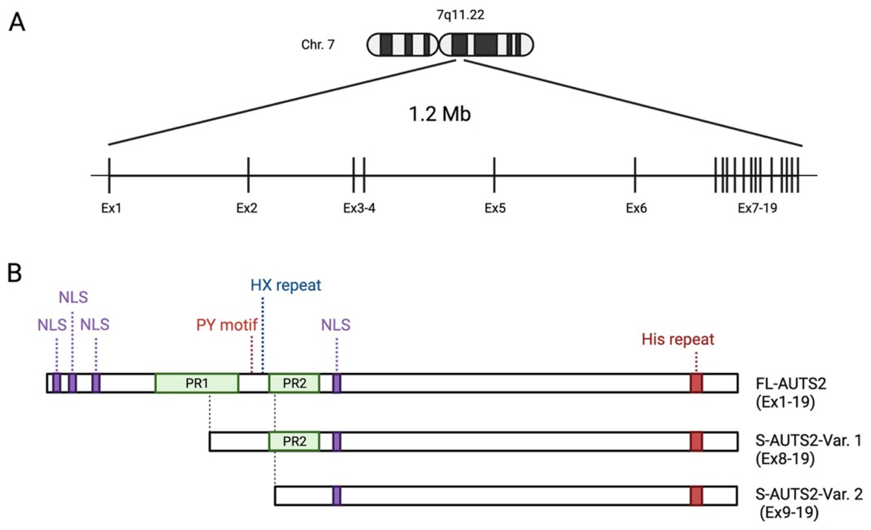
2. Structure and Expression of the AUTS2 Gene

AUTS2 is a large gene spanning 1.2 M bases on human chromosome 7q11.22 (Figure 1A) [12,23]. It consists of 19 exons, the first 6 of which are separated by long introns at the 5′ end, whilst the remaining 13 are compact with clustered smaller introns at the 3′ end. The full-length AUTS2 transcript encodes a protein with 1259 amino acids (aa) in humans (NM_015570) and 1261 aa in mice (NM_177047), although various isoforms are generated by alternative splicing and multiple transcription start sites (TSS). In the human brain, a short 3′ AUTS2 transcript generated from TSS within exon 9 has been identified by 5′ RACE (5′ rapid amplification of cDNA ends) [14]. In the developing mouse brain, two short C-terminal AUTS2 isoforms translated from exon 8 (S-AUTS2-Var1; ~88 kDa) and exon 9 (S-AUTS2-Var2; ~78 kDa), as well as the full-length AUTS2 (FL-AUTS2; ~170 kDa) were detected by 5′ RACE and immunoblot experiments [24,25]. Additional alternative splicing AUTS2 variants skipping exons 10 and 12 in humans (corresponding to exons 11 and 13 in mice) were also found in the fetal brain [26].
In the mouse cerebral cortex, FL-AUTS2 and S-AUTS2-Var1 appear from the early embryonic stages [24,27]. The long isoform shows the highest expression level at later embryonic stages around embryonic days 16–18, and is also present postnatally; although, it gradually decreases thereafter. In contrast, the short isoform is transiently expressed in embryonic brains and disappear postnatally. Interestingly, in mouse embryonic stem cells (mESCs), an isoform switch from the long to short AUTS2 isoform occurs during the transition from undifferentiated to differentiated cortical neuronal states [26].
In the normal mouse brain, S-AUTS2-Var2 is barely detectable compared to other AUTS2 isoforms. In contrast, the expression of this shortest isoform was abnormally increased in mutant mice lacking exon 8 of Auts2 [24,25]. These findings suggest that there are internal enhancer/promoter regions for the alternative transcription of S-AUTS2, tightly regulating the temporal expression pattern of AUTS2 isoforms in the brain. Several potential enhancer regions have been identified within the introns of this gene in zebrafish and mice [12]. Furthermore, Kondrychyn et al. demonstrated that the AUTS2 ortholog in zebrafish, auts2, potentially has 13 unique TSSs, and more than 20 alternative transcripts were found to be produced from this gene locus [28]. Therefore, the complexity of this gene may be substantially higher in mammals as compared to other species than previously identified. In humans, some of the genomic variants were identified within intronic regions of the AUTS2 locus in patients with NDDs, implying that disorganized expression of AUTS2 may be associated with the onset of these disorders [21]. Auts2 expression is reportedly regulated by T-box brain protein 1 (Tbr1), a transcription involved in the development of cortical deep-layer projection neurons and also implicated in ASD [29,30]. In addition, methyl CpG binding protein 2 (MECP2), whose gene mutations are associated with Rett syndrome and ASD, suppresses AUTS2 expression [31,32].
Auts2 is widely expressed in multiple regions of the developing mouse brain, but particularly strong levels are observed within areas related to higher cognitive functions, such as the cerebral cortex, the Ammon’s horn and dentate gyrus of the hippocampus, and the cerebellum [33]. In postnatal and fully developed mouse brains, Auts2 expression is restricted to a few types of neurons, but is strong in glutamatergic projection neurons in the prefrontal cortex, pyramidal CA neurons in the hippocampus, granule neurons of the dentate gyrus, and cerebellar Purkinje and Golgi cells [33,34].

3. Genotype–Phenotype Correlations of AUTS2 Syndrome

As noted, a wide range of neurodevelopmental phenotypes in the patients with AUTS2 syndrome have been reported in individual studies; however, few studies have systematically reviewed the relationship between genotype and phenotype using a relatively large cohort of patients with AUTS2 mutations. In 2013, Beunders et al. collected the clinical data of patients (17 individuals and 4 family members) carrying exonic AUTS2 deletions from a large cohort (49,684 samples) with ID and/or multiple congenital anomalies, and examined the genotype–phenotype correlations in AUTS2 syndrome [14]. In that study, they originally established an AUTS2 syndrome severity score (ASSS) system, which was based on the sum of 32 clinical features observed with a frequency of over 10% in the first cohort of individuals with AUTS2 mutations. The ASSS system includes physical (growth, feeding, dysmorphic features, and skeletal disorders) and neuropsychiatric (neurological and neurodevelopmental disorders) phenotypes and congenital anomalies. Their paradigm revealed that the patients with deletions of the conserved C-terminal regions of AUTS2 (exons at the 3′ of the AUTS2 gene) showed severe AUTS2 syndrome phenotypes including neurodevelopmental disorders and dysmorphic features. In contrast, the individuals with small in-frame deletions of the different combinations of exons 2–5 at a non-conserved 5′ region (N-terminus of AUTS2) displayed lower ASSS values and milder pathological features [14].
After the initial description by Beunders et al., a meta-analysis using the data of 31 patients with AUTS2 syndrome obtained from seven studies, in addition to five newly identified clinical cases, supported that the patients with ADHD and/or ASDs, who carry mutations at 3′ end of the AUTS2 locus showed higher ASSS values compared with individuals with AUTS2 aberrations at the 5′ end of the locus [35]. These findings imply that the ablation of C-terminal region of AUTS2 may contribute to the onset of AUTS2 syndrome. Meanwhile, the individuals carrying a frame shift microdeletion within exon 6 or 7, which would only disrupt full-length AUTS2 transcript but were unlikely to affect the C-terminal AUTS2 short transcripts [36], have reportedly exhibited severe AUTS2 syndrome phenotypes. Taken together, this evidence implies that the deletion of the C-terminal region of full-length AUTS2 transcript and/or the loss of expression of C-terminal AUTS2 short transcripts may contribute to the onset of AUTS2 syndrome.

4. Protein Structure of AUTS2

Protein primary structure analysis predicts that AUTS2 possesses several putative nuclear localization signal sequences and two proline-rich domains (PR1 and PR2) (Figure 1B) [11,21]. Other predicted protein motifs include a PY (Pro-Tyr) motif (PPPY), eight CAG (His) repeats, several protein kinase phosphorylation sites, and putative SH2- and SH3-binding domains; however, the exact role of these motifs has not been well analyzed. The PY motif could act as a binding motif to a WW domain that is present in the activation domain of several transcription factors [37]. The His repeat is found in some nuclear proteins, which have been reported to function in the targeting of proteins to subnuclear compartments, such as nuclear speckles [38]. AUTS2 is also predicted to contain several conserved domains that may be involved in RNA metabolism. Castanza et al. found that AUTS2 binds to RNA transcripts related to various biological processes [39]. Proteomic analyses identified several RNA-binding proteins (RBPs) as the AUTS2-binding molecules in the mouse neonatal cerebral cortex, such as RNA splicing factors (splicing factor proline-glutamine rich (SFPQ), serine-rich splicing factor 3 (SRSF3)), the RNA helicases (DDX5 and 17), and the RBP, NONO [39]. Although the physiological significance of the interaction of AUTS2 with RNAs or RBPs remains to be clarified, it is conceivable that AUTS2 may control protein expression through RNA metabolism in addition to transcriptional regulation, as described below.
AUTS2 has an additional characteristic histidine-rich sequence (HX repeats) between the PR domains. Individuals carrying de novo in-frame microdeletion within the region corresponding to HX repeats in the AUTS2 locus display NDDs, including cognitive delay, microcephaly, and craniofacial dysmorphisms [40,41]. Liu et al. recently found that AUTS2 interacts with a histone acetyltransferase P300/CBP via its HX repeats motif, which is required for the differentiation of neural progenitor cells into neurons [41].

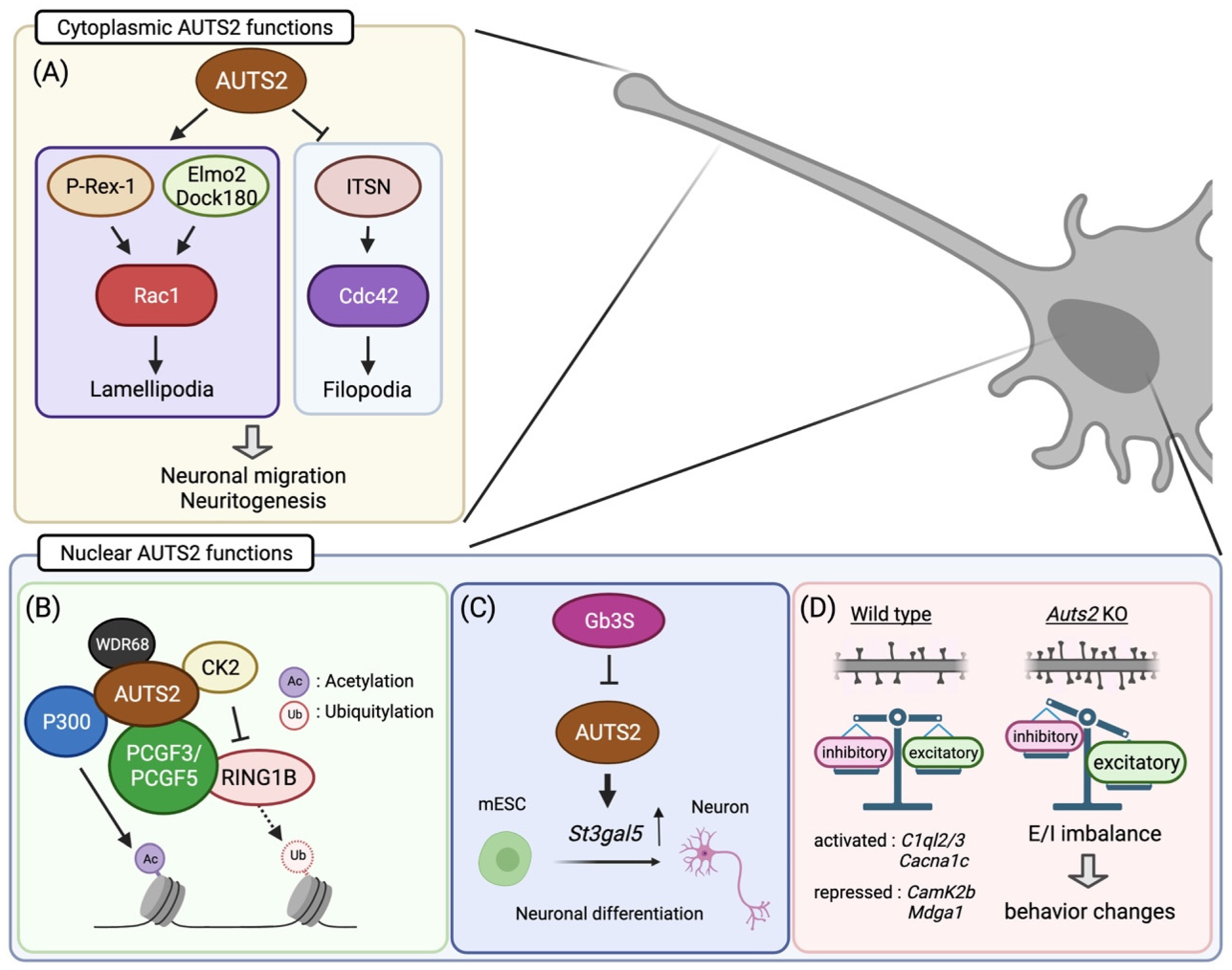
5. Cytoplasmic AUTS2 Functions in Cytoskeletal Organization

AUTS2 is found in the cell nuclei of glutamatergic neurons in the mouse cerebral cortex during early developmental stages [24]. As neural development proceeds, it also appears in the cytoplasm of differentiated neurons, including dendrites, axons, and cell bodies. Subcellular localization analyses showed that FL-AUTS2 was localized in both the nucleus and cytoplasm, whereas the S-AUTS2 isoforms were exclusively nuclear [24]. Cytoplasmic AUTS2 is involved in cell motility and morphogenesis by regulating reorganization of the actin cytoskeleton (Figure 2A) [24]. AUTS2 activates the Rho-family small GTPase Rac1 via guanine exchange factors (GEFs), such as P-Rex1 or the Elmo2/Dock180 complex, leading to lamellipodia formation in neuroblastoma cell lines. In contrast, AUTS2 inhibits filopodia formation by suppressing the activities of another Rho-family G protein, Cdc42, through interaction with its GEF, intersectin 1 and 2.

6. Transcriptional Regulation by Nuclear AUTS2

Nuclear AUTS2 functions as a regulator of gene transcription during neurodevelopment. Genomic profiling of AUTS2 by chromatin immunoprecipitation sequencing, in combination with transcriptome analysis (RNA sequencing), revealed that it interacts with the promoter and enhancer regions of the genes related to brain development as well as those associated with NDDs, many of which are actively expressed in the developing mouse forebrain [42]. In addition, AUTS2 shares DNA-binding motifs with some transcription factors that are involved in neural development, including TCF3, Pitx3, and FOXO3, implying that AUTS2 functions in transcriptional activation [42]. Gao et al. found that AUTS2 interacts with an epigenetic regulator type I Polycomb repressive complex (PRC) 1 (Figure 2B) [27]. PRC1 canonically functions as a transcriptional repressor to maintain gene silencing through the compaction of local chromatin by depositing monoubiquitylation of histone H2A at lysine-119 (H2AK119ub1) via its core component RING1A/B [43]. Incorporation of AUTS2 into the PRC1 complex converts this complex to a transcriptional activator by recruiting two other components, casein kinase 2 (CK2), and histone acetyltransferase P300/CBP [27]. CK2 inhibits the monoubiquitylation activity of RING1 proteins, while P300 promotes transcription by acetylating histone H3 at lysine-27 (H3K27ac). In mESCs, the WD40-repeats-containing protein WDR68 mediates transcriptional activation by induction of the AUTS2-PRC1 complex [44]. On the other hand, Monderer-Rothkoff et al. demonstrated that in the luciferase reporter assay, while both FL-AUTS2 and S-AUTS2 activated gene transcription in non-neuronal HEK293 cells, FL-AUTS2 or N-terminal AUTS2 fragment (encoded by exons 1–9) can function as transcriptional repressors in neuroblastoma Neuro2A cells [26]. Whether AUTS2 acts as a transcriptional activator or repressor may therefore depend on the intracellular context, that is, which AUTS2-binding co-factor(s) are expressed.

7. The Role of AUTS2 in Neurogenesis

Since the first report by Sultana et al. in 2002, a number of clinical genomic analyses have demonstrated the association of AUTS2 mutations with NDDs, albeit the molecular and biological functions of this gene have long remained unknown. In 2013, two different groups attempted to determine the function of AUTS2 by means of knockdown experiments using morpholino in a zebrafish model [12,14]. The auts2 morphants displayed developmental abnormalities, including microcephaly and craniofacial dysmorphisms (smaller jaw size). In the morphant brains, neuronal cells were markedly decreased in various regions, such as the cerebellum, optic tectum, and retina, which was likely based on an increase in apoptosis and/or a decrease in neuronal differentiation [12,14]. Co-introduction of full-length human AUTS2 mRNA together with the auts2 morpholino restored morphant phenotypes back to normal. Moreover, the 3′-end short transcript corresponding to a human AUTS2 short isoform could also rescue the phenotype, suggesting that the C-terminal region of AUTS2 contains the domains crucially involved in neural development. This is likely correlated with the observation that the severity of the symptoms of AUTS2 syndrome is prominent in individuals with disruption at the 3′ end of the AUTS2 locus [14]. Mice lacking Auts2 exhibit hypoplasia in the dentate gyrus as well as in the cerebellum [34,39,45]. These findings suggest that AUTS2 plays a key role in neural development in the brain.
In addition, the in vitro analyses using mESCs demonstrated that AUTS2 is involved in neuronal differentiation through histone modifications together with the aforementioned PRC1 complex, including P300, CK2, and WDR68 [41,44,46]. Similar to the findings in the zebrafish model, disrupting the Auts2 gene in mESC led to increased cell death during differentiation into neurons, which was rescued by expressing the C-terminal AUTS2 shot isoform as well as FL-AUTS2 [26]. Meanwhile, forced expression of FL-AUTS2 in mESCs leads to the delay of neuronal differentiation [34]. Furthermore, Russo et al. found that AUTS2 promotes neuronal differentiation through transcriptional activation of the sphingolipid-modifying enzyme GM3S (Figure 2C) [46]. Taken together, these findings suggest that AUT2 controls neuronal differentiation by transcriptional regulation via histone modification, an epigenetic mechanism.

8. AUTS2 Regulates Neuronal Migration and Neuritogenesis during Corticogenesis

In primary cultured hippocampal neurons, forced expression of FL-AUTS2 promotes neurite outgrowth, whereas co-expression of dominant-negative forms of Rac1 or Elmo2 blocks this effect [24]. Furthermore, loss of Auts2 results in an impairment of axonal elongation of cortical commissural neurons in the developing mouse cerebral cortex, which is restored by expression of wild-type Rac1 in Auts2-deficient neurons; this suggests that the AUTS2-Rac1 signaling pathway plays an important role in the neuritogenesis of cortical neurons (Figure 2A) [24].
The AUTS2-Rac1 pathway is also required for neuronal migration (Figure 2A). Disruption of Auts2 leads to a delay in neuronal migration in the developing mouse cerebral cortex [24]. In Auts2-deficient migrating neurons, the activity of c-Jun N-terminal kinase, which is a Rac1 downstream target, is markedly decreased. However, the observed defects in cell migration were restored by the introduction of FL-AUTS2 or wild-type Rac1, but not a short isoform of AUTS2 [24]. In addition, nuclear export sequence (NES)-tagged FL-AUTS2 also rescued the defects in migration, suggesting that cytosolic FL-AUTS2 functions in cortical neuronal migration and subsequent neurite formation by regulating cytoskeletal rearrangements through activation of the Rac1 signaling pathway.
Neuronal migration and subsequent neurite formation are important initial steps in fetal neurodevelopmental processes to properly assemble cortical structures as well as functional neural circuits. Dysregulation of cortical neuronal migration has been associated with NDDs, including ASD [47]. In humans, individuals with AUTS2 mutations exhibit a wide range of pathological features, including microcephaly, corpus callosum hypoplasia, and psychiatric illnesses. Therefore, defects in neuronal migration and neuritogenesis due to loss of AUTS2 function in the cytoplasm may partly reflect the complex phenotypes of AUTS2 syndrome.

9. AUTS2 Restricts the Number of Excitatory Synapses to Regulate the E/I Balance

Synaptogenesis is a critical neurodevelopmental process for the establishment of the basis underlying higher cognitive brain functions. During postnatal development and in adulthood, the formation and maintenance of an appropriate ratio of excitatory and inhibitory synapses ensures proper excitation/inhibition (E/I) balance in neural circuits [48,49,50]. In contrast, a disturbed E/I balance within the neural circuitry is thought to be associated with NDDs, including ASD, ADHD, schizophrenia, and epilepsy [51,52].
AUTS2 modulates E/I balance by restricting the number of excitatory synapses, suppressing newly formed dendritic spines, and/or promoting the pruning of existing spines during postnatal brain development (Figure 2D) [25]. Loss of Auts2 leads to increased excitatory synapse formation in pyramidal neurons in multiple forebrain areas, including the medial prefrontal cortex (mPFC), auditory cortex, and hippocampus in the mouse brain as well as primary cultured mouse hippocampal neurons [25]. Interestingly, targeted disruption of Auts2 in forebrain neurons during late postnatal stages results in excess spine formation, suggesting the involvement of AUTS2 in the regulation of synaptic homeostasis in mature brains. In contrast, AUTS2 does not affect the number of inhibitory synapses. In the Auts2 mutant hippocampus, excitatory synaptic inputs were increased, while inhibitory synaptic inputs were not altered, resulting in increased excitability in local neural circuits [25]. The aberrant excitatory synapse formation in Auts2-deficient neurons is rescued by re-expression of FL-AUTS2 but not S-AUTS2 isoforms. Moreover, the cytoplasmic form of NES-FL-AUTS2 does not restore the synaptic phenotype. Therefore, it is suggested that AUTS2 can restrict the number of excitatory synapses by regulating the transcription of genes related to synapse development by acting as a component of PRC1 or by other regulatory mechanisms. Transcriptome analyses revealed several candidate AUTS2 downstream target genes related to synapse formation and/or functions such as Mdga1, Reln, Camk2b, Cacna1c, and C1ql2/3 [25]. However, it has not been clarified which of these genes are directly regulated by AUTS2 and responsible for the synaptic pathogenesis caused by Auts2 mutations.

10. AUTS2 Is Involved in Cerebellar Development

The cerebellum has traditionally been thought to function in voluntary motor coordination, but accumulating evidence suggests that it is associated with higher-order brain functions, such as social cognition, the reward system, and emotional processing [53,54,55,56]. Among neurons in the cerebellar cortex, Purkinje cells (PCs) play a pivotal role as the sole output neurons to transmit various information processed in the cerebellum to higher brain regions [57,58,59].
Auts2 is expressed in the cerebellar primordium during early prenatal developmental stages [33]. In adult stages, the expression of AUTS2 is confined to PCs and Golgi cells in the granule cell (GC) layer in the cerebellar cortices [34]. Loss of Auts2 was shown to result in reduced cerebellar size in mice [34]. In Auts2 cerebellar conditional knockout (cKO) mice, the number of PCs and GCs is diminished [34]. Coincidently, some patients with AUTS2 mutations exhibit smaller cerebella [40,41]. Given that AUTS2 is expressed in PCs but not GCs, AUTS2 may directly regulate PC neurogenesis. It is also known that PCs promote the amplification of GC progenitors by secreting sonic hedgehog (SHH), eventually contributing to cerebellar size expansion [60]. Therefore, in Auts2 cKO mice, GCs might fail to proliferate because of the decrease in the overall amount of SHH along with the reduction in PC numbers, thus causing a reduction in cerebellar size.
The PCs in Auts2 cKO cerebella show impaired dendrite maturation, including a delay in the regression of surplus dendrites from the cell soma during the early postnatal stages and subsequent dendrite outgrowth, resulting in diminished molecular layer thickness [34]. How exactly AUTS2 promotes the maturation of PC dendrites is unknown; however, since the Rac signaling pathway has been reportedly involved in the dendrite morphogenesis of PCs [61], it is possible that cytoplasmic AUTS2 can be directly involved in these processes via cytoskeletal rearrangements through Rac1 activation, as observed in cerebral cortical neurons. Alternatively, nuclear AUTS2 may control the maturation of PC dendrites by regulating the expression of genes involved in dendrite development.
In the postnatal stages, PCs receive excitatory presynaptic inputs from a single climbing fiber (CF) innervated from the inferior olive nucleus neurons outside the cerebellum, in addition to accepting other excitatory inputs from multiple parallel fibers (PFs) extended from GCs [62]. During the early postnatal period, multiple CFs project to each PC cell body. Subsequently, a single CF with the strongest excitatory transmission is selected to translocate along the primary PC dendrites to form CF synapses, while the remaining CFs are removed. Auts2 cKO mice show impairments in CF synapse translocation during development, resulting in a decreased number of CF synapses in adulthood [34]. Moreover, electrophysiological experiments revealed that the majority of Auts2 cKO PCs still received multiple CF inputs even in adulthood, suggesting that the excess CF elimination process is impaired by loss of Auts2 [34].
In contrast, AUTS2 restricts the number of PF synapses in PCs. Loss of Auts2 leads to increased PF synapse formation [34]. Furthermore, Auts2-deficient PCs exhibit enhanced excitatory neurotransmission in PF synapses. No such alterations were observed in inhibitory synapses. This is evidence implying that the E/I balance in PCs could be disturbed in Auts2 cKO mice. Moreover, Auts2-deficient PCs show reduced expression of the P/Q-type voltage-dependent Ca2+ channel gene Cacna1a, which has been reported to regulate excitatory synapse development, including CF synapse elimination and PF synapse boundary formation [63,64,65]. Therefore, it is possible that AUTS2 can regulate excitatory synapse development in PCs through transcriptional regulation of the expression of relevant genes with the same mechanism as observed in telencephalon [25].
Emerging evidence has pointed out a link between cerebellar dysfunction and NDDs, such as ASD, and studies in both human and animal models have shown a decrease in the number or disrupted function of PCs [66,67]. AUTS2 is a key regulator involved in a wide range of processes during PC development, including neurogenesis, dendrite maturation, and PC-centric neural circuit assembly. However, the molecular mechanisms underlying the regulation of AUTS2 in PC development are not well understood. In addition, the function of this molecule in other AUTS2-expressing cells, such as Golgi cells, has not been determined, and how Auts2 loss affects the development or function of these cells remains to be clarified.

11. Behavioral Abnormalities in Auts2 Mutant Mice

Behavioral studies have been conducted using several different types of Auts2 mutant mice. Homozygous pups with neuron-specific deletion of Auts2 exhibit defects in motor abilities, such as geotaxis and righting reflex [27]. Adult homozygotes with cerebellar ablation of Auts2 also showed impaired balance control and motor learning [34]. Moreover, Auts2 heterozygous conventional KO mice exhibit autistic-like behaviors, such as social deficits and altered vocal communication [25,68]. Intriguingly, loss of Auts2 not only reduces the number of ultrasonic vocalizations (USVs) but also the complexity of USV syllables emitted from male mice during courtship behaviors [25]. Defects in social behaviors and vocal communication are well recapitulated by selective deletion of Auts2 in excitatory neurons in the adult forebrain [25], implying that the synaptic regulation by AUTS2 significantly contributes to social behaviors in mice. Furthermore, Auts2 mutant mice manifest altered anxiety as well as abnormal startle responses to auditory and nociceptive stimuli [25,68], which may be due to the disturbed E/I balance in sensory cortices. Both phenotypes are often observed in individuals with ASD, in addition to other impairments [3]. In addition, loss of Auts2 results in defects in cognitive and memory function, suggesting that AUTS2 contributes to neurocognitive brain functions.

12. Conclusions and Perspectives

Over the last few decades, multiple mutations within the AUTS2 locus have been identified in patients with NDDs, identifying it as a causative factor for neurodevelopmental and psychological disorders such as ASD, ID, ADHD, and schizophrenia. Accumulating evidence has revealed that AUTS2 is a key regulator involved in a wide range of neurodevelopmental processes, from prenatal neurogenesis to the assembly of neural circuits in mature brains. In cell nuclei, AUTS2 participates in the regulation of gene expression through epigenetic modulation. Cytoplasmic AUTS2 contributes to the organization of cytoskeletal rearrangements to regulate prenatal cortical development, including neuronal migration and neurite morphogenesis. Postnatally, AUTS2 modulates the E/I balance of synaptic inputs by restricting the number of excitatory synapses.
Nevertheless, some key issues remain to be addressed. Although several lines of evidence have suggested that multiple types of AUTS2 isoforms are potentially expressed in various brain regions, the exact mechanisms by which their expression is regulated and their precise spatiotemporal organization has not been analyzed. In addition, the biological function of the individual AUTS2 isoforms is not well understood, and how the loss- or gain-of-function of these isoforms affects neurodevelopment as well as behaviors remains unknown. Moreover, the specific AUTS2 downstream target genes and molecular pathways involved in neurodevelopment, including neurogenesis and synaptogenesis, have not been well determined. Further elucidation of the molecular function of AUTS2, as well as a more comprehensive understanding of the AUTS2-mediated gene expression network pathways in neural development will help to unravel the complex pathological mechanisms of NDDs and provide promising insights for new therapeutic strategies to treat these disorders.

Author Contributions

Conceptualization, K.H. and M.H.; writing—original draft preparation, K.H.; visualization, K.S. All authors have read and agreed to the published version of the manuscript.

Funding

This work was supported by Grants-in-Aid for Scientific Research, KAKENHI (Grant 16H06528 and 18H02538 for M.H.); AMED (19dm0107085; JP21wm0425005 to M.H.), The Mother and Child Health Foundation, an Intramural Research Grant of NCNP (Grants 30-9 and 1-4 for M.H.), and the \Japan Health Research Promotion Bureau (JH) under Research Fund (Grant 2020-B-07 for M.H).

Institutional Review Board Statement

Not applicable.

Informed Consent Statement

Not applicable.

Data Availability Statement

Not applicable.

Acknowledgments

We acknowledge the work of the present members of our laboratory. Figures were created with BioRender.com.

Conflicts of Interest

The authors declare no conflict of interest.

References

  1. Parenti, I.; Rabaneda, L.G.; Schoen, H.; Novarino, G. Neurodevelopmental Disorders: From Genetics to Functional Pathways. Trends Neurosci. 2020, 43, 608–621. [Google Scholar] [CrossRef] [PubMed]
  2. Niemi, M.E.K.; Martin, H.C.; Rice, D.L.; Gallone, G.; Gordon, S.; Kelemen, M.; McAloney, K.; McRae, J.; Radford, E.J.; Yu, S.; et al. Common genetic variants contribute to risk of rare severe neurodevelopmental disorders. Nature 2018, 562, 268–271. [Google Scholar] [CrossRef] [PubMed] [Green Version]
  3. American Psychiatric Association. Diagnostic and Statistical Manual of Mental Disorders, 5th ed.; American Psychiatric Association: Washington, DC, USA, 2013. [Google Scholar]
  4. Tarlungeanu, D.C.; Novarino, G. Genomics in neurodevelopmental disorders: An avenue to personalized medicine. Exp. Mol. Med. 2018, 50, 1–7. [Google Scholar] [CrossRef] [PubMed] [Green Version]
  5. Mardis, E.R. A decade’s perspective on DNA sequencing technology. Nature 2011, 470, 198–203. [Google Scholar] [CrossRef]
  6. Ng, S.B.; Buckingham, K.J.; Lee, C.; Bigham, A.W.; Tabor, H.K.; Dent, K.M.; Huff, C.D.; Shannon, P.T.; Jabs, E.W.; Nickerson, D.A.; et al. Exome sequencing identifies the cause of a mendelian disorder. Nat. Genet. 2010, 42, 30–35. [Google Scholar] [CrossRef] [PubMed] [Green Version]
  7. Satterstrom, F.K.; Kosmicki, J.A.; Wang, J.; Breen, M.S.; De Rubeis, S.; An, J.Y.; Peng, M.; Collins, R.; Grove, J.; Klei, L.; et al. Large-Scale Exome Sequencing Study Implicates Both Developmental and Functional Changes in the Neurobiology of Autism. Cell 2020, 180, 568–584. [Google Scholar] [CrossRef]
  8. Sanders, S.J.; He, X.; Willsey, A.J.; Ercan-Sencicek, A.G.; Samocha, K.E.; Cicek, A.E.; Murtha, M.T.; Bal, V.H.; Bishop, S.L.; Dong, S.; et al. Insights into Autism Spectrum Disorder Genomic Architecture and Biology from 71 Risk Loci. Neuron 2015, 87, 1215–1233. [Google Scholar] [CrossRef] [Green Version]
  9. Ben-David, E.; Shifman, S. Combined analysis of exome sequencing points toward a major role for transcription regulation during brain development in autism. Mol. Psychiatry 2013, 18, 1054–1056. [Google Scholar] [CrossRef] [Green Version]
  10. De Rubeis, S.; He, X.; Goldberg, A.P.; Poultney, C.S.; Samocha, K.; Cicek, A.E.; Kou, Y.; Liu, L.; Fromer, M.; Walker, S.; et al. Synaptic, transcriptional and chromatin genes disrupted in autism. Nature 2014, 515, 209–215. [Google Scholar] [CrossRef]
  11. Sultana, R.; Yu, C.E.; Yu, J.; Munson, J.; Chen, D.; Hua, W.; Estes, A.; Cortes, F.; de la Barra, F.; Yu, D.; et al. Identification of a novel gene on chromosome 7q11.2 interrupted by a translocation breakpoint in a pair of autistic twins. Genomics 2002, 80, 129–134. [Google Scholar] [CrossRef]
  12. Oksenberg, N.; Stevison, L.; Wall, J.D.; Ahituv, N. Function and regulation of AUTS2, a gene implicated in autism and human evolution. PLoS Genet. 2013, 9, e1003221. [Google Scholar] [CrossRef] [Green Version]
  13. Amarillo, I.E.; Li, W.L.; Li, X.; Vilain, E.; Kantarci, S. De novo single exon deletion of AUTS2 in a patient with speech and language disorder: A review of disrupted AUTS2 and further evidence for its role in neurodevelopmental disorders. Am. J. Med. Genet. A 2014, 164A, 958–965. [Google Scholar] [CrossRef]
  14. Beunders, G.; Voorhoeve, E.; Golzio, C.; Pardo, L.M.; Rosenfeld, J.A.; Talkowski, M.E.; Simonic, I.; Lionel, A.C.; Vergult, S.; Pyatt, R.E.; et al. Exonic deletions in AUTS2 cause a syndromic form of intellectual disability and suggest a critical role for the C terminus. Am. J. Hum. Genet. 2013, 92, 210–220. [Google Scholar] [CrossRef] [Green Version]
  15. Jolley, A.; Corbett, M.; McGregor, L.; Waters, W.; Brown, S.; Nicholl, J.; Yu, S. De novo intragenic deletion of the autism susceptibility candidate 2 (AUTS2) gene in a patient with developmental delay: A case report and literature review. Am. J. Med. Genet. A 2013, 161A, 1508–1512. [Google Scholar] [CrossRef] [PubMed]
  16. Zhang, B.; Xu, Y.H.; Wei, S.G.; Zhang, H.B.; Fu, D.K.; Feng, Z.F.; Guan, F.L.; Zhu, Y.S.; Li, S.B. Association Study Identifying a New Susceptibility Gene (AUTS2) for Schizophrenia. Int. J. Mol. Sci. 2014, 15, 19406–19416. [Google Scholar] [CrossRef] [PubMed] [Green Version]
  17. Elia, J.; Gai, X.; Xie, H.M.; Perin, J.C.; Geiger, E.; Glessner, J.T.; D'Arcy, M.; deBerardinis, R.; Frackelton, E.; Kim, C.; et al. Rare structural variants found in attention-deficit hyperactivity disorder are preferentially associated with neurodevelopmental genes. Mol. Psychiatry 2010, 15, 637–646. [Google Scholar] [CrossRef]
  18. Girirajan, S.; Brkanac, Z.; Coe, B.P.; Baker, C.; Vives, L.; Vu, T.H.; Shafer, N.; Bernier, R.; Ferrero, G.B.; Silengo, M.; et al. Relative burden of large CNVs on a range of neurodevelopmental phenotypes. PLoS Genet. 2011, 7, e1002334. [Google Scholar] [CrossRef] [PubMed]
  19. Mefford, H.C.; Muhle, H.; Ostertag, P.; von Spiczak, S.; Buysse, K.; Baker, C.; Franke, A.; Malafosse, A.; Genton, P.; Thomas, P.; et al. Genome-wide copy number variation in epilepsy: Novel susceptibility loci in idiopathic generalized and focal epilepsies. PLoS Genet. 2010, 6, e1000962. [Google Scholar] [CrossRef] [PubMed]
  20. Myung, W.; Kim, J.; Lim, S.W.; Shim, S.; Won, H.H.; Kim, S.; Kim, S.; Lee, M.S.; Chang, H.S.; Kim, J.W.; et al. A genome-wide association study of antidepressant response in Koreans. Transl. Psychiatry 2015, 5, e633. [Google Scholar] [CrossRef] [Green Version]
  21. Oksenberg, N.; Ahituv, N. The role of AUTS2 in neurodevelopment and human evolution. Trends Genet. 2013, 29, 600–608. [Google Scholar] [CrossRef] [Green Version]
  22. Beunders, G.; van de Kamp, J.; Vasudevan, P.; Morton, J.; Smets, K.; Kleefstra, T.; de Munnik, S.A.; Schuurs-Hoeijmakers, J.; Ceulemans, B.; Zollino, M.; et al. A detailed clinical analysis of 13 patients with AUTS2 syndrome further delineates the phenotypic spectrum and underscores the behavioural phenotype. J. Med. Genet. 2016, 53, 523–532. [Google Scholar] [CrossRef]
  23. Hori, K.; Hoshino, M. Neuronal Migration and AUTS2 Syndrome. Brain Sci. 2017, 7, 54. [Google Scholar] [CrossRef] [PubMed] [Green Version]
  24. Hori, K.; Nagai, T.; Shan, W.; Sakamoto, A.; Taya, S.; Hashimoto, R.; Hayashi, T.; Abe, M.; Yamazaki, M.; Nakao, K.; et al. Cytoskeletal regulation by AUTS2 in neuronal migration and neuritogenesis. Cell Rep. 2014, 9, 2166–2179. [Google Scholar] [CrossRef] [Green Version]
  25. Hori, K.; Yamashiro, K.; Nagai, T.; Shan, W.; Egusa, S.F.; Shimaoka, K.; Kuniishi, H.; Sekiguchi, M.; Go, Y.; Tatsumoto, S.; et al. AUTS2 Regulation of Synapses for Proper Synaptic Inputs and Social Communication. iScience 2020, 23, 101183. [Google Scholar] [CrossRef]
  26. Monderer-Rothkoff, G.; Tal, N.; Risman, M.; Shani, O.; Nissim-Rafinia, M.; Malki-Feldman, L.; Medvedeva, V.; Groszer, M.; Meshorer, E.; Shifman, S. AUTS2 isoforms control neuronal differentiation. Mol. Psychiatry 2019, 26, 666–681. [Google Scholar] [CrossRef] [PubMed]
  27. Gao, Z.; Lee, P.; Stafford, J.M.; von Schimmelmann, M.; Schaefer, A.; Reinberg, D. An AUTS2-Polycomb complex activates gene expression in the CNS. Nature 2014, 516, 349–354. [Google Scholar] [CrossRef] [PubMed] [Green Version]
  28. Kondrychyn, I.; Robra, L.; Thirumalai, V. Transcriptional Complexity and Distinct Expression Patterns of auts2 Paralogs in Danio rerio. G3 2017, 7, 2577–2593. [Google Scholar] [CrossRef] [Green Version]
  29. Bedogni, F.; Hodge, R.D.; Elsen, G.E.; Nelson, B.R.; Daza, R.A.; Beyer, R.P.; Bammler, T.K.; Rubenstein, J.L.; Hevner, R.F. Tbr1 regulates regional and laminar identity of postmitotic neurons in developing neocortex. Proc. Natl. Acad. Sci. USA 2010, 107, 13129–13134. [Google Scholar] [CrossRef] [PubMed] [Green Version]
  30. Notwell, J.H.; Heavner, W.E.; Darbandi, S.F.; Katzman, S.; McKenna, W.L.; Ortiz-Londono, C.F.; Tastad, D.; Eckler, M.J.; Rubenstein, J.L.; McConnell, S.K.; et al. TBR1 regulates autism risk genes in the developing neocortex. Genome Res. 2016, 26, 1013–1022. [Google Scholar] [CrossRef] [Green Version]
  31. Renthal, W.; Boxer, L.D.; Hrvatin, S.; Li, E.; Silberfeld, A.; Nagy, M.A.; Griffith, E.C.; Vierbuchen, T.; Greenberg, M.E. Characterization of human mosaic Rett syndrome brain tissue by single-nucleus RNA sequencing. Nat. Neurosci. 2018, 21, 1670–1679. [Google Scholar] [CrossRef]
  32. Ben-Shachar, S.; Chahrour, M.; Thaller, C.; Shaw, C.A.; Zoghbi, H.Y. Mouse models of MeCP2 disorders share gene expression changes in the cerebellum and hypothalamus. Hum. Mol. Genet. 2009, 18, 2431–2442. [Google Scholar] [CrossRef] [PubMed] [Green Version]
  33. Bedogni, F.; Hodge, R.D.; Nelson, B.R.; Frederick, E.A.; Shiba, N.; Daza, R.A.; Hevner, R.F. Autism susceptibility candidate 2 (Auts2) encodes a nuclear protein expressed in developing brain regions implicated in autism neuropathology. Gene Expr. Patterns 2010, 10, 9–15. [Google Scholar] [CrossRef] [PubMed] [Green Version]
  34. Yamashiro, K.; Hori, K.; Lai, E.S.K.; Aoki, R.; Shimaoka, K.; Arimura, N.; Egusa, S.F.; Sakamoto, A.; Abe, M.; Sakimura, K.; et al. AUTS2 Governs Cerebellar Development, Purkinje Cell Maturation, Motor Function and Social Communication. iScience 2020, 23, 101820. [Google Scholar] [CrossRef] [PubMed]
  35. Sanchez-Jimeno, C.; Blanco-Kelly, F.; Lopez-Grondona, F.; Losada-Del Pozo, R.; Moreno, B.; Rodrigo-Moreno, M.; Martinez-Cayuelas, E.; Riveiro-Alvarez, R.; Fenollar-Cortes, M.; Ayuso, C.; et al. Attention Deficit Hyperactivity and Autism Spectrum Disorders as the Core Symptoms of AUTS2 Syndrome: Description of Five New Patients and Update of the Frequency of Manifestations and Genotype-Phenotype Correlation. Genes 2021, 12, 1360. [Google Scholar] [CrossRef]
  36. Beunders, G.; de Munnik, S.A.; Van der Aa, N.; Ceulemans, B.; Voorhoeve, E.; Groffen, A.J.; Nillesen, W.M.; Meijers-Heijboer, E.J.; Frank Kooy, R.; Yntema, H.G.; et al. Two male adults with pathogenic AUTS2 variants, including a two-base pair deletion, further delineate the AUTS2 syndrome. Eur. J. Hum. Genet. 2015, 23, 803–807. [Google Scholar] [CrossRef] [PubMed] [Green Version]
  37. Yagi, R.; Chen, L.F.; Shigesada, K.; Murakami, Y.; Ito, Y. A WW domain-containing yes-associated protein (YAP) is a novel transcriptional co-activator. EMBO J. 1999, 18, 2551–2562. [Google Scholar] [CrossRef] [Green Version]
  38. Salichs, E.; Ledda, A.; Mularoni, L.; Alba, M.M.; de la Luna, S. Genome-wide analysis of histidine repeats reveals their role in the localization of human proteins to the nuclear speckles compartment. PLoS Genet. 2009, 5, e1000397. [Google Scholar] [CrossRef] [PubMed] [Green Version]
  39. Castanza, A.S.; Ramirez, S.; Tripathi, P.P.; Daza, R.A.M.; Kalume, F.K.; Ramirez, J.M.; Hevner, R.F. AUTS2 Regulates RNA Metabolism and Dentate Gyrus Development in Mice. Cereb. Cortex 2021, 31, 4808–4824. [Google Scholar] [CrossRef]
  40. Palumbo, P.; Di Muro, E.; Accadia, M.; Benvenuto, M.; Di Giacomo, M.C.; Castellana, S.; Mazza, T.; Castori, M.; Palumbo, O.; Carella, M. Whole Exome Sequencing Reveals a Novel AUTS2 In-Frame Deletion in a Boy with Global Developmental Delay, Absent Speech, Dysmorphic Features, and Cerebral Anomalies. Genes 2021, 12, 229. [Google Scholar] [CrossRef] [PubMed]
  41. Liu, S.; Aldinger, K.A.; Cheng, C.V.; Kiyama, T.; Dave, M.; McNamara, H.K.; Zhao, W.; Caraffi, S.G.; Ivanovski, I.; Errichiello, E.; et al. NRF1 association with AUTS2-Polycomb mediates specific gene activation in the brain. Mol. Cell 2021, 81, P4663–P4676. [Google Scholar] [CrossRef]
  42. Oksenberg, N.; Haliburton, G.D.; Eckalbar, W.L.; Oren, I.; Nishizaki, S.; Murphy, K.; Pollard, K.S.; Birnbaum, R.Y.; Ahituv, N. Genome-wide distribution of Auts2 binding localizes with active neurodevelopmental genes. Transl. Psychiatry 2014, 4, e431. [Google Scholar] [CrossRef] [Green Version]
  43. Geng, Z.; Gao, Z. Mammalian PRC1 Complexes: Compositional Complexity and Diverse Molecular Mechanisms. Int. J. Mol. Sci. 2020, 21, 8594. [Google Scholar] [CrossRef]
  44. Wang, Q.; Geng, Z.; Gong, Y.; Warren, K.; Zheng, H.; Imamura, Y.; Gao, Z. WDR68 is essential for the transcriptional activation of the PRC1-AUTS2 complex and neuronal differentiation of mouse embryonic stem cells. Stem Cell Res 2018, 33, 206–214. [Google Scholar] [CrossRef]
  45. Weisner, P.A.; Chen, C.Y.; Sun, Y.; Yoo, J.; Kao, W.C.; Zhang, H.; Baltz, E.T.; Troy, J.M.; Stubbs, L. A Mouse Mutation That Dysregulates Neighboring Galnt17 and Auts2 Genes Is Associated with Phenotypes Related to the Human AUTS2 Syndrome. G3 2019, 9, 3891–3906. [Google Scholar] [CrossRef] [Green Version]
  46. Russo, D.; Della Ragione, F.; Rizzo, R.; Sugiyama, E.; Scalabri, F.; Hori, K.; Capasso, S.; Sticco, L.; Fioriniello, S.; De Gregorio, R.; et al. Glycosphingolipid metabolic reprogramming drives neural differentiation. EMBO J. 2018, 37, e97674. [Google Scholar] [CrossRef]
  47. Reiner, O.; Karzbrun, E.; Kshirsagar, A.; Kaibuchi, K. Regulation of neuronal migration, an emerging topic in autism spectrum disorders. J. Neurochem. 2016, 136, 440–456. [Google Scholar] [CrossRef]
  48. Davis, G.W. Homeostatic signaling and the stabilization of neural function. Neuron 2013, 80, 718–728. [Google Scholar] [CrossRef] [PubMed] [Green Version]
  49. Tien, N.W.; Kerschensteiner, D. Homeostatic plasticity in neural development. Neural Dev. 2018, 13, 9. [Google Scholar] [CrossRef] [PubMed]
  50. Wefelmeyer, W.; Puhl, C.J.; Burrone, J. Homeostatic Plasticity of Subcellular Neuronal Structures: From Inputs to Outputs. Trends Neurosci. 2016, 39, 656–667. [Google Scholar] [CrossRef] [Green Version]
  51. Gao, R.; Penzes, P. Common mechanisms of excitatory and inhibitory imbalance in schizophrenia and autism spectrum disorders. Curr. Mol. Med. 2015, 15, 146–167. [Google Scholar] [CrossRef] [PubMed]
  52. Penzes, P.; Cahill, M.E.; Jones, K.A.; VanLeeuwen, J.E.; Woolfrey, K.M. Dendritic spine pathology in neuropsychiatric disorders. Nat. Neurosci. 2011, 14, 285–293. [Google Scholar] [CrossRef] [Green Version]
  53. Carta, I.; Chen, C.H.; Schott, A.L.; Dorizan, S.; Khodakhah, K. Cerebellar modulation of the reward circuitry and social behavior. Science 2019, 363, eaav0581. [Google Scholar] [CrossRef] [PubMed]
  54. Tsai, P.T.; Hull, C.; Chu, Y.; Greene-Colozzi, E.; Sadowski, A.R.; Leech, J.M.; Steinberg, J.; Crawley, J.N.; Regehr, W.G.; Sahin, M. Autistic-like behaviour and cerebellar dysfunction in Purkinje cell Tsc1 mutant mice. Nature 2012, 488, 647–651. [Google Scholar] [CrossRef] [Green Version]
  55. Schmahmann, J.D.; Caplan, D. Cognition, emotion and the cerebellum. Brain 2006, 129, 290–292. [Google Scholar] [CrossRef]
  56. Van Overwalle, F.; Baetens, K.; Marien, P.; Vandekerckhove, M. Social cognition and the cerebellum: A meta-analysis of over 350 fMRI studies. Neuroimage 2014, 86, 554–572. [Google Scholar] [CrossRef]
  57. White, J.J.; Sillitoe, R.V. Development of the cerebellum: From gene expression patterns to circuit maps. Wiley Interdiscip. Rev. Dev. Biol. 2013, 2, 149–164. [Google Scholar] [CrossRef]
  58. Xiao, L.; Bornmann, C.; Hatstatt-Burkle, L.; Scheiffele, P. Regulation of striatal cells and goal-directed behavior by cerebellar outputs. Nat. Commun. 2018, 9, 3133. [Google Scholar] [CrossRef] [PubMed] [Green Version]
  59. Kelly, E.; Meng, F.; Fujita, H.; Morgado, F.; Kazemi, Y.; Rice, L.C.; Ren, C.; Escamilla, C.O.; Gibson, J.M.; Sajadi, S.; et al. Regulation of autism-relevant behaviors by cerebellar-prefrontal cortical circuits. Nat. Neurosci. 2020, 23, 1102–1110. [Google Scholar] [CrossRef] [PubMed]
  60. Dahmane, N.; Ruiz i Altaba, A. Sonic hedgehog regulates the growth and patterning of the cerebellum. Development 1999, 126, 3089–3100. [Google Scholar] [CrossRef]
  61. Donald, S.; Humby, T.; Fyfe, I.; Segonds-Pichon, A.; Walker, S.A.; Andrews, S.R.; Coadwell, W.J.; Emson, P.; Wilkinson, L.S.; Welch, H.C. P-Rex2 regulates Purkinje cell dendrite morphology and motor coordination. Proc. Natl. Acad. Sci. USA 2008, 105, 4483–4488. [Google Scholar] [CrossRef] [Green Version]
  62. Kano, M.; Watanabe, T.; Uesaka, N.; Watanabe, M. Multiple Phases of Climbing Fiber Synapse Elimination in the Developing Cerebellum. Cerebellum 2018, 17, 722–734. [Google Scholar] [CrossRef]
  63. Hashimoto, K.; Tsujita, M.; Miyazaki, T.; Kitamura, K.; Yamazaki, M.; Shin, H.S.; Watanabe, M.; Sakimura, K.; Kano, M. Postsynaptic P/Q-type Ca2+ channel in Purkinje cell mediates synaptic competition and elimination in developing cerebellum. Proc. Natl. Acad. Sci. USA 2011, 108, 9987–9992. [Google Scholar] [CrossRef] [Green Version]
  64. Miyazaki, T.; Hashimoto, K.; Shin, H.S.; Kano, M.; Watanabe, M. P/Q-type Ca2+ channel alpha1A regulates synaptic competition on developing cerebellar Purkinje cells. J. Neurosci. 2004, 24, 1734–1743. [Google Scholar] [CrossRef] [Green Version]
  65. Miyazaki, T.; Yamasaki, M.; Hashimoto, K.; Yamazaki, M.; Abe, M.; Usui, H.; Kano, M.; Sakimura, K.; Watanabe, M. Cav2.1 in cerebellar Purkinje cells regulates competitive excitatory synaptic wiring, cell survival, and cerebellar biochemical compartmentalization. J. Neurosci. 2012, 32, 1311–1328. [Google Scholar] [CrossRef] [PubMed] [Green Version]
  66. Amaral, D.G.; Schumann, C.M.; Nordahl, C.W. Neuroanatomy of autism. Trends Neurosci. 2008, 31, 137–145. [Google Scholar] [CrossRef] [PubMed]
  67. Bauman, M.L.; Kemper, T.L. Neuroanatomic observations of the brain in autism: A review and future directions. Int. J. Dev. Neurosci. 2005, 23, 183–187. [Google Scholar] [CrossRef]
  68. Hori, K.; Nagai, T.; Shan, W.; Sakamoto, A.; Abe, M.; Yamazaki, M.; Sakimura, K.; Yamada, K.; Hoshino, M. Heterozygous Disruption of Autism susceptibility candidate 2 Causes Impaired Emotional Control and Cognitive Memory. PLoS ONE 2015, 10, e0145979. [Google Scholar] [CrossRef] [PubMed]
Figure 1. Schematic showing the human AUTS2 gene genomic region and the structure of AUTS2 protein isoforms: (A) Genomic structure of human AUTS2 locus on chromosome 7q11.2 (Chr. 7), consisting of 19 exons (Ex). (B) Schematic representation of the protein structure of full-length and C-terminal short AUTS2 isoforms. The location of predicted domains, motifs, and other characteristic sequences are shown. NLS: nuclear localization signal sequence; PR: proline-rich domains. Illustration created with BioRender.com.
Cells 11 00011 g001
Figure 2. Summary of the molecular and cellular functions of AUTS2 in neurodevelopment: (A) Cytoplasmic AUTS2 activates the Rac1 signaling pathway via P-Rex1 and Elmo2/Dock180 complex, promoting neuronal migration and neurite extension. In contrast, AUTS2 inhibits Cdc42 activities to suppress filopodia formation. (BD) In cell nuclei, AUTS2 activates gene transcription through histone modification by interacting with the PRC1 complex (B). AUTS2 represses the monoubiquitylation activity of RING1B by recruiting CK2 while promoting histone acetylation via P300. (C) AUTS2 controls neuronal differentiation from mouse embryonic stem cells (mESCs) through transcriptional activation of the sphingolipid-producing enzyme gene, St3gal5. In mESC, AUTS2 expression is repressed by globo-series glycosphingolipids generated from Gb3 synthase, Gb3S. (D) AUTS2 modulates the E/I balance by limiting the number of excitatory synapses. Loss of Auts2 leads to increased dendritic spine formation, disturbing the E/I balance within neural circuits. AUTS2 regulates the expression of the gene related to synapse development and functions. Moreover, Auts2 mutant mice display behavioral abnormalities in social interaction, vocal communication, cognition, and motor skills. Illustration created with BioRender.com.
Cells 11 00011 g002
Publisher’s Note: MDPI stays neutral with regard to jurisdictional claims in published maps and institutional affiliations.

