Review

Phospholipase A2 Drives Tumorigenesis and Cancer Aggressiveness through Its Interaction with Annexin A1

by
Lara Vecchi
1,
Thaise Gonçalves Araújo
1,2,
Fernanda Van Petten de Vasconcelos Azevedo
1,
Sara Teixeria Soares Mota
1,
Veridiana de Melo Rodrigues Ávila
3,
Matheus Alves Ribeiro
2 and
Luiz Ricardo Goulart
1,*
1
Laboratory of Nanobiotechnology, Federal University of Uberlandia, Uberlandia 38400-902, MG, Brazil
2
Laboratory of Genetics and Biotechnology, Federal University of Uberlandia, Patos de Minas 387400-128, MG, Brazil
3
Laboratory of Biochemistry and Animal Toxins, Institute of Biotechnology, Federal University of Uberlandia, Uberlandia 38400-902, MG, Brazil
*
Author to whom correspondence should be addressed.
Cells 2021, 10(6), 1472; https://doi.org/10.3390/cells10061472
Submission received: 12 May 2021 / Revised: 3 June 2021 / Accepted: 8 June 2021 / Published: 11 June 2021
(This article belongs to the Section Intracellular and Plasma Membranes)

Abstract

:
Phospholipids are suggested to drive tumorigenesis through their essential role in inflammation. Phospholipase A2 (PLA2) is a phospholipid metabolizing enzyme that releases free fatty acids, mostly arachidonic acid, and lysophospholipids, which contribute to the development of the tumor microenvironment (TME), promoting immune evasion, angiogenesis, tumor growth, and invasiveness. The mechanisms mediated by PLA2 are not fully understood, especially because an important inhibitory molecule, Annexin A1, is present in the TME but does not exert its action. Here, we will discuss how Annexin A1 in cancer does not inhibit PLA2 leading to both pro-inflammatory and pro-tumoral signaling pathways. Moreover, Annexin A1 promotes the release of cancer-derived exosomes, which also lead to the enrichment of PLA2 and COX-1 and COX-2 enzymes, contributing to TME formation. In this review, we aim to describe the role of PLA2 in the establishment of TME, focusing on cancer-derived exosomes, and modulatory activities of Annexin A1. Unraveling how these proteins interact in the cancer context can reveal new strategies for the treatment of different tumors. We will also describe the possible strategies to inhibit PLA2 and the approaches that could be used in order to resume the anti-PLA2 function of Annexin A1.

1. Introduction

Cancer incidence and mortality have been growing rapidly worldwide. According to the World Health Organization (WHO), in 2020, except for non-melanoma skin cancer, 18 million new cases of the disease and 9.8 million deaths were recorded worldwide, making cancer the second cause of death in the population. The term cancer covers more than 200 diseases, which are histologically and molecularly different. Overall, according to the latest WHO estimate, the most diagnosed tumors in 2020 were: breast (2.26 million cases), lung (2.20 million cases), colorectal (1.93 million cases), prostate (1.41 million), and stomach (1.08 million cases). For the year 2040, an increase of approximately 11.4 million cases (63.4%) is estimated [1]. In the United States, around 1.8 million new cancers will be diagnosed in 2020, with 606,000 deaths [2]. Carcinogenesis is a dynamic process in which transformed cells express different hallmarks during their evolution [3]. Hanahan and Weinberg proposed common and fundamental characteristics for the promotion and progression of tumors, which, despite being a unifying set of organizing principles, must be analyzed as closely related aspects that promote the transformation of normal cells and the stochastic advance of the disease [3,4]. The sustaining proliferative signaling is the main hallmark, once the homeostasis of the cell cycle is interrupted during tumorigenesis. The persistence of non-canonical stimuli, genomic instability, overexpression of receptors, recycling of markers, and epigenetic alterations are events responsible for conferring survival advantages and the selection of clones of malignant cells, which do not follow a fixed path that guarantees a highly heterogeneous environment [5,6].
However, malignant transformation requires the destabilization of multiple signals, which allows not only the survival of tumor cells but also their ability to progress and even to resist therapeutic strategies [7]. The evasion of growth suppression and cell death signals are also crucial in determining these phenotypes with the inactivation of tumor suppressor genes, imbalance in the repair mechanisms, and alterations in the dynamics of the telomeres. Associated with these characteristics, metabolic rewriting immune modulation and abetting microenvironment support continuous cell growth and proliferation. Finally, vascularization and activation of tissue invasion and metastasis establish the ability of these cells to invade surrounding tissue and seed distant sites, which defines an advanced and generally fatal stage of the disease [4,6].
Although the model of clonal evolution and/or selection framework mode proposed by Peter Nowell in 1976 [8] is widely used, it is important to consider that intratumoral heterogeneity also stems from branched evolution and a cooperative environment between different subclones and cell types [7,9]. In this scenario, the molecular make-up of cancer cells is different even at the same tumor site, showing the fundamental role played by the tumor microenvironment (TME) in the initiation and progression of the disease [7]. Abnormal lipid metabolism occurs both in tumor cells and in the TME contributing to the escape from immune system recognition. In this review, we aimed to describe the role of phospholipase A2 (PLA2), and its interaction with the protein Annexin A1 (AnxA1), in cancer development and progression. We will focus on how such interaction could promote the establishment of an inflammatory and immunosuppressive TME.

2. The TME

A tumor is not just an isolated set of molecularly deregulated cells. It is a heterogeneous environment maintained and sustained by dynamism and cooperation between tumor cells at different stages of differentiation and resident host cells, secreted factors, and extracellular matrix (ECM) [10]. TME is a complex and continuously evolving entity that regulates carcinogenesis, controls the initiation, development, and progression of tumors, and impacts therapeutic efficacy. Moreover, the interplay between TME components is mediated by cell-to-cell contact, soluble factors, and extracellular vesicles (EVs) such as exosomes [11]. As proposed by Steven Paget, TME is the fertile “soil” that sustains tumor cells being composed of ECM, fibroblasts, adipocytes, blood vessels, growth factors, hormones, cytokines, and immune cells [10,12]. Extracellular matrix (ECM) represents the non-cellular component of TME, which is secreted by cells and organize in a structure that provides physical support. ECM components are mainly represented by water, polysaccharides, proteoglycans, and proteins such as collagen, fibronectin, and laminin [12,13]. Malignant cells manipulate the ECM by promoting enrichment in matrix remodeling enzymes, such as matrix metalloproteinases (MMPs), and by recruiting cancer-associated fibroblasts (CAFs) [14,15]. This environment established by cancer cells influences the ability of malignant cells to metastasize [16]. Moreover, proteins and transcriptional factors associated with epithelial–mesenchymal transition (EMT) are also activated to reprogram the cellular architecture. Transforming growth factor β (TGF-β) and bone morphogenetic protein (BMP) display essential functions in the EMT switch of tumor cells, stimulating migration and invasion [10].
Blood vessels in TME are responsible to satisfy tumor cell demands, driving angiogenesis, a process by which new blood vessels are developed from a pre-existing vascular network [17]. Hypoxia and the acidic microenvironment trigger this event to restore metabolic equilibrium, coordinated by endothelial cells and regulated by the expression of genes involved in angiogenesis, cell survival, metabolism, and inflammation [3,18]. Vascular endothelial growth factor A (VEGFA) is one of the main mediators of angiogenesis, secreted by tumor cells, together with platelet-derived growth factor (PDGF), and epidermal growth factor (EGF). However, it is the dynamic cooperation present in the TME that sustains the process, involving tumor-associated stromal cells (TASCs), ECM, and leukocytes [17].
The immune cells within TME include neutrophils, macrophages, mast cells, dendritic cells, myeloid-derived suppressor cells, and natural killer cells (innate response), as well as T and B lymphocytes (adaptative response), which may develop tumor-antagonizing or tumor-promoting functions [10,19]. Tumor-infiltrating immune cells represent a prognostic factor in different cancer types, although the complexity of their cellular constituents, cytokines, and mediators determine the course of the disease and even its response to therapy [20]. The TME is characterized for being an immunosuppressive environment where immune regulatory cells and molecules inhibit the activation of T cells [21,22]. TME also presents an abnormal lipid metabolism, with an increase in fatty acid oxidation and a release of eicosanoids from arachidonic acid (AA) [23,24]. Eicosanoids are potent inflammatory mediators that are crucial in the establishment of a highly inflammatory TME, which is a hallmark of cancer. Such lipid metabolism abnormalities confer cancer cells’ resistance to chemotherapy and radiotherapy [24]. Therefore, it is evident that, during tumor promotion and progression, the oxidative damage leads to an imbalance of inflammatory and immunosuppression signaling which results in loss of control of TME.

Immunosuppressive and Inflammatory Properties of the TME

Although immune cells are essential for tumor control, cancer cells can evade the immune system by modulating markers and signaling pathways [25]. The immunological response relies on the processing of tumoral antigens by antigen-presenting cells (APC) and their subsequent presentation on MHC molecules. APCs are subdivided into dendritic cells (DCs), macrophages, and B cells, being DCs the most potent APCs. In order to get activated, naïve T cells need to receive a co-stimulation through the binding of CD28, expressed on T cells, to B7, which is expressed on APCs. Once activated, T cells proliferate and differentiate into effector T cells. Effector T cells comprise the three types of CD4+ T helper cells (TH), the TH1, TH2, and TH17 types, and the cytotoxic T lymphocytes (CTLs). The distinction between TH1 and TH2 cells is based on the profile of cytokine expression. TH1 response is involved in pathogen clearance and produces IFN-γ, while TH2 cells control parasitic infections and express IL-4, IL-5, and IL-13 [26]. TH17 are pro-inflammatory T cells that produce interleukin 17 (IL-17); they are involved in inflammation, protection against pathogens, and pathogens clearance [27]. It is well known that TH17 cells are actively recruited in the TME of different types of cancer where they can either promote or suppress tumor progression [28,29]. By producing interferon-gamma (IFN-γ), tumor necrosis factor-alpha (TNF-α), and granzymes, CTLs can destroy virus-infected cells and cancer cells [30].
In addition to activating an appropriate T cell profile, an efficient anti-tumor response relies also on the activity of natural killer (NK) cells. They belong to the innate immune system and are specialized in killing cells infected by pathogens. Indeed, NKs express perforins that, by creating holes on the cell membrane, allow the release of granzymes with cytotoxic activity [30,31]. Viral infection and malignant transformation downregulate MHC molecules, and, as a consequence of this event, the cytotoxic activity of NKs is activated [32]. Therefore, NKs expand whether there is an infection or inflammation or whether cancer cells are present.
The TME is an immune suppressive environment characterized by a dysfunction of APCs and by the presence of immune regulatory cells that inhibit T cell priming and suppress CTL function [33,34]. Soluble factors present in TME drive DC tolerization, a process that generates the so-called tolerogenic DCs that lack co-stimulatory molecules on the cell surface and hence, do not activate T cells. In fact, tolerogenic DCs come into contact with T cells inducing their anergy or differentiation into regulatory T cells (Tregs) [35,36]. While Tregs (CD4+ CD25+ Foxp3+) are essential to avoid autoimmunity in normal conditions, in a cancer context they greatly contribute to the immunosuppression in TME [37,38]. In order to exert their regulatory function, Tregs secrete immune suppressive cytokines, such as TGF-β, which inhibit CTL and T cell activation, limiting DC motility [39,40]. TGF-β acts in coordination with chemokines produced by cancer cells and with the local inflammation, recruiting monocytes to the TME and promoting their differentiation into tumor-associated macrophages (TAMs) [41,42]. Macrophages, in turn, can be classified into M1 and M2 macrophages [43]. The signaling through Toll-like receptors (TLRs) and IFN-γ induces the macrophages M1, also called classically activated macrophages. They stimulate immune response and produce pro-inflammatory cytokines. On the contrary, the M2 macrophages, or alternatively activated macrophages, are immunosuppressive being induced by IL-4 and IL-13 and express IL-10. It has been reported that TAMs usually display an M2 phenotype, promoting angiogenesis [43]. TAMs are important mediators of the inflammation in the TME [44] together with tumor-associated stromal cells (TASCs). The main cellular component of the stroma of solid tumors is represented by cancer-associated fibroblasts (CAFs) that cooperate with TAMs to supply the TME with inflammatory mediators and to aid the recruitment of inflammatory cells [45,46]. Moreover, it has been demonstrated that CAFs, TAMs, and APCs in the TME promote the differentiation and expansion of the inflammatory TH17 cells [47,48]. Once in the TME, TH17 cells could eventually differentiate into Tregs [49].
Myeloid-derived suppressive cells (MDCSs) represent another class of cells that are likely recruited by TH17 lymphocytes and activated by the inflammatory microenvironment of TME [50,51]. MDSCs are a group of myeloid progenitors and immature mononuclear cells comparable to monocytes and immature polymorphonucleates [52] that regulate the immune system in both physiological and pathological conditions. Their expansion is particularly enhanced in the presence of cancer cells, inflammation, or infection [53]. MDSCs exert their immunosuppressive function due to the production of IL-10 and TGF-β and due to their activity in inducing Tregs and T cell anergy [54,55]. To summarize, in TME APCs display a reduced antigen presentation function, resulting in decreased T cell activation. In addition, cancer cells do not express the co-stimulatory molecule, B7, contributing to T cell anergy [56]. Finally, certain types of cancer cells express high levels of the protein “programmed death ligand-1” (PDL-1), which displays a suppressive function. Indeed, when PDL-1 binds to its receptor PD1, expressed on T cells, it elicits a signaling cascade that results in T cell inactivation, reduced T cell proliferation, and reduced apoptosis of Tregs [57,58]. However, the cancer-immune evasion is also mediated by several soluble factors and molecules released into EVs, present in TME. In this scenario, PLA2 seems to play an essential role in tumorigenesis, cancer progression, and immunosuppression. In particular, the release of AA-induced by PLA2 supplies the TME with the inflammatory prostaglandin E2 (PGE2) whose activity is crucial in enhancing inflammation, and immunosuppression [23]. Moreover, changes in the behavior of the endogenous PLA2 inhibitor, the protein Annexin A1 (AnxA1), contribute to cancer progression [59]. In particular, the loss of the anti-PLA2 activity of AnxA1 could represent a hallmark of cancer aggressiveness.

3. Phospholipases: Classification and General Properties

Phospholipases (PLs) are a ubiquitous group of enzymes that share the property of hydrolyzing phospholipids, which are essential components of cell membranes [60]. In nature, phospholipases are widespread and play different roles such as signal transduction, production of lipid mediators and second messengers, digestion of metabolites in humans, and different pharmacological actions in snake venoms. These enzymes vary considerably in structure, function, regulation, and mode of action [61]. The majority of cells contain large amounts of phospholipases that can exist as secreted forms, associated with the membrane or located intracellularly [62]. Thus, PLs belong to classes of hydrolases that catalyze the hydrolysis of ester bonds and phosphate esters in phospholipids, predominantly on glycerophospholipids, also degrading neutral lipids [60]. Depending on where the hydrolytic cleavage of the phospholipid molecule is stimulated by the enzyme, PLs are divided into four classes: PLA (PLA1 and PLA2), PLB, PLC, and PLD. The phospholipase A1 (PLA1) and phospholipase A2 (PLA2) are acyl-hydrolases that are responsible for removing fatty acids from the glycerol structure of the target phospholipid. PLA1 also generates a 2-acyl lysophospholipid, by acting on the sn-1 position, while PLA2 acts on the sn-2 position releasing a 1-acyl lysophospholipid. The phospholipase B (PLB) hydrolyzes both of these acyl groups and frequently displays also lysophospholipase activity, thus removing the remaining acyl portion in lysophospholipids. In addition to this, some fungal PLBs also exert transacylase activity, thus leading to phospholipid generation from free fatty acids and lysophospholipids. Finally, phospholipase C (PLC) and phospholipase D (PLD) are phosphodiesterases [61,63].
Phospholipases A2 (PLA2 EC 3.1.1.4) belong to a PLA superfamily of enzymes, widely distributed in living organisms. PLA2s catalyze the hydrolysis of acyl-ester bonds at the sn-2 position of the phospholipids present in the cell membrane [64,65]. The hydrolysis reaction of membrane phospholipids depends on calcium ions, on the catalytic unit of PLA2, which is formed by the amino acids Histidine at position 48 and Aspartic acid at position 49, and on a water molecule. The products generated by the catalysis of these enzymes are, on one hand, polyunsaturated fatty acids, mostly AA [66], and, on the other hand, lysophospholipids. In this context, PLA2 is involved in determining the phospholipid composition of membranes, in supporting a balance between saturated and unsaturated fatty acids, and in generating epidermal lipid barriers [67,68]. In addition to this, PLA2 exerts an important function in producing energy thanks to the release of fatty acids that enter the β-oxidation metabolic pathway [69]. Lysophospholipids function as extracellular mediators, elicit specific G-protein-coupled receptors’ signaling pathways, which are involved in Ca+2 homeostasis and various cellular processes, such as proliferation, survival, migration, and adhesion [70]. In this manner, lysophospholipids contribute to different biological processes such as regulation of the immune system, inflammation, and cancer. An important lysophospholipid generated by the action of PLA2 is lysophosphatidylcholine (LPC) [71] that increases cancer metastases [72]. Importantly, lysophospholipids can be further processed by Autotaxin, a PLD enzyme that cleaves the serine/choline groups from lysophosphatidylcholine and lysophosphatidylserine releasing lysophosphatidic acid (LPA) [73]. LPA functions as a mitogen activating the G protein-coupled receptors, LPAR1-6. LPA signaling is frequently dysregulated in cancer being responsible for oncogenesis, cancer cells’ proliferation, and metastasis formation [74]. Once released by the action of PLA2, AA is metabolized by various enzymes and participates in the synthesis of eicosanoids such as prostaglandins, prostacyclins, and leukotrienes. Through the action of the enzymes cyclooxygenases (COXs), AA is transformed into the prostaglandin H2 (PGH2) that is the precursor of the highly inflammatory, PGE2, and of thromboxanes that display vasoconstrictor activities [75,76]. Moreover, AA can be oxidized by the enzyme 5-lipoxygenase (LOX) to produce leukotrienes, an additional class of inflammatory mediators. In addition, when AA is metabolized by cytochrome P450, epoxides are produced and act in lowering blood pressure (Figure 1) [77].
The PLA2 superfamily comprises several highly different proteins that can be distributed throughout six major classes: cytosolic (cPLA2), secreted (sPLA2), calcium-independent (iPLA2), platelet activator acetyl-hydrolase (PAF-AH), lysosomal (LPLA2), and adipose tissue-specific PLA2s (AdPLA). Each member of this superfamily has been implicated in lipid metabolism and the first two classes are highly expressed in tumor cells [78,79].
cPLA2 hydrolyzes mainly glycerophospholipids including phosphatidylcholines, phosphatidylethanolamines, and or AA in the sn-2 position, it has a catalytic effect dependent on Ca2+ and its high molecular mass can vary between 61 and 114 kDa [66]. These enzymes are widely distributed in most types of human tissue and are responsible for different disturbances including allergic responses and inflammatory damage induced in lung and brain cancer models [80].
The sPLA2 group was the first type of PLA2 discovered. These enzymes are found in animal venoms, synovial fluid, and various mammalian tissues. sPLA2s are classified into 18 main groups (IA, IB, IIA, IIB, IIC, IID, IIE, IIF, III, V, IX, X, XIA, XIB, XIIA, XIIB, XIII, and XIV) and various subgroups according to the homology of sequence [78]. sPLA2s have been described as carcinogenic mediators due to the metabolic activity of their reaction products, in particular eicosanoids. These eicosanoids are directly involved in proliferation, survival, differentiation, and inflammation, besides contributing to the establishment and maintenance of important stages of tumor growth and metastasis [81,82]. In addition, it is known that the catalytic activity of PLA2s also leads to the production of the platelet-activating factor (PAF), characterized as an important mediator of the inflammatory process during platelet aggregation [83]. In addition to the catalytic role of PLA2 in releasing AA, it has been demonstrated that sPLA2 displays non-catalytic functions. Indeed, sPLA2 can activate membrane receptors expressed on tumor cells triggering intracellular responses that promote cell growth, proliferation, and resistance to metabolic stress and apoptosis. Therefore, understanding the role of sPLA2 in the molecular biology of cancer may contribute substantially to the development of additional strategies to control different tumors [81,84].

PLA2 in TME

PLA2 regulates lipid metabolism by releasing AA from membrane phospholipids and by promoting the synthesis of eicosanoids [85,86]. In fact, the importance of PLA2 in cancer has been described with much effort devoted to depicting the role of sPLA2. Different isoforms of sPLA2 exist and, among them, sPLA2-IIA is upregulated in the lung, prostate, colon, gastric, and breast cancers [87]. sPLA2-IIA favors tumorigenesis, proliferation, cell survival, and increases the local inflammation, angiogenesis [81]. This isoform supports the cancer stem cell (CSC) phenotype of lung and prostate cancer cells [88] and correlates with the aggressive castration-resistant prostate cancer (CRPC) [89]. sPLA2-IIA has also been found to play an essential role in TME of prostate and lung cancer [90,91]. Increased levels of sPLA2 in the TME of prostate cancer patients correlates with a poor prognosis [87,89]. Regarding the isoform sPLA2 IID, Miki and collaborators demonstrated that this enzyme acts as an immunosuppressive molecule in skin cancer by increasing the polarization of macrophages towards the M2 phenotype and by diminishing CTL activity [92].
cPLA2 and iPLA2 also display important roles in cancer. In particular, the expression of cPLA2 has been correlated with a worse prognosis in several types of cancer [93] and with angiogenesis in colorectal cancer [94]. Moreover, CRPC expresses higher levels of cPLA2 [95]. According to Weiser-Evans and collaborators, deletion of cPLA2 alters the TME in such a way that progression of lung cancer is inhibited through macrophage modulation [96]. Regarding iPLA2, some studies demonstrated its involvement in ovarian cancer [97,98] and a pro-tumoral role of extracellular and exosome-free iPLA2 and cPLA2 [99].
In addition to these findings, PLA2 acts indirectly as an immunosuppressive molecule through the synthesis of PGE2 and LPA. PGE2 is a highly immunosuppressive molecule, which is significantly expressed in colon, lung, breast, and head and neck cancers [100]. It has been described that PGE2 acts inhibiting NK cells and promoting the expansion of regulatory cells [101,102]. Indeed, it has been demonstrated that PGE2 is one of the major inducers of tolerogenic DCs [103,104] and of MDSC that inhibit the anti-tumor response [51,105]. In addition to this, PGE2 enhances the proliferation and function of Tregs by inducing the expression of the transcription factor, FOXP3, whose activity is necessary for the development of the immunosuppressive functions of Tregs [106]. Moreover, by increasing IL-17 expression, PGE2 promotes the recruitment of macrophages in TME and stimulates their polarization towards the M2 phenotype [107]. On the other side, although little is known about the immunosuppressive activities of LPA, it seems that this molecule can inhibit the anti-tumor effector functions of CTLs [108] and that, its signaling pathway through LPAR supports TAM development [109].

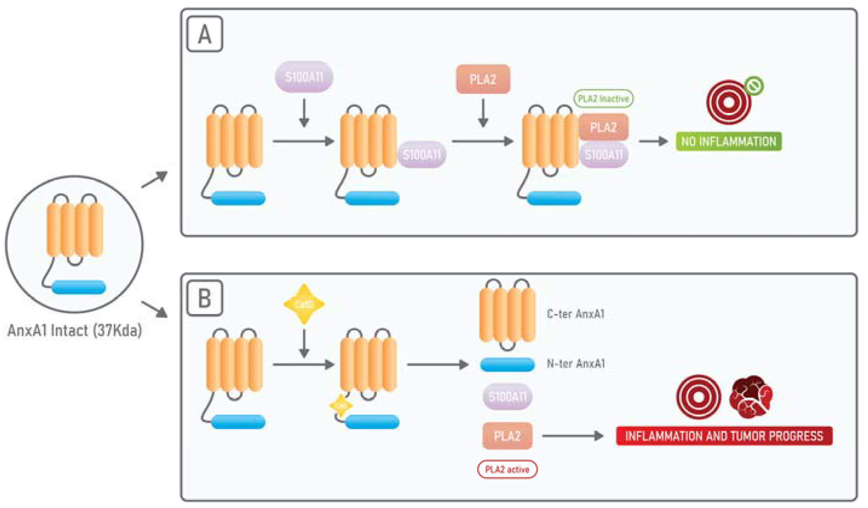
4. Annexin A1: An Endogenous PLA2 Inhibitor

It is well accepted that PLA2 is one of the major players in the establishment of an inflammatory environment and, consequently, it is crucially involved in tumorigenesis and tumor progression [81]. On the other side, a key mediator of the anti-inflammatory response is the 37 kDa protein Annexin A1 (AnxA1). AnxA1 is a phospholipid-binding protein expressed in many tissues and cell types including leukocytes, lymphocytes, endothelial and epithelial cells [110]. AnxA1 is one of the mediators of the anti-inflammatory activity of glucocorticoids (GCs) [111], and exerts its anti-inflammatory activities by inhibiting PLA2 in the cytoplasm [112]. AnxA1 also regulates different processes including membrane trafficking, proliferation, differentiation, and apoptosis [113,114].
AnxA1 can be found in its 37 kDa intact form that displays an anti-PLA2 activity and in two cleaved forms of 33 and 36 kDa. The 33 kDa cleaved form was described to be pro-inflammatory [115,116] whereas the 36 kDa cleaved form was associated with monocyte recruitment and prevention of inflammation [116]. AnxA1 cleavage is due to elastases, metalloproteases, or proteinases and leads to the release of the AnxA1 N-terminal biological active peptide [117] that signals through the “Formyl peptide receptors” (FPRs) [118]. FPRs are Gi protein-coupled receptors involved in the chemotaxis of leukocytes towards the bacterial chemotactic peptide N-formyl-methionyl-leucyl-phenylalanine (fMLF) [119]. By binding to FPRs, N-formylated peptides elicit a signal cascade involving PI3K and MAPK [120,121]. The anti-inflammatory activity of AnxA1 is exerted by binding to the FPR2 that is expressed on fibroblasts, endothelial cells, stromal cells, and is highly abundant in leukocytes [120,122]. The anti-inflammatory activity of the N-terminal peptide of AnxA1 is, however, 20-fold less potent, a fact that raised the hypothesis that such a peptide could display the role of limiting the action of the intact form of AnxA1 [123]. Nevertheless, AnxA1 has a pro-inflammatory role in certain circumstances. In fact, once phosphorylated by PKC, AnxA1 migrates to nuclei where it induces the expression of pro-inflammatory cytokines [124]. Moreover, the 33 kDa cleaved form of AnxA1 generated by Calpain 1 increases the immobilization of neutrophils on endothelial cells thus facilitating the trans-endothelial migration and promoting inflammation [115]. Finally, it has been shown that in cells infected by influenza virus A the N-terminal peptide of AnxA1 enhances the inflammatory response by activating FPR2 [125].
AnxA1 binds, through its N-terminal portion, to S100A11 another calcium-binding protein and, when AnxA1 is bound to S100A11, it displays a high affinity for cPLA2. On the other side, it has been shown that, in squamous carcinoma cells, when there is an exposure to EGF, EGFR phosphorylates AnxA1 in its tyrosine 21 residue and subsequently AnxA1 suffers a proteolytic cleavage at its tryptophan 12 residue by Cathepsin D. Such cleavage results in the dissociation from S100A11 and cPLA2. In this way, the anti-cPLA2 activity of AnxA1 is abolished and cPLA2 can promote tumor growth (Figure 2) [59].

AnxA1 in TME

AnxA1 has been described as an essential player in several aspects of cancer, such as proliferation, chemoresistance, invasion, and metastasis formation [126]. In fact, AnxA1 plays important roles in the progression of several types of tumors, including astrocitomas, glioblastomas, melanomas, and those affecting the lung, breast, and pancreas [127,128]. In breast cancer, it has been described that AnxA1 supports the metastatic process by promoting the TGF-β/Smad signaling and the subsequent EMT [129]. Interestingly AnxA1 can be found in a secreted form in breast, prostate, and pancreatic cancers. This secreted form of AnxA1 elicits an autocrine signaling cascade through FPR1 that stimulates migration and invasion properties of these tumors [130,131]. The pivotal role of FPR1 signaling has also been described for astrocytomas [132] and neuroblastomas [121].
Recently, AnxA1 has been described to play immunosuppressive roles in the cancer context. It has been shown that AnxA1 promotes the polarization of macrophages towards the M2 phenotype and induces the expression of IL-10 thus facilitating breast cancer progression and metastasis [133,134]. In hepatocellular carcinoma (HCC), it has been shown that the AnxA1 N-terminal peptide is responsible for the polarization of macrophages towards the M2 phenotype, by signaling through FPR2 and by eliciting the activation of ERK, Akt, and NFkB [135]. Indeed, a previous study had shown that a deficiency in FPR2 sustains an M1 phenotype in HCC [136]. It was also reported that AnxA1 plays an essential role in the induction of Tregs in the TME of triple-negative breast cancer models [137].

5. PLA2 and Annexin A1 in Cancer-Derived Extracellular Vesicles

EVs are lipid bilayer delimited particles that are released from cells. They can be found in different biological fluids regulating inflammation and tissue repair [138], and modulating the immune response, viral pathogenicity, and cancer progression [139,140]. EVs promote intercellular communication through contacting membranes of target cells or by transferring EVs’ cargos, which can be lipids, proteins, and nucleic acids [141]. EVs are classified into endosomal-derived exosomes and plasma membrane-derived MVs, Exosomes originate from multivesicular bodies (MVBs) which are endosomes that contain intraluminal vesicles (ILVs). When MVBs fuse with the plasmatic membrane ILVs are released as exosomes [139,140]. Cancer cells produce copious amounts of both MVs and exosomes that can be found in all biological fluids altering the phenotype of cells with which they come in contact, promoting a pro-tumoral gene expression [142,143]. Tumor-derived exosomes (TDEs) are involved in the increased proliferation and chemoresistance of cancer cells [144], angiogenesis [145], and metastasis [146]. Moreover, the immunosuppressive roles of exosomes have been described. In this scenario, exosomes induce T cell apoptosis [147], decrease DC differentiation [148], and suppress NK cytotoxic response [149]. MVs have also been linked with several pro-tumoral functions such as proliferation, angiogenesis, metastasis, chemoresistance, and immunomodulation [142]. It has been shown that MVs, due to the presence of TGF-β on their surface, can interact with immune cells inducing NK and T cell suppression [150].
sPLA2 is present in the extracellular milieu either within exosomes or as an exosome-free secreted form [151]. It has been shown that both of these PLA2 forms act on phospholipids present on MVs promoting the release of AA and therefore amplifying the inflammatory process [152,153]. Hence, although exosomes and MVs represent distinct structures, these EVs can cooperate in the induction of the immunosuppression and inflammation of TME (Figure 3).
It has been found that TDEs contain cPLA2, iPLA2, sPLA2, COX-1, and COX-2. Moreover, TDEs are enriched in free fatty acids, including AA, and the immunosuppressive molecule PGE2 [151]. TEDs-associated PGE2 promotes tumorigenesis by increasing the expression of cell death protein-ligand (PDL-1), a molecule responsible for immune escape [154]. In breast cancer, TEDs-associated PGE2 is responsible for the release of pro-inflammatory cytokines that leads to the accumulation of MDSC in TME [155,156]. Regarding AnxA1, it is released within EVs [157,158]. Recently, AnxA1 has started to be considered a specific marker of MVs [159] that is localized on the surface of these structures. However, the mechanism through which this happens remains unresolved [160]. Calcium promotes the interaction of AnxA1 with cellular membranes [161] from which it may be loaded on budding MVs [160]. AnxA1 present in EVs promotes the activation of keratinocytes through the activation of FPRs in an autocrine loop [162], which promotes cancer cell motility [157]. Leoni and collaborators also demonstrated that the inhibition of FPR1 and FPR2 abrogated the pro-healing effect of AnxA1 containing EVs [158]. Moreover, EVs containing AnxA1 secreted by prostate epithelial cells may contribute to the suppression of the immune response into the male tract [163]. Finally, AnxA1 has been also described as being essential for the release of exosomes from MVBs [164,165]. Therefore, AnxA1 can promote the release of PLA2-enriched exosomes from cancer cells, leading to increased PGE2 levels and subsequently to an increase in the inflammatory response and immunosuppression in TME.

6. Use of PLA2 Inhibitors to Control Cancer Progression

The use of non-steroidal anti-inflammatory drugs (NSAIDs) or COX-2 inhibitors (COXIBs) has been widely explored during the last years as cancer prevention and treatment strategies. Studies have shown either a decrease incidence of cancer in chronic users or a decrease in mortality rates in cancer patients treated with these drugs [166,167]. Despite that COXIBs display the advantage of non-inducing toxicity in the gastrointestinal tract, their clinical long-term use is limited by their significant cardiotoxicity. Since such side effect is due to a shunting towards leukotriene production [168], growing evidence has shown that the dual inhibition of COX and LOX enzymes would be an efficient and safer option compared to COXIBs alone [169]. In addition to this, the anti-inflammatory activity of corticosteroids drugs is mediated by AnxA1. Therefore, AnxA1 action could be limited in TME due to the presence of its cleaved and pro-inflammatory form. In fact, AnxA1 cleavage could explain why corticosteroids can display a pro-tumoral or an anti-tumoral effect depending on the type of cancer [170]. However, AnxA1 regulates the functions of PLA2. Hence, PLA2 inhibitors are also interesting as anti-cancer therapeutic strategies. Indeed, sPLA2 inhibition, besides avoiding the release of AA and the subsequent production of PGE2, could also interfere with the non-catalytic activities of PLA2 in signal transduction pathways that support tumor growth and progression.
Recently, it has been suggested that PGE2 enhances cancer cells’ invasion by increasing the expression of adhesion molecules, such as intercellular adhesion molecule-1 (ICAM-1), and increasing the phosphorylation of anti-apoptotic transcription factor “signal transducer and activator of transcription” (STAT-3). Authors achieved a significant reduction in PGE2 and ICAM-1 expression levels, as well as a reduction in STAT-3 phosphorylation levels by inhibiting sPLA2 in lung cancer cells [171,172]. Similarly, the treatment of human esophageal adenocarcinoma cells with sPLA2 inhibitor attenuates the expression of ICAM-1 [173] and decreases viability and proliferation of this type of cancer cell [174]. Therefore, the use of molecules and drugs able to inhibit PLA2 and especially with anti sPLA2 activities could represent a good strategy in order to improve cancer patients’ outcomes.
Varespladib (LY315920) and its orally available form methyl-Varespladib (LY333013) are promising PLA2 inhibitors. Those compounds have been developed by the pharmaceutical industry to treat inflammatory diseases. Both are potent and selective inhibitors of the human sPLA2 and their inhibitory activity on sPLA2 occurs at nano-and picomolar concentrations. Interestingly, these drugs display inhibitory activity against sPLA2s present in 28 snake venoms [175,176]. In animal models, Varespladib and methyl-Varespladib have been shown to inhibit atherogenesis since they demonstrated to significantly decrease total cholesterol and to reduce aneurysm formation [177,178]. However, for the treatment of patients with acute coronary syndrome and other inflammatory diseases, such as sepsis and rheumatoid arthritis, these inhibitors failed to show efficacy [176,179].
Another synthetic sPLA2 inhibitor is the molecule S3319 that, in lung cancer cells, decreases ICAM-1 expression levels and, subsequently, reduces cancer cell invasion [171]. Different studies indicated that sPLA2 could be used as a biomarker and as an important therapeutic target in prostate cancer [180]. The sPLA2 mRNA levels were 22-fold overexpressed in prostate cancer cells when compared to normal cells. Two inhibitors of sPLA2, cFLSYR, and c(2Nap)LS(2Nap)R, proved to be efficient in attenuating the proliferation of sPLA2-positive LNCaP and PC-3 cell lines but not the sPLA2-negative DU145 cell line. Curiously, sPLA2 is overexpressed in androgen-independent prostate cancer PC3 cells when compared to the androgen-dependent LNCaP cell lines. Therefore, PLA2 inhibitors could be used as alternative strategies in the treatment of prostatic tumors and diagnosis of prostate cancer. The use of sPLA2 inhibitors could be of particular interest for those prostate cancers that are positive for sPLA2 and are non-responsive to androgen due to their androgen-independency.
A natural inhibitor of sPLA2-IIA, ochnaflavone, has been shown to strongly inhibit sPLA2-IIA activity and, as a result, it could be an interesting molecule in the treatment of inflammatory diseases and cancer [181]. In human aortic smooth muscle cells, the treatment with ochnaflavone inhibited DNA synthesis, and downregulated cyclins and cyclin-dependent kinases (CDKs) thus leading to G1-phase cell cycle arrest [182,183]. Another potent inhibitor of PLA2 is the marine natural product scalaradial that showed cytotoxic activity on various cancer cell lines, specifically against HepG2, MCF-7, HeLa, and HCT-116 cells [184].
Maslinic acid, a natural pentacyclic triterpenoid, was proved to inhibit the sPLA2 enzyme activity in a concentration-dependent manner. In addition, maslinic acid inhibits the inflammation induced by sPLA2, including PGE2 production and differentiation and migration of inflammatory cells [185,186]. Indeed, maslinic acid induces different anti-cancer effects in multiple tumors like those affecting the breast, prostate, pancreas, kidneys, lungs, and gastro-intestinal tract [187,188].
Sulforaphane, a natural isothiocyanate present in cruciferous vegetables, also showed to potently inhibit the expression and activity of sPLA2. It exhibited chemoprevention properties and therapeutic potential against several types of cancer including oral, prostate, breast, colon, skin, and urinary bladder cancers. In breast cancer, this bioactive compound has been studied extensively as an anti-cancer agent inhibiting the expression of anti-apoptotic genes and inducing G2-M cell cycle arrest by stabilizing microtubules [175].
Several PLA2 inhibitory proteins were purified from the plasma of different species of snakes and are classified into alpha (α), beta (β), and gamma (γ) types, according to their structural features. Thereupon, Gimenes et. al. (2017) demonstrated that γCdcPLI, a sPLA2 inhibitor from Crotalus durissus collilineatus, has anti-tumor, antimetastatic, and anti-angiogenic properties in MDA-MB-231 breast cancer cells [189]. This inhibitor modulates important mediators of the apoptotic pathway and reduces the production of vascular endothelial growth factor (VEGF) [189].
We already pointed that AnxA1 is an endogenous PLA2 inhibitor that can be cleaved by different proteases, including elastase, calpain, plasmin, proteinase 3, and Cathepsin D. Of particular interest is the cleavage of AnxA1 by the soluble lysosomal aspartic endopeptidase (EC 3.4.23.5), Cathepsin D, which is found to be highly expressed in various types of cancers and correlated with metastasis [190]. Cathepsin D cleaves AnxA1 at Trp12 residue and unlocks its binding to S100A11, which is essential in order to bind and inhibit PLA2. Therefore, upon this cleavage AnxA1 is no longer able to inhibit PLA2 [59]. By using the inhibitor of Cathepsin D, Pepstatin A, it was possible to inhibit the amount of cleaved AnxA1, to induce apoptosis, and to decrease the invasion and migration of triple-negative breast cancer cells. These anti-tumorigenic effects were, at least in part, due to resumed inhibition of PLA2 by the intact form of AnxA1 [191].

7. Final Considerations

The activity of PLA2 in TME plays crucial roles in tumor development and progression. PLA2 can be found in TME either in a free form that acts on lipids present in MVs or within exosomes. An endogenous inhibitor of PLA2 is the anti-inflammatory protein, AnxA1. However, once cleaved by proteases, AnxA1 is no longer able to inhibit PLA2 and in this way, it promotes tumor progression. Interestingly, AnxA1 can be found in MVs in its cleaved form that probably supports the action of PLA2 on MVs’ lipids. Therefore, the direct inhibition of PLA2 or the inhibition of AnxA1 cleavage, with the subsequent resumed anti-PLA2 activity, could represent interesting therapeutic strategies in the cancer context. The knowledge presented in this review emphasizes the importance of validating these strategies in order to open the possibility of designing innovative approaches to improve cancer patients’ outcomes.

Author Contributions

L.R.G. conceptualization, writing and revision of the manuscript; L.V.; S.T.S.M.; F.V.P.d.V.A.; V.d.M.R.Á.; T.G.A.; and M.A.R. writing and revision of the manuscript, designing of figures. All authors have read and agreed to the published version of the manuscript.

Funding

This work was supported by funding from the following institutions: FAPEMIG, CNPq, and CAPES (23038007281201161).

Institutional Review Board Statement

Not applicable.

Informed Consent Statement

Not applicable.

Data Availability Statement

No new data were created or analyzed in this study. Data sharing is not applicable to this article.

Conflicts of Interest

The authors declare no conflict of interest. The funders had no role in the design of the study; in the collection, analyses, or interpretation of data; in the writing of the manuscript, or in the decision to publish the results.

References

  1. World Health Organization. Breast Cancer: Prevention and Control. 2018. Available online: https://www.who.int/cancer/detection/breastcancer/en/ (accessed on 23 April 2021).
  2. Siegel, R.L.; Miller, K.D.; Jemal, A. Cancer statistics, 2020. CA Cancer J. Clin. 2020, 70, 7–30. [Google Scholar] [CrossRef]
  3. Fouad, Y.A.; Aanei, C. Revisiting the hallmarks of cancer. Am. J. Cancer Res. 2017, 7, 1016–1036. [Google Scholar]
  4. Hanahan, D.; Weinberg, R.A. Hallmarks of cancer: The next generation. Cell 2011, 144, 646–674. [Google Scholar] [CrossRef] [Green Version]
  5. Biswas, S.; Rao, C.M. Epigenetics in cancer: Fundamentals and Beyond. Pharm. Ther. 2017, 173, 118–134. [Google Scholar] [CrossRef]
  6. Vogelstein, B.; Papadopoulos, N.; Velculescu, V.E.; Zhou, S.; Diaz, L.A., Jr.; Kinzler, K.W. Cancer genome landscapes. Science 2013, 339, 1546–1558. [Google Scholar] [CrossRef] [PubMed]
  7. Cleary, A.S.; Leonard, T.L.; Gestl, S.A.; Gunther, E.J. Tumour cell heterogeneity maintained by cooperating subclones in Wnt-driven mammary cancers. Nature 2014, 508, 113–117. [Google Scholar] [CrossRef] [PubMed]
  8. Nowell, P.C. The clonal evolution of tumor cell populations. Science 1976, 194, 23–28. [Google Scholar] [CrossRef] [PubMed]
  9. Baca, S.C.; Prandi, D.; Lawrence, M.S.; Mosquera, J.M.; Romanel, A.; Drier, Y.; Park, K.; Kitabayashi, N.; MacDonald, T.Y.; Ghandi, M.; et al. Punctuated evolution of prostate cancer genomes. Cell 2013, 153, 666–677. [Google Scholar] [CrossRef] [PubMed] [Green Version]
  10. Anderson, N.M.; Simon, M.C. The tumor microenvironment. Curr. Biol. 2020, 30, R921–R925. [Google Scholar] [CrossRef]
  11. Garcia-Gomez, A.; Rodríguez-Ubreva, J.; Ballestar, E. Epigenetic interplay between immune, stromal and cancer cells in the tumor microenvironment. Clin. Immunol. 2018, 196, 64–71. [Google Scholar] [CrossRef]
  12. Wu, T.; Dai, Y. Tumor microenvironment and therapeutic response. Cancer Lett. 2017, 387, 61–68. [Google Scholar] [CrossRef]
  13. Mohan, V.; Das, A.; Sagi, I. Emerging roles of ECM remodeling processes in cancer. Semin. Cancer Biol. 2020, 62, 192–200. [Google Scholar] [CrossRef]
  14. Erdogan, B.; Webb, D.J. Cancer-associated fibroblasts modulate growth factor signaling and extracellular matrix remodeling to regulate tumor metastasis. Biochem. Soc. Trans. 2017, 45, 229–236. [Google Scholar] [CrossRef] [Green Version]
  15. Rice, A.J.; Cortes, E.; Lachowski, D.; Cheung, B.C.H.; Karim, S.A.; Morton, J.P.; Del Rio Hernandez, A. Matrix stiffness induces epithelial-mesenchymal transition and promotes chemoresistance in pancreatic cancer cells. Oncogenesis 2017, 6, e352. [Google Scholar] [CrossRef] [Green Version]
  16. Lambert, A.W.; Pattabiraman, D.R.; Weinberg, R.A. Emerging Biological Principles of Metastasis. Cell 2017, 168, 670–691. [Google Scholar] [CrossRef] [PubMed] [Green Version]
  17. De Palma, M.; Biziato, D.; Petrova, T.V. Microenvironmental regulation of tumour angiogenesis. Nat. Rev. Cancer 2017, 17, 457–474. [Google Scholar] [CrossRef] [PubMed]
  18. Carmeliet, P.; Jain, R.K. Angiogenesis in cancer and other diseases. Nature 2000, 407, 249–257. [Google Scholar] [CrossRef] [PubMed]
  19. Lei, X.; Lei, Y.; Li, J.-K.; Du, W.-X.; Li, R.-G.; Yang, J.; Li, J.; Li, F.; Tan, H.-B. Immune cells within the tumor microenvironment: Biological functions and roles in cancer immunotherapy. Cancer Lett. 2020, 470, 126–133. [Google Scholar] [CrossRef]
  20. Galli, F.; Aguilera, J.V.; Palermo, B.; Markovic, S.N.; Nistico, P.; Signore, A. Relevance of immune cell and tumor microenvironment imaging in the new era of immunotherapy. J. Exp. Clin. Cancer Res. 2020, 39, 89. [Google Scholar] [CrossRef]
  21. Zhang, Y.; Ertl, H.C. Starved and Asphyxiated: How Can CD8(+) T Cells within a Tumor Microenvironment Prevent Tumor Progression. Front. Immunol. 2016, 7, 32. [Google Scholar] [CrossRef] [Green Version]
  22. Topalian, S.L.; Drake, C.G.; Pardoll, D.M. Immune checkpoint blockade: A common denominator approach to cancer therapy. Cancer Cell 2015, 27, 450–461. [Google Scholar] [CrossRef] [Green Version]
  23. Johnson, A.M.; Kleczko, E.K.; Nemenoff, R.A. Eicosanoids in Cancer: New Roles in Immunoregulation. Front. Pharm. 2020, 11, 595498. [Google Scholar] [CrossRef] [PubMed]
  24. Corn, K.C.; Windham, M.A.; Rafat, M. Lipids in the tumor microenvironment: From cancer progression to treatment. Prog. Lipid Res. 2020, 80, 101055. [Google Scholar] [CrossRef]
  25. Tang, S.; Ning, Q.; Yang, L.; Mo, Z.; Tang, S. Mechanisms of immune escape in the cancer immune cycle. Int. Immunopharmacol. 2020, 86, 106700. [Google Scholar] [CrossRef]
  26. Bretscher, P. On Analyzing How the Th1/Th2 Phenotype of an Immune Response Is Determined: Classical Observations Must Not Be Ignored. Front. Immunol. 2019, 10, 1024. [Google Scholar] [CrossRef] [PubMed] [Green Version]
  27. Zambrano-Zaragoza, J.F.; Romo-Martinez, E.J.; Duran-Avelar Mde, J.; Garcia-Magallanes, N.; Vibanco-Perez, N. Th17 cells in autoimmune and infectious diseases. Int. J. Inflam. 2014, 2014, 651503. [Google Scholar] [CrossRef]
  28. Kryczek, I.; Wei, S.; Szeliga, W.; Vatan, L.; Zou, W. Endogenous IL-17 contributes to reduced tumor growth and metastasis. Blood 2009, 114, 357–359. [Google Scholar] [CrossRef] [PubMed] [Green Version]
  29. Zou, W.; Restifo, N.P. T(H)17 cells in tumour immunity and immunotherapy. Nat. Rev. Immunol. 2010, 10, 248–256. [Google Scholar] [CrossRef]
  30. Williams, M.A.; Bevan, M.J. Effector and memory CTL differentiation. Annu. Rev. Immunol. 2007, 25, 171–192. [Google Scholar] [CrossRef] [PubMed]
  31. Jost, S.; Altfeld, M. Control of human viral infections by natural killer cells. Annu. Rev. Immunol. 2013, 31, 163–194. [Google Scholar] [CrossRef]
  32. Vivier, E.; Raulet, D.H.; Moretta, A.; Caligiuri, M.A.; Zitvogel, L.; Lanier, L.L.; Yokoyama, W.M.; Ugolini, S. Innate or adaptive immunity? The example of natural killer cells. Science 2011, 331, 44–49. [Google Scholar] [CrossRef] [PubMed] [Green Version]
  33. Vander Heiden, M.G.; Cantley, L.C.; Thompson, C.B. Understanding the Warburg effect: The metabolic requirements of cell proliferation. Science 2009, 324, 1029–1033. [Google Scholar] [CrossRef] [PubMed] [Green Version]
  34. Luo, Y.; Zheng, S.G. Hall of Fame among Pro-inflammatory Cytokines: Interleukin-6 Gene and Its Transcriptional Regulation Mechanisms. Front. Immunol. 2016, 7, 604. [Google Scholar] [CrossRef]
  35. Gabrilovich, D. Mechanisms and functional significance of tumour-induced dendritic-cell defects. Nat. Rev. Immunol. 2004, 4, 941–952. [Google Scholar] [CrossRef] [PubMed]
  36. Melief, C.J. Cancer immunotherapy by dendritic cells. Immunity 2008, 29, 372–383. [Google Scholar] [CrossRef] [PubMed] [Green Version]
  37. Budhu, S.; Schaer, D.A.; Li, Y.; Toledo-Crow, R.; Panageas, K.; Yang, X.; Zhong, H.; Houghton, A.N.; Silverstein, S.C.; Merghoub, T.; et al. Blockade of surface-bound TGF-beta on regulatory T cells abrogates suppression of effector T cell function in the tumor microenvironment. Sci. Signal. 2017, 10. [Google Scholar] [CrossRef] [Green Version]
  38. Nishikawa, H.; Sakaguchi, S. Regulatory T cells in tumor immunity. Int. J. Cancer 2010, 127, 759–767. [Google Scholar] [CrossRef]
  39. Ito, T.; Wang, Y.H.; Duramad, O.; Hanabuchi, S.; Perng, O.A.; Gilliet, M.; Qin, F.X.; Liu, Y.J. OX40 ligand shuts down IL-10-producing regulatory T cells. Proc. Natl. Acad. Sci. USA 2006, 103, 13138–13143. [Google Scholar] [CrossRef] [Green Version]
  40. Weber, F.; Byrne, S.N.; Le, S.; Brown, A.; Breit, S.N.; Scolyer, R.A.; Halliday, G.M. Transforming growth factor-beta1 immobilises dendritic cells within skin tumours and facilitates tumour escape from the immune system. Cancer Immunol. Immunother. 2005, 54, 898–906. [Google Scholar] [CrossRef]
  41. Ruffell, B.; Affara, N.I.; Coussens, L.M. Differential macrophage programming in the tumor microenvironment. Trends Immunol. 2012, 33, 119–126. [Google Scholar] [CrossRef] [PubMed] [Green Version]
  42. Chen, J.J.; Lin, Y.C.; Yao, P.L.; Yuan, A.; Chen, H.Y.; Shun, C.T.; Tsai, M.F.; Chen, C.H.; Yang, P.C. Tumor-associated macrophages: The double-edged sword in cancer progression. J. Clin. Oncol. 2005, 23, 953–964. [Google Scholar] [CrossRef]
  43. Sica, A.; Allavena, P.; Mantovani, A. Cancer related inflammation: The macrophage connection. Cancer Lett. 2008, 267, 204–215. [Google Scholar] [CrossRef]
  44. Mantovani, A.; Schioppa, T.; Porta, C.; Allavena, P.; Sica, A. Role of tumor-associated macrophages in tumor progression and invasion. Cancer Metastasis. Rev. 2006, 25, 315–322. [Google Scholar] [CrossRef]
  45. Schoppmann, S.F.; Birner, P.; Stockl, J.; Kalt, R.; Ullrich, R.; Caucig, C.; Kriehuber, E.; Nagy, K.; Alitalo, K.; Kerjaschki, D. Tumor-associated macrophages express lymphatic endothelial growth factors and are related to peritumoral lymphangiogenesis. Am. J. Pathol. 2002, 161, 947–956. [Google Scholar] [CrossRef] [Green Version]
  46. Iijima, J.; Konno, K.; Itano, N. Inflammatory alterations of the extracellular matrix in the tumor microenvironment. Cancers 2011, 3, 3189–3205. [Google Scholar] [CrossRef] [PubMed] [Green Version]
  47. Kryczek, I.; Banerjee, M.; Cheng, P.; Vatan, L.; Szeliga, W.; Wei, S.; Huang, E.; Finlayson, E.; Simeone, D.; Welling, T.H.; et al. Phenotype, distribution, generation, and functional and clinical relevance of Th17 cells in the human tumor environments. Blood 2009, 114, 1141–1149. [Google Scholar] [CrossRef] [PubMed] [Green Version]
  48. Miyahara, Y.; Odunsi, K.; Chen, W.; Peng, G.; Matsuzaki, J.; Wang, R.F. Generation and regulation of human CD4+ IL-17-producing T cells in ovarian cancer. Proc. Natl. Acad. Sci. USA 2008, 105, 15505–15510. [Google Scholar] [CrossRef] [Green Version]
  49. Guery, L.; Hugues, S. Th17 Cell Plasticity and Functions in Cancer Immunity. BioMed Res. Int. 2015, 2015, 314620. [Google Scholar] [CrossRef] [Green Version]
  50. Veglia, F.; Perego, M.; Gabrilovich, D. Myeloid-derived suppressor cells coming of age. Nat. Immunol. 2018, 19, 108–119. [Google Scholar] [CrossRef]
  51. Bunt, S.K.; Sinha, P.; Clements, V.K.; Leips, J.; Ostrand-Rosenberg, S. Inflammation induces myeloid-derived suppressor cells that facilitate tumor progression. J. Immunol. 2006, 176, 284–290. [Google Scholar] [CrossRef] [Green Version]
  52. Marvel, D.; Gabrilovich, D.I. Myeloid-derived suppressor cells in the tumor microenvironment: Expect the unexpected. J. Clin. Investig. 2015, 125, 3356–3364. [Google Scholar] [CrossRef]
  53. Gabrilovich, D.I.; Nagaraj, S. Myeloid-derived suppressor cells as regulators of the immune system. Nat. Rev. Immunol. 2009, 9, 162–174. [Google Scholar] [CrossRef]
  54. Lindau, D.; Gielen, P.; Kroesen, M.; Wesseling, P.; Adema, G.J. The immunosuppressive tumour network: Myeloid-derived suppressor cells, regulatory T cells and natural killer T cells. Immunology 2013, 138, 105–115. [Google Scholar] [CrossRef]
  55. Huang, B.; Pan, P.Y.; Li, Q.; Sato, A.I.; Levy, D.E.; Bromberg, J.; Divino, C.M.; Chen, S.H. Gr-1+CD115+ immature myeloid suppressor cells mediate the development of tumor-induced T regulatory cells and T-cell anergy in tumor-bearing host. Cancer Res. 2006, 66, 1123–1131. [Google Scholar] [CrossRef] [Green Version]
  56. Zippelius, A.; Batard, P.; Rubio-Godoy, V.; Bioley, G.; Lienard, D.; Lejeune, F.; Rimoldi, D.; Guillaume, P.; Meidenbauer, N.; Mackensen, A.; et al. Effector function of human tumor-specific CD8 T cells in melanoma lesions: A state of local functional tolerance. Cancer Res. 2004, 64, 2865–2873. [Google Scholar] [CrossRef] [Green Version]
  57. Chemnitz, J.M.; Parry, R.V.; Nichols, K.E.; June, C.H.; Riley, J.L. SHP-1 and SHP-2 associate with immunoreceptor tyrosine-based switch motif of programmed death 1 upon primary human T cell stimulation, but only receptor ligation prevents T cell activation. J. Immunol. 2004, 173, 945–954. [Google Scholar] [CrossRef]
  58. Driessens, G.; Kline, J.; Gajewski, T.F. Costimulatory and coinhibitory receptors in anti-tumor immunity. Immunol. Rev. 2009, 229, 126–144. [Google Scholar] [CrossRef] [PubMed]
  59. Sakaguchi, M.; Murata, H.; Sonegawa, H.; Sakaguchi, Y.; Futami, J.; Kitazoe, M.; Yamada, H.; Huh, N.H. Truncation of annexin A1 is a regulatory lever for linking epidermal growth factor signaling with cytosolic phospholipase A2 in normal and malignant squamous epithelial cells. J. Biol. Chem. 2007, 282, 35679–35686. [Google Scholar] [CrossRef] [PubMed] [Green Version]
  60. Dennis, E.A. Introduction to Thematic Review Series: Phospholipases: Central Role in Lipid Signaling and Disease. J. Lipid Res. 2015, 56, 1245–1247. [Google Scholar] [CrossRef] [PubMed] [Green Version]
  61. Wilton, D.C. CHAPTER 11—Phospholipases. In Biochemistry of Lipids, Lipoproteins and Membranes, 5th ed.; Vance, D.E., Vance, J.E., Eds.; Elsevier: San Diego, CA, USA, 2008; pp. 305–329. [Google Scholar]
  62. Aloulou, A.; Rahier, R.; Arhab, Y.; Noiriel, A.; Abousalham, A. Phospholipases: An Overview. Methods Mol. Biol. 2018, 1835, 69–105. [Google Scholar]
  63. Brown, W.J.; Chambers, K.; Doody, A. Phospholipase A2 (PLA2) enzymes in membrane trafficking: Mediators of membrane shape and function. Traffic 2003, 4, 214–221. [Google Scholar] [CrossRef]
  64. Azevedo, F.V.; Lopes, D.S.; Cirilo Gimenes, S.N.; Ache, D.C.; Vecchi, L.; Alves, P.T.; Guimaraes Dde, O.; Rodrigues, R.S.; Goulart, L.R.; Rodrigues, V.d.M.; et al. Human breast cancer cell death induced by BnSP-6, a Lys-49 PLA(2) homologue from Bothrops pauloensis venom. Int. J. Biol. Macromol. 2016, 82, 671–677. [Google Scholar] [CrossRef] [PubMed]
  65. de Vasconcelos Azevedo, F.V.P.; Zoia, M.A.P.; Lopes, D.S.; Gimenes, S.N.; Vecchi, L.; Alves, P.T.; Rodrigues, R.S.; Silva, A.C.A.; Yoneyama, K.A.G.; Goulart, L.R.; et al. Antitumor and antimetastatic effects of PLA2-BthTX-II from Bothrops jararacussu venom on human breast cancer cells. Int. J. Biol. Macromol. 2019, 135, 261–273. [Google Scholar] [CrossRef]
  66. Burke, J.E.; Dennis, E.A. Phospholipase A2 biochemistry. Cardiovasc. Drugs Ther. 2009, 23, 49–59. [Google Scholar] [CrossRef] [PubMed] [Green Version]
  67. Ilic, D.; Bollinger, J.M.; Gelb, M.; Mauro, T.M. sPLA2 and the epidermal barrier. Biochim. Biophys. Acta 2014, 1841, 416–421. [Google Scholar] [CrossRef] [PubMed] [Green Version]
  68. Murakami, M.; Sato, H.; Taketomi, Y. Updating Phospholipase A2 Biology. Biomolecules 2020, 10, 1457. [Google Scholar] [CrossRef] [PubMed]
  69. Slatter, D.A.; Aldrovandi, M.; O’Connor, A.; Allen, S.M.; Brasher, C.J.; Murphy, R.C.; Mecklemann, S.; Ravi, S.; Darley-Usmar, V.; O’Donnell, V.B. Mapping the Human Platelet Lipidome Reveals Cytosolic Phospholipase A2 as a Regulator of Mitochondrial Bioenergetics during Activation. Cell Metab. 2016, 23, 930–944. [Google Scholar] [CrossRef] [PubMed] [Green Version]
  70. Ishii, I.; Fukushima, N.; Ye, X.; Chun, J. Lysophospholipid receptors: Signaling and biology. Annu. Rev. Biochem. 2004, 73, 321–354. [Google Scholar] [CrossRef] [PubMed] [Green Version]
  71. Meyer zu Heringdorf, D.; Jakobs, K.H. Lysophospholipid receptors: Signalling, pharmacology and regulation by lysophospholipid metabolism. Biochim. Biophys. Acta 2007, 1768, 923–940. [Google Scholar] [CrossRef] [Green Version]
  72. Law, S.H.; Chan, M.L.; Marathe, G.K.; Parveen, F.; Chen, C.H.; Ke, L.Y. An Updated Review of Lysophosphatidylcholine Metabolism in Human Diseases. Int. J. Mol. Sci. 2019, 20, 1149. [Google Scholar] [CrossRef] [Green Version]
  73. Tokumura, A.; Majima, E.; Kariya, Y.; Tominaga, K.; Kogure, K.; Yasuda, K.; Fukuzawa, K. Identification of human plasma lysophospholipase D, a lysophosphatidic acid-producing enzyme, as autotaxin, a multifunctional phosphodiesterase. J. Biol. Chem. 2002, 277, 39436–39442. [Google Scholar] [CrossRef] [PubMed] [Green Version]
  74. Benesch, M.G.; Ko, Y.M.; McMullen, T.P.; Brindley, D.N. Autotaxin in the crosshairs: Taking aim at cancer and other inflammatory conditions. FEBS Lett. 2014, 588, 2712–2727. [Google Scholar] [CrossRef] [PubMed] [Green Version]
  75. Smith, W.L.; DeWitt, D.L.; Garavito, R.M. Cyclooxygenases: Structural, cellular, and molecular biology. Annu. Rev. Biochem. 2000, 69, 145–182. [Google Scholar] [CrossRef] [PubMed] [Green Version]
  76. Smith, W.L.; Urade, Y.; Jakobsson, P.J. Enzymes of the cyclooxygenase pathways of prostanoid biosynthesis. Chem. Rev. 2011, 111, 5821–5865. [Google Scholar] [CrossRef] [PubMed] [Green Version]
  77. Wang, B.; Wu, L.; Chen, J.; Dong, L.; Chen, C.; Wen, Z.; Hu, J.; Fleming, I.; Wang, D.W. Metabolism pathways of arachidonic acids: Mechanisms and potential therapeutic targets. Signal. Transduct. Target. Ther. 2021, 6, 94. [Google Scholar] [CrossRef] [PubMed]
  78. Six, D.A.; Dennis, E.A. The expanding superfamily of phospholipase A(2) enzymes: Classification and characterization. Biochim. Biophys. Acta 2000, 1488, 1–19. [Google Scholar] [CrossRef]
  79. Schaloske, R.H.; Dennis, E.A. The phospholipase A2 superfamily and its group numbering system. Biochim. Biophys. Acta 2006, 1761, 1246–1259. [Google Scholar] [CrossRef]
  80. Leslie, C.C. Cytosolic phospholipase A(2): Physiological function and role in disease. J. Lipid Res. 2015, 56, 1386–1402. [Google Scholar] [CrossRef] [Green Version]
  81. Brglez, V.; Lambeau, G.; Petan, T. Secreted phospholipases A2 in cancer: Diverse mechanisms of action. Biochimie 2014, 107, 114–123. [Google Scholar] [CrossRef]
  82. Cummings, B.S.; McHowat, J.; Schnellmann, R.G. Phospholipase A(2)s in cell injury and death. J. Pharmacol. Exp. Ther. 2000, 294, 793–799. [Google Scholar]
  83. Valentin, E.; Lambeau, G. Increasing molecular diversity of secreted phospholipases A(2) and their receptors and binding proteins. Biochim. Biophys. Acta 2000, 1488, 59–70. [Google Scholar] [CrossRef]
  84. Hunter, K.W.; Crawford, N.P.; Alsarraj, J. Mechanisms of metastasis. Breast Cancer Res. BCR 2008, 10, S2. [Google Scholar] [CrossRef] [Green Version]
  85. Scott, K.F.; Sajinovic, M.; Hein, J.; Nixdorf, S.; Galettis, P.; Liauw, W.; de Souza, P.; Dong, Q.; Graham, G.G.; Russell, P.J. Emerging roles for phospholipase A2 enzymes in cancer. Biochimie 2010, 92, 601–610. [Google Scholar] [CrossRef]
  86. Peng, Z.; Chang, Y.; Fan, J.; Ji, W.; Su, C. Phospholipase A2 superfamily in cancer. Cancer Lett. 2021, 497, 165–177. [Google Scholar] [CrossRef] [PubMed]
  87. Jiang, J.; Neubauer, B.L.; Graff, J.R.; Chedid, M.; Thomas, J.E.; Roehm, N.W.; Zhang, S.; Eckert, G.J.; Koch, M.O.; Eble, J.N.; et al. Expression of group IIA secretory phospholipase A2 is elevated in prostatic intraepithelial neoplasia and adenocarcinoma. Am. J. Pathol. 2002, 160, 667–671. [Google Scholar] [CrossRef] [Green Version]
  88. Lu, S.; Dong, Z. Overexpression of secretory phospholipase A2-IIa supports cancer stem cell phenotype via HER/ERBB-elicited signaling in lung and prostate cancer cells. Int. J. Oncol. 2017, 50, 2113–2122. [Google Scholar] [CrossRef]
  89. Sved, P.; Scott, K.F.; McLeod, D.; King, N.J.; Singh, J.; Tsatralis, T.; Nikolov, B.; Boulas, J.; Nallan, L.; Gelb, M.H.; et al. Oncogenic action of secreted phospholipase A2 in prostate cancer. Cancer Res. 2004, 64, 6934–6940. [Google Scholar] [CrossRef] [PubMed] [Green Version]
  90. Dong, Y.; Lu, B.; Zhang, X.; Zhang, J.; Lai, L.; Li, D.; Wu, Y.; Song, Y.; Luo, J.; Pang, X.; et al. Cucurbitacin E, a tetracyclic triterpenes compound from Chinese medicine, inhibits tumor angiogenesis through VEGFR2-mediated Jak2-STAT3 signaling pathway. Carcinogenesis 2010, 31, 2097–2104. [Google Scholar] [CrossRef] [Green Version]
  91. Oleksowicz, L.; Liu, Y.; Bracken, R.B.; Gaitonde, K.; Burke, B.; Succop, P.; Levin, L.; Dong, Z.; Lu, S. Secretory phospholipase A2-IIa is a target gene of the HER/HER2-elicited pathway and a potential plasma biomarker for poor prognosis of prostate cancer. Prostate 2012, 72, 1140–1149. [Google Scholar] [CrossRef] [PubMed] [Green Version]
  92. Miki, Y.; Kidoguchi, Y.; Sato, M.; Taketomi, Y.; Taya, C.; Muramatsu, K.; Gelb, M.H.; Yamamoto, K.; Murakami, M. Dual Roles of Group IID Phospholipase A2 in Inflammation and Cancer. J. Biol. Chem. 2016, 291, 15588–15601. [Google Scholar] [CrossRef] [Green Version]
  93. Caiazza, F.; McCarthy, N.S.; Young, L.; Hill, A.D.; Harvey, B.J.; Thomas, W. Cytosolic phospholipase A2-alpha expression in breast cancer is associated with EGFR expression and correlates with an adverse prognosis in luminal tumours. Br. J. Cancer 2011, 104, 338–344. [Google Scholar] [CrossRef] [Green Version]
  94. Wendum, D.; Svrcek, M.; Rigau, V.; Boelle, P.Y.; Sebbagh, N.; Parc, R.; Masliah, J.; Trugnan, G.; Flejou, J.F. COX-2, inflammatory secreted PLA2, and cytoplasmic PLA2 protein expression in small bowel adenocarcinomas compared with colorectal adenocarcinomas. Mod. Pathol. 2003, 16, 130–136. [Google Scholar] [CrossRef] [Green Version]
  95. Patel, M.I.; Singh, J.; Niknami, M.; Kurek, C.; Yao, M.; Lu, S.; Maclean, F.; King, N.J.; Gelb, M.H.; Scott, K.F.; et al. Cytosolic phospholipase A2-alpha: A potential therapeutic target for prostate cancer. Clin. Cancer Res. 2008, 14, 8070–8079. [Google Scholar] [CrossRef] [Green Version]
  96. Weiser-Evans, M.C.; Wang, X.Q.; Amin, J.; Van Putten, V.; Choudhary, R.; Winn, R.A.; Scheinman, R.; Simpson, P.; Geraci, M.W.; Nemenoff, R.A. Depletion of cytosolic phospholipase A2 in bone marrow-derived macrophages protects against lung cancer progression and metastasis. Cancer Res. 2009, 69, 1733–1738. [Google Scholar] [CrossRef] [Green Version]
  97. Li, H.; Zhao, Z.; Wei, G.; Yan, L.; Wang, D.; Zhang, H.; Sandusky, G.E.; Turk, J.; Xu, Y. Group VIA phospholipase A2 in both host and tumor cells is involved in ovarian cancer development. FASEB J. 2010, 24, 4103–4116. [Google Scholar] [CrossRef] [Green Version]
  98. Xu, Y.; Xiao, Y.J.; Zhu, K.; Baudhuin, L.M.; Lu, J.; Hong, G.; Kim, K.S.; Cristina, K.L.; Song, L.; Williams, F.S.; et al. Unfolding the pathophysiological role of bioactive lysophospholipids. Endocr. Metabol. Disord. 2003, 3, 23–32. [Google Scholar]
  99. Cai, Q.; Zhao, Z.; Antalis, C.; Yan, L.; Del Priore, G.; Hamed, A.H.; Stehman, F.B.; Schilder, J.M.; Xu, Y. Elevated and secreted phospholipase A(2) activities as new potential therapeutic targets in human epithelial ovarian cancer. FASEB J. 2012, 26, 3306–3320. [Google Scholar] [CrossRef] [PubMed] [Green Version]
  100. Wang, D.; Dubois, R.N. Eicosanoids and cancer. Nat. Rev. Cancer 2010, 10, 181–193. [Google Scholar] [CrossRef]
  101. Rodriguez-Vita, J.; Lawrence, T. The resolution of inflammation and cancer. Cytokine Growth Factor Rev. 2010, 21, 61–65. [Google Scholar] [CrossRef]
  102. Holt, D.M.; Ma, X.; Kundu, N.; Collin, P.D.; Fulton, A.M. Modulation of host natural killer cell functions in breast cancer via prostaglandin E2 receptors EP2 and EP4. J. Immunother. 2012, 35, 179–188. [Google Scholar] [CrossRef] [PubMed] [Green Version]
  103. Korkaya, H.; Liu, S.; Wicha, M.S. Breast cancer stem cells, cytokine networks, and the tumor microenvironment. J. Clin. Investig. 2011, 121, 3804–3809. [Google Scholar] [CrossRef]
  104. Liu, Z.; Li, Z.; Mao, K.; Zou, J.; Wang, Y.; Tao, Z.; Lin, G.; Tian, L.; Ji, Y.; Wu, X.; et al. Dec2 promotes Th2 cell differentiation by enhancing IL-2R signaling. J. Immunol. 2009, 183, 6320–6329. [Google Scholar] [CrossRef] [PubMed] [Green Version]
  105. Sinha, P.; Clements, V.K.; Fulton, A.M.; Ostrand-Rosenberg, S. Prostaglandin E2 promotes tumor progression by inducing myeloid-derived suppressor cells. Cancer Res. 2007, 67, 4507–4513. [Google Scholar] [CrossRef] [PubMed] [Green Version]
  106. Baratelli, F.; Lin, Y.; Zhu, L.; Yang, S.C.; Heuze-Vourc’h, N.; Zeng, G.; Reckamp, K.; Dohadwala, M.; Sharma, S.; Dubinett, S.M. Prostaglandin E2 induces FOXP3 gene expression and T regulatory cell function in human CD4+ T cells. J. Immunol. 2005, 175, 1483–1490. [Google Scholar] [CrossRef] [Green Version]
  107. Liu, L.; Ge, D.; Ma, L.; Mei, J.; Liu, S.; Zhang, Q.; Ren, F.; Liao, H.; Pu, Q.; Wang, T.; et al. Interleukin-17 and prostaglandin E2 are involved in formation of an M2 macrophage-dominant microenvironment in lung cancer. J. Thorac. Oncol. 2012, 7, 1091–1100. [Google Scholar] [CrossRef] [Green Version]
  108. Mathew, D.; Kremer, K.N.; Strauch, P.; Tigyi, G.; Pelanda, R.; Torres, R.M. LPA5 Is an Inhibitory Receptor That Suppresses CD8 T-Cell Cytotoxic Function via Disruption of Early TCR Signaling. Front. Immunol. 2019, 10, 1159. [Google Scholar] [CrossRef] [PubMed] [Green Version]
  109. Feng, Y.; Xiao, M.; Zhang, Z.; Cui, R.; Jiang, X.; Wang, S.; Bai, H.; Liu, C.; Zhang, Z. Potential interaction between lysophosphatidic acid and tumor-associated macrophages in ovarian carcinoma. J. Inflamm. 2020, 17, 23. [Google Scholar] [CrossRef]
  110. Kamal, A.M.; Flower, R.J.; Perretti, M. An overview of the effects of annexin 1 on cells involved in the inflammatory process. Mem. Inst. Oswaldo. Cruz. 2005, 100, 39–47. [Google Scholar] [CrossRef] [Green Version]
  111. Yang, Y.H.; Morand, E.; Leech, M. Annexin A1: Potential for glucocorticoid sparing in RA. Nat. Rev. Rheumatol. 2013, 9, 595–603. [Google Scholar] [CrossRef] [PubMed]
  112. Sheikh, M.H.; Solito, E. Annexin A1: Uncovering the Many Talents of an Old Protein. Int. J. Mol. Sci. 2018, 19, 1045. [Google Scholar] [CrossRef] [Green Version]
  113. Barbosa, C.M.V.; Fock, R.A.; Hastreiter, A.A.; Reutelingsperger, C.; Perretti, M.; Paredes-Gamero, E.J.; Farsky, S.H.P. Extracellular annexin-A1 promotes myeloid/granulocytic differentiation of hematopoietic stem/progenitor cells via the Ca(2+)/MAPK signalling transduction pathway. Cell Death Discov. 2019, 5, 135. [Google Scholar] [CrossRef] [Green Version]
  114. Bizzarro, V.; Fontanella, B.; Franceschelli, S.; Pirozzi, M.; Christian, H.; Parente, L.; Petrella, A. Role of Annexin A1 in mouse myoblast cell differentiation. J. Cell Physiol. 2010, 224, 757–765. [Google Scholar] [CrossRef]
  115. Williams, S.L.; Milne, I.R.; Bagley, C.J.; Gamble, J.R.; Vadas, M.A.; Pitson, S.M.; Khew-Goodall, Y. A proinflammatory role for proteolytically cleaved annexin A1 in neutrophil transendothelial migration. J. Immunol. 2010, 185, 3057–3063. [Google Scholar] [CrossRef] [Green Version]
  116. Blume, K.E.; Soeroes, S.; Keppeler, H.; Stevanovic, S.; Kretschmer, D.; Rautenberg, M.; Wesselborg, S.; Lauber, K. Cleavage of annexin A1 by ADAM10 during secondary necrosis generates a monocytic “find-me” signal. J. Immunol. 2012, 188, 135–145. [Google Scholar] [CrossRef] [PubMed]
  117. Rescher, U.; Goebeler, V.; Wilbers, A.; Gerke, V. Proteolytic cleavage of annexin 1 by human leukocyte elastase. Biochim. Biophys. Acta 2006, 1763, 1320–1324. [Google Scholar] [CrossRef] [PubMed] [Green Version]
  118. Gavins, F.N.; Yona, S.; Kamal, A.M.; Flower, R.J.; Perretti, M. Leukocyte antiadhesive actions of annexin 1: ALXR- and FPR-related anti-inflammatory mechanisms. Blood 2003, 101, 4140–4147. [Google Scholar] [CrossRef] [Green Version]
  119. Le, Y.; Murphy, P.M.; Wang, J.M. Formyl-peptide receptors revisited. Trends Immunol. 2002, 23, 541–548. [Google Scholar] [CrossRef]
  120. Cattaneo, F.; Parisi, M.; Ammendola, R. Distinct signaling cascades elicited by different formyl peptide receptor 2 (FPR2) agonists. Int. J. Mol. Sci. 2013, 14, 7193–7230. [Google Scholar] [CrossRef] [Green Version]
  121. Snapkov, I.; Oqvist, C.O.; Figenschau, Y.; Kogner, P.; Johnsen, J.I.; Sveinbjornsson, B. The role of formyl peptide receptor 1 (FPR1) in neuroblastoma tumorigenesis. BMC Cancer 2016, 16, 490. [Google Scholar] [CrossRef] [Green Version]
  122. Dufton, N.; Hannon, R.; Brancaleone, V.; Dalli, J.; Patel, H.B.; Gray, M.; D’Acquisto, F.; Buckingham, J.C.; Perretti, M.; Flower, R.J. Anti-inflammatory role of the murine formyl-peptide receptor 2: Ligand-specific effects on leukocyte responses and experimental inflammation. J. Immunol. 2010, 184, 2611–2619. [Google Scholar] [CrossRef] [Green Version]
  123. Purvis, G.S.D.; Solito, E.; Thiemermann, C. Annexin-A1: Therapeutic Potential in Microvascular Disease. Front. Immunol. 2019, 10, 938. [Google Scholar] [CrossRef] [PubMed] [Green Version]
  124. Zhao, B.; Wang, J.; Liu, L.; Li, X.; Liu, S.; Xia, Q.; Shi, J. Annexin A1 translocates to nucleus and promotes the expression of pro-inflammatory cytokines in a PKC-dependent manner after OGD/R. Sci. Rep. 2016, 6, 27028. [Google Scholar] [CrossRef] [PubMed] [Green Version]
  125. Tcherniuk, S.; Cenac, N.; Comte, M.; Frouard, J.; Errazuriz-Cerda, E.; Galabov, A.; Morange, P.E.; Vergnolle, N.; Si-Tahar, M.; Alessi, M.C.; et al. Formyl Peptide Receptor 2 Plays a Deleterious Role During Influenza A Virus Infections. J. Infect. Dis. 2016, 214, 237–247. [Google Scholar] [CrossRef] [PubMed] [Green Version]
  126. Foo, S.L.; Yap, G.; Cui, J.; Lim, L.H.K. Annexin-A1—A Blessing or a Curse in Cancer? Trends Mol. Med. 2019, 25, 315–327. [Google Scholar] [CrossRef]
  127. Araujo, T.G.; Marangoni, K.; Rocha, R.M.; Maia, Y.C.; Araujo, G.R.; Alcantar, T.M.; Alves, P.T.; Calabria, L.; Neves, A.F.; Soares, F.A.; et al. Dynamic dialog between cytokeratin 18 and annexin A1 in breast cancer: A transcriptional disequilibrium. Acta. Histochem. 2014, 116, 1178–1184. [Google Scholar] [CrossRef] [PubMed]
  128. Gibbs, L.D.; Vishwanatha, J.K. Prognostic impact of AnxA1 and AnxA2 gene expression in triple-negative breast cancer. Oncotarget 2018, 9, 2697–2704. [Google Scholar] [CrossRef] [Green Version]
  129. de Graauw, M.; van Miltenburg, M.H.; Schmidt, M.K.; Pont, C.; Lalai, R.; Kartopawiro, J.; Pardali, E.; Le Devedec, S.E.; Smit, V.T.; van der Wal, A.; et al. Annexin A1 regulates TGF-beta signaling and promotes metastasis formation of basal-like breast cancer cells. Proc. Natl. Acad. Sci. USA 2010, 107, 6340–6345. [Google Scholar] [CrossRef] [Green Version]
  130. Vecchi, L.; Alves Pereira Zoia, M.; Goss Santos, T.; de Oliveira Beserra, A.; Colaco Ramos, C.M.; Franca Matias Colombo, B.; Paiva Maia, Y.C.; Piana de Andrade, V.; Teixeira Soares Mota, S.; Goncalves de Araujo, T.; et al. Inhibition of the AnxA1/FPR1 autocrine axis reduces MDA-MB-231 breast cancer cell growth and aggressiveness in vitro and in vivo. Biochim. Biophys. Acta Mol. Cell Res. 2018, 1865, 1368–1382. [Google Scholar] [CrossRef]
  131. Mota, S.T.S.; Vecchi, L.; Alves, D.A.; Cordeiro, A.O.; Guimaraes, G.S.; Campos-Fernandez, E.; Maia, Y.C.P.; Dornelas, B.C.; Bezerra, S.M.; de Andrade, V.P.; et al. Annexin A1 promotes the nuclear localization of the epidermal growth factor receptor in castration-resistant prostate cancer. Int. J. Biochem. Cell Biol. 2020, 127, 105838. [Google Scholar] [CrossRef]
  132. Boer, J.C.; Domanska, U.M.; Timmer-Bosscha, H.; Boer, I.G.; de Haas, C.J.; Joseph, J.V.; Kruyt, F.A.; de Vries, E.G.; den Dunnen, W.F.; van Strijp, J.A.; et al. Inhibition of formyl peptide receptor in high-grade astrocytoma by CHemotaxis Inhibitory Protein of S. aureus. Br. J. Cancer 2013, 108, 587–596. [Google Scholar] [CrossRef] [Green Version]
  133. Locatelli, I.; Sutti, S.; Jindal, A.; Vacchiano, M.; Bozzola, C.; Reutelingsperger, C.; Kusters, D.; Bena, S.; Parola, M.; Paternostro, C.; et al. Endogenous annexin A1 is a novel protective determinant in nonalcoholic steatohepatitis in mice. Hepatology 2014, 60, 531–544. [Google Scholar] [CrossRef] [PubMed] [Green Version]
  134. Moraes, L.A.; Kar, S.; Foo, S.L.; Gu, T.; Toh, Y.Q.; Ampomah, P.B.; Sachaphibulkij, K.; Yap, G.; Zharkova, O.; Lukman, H.M.; et al. Annexin-A1 enhances breast cancer growth and migration by promoting alternative macrophage polarization in the tumour microenvironment. Sci. Rep. 2017, 7, 17925. [Google Scholar] [CrossRef]
  135. Ampomah, P.B.; Moraes, L.A.; Lukman, H.M.; Lim, L.H.K. Formyl peptide receptor 2 is regulated by RNA mimics and viruses through an IFN-beta-STAT3-dependent pathway. FASEB J. 2018, 32, 1468–1478. [Google Scholar] [CrossRef] [Green Version]
  136. Li, Y.; Cai, L.; Wang, H.; Wu, P.; Gu, W.; Chen, Y.; Hao, H.; Tang, K.; Yi, P.; Liu, M.; et al. Pleiotropic regulation of macrophage polarization and tumorigenesis by formyl peptide receptor-2. Oncogene 2011, 30, 3887–3899. [Google Scholar] [CrossRef] [Green Version]
  137. Bai, F.; Zhang, P.; Fu, Y.; Chen, H.; Zhang, M.; Huang, Q.; Li, D.; Li, B.; Wu, K. Targeting ANXA1 abrogates Treg-mediated immune suppression in triple-negative breast cancer. J. Immunother. Cancer 2020, 8, e000169. [Google Scholar] [CrossRef]
  138. Oggero, S.; Austin-Williams, S.; Norling, L.V. The Contrasting Role of Extracellular Vesicles in Vascular Inflammation and Tissue Repair. Front. Pharmacol. 2019, 10, 1479. [Google Scholar] [CrossRef] [PubMed]
  139. Kalluri, R.; LeBleu, V.S. The biology, function, and biomedical applications of exosomes. Science 2020, 367. [Google Scholar] [CrossRef] [PubMed]
  140. van Niel, G.; D’Angelo, G.; Raposo, G. Shedding light on the cell biology of extracellular vesicles. Nat. Rev. Mol. Cell Biol. 2018, 19, 213–228. [Google Scholar] [CrossRef] [PubMed]
  141. Sullivan, R.; Maresh, G.; Zhang, X.; Salomon, C.; Hooper, J.; Margolin, D.; Li, L. The Emerging Roles of Extracellular Vesicles As Communication Vehicles within the Tumor Microenvironment and Beyond. Front. Endocrinol. 2017, 8, 194. [Google Scholar] [CrossRef] [PubMed] [Green Version]
  142. Bian, X.; Xiao, Y.T.; Wu, T.; Yao, M.; Du, L.; Ren, S.; Wang, J. Microvesicles and chemokines in tumor microenvironment: Mediators of intercellular communications in tumor progression. Mol. Cancer 2019, 18, 50. [Google Scholar] [CrossRef] [PubMed]
  143. Czystowska-Kuzmicz, M.; Whiteside, T.L. The potential role of tumor-derived exosomes in diagnosis, prognosis, and response to therapy in cancer. Expert. Opin. Biol. Ther. 2021, 21, 241–258. [Google Scholar] [CrossRef] [PubMed]
  144. Soldevilla, B.; Rodriguez, M.; San Millan, C.; Garcia, V.; Fernandez-Perianez, R.; Gil-Calderon, B.; Martin, P.; Garcia-Grande, A.; Silva, J.; Bonilla, F.; et al. Tumor-derived exosomes are enriched in DeltaNp73, which promotes oncogenic potential in acceptor cells and correlates with patient survival. Hum. Mol. Genet. 2014, 23, 467–478. [Google Scholar] [CrossRef] [PubMed] [Green Version]
  145. Ahmadi, M.; Rezaie, J. Tumor cells derived-exosomes as angiogenenic agents: Possible therapeutic implications. J. Transl. Med. 2020, 18, 249. [Google Scholar] [CrossRef] [PubMed]
  146. Peinado, H.; Aleckovic, M.; Lavotshkin, S.; Matei, I.; Costa-Silva, B.; Moreno-Bueno, G.; Hergueta-Redondo, M.; Williams, C.; Garcia-Santos, G.; Ghajar, C.; et al. Melanoma exosomes educate bone marrow progenitor cells toward a pro-metastatic phenotype through MET. Nat. Med. 2012, 18, 883–891. [Google Scholar] [CrossRef] [PubMed] [Green Version]
  147. Abusamra, A.J.; Zhong, Z.; Zheng, X.; Li, M.; Ichim, T.E.; Chin, J.L.; Min, W.P. Tumor exosomes expressing Fas ligand mediate CD8+ T-cell apoptosis. Blood Cells Mol. Dis. 2005, 35, 169–173. [Google Scholar] [CrossRef] [PubMed]
  148. Clayton, A.; Mason, M.D. Exosomes in tumour immunity. Curr. Oncol. 2009, 16, 46–49. [Google Scholar] [CrossRef]
  149. Clayton, A.; Mitchell, J.P.; Court, J.; Mason, M.D.; Tabi, Z. Human tumor-derived exosomes selectively impair lymphocyte responses to interleukin-2. Cancer Res. 2007, 67, 7458–7466. [Google Scholar] [CrossRef] [Green Version]
  150. Szczepanski, M.J.; Szajnik, M.; Welsh, A.; Whiteside, T.L.; Boyiadzis, M. Blast-derived microvesicles in sera from patients with acute myeloid leukemia suppress natural killer cell function via membrane-associated transforming growth factor-beta1. Haematologica 2011, 96, 1302–1309. [Google Scholar] [CrossRef]
  151. Subra, C.; Grand, D.; Laulagnier, K.; Stella, A.; Lambeau, G.; Paillasse, M.; De Medina, P.; Monsarrat, B.; Perret, B.; Silvente-Poirot, S.; et al. Exosomes account for vesicle-mediated transcellular transport of activatable phospholipases and prostaglandins. J. Lipid Res. 2010, 51, 2105–2120. [Google Scholar] [CrossRef] [Green Version]
  152. Boilard, E.; Nigrovic, P.A.; Larabee, K.; Watts, G.F.; Coblyn, J.S.; Weinblatt, M.E.; Massarotti, E.M.; Remold-O’Donnell, E.; Farndale, R.W.; Ware, J.; et al. Platelets amplify inflammation in arthritis via collagen-dependent microparticle production. Science 2010, 327, 580–583. [Google Scholar] [CrossRef] [Green Version]
  153. Boilard, E. Extracellular vesicles and their content in bioactive lipid mediators: More than a sack of microRNA. J. Lipid Res. 2018, 59, 2037–2046. [Google Scholar] [CrossRef] [Green Version]
  154. Prima, V.; Kaliberova, L.N.; Kaliberov, S.; Curiel, D.T.; Kusmartsev, S. COX2/mPGES1/PGE2 pathway regulates PD-L1 expression in tumor-associated macrophages and myeloid-derived suppressor cells. Proc. Natl. Acad. Sci. USA 2017, 114, 1117–1122. [Google Scholar] [CrossRef] [Green Version]
  155. Xiang, X.; Poliakov, A.; Liu, C.; Liu, Y.; Deng, Z.B.; Wang, J.; Cheng, Z.; Shah, S.V.; Wang, G.J.; Zhang, L.; et al. Induction of myeloid-derived suppressor cells by tumor exosomes. Int. J. Cancer 2009, 124, 2621–2633. [Google Scholar] [CrossRef] [Green Version]
  156. Kumar, V.; Patel, S.; Tcyganov, E.; Gabrilovich, D.I. The Nature of Myeloid-Derived Suppressor Cells in the Tumor Microenvironment. Trends Immunol. 2016, 37, 208–220. [Google Scholar] [CrossRef] [PubMed] [Green Version]
  157. Pessolano, E.; Belvedere, R.; Bizzarro, V.; Franco, P.; Marco, I.; Porta, A.; Tosco, A.; Parente, L.; Perretti, M.; Petrella, A. Annexin A1 May Induce Pancreatic Cancer Progression as a Key Player of Extracellular Vesicles Effects as Evidenced in the In Vitro MIA PaCa-2 Model System. Int. J. Mol. Sci. 2018, 19, 3878. [Google Scholar] [CrossRef] [PubMed] [Green Version]
  158. Leoni, G.; Neumann, P.A.; Kamaly, N.; Quiros, M.; Nishio, H.; Jones, H.R.; Sumagin, R.; Hilgarth, R.S.; Alam, A.; Fredman, G.; et al. Annexin A1-containing extracellular vesicles and polymeric nanoparticles promote epithelial wound repair. J. Clin. Investig. 2015, 125, 1215–1227. [Google Scholar] [CrossRef] [PubMed] [Green Version]
  159. Jeppesen, D.K.; Fenix, A.M.; Franklin, J.L.; Higginbotham, J.N.; Zhang, Q.; Zimmerman, L.J.; Liebler, D.C.; Ping, J.; Liu, Q.; Evans, R.; et al. Reassessment of Exosome Composition. Cell 2019, 177, 428–445.e418. [Google Scholar] [CrossRef] [PubMed] [Green Version]
  160. Rogers, M.A.; Buffolo, F.; Schlotter, F.; Atkins, S.K.; Lee, L.H.; Halu, A.; Blaser, M.C.; Tsolaki, E.; Higashi, H.; Luther, K.; et al. Annexin A1-dependent tethering promotes extracellular vesicle aggregation revealed with single-extracellular vesicle analysis. Sci. Adv. 2020, 6, eabb1244. [Google Scholar] [CrossRef]
  161. Draeger, A.; Wray, S.; Babiychuk, E.B. Domain architecture of the smooth-muscle plasma membrane: Regulation by annexins. Biochem. J. 2005, 387, 309–314. [Google Scholar] [CrossRef] [Green Version]
  162. Pessolano, E.; Belvedere, R.; Bizzarro, V.; Franco, P.; Marco, I.; Petrella, F.; Porta, A.; Tosco, A.; Parente, L.; Perretti, M.; et al. Annexin A1 Contained in Extracellular Vesicles Promotes the Activation of Keratinocytes by Mesoglycan Effects: An Autocrine Loop Through FPRs. Cells 2019, 8, 753. [Google Scholar] [CrossRef] [Green Version]
  163. Aalberts, M.; Stout, T.A.; Stoorvogel, W. Prostasomes: Extracellular vesicles from the prostate. Reproduction 2014, 147, R1-14. [Google Scholar] [CrossRef] [PubMed] [Green Version]
  164. Eden, E.R.; Sanchez-Heras, E.; Tsapara, A.; Sobota, A.; Levine, T.P.; Futter, C.E. Annexin A1 Tethers Membrane Contact Sites that Mediate ER to Endosome Cholesterol Transport. Dev. Cell 2016, 37, 473–483. [Google Scholar] [CrossRef] [Green Version]
  165. Rentero, C.; Blanco-Munoz, P.; Meneses-Salas, E.; Grewal, T.; Enrich, C. Annexins-Coordinators of Cholesterol Homeostasis in Endocytic Pathways. Int. J. Mol. Sci. 2018, 19, 1444. [Google Scholar] [CrossRef] [Green Version]
  166. Bardou, M.; Barkun, A.N.; Ghosn, J.; Hudson, M.; Rahme, E. Effect of chronic intake of NSAIDs and cyclooxygenase 2-selective inhibitors on esophageal cancer incidence. Clin. Gastroenterol. Hepatol. Off. Clin. Pract. J. Am. Gastroenterol. Assoc. 2004, 2, 880–887. [Google Scholar] [CrossRef]
  167. Rayburn, E.R.; Ezell, S.J.; Zhang, R. Anti-Inflammatory Agents for Cancer Therapy. Mol. Cell. Pharmacol. 2009, 1, 29–43. [Google Scholar] [CrossRef] [PubMed]
  168. de Gaetano, G.; Donati, M.B.; Cerletti, C. Prevention of thrombosis and vascular inflammation: Benefits and limitations of selective or combined COX-1, COX-2 and 5-LOX inhibitors. Trends Pharmacol. Sci. 2003, 24, 245–252. [Google Scholar] [CrossRef]
  169. Liaras, K.; Fesatidou, M.; Geronikaki, A. Thiazoles and Thiazolidinones as COX/LOX Inhibitors. Molecules 2018, 23, 685. [Google Scholar] [CrossRef] [PubMed] [Green Version]
  170. Lin, K.T.; Wang, L.H. New dimension of glucocorticoids in cancer treatment. Steroids 2016, 111, 84–88. [Google Scholar] [CrossRef] [Green Version]
  171. Yu, J.A.; Sadaria, M.R.; Meng, X.; Mitra, S.; Ao, L.; Fullerton, D.A.; Weyant, M.J. Lung cancer cell invasion and expression of intercellular adhesion molecule-1 (ICAM-1) are attenuated by secretory phospholipase A(2) inhibition. J. Thorac. Cardiovasc. Surg. 2012, 143, 405–411. [Google Scholar] [CrossRef] [PubMed] [Green Version]
  172. Makrilia, N.; Kollias, A.; Manolopoulos, L.; Syrigos, K. Cell adhesion molecules: Role and clinical significance in cancer. Cancer Investig. 2009, 27, 1023–1037. [Google Scholar] [CrossRef]
  173. Sadaria, M.R.; Meng, X.; Fullerton, D.A.; Reece, T.B.; Shah, R.R.; Grover, F.L.; Weyant, M.J. Secretory phospholipase A2 inhibition attenuates intercellular adhesion molecule-1 expression in human esophageal adenocarcinoma cells. Ann. Thorac. Surg. 2011, 91, 1539–1545. [Google Scholar] [CrossRef] [PubMed]
  174. Sadaria, M.R.; Yu, J.A.; Meng, X.; Fullerton, D.A.; Reece, T.B.; Weyant, M.J. Secretory phospholipase A2 mediates human esophageal adenocarcinoma cell growth and proliferation via ERK 1/2 pathway. Anticancer Res. 2013, 33, 1337–1342. [Google Scholar] [PubMed]
  175. Nikolaou, A.; Kokotou, M.G.; Vasilakaki, S.; Kokotos, G. Small-molecule inhibitors as potential therapeutics and as tools to understand the role of phospholipases A2. Biochim. Biophys. Acta BBA Mol. Cell Biol. Lipids 2019, 1864, 941–956. [Google Scholar] [CrossRef]
  176. Rosenson, R.S.; Hislop, C.; Elliott, M.; Stasiv, Y.; Goulder, M.; Waters, D. Effects of varespladib methyl on biomarkers and major cardiovascular events in acute coronary syndrome patients. J. Am. Coll. Cardiol. 2010, 56, 1079–1088. [Google Scholar] [CrossRef] [Green Version]
  177. Suckling, K. Phospholipase A2s: Developing drug targets for atherosclerosis. Atherosclerosis 2010, 212, 357–366. [Google Scholar] [CrossRef] [PubMed]
  178. Fraser, H.; Hislop, C.; Christie, R.M.; Rick, H.L.; Reidy, C.A.; Chouinard, M.L.; Eacho, P.I.; Gould, K.E.; Trias, J. Varespladib (A-002), a secretory phospholipase A2 inhibitor, reduces atherosclerosis and aneurysm formation in ApoE-/- mice. J. Cardiovasc. Pharmacol. 2009, 53, 60–65. [Google Scholar] [CrossRef]
  179. Nicholls, S.J.; Kastelein, J.J.; Schwartz, G.G.; Bash, D.; Rosenson, R.S.; Cavender, M.A.; Brennan, D.M.; Koenig, W.; Jukema, J.W.; Nambi, V.; et al. Varespladib and cardiovascular events in patients with an acute coronary syndrome: The VISTA-16 randomized clinical trial. Jama 2014, 311, 252–262. [Google Scholar] [CrossRef]
  180. Dong, Z.; Liu, Y.; Scott, K.F.; Levin, L.; Gaitonde, K.; Bracken, R.B.; Burke, B.; Zhai, Q.J.; Wang, J.; Oleksowicz, L.; et al. Secretory phospholipase A2-IIa is involved in prostate cancer progression and may potentially serve as a biomarker for prostate cancer. Carcinogenesis 2010, 31, 1948–1955. [Google Scholar] [CrossRef]
  181. Moon, T.C.; Hwang, H.S.; Quan, Z.; Son, K.H.; Kim, C.H.; Kim, H.P.; Kang, S.S.; Son, J.K.; Chang, H.W. Ochnaflavone, naturally occurring biflavonoid, inhibits phospholipase A2 dependent phosphatidylethanolamine degradation in a CCl4-induced rat liver microsome. Biol. Pharm. Bull. 2006, 29, 2359–2361. [Google Scholar] [CrossRef] [Green Version]
  182. Suh, S.J.; Jin, U.H.; Kim, S.H.; Chang, H.W.; Son, J.K.; Lee, S.H.; Son, K.H.; Kim, C.H. Ochnaflavone inhibits TNF-alpha-induced human VSMC proliferation via regulation of cell cycle, ERK1/2, and MMP-9. J. Cell. Biochem. 2006, 99, 1298–1307. [Google Scholar] [CrossRef]
  183. Suh, S.J.; Chung, T.W.; Son, M.J.; Kim, S.H.; Moon, T.C.; Son, K.H.; Kim, H.P.; Chang, H.W.; Kim, C.H. The naturally occurring biflavonoid, ochnaflavone, inhibits LPS-induced iNOS expression, which is mediated by ERK1/2 via NF-kappaB regulation, in RAW264.7 cells. Arch. Biochem. Biophys. 2006, 447, 136–146. [Google Scholar] [CrossRef] [PubMed]
  184. Elhady, S.S.; El-Halawany, A.M.; Alahdal, A.M.; Hassanean, H.A.; Ahmed, S.A. A New Bioactive Metabolite Isolated from the Red Sea Marine Sponge Hyrtios erectus. Molecules 2016, 21, 82. [Google Scholar] [CrossRef] [PubMed] [Green Version]
  185. Yap, W.H.; Ahmed, N.; Lim, Y.M. Inhibition of Human Group IIA-Secreted Phospholipase A2 and THP-1 Monocyte Recruitment by Maslinic Acid. Lipids 2016, 51, 1153–1159. [Google Scholar] [CrossRef]
  186. Yap, W.H.; Ooi, B.K.; Ahmed, N.; Lim, Y.M. Maslinic acid modulates secreted phospholipase A2-IIA (sPLA2-IIA)-mediated inflammatory effects in macrophage foam cells formation. J. Biosci. 2018, 43, 277–285. [Google Scholar] [CrossRef] [PubMed]
  187. Wei, Q.; Zhang, B.; Li, P.; Wen, X.; Yang, J. Maslinic Acid Inhibits Colon Tumorigenesis by the AMPK-mTOR Signaling Pathway. J. Agric. Food Chem. 2019, 67, 4259–4272. [Google Scholar] [CrossRef] [PubMed]
  188. Jain, R.; Grover, A. Maslinic acid differentially exploits the MAPK pathway in estrogen-positive and triple-negative breast cancer to induce mitochondrion-mediated, caspase-independent apoptosis. Apoptosis 2020, 25, 817–834. [Google Scholar] [CrossRef] [PubMed]
  189. Gimenes, S.N.C.; Lopes, D.S.; Alves, P.T.; Azevedo, F.; Vecchi, L.; Goulart, L.R.; Rodrigues, T.C.S.; Santos, A.L.Q.; Brites, V.L.C.; Teixeira, T.L.; et al. Antitumoral effects of gammaCdcPLI, a PLA2 inhibitor from Crotalus durissus collilineatus via PI3K/Akt pathway on MDA-MB-231 breast cancer cell. Sci. Rep. 2017, 7, 7077. [Google Scholar] [CrossRef] [Green Version]
  190. Zhang, M.; Wu, J.S.; Yang, X.; Pang, X.; Li, L.; Wang, S.S.; Wu, J.B.; Tang, Y.J.; Liang, X.H.; Zheng, M.; et al. Overexpression Cathepsin D Contributes to Perineural Invasion of Salivary Adenoid Cystic Carcinoma. Front. Oncol. 2018, 8, 492. [Google Scholar] [CrossRef]
  191. Zoia, M.A.P.; Azevedo, F.V.P.; Vecchi, L.; Mota, S.T.S.; Rodovalho, V.R.; Cordeiro, A.O.; Correia, L.I.V.; Silva, A.C.A.; Avila, V.M.R.; Araujo, T.G.; et al. Inhibition of Triple-Negative Breast Cancer Cell Aggressiveness by Cathepsin D Blockage: Role of Annexin A1. Int. J. Mol. Sci. 2019, 20, 1337. [Google Scholar] [CrossRef] [Green Version]
Figure 1. Schematic representation of PLA2 cascade. PLA2 acts on membrane phospholipids catalyzing the hydrolysis of acyl-ester bonds at the sn-2 position. Upon this hydrolysis, free fatty acids, mainly arachidonic acid (AA), are released. AA is therefore metabolized by COX enzymes to release PGH2, the precursor of thromboxanes and of the highly inflammatory PGE2. AA is also metabolized by lipoxygenase to produce leukotrienes, another class of inflammatory molecules, or by cytochrome P450 to release epoxides.
Cells 10 01472 g001
Figure 2. Schematic representation of AnxA1 cleavage and subsequent lack of inhibition of PLA2. (A) When AnxA1 is present in its intact form of 37 kDa, it binds to S100A11 and this complex is able to bind and inhibit PLA2, resulting in inflammation prevention and/or resolution. (B) When AnxA1 is cleaved by the protease Cathepsin D (CatD), the release of its N-terminal peptide and its 33 kDa C-terminal portion takes place. Once cleaved in this way, AnxA1 is no longer able to bind to S100A11. The disruption of such complex results in the inability to bind and inhibit PLA2. Hence, PLA2 remains functional and promotes inflammation and tumor progression.
Cells 10 01472 g002
Figure 3. Schematic representation of PLA2 and AnxA1 immunosuppressive functions in TME. (A) Tumors release AnxA1 cleaved form in microvesicles and either free sPLA2 or PLA2 localized within tumor-derived exosomes. Such type of release promotes immunosuppression by inhibiting NK and CTL activity while enhancing Tregs and MDSCs expansion and M2 polarization of macrophages. (B) Free sPLA2 acts on lipids of microvesicles, releasing arachidonic acid (AA). The presence of the AnxA1 cleaved form in microvesicles probably supports sPLA2 action.
Cells 10 01472 g003
Publisher’s Note: MDPI stays neutral with regard to jurisdictional claims in published maps and institutional affiliations.

