Review

The Orai1-AC8 Interplay: How Breast Cancer Cells Escape from Orai1 Channel Inactivation

by
José Sánchez-Collado
,
José J. López
* and
Juan A. Rosado
*
Cellular Physiology Research Group, Department of Physiology, Institute of Molecular Pathology Biomarkers, University of Extremadura, 10003 Caceres, Spain
*
Authors to whom correspondence should be addressed.
Cells 2021, 10(6), 1308; https://doi.org/10.3390/cells10061308
Submission received: 12 April 2021 / Revised: 17 May 2021 / Accepted: 20 May 2021 / Published: 25 May 2021
(This article belongs to the Collection Advance in Ion Channel Signaling in Cancer Cells)

Abstract

:
The interplay between the Ca2+-sensitive adenylyl cyclase 8 (AC8) and Orai1 channels plays an important role both in the activation of the cAMP/PKA signaling and the modulation of Orai1-dependent Ca2+ signals. AC8 interacts with a N-terminal region that is exclusive to the Orai1 long variant, Orai1α. The interaction between both proteins allows the Ca2+ that enters the cell through Orai1α to activate the generation of cAMP by AC8. Subsequent PKA activation results in Orai1α inactivation by phosphorylation at serine-34, thus shaping Orai1-mediated cellular functions. In breast cancer cells, AC8 plays a relevant role supporting a variety of cancer hallmarks, including proliferation and migration. Breast cancer cells overexpress AC8, which shifts the AC8-Orai1 stoichiometry in favor of the former and leads to the impairment of PKA-dependent Orai1α inactivation. This mechanism contributes to the enhanced SOCE observed in triple-negative breast cancer cells. This review summarizes the functional interaction between AC8 and Orai1α in normal and breast cancer cells and its relevance for different cancer features.

1. Introduction

After the activation of cell membrane receptors, a cascade of intracellular signals ensures the connection between the stimulus and the cellular responses. Calcium and cAMP signals constitute the axis of two ubiquitous signaling mechanisms and control a myriad of cellular functions. Cytosolic cAMP concentration is regulated by the equilibrium between the activity of adenylyl cyclases (AC) and phosphodiesterases. However, Ca2+ is not generated or metabolized; the changes in cytosolic free-Ca2+ concentration ([Ca2+]c) are mediated by Ca2+ mobilization through Ca2+ channels and transporters. Resting cytosolic Ca2+ concentration is maintained at about 100 nM, while Ca2+ concentration in the endoplasmic reticulum (ER) is in the micromolar range, and that in the extracellular fluid ranges from 1.5 to 2.0 mM [1]. The resulting concentration gradient ensures sufficient Ca2+ release from the ER and Ca2+ influx from the extracellular medium for signal transduction when Ca2+ permeable channels open. Cell regulation of the magnitude and the spatiotemporal properties of the Ca2+ signals is vital for the versatility of this universal pathway [2]. Calcium signals’ shape and localization is managed by modulating the activity of Ca2+ channels, pumps and exchangers, which usually involves the concomitant activation of another signaling pathway, such as cAMP [3]. Apart from the reciprocal negative regulation between Ca2+ and cAMP, the interaction between these signaling factors originates cases of synergism where common effects are promoted [4].

2. Overview of Store-Operated Ca2+ Entry
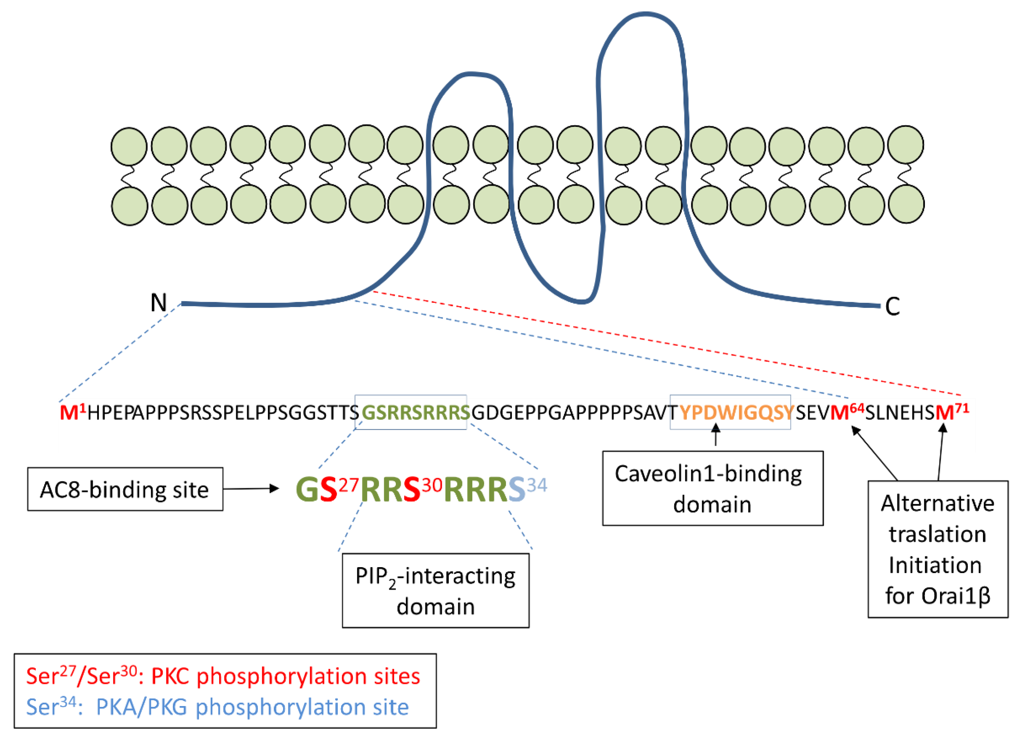
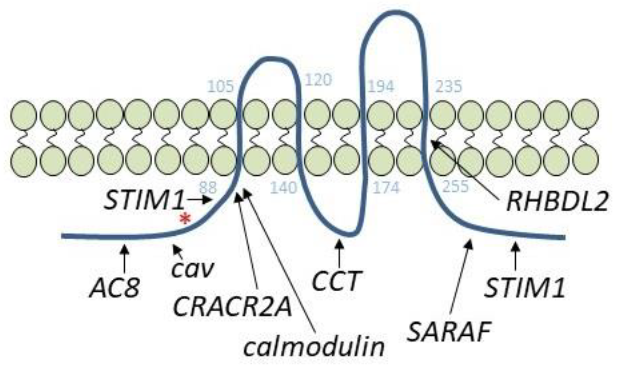
Store Operated Ca2+ Entry (SOCE) is a ubiquitous signaling mechanism initiated after the depletion of ER Ca2+ stores. Under physiological conditions, SOCE is associated with the activation of membrane receptors, which causes the mobilization of ER Ca2+ reservoirs via IP3-receptor activation [5,6]. The first store-operated current identified, the Ca2+ release-activated current (ICRAC), is non-voltage-activated, inwardly rectifying, and highly selective for Ca2+ [5,6]. The key molecular components of SOCE are STIM and Orai proteins. STIM1 and its homolog STIM2 act as ER Ca2+ sensors and play an essential role in SOCE. The STIM proteins contain a cytosolic STIM-Orai-activating region (SOAR), which is the minimal sequence sufficient to activate Orai channels [7,8,9,10,11]. Upon store depletion, STIM proteins trigger Ca2+ influx from the extracellular medium by contacting and activating the store-operated Ca2+ channels [12]. The Ca2+-released activated Ca2+ (CRAC) channels are comprised of Orai1 [13,14,15,16], which is highly selective to Ca2+. Initially, the involvement of the Orai1 homologs, Orai2 and Orai3, in CRAC channels was unclear; however, recently, native CRAC channels have been described as hexameric structures formed by the association between different Orai family members, leading to channels that fine-tune the extent of SOCE to match the strength of the stimulation with different concentrations of physiological agonists [17,18,19]. In addition to ICRAC, another store-operated current, less selective for Ca2+, has been described, ISOC, which involves the participation of Orai1, the canonical transient receptor potential (TRP) channel TRPC1 and STIM1 [20,21,22]. Whether TRPC1 is able to operate as a store-operated channel in the absence of Orai1 still remains controversial. While there is some overlap between the Ca2+ currents generated by Orai1 and TRPC1, they have been reported to regulate different cell functions [23] and, in this review, we will focus on the regulation of Orai1 channels.
In its resting state, STIM1 exists as a dimer, in a quiescent configuration, with the SOAR domain occluded in the folded C-terminal region. Calcium store depletion leads to dissociation of Ca2+ from the STIM1 EF-hand domain, leading to STIM1 multimerization and translocation to puncta within the ER membrane located in ER-plasma membrane (PM) junctions [24]. Subsequently, STIM1 undergoes a conformational change, leading to association of the EF-hand/SAM domains in the intraluminal N-terminal region of the STIM1 dimer [25]. The conformational change is transferred through the transmembrane domains and unbends the cytosolic region, leading to exposure of the SOAR to bind to and activate the Orai1 channel. The molecular model for the interaction with and activation of Orai1 by STIM1 predicts the unimolecular coupling between STIM1 and Orai1 and suggests that the available STIM1 subunit of the dimer might undergo inter-hexameric Orai1 channel crosslinking [26]. The Orai1 leucine-273 located in the Orai1 C-terminal STIM1-binding domain has been reported to play an essential role in Orai1 gating by STIM1 [27], propagating the STIM1-binding signal through the Orai1 transmembrane domains to the pore-forming helices to induce channel gating [28].
Native CRAC channels have been reported to be formed by the heterogeneous association of Orai proteins, which confer CRAC channel differences in their biophysical properties. For instance, Orai3 has been reported to modulate inhibition of ICRAC by reactive oxygen species [29,30,31]. In immune cell types, Orai2, together with Orai1, is a primary component of the CRAC channels and plays a relevant functional role [32,33]. Furthermore, Orai2 and, especially, Orai3 exhibit more prominent fast Ca2+-dependent inactivation than Orai1 [34]. Based on the stronger Ca2+-dependent inactivation, Orai2 and Orai3 negatively regulate CRAC currents and might modulate native Ca2+ signals and the subsequent cellular responses, emphasizing Orai proteins’ expression and stoichiometry as crucial factors to understand the role of SOCE in a given cell type [18,19].

3. Orai1 Variants

In 2012, Fukushima et al. [35] reported the existence of two Orai1 variants at the protein level in mammalian cells (reviewed in [36]). The longer variant, Orai1α, is the full-length Orai1 with 301 amino acids, while the short variant, termed Orai1β (231–238 amino acids), arises by a process of alternative translation initiation from a methionine at position 64 (probably also 71) in the Orai1α variant. Therefore, Orai1β lacks the N-terminal amino acids 1 to 63 or 70 that exist in Orai1α, which exhibits different motifs with potential functional relevance (Figure 1). Specifically, the sequence between amino acids 26–34 in Orai1α is involved in its interaction with the Ca2+-regulated adenylyl cyclase 8 (AC8) [37]. In addition, the serine residues at positions 27 and 30 are PKC phosphorylation sites, and phosphorylation of Orai1 at serines 27 and 30 has been associated with channel inactivation [38]. Serine-34 has been reported as a PKG [39], as well as PKA [40], phosphorylation site; phosphorylation of this residue is also involved in Orai1α inactivation. There is a predicted PIP2-binding domain in the polyarginine sequence between amino acids 28–33 [38]. Finally, the sequence between amino acids 52–60 forms a caveolin-binding domain [41]. While Orai1α and Orai1β show similar subcellular localization and both isoforms are able to support both ICRAC and ISOC currents [20,35], they exhibit some functional differences. Among them, Orai1α, but not Orai1β, is able to support the IARC current [20], a non-capacitive Ca2+ current through the arachidonate acid-regulated Ca2+ (ARC) channel and involving the participation of Orai1, Orai3 and the minor pool of plasma membrane resident STIM1 [42,43,44]. Furthermore, Orai1α exhibits a more pronounced fast Ca2+-dependent inactivation [20], and FRAP recovery experiments have revealed that Orai1α shows slower plasma membrane mobility than Orai1β [35].
Orai1α and Orai1β have been reported to activate NFAT1 nuclear translocation upon cell stimulation with thapsigargin [40]. However, a recent study has revealed that Orai1α interacts with the scaffold protein A-kinase anchoring protein (AKAP)-79, while Orai1β shows a weak association with this protein [45]. AKAP-79 has been reported to allow colocalization between NFAT1 and the Ca2+-dependent phosphatase calcineurin, which facilitates NFAT1 dephosphorylation [46]. The findings by Kar and coworkers challenge the participation of Orai1β in NFAT1 nuclear translocation or suggest an alternative pathway for Orai1β-mediated NFAT1 activation [45]. The presence of two Orai1 variants with slightly different biophysical properties, together with the different expression of Orai2, Orai3 and STIM isoforms and variants, unveils the heterogeneity of agonist-stimulated Ca2+ signals.

4. Orai1-Interacting Proteins: Adenylyl Cyclase 8

Besides STIM1, different proteins have been reported to interact with and regulate Orai1 activation and function (Figure 2). Among them, CRACR2A (also known as CRAC regulator 2A, EFCAB4B or FLJ33805) is a cytosolic protein that contains two EF-hand domains in the N-terminal region and stabilizes the STIM1-Orai1 interaction. CRACR2A interacts with the N-terminal region of Orai1, involving lysines at positions 85 and 87, and this interaction plays a relevant role in Orai1 clustering and activity [47]. Nevertheless, the more recent publication of the crystal structure of the full Drosophila melanogaster Orai1, showing that the pore of the hexameric channel, which extends > 20 Å into the cytosol, includes lysines 85 and 87 [17], challenges the possibility that CRACR2A directly interacts with those residues according to the structure of the Drosophila Orai1. The chaperonin-containing TCP-1 (CCT) is a molecular chaperone that interacts with the sequence between amino acids 157–167, located in the Orai1 intracellular loop; this increases PM residence of Orai1, leading to faster STIM1-Orai1 puncta formation [48]. The STIM1-modulator SARAF (SOCE-associated regulatory factor) has been demonstrated to activate Orai1 by its interaction with the C-terminus of Orai1 [49]. SARAF is a 339-amino-acid-long protein that has been identified as a STIM1-interacting protein that modulates SOCE and prevents Ca2+ overload. SARAF interacts with the C-terminal inhibitory domain of STIM1, preventing spontaneous activation of STIM1 and modulating basal ER and cytosolic Ca2+ concentrations [50]. The dynamic interaction between SARAF and STIM1 has been shown to be modulated by the Ca2+-binding protein EFHB (EF-hand domain family member B [51]); Caveolin-1 has also found to interact with and regulate Orai1 function. A number of studies have provided evidence for a positive role of caveolin-1 in the activation of SOCE [52,53]. Two caveolin-binding sites have been reported in Orai1 located between amino acids 52 and 60, in the N-terminal region, and at residues 250 to 253, located in the fourth transmembrane domain [41,54]. Orai1 internalization has been found to involve the caveolin-binding site located between residues 52 and 60, that exists in Orai1α exclusively [55], and a more recent study has revealed that the cytosolic C-terminal sequence between amino acids 260–275 is also essential for Orai1 internalization in Xenopus oocytes during meiosis [56]. A different mechanism for Orai1 endocytosis has been described in renal proximal tubular epithelial cells, where Orai1 colocalizes with clathrin instead of caveolin after interaction with amnionless, a protein associated with receptor endocytosis, leading to clathrin-mediated endocytosis of Orai1/STIM1 complexes and albumin upon Ca2+ store depletion [57]. Other proteins that regulate the Orai1 channel function include ubiquilin, which downregulates SOCE by promoting the ubiquitination and lysosomal degradation of Orai1 [58], and the recently reported RHBDL2 protein, a rhomboid intramembrane protease that interacts with the fourth transmembrane domain of Orai1 and degrades inappropriately activated CRAC channels in non-stimulated cells [59]. In contrast to the molecular mechanisms leading to a reduction of Orai1 plasma membrane expression, different pathways have been reported to enhance Orai1 surface exposure. In this context, secretory pathway Ca2+-ATPase (SPCA2) has been shown to interact with Orai1 and promote its translocation to the plasma membrane, leading to constitutive channel activation [60,61,62].
Recently, Zhang et al. have described the role of AC8 in Orai1 Ca2+-dependent inactivation [40]. As mentioned above, the N-terminal domain of AC8 interacts with the sequence GSRRSRRRS (amino acids 26–34) located in the N-terminal region of Orai1 [37]. The AC8-binding region is absent in the short Orai1 variant, Orai1β, since unlike the long Orai1 variant, Orai1α, it lacks the N-terminal 63 amino acids. As a consequence, AC8 does not interact with Orai1β [63]. Calcium entry through Orai1 triggers AC8 activity, which, in turn, increases cytosolic cAMP concentration in the channel microenvironment [64], thus providing a point of convergence for Ca2+ and cAMP signals.
AC8 is among the Ca2+-sensitive adenylyl cyclases. Despite the lack of EF-hand motifs in AC8, it contains two calmodulin (CaM)-binding domains, located in the N-terminal domain and the C2b region [65]. In resting cells, AC8 is in a quiescent/autoinhibited state supported by steric hindrance caused by the C-terminal domain that prevents ATP interaction with the ATP-binding site, and upon cell stimulation the enzyme is activated by a Ca2+ and CaM-dependent conformational change [66]. The first evidence supporting the activation of AC8 by Ca2+ entry was provided by Fagan et al. [67], reporting that AC8 was robustly stimulated by SOCE but not by Ca2+ released from the intracellular stores or by Ca2+ influx elicited by Ca2+ ionophores. Subsequent studies demonstrated that the activation of AC8 by SOCE occurred both in electrically excitable and non-excitable cells, the former also showing activation of AC8 by Ca2+ influx elicited by the opening of L-type (Cav1.2) Ca2+ channels [68,69]. Nevertheless, in non-excitable cells AC8 activation has been reported to occur exclusively upon activation of SOCE, since non-capacitative Ca2+ influx through the ARC channels or diacylglycerol-activated TRPC channels failed to activate AC8 [70,71].
Once activated, AC8 induces local increases in cAMP, which, in turn, activate PKA, leading to the phosphorylation of Orai1 at serine-34, a mechanism specific to Orai1α. Co-localization of PKA with Orai1 is mediated by AKAP-79 [40]. Phosphorylation of Orai1α at serine-34 leads to channel inactivation and shapes the Orai1-mediated Ca2+ signals and function. Specifically, while both Orai1 variants can activate nuclear translocation and transcriptional activity of NFAT4, an NFAT isoform sensitive to small changes in [Ca2+]c, upon stimulation with physiological agonist concentrations Orai1β-mediated Ca2+ signals supported faster and more robust NFAT4 translocation than those mediated by Orai1α [40]. These findings suggest that the relative expression of Orai1α and Orai1β, and the abundance of AC8, in a given cell type might significantly remodel Ca2+ signals and functions evoked by physiological concentrations of agonists.

5. Orai1-Adenylyl Cyclase 8 in Cancer Cells

Cancer cells are characterized by the disturbance of the fine balance of the cell cycle, shifting this balance towards excessive proliferation while attenuating pathways leading to cell death. In cancer cells, crucial processes leading to proliferation—but also migration, invasion and drug or apoptosis resistance—are mediated by Ca2+ and cAMP [63,72]. Some cancers, in order to orchestrate the oncogenic machinery, tune the signaling pathways by modifying the expression of key regulators or by favoring specific post-translational protein modifications [73], thus remodeling the signal transduction mechanisms. Breast cancer is a heterogeneous disease that is commonly classified by its histological and molecular features and gene expression profile into luminal, HER2 and triple-negative types [74]. The latter has been stratified into six subtypes known as basal-like 1 (BL1), basal-like 2 (BL2), mesenchymal (M), mesenchymal stem-like (MSL), immunomodulatory (IM), and luminal androgen receptor (LAR) [75,76].
In recent decades, numerous studies have described the link between Ca2+ signals and the hallmarks of breast cancer, revealing changes in protein expression and activity with a clear heterogeneity among cancer subtypes. In the context of SOCE, Ca2+ entry through CRAC channels promotes epithelial to mesenchymal transition, cell proliferation, angiogenesis, metastasis and resistance to chemotherapy in breast cancer cells [77]. Supporting this crucial role, Orai1 overexpression has been described in a variety of breast cancer subtypes, showing a dominant function in triple-negative breast cancer (TNBC) cells [78,79]. Conversely, STIM1 expression level shows a wide heterogeneity which embraces differences among cell lines from the same cancer subtype. Interestingly, in breast cancer cells of the luminal subtype (estrogen receptor positive (ER+) breast cancer cells), SOCE is strongly dependent on Orai3 channels, whose expression depends on estrogen receptor-α in these cells [80], with Orai1 playing a minor role [78]. These observations illustrate the heterogeneity of Ca2+ handling in cells from different breast cancer subtypes.
Based on the previous studies by Willoughby [37] and Zhang [40] in non-tumoral cells, in 2019 we provided evidence for the remodeling of the Orai1-AC8 interplay in different subtypes of TNBC cells and its role in the promotion of breast cancer hallmarks [63]. As mentioned earlier, AC8-Orai1 coupling allows two sequential functions: (1) the activation of AC8 upon Ca2+ influx through the channel, leading to the activation of the cAMP signaling pathway, and (2) the subsequent inactivation of Orai1 by PKA phosphorylation at serine-34. Concerning the regulation of Orai1 by the AC8/PKA pathway, two considerations should be taken into account. First of all, Willoughby and coworkers observed that the interaction of AC8 with Orai1 occurs at the AC8-binding site in the unique N-terminal region of Orai1α [37], and that the phosphorylation sites of PKC, PKA and PKG at serines 27, 30 and 34, with relevant roles for channel inactivation, are also found at the exclusive N-terminus of Orai1α [38,39,40]. These findings reveal that AC8 does not interact with the short Orai1 variant, Orai1β, as demonstrated experimentally [63], and that Orai1β is not susceptible to inactivation by phosphorylation of the mentioned serine residues. The AC8-mediated Orai1 inactivation is expected to solely affect the Orai1α-forming CRAC channels; the existence of Orai1α:Orai1β heteromeric channels remains to be elucidated. Second, the AC8-binding site overlaps with serines-27, 30 and 34 (see Figure 1), and, therefore, AC8 binding might interfere with phosphorylation at the mentioned serines. In triple-negative MDA-MB-231 breast cancer cells of the MSL subtype [75], both Orai1 variants as well as AC8 show an enhanced expression at the protein level, but these cells predominantly express AC8, which shifts the Orai1α/AC8 stoichiometry in favor of AC8 [63]. In MDA-MB-231 cells, determination of the serine phosphorylation status of Orai1 at native conditions revealed that the AC8-bound Orai1 subunits (Orai1 that co-immunoprecipitates with AC8) are not phosphorylated at serine residues, while the AC8-free Orai1 subunits show a significant serine phosphorylation, thus providing evidence for the impairment of Orai1 serine phosphorylation upon AC8 binding [63]. These findings were confirmed in cells transfected with siRNA for AC8 gene silencing or AC8 overexpression plasmid, where AC8 knockdown enhances Orai1 serine phosphorylation while AC8 overexpression abolishes this process. In a model where interaction with AC8 prevents PKA-dependent phosphorylation and inactivation of a given Orai1 subunit, one would postulate that overexpression of AC8, as observed in MDA-MB-231 cells, might decrease the number of channels not associated with this cyclase, thus impairing phosphorylation-dependent CDI and subsequently enhancing Ca2+ influx (Figure 3). Our results indicated that AC8 knockdown in MDA-MB-231 cells significantly attenuated TG-induced SOCE as well as Ca2+ influx evoked by co-expression of Orai1α and the Orai1-activating small fragment (OASF) region of STIM1 [9], which activates CRAC channels independently of Ca2+ store depletion, and, conversely, AC8 overexpression slightly but significantly enhances SOCE and Ca2+ influx mediated by co-expression of OASF and Orai1α in these cells [63]. These findings indicate that, while in normal cells the interaction of AC8 with Orai1α leads to net channel inactivation [40], in TNBC cells of the MSL subtype, overexpression of AC8 interferes with Orai1α phosphorylation and inactivation, resulting in enhanced SOCE.
Remodeling of the AC8 expression, and thus AC8-Orai1α stoichiometry, was proved to be important for the support of certain SOCE-dependent cancer hallmarks. AC8 expression attenuation reduced proliferation in the MSL-TNBC cell lines MDA-MB-231 and Hs578T, and inhibited MDA-MB-231 cell migration by inhibition of tyrosine phosphorylation of the focal adhesion kinase (FAK) [63]. Although the effect observed after AC8 silencing might be attributed to the regulation of SOCE, a functional role for the cAMP pathway in these processes cannot be ruled out. There is no information about the functional interaction between Orai1α and AC8 in other subtypes of TNBC cells, and the different gene expression profile between subtypes prevents the generalization of the findings observed in MSL cells, but given the potential interest of these findings to identify new pharmacological targets for the treatment of TNBC, the analysis of the interaction between Orai1 and AC8 in other TNBC subtypes deserves further study.
AC8 overexpression also occurs in estrogen receptor positive (ER+), luminal A, breast cancer MCF7 cells, together with a high expression of Orai1α and Orai1β [63]. Nevertheless, in these cells, Orai3 is sufficient to conduct SOCE, as Orai3 knockdown impairs TG-induced Ca2+ influx, while knockdown of either Orai1 or Orai2 was without effect on SOCE in MCF7 cells [78]. It is unclear whether Orai1 or Orai2 are constituents of the CRAC channels in MCF7, with Orai3 as the predominant subunit, but if this proves to be the case, their role might be redundant. However, a role for Orai1 has been reported in constitutive Ca2+ entry in these cells stimulated by SPCA [60]. Whether SPCA can indistinctly interact with Orai1α and Orai1β, as well as the possible role of AC8 in the regulation of constitutive Ca2+ entry mediated by Orai1, remains to be elucidated. As the AC8-binding site is exclusive for Orai1α and, therefore, AC8 is unable to associate to Orai2 and Orai3 [37], it is quite unlikely that AC8 regulates SOCE in MCF7 cells and, therefore, the mechanism described above is cancer subtype-specific. Nevertheless, AC8 plays a functional role in MCF7 cells, as we have found that AC8 knockdown leads to attenuation of MCF7 cell migration to a similar extent as pharmacological inhibition of PKA both in resting conditions and upon stimulation with carbachol, a Ca2+-mobilizing agonist [63]. These findings indicate that AC8 can still be activated in MCF7, probably by interaction with Orai1-forming, constitutively open Ca2+ channels or by proximity to Orai3-forming CRAC channels (Figure 3). Activated AC8 might result in the generation of cAMP and subsequent activation of PKA and other downstream effectors, including the transcription factors CREB and CREM or the exchange protein directly activated by cAMP (Epac) [81]. We have found that the increase in cytosolic cAMP using the cell-permeant 8-Br-cAMP enhances MCF7 cell migration, and, conversely, inactivation of PKA by KT-5720 attenuates cell migration [63]. Furthermore, pharmacological inhibition of Epac has been reported to disrupt its association with the microtubule cytoskeleton, to induce delocalization of AKAP9 from the centrosome, and to result in attenuation of MCF7 cell migration, leading to cell death [82]. Altogether, these findings indicate that activation of the cAMP pathway in estrogen-receptor-positive MCF7 breast cancer cells plays a relevant role in cell migration and tumorigenesis.

6. Conclusions

In summary, the AC8-Orai1α interplay is a relevant mechanism for CRAC channel inactivation and for shaping the Ca2+ signals in response to physiological agonists. In TNBC cells of the MSL subtype, the expression of AC8 and Orai1α is altered to shift the AC8-Orai1α stoichiometry in favor of the former, thus reducing the number of non-AC8-bound Orai1α subunits susceptible to inactivation and, as a result, enhancing SOCE and supporting a variety of cancer hallmarks. While a role for AC8 in the regulation of SOCE in estrogen receptor positive breast cancer cells is unlikely, AC8 is overexpressed in these cells, and its activation by Ca2+-dependent mechanisms plays a relevant role in the development of cancer features. Unveiling the mechanisms underlying breast cancer cell biology will shed a light on the identification of novel pharmacological opportunities for cancer therapy, inspiring the development of specific anti-tumoral strategies.

Author Contributions

Conceptualization, J.S.-C., J.J.L. and J.A.R.; Writing—Original Draft Preparation, J.S.-C. and J.A.R.; Writing—Review and Editing, J.J.L.; Funding Acquisition, J.A.R. and J.J.L. All authors have read and agreed to the published version of the manuscript.

Funding

This research was funded by Agencia Estatal de Investigación PID2019-104084GB-C21/AEI/10.13039/501100011033, MICINN (Grant BFU2016-74932-C2) and Junta de Extremadura-Fondo Europeo de Desarrollo Regional (FEDER; Grants IB16046, TA18011 and GR18061) to J.A.R. and by Junta de Extremadura (TA18011) to J.J.L. J.J.L. is supported by a contract from Junta de Extremadura (TA18011). J.S.-C. is supported by a contract from the Ministry of Science, Innovation, and Universities, Spain.

Institutional Review Board Statement

Not applicable.

Informed Consent Statement

Not applicable.

Data Availability Statement

The study does not report any new data.

Conflicts of Interest

The authors declare no conflict of interest.

References

  1. Berridge, M.J.; Lipp, P.; Bootman, M.D. The versatility and universality of calcium signalling. Nat. Revs. Mol. Cell Biol. 2000, 1, 11–21. [Google Scholar] [CrossRef] [PubMed]
  2. Kar, P.; Parekh, A.B. Distinct spatial Ca2+ signatures selectively activate different NFAT transcription factor isoforms. Mol. Cell 2015, 58, 232–243. [Google Scholar] [CrossRef] [PubMed] [Green Version]
  3. Bruce, J.I.; Straub, S.V.; Yule, D.I. Crosstalk between cAMP and Ca2+ signaling in non-excitable cells. Cell Calcium 2003, 34, 431–444. [Google Scholar] [CrossRef]
  4. Bruce, J.I.; Shuttleworth, T.J.; Giovannucci, D.R.; Yule, D.I. Phosphorylation of inositol 1,4,5-trisphosphate receptors in parotid acinar cells. A mechanism for the synergistic effects of cAMP on Ca2+ signaling. J. Biol. Chem. 2002, 277, 1340–1348. [Google Scholar] [CrossRef] [Green Version]
  5. Putney, J.W., Jr. A model for receptor-regulated calcium entry. Cell Calcium 1986, 7, 1–12. [Google Scholar] [CrossRef]
  6. Hoth, M.; Penner, R. Depletion of intracellular calcium stores activates a calcium current in mast cells. Nature 1992, 355, 353–356. [Google Scholar] [CrossRef]
  7. Yuan, J.P.; Zeng, W.; Dorwart, M.R.; Choi, Y.J.; Worley, P.F.; Muallem, S. SOAR and the polybasic STIM1 domains gate and regulate Orai channels. Nat. Cell Biol. 2009, 11, 337–343. [Google Scholar] [CrossRef] [PubMed]
  8. Park, C.Y.; Hoover, P.J.; Mullins, F.M.; Bachhawat, P.; Covington, E.D.; Raunser, S.; Walz, T.; Garcia, K.C.; Dolmetsch, R.E.; Lewis, R.S. STIM1 clusters and activates CRAC channels via direct binding of a cytosolic domain to Orai1. Cell 2009, 136, 876–890. [Google Scholar] [CrossRef] [Green Version]
  9. Muik, M.; Fahrner, M.; Derler, I.; Schindl, R.; Bergsmann, J.; Frischauf, I.; Groschner, K.; Romanin, C. A Cytosolic Homomerization and a Modulatory Domain within STIM1 C Terminus Determine Coupling to ORAI1 Channels. J. Biol. Chem. 2009, 284, 8421–8426. [Google Scholar] [CrossRef] [Green Version]
  10. Kawasaki, T.; Lange, I.; Feske, S. A minimal regulatory domain in the C terminus of STIM1 binds to and activates ORAI1 CRAC channels. Biochem. Biophys. Res. Commun. 2009, 385, 49–54. [Google Scholar] [CrossRef] [Green Version]
  11. Wang, X.; Wang, Y.; Zhou, Y.; Hendron, E.; Mancarella, S.; Andrake, M.D.; Rothberg, B.S.; Soboloff, J.; Gill, D.L. Distinct Orai-coupling domains in STIM1 and STIM2 define the Orai-activating site. Nat. Commun. 2014, 5, 3183. [Google Scholar] [CrossRef] [PubMed]
  12. Zhang, S.L.; Yu, Y.; Roos, J.; Kozak, J.A.; Deerinck, T.J.; Ellisman, M.H.; Stauderman, K.A.; Cahalan, M.D. STIM1 is a Ca2+ sensor that activates CRAC channels and migrates from the Ca2+ store to the plasma membrane. Nature 2005, 437, 902–905. [Google Scholar] [CrossRef]
  13. Feske, S.; Gwack, Y.; Prakriya, M.; Srikanth, S.; Puppel, S.H.; Tanasa, B.; Hogan, P.G.; Lewis, R.S.; Daly, M.; Rao, A. A mutation in Orai1 causes immune deficiency by abrogating CRAC channel function. Nature 2006, 441, 179–185. [Google Scholar] [CrossRef]
  14. Vig, M.; Beck, A.; Billingsley, J.M.; Lis, A.; Parvez, S.; Peinelt, C.; Koomoa, D.L.; Soboloff, J.; Gill, D.L.; Fleig, A.; et al. CRACM1 multimers form the ion-selective pore of the CRAC channel. Curr. Biol. 2006, 16, 2073–2079. [Google Scholar] [CrossRef] [PubMed] [Green Version]
  15. Peinelt, C.; Vig, M.; Koomoa, D.L.; Beck, A.; Nadler, M.J.; Koblan-Huberson, M.; Lis, A.; Fleig, A.; Penner, R.; Kinet, J.P. Amplification of CRAC current by STIM1 and CRACM1 (Orai1). Nat. Cell. Biol. 2006, 8, 771–773. [Google Scholar] [CrossRef] [PubMed] [Green Version]
  16. Soboloff, J.; Spassova, M.A.; Tang, X.D.; Hewavitharana, T.; Xu, W.; Gill, D.L. Orai1 and STIM reconstitute store-operated calcium channel function. J. Biol. Chem. 2006, 281, 20661–20665. [Google Scholar] [CrossRef] [PubMed] [Green Version]
  17. Hou, X.; Pedi, L.; Diver, M.M.; Long, S.B. Crystal structure of the calcium release-activated calcium channel Orai. Science 2012, 338, 1308–1313. [Google Scholar] [CrossRef] [Green Version]
  18. Vaeth, M.; Yang, J.; Yamashita, M.; Zee, I.; Eckstein, M.; Knosp, C.; Kaufmann, U.; Karoly Jani, P.; Lacruz, R.S.; Flockerzi, V.; et al. ORAI2 modulates store-operated calcium entry and T cell-mediated immunity. Nat. Commun. 2017, 8, 14714. [Google Scholar] [CrossRef] [Green Version]
  19. Yoast, R.E.; Emrich, S.M.; Zhang, X.; Xin, P.; Johnson, M.T.; Fike, A.J.; Walter, V.; Hempel, N.; Yule, D.I.; Sneyd, J.; et al. The native ORAI channel trio underlies the diversity of Ca(2+) signaling events. Nat. Commun. 2020, 11, 2444. [Google Scholar] [CrossRef]
  20. Desai, P.N.; Zhang, X.; Wu, S.; Janoshazi, A.; Bolimuntha, S.; Putney, J.W.; Trebak, M. Multiple types of calcium channels arising from alternative translation initiation of the Orai1 message. Sci. Signal. 2015, 8, ra74. [Google Scholar] [CrossRef] [Green Version]
  21. Huang, G.N.; Zeng, W.; Kim, J.Y.; Yuan, J.P.; Han, L.; Muallem, S.; Worley, P.F. STIM1 carboxyl-terminus activates native SOC, I(crac) and TRPC1 channels. Nat. Cell Biol. 2006, 8, 1003–1010. [Google Scholar] [CrossRef] [PubMed]
  22. Jardin, I.; Lopez, J.J.; Salido, G.M.; Rosado, J.A. Orai1 mediates the interaction between STIM1 and hTRPC1 and regulates the mode of activation of hTRPC1-forming Ca2+ channels. J. Biol. Chem. 2008, 283, 25296–25304. [Google Scholar] [CrossRef] [Green Version]
  23. Ong, H.L.; Subedi, K.P.; Son, G.Y.; Liu, X.; Ambudkar, I.S. Tuning store-operated calcium entry to modulate Ca2+-dependent physiological processes. Biochim. Biophys. Acta Mol. Cell Res. 2019, 1866, 1037–1045. [Google Scholar] [CrossRef] [PubMed]
  24. Baba, Y.; Hayashi, K.; Fujii, Y.; Mizushima, A.; Watarai, H.; Wakamori, M.; Numaga, T.; Mori, Y.; Iino, M.; Hikida, M.; et al. Coupling of STIM1 to store-operated Ca2+ entry through its constitutive and inducible movement in the endoplasmic reticulum. Proc. Natl. Acad. Sci. USA 2006, 103, 16704–16709. [Google Scholar] [CrossRef] [PubMed] [Green Version]
  25. Stathopulos, P.B.; Zheng, L.; Li, G.Y.; Plevin, M.J.; Ikura, M. Structural and mechanistic insights into STIM1-mediated initiation of store-operated calcium entry. Cell 2008, 135, 110–122. [Google Scholar] [CrossRef] [Green Version]
  26. Zhou, Y.; Wang, X.; Loktionova, N.A.; Cai, X.; Nwokonko, R.M.; Vrana, E.; Wang, Y.; Rothberg, B.S.; Gill, D.L. STIM1 dimers undergo unimolecular coupling to activate Orai1 channels. Nat. Commun. 2015, 6, 8395. [Google Scholar] [CrossRef] [Green Version]
  27. Cai, X.; Nwokonko, R.M.; Loktionova, N.A.; Abdulqadir, R.; Baraniak, J.H., Jr.; Wang, Y.; Trebak, M.; Zhou, Y.; Gill, D.L. Pore properties of Orai1 calcium channel dimers and their activation by the STIM1 ER calcium sensor. J. Biol. Chem. 2018, 293, 12962–12974. [Google Scholar] [CrossRef] [Green Version]
  28. Baraniak, J.H., Jr.; Zhou, Y.; Nwokonko, R.M.; Gill, D.L. The Intricate Coupling Between STIM Proteins and Orai Channels. Curr. Opin. Physiol. 2020, 17, 106–114. [Google Scholar] [CrossRef]
  29. Bogeski, I.; Kummerow, C.; Al-Ansary, D.; Schwarz, E.C.; Koehler, R.; Kozai, D.; Takahashi, N.; Peinelt, C.; Griesemer, D.; Bozem, M.; et al. Differential redox regulation of ORAI ion channels: A mechanism to tune cellular calcium signaling. Sci. Signal. 2010, 3, ra24. [Google Scholar] [CrossRef] [Green Version]
  30. Alansary, D.; Bogeski, I.; Niemeyer, B.A. Facilitation of Orai3 targeting and store-operated function by Orai1. Biochim. Biophys. Acta 2015, 1853, 1541–1550. [Google Scholar] [CrossRef] [Green Version]
  31. Saul, S.; Gibhardt, C.S.; Schmidt, B.; Lis, A.; Pasieka, B.; Conrad, D.; Jung, P.; Gaupp, R.; Wonnenberg, B.; Diler, E.; et al. A calcium-redox feedback loop controls human monocyte immune responses: The role of ORAI Ca2+ channels. Sci. Signal. 2016, 9, ra26. [Google Scholar] [CrossRef] [PubMed]
  32. Grimes, D.; Johnson, R.; Pashos, M.; Cummings, C.; Kang, C.; Sampedro, G.R.; Tycksen, E.; McBride, H.J.; Sah, R.; Lowell, C.A.; et al. ORAI1 and ORAI2 modulate murine neutrophil calcium signaling, cellular activation, and host defense. Proc. Natl. Acad. Sci. USA 2020, 117, 24403–24414. [Google Scholar] [CrossRef] [PubMed]
  33. Diez-Bello, R.; Jardin, I.; Salido, G.M.; Rosado, J.A. Orai1 and Orai2 mediate store-operated calcium entry that regulates HL60 cell migration and FAK phosphorylation. Biochim. Biophys. Acta 2017, 1864, 1064–1070. [Google Scholar] [CrossRef] [PubMed]
  34. Lee, K.P.; Yuan, J.P.; Zeng, W.; So, I.; Worley, P.F.; Muallem, S. Molecular determinants of fast Ca2+-dependent inactivation and gating of the Orai channels. Proc. Natl. Acad. Sci. USA 2009, 106, 14687–14692. [Google Scholar] [CrossRef] [PubMed] [Green Version]
  35. Fukushima, M.; Tomita, T.; Janoshazi, A.; Putney, J.W. Alternative translation initiation gives rise to two isoforms of Orai1 with distinct plasma membrane mobilities. J. Cell Sci. 2012, 125, 4354–4361. [Google Scholar] [CrossRef] [Green Version]
  36. Trebak, M.; Putney, J.W., Jr. ORAI Calcium Channels. Physiology 2017, 32, 332–342. [Google Scholar] [CrossRef] [PubMed]
  37. Willoughby, D.; Everett, K.L.; Halls, M.L.; Pacheco, J.; Skroblin, P.; Vaca, L.; Klussmann, E.; Cooper, D.M. Direct binding between Orai1 and AC8 mediates dynamic interplay between Ca2+ and cAMP signaling. Sci. Signal. 2012, 5, ra29. [Google Scholar] [CrossRef]
  38. Kawasaki, T.; Ueyama, T.; Lange, I.; Feske, S.; Saito, N. Protein kinase C-induced phosphorylation of Orai1 regulates the intracellular Ca2+ level via the store-operated Ca2+ channel. J. Biol. Chem. 2010, 285, 25720–25730. [Google Scholar] [CrossRef] [Green Version]
  39. Wang, Y.; Li, Z.C.; Zhang, P.; Poon, E.; Kong, C.W.; Boheler, K.R.; Huang, Y.; Li, R.A.; Yao, X. Nitric Oxide-cGMP-PKG Pathway Acts on Orai1 to Inhibit the Hypertrophy of Human Embryonic Stem Cell-Derived Cardiomyocytes. Stem Cells 2015, 33, 2973–2984. [Google Scholar] [CrossRef]
  40. Zhang, X.; Pathak, T.; Yoast, R.; Emrich, S.; Xin, P.; Nwokonko, R.M.; Johnson, M.; Wu, S.; Delierneux, C.; Gueguinou, M.; et al. A calcium/cAMP signaling loop at the ORAI1 mouth drives channel inactivation to shape NFAT induction. Nat. Commun. 2019, 10, 1971. [Google Scholar] [CrossRef] [PubMed]
  41. Yeh, Y.C.; Parekh, A.B. Distinct structural domains of caveolin-1 independently regulate Ca2+ release-activated Ca2+ channels and Ca2+ microdomain-dependent gene expression. Mol. Cell Biol. 2015, 35, 1341–1349. [Google Scholar] [CrossRef] [Green Version]
  42. Mignen, O.; Thompson, J.L.; Shuttleworth, T.J. Both Orai1 and Orai3 are essential components of the arachidonate-regulated Ca2+-selective (ARC) channels. J. Physiol. 2008, 586, 185–195. [Google Scholar] [CrossRef]
  43. Mignen, O.; Thompson, J.L.; Shuttleworth, T.J. STIM1 regulates Ca2+ entry via arachidonate-regulated Ca2+-selective (ARC) channels without store depletion or translocation to the plasma membrane. J. Physiol. 2007, 579, 703–715. [Google Scholar] [CrossRef] [PubMed]
  44. Zhang, X.; Zhang, W.; Gonzalez-Cobos, J.C.; Jardin, I.; Romanin, C.; Matrougui, K.; Trebak, M. Complex role of STIM1 in the activation of store-independent Orai1/3 channels. J. Gen. Physiol. 2014, 143, 345–359. [Google Scholar] [CrossRef] [PubMed] [Green Version]
  45. Kar, P.; Lin, Y.P.; Bhardwaj, R.; Tucker, C.J.; Bird, G.S.; Hediger, M.A.; Monico, C.; Amin, N.; Parekh, A.B. The N terminus of Orai1 couples to the AKAP79 signaling complex to drive NFAT1 activation by local Ca2+ entry. Proc. Natl. Acad. Sci. USA 2021, 118, e2012908118. [Google Scholar] [CrossRef] [PubMed]
  46. Kar, P.; Samanta, K.; Kramer, H.; Morris, O.; Bakowski, D.; Parekh, A.B. Dynamic assembly of a membrane signaling complex enables selective activation of NFAT by Orai1. Curr. Biol. 2014, 24, 1361–1368. [Google Scholar] [CrossRef] [PubMed] [Green Version]
  47. Srikanth, S.; Jung, H.J.; Kim, K.D.; Souda, P.; Whitelegge, J.; Gwack, Y. A novel EF-hand protein, CRACR2A, is a cytosolic Ca2+ sensor that stabilizes CRAC channels in T cells. Nat. Cell Biol. 2010, 12, 436–446. [Google Scholar] [CrossRef] [Green Version]
  48. Hodeify, R.; Nandakumar, M.; Own, M.; Courjaret, R.J.; Graumann, J.; Hubrack, S.Z.; Machaca, K. The CCT chaperonin is a novel regulator of Ca2+ signaling through modulation of Orai1 trafficking. Sci. Adv. 2018, 4, eaau1935. [Google Scholar] [CrossRef] [PubMed] [Green Version]
  49. Albarran, L.; Lopez, J.J.; Amor, N.B.; Martin-Cano, F.E.; Berna-Erro, A.; Smani, T.; Salido, G.M.; Rosado, J.A. Dynamic interaction of SARAF with STIM1 and Orai1 to modulate store-operated calcium entry. Sci. Rep. 2016, 6, 24452. [Google Scholar] [CrossRef]
  50. Palty, R.; Raveh, A.; Kaminsky, I.; Meller, R.; Reuveny, E. SARAF inactivates the store operated calcium entry machinery to prevent excess calcium refilling. Cell 2012, 149, 425–438. [Google Scholar] [CrossRef] [Green Version]
  51. Albarran, L.; Lopez, J.J.; Jardin, I.; Sanchez-Collado, J.; Berna-Erro, A.; Smani, T.; Camello, P.J.; Salido, G.M.; Rosado, J.A. EFHB is a Novel Cytosolic Ca2+ Sensor That Modulates STIM1-SARAF Interaction. Cell Physiol. Biochem. 2018, 51, 1164–1178. [Google Scholar] [CrossRef]
  52. Zhu, H.; Weisleder, N.; Wu, P.; Cai, C.; Chen, J.W. Caveolae/caveolin-1 are important modulators of store-operated calcium entry in Hs578/T breast cancer cells. J. Pharmacol. Sci. 2008, 106, 287–294. [Google Scholar] [CrossRef] [Green Version]
  53. Prakash, Y.S.; Thompson, M.A.; Vaa, B.; Matabdin, I.; Peterson, T.E.; He, T.; Pabelick, C.M. Caveolins and intracellular calcium regulation in human airway smooth muscle. Am. J. Physiol. Lung Cell Mol. Physiol. 2007, 293, L1118–L1126. [Google Scholar] [CrossRef] [PubMed] [Green Version]
  54. Yu, F.; Sun, L.; Machaca, K. Constitutive recycling of the store-operated Ca2+ channel Orai1 and its internalization during meiosis. J. Cell Biol. 2010, 191, 523–535. [Google Scholar] [CrossRef] [PubMed] [Green Version]
  55. Yu, F.; Sun, L.; Machaca, K. Orai1 internalization and STIM1 clustering inhibition modulate SOCE inactivation during meiosis. Proc. Natl. Acad. Sci. USA 2009, 106, 17401–17406. [Google Scholar] [CrossRef] [Green Version]
  56. Hodeify, R.; Dib, M.; Alcantara-Adap, E.; Courjaret, R.; Nader, N.; Reyes, C.Z.; Hammad, A.S.; Hubrack, S.; Yu, F.; Machaca, K. The carboxy terminal coiled-coil modulates Orai1 internalization during meiosis. Sci. Rep. 2021, 11, 2290. [Google Scholar] [CrossRef] [PubMed]
  57. Zeng, B.; Chen, G.L.; Garcia-Vaz, E.; Bhandari, S.; Daskoulidou, N.; Berglund, L.M.; Jiang, H.; Hallett, T.; Zhou, L.P.; Huang, L.; et al. ORAI channels are critical for receptor-mediated endocytosis of albumin. Nat. Commun. 2017, 8, 1920. [Google Scholar] [CrossRef] [Green Version]
  58. Lee, J.E.; Jeon, I.S.; Han, N.E.; Song, H.J.; Kim, E.G.; Choi, J.W.; Song, K.D.; Lee, H.K.; Choi, J.K. Ubiquilin 1 interacts with Orai1 to regulate calcium mobilization. Mol. Cells 2013, 35, 41–46. [Google Scholar] [CrossRef] [Green Version]
  59. Grieve, A.G.; Yeh, Y.C.; Zarcone, L.; Breuning, J.; Johnson, N.; Stříšovský, K.; Brown, M.H.; Parekh, A.B.; Freeman, M. Conformational surveillance of Orai1 by a rhomboid intramembrane protease prevents inappropriate CRAC channel activation. BioRxiv 2020. [CrossRef] [Green Version]
  60. Feng, M.; Grice, D.M.; Faddy, H.M.; Nguyen, N.; Leitch, S.; Wang, Y.; Muend, S.; Kenny, P.A.; Sukumar, S.; Roberts-Thomson, S.J.; et al. Store-independent activation of Orai1 by SPCA2 in mammary tumors. Cell 2010, 143, 84–98. [Google Scholar] [CrossRef] [Green Version]
  61. Cross, B.M.; Hack, A.; Reinhardt, T.A.; Rao, R. SPCA2 regulates Orai1 trafficking and store independent Ca2+ entry in a model of lactation. PLoS ONE 2013, 8, e67348. [Google Scholar] [CrossRef] [PubMed]
  62. Cantonero, C.; Sanchez-Collado, J.; Gonzalez-Nunez, M.A.; Salido, G.M.; Lopez, J.J.; Jardin, I.; Rosado, J.A. Store-independent Orai1-mediated Ca2+ entry and cancer. Cell Calcium 2019, 80, 1–7. [Google Scholar] [CrossRef] [PubMed]
  63. Sanchez-Collado, J.; Lopez, J.J.; Jardin, I.; Camello, P.J.; Falcon, D.; Regodon, S.; Salido, G.M.; Smani, T.; Rosado, J.A. Adenylyl Cyclase Type 8 Overexpression Impairs Phosphorylation-Dependent Orai1 Inactivation and Promotes Migration in MDA-MB-231 Breast Cancer Cells. Cancers 2019, 11, 1624. [Google Scholar] [CrossRef] [Green Version]
  64. Martin, A.C.; Willoughby, D.; Ciruela, A.; Ayling, L.J.; Pagano, M.; Wachten, S.; Tengholm, A.; Cooper, D.M. Capacitative Ca2+ entry via Orai1 and stromal interacting molecule 1 (STIM1) regulates adenylyl cyclase type 8. Mol. Pharmacol. 2009, 75, 830–842. [Google Scholar] [CrossRef] [PubMed] [Green Version]
  65. Gu, C.; Cooper, D.M. Calmodulin-binding sites on adenylyl cyclase type VIII. J. Biol. Chem. 1999, 274, 8012–8021. [Google Scholar] [CrossRef] [PubMed] [Green Version]
  66. Simpson, R.E.; Ciruela, A.; Cooper, D.M.F. The role of calmodulin recruitment in Ca2+ stimulation of adenylyl cyclase type 8. J. Biol. Chem. 2006, 281, 17379–17389. [Google Scholar] [CrossRef] [PubMed] [Green Version]
  67. Fagan, K.A.; Mahey, R.; Cooper, D.M. Functional co-localization of transfected Ca2+-stimulable adenylyl cyclases with capacitative Ca2+ entry sites. J. Biol. Chem. 1996, 271, 12438–12444. [Google Scholar] [CrossRef] [Green Version]
  68. Fagan, K.A.; Graf, R.A.; Tolman, S.; Schaack, J.; Cooper, D.M. Regulation of a Ca2+-sensitive adenylyl cyclase in an excitable cell. Role of voltage-gated versus capacitative Ca2+ entry. J. Biol. Chem. 2000, 275, 40187–40194. [Google Scholar] [CrossRef] [Green Version]
  69. Everett, K.L.; Cooper, D.M. An improved targeted cAMP sensor to study the regulation of adenylyl cyclase 8 by Ca2+ entry through voltage-gated channels. PLoS ONE 2013, 8, e75942. [Google Scholar] [CrossRef] [Green Version]
  70. Shuttleworth, T.J.; Thompson, J.L. Discriminating between capacitative and arachidonate-activated Ca2+ entry pathways in HEK293 cells. J. Biol. Chem. 1999, 274, 31174–31178. [Google Scholar] [CrossRef] [Green Version]
  71. Martin, A.C.; Cooper, D.M. Capacitative and 1-oleyl-2-acetyl-sn-glycerol-activated Ca2+ entry distinguished using adenylyl cyclase type 8. Mol. Pharmacol. 2006, 70, 769–777. [Google Scholar] [CrossRef] [PubMed] [Green Version]
  72. Wehbe, N.; Slika, H.; Mesmar, J.; Nasser, S.A.; Pintus, G.; Baydoun, S.; Badran, A.; Kobeissy, F.; Eid, A.H.; Baydoun, E. The Role of Epac in Cancer Progression. Int. J. Mol. Sci. 2020, 21, 6489. [Google Scholar] [CrossRef]
  73. Roberts-Thomson, S.J.; Chalmers, S.B.; Monteith, G.R. The Calcium-Signaling Toolkit in Cancer: Remodeling and Targeting. Cold Spring Harb. Perspect. Biol. 2019, 11, a035204. [Google Scholar] [CrossRef] [Green Version]
  74. Ahn, S.G.; Kim, S.J.; Kim, C.; Jeong, J. Molecular Classification of Triple-Negative Breast Cancer. J. Breast Cancer 2016, 19, 223–230. [Google Scholar] [CrossRef]
  75. Lehmann, B.D.; Bauer, J.A.; Chen, X.; Sanders, M.E.; Chakravarthy, A.B.; Shyr, Y.; Pietenpol, J.A. Identification of human triple-negative breast cancer subtypes and preclinical models for selection of targeted therapies. J. Clin. Investig. 2011, 121, 2750–2767. [Google Scholar] [CrossRef] [PubMed] [Green Version]
  76. Wang, D.Y.; Jiang, Z.; Zacksenhaus, E. Stratifying the stratifiers of triple negative breast cancer. Oncotarget 2020, 11, 306–308. [Google Scholar] [CrossRef]
  77. So, C.L.; Saunus, J.M.; Roberts-Thomson, S.J.; Monteith, G.R. Calcium signalling and breast cancer. Sem. Cell Dev. Biol. 2019, 94, 74–83. [Google Scholar] [CrossRef]
  78. Motiani, R.K.; Abdullaev, I.F.; Trebak, M. A novel native store-operated calcium channel encoded by Orai3: Selective requirement of Orai3 versus Orai1 in estrogen receptor-positive versus estrogen receptor-negative breast cancer cells. J. Biol. Chem. 2010, 285, 19173–19183. [Google Scholar] [CrossRef] [PubMed] [Green Version]
  79. McAndrew, D.; Grice, D.M.; Peters, A.A.; Davis, F.M.; Stewart, T.; Rice, M.; Smart, C.E.; Brown, M.A.; Kenny, P.A.; Roberts-Thomson, S.J.; et al. ORAI1-mediated calcium influx in lactation and in breast cancer. Mol. Cancer Ther. 2011, 10, 448–460. [Google Scholar] [CrossRef] [PubMed] [Green Version]
  80. Motiani, R.K.; Zhang, X.; Harmon, K.E.; Keller, R.S.; Matrougui, K.; Bennett, J.A.; Trebak, M. Orai3 is an estrogen receptor alpha-regulated Ca2+ channel that promotes tumorigenesis. FASEB J. 2013, 27, 63–75. [Google Scholar] [CrossRef] [Green Version]
  81. de Rooij, J.; Zwartkruis, F.J.; Verheijen, M.H.; Cool, R.H.; Nijman, S.M.; Wittinghofer, A.; Bos, J.L. Epac is a Rap1 guanine-nucleotide-exchange factor directly activated by cyclic AMP. Nature 1998, 396, 474–477. [Google Scholar] [CrossRef] [PubMed]
  82. Kumar, N.; Gupta, S.; Dabral, S.; Singh, S.; Sehrawat, S. Role of exchange protein directly activated by cAMP (EPAC1) in breast cancer cell migration and apoptosis. Mol. Cell Biochem. 2017, 430, 115–125. [Google Scholar] [CrossRef] [PubMed]
Figure 1. Amino acid sequence of the N-terminal region unique to Orai1α. Within the 63 or 70 residues unique for the N-terminal region of Orai1α there are several functional domains, including one of the caveolin1-binding domains and the AC8-binding site, which overlap with the PIP2-interacting region and the PKC and PKA/PKG phosphorylation sites (serines 27, 30 and 34, respectively).
Cells 10 01308 g001
Figure 2. Cartoon depicting the most relevant Orai1-interacting proteins: AC8, adenylyl cyclase isoform 8; cav, caveolin-1; CRACR2A, CRAC regulator 2A; CCT, chaperonin-containing TCP-1; SARAF, SOCE-associated regulatory factor; RHBDL2, rhomboid-like 2. The residues that delimit the transmembrane domains are represented in light blue. The asterisk indicates the start of Orai1β.
Cells 10 01308 g002
Figure 3. Cartoon depicting the AC8-Orai interplay in normal and breast cancer cells. Only one Orai subunit of the CRAC channel hexameric structure is depicted for clarity. In normal cells, Orai1 plays a predominant role in CRAC channels. Ca2+ influx through Orai1α leads to the activation of AC8, which in turn results in the activation of cAMP-dependent protein kinase (PKA) and inactivation of the AC8-free Orai1α subunits by phosphorylation at serine-34, thus moderating SOCE. In mesenchymal stem-like (MSL) TNBC cells Orai1 is the major component of CRAC channels. In these cells, AC8 overexpression decreases the number of AC8-free Orai1α subunits susceptible to phosphorylation and inactivation, leading to enhanced SOCE. In ER+ breast cancer cells, SOCE is mediated by Orai3, which is unable to bind AC8, and Orai1 participates in SPCA-dependent constitutive Ca2+ entry (CCaC). AC8 activation is activated by still unknown mechanisms that might involve Ca2+ influx via Orai1 or Orai3 in the close vicinity of Orai1 channels. The possible inactivation of Orai1-forming CCaCs by PKA remains unknown.
Cells 10 01308 g003
Publisher’s Note: MDPI stays neutral with regard to jurisdictional claims in published maps and institutional affiliations.

