Review

Not So Dead Genes—Retrocopies as Regulators of Their Disease-Related Progenitors and Hosts

by
Joanna Ciomborowska-Basheer
,
Klaudia Staszak
,
Magdalena Regina Kubiak
and
Izabela Makałowska
*
Institute of Human Biology and Evolution, Faculty of Biology, Adam Mickiewicz University Poznań, 61-614 Poznań, Poland
*
Author to whom correspondence should be addressed.
These authors contributed equally to this work.
Cells 2021, 10(4), 912; https://doi.org/10.3390/cells10040912
Submission received: 7 March 2021 / Revised: 30 March 2021 / Accepted: 13 April 2021 / Published: 15 April 2021
(This article belongs to the Special Issue Long Noncoding RNAs in Disease)

Abstract

:
Retroposition is RNA-based gene duplication leading to the creation of single exon nonfunctional copies. Nevertheless, over time, many of these duplicates acquire transcriptional capabilities. In human in most cases, these so-called retrogenes do not code for proteins but function as regulatory long noncoding RNAs (lncRNAs). The mechanisms by which they can regulate other genes include microRNA sponging, modulation of alternative splicing, epigenetic regulation and competition for stabilizing factors, among others. Here, we summarize recent findings related to lncRNAs originating from retrocopies that are involved in human diseases such as cancer and neurodegenerative, mental or cardiovascular disorders. Special attention is given to retrocopies that regulate their progenitors or host genes. Presented evidence from the literature and our bioinformatics analyses demonstrates that these retrocopies, often described as unimportant pseudogenes, are significant players in the cell’s molecular machinery.

1. Introduction

Retrosequences, previously described as meaningless and biologically unimportant elements, are now recognized as evolutionarily significant, and their roles in shaping genomes, transcriptomes and proteomes have become increasingly evident [1,2,3]. This type of RNA-based gene duplicate is created through retroposition, which, together with DNA-based duplication, is known to be one of the major sources of new genes [2,4,5]. Formation of a retrocopy starts with transcription of the multiexonic parental gene (Figure 1). The mature mRNA is transported to the cytoplasm where in mammals proteins from LINE1 (Long interspersed nuclear elements 1), i.e., reverse transcriptase and endonuclease, accompanied by chaperones bind to the polyA tail. This complex is transported back to the nucleus where it anneals to the broken DNA ends and undergoes reverse transcription. Created cDNA is incorporated into new genomic surroundings. The final step includes creating short flanking repeats at insertion site, so called target site duplication (TDS). The presence of the 3′ polyA tail, and flanking sequences constitute signature of LINE-mediated retrotransposition [6,7]. These copies are regarded as “dead on arrival” pseudo(retro)genes, which usually lack introns, core promoters and other regulatory elements. Retrocopies are highly represented in placental mammals, especially primates [8]. In other genomes, Drosophila for example, the number of retroposed genes is relatively low [9,10]. In early studies of duplicated genes evolution, it was postulated that usually one of the duplicates accumulates mutations and becomes nonfunctional [11,12]. However, it occurred that “relaxed” selection and evolutionary freedom, which are characteristic of the majority of duplicates, may lead not only to pseudogenization but also to the acquisition of new functions [13,14]. Over time, two new phenomena related to functional evolution after duplication have been described: (i) neofunctionalization, where one copy acquires a new function and the other one keeps the original one [15], and (ii) subfunctionalization, where maintained function is shared between duplicated genes [16,17]. Additionally, as our and other studies showed, it is also possible that the retrogene (functional retrocopy) replaces its progenitor [18,19]. In the case of retrocopies, the first step needs to be obtaining regulatory elements, and there is growing evidence that many retrocopies gained the capability to be expressed over time [4,20,21].
Regardless of being described as “junk DNA” for a long time, there are numerous examples demonstrating that retrocopies may successfully work as regulatory sequences as well as crucial protein coding genes [22,23,24]. A spectacular example of retrocopy function is the TP53 gene, a well-known tumor suppressor, and its retrocopies in elephants. Elephants have a lower-than-expected rate of cancer. It has been proposed that multiple functional retrocopies of TP53 are involved in an increased apoptotic response by compensating for the function of their progenitor [25,26]. This compensation mechanism, in turn, might underlie the cancer resistance observed in these animals. Nevertheless, in human protein coding is relatively rare among retrogenes. For example, in RetrogeneDB2 only 106 retrocopies, out of 4611, were identified as known protein coding genes, and only 847 (18%) has intact ORF (Open Reading Frame) inherited from parental gene. Interestingly, it is quite opposite in Drosophila where out of 83 identified in RetrogeneDB retrocopies, as many as 81 are annotated as known protein coding genes [27]. It was found that 256 retrocopies overlaps in the human genome with annotated lncRNAs and additional 230 may act as competing endogenous RNA since they share microRNA (miRNA) targets and have correlated expression with transcripts of 232 protein-coding genes [3]. Accumulating evidence suggests that substantial number of transcriptionally active retrocopies in human act as long noncoding RNAs (lncRNAs) [14,28]. Due to their high sequence similarity, they have a natural ability to regulate, via various mechanisms, their parental genes. Additionally, since almost 40% of retrocopies are located in introns of other genes, they possess great potential to control, as antisense transcripts, their host genes.
There are a number of ways in which retrocopies may regulate their progenitors or hosts. Retrocopies can be transcribed from the antisense strand and act as natural antisense transcripts (NATs) [29]. These NATs could be involved in multiple molecular processes, including epigenetic regulation (Figure 2A), chromatin remodeling [30], or, by forming RNA:RNA duplexes, stability control, RNA editing and processing (Figure 2B) [31]. Many retrocopies work as competing endogenous RNAs (ceRNAs), also known as microRNA sponges (Figure 2C) [15,32], while others can be a source of small RNAs [33]. Retrocopies can also compete with parental genes for other molecules, such as stabilizing factors (Figure 2D) [34] or translational machinery [35]. They may also influence the splicing of the host gene as potential factors that facilitate transcriptional interference [3,36,37,38]. The impact of retrocopies on the DNA level is also noticeable since they may be involved in nonallelic homologous recombination, resulting in the formation of chimeric transcripts (Figure 2E) [3].
In light of the variety of possible functions, lncRNAs originating from retrocopies (retro-lncRNAs) can play a significant role in the cell regulatory machinery. This is especially important when their progenitors or host genes are critical in disease pathogenesis. In this review, we focus on such examples in human disorders, considering possible mechanisms of retro-lncRNA action.

2. Retro-lncRNAs in Cancer

Cancer constitutes a heterogeneous phenomenon with varied forms and a multifactorial basis. The role of retrocopies’ involvement in cancer has been described numerous times, and most of these retrocopies act as lncRNAs [14]. They are multifunctional, showing both oncogenic and suppressor effects [39]. In the cancer literature, we can find retro-lncRNAs that derive from parental genes referred to as “drivers”. Genetic changes within their sequences, such as mutations, give a selective growth advantage for cancer cells and thus drive cancer development [40]. Such an example is the HMGA1 gene, which can act as a driver in liver carcinogenesis [41]. Retrocopies with no coding potential, HMGA1P6 and HMGA1P7, have been indicated to be upregulated in endometrial cancer [42], ovarian cancer and thyroid cancer [43]. These additional copies can act as decoys for common microRNAs and thus regulate the expression of the parental HMGA1 gene [43]. Another example constitutes a well-known KRAS gene showing driver traits in pancreatic cancer [44] and lung cancer [45], among others. Its retrocopy, KRASP1, which has been found to be highly expressed in prostate cancer, most likely regulates parental gene expression by sequestering microRNA [15].
Examples of retrogene-derived lncRNAs that arose from the parental gene with oncogenic function are POU5F1P4 (OCT4-pg4) and POU5F1P5 (OCT4-pg5), noncoding copies of the POU5F1 (OCT4) gene. The POU5F1 gene has been correlated with the occurrence of cancer stem cell populations, cell fractions increasing the risk of metastasis and recurrence in colorectal cancer samples [46,47]. POU5F1P4 and POU5F1P5 have been shown to be involved in the pathogenesis of hepatocellular carcinoma [48] and endometrial carcinoma [49], respectively. Most likely, these retro-lncRNAs act as microRNA sponges [48,49]. Furthermore, a regulatory mechanism based on antisense RNA-mediated epigenetic silencing of parental gene transcription has also been proposed. OCT4-pg5, together with other factors including G9a and Ezh2, create a silencing complex that inhibits parental gene transcription. Transcriptional inhibition could be blocked when antisense RNA is bound and sequestered by proteins such as PURA and NCL (Figure 2A) [50,51]. Another example of an oncogene-derived retro-lncRNA is SUMO1P3. Its increased expression has been associated with tumor size, lymphatic metastasis, differentiation and invasion in gastric cancer patients [52]. In gastric cancer, silencing of parental SUMO1 resulted in inhibited proliferation and supported apoptosis [53]. Functional analysis showed the potential role of SUMO1P3 in microRNA sponging and cis-NAT regulation of its host gene COPA [3].
Correlation between the expression level of lncRNA RACGAP1P and the promotion of early hepatocellular carcinoma recurrence [54] or breast cancer progression [55] has also been described. Moreover, the parental gene RACGAP1 has been correlated with an aggressive phenotype in multiple cancers, including breast cancer [56] and ovarian cancer [57]. The potential mechanism is explained by RACGAP1P sponging of miR-15-5p (Figure 2C) [54]. Another example of a retrocopy associated with cancer is ANXA2P2. Cell culture studies have indicated that the ANXA2 gene promotes the invasion of breast cancer cells [58], and elevated expression of its copy, ANXA2P2, has been related to an aggressive phenotype in the progression of hepatocellular carcinoma [59]. In turn, upregulation of UBE2CP3 has been highlighted as linked to the induction of the epithelial-mesenchymal transition and thus metastasis promotion in hepatocellular carcinoma [60]. Their mechanisms of action remain unclear.
In the literature, there are also examples of retro-lncRNAs that arise from reverse transcription of suppressor genes. In examples described below, all lncRNAs mirror their parental genes and exert suppressor effects. The long noncoding RNA INTS6P1, along with its parental gene INTS6, have been termed cancer suppressors in hepatocellular carcinoma. The mechanism of their action has been connected with competition for oncogenic miR-17-5p [61]. Low expression levels of PTENP1 are related to the cancer phenotype, and overexpression of this retro-lncRNA has been demonstrated to inhibit cancer cell proliferation [62]. Interestingly, this retrocopy is transcribed in sense as well as in antisense orientation. Under normal condition the sense transcript of PTENP1 protects the parental gene from microRNA binding and therefore from translation inhibition. In cancer cells, down-regulation of PTENP1 expression leads to miRNA-driven degradation of PTEN (Figure 2C) [15,63]. The antisense isoform of PTENP1 may play a role as an epigenetic regulator by binding to the PTEN promoter and modulating its transcription [30]. Finally, downregulation of TUSC2P1 along with its parental gene, TUSC2, has been correlated with the promotion of apoptosis in cancer cells, which confirms their suppressor activity [64].
In addition to these published examples, our recent analysis of RNA-seq data has shown that some cancer cell lines, including hepatocyte carcinoma (HepG2) and chronic myelogenous leukemia (K562), have particularly high numbers of expressed retrocopies [3]. We also identified three retrocopies, AC107983.1, SYPL1P2, and NDUFB1P1, whose expression occurred in all analyzed cancer libraries but not in normal tissues. In the case of two of them, AC107983.1 and NDUFB1P1, genes localization and expression correlation suggest mechanisms of action based on cis-NAT regulation of the host genes CCDC144B and CDC25A, respectively. LncRNA AC107983.1 also demonstrated microRNA sponging capability. This assumption was made based on shared miRNA targets and negative correlation of the expression [3].
Retro-lncRNAs associated with diseases described in this review, together with some additional published cases, are presented in Table 1.

3. Retrocopies as lncRNAs in Neurodegenerative Disorders

Neurodegenerative diseases constitute complex and heterogeneous conditions that are based on neurons devastating and mainly affect elderly people. This group, among others, involves Huntington’s disease (HD), Parkinson’s disease (PD) and Alzheimer’s disease (AD). Symptoms of these disorders are not clear-cut, and thus, the correct diagnosis is quite a challenge [87]. Our knowledge of neurodegenerative pathogenesis remains incomplete and to tackle these challenges, an increasing number of studies of neurodegenerative diseases take into account lncRNAs [88,89]. This is supported by the fact that some lncRNAs are exceptionally enriched in specific brain regions [90,91].
Costa et al. analyzed the differential expression of pseudogenes in neurodegenerative diseases. This analysis included retroposition-derived lncRNAs. In the case of Huntington’s disease, an interesting example is a group of three retrocopies, HMGB1P1, HMGB1P5, and HMGB1P10, which are related to disease phenotype and share microRNA binding sites with their progenitor [78]. Their parental gene, HMGB1, also plays a role in the neurodegeneration process. HMGB1 protein interacts directly with huntingtin protein, and the overexpression of HMGB1 results in the inhibition of HD progression [92,93]. Complex analyses of RNA-seq data showed that one of HMGB1 retrocopies, HMGB1P10, can act through microRNA sponging and may also regulate the TPST2 gene in the cis-NAT configuration [3]. Another example of a dysregulated lncRNA is TLK2P1 [78], which originates from the TLK2 gene which has been connected with intellectual disabilities [94]. Our analysis suggested the action of TLK2P1 as a microRNA sponge [3]. The expression level of the VDAC1P1 retrocopy has also been deregulated in HD. On the basis of sharing binding sites with its parental gene, the role of the microRNA sponge can be inferred [78]. Although the involvement of its parental gene in HD was not reported, upregulation of the VDAC1 gene was discovered in postmortem AD brains [95].
Explanations for the functionality of some HD-related retro-lncRNAs can be found in the role of their progenitors in brain development. Studies in mouse models have shown that HMGN2, the parental gene of HMGN2P3, is an important molecule involved in embryonic/postnatal brain development and that the loss of HMGN2 is associated with microcephaly [96]. Another example of retrocopy with progenitors in brain formation is FABP5P1, where the FABP5 gene participates in postnatal neurogenesis [97]. It has also been discovered that the expression of HIGD1A, a parental gene of HIGD1AP14, is widely distributed but uneven in the brain [98]. These two retro-lncRNAs, FABP5P1 and HIGD1AP14, most likely constitute microRNA sponges, as they share common microRNA binding sites with their parental genes [78].
In the search for therapy, researchers of neurodegenerative diseases mainly focus on the use of induced pluripotent stem cells [99]. Costa et al. indicated that POU5F1P4 is deregulated in HD [78]. The cognate gene POU5F1 (OCT4) is one of the main pluripotent genes [100]. Furthermore, the parental gene of RBBP4P4, RBBP4, is also required for pluripotency maintenance [101]. Considering this fact, the occurrence of additional retrocopies of genes responsible for controlling this phenomenon may be essential to the outcome of therapy studies.
In the Parkinson’s disease dataset, also analyzed by Costa et al., three retroposition-derived lncRNAs, PHC1P1, RBMXP2 and CHCHD2P2, were identified [78]. The parental gene of PHC1P1, PHC1, has been listed as involved in the neuroinflammation that underlies neurodegenerative diseases [102], and according to our analyses, its retrocopy may potentially compete for microRNAs [3]. The progenitor of RBMXP2, gene RBMX, is crucial during the development of the zebrafish brain [103] and has been related to X-linked intellectual disability [104]. The last retro-lncRNA, CHCHD2P2, showed an altered level of expression in HD as well as in PD. In the literature, there is a reference of its parental gene CHCHD2 to the pathogenesis of Parkinson’s disease [105].
It is broadly discussed whether there is any relationship between neurodegenerative diseases and cancer. Both are age-related disorders, and they share the competitive endogenous RNA (ceRNA) mechanism involved in pathogenesis. In turn, these conditions differ in some contexts, as cancer is associated with cell death inhibition, while neurodegenerative diseases are connected with neuronal apoptosis [106]. These disorders share molecules dysregulated during disease progression, but sometimes changes in expression levels occur in opposite directions. It is worth noting that a negative correlation between the incidence of individual neoplasms and the risk of developing Alzheimer’s disease was reported [107]. Based on the results of Costa et al., who analyzed the differential expression of pseudogenes in neurodegenerative disease data [78], one may select retro-lncRNAs, which are common in both conditions. Examples of such are POU5F1P4 and PTENP1 as well as RBMS1P1, RBMXP2 and RHOQP2, which were identified in our laboratory in the differential expression analysis of breast cancer RNA-seq data [108].

4. Retro-lncRNAs and Other Diseases

4.1. Cardiovascular Diseases

Cardiovascular diseases are described as disorders related to the heart and blood vessels, and they are the largest cause of all deaths worldwide [109]. This group of diseases includes peripheral arterial disease, coronary heart disease, rheumatic heart disease, congenital heart disease and others. These conditions include heart attack, stroke, essential hypertension (EH), aortic dissection and atrial fibrillation (AF). Although the most important risk factors are well known, we are still far from understanding the molecular basis of these diseases and finding effective treatments. Recently, lncRNAs have become one of the most promising targets in cardiovascular disease research.
LOC646616 and LAP3P2, two retrogene-derived lncRNAs, have recently been described, together with two circRNAs, as candidates for therapeutic targets in the case of essential hypertension [79]. EH is defined as chronic high blood pressure without any definite cause, and it covers approximately 95% of all hypertension cases. LOC646616 was identified as upregulated in EH patients and acted as a sponge for miR-637. As a result, the WNT/β-catenin signaling pathway is activated. This pathway is related to the regulation of stem cell pluripotency and cell fate during development. The second retro-lncRNA, LAP3P2, was highly coexpressed with mRNAs of crucial elements of the same WNT/β-catenin signaling pathway, WNT and CAMK2N2 genes. It shares microRNA miR-637 binding sites with transcripts of these two genes and acts as competing endogenous RNA.
Another study related to cardiovascular diseases presented VDAC2P2 as a lncRNA potentially involved in atrial fibrillation (AF). AF is one of the most common and complex types of arrhythmia and is related to the risk of stroke and heart failure. VDAC2P2 together with two circRNAs were found to be significantly differentially expressed in atrial tissues in AF patients [80]. It was also shown that VDAC2P2 may regulate its parental gene VDAC2 as well as the neighboring gene KLRG1. Both parental gene and retrogene are related to the process of metabolite diffusion through the mitochondrial outer membrane. An association between mitochondrial dysfunction and atrial fibrillation has been previously postulated [110,111]. The function of VDAC2P2 is not known, but it may be involved in homologous recombination and gene conversion or serve as an antisense RNA [80].
The differentiation, proliferation and apoptosis of smooth muscle cells (SMCs) are important factors related to the development of aortic dissection and aortic aneurysm (AA) [112]. LncRNAs are known to modulate this type of cell, and one of them is PTENP1. This is yet another molecular process in which this lncRNA is involved in addition to cancers and neurodegenerative disorders, as described above. PTENP1 and its parental gene, PTEN, were found to be upregulated in human aortic dissection samples. In this case PTENP1 also works as a microRNA sponge competing for miR-21 with parental gene transcripts, and overexpression of lncRNA results in elevated level of PTEN protein [81].

4.2. Mental Disorders

Mental disorders are diverse diseases, but the most common characteristics include problems with perceptions, emotions, behavior and relationships with other people. This group includes depression, schizophrenia and other psychoses, dementia, bipolar disorder and autism spectrum disorders [109]. Recent studies have demonstrated that lncRNAs originating from retrocopies are also associated with this type of diseases. One of the examples is NDUFV2P1, which is related to schizophrenia (SZ), in which atypical neuronal transmission and dysregulation of brain energy metabolism were reported [113,114]. Last year, studies revealed a new basis of schizophrenia that is related to the abnormal functioning of mitochondria. The NDUFV2 protein, encoded by the NDUFV2P1 progenitor, is a crucial subunit of the cytochrome C oxidase I (CoI) complex of the mitochondrial respiratory chain, and its level can be reduced due to increased retrocopy expression, which leads to mitochondrial dysfunction [82]. Interestingly, deterioration of the CoI complex was also observed in bipolar disorder and Parkinson’s disease [115,116].
Autism spectrum disorder (ASD) is diagnosed in one in 160 children worldwide and sometimes influences adolescents and adults. It is quite common to observe coexisting diseases, such as epilepsy, depression or attention deficit hyperactivity disorder (ADHD) [109]. The development of ASD is caused by multiple factors, among which there are more than 130 genes [83]. It has been reported that some lncRNAs may also be involved in ASD pathogenesis, especially at the epigenetic level [117]. One of them is MSNP1AS, an antisense transcript of MSNP1, a retrocopy that originated from the MSN gene. Downregulation of MSN protein expression leads to inhibited activation of the PI3K/Akt signaling pathway. MNSP1AS is one of the major factors influencing the PI3K/Akt pathway, and the RhoA and Rac1 pathways are important for neuronal structure and survival [83]. It was reported that MSNP1AS and MSN are able to form dsRNA (double stranded RNA), which suppresses the expression of the protein encoded by the parental gene [83,118,119]. Furthermore, overexpression of MSNP1AS in ASD patients has an impact on smaller amounts and lengths of neurites in human neural progenitor cell lines [118,119].

4.3. Other Diseases

Preeclampsia (PE) is a specific disease during pregnancy characterized by hypertension, sometimes accompanied by proteinuria. Severe preeclampsia (SPE) is a major cause of maternal death and perinatal mortality worldwide. Implantation, placentation and decidua formation are the key processes in early pregnancy, and disturbances are thought to be the major cause of PE [84]. Two interesting examples of retro-lncRNAs involved in PE are HK2P1 [85] and PGK1P2 [84]. HK2P1 and its parental gene HK2 were found to be downregulated in human endometrial stromal cells, which inhibits their proliferation and differentiation and causes preeclampsia. The retrocopy works as a ceRNA and regulates the expression of the parental gene through competition for miR-6887-3p [85]. A very similar mechanism of interaction was described for PGK1P2 and PGK1. Deficiency of their mRNA levels and PGK1 protein in the decidua deregulates the glycolytic pathway, which is crucial for changes in the endometrium during pregnancy and the occurrence of PE. Additionally, retrocopy regulates the parental gene level by sponging microRNA, in this case miR-300-5p [84].
Biliary atresia (BA) is related to the fibrosis of extrahepatic bile ducts and is the major cause of cholestasis in children, which is the main reason for liver transplantation among children. The pathogenesis of biliary atresia is unclear, but some studies have shown an association between lncRNA deregulation and the development of fibrosis. One such lncRNA is ANXA2P3, which originates from a retrocopy of the annexin 2 gene ANXA2. Increased expression levels of ANXA2P3 and ANXA2 have a positive effect on cell proliferation and inhibit cell apoptosis. Transcripts of both genes are considered targets in treatments preventing liver injury and as future biomarkers in patients with BA [86].
Diabetes is a metabolic disease characterized by elevated levels of blood glucose resulting from the inefficient production or usage of endogenous insulin. It has a significant impact on human populations, as the number of people with diabetes has increased 4-fold over the last 40 years [109]. In some individuals affected by this disease, retrocopy-derived HMGA1P8 lncRNA was shown to play an important role [69]. HMGA1 is an architectural nuclear protein that functions mainly as a specific cofactor for activation of the insulin receptor gene (INSR). Case studies of two unrelated patients affected by type 2 diabetes showed that the expression of the HMGA1 gene was markedly reduced, while its retrocopy HMGA1P8 was overexpressed. A more in-depth study indicated that enhanced expression of lncRNA results in destabilization of parental gene mRNA by effective competition for a trans-acting cytoplasmic protein critical to mRNA stability (Figure 2D). Consequently, the expression of the INSR gene is suppressed, which in turn results in insulin resistance [34].

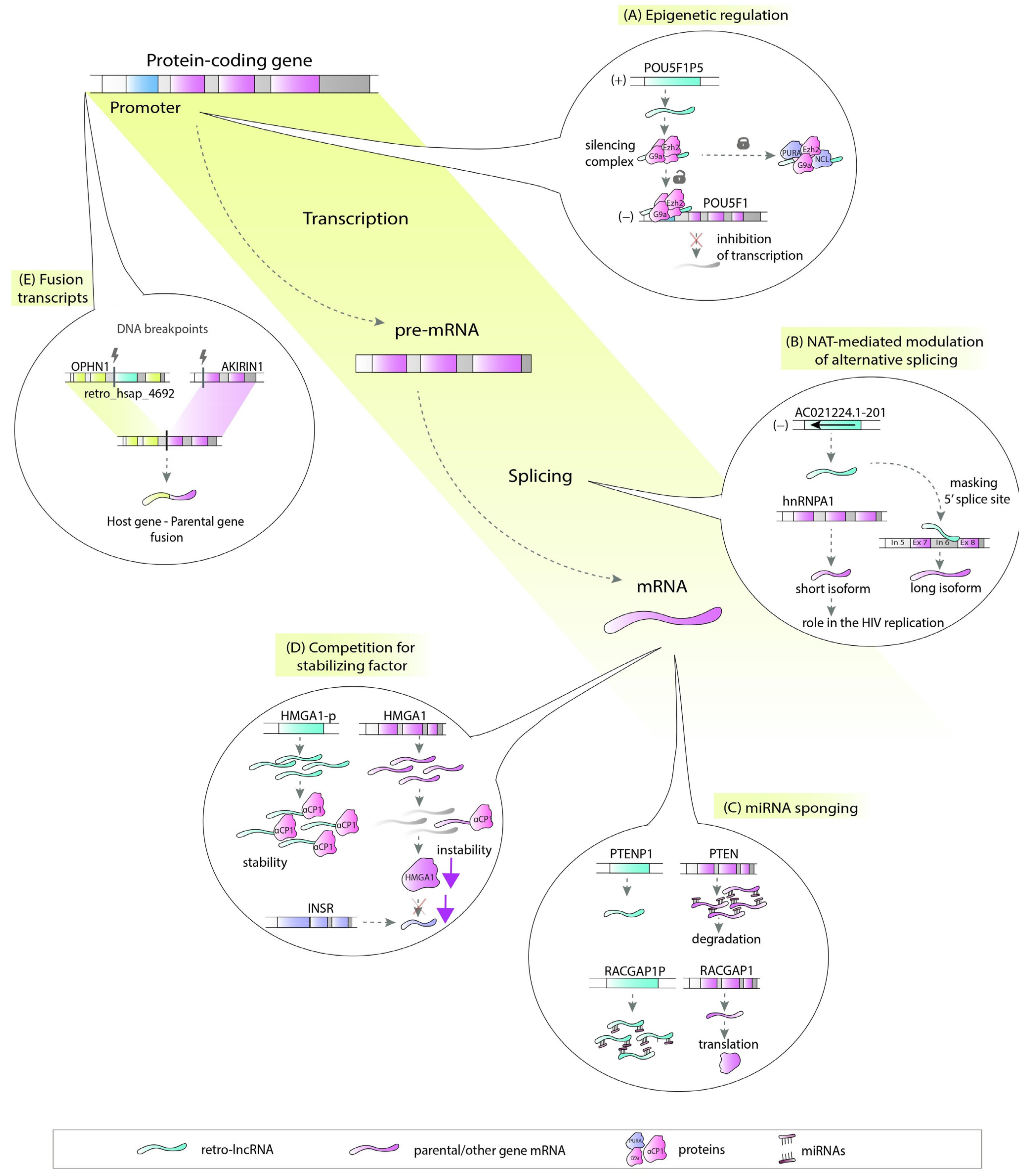
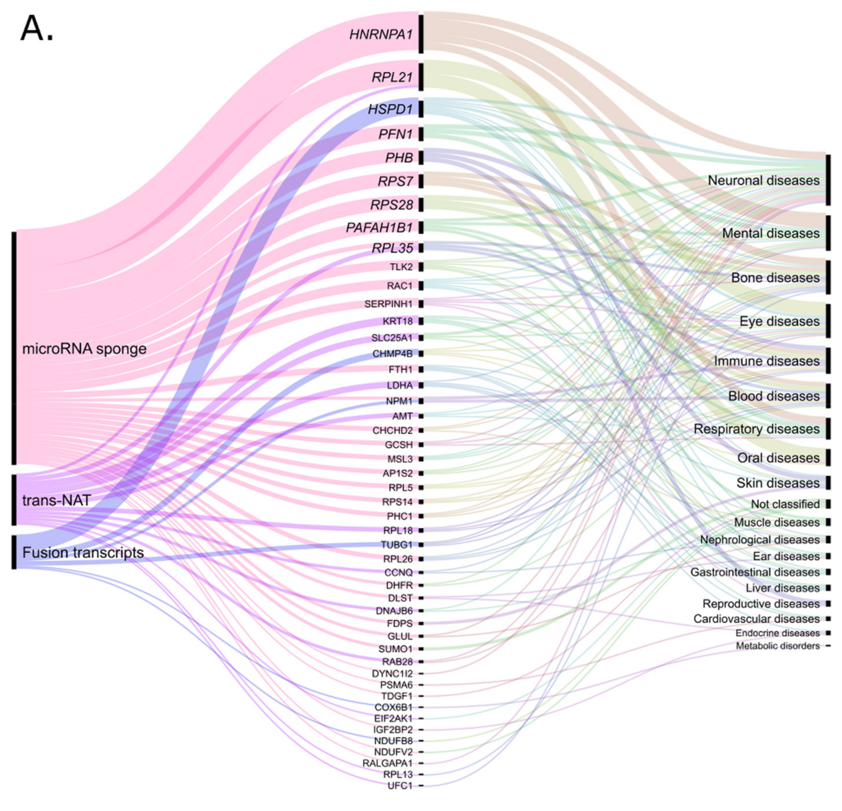
5. Noncoding Retrocopies as Putative Players in Pathogenic Processes

The numerous examples of the link between retrocopies and various pathogenic processes presented above are undoubtedly important evidence of their underestimated roles. These copies may be important for maintaining the proper functioning of the cell or, on the other hand, their expression may be deleterious. An increasing amount of data from a variety of high-throughput experiments makes it feasible to identify promising candidates that may regulate or interrupt the expression of other genes. In our recent work, we were able to propose the possible mode of action for approximately 43% of human retrocopies annotated in RetrogeneDB2 [3,27]. Utilizing these data we investigated whether this putatively regulated by retro-lncRNAs parental and host genes, are related to human diseases (Supplementary File S1). The input dataset included genes potentially regulated by retrocopies identified as (a) miRNA sponges; (b) trans natural antisense transcripts; (c) cis natural antisense transcripts; (d) factors of transcriptional interference and (e) source of fusion transcripts. These genes were selected based on genomic localization, RNA-seq data analysis, expression correlation analysis, identification of miRNA targets, sequence complementarity. All methodological aspects of these analyses are described in detail in original paper [3]. For each gene identified as putatively regulated by retrocopy, we retrieved the “MIM morbid accession” using Ensembl BioMart [120,121,122]. The resulting list of disease-related genes was then expanded by disease characteristics. To this goal, we classified the diseases according to the MalaCards global and anatomical categories [123]. Remarkably, both the parental and host genes are mainly associated with neuronal diseases. Bone, eye, and mental diseases were also well represented in the analyzed groups (Figure 3).
We found that 48 parental genes potentially regulated by 71 retrocopies were associated with 52 human diseases (Supplementary File S1: Table S1). In this group, microRNA sponge activity is the most popular mode of action (Figure 3A). This function was proposed for over three-quarters of the retrocopies. Among the identified parental genes, CHCHD2 seems to be an interesting example. Recent studies suggest that CHCHD2 regulates the functions of cytochrome c and that the loss of this regulation is associated with Parkinson’s disease [124]. We identified retro_hsap_116, also called CHCHD2P6, as a potential competitive endogenous RNA since it shares microRNA target sequences with the parental gene. Interestingly, CHCHD2 may also be regulated in a similar way by another retrocopy, CHCHD2P2 [78]. The predictions may be worth further analysis, especially considering the fact that recent studies showed specific changes in microRNA expression in Parkinson’s disease [125].
Retrocopies can also be a source of trans natural antisense transcripts for parental genes. From the analyzed dataset, 13 transcripts with retrocopy-derived exons in antisense orientation might regulate the expression of retrocopy progenitors. The retrocopy retro_hsap_2353, known as KRT18P29, embedded in the PPP1R1C gene can illustrate this phenomenon. Two noncoding isoforms of the host gene incorporated the 63 bp antisense sequence of KRT18P29 retrocopy as a new exon. By definition, the exon is also antisense to KRT18, a parental gene that was found to be linked with liver cirrhosis. Interestingly, both parental and host genes were differentially expressed in the EGR1-overexpressing cell line used in a study on the malignancy of human non-small cell lung carcinoma [126]. However, when the expression of KRT18 was upregulated, the expression of PPP1R1C was downregulated. In our previous studies, we found another intriguing example, an antisense transcript of retrocopy AC021224.1-201, which could be involved in splicing regulation of its progenitor hnRNPA1 [29]. The analysis of RNA:RNA duplexes formed between lncRNAs and pre-mRNA sequences and predicted based on base-pairing analysis, suggested that this lncRNA is able to mask the 5′ splice site in the sixth intron of the parental gene (Figure 2B). When the interaction does not occur, a shorter isoform of hnRNPA1 is expressed. This transcript was shown to play regulatory roles in human immunodeficiency virus splicing and replication [29,127].
Staying with the topic of natural antisense transcripts, it should be noted that the interaction of retrocopies in cis on host genes is the most represented group of their potential function (Figure 3B). In total, we found 186 retrocopies regulating 174 host genes associated with 247 diseases (Supplementary File S1: Table S2). A noteworthy subgroup is represented by intronic retrocopies proposed as transcriptional interference factors [3]. One example is the ERLIN2 gene associated with spastic paraplegia type 18. The expression of embedded in this gene retrocopy (retro_hsap_4044) is positively correlated with the expression of two short splice variants of the host gene, which might suggest facilitating early transcription termination [3,36]. Moreover, ERLIN2 was also studied in the context of other human diseases, such as mental retardation [128], lateral sclerosis [129] and breast cancer [130], which makes this gene worthy of further investigation.
In addition to the abovementioned functions, the nonallelic homologous recombination between a retrocopy and its progenitor, or two retrocopies of the same gene, may be considered as another important disease-related level of regulation. For example, a high sequence similarity between the EIF2AK1 parental gene and retro_hsap_2713 embedded in the ATR gene may explain the generation of a fusion transcript containing parental and host gene exons found in cancer cells [131]. In our data, nine retrocopies can be considered to be involved in the recombination of parental and/or host genes associated with diseases. We found, inter alia, a chimeric transcript associated with cataract and formed by CHMP4B, and FBXO34, the parental gene and host gene of retro_hsap_1339, respectively. Another example is a fusion of OPHN1 related to the X-linked mental retardation host of retro_hsap_4692, and AKIRIN1 (Figure 2E), the retrogene progenitor. Moreover, retrocopies themselves can be a part of chimeric transcripts. An example could be two chimeric transcripts found in acute myeloid leukemia. One resulted from the fusion of retro_hsap_1547 and its parental gene RPL32, and another resulted from retro_hsap_4032 and the parental gene COX6B1 [3,132].

6. Conclusions

In this review, we described multiple examples of retrocopy-derived lncRNAs related to human diseases. These examples demonstrate how important players in the whole cell machinery are copies commonly described as pseudogenes. They work in various manners, including microRNA sponging, chimeric transcript formation, as NATs or by influencing splicing. Their functions have been associated with many disorders, such as cancer, neurodegenerative and mental diseases, and cardiovascular diseases. Involvement of retrocopies in those pathogeneses, for a long time underrated, has proven to be crucial and confirms the role of retrocopies in many molecular mechanisms. Numerous studies have also demonstrated their potential as therapeutic targets. Apart from the literature-based examples, we report here additional candidates selected based on our bioinformatics analysis, that may regulate their parental and host genes. This gives many promising candidates for further studies and helps to understand retroposed gene involvement in human diseases.

Supplementary Materials

The following are available online at https://www.mdpi.com/article/10.3390/cells10040912/s1, Table S_Table_1, S_Table_2; Supplementary_file_1.xlsx.

Author Contributions

Conceptualization, J.C.-B., K.S., M.R.K. and I.M.; re-analysis of data, M.R.K.; writing—original draft preparation, J.C.-B., K.S. and M.R.K.; writing—review and editing, J.C.-B., K.S., M.R.K. and I.M.; visualization, K.S. and M.R.K.; supervision, I.M.; funding acquisition, M.R.K. All authors have read and agreed to the published version of the manuscript.

Funding

This work was supported by the National Science Centre of Poland grant number 2017/27/N/NZ2/00174 to M.R.K.

Institutional Review Board Statement

Not applicable.

Conflicts of Interest

The authors declare no conflict of interest.

References

  1. Brosius, J. RNAs from all categories generate retrosequences that may be exapted as novel genes or regulatory elements. Gene 1999, 238, 115–134. [Google Scholar] [CrossRef]
  2. Casola, C.; Betrán, E. the genomic impact of gene retrocopies: What have we learned from comparative genomics, population genomics, and transcriptomic analyses? Genome Biol. Evol. 2017, 9, 1351–1373. [Google Scholar] [CrossRef] [PubMed]
  3. Kubiak, M.R.; Szcześniak, M.W.; Makałowska, I. Complex Analysis of Retroposed Genes’ Contribution to Human Genome, Proteome and Transcriptome. Genes 2020, 11, 542. [Google Scholar] [CrossRef] [PubMed]
  4. Carelli, F.N.; Hayakawa, T.; Go, Y.; Imai, H.; Warnefors, M.; Kaessmann, H. The life history of retrocopies illuminates the evolution of new mammalian genes. Genome Res. 2016, 26, 301–314. [Google Scholar] [CrossRef] [Green Version]
  5. Betran, E.; Wang, W.; Jin, L.; Long, M. Evolution of the phosphoglycerate mutase processed gene in human and chimpanzee revealing the origin of a new primate gene. Mol. Biol. Evol. 2002, 654–663. [Google Scholar] [CrossRef] [Green Version]
  6. Vanin, E.F. Processed pseudogenes: Characteristics and evolution. Annu. Rev. Genet. 1985, 19, 253–272. [Google Scholar] [CrossRef]
  7. Esnault, C.; Maestre, J.; Heidmann, T. Human LINE retrotransposons generate processed pseudogenes. Nat. Genet. 2000, 363–367. [Google Scholar] [CrossRef]
  8. Mighell, A.J.; Smith, N.R.; Robinson, P.A.; Markham, A.F. Vertebrate pseudogenes. Febs Lett. 2000, 468, 109–114. [Google Scholar] [CrossRef] [Green Version]
  9. Betran, E. Retroposed new genes out of the X in drosophila. Genome Res. 2002, 12, 1854–1859. [Google Scholar] [CrossRef] [Green Version]
  10. Bai, Y.; Casola, C.; Feschotte, C.; Betrán, E. Comparative genomics reveals a constant rate of origination and convergent acquisition of functional retrogenes in drosophila. Genome Biol. 2007, 8, R11. [Google Scholar] [CrossRef] [Green Version]
  11. Haldane, J. The part played by recurent mutation in evolution. Am. Nat. 1933, 67, 5–19. [Google Scholar] [CrossRef]
  12. Fisher, R.A. The sheltering of lethals. Am. Nat. 1935, 69, 446–455. [Google Scholar] [CrossRef]
  13. Nei, M. Gene duplication and nucleotide substitution in evolution. Nature 1969, 221, 40–42. [Google Scholar] [CrossRef]
  14. Lou, W.; Ding, B.; Fu, P. Pseudogene-Derived lncRNAs and their mirna sponging mechanism in human cancer. Front. Cell Dev. Biol. 2020, 8, 85. [Google Scholar] [CrossRef] [Green Version]
  15. Poliseno, L.; Salmena, L.; Zhang, J.; Carver, B.; Haveman, W.J.; Pandolfi, P.P. A coding-independent function of gene and pseudogene mRNAs regulates tumour biology. Nature 2010, 465, 1033–1038. [Google Scholar] [CrossRef] [Green Version]
  16. Ohno, S. Evolution by Gene Duplication; Springer: New York, NY, USA, 1970. [Google Scholar]
  17. Force, A.; Lynch, M.; Pickett, F.B.; Amores, A.; Yan, Y.L.; Postlethwait, J. Preservation of duplicate genes by complementary, degenerative mutations. Genetics 1999, 151, 1531–1545. [Google Scholar]
  18. Ciomborowska, J.; Rosikiewicz, W.; Szklarczyk, D.; Makałowski, W.; Makałowska, I. “Orphan” retrogenes in the human genome. Mol. Biol. Evol. 2013, 30, 384–396. [Google Scholar] [CrossRef]
  19. Krasnov, A.N. A retrocopy of a gene can functionally displace the source gene in evolution. Nucleic Acids Res. 2005, 33, 6654–6661. [Google Scholar] [CrossRef] [Green Version]
  20. Bai, Y.; Casola, C.; Betrán, E. Evolutionary origin of regulatory regions of retrogenes in drosophila. BMC Genom. 2008. [Google Scholar] [CrossRef] [Green Version]
  21. Sarda, S.; Hannenhalli, S. Orphan CpG islands as alternative promoters. Transcription 2018, 9, 171–176. [Google Scholar] [CrossRef]
  22. Devor, E.J. Primate microRNAs miR-220 and miR-492 lie within processed pseudogenes. J. Hered. 2006, 97, 186–190. [Google Scholar] [CrossRef] [PubMed] [Green Version]
  23. Parker, H.G.; VonHoldt, B.M.; Quignon, P.; Margulies, E.H.; Shao, S.; Mosher, D.S.; Spady, T.C.; Elkahloun, A.; Cargill, M.; Jones, P.G.; et al. An expressed fgf4 retrogene is associated with breed-defining chondrodysplasia in domestic dogs. Science 2009, 325, 995–998. [Google Scholar] [CrossRef] [PubMed] [Green Version]
  24. Kubiak, M.R.; Makałowska, I. Protein-Coding Genes’ Retrocopies and Their Functions. Viruses 2017, 9, 80. [Google Scholar] [CrossRef] [Green Version]
  25. Abegglen, L.M.; Caulin, A.F.; Chan, A.; Lee, K.; Robinson, R.; Campbell, M.S.; Kiso, W.K.; Schmitt, D.L.; Waddell, P.J.; Bhaskara, S.; et al. Potential Mechanisms for Cancer Resistance in Elephants and Comparative Cellular Response to DNA Damage in Humans. JAMA 2015, 314, 1850–1860. [Google Scholar] [CrossRef]
  26. Sulak, M.; Fong, L.; Mika, K.; Chigurupati, S.; Yon, L.; Mongan, N.P.; Emes, R.D.; Lynch, V.J. TP53 copy number expansion is associated with the evolution of increased body size and an enhanced DNA damage response in elephants. eLife 2016, 5. [Google Scholar] [CrossRef]
  27. Rosikiewicz, W.; Kabza, M.; Kosinski, J.G.; Ciomborowska-Basheer, J.; Kubiak, M.R.; Makalowska, I. RetrogeneDB-a database of plant and animal retrocopies. Database J. Biol. Databases Curation 2017, 2017. [Google Scholar] [CrossRef] [PubMed]
  28. Glenfield, C.; McLysaght, A. pseudogenes provide evolutionary evidence for the competitive endogenous RNA hypothesis. Mol. Biol. Evol. 2018. [Google Scholar] [CrossRef]
  29. Bryzghalov, O.; Szcześniak, M.W.; Makałowska, I. Retroposition as a source of antisense long non-coding RNAs with possible regulatory functions. Acta Biochim. Pol. 2016, 63, 825–833. [Google Scholar] [CrossRef] [PubMed]
  30. Johnsson, P.; Ackley, A.; Vidarsdottir, L.; Lui, W.-O.; Corcoran, M.; Grandér, D.; Morris, K.V. A pseudogene long-noncoding-RNA network regulates PTEN transcription and translation in human cells. Nat. Struct. Mol. Biol. 2013, 20, 440–446. [Google Scholar] [CrossRef] [Green Version]
  31. Szcześniak, M.W.; Makałowska, I. lncRNA-RNA Interactions across the Human Transcriptome. PLoS ONE 2016, 11, e0150353. [Google Scholar] [CrossRef]
  32. Stambolic, V.; Suzuki, A.; de la Pompa, J.L.; Brothers, G.M.; Mirtsos, C.; Sasaki, T.; Ruland, J.; Penninger, J.M.; Siderovski, D.P.; Mak, T.W. Negative regulation of PKB/Akt-dependent cell survival by the tumor suppressor PTEN. Cell 1998, 95, 29–39. [Google Scholar] [CrossRef] [Green Version]
  33. Griffiths-Jones, S.; Grocock, R.J.; van Dongen, S.; Bateman, A.; Enright, A.J. Mirbase: MicroRNA sequences, targets and gene nomenclature. Nucleic Acids Res. 2006, 34, D140–D144. [Google Scholar] [CrossRef]
  34. Chiefari, E.; Iiritano, S.; Paonessa, F.; Le Pera, I.; Arcidiacono, B.; Filocamo, M.; Foti, D.; Liebhaber, S.A.; Brunetti, A. Pseudogene-mediated posttranscriptional silencing of HMGA1 can result in insulin resistance and type 2 diabetes. Nat. Commun. 2010, 1, 40. [Google Scholar] [CrossRef] [Green Version]
  35. Bier, A.; Oviedo-Landaverde, I.; Zhao, J.; Mamane, Y.; Kandouz, M.; Batist, G. Connexin43 pseudogene in breast cancer cells offers a novel therapeutic target. Mol. Cancer Ther. 2009, 8, 786–793. [Google Scholar] [CrossRef] [Green Version]
  36. Kaer, K.; Branovets, J.; Hallikma, A.; Nigumann, P.; Speek, M. Intronic L1 retrotransposons and nested genes cause transcriptional interference by inducing intron retention, exonization and cryptic polyadenylation. PLoS ONE 2011, 6, e26099. [Google Scholar] [CrossRef]
  37. Shearwin, K.E.; Callen, B.P.; Egan, J.B. Transcriptional interference--a crash course. Trends Genet. TIG 2005, 21, 339–345. [Google Scholar] [CrossRef] [Green Version]
  38. Long, M.; Langley, C.H. Natural selection and the origin of jingwei, a chimeric processed functional gene in Drosophila. Science 1993, 260, 91–95. [Google Scholar] [CrossRef]
  39. Staszak, K.; Makałowska, I. Cancer, retrogenes, and evolution. Life 2021, 11, 72. [Google Scholar] [CrossRef]
  40. Pon, J.R.; Marra, M.A. Driver and passenger mutations in cancer. Annu. Rev. Pathol. 2015, 10, 25–50. [Google Scholar] [CrossRef]
  41. Andreozzi, M.; Quintavalle, C.; Benz, D.; Quagliata, L.; Matter, M.; Calabrese, D.; Tosti, N.; Ruiz, C.; Trapani, F.; Tornillo, L.; et al. HMGA1 expression in human hepatocellular carcinoma correlates with poor prognosis and promotes tumor growth and migration in in vitro models. Neoplasia 2016, 18, 724–731. [Google Scholar] [CrossRef] [Green Version]
  42. Palumbo Júnior, A.; de Sousa, V.P.L.; Esposito, F.; De Martino, M.; Forzati, F.; Moreira, F.C.d.B.; Simão, T.d.A.; Nasciutti, L.E.; Fusco, A.; Ribeiro Pinto, L.F.; et al. Overexpression of HMGA1 Figures as a potential prognostic factor in Endometrioid Endometrial Carcinoma (EEC). Genes 2019, 10, 372. [Google Scholar] [CrossRef] [PubMed] [Green Version]
  43. Esposito, F.; De Martino, M.; Petti, M.G.; Forzati, F.; Tornincasa, M.; Federico, A.; Arra, C.; Pierantoni, G.M.; Fusco, A. HMGA1 pseudogenes as candidate proto-oncogenic competitive endogenous RNAs. Oncotarget 2014, 5, 8341–8354. [Google Scholar] [CrossRef] [PubMed] [Green Version]
  44. Waters, A.M.; Der, C.J. KRAS: The critical driver and therapeutic target for pancreatic cancer. Cold Spring Harb. Perspect. Med. 2018, 8. [Google Scholar] [CrossRef] [PubMed]
  45. Grzeskowiak, C.L.; Kundu, S.T.; Mo, X.; Ivanov, A.A.; Zagorodna, O.; Lu, H.; Chapple, R.H.; Tsang, Y.H.; Moreno, D.; Mosqueda, M.; et al. In vivo screening identifies GATAD2B as a metastasis driver in KRAS-driven lung cancer. Nat. Commun. 2018, 9, 2732. [Google Scholar] [CrossRef]
  46. Miyoshi, N.; Fujino, S.; Ohue, M.; Yasui, M.; Takahashi, Y.; Sugimura, K.; Tomokuni, A.; Akita, H.; Kobayashi, S.; Takahashi, H.; et al. The POU5F1 gene expression in colorectal cancer: A novel prognostic marker. Surg. Today 2018, 48, 709–715. [Google Scholar] [CrossRef]
  47. Lathia, J.; Liu, H.; Matei, D. The clinical impact of cancer stem cells. Oncologist 2020, 25, 123–131. [Google Scholar] [CrossRef] [Green Version]
  48. Wang, L.; Guo, Z.-Y.; Zhang, R.; Xin, B.; Chen, R.; Zhao, J.; Wang, T.; Wen, W.-H.; Jia, L.-T.; Yao, L.-B.; et al. Pseudogene OCT4-pg4 functions as a natural micro RNA sponge to regulate OCT4 expression by competing for miR-145 in hepatocellular carcinoma. Carcinogenesis 2013, 34, 1773–1781. [Google Scholar] [CrossRef] [Green Version]
  49. Bai, M.; Yuan, M.; Liao, H.; Chen, J.; Xie, B.; Yan, D.; Xi, X.; Xu, X.; Zhang, Z.; Feng, Y. OCT4 pseudogene 5 upregulates OCT4 expression to promote proliferation by competing with miR-145 in endometrial carcinoma. Oncol. Rep. 2015, 33, 1745–1752. [Google Scholar] [CrossRef] [Green Version]
  50. Hawkins, P.G.; Morris, K.V. Transcriptional regulation of Oct4 by a long non-coding RNA antisense to Oct4-pseudogene 5. Transcription 2010, 1, 165–175. [Google Scholar] [CrossRef] [Green Version]
  51. Scarola, M.; Comisso, E.; Pascolo, R.; Chiaradia, R.; Marion, R.M.; Schneider, C.; Blasco, M.A.; Schoeftner, S.; Benetti, R. Epigenetic silencing of Oct4 by a complex containing SUV39H1 and Oct4 pseudogene lncRNA. Nat. Commun. 2015, 6, 7631. [Google Scholar] [CrossRef] [Green Version]
  52. Mei, D.; Song, H.; Wang, K.; Lou, Y.; Sun, W.; Liu, Z.; Ding, X.; Guo, J. Up-regulation of SUMO1 pseudogene 3 (SUMO1P3) in gastric cancer and its clinical association. Med. Oncol. 2013, 30, 709. [Google Scholar] [CrossRef]
  53. Jin, L.; Shen, K.; Chen, T.; Yu, W.; Zhang, H. SUMO-1 Gene Silencing Inhibits Proliferation and Promotes Apoptosis of Human Gastric Cancer SGC-7901 Cells. Cell. Physiol. Biochem. Int. J. Exp. Cell. Physiol. Biochem. Pharmacol. 2017, 41, 987–998. [Google Scholar] [CrossRef]
  54. Wang, M.-Y.; Chen, D.-P.; Qi, B.; Li, M.-Y.; Zhu, Y.-Y.; Yin, W.-J.; He, L.; Yu, Y.; Li, Z.-Y.; Lin, L.; et al. Pseudogene RACGAP1P activates RACGAP1/Rho/ERK signalling axis as a competing endogenous RNA to promote hepatocellular carcinoma early recurrence. Cell Death Dis. 2019, 10, 426. [Google Scholar] [CrossRef]
  55. Zhou, D.; Ren, K.; Wang, M.; Wang, J.; Li, E.; Hou, C.; Su, Y.; Jin, Y.; Zou, Q.; Zhou, P.; et al. Long non-coding RNA RACGAP1P promotes breast cancer invasion and metastasis via miR-345-5p/RACGAP1-mediated mitochondrial fission. Mol. Oncol. 2021, 15, 543–559. [Google Scholar] [CrossRef]
  56. Pliarchopoulou, K.; Kalogeras, K.T.; Kronenwett, R.; Wirtz, R.M.; Eleftheraki, A.G.; Batistatou, A.; Bobos, M.; Soupos, N.; Polychronidou, G.; Gogas, H.; et al. Prognostic significance of RACGAP1 mRNA expression in high-risk early breast cancer: A study in primary tumors of breast cancer patients participating in a randomized Hellenic Cooperative Oncology Group trial. Cancer Chemother. Pharmacol. 2013, 71, 245–255. [Google Scholar] [CrossRef]
  57. Wang, C.; Wang, W.; Liu, Y.; Yong, M.; Yang, Y.; Zhou, H. Rac GTPase activating protein 1 promotes oncogenic progression of epithelial ovarian cancer. Cancer Sci. 2018, 109, 84–93. [Google Scholar] [CrossRef] [PubMed] [Green Version]
  58. Wu, B.; Zhang, F.; Yu, M.; Zhao, P.; Ji, W.; Zhang, H.; Han, J.; Niu, R. Up-regulation of Anxa2 gene promotes proliferation and invasion of breast cancer MCF-7 cells. Cell Prolif. 2012, 45, 189–198. [Google Scholar] [CrossRef] [PubMed]
  59. Wang, Q.-S.; Shi, L.-L.; Sun, F.; Zhang, Y.-F.; Chen, R.-W.; Yang, S.-L.; Hu, J.-L. High expression of ANXA2 pseudogene ANXA2P2 promotes an aggressive phenotype in hepatocellular carcinoma. Dis. Markers 2019, 2019, 9267046. [Google Scholar] [CrossRef] [PubMed]
  60. Cao, S.-W.; Huang, J.-L.; Chen, J.; Hu, Y.-W.; Hu, X.-M.; Ren, T.-Y.; Zheng, S.-H.; Lin, J.-D.; Tang, J.; Zheng, L.; et al. Long non-coding RNA UBE2CP3 promotes tumor metastasis by inducing epithelial-mesenchymal transition in hepatocellular carcinoma. Oncotarget 2017, 8, 65370–65385. [Google Scholar] [CrossRef] [Green Version]
  61. Peng, H.; Ishida, M.; Li, L.; Saito, A.; Kamiya, A.; Hamilton, J.P.; Fu, R.; Olaru, A.V.; An, F.; Popescu, I.; et al. Pseudogene INTS6P1 regulates its cognate gene INTS6 through competitive binding of miR-17-5p in hepatocellular carcinoma. Oncotarget 2015, 6, 5666–5677. [Google Scholar] [CrossRef] [Green Version]
  62. Hu, S.; Xu, L.; Li, L.; Luo, D.; Zhao, H.; Li, D.; Peng, B. Overexpression of lncRNA PTENP1 suppresses glioma cell proliferation and metastasis in vitro. Oncotargets Ther. 2019, 12, 147–156. [Google Scholar] [CrossRef] [Green Version]
  63. Vitiello, M.; Evangelista, M.; Zhang, Y.; Salmena, L.; Pandolfi, P.P.; Poliseno, L. PTENP1 is a ceRNA for PTEN: It’s CRISPR clear. J. Hematol. Oncol. 2020, 13, 73. [Google Scholar] [CrossRef]
  64. Rutnam, Z.J.; Du, W.W.; Yang, W.; Yang, X.; Yang, B.B. The pseudogene TUSC2P promotes TUSC2 function by binding multiple microRNAs. Nat. Commun. 2014, 5, 2914. [Google Scholar] [CrossRef]
  65. Wu, S.; Chen, S.; Lin, N.; Yang, J. Long non-coding RNA SUMO1P3 promotes hepatocellular carcinoma progression through activating Wnt/β-catenin signalling pathway by targeting miR-320a. J. Cell. Mol. Med. 2020, 24, 3108–3116. [Google Scholar] [CrossRef] [Green Version]
  66. Qian, Y.-Y.; Li, K.; Liu, Q.-Y.; Liu, Z.-S. Long non-coding RNA PTENP1 interacts with miR-193a-3p to suppress cell migration and invasion through the PTEN pathway in hepatocellular carcinoma. Oncotarget 2017, 8, 107859–107869. [Google Scholar] [CrossRef] [Green Version]
  67. Liu, F.; Gong, R.; He, B.; Chen, F.; Hu, Z. TUSC2P suppresses the tumor function of esophageal squamous cell carcinoma by regulating TUSC2 expression and correlates with disease prognosis. BMC Cancer 2018, 18, 894. [Google Scholar] [CrossRef]
  68. Kong, Y.; Zhang, L.; Huang, Y.; He, T.; Zhang, L.; Zhao, X.; Zhou, X.; Zhou, D.; Yan, Y.; Zhou, J.; et al. Pseudogene PDIA3P1 promotes cell proliferation, migration and invasion, and suppresses apoptosis in hepatocellular carcinoma by regulating the p53 pathway. Cancer Lett. 2017, 407, 76–83. [Google Scholar] [CrossRef]
  69. Xu, T.; Li, D.; He, Y.; Zhang, F.; Qiao, M.; Chen, Y. The expression level of CSDAP1 in lung cancer and its clinical significance. Oncol. Lett. 2018, 16, 4361–4366. [Google Scholar] [CrossRef] [Green Version]
  70. Xu, H.; Chen, B.; Xing, J.; Wei, Z.; Liu, C.; Qiu, Y.; Lin, Y.; Ren, L. Upregulation of LGMNP1 confers radiotherapy resistance in glioblastoma. Oncol. Rep. 2019, 41, 3435–3443. [Google Scholar] [CrossRef]
  71. Lou, W.; Ding, B.; Fan, W. High Expression of pseudogene PTTG3P indicates a poor prognosis in human breast cancer. Mol. Ther. Oncolytics 2019, 14, 15–26. [Google Scholar] [CrossRef] [Green Version]
  72. Liu, Y.; Wang, W.; Li, Y.; Sun, F.; Lin, J.; Li, L. CKS1BP7, a Pseudogene of CKS1B, is Co-Amplified with IGF1R in Breast Cancers. Pathol. Oncol. Res. POR 2018, 24, 223–229. [Google Scholar] [CrossRef] [PubMed]
  73. Chen, B.; Wang, C.; Zhang, J.; Zhou, Y.; Hu, W.; Guo, T. New insights into long noncoding RNAs and pseudogenes in prognosis of renal cell carcinoma. Cancer Cell Int. 2018, 18, 157. [Google Scholar] [CrossRef] [PubMed]
  74. Chen, X.; Zhu, H.; Wu, X.; Xie, X.; Huang, G.; Xu, X.; Li, S.; Xing, C. Downregulated pseudogene CTNNAP1 promote tumor growth in human cancer by downregulating its cognate gene CTNNA1 expression. Oncotarget 2016, 7, 55518–55528. [Google Scholar] [CrossRef] [PubMed] [Green Version]
  75. Wang, S.; Yu, J. Long non-coding RNA transcribed from pseudogene PPIAP43 is associated with radiation sensitivity of small cell lung cancer cells. Oncol. Lett. 2019, 18, 4583–4592. [Google Scholar] [CrossRef] [Green Version]
  76. Wang, R.; Zhang, T.; Yang, Z.; Jiang, C.; Seng, J. Long non-coding RNA FTH1P3 activates paclitaxel resistance in breast cancer through miR-206/ABCB1. J. Cell. Mol. Med. 2018, 22, 4068–4075. [Google Scholar] [CrossRef]
  77. Pan, Y.; Sun, C.; Huang, M.; Liu, Y.; Qi, F.; Liu, L.; Wen, J.; Liu, J.; Xie, K.; Ma, H.; et al. A genetic variant in pseudogene E2F3P1 contributes to prognosis of hepatocellular carcinoma. J. Biomed. Res. 2014, 28, 194–200. [Google Scholar] [CrossRef] [Green Version]
  78. Costa, V.; Esposito, R.; Aprile, M.; Ciccodicola, A. Non-coding RNA and pseudogenes in neurodegenerative diseases: “The (un)Usual Suspects”. Front. Genet. 2012, 3, 231. [Google Scholar] [CrossRef] [Green Version]
  79. Yin, L.; Yao, J.; Deng, G.; Wang, X.; Cai, W.; Shen, J. Identification of candidate lncRNAs and circRNAs regulating WNT3/β-catenin signaling in essential hypertension. Aging 2020, 12, 8261–8288. [Google Scholar] [CrossRef]
  80. Wu, N.; Li, J.; Chen, X.; Xiang, Y.; Wu, L.; Li, C.; Zhang, H.; Tong, S.; Zhong, L.; Li, Y. Identification of long non-coding RNA and circular RNA expression profiles in atrial fibrillation. Heart Lung Circ. 2020, 29, e157–e167. [Google Scholar] [CrossRef]
  81. Lai, Y.; Li, J.; Zhong, L.; He, X.; Si, X.; Sun, Y.; Chen, Y.; Zhong, J.; Hu, Y.; Li, B.; et al. The pseudogene PTENP1 regulates smooth muscle cells as a competing endogenous RNA. Clin. Sci. 2019, 133, 1439–1455. [Google Scholar] [CrossRef]
  82. Bergman, O.; Karry, R.; Milhem, J.; Ben-Shachar, D. NDUFV2 pseudogene (NDUFV2P1) contributes to mitochondrial complex I deficits in schizophrenia. Mol. Psychiatry 2020, 25, 805–820. [Google Scholar] [CrossRef]
  83. Luo, T.; Ou, J.-N.; Cao, L.-F.; Peng, X.-Q.; Li, Y.-M.; Tian, Y.-Q. The autism-related lncRNA MSNP1AS regulates moesin protein to influence the RhoA, Rac1, and PI3K/Akt pathways and regulate the structure and survival of neurons. Autism Res. Off. J. Int. Soc. Autism Res. 2020, 13, 2073–2082. [Google Scholar] [CrossRef]
  84. Tong, J.; Yang, J.; Lv, H.; Lv, S.; Zhang, C.; Chen, Z.-J. Dysfunction of pseudogene PGK1P2 is involved in preeclampsia by acting as a competing endogenous RNA of PGK1. Pregnancy Hypertens. 2018, 13, 37–45. [Google Scholar] [CrossRef]
  85. Lv, H.; Tong, J.; Yang, J.; Lv, S.; Li, W.-P.; Zhang, C.; Chen, Z.-J. Dysregulated pseudogene HK2P1 may contribute to preeclampsia as a competing endogenous RNA for hexokinase 2 by impairing decidualization. Hypertension 2018, 71, 648–658. [Google Scholar] [CrossRef]
  86. Nuerzhati, Y.; Dong, R.; Song, Z.; Zheng, S. Role of the long non-coding RNA-Annexin A2 pseudogene 3/Annexin A2 signaling pathway in biliary atresia-associated hepatic injury. Int. J. Mol. Med. 2019, 43, 739–748. [Google Scholar] [CrossRef] [Green Version]
  87. Erkkinen, M.G.; Kim, M.-O.; Geschwind, M.D. Clinical neurology and epidemiology of the major neurodegenerative diseases. Cold Spring Harb. Perspect. Biol. 2018, 10. [Google Scholar] [CrossRef] [Green Version]
  88. Riva, P.; Ratti, A.; Venturin, M. The Long Non-Coding RNAs in neurodegenerative diseases: Novel mechanisms of pathogenesis. Curr. Alzheimer Res. 2016, 13, 1219–1231. [Google Scholar] [CrossRef]
  89. Yan, W.; Chen, Z.-Y.; Chen, J.-Q.; Chen, H.-M. LncRNA NEAT1 promotes autophagy in MPTP-induced Parkinson’s disease through stabilizing PINK1 protein. Biochem. Biophys. Res. Commun. 2018, 496, 1019–1024. [Google Scholar] [CrossRef]
  90. Mercer, T.R.; Dinger, M.E.; Sunkin, S.M.; Mehler, M.F.; Mattick, J.S. Specific expression of long noncoding RNAs in the mouse brain. Proc. Natl. Acad. Sci. USA 2008, 105, 716–721. [Google Scholar] [CrossRef] [Green Version]
  91. Kadakkuzha, B.M.; Liu, X.-A.; McCrate, J.; Shankar, G.; Rizzo, V.; Afinogenova, A.; Young, B.; Fallahi, M.; Carvalloza, A.C.; Raveendra, B.; et al. Transcriptome analyses of adult mouse brain reveal enrichment of lncRNAs in specific brain regions and neuronal populations. Front. Cell. Neurosci. 2015, 9, 63. [Google Scholar] [CrossRef] [Green Version]
  92. Fang, P.; Schachner, M.; Shen, Y.-Q. HMGB1 in development and diseases of the central nervous system. Mol. Neurobiol. 2012, 45, 499–506. [Google Scholar] [CrossRef] [PubMed]
  93. Angelopoulou, E.; Paudel, Y.N.; Piperi, C. Exploring the role of high-mobility group box 1 (HMGB1) protein in the pathogenesis of Huntington’s disease. J. Mol. Med. 2020, 98, 325–334. [Google Scholar] [CrossRef] [PubMed]
  94. Lelieveld, S.H.; Reijnders, M.R.F.; Pfundt, R.; Yntema, H.G.; Kamsteeg, E.-J.; de Vries, P.; de Vries, B.B.A.; Willemsen, M.H.; Kleefstra, T.; Löhner, K.; et al. Meta-analysis of 2,104 trios provides support for 10 new genes for intellectual disability. Nat. Neurosci. 2016, 19, 1194–1196. [Google Scholar] [CrossRef] [PubMed]
  95. Shoshan-Barmatz, V.; Nahon-Crystal, E.; Shteinfer-Kuzmine, A.; Gupta, R. VDAC1, mitochondrial dysfunction, and Alzheimer’s disease. Pharmacol. Res. 2018, 131, 87–101. [Google Scholar] [CrossRef]
  96. Gao, X.-L.; Tian, W.-J.; Liu, B.; Wu, J.; Xie, W.; Shen, Q. High-mobility group nucleosomal binding domain 2 protects against microcephaly by maintaining global chromatin accessibility during corticogenesis. J. Biol. Chem. 2020, 295, 468–480. [Google Scholar] [CrossRef]
  97. Matsumata, M.; Sakayori, N.; Maekawa, M.; Owada, Y.; Yoshikawa, T.; Osumi, N. The effects of Fabp7 and Fabp5 on postnatal hippocampal neurogenesis in the mouse. Stem Cells 2012, 30, 1532–1543. [Google Scholar] [CrossRef]
  98. López, L.; Zuluaga, M.J.; Lagos, P.; Agrati, D.; Bedó, G. The expression of Hypoxia-Induced Gene 1 (Higd1a) in the central nervous system of male and female rats differs according to age. J. Mol. Neurosci. 2018, 66, 462–473. [Google Scholar] [CrossRef]
  99. Csobonyeiova, M.; Polak, S.; Danisovic, L. recent overview of the use of iPSCs huntington’s disease modeling and therapy. Int. J. Mol. Sci. 2020, 21, 2239. [Google Scholar] [CrossRef] [Green Version]
  100. Yamanaka, S. Induction of pluripotent stem cells from mouse fibroblasts by four transcription factors. Cell Prolif. 2008, 41 (Suppl. 1), 51–56. [Google Scholar] [CrossRef]
  101. O’Connor, M.D.; Wederell, E.; Robertson, G.; Delaney, A.; Morozova, O.; Poon, S.S.S.; Yap, D.; Fee, J.; Zhao, Y.; McDonald, H.; et al. Retinoblastoma-binding proteins 4 and 9 are important for human pluripotent stem cell maintenance. Exp. Hematol. 2011, 39, 866–879.e861. [Google Scholar] [CrossRef] [Green Version]
  102. Nuzziello, N.; Liguori, M. The MicroRNA Centrism in the Orchestration of Neuroinflammation in Neurodegenerative Diseases. Cells 2019, 8, 1193. [Google Scholar] [CrossRef] [Green Version]
  103. Tsend-Ayush, E.; O’Sullivan, L.A.; Grützner, F.S.; Onnebo, S.M.N.; Lewis, R.S.; Delbridge, M.L.; Marshall Graves, J.A.; Ward, A.C. RBMX gene is essential for brain development in zebrafish. Dev. Dyn. Off. Publ. Am. Assoc. Anat. 2005, 234, 682–688. [Google Scholar] [CrossRef]
  104. Shashi, V.; Xie, P.; Schoch, K.; Goldstein, D.B.; Howard, T.D.; Berry, M.N.; Schwartz, C.E.; Cronin, K.; Sliwa, S.; Allen, A.; et al. The RBMX gene as a candidate for the Shashi X-linked intellectual disability syndrome. Clin. Genet. 2015, 88, 386–390. [Google Scholar] [CrossRef]
  105. Liu, G.; Li, K. CHCHD2 and Parkinson’s disease. Lancet. Neurol. 2015, 14, 679–680. [Google Scholar] [CrossRef] [Green Version]
  106. Seo, J.; Park, M. Molecular crosstalk between cancer and neurodegenerative diseases. Cell. Mol. Life Sci. CMLS 2020, 77, 2659–2680. [Google Scholar] [CrossRef] [Green Version]
  107. Driver, J.A.; Beiser, A.; Au, R.; Kreger, B.E.; Splansky, G.L.; Kurth, T.; Kiel, D.P.; Lu, K.P.; Seshadri, S.; Wolf, P.A. Inverse association between cancer and Alzheimer’s disease: Results from the Framingham Heart Study. Br. Med. J. Clin. Res. Ed. 2012, 344, e1442. [Google Scholar] [CrossRef] [Green Version]
  108. Marciniak, P. Retropozycja Genów w Tkankach Nowotworowych. Master’s Thesis, Adam Mickiewicz University, Poznań, Poland, 2018. [Google Scholar]
  109. World Healh Organization. Available online: https://http://www.who.int/ (accessed on 15 January 2021).
  110. Montaigne, D.; Marechal, X.; Lefebvre, P.; Modine, T.; Fayad, G.; Dehondt, H.; Hurt, C.; Coisne, A.; Koussa, M.; Remy-Jouet, I.; et al. Mitochondrial dysfunction as an arrhythmogenic substrate: A translational proof-of-concept study in patients with metabolic syndrome in whom post-operative atrial fibrillation develops. J. Am. Coll. Cardiol. 2013, 62, 1466–1473. [Google Scholar] [CrossRef] [Green Version]
  111. Jeganathan, J.; Saraf, R.; Mahmood, F.; Pal, A.; Bhasin, M.K.; Huang, T.; Mittel, A.; Knio, Z.; Simons, R.; Khabbaz, K.; et al. Mitochondrial dysfunction in atrial tissue of patients developing postoperative atrial fibrillation. Ann. Thorac. Surg. 2017, 104, 1547–1555. [Google Scholar] [CrossRef] [Green Version]
  112. Rowe, V.L.; Stevens, S.L.; Reddick, T.T.; Freeman, M.B.; Donnell, R.; Carroll, R.C.; Goldman, M.H. Vascular smooth muscle cell apoptosis in aneurysmal, occlusive, and normal human aortas. J. Vasc. Surg. 2000, 31, 567–576. [Google Scholar] [CrossRef]
  113. Javitt, D.C. Glutamate as a therapeutic target in psychiatric disorders. Mol. Psychiatry 2004, 9, 984–997. [Google Scholar] [CrossRef] [Green Version]
  114. Konradi, C.; Öngür, D. Role of mitochondria and energy metabolism in schizophrenia and psychotic disorders. Schizophr. Res. 2017, 187, 1–2. [Google Scholar] [CrossRef]
  115. Scola, G.; Kim, H.K.; Young, L.T.; Andreazza, A.C. A fresh look at complex I in microarray data: Clues to understanding disease-specific mitochondrial alterations in bipolar disorder. Biol. Psychiatry 2013, 73, e4–e5. [Google Scholar] [CrossRef]
  116. Keeney, P.M.; Xie, J.; Capaldi, R.A.; Bennett, J.P. Parkinson’s disease brain mitochondrial complex I has oxidatively damaged subunits and is functionally impaired and misassembled. J. Neurosci. Off. J. Soc. Neurosci. 2006, 26, 5256–5264. [Google Scholar] [CrossRef]
  117. Tang, J.; Yu, Y.; Yang, W. Long noncoding RNA and its contribution to autism spectrum disorders. CNS Neurosci. Ther. 2017, 23, 645–656. [Google Scholar] [CrossRef] [Green Version]
  118. Kerin, T.; Ramanathan, A.; Rivas, K.; Grepo, N.; Coetzee, G.A.; Campbell, D.B. A noncoding RNA antisense to moesin at 5p14.1 in autism. Sci. Transl. Med. 2012, 4, 128ra140. [Google Scholar] [CrossRef]
  119. DeWitt, J.J.; Grepo, N.; Wilkinson, B.; Evgrafov, O.V.; Knowles, J.A.; Campbell, D.B. Impact of the autism-associated long noncoding RNA MSNP1AS on neuronal architecture and gene expression in human neural progenitor cells. Genes 2016, 7, 76. [Google Scholar] [CrossRef]
  120. Amberger, J.S.; Bocchini, C.A.; Scott, A.F.; Hamosh, A. OMIM.org: Leveraging knowledge across phenotype-gene relationships. Nucleic Acids Res. 2019, 47, D1038–D1043. [Google Scholar] [CrossRef] [PubMed] [Green Version]
  121. Howe, K.L.; Achuthan, P.; Allen, J.; Allen, J.; Alvarez-Jarreta, J.; Amode, M.R.; Armean, I.M.; Azov, A.G.; Bennett, R.; Bhai, J.; et al. Ensembl 2021. Nucleic Acids Res. 2021, 49, D884–D891. [Google Scholar] [CrossRef] [PubMed]
  122. Kinsella, R.J.; Kähäri, A.; Haider, S.; Zamora, J.; Proctor, G.; Spudich, G.; Almeida-King, J.; Staines, D.; Derwent, P.; Kerhornou, A.; et al. Ensembl BioMarts: A hub for data retrieval across taxonomic space. Database J. Biol. Databases Curation 2011, 2011, bar030. [Google Scholar] [CrossRef] [PubMed]
  123. Rappaport, N.; Twik, M.; Plaschkes, I.; Nudel, R.; Iny Stein, T.; Levitt, J.; Gershoni, M.; Morrey, C.P.; Safran, M.; Lancet, D. MalaCards: An amalgamated human disease compendium with diverse clinical and genetic annotation and structured search. Nucleic Acids Res. 2017, 45, D877–D887. [Google Scholar] [CrossRef] [PubMed] [Green Version]
  124. Meng, H.; Yamashita, C.; Shiba-Fukushima, K.; Inoshita, T.; Funayama, M.; Sato, S.; Hatta, T.; Natsume, T.; Umitsu, M.; Takagi, J.; et al. Loss of parkinson’s disease-associated protein CHCHD2 affects mitochondrial crista structure and destabilizes cytochrome c. Nat. Commun. 2017, 8, 15500. [Google Scholar] [CrossRef] [Green Version]
  125. Uwatoko, H.; Hama, Y.; Iwata, I.T.; Shirai, S.; Matsushima, M.; Yabe, I.; Utsumi, J.; Sasaki, H. Identification of plasma microRNA expression changes in multiple system atrophy and Parkinson’s disease. Mol. Brain 2019, 12, 49. [Google Scholar] [CrossRef] [Green Version]
  126. Zhang, H.; Chen, X.; Wang, J.; Guang, W.; Han, W.; Zhang, H.; Tan, X.; Gu, Y. EGR1 decreases the malignancy of human non-small cell lung carcinoma by regulating KRT18 expression. Sci. Rep. 2014, 4, 5416. [Google Scholar] [CrossRef] [Green Version]
  127. Mayeda, A.; Munroe, S.H.; Cáceres, J.F.; Krainer, A.R. Function of conserved domains of hnRNP A1 and other hnRNP A/B proteins. Embo J. 1994, 13, 5483–5495. [Google Scholar] [CrossRef]
  128. Yıldırım, Y.; Orhan, E.K.; Iseri, S.A.U.; Serdaroglu-Oflazer, P.; Kara, B.; Solakoğlu, S.; Tolun, A. A frameshift mutation of ERLIN2 in recessive intellectual disability, motor dysfunction and multiple joint contractures. Hum. Mol. Genet. 2011, 20, 1886–1892. [Google Scholar] [CrossRef] [Green Version]
  129. Al-Saif, A.; Bohlega, S.; Al-Mohanna, F. Loss of ERLIN2 function leads to juvenile primary lateral sclerosis. Ann. Neurol. 2012, 72, 510–516. [Google Scholar] [CrossRef]
  130. Li, W.; Liu, J.; Zhang, B.; Bie, Q.; Qian, H.; Xu, W. Transcriptome Analysis Reveals Key Genes and Pathways Associated with Metastasis in Breast Cancer. Oncotargets Ther. 2020, 13, 323–335. [Google Scholar] [CrossRef] [Green Version]
  131. Wang, Y.; Wang, Y.; Liu, Q.; Xu, G.; Mao, F.; Qin, T.; Teng, H.; Cai, W.; Yu, P.; Cai, T.; et al. Comparative RNA-seq analysis reveals potential mechanisms mediating the conversion to androgen independence in an LNCaP progression cell model. Cancer Lett. 2014, 342, 130–138. [Google Scholar] [CrossRef]
  132. Kim, P.; Zhou, X. FusionGDB: Fusion gene annotation DataBase. Nucleic Acids Res. 2019, 47, D994–D1004. [Google Scholar] [CrossRef]
  133. Mauri, M.; Elli, T.; Caviglia, G.; Uboldi, G.; Azzi, M. RAWGraphs: A visualisation platform to create open outputs. In Proceedings of the Chitaly ‘17: 12th Biannual Conference of the Italian SIGCHI Chapter, Cagliari, Italy, 18 September 2017; pp. 1–5, Copyright (c), 2013-2021 DensityDesign Lab, Calibro, INMAGIK <[email protected]>. [Google Scholar]
Figure 1. Retrotransposition of protein coding genes. The parental gene is transcribed and transported to the cytoplasm where LINE1-derived proteins bind to it. This complex is transported back to the nucleus and anneals to the broken DNA ends. Next, the reverse transcription process takes place and cDNA is inserted in the genome along with short flanking repeats. Transcription of created retrocopy can results in coding or non-coding RNA. Transcripts of retroposition-derived genes may be involved in pathogenesis of many human diseases.
Cells 10 00912 g001
Figure 2. Examples of functions of human disease-related retrocopies. (A) RNA-mediated epigenetic regulation. POU5F1P5 along with G9a and Ezh2 proteins create silencing complex that inhibits transcription of POU5F1. The complex can become blocked when proteins PURA and NCL bind to the POU5F1P5. (B) Splicing regulation. Antisense transcript of retrocopy AC021224.1-201 can bind to the parental gene hnRNPA1 and mask the 5′ splice site in the sixth intron. (C) Sponging miRNA. Under cancer condition, decreased expression level of retrocopy PTENP1 contributes to increased miRNA binding to the PTEN and drives the suppressor gene on the degradation pathway. In turn, binding miRNAs to the highly expressed RACGAP1P allows for expression of oncogene RACGAP1. (D) Competition for stabilizing factors. Elevated expression of HMGA1-p (HMGA1P8) results in destabilization of parental gene mRNA by effective competition for a trans-acting cytoplasmic protein critical to mRNA stability. Low expression level of HMGA1 gene contributes to decreased expression of the INSR gene which consequently manifests itself in insulin resistance. (E) Fusion transcripts. High sequence similarity between AKIRIN1 and its retrocopy retro_hsap_4692, nested in the host gene OPHN1 may lead to non-allelic recombination and fusion transcript formed by AKIRIN1 and OPHN1.
Cells 10 00912 g002
Figure 3. Classification of diseases associated with (A) parental genes and (B) host genes. (Plots created with RAWGraph [133]).
Cells 10 00912 g003aCells 10 00912 g003b
Table 1. Retro-long noncoding RNAs (lncRNAs) in human diseases.
Retro-lncRNAVerified or Putative MechanismDiseasesParental Gene
Cancer
HMGA1P6miRNA sponge [43]endometrial cancer [42], ovarian cancer, thyroid cancer [43]HMGA1
HMGA1P7miRNA sponge [43]HMGA1
KRASP1miRNA sponge [15]prostate cancer [15]KRAS
POU5F1P4 (Oct4-pg4)miRNA sponge [48]hepatocellular carcinoma [48]POU5F1 (OCT4)
POU5F1P5 (Oct4-pg5)miRNA sponge [49], epigenetic regulation [50]endometrial carcinoma [49]POU5F1 (OCT4)
SUMO1P3miRNA sponge [3,65]; cis-NAT for host gene [3]gastric cancer [52], hepatocellular carcinoma [65]SUMO1
RACGAP1PmiRNA sponge [54,55]hepatocellular carcinoma [54], breast cancer [55]RACGAP1
ANXA2P2-hepatocellular carcinoma [59]ANXA2
UBE2CP3-hepatocellular carcinoma [60]UBE2CP3
INTS6P1miRNA sponge [61]hepatocellular carcinoma [61]INTS6
PTENP1miRNA sponge [66], epigenetic regulation [30]hepatocellular carcinoma [66], glioma [62]PTEN
TUSC2P1miRNA sponge [64]esophageal squamous cell carcinoma [67]TUSC2
PDIA3P1miRNA sponge, cis-NAT for host gene [3]hepatocellular carcinoma [68]PDIA3
CSDAP1 (YBX3P1)-lung cancer [69]CSDA (YBX3)
LGMNP1-glioblastoma [70]LGMN
PTTG3PmiRNA sponge [71]breast cancer [71]PTTG1
CKS1BP7-breast cancer [72]CKS1B
MSL3P1miRNA sponge [3]renal cell carcinoma [73]MSL3
CTNNA1P1miRNA sponge [74]colorectal cancer [74]CTNNA1
PPIAP43miRNA sponge [75]lung cancer [75]PPIA
FTH1P3miRNA sponge [76]breast cancer [76]FTH1
E2F3P1-hepatocellular carcinoma [77]E2F3
AC107983.1miRNA sponge, cis-NATs for host gene [3]cancer cell lines [3]RPS28
SYPL1P2-SYPL1
NDUFB1P1cis-NATs for host gene [3]NDUFB1
Neurodegenerative Disorders
BZW1P2miRNA sponge [78]Huntington’s disease [78]BZW1
COX7A2P2miRNA sponge [78]COX7A2
DGKZP1miRNA sponge [78]DGKZ
EEF1A1P5miRNA sponge [78], cis-NAT for host gene [3]EEF1A1
EIF2S2P4miRNA sponge [78], fusion transcript [3] EIF2S2
ETF1P1miRNA sponge [78]ETF1
FABP5P1miRNA sponge [78]FABP5
HIGD1AP14miRNA sponge [78]HIGD1A
HMGB1P1miRNA sponge [78]HMGB1
HMGB1P10miRNA sponge [3,78]; cis-NAT for host gene [3]HMGB1
HMGB1P5miRNA sponge [78]HMGB1
HMGN1P36miRNA sponge [78]HMGN1
HMGN2P3miRNA sponge [78]HMGN2
HNRNPA3P1miRNA sponge [78]HNRNPA3
HTR7P1miRNA sponge [78]HTR7
POU5F1P4 (Oct4-pg4)miRNA sponge [78]POU5F1(OCT4)
PTENP1miRNA sponge [78]PTEN
RBBP4P4miRNA sponge [78]RBBP4
RBMS1P1miRNA sponge [3,78], cis-NAT for host gene [3]RBMS1
RHOQP2miRNA sponge [78]RHOQ
RPLP0P6miRNA sponge [78]RPLP0
S100A11P1miRNA sponge [78]S100A11
SKP1P1miRNA sponge [78]SKP1
TLK2P1miRNA sponge [3,78]TLK2
VDAC1P1miRNA sponge [78]VDAC1
VEZF1P1miRNA sponge [78]VEZF1
YWHAZP3miRNA sponge [78]YWHAZ
ZFAND6P1miRNA sponge [78]ZFAND6
CHCHD2P2miRNA sponge [78]Parkinson’s disease [78]CHCHD2
PHC1P1miRNA sponge [3,78]PHC1
RBMXP2miRNA sponge [78]RBMX
CHCHD2P2miRNA sponge [78]Huntington’s disease, Parkinson’s disease [78]CHCHD2
Other Diseses
LOC646616miRNA sponge [79]essential hypertension [79]TMEM183A
LAP3P2LAP3
VDAC2P2-atrial fibrillation [80]VDAC2
PTENP1miRNA sponge [81]aortic dissection [81]PTEN
NDUFV2P1miRNA sponge [3]shizophrenia [82]NDUFV2
MSNP1AS-autism spectrum disorder [83]MSN
PGK1P2miRNA sponge [84](severe) preeclampsia [84]PGK1
HK2P1miRNA sponge [85](severe) preeclampsia [85]HK2
ANXA2P3-biliary atresia [86]ANXA2
HMGA1P8competition for factor [34]diabetes [34]HMGA1
Publisher’s Note: MDPI stays neutral with regard to jurisdictional claims in published maps and institutional affiliations.

