Review

Alteration of STIM1/Orai1-Mediated SOCE in Skeletal Muscle: Impact in Genetic Muscle Diseases and Beyond

Department of Pharmacy-Drug Sciences, University of Bari “Aldo Moro”, 70125 Bari, Italy
*
Authors to whom correspondence should be addressed.
Cells 2021, 10(10), 2722; https://doi.org/10.3390/cells10102722
Submission received: 11 August 2021 / Revised: 5 October 2021 / Accepted: 8 October 2021 / Published: 12 October 2021
(This article belongs to the Special Issue Skeletal Muscle Ion Channels in Health and Diseases)

Abstract

:
Intracellular Ca2+ ions represent a signaling mediator that plays a critical role in regulating different muscular cellular processes. Ca2+ homeostasis preservation is essential for maintaining skeletal muscle structure and function. Store-operated Ca2+ entry (SOCE), a Ca2+-entry process activated by depletion of intracellular stores contributing to the regulation of various function in many cell types, is pivotal to ensure a proper Ca2+ homeostasis in muscle fibers. It is coordinated by STIM1, the main Ca2+ sensor located in the sarcoplasmic reticulum, and ORAI1 protein, a Ca2+-permeable channel located on transverse tubules. It is commonly accepted that Ca2+ entry via SOCE has the crucial role in short- and long-term muscle function, regulating and adapting many cellular processes including muscle contractility, postnatal development, myofiber phenotype and plasticity. Lack or mutations of STIM1 and/or Orai1 and the consequent SOCE alteration have been associated with serious consequences for muscle function. Importantly, evidence suggests that SOCE alteration can trigger a change of intracellular Ca2+ signaling in skeletal muscle, participating in the pathogenesis of different progressive muscle diseases such as tubular aggregate myopathy, muscular dystrophy, cachexia, and sarcopenia. This review provides a brief overview of the molecular mechanisms underlying STIM1/Orai1-dependent SOCE in skeletal muscle, focusing on how SOCE alteration could contribute to skeletal muscle wasting disorders and on how SOCE components could represent pharmacological targets with high therapeutic potential.

Graphical Abstract

1. Introduction

In skeletal muscle fibers, intracellular Ca2+ ions are essential signaling mediators that play a critical role in contraction and muscle plasticity mechanisms by regulating protein synthesis and degradation, fiber type shifting, calcium-regulated proteases and transcription factors and mitochondrial adaptations [1]. Ca2+ homeostasis alteration has been observed in a growing number of muscle diseases, such as muscular hypotonia and myopathies [2,3,4], muscular dystrophies [5,6,7], cachexia [8] and age-related sarcopenia [9,10,11,12,13]. For this reason, the preservation of Ca2+ homeostasis is an important and necessary requisite for maintaining skeletal muscle structure and function. Cellular Ca2+ homeostasis is maintained through the precise and coordinated function of Ca2+ transport molecules, Ca2+ buffer/binding proteins such as calsequestrin or calreticulin, and several calcium channels. These include the plasma membrane calcium ATPases (PMCAs) that actively pump Ca2+ out of the cell [14]; the Ca2+-release-activated-Ca2+ (CRAC) channel located in the plasma membrane (PM) and activated by the endoplasmic/sarcoplasmic reticulum (ER/SR)-Ca2+ release; and the sarco-/endoplasmic reticular calcium ATPase (SERCA) located in the ER/SR that transport Ca2+ back into the ER/SR [15]. In skeletal muscle, calcium homeostasis is achieved when there is a balance between the calcium channels/buffers functions and three muscle principal systems: excitation–contraction (EC) coupling, excitation-coupled Ca2+ entry (ECCE), and store-operated Ca2+ entry (SOCE). EC coupling is the process mediated by mechanical coupling between the dihydropyridine receptor (DHPR) in the transverse tubule membrane, specialized invaginations of the sarcolemma, and the ryanodine receptor type 1 (RYR1) ion channel located in the ER/SR membrane. In this process, an action potential in the transverse tubule and the voltage-dependent conformational change of DHPR trigger the release of Ca2+ from the sarcoplasmic reticulum to drive muscle contraction [16]. ECCE is a store-independent Ca2+ entry pathway mediated by the DHPR, RYR1, and by a yet to be identified Ca2+ entry channel with properties corresponding to those of store-operated Ca2+ channels. It is triggered by sustained or repetitive depolarization and contributes to muscle contractility [17,18,19]. SOCE is a Ca2+-entry process activated by depletion of intracellular stores that contributes to the regulation of various functions in many cell types. It is mediated by the interaction between stromal-interacting molecule-1 (STIM1), the Ca2+ sensor of ER/SR [20], and Orai1, the key CRAC channel located in the transverse tubules [21]. Aberrant SOCE can trigger a change of intracellular Ca2+ signaling in skeletal muscle, thus causing or contributing to the pathogenesis of various skeletal muscle disorders. Therefore, therapies focused on restoring SOCE mechanism and targeting SOCE-associated proteins are promising for the treatment of SOCE-related skeletal muscle disorders. The present review aims to provide a brief overview of the molecular mechanisms underlying STIM1/Orai1-dependent SOCE in skeletal muscle, focusing on how SOCE alteration may contribute to muscle diseases.

2. Molecular Components of SOCE

2.1. Store-Operated-Calcium Channels

Store-operated-calcium channels (SOCCs) are plasma membrane Ca2+ channels regulated by Ca2+ content in intracellular deposits. Due to their strong functional connections to ER/SR and their small but selective conductance for Ca2+, they have preferential access to Ca2+ response pathways and provide Ca2+ to refill the ER/SR after Ca2+ is released and pumped through the plasma membrane [22]. Changes in Ca2+ concentration within the ER/SR provide a signal for SOCCs activation at the sarcolemmal membrane, which play an important role in maintaining Ca2+ homeostasis in physiology, as well as in determining calcium homeostasis dysregulation in pathological condition. The critical components of SOCCs responsible for the SOCE mechanism are: the stromal interaction molecule-1 (STIM1) protein located in ER/SR [23,24], and Orai1 channel, the key element of CRAC channel, located in transverse tubule of plasma membrane [21,25,26].

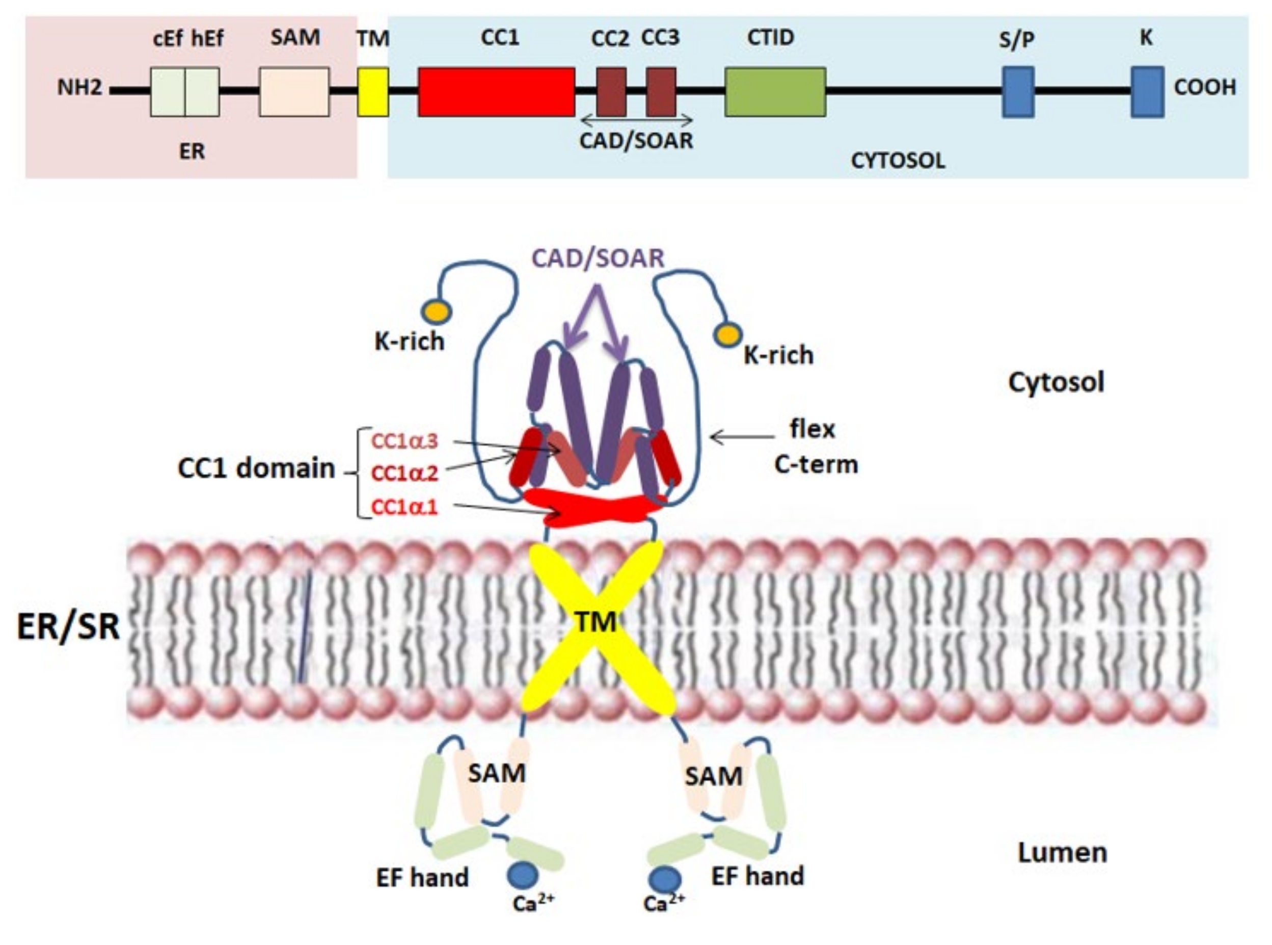
2.2. STIM1 Protein: The Ca2+ Sensor for SOCE

Stromal interaction molecule (STIM) proteins are single-pass transmembrane proteins located in the ER/SR, where they act as ER/SR Ca2+ sensors for SOCE. STIM1 knockdown and mutagenesis studies strongly contributed to clarify the Ca2+ sensor property associated with these proteins [27,28]. In mammals, the STIM protein family includes two homologs, STIM1 and STIM2, with three variants for STIM2, (STIM 2.1, STIM 2.2, and STIM 2.3) [29]. The Ca2+ sensing domain is located at the N-terminus region of STIM1, facing the ER/SR luminal side, and consists of a canonical EF-hand (cEFh), a non-canonical EF-hand (ncEFh), and sterile-motif (SAM) domains. SAM is followed by the transmembrane (TM) domain. Although Ca2+ binds only to the cEF-domain, the stability of the whole EF-hand-SAM domain is important for its Ca2+ sensing role [30,31]. Furthermore, negatively charged acid residues D76, D84, and E87 in the cEF-hand are pivotal for sensing Ca2+ levels in the ER/SR [24,32]. The essential sites for coupling to Orai1 are located in the STIM1 C-terminus region, placed in the cytoplasmic side of ER/SR. These binding sites include: three conserved cytosolic coiled-coil (CC) domains (CC1, CC2, CC3), a proline/serine-rich domain and, at the very end of the C-terminus, a lysine-rich domain, which participates in Orai1-independent plasma membrane targeting of STIM1 [33,34]. The CC1 domain can be separated into CC1α1, CC1α2, and CC1α3, and participates in the self-oligomerization of STIM1 at rest [35]. Furthermore, CC2 and CC3 domains, which comprise a CRAC activation domain/STIM1–Orai1 activating region domain (CAD/SOAR domain), interacts and activates Orai1 [36]. The CAD/SOAR domain also participates in the self-oligomerization of STIM1 [37]. Furthermore, the STIM1 C-terminus region includes the C-terminal inhibitory domain (CTID), which interacts with the Ca2+ entry regulatory protein SARAF in the resting state and is responsible for the regulation of the slow Ca2+ inactivation dependent on Orai1 [38] (Figure 1). To date, it is known that, in addition to SARAF, there are several auxiliary proteins which, through direct interactions with STIM1 and/or Orai1, favor or reduce the influx of Ca2+. For example, several studies have shown that STIMATE (STIM-activating enhancer), an ER/SR transmembrane protein encoded by the TMEM110 gene, interacts directly with STIM1, favoring the conformational change of STIM1 and contributing to maintaining the correct structure of the ER/SR-PM junctions [39,40,41]. Furthermore, it has been shown that STIMATE depletion reduces the formation of STIM1 points at the ER-junctions [39,40,41]. Moreover, in skeletal muscle cells, an alternatively spliced variant of STIM1 is also expressed. STIM1L (L for long, as it encodes an extra 106 amino acids) is a longer version of STIM1 that contributes to the skeletal muscle SOCE activation. Unlike the diffuse distribution of STIM1 at the resting state, STIM1L appears to be pre-localized at the ER/SR-PM junctions where it interacts with cytoskeletal actin and forms a permanent cluster with Orai1 [42]. This pre-formed STIM1L-Orai1 cluster can potentially explain the faster SOCE activation and extracellular Ca2+ entry in skeletal muscle compared with other cell types [43,44]. It has also been reported that STIM1L can interact with TRPC1 and TRPC4 [34,45]. In particular, a recent study demonstrates that STIM1L interacts preferentially with TRPC1 while being less efficient in Orai1 gating, then defining independent and specific interactions and functions of the two sliced forms [45]. Further focused studies are needed to gain better insight into the interactions between these proteins.
STIM1 and STIM2 are characterized by a > 74% sequence similarity (66% sequence identity) between their key domains (EF/SAM domains, CC1, SOAR), but work differently as Ca2+ sensors and activators of SOCE [46]. Although STIM2 is an analogue protein of STIM1, its functional role and contribution to the whole SOCE-mediated Ca2+ signaling in skeletal muscle are not clear. An initial study on the role of STIM2 in SOCE demonstrated that STIM2 was a weaker Orai1 activator and a slow responder to ER luminal Ca2+ changes compared to STIM1 [47]. Successively, Ong et al. reported that STIM2 is activated under a mild depletion of Ca2+ stores and is able to form heterodimers with STIM1, thus increasing the recruitment of STIM1 to the ER/SR-PM junction and facilitating its activation [48]. A subsequent study showed that, in STIM2-knockdown mouse primary skeletal myotubes, STIM2 is able to interact with SERCA1a, causing a reduction of its activity during skeletal muscle contraction [49]. In addition, SOCE is significantly reduced after STIM2-knockdown, suggesting that STIM2 also contributes to SOCE in skeletal muscle [50]. Furthermore, STIM2 variants have different roles in the modulation of SOCE; STIM2.1 and STIM2.2 have been described to play as an inhibitor and an activator of SOCE, respectively, while the role of STIM2.3 still remains unclear [50].

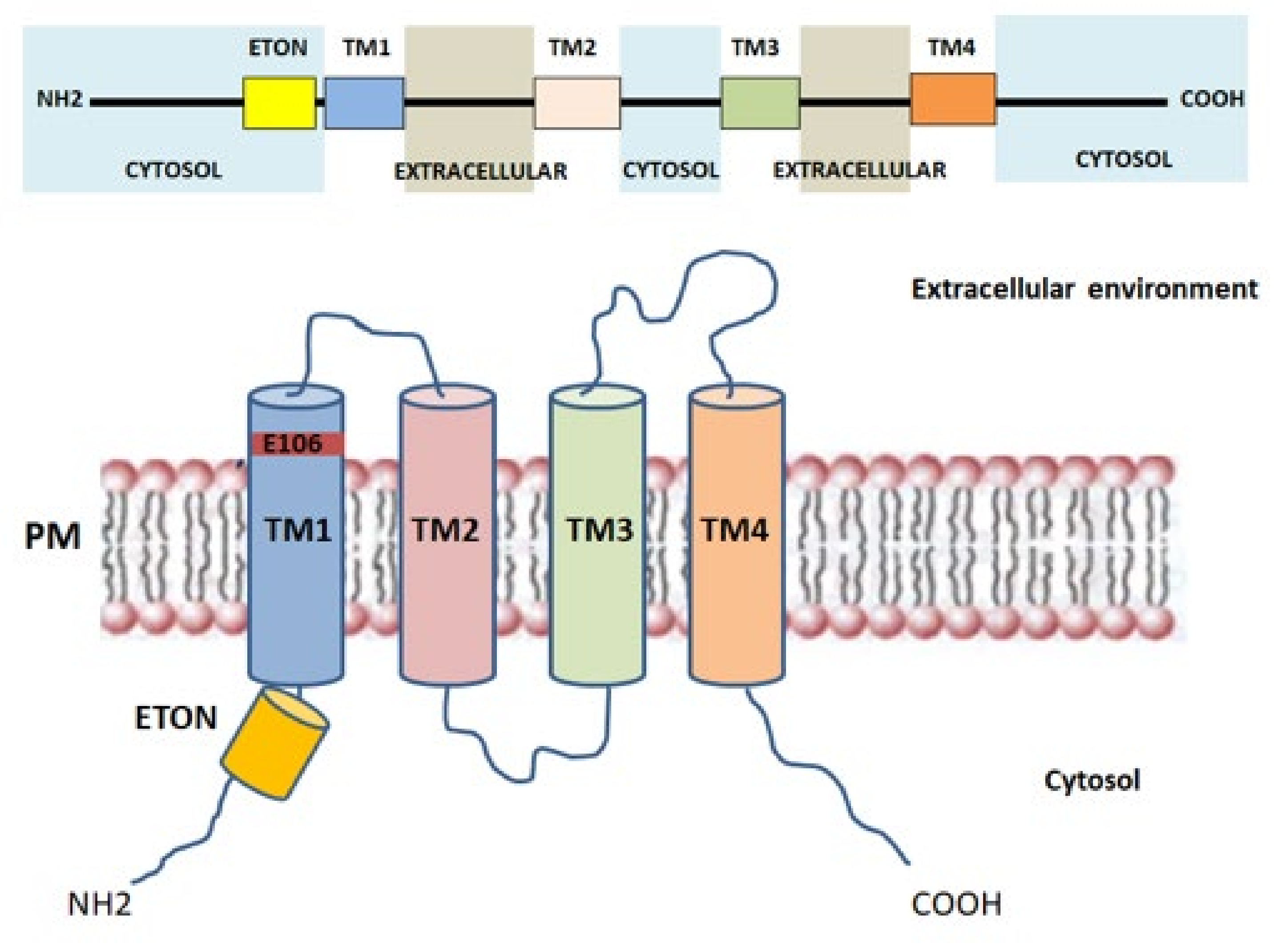
2.3. Orai1: The Key Component of CRAC Current
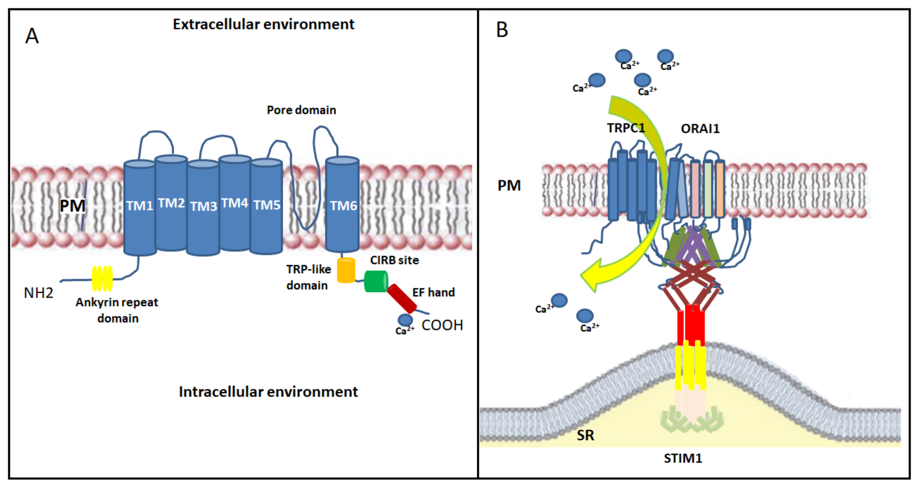
Orai proteins have been identified as key components of the Ca2+ release-activated Ca2+ channel (CRAC channel) [21,51] and are considered the major SOCE-mediating channels in skeletal muscle cells [52,53]. Particularly, ORAI (also called CRACM) proteins are located in the transverse tubules of PM and are responsible for the formation of the Ca2+-selective ion pores. Three Orai isoforms (Orai1-3, or CRACM1-3) encoded by homologous genes and two versions of Orai1, Orai1α and Orai1β, arising from alternative translation initiation [54], were identified in the human genome [55]. The presence of a point mutation (R91W) in Orai1, leading to loss of ICRAC current in human T cells, suggested the link between Orai1, in both Orai1α and β isoforms, and CRAC channel function [21,56,57,58]. Orai channels form hexameric complexes arranged around a central highly Ca2+-selective pore [59]. Each Orai subunit is composed of four transmembrane helices (TM1-TM4) connected by one intracellular (TM2-TM3) and two extracellular loops (TM1-TM2, TM3-TM4) with the N- and C-regions facing the cytoplasm that mediate the interaction with STIM1, STIM2, and other regulatory proteins [25] (Figure 2). The Ca2+ pore is formed by six TM1 domains surrounded by TM2-TM3, which provide stability to the structure [60], and by a cytosolic C-terminus. The glutamate at position 106, situated at the extracellular end of TM1, provides the binding site for Ca2+ ions inside the channel and confers the high Ca2+ selectivity to the CRAC channel [55,61]. Close to TM1 region, a conserved sequence called extended transmembrane Orai1 N-terminal (ETON) region is present. This region contributes to the interaction between the N-terminus of Orai1 and STIM1 [62]. Indeed, Orai1 mutants that lack the ETON region result in a reduced interaction with STIM1 [62]. The TM4 outer ring transduces the gating signal generated by the binding of STIM1 to the ORAI1 C-terminus [52,63]. Evidence demonstrates that the TM3-TM4 loop is pivotal for STIM1-mediated Orai1 gating [64] as it controls the interaction with Orai N-terminus and promotes a permissive conformation of Orai channel [65]. Orai1 gating depends on the folding state of STIM1 located in the ER/SR membrane. The interaction between Orai1 and STIM1 leads to conformational changes of the C-terminus in the Orai1 complex, that are subsequently transmitted to the pore region [66]. Furthermore, it has been shown that, in mouse skeletal myotubes, Orai1 was able to interact with Mitsugumin 53, a protein responsible for membrane repair of various cells, finally increasing Ca2+ entry through Orai1 via a SOCE mechanism [67]. The physiological roles of Orai2 and Orai3 in skeletal muscle are still poorly defined. The biophysical properties of Orai2 and Orai3, including Ca2+ permeation, are different from those of Orai1, suggesting that these proteins are little involved in SOCE [68]. Recently, Yoast et al. demonstrated that native Orai2-3 heteromerize with Orai1 to form a native CRAC channel regulating the activation of NFAT1 and NFAT4 isoforms. These results suggest that the levels of Orai2-3 relative to Orai1 can alter the magnitude of SOCE and downstream signaling [69].

2.4. TRP Channels

In skeletal muscle, the non-selective transient receptor potential channels (TRPCs) have also been proposed as emerging SOCE channels [70]. Indeed, STIM1, ORAI1 and TRP channels act efficiently and are located near the triad junction [71]. TRPCs are activated downstream of the PLC/ IP3/DAG pathway; they contribute to the entry of extracellular Ca2+ with moderate Ca2+ selectivity [72]. TRP channels include seven isoforms (TRPC1-7), but only TRPC1 and TRPC3-6 are expressed in skeletal muscle [73,74]. TRPC consist of six transmembrane domains (TM1-TM6) with the TM5-TM6 loop forming the channel pore and with the N- and C-regions facing into the cytoplasm [75]. The cytosolic N-terminus contains an ankyrin repeat domain; while the cytosolic C-terminus contains a TRP-like domain for phosphatidylinositol biphosphate (PIP2) regulation necessary for channel activity and desensitization, a calmodulin/inositol 1,4,5-trisphosphate receptor-binding (CIRB) site and a Ca2+ binding EF-hand [76] (Figure 3A). Among all skeletal muscle isoforms, the best characterized is TRPC1, which has a role in Ca2+ homeostasis during sustained contractile muscle activity and is considered the best candidate to conduct Ca2+ influx during SOCE. TRPC1, which acts as a SOCE channel, participates in myoblasts migration and fusion via calpain activation during the terminal differentiation process [76]. Indeed, Antigny and colleagues demonstrated that TRPC1 and TRPC4-mediated Ca2+ entry was important to produce normal-sized myotubes. Conversely, overexpression of STIM1 with TRPC4 or TRPC1 increased SOCE, accelerated myoblast fusion, and produced hypertrophic myotubes [77]. Additionally, the Ca2+ entry through TRPC1 has been shown to play a role in myoblast differentiation and in muscle regeneration by modulating the PI3K/Akt/mTOR/p70S6K pathway. In fact, in myoblasts derived from Trpc1−/− mice, the phosphorylation of both Akt and p70S6K proteins, as well as the activation of PI3K, were inhibited. This evidence suggests that Ca2+ entry through TRPC1 plays a role in the activation of this pathway [78]. TRPC1 or TRPC4 has been shown to complex with STIM1 and STIM1L, thus contributing to the activation of skeletal muscle SOCE associated with sustained Ca2+ transients during repetitive membrane depolarizations. This process, in turn, promotes myogenesis and maintains a fast repetitive Ca2+ release in human skeletal myotubes [45]. TRPC1 seems to be activated by both STIM1 isoforms, with STIM1L more efficient in activating TRPC1than STIM1 [45,46]. Furthermore, recent studies reported that the TRPC1 mediated currents did not reproduce the biophysical properties of ICRAC [29], defining TRPC1 current as Isoc. However, this observation does not rule out the TRPC channels’ involvement in SOCE. Experimental studies have shown that STIM1 interacts with and directly activates TRPC1 channels not only by binding through its SOAR region with TRPC1, but also through electrostatic interactions between highly positively charged lysines located in the STIM1 lysine-rich domain and the TRPC1 aspartate residues [70,79]. STIM1 residues involved in TRPC1 gating are distinct from those involved in Orai1 activation. In addition, a different mechanism of TRPCs versus Orai channels by STIM1 is suggested [79]. However, activation of TRPC1 requires an additional crucial functional interaction with Orai1. This observation was confirmed by data showing that Orai1-mediated Ca2+ entry triggers the recruitment of TRPC1 into the PM where it is then activated by STIM1 [80,81]. In human skeletal muscle the expression of TRPC3 and TRPC6 isoforms is firmly established, and it has been found that Orai1 is able to bind to the N- and C- terminals of TRPC3 or TRPC6. Actually, it is not clear if they are able to act as SOCE channels without Orai1 interaction [82]. Furthermore, in skeletal muscle, STIM1L, like Orai1, is known to interact with TRPC3 and TRPC6 [45,83], but it is not clear whether this interaction is fundamental for SOCE mechanism.
Finally, the overall findings on STIM1, Orai1 and TRPC suggest the formation of a dynamic STIM1-Orai1-TRPC1 ternary complex as the SOC channels; after store depletion, STIM1 oligomers transfer to the ER/SR-PM junctions where they bind Orai1 and TRPC1 in lipid raft domains, activating both Ca2+ channels [29] (Figure 3B). Further studies on the relationships between TRPCs, Orais and STIM1 are needed to examine this and other scenarios.

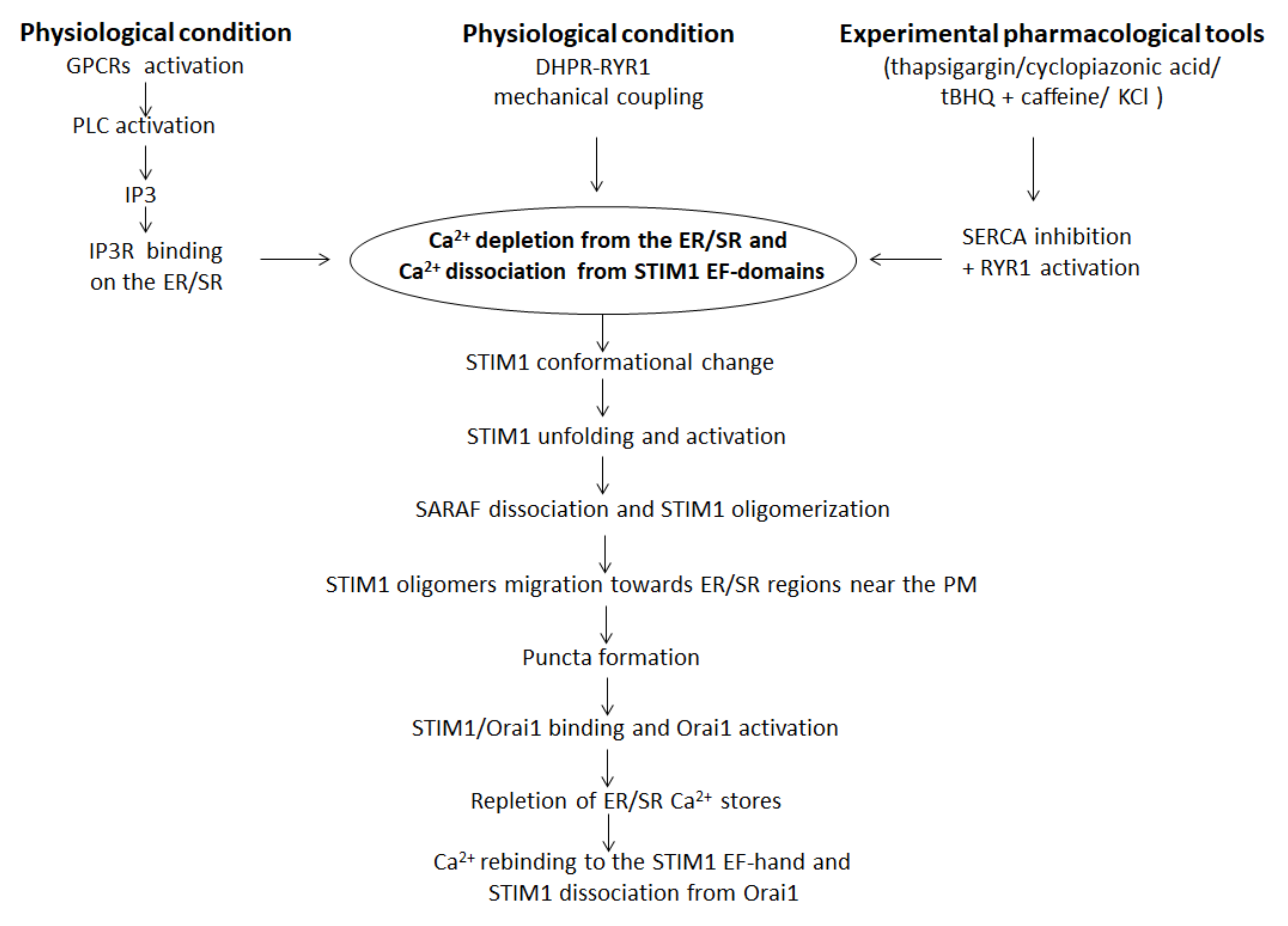
3. SOCE Mechanism in Skeletal Muscle: An Overview

SOCE, also known as capacitive calcium entry (CCE), consists in a Ca2+ influx downstream of ER/SR Ca2+ stores and it is a pivotal mechanism in cellular calcium signaling and in maintaining cellular calcium homeostasis [84]. The concept of SOCE was first postulated by Putney in 1986 who demonstrated that in salivary gland cells the depletion of internal Ca2+ stores controlled the extent of Ca2+ influx [85]. Successively, in 2001, SOCE was identified in skeletal muscle fibers of adult mice for the first time [86]. Generally, SOCE is activated in response to the emptying of the SR/ER-Ca2+ stores and is followed by a limited increase in [Ca2+]i due to the smaller SR/ER volume. Two major families of intracellular Ca2+ channels are responsible for linking extracellular stimuli to the release of Ca2+ from the ER/SR: RyRs, which are opened by their direct interaction with the voltage-sensing dihydropyridine receptor in the PM, and the inositol 1,4,5-trisphosphate receptors (IP3R). In the second case, depletion of the ER/SR store essentially occurs following the stimulation of plasma membrane G protein-coupled receptors (GPCRs) through the activation of phospholipase C (PLC) subtypes and the related release of inositol 1,4,5-triphosphate (IP3); IP3 binds to the IP3R on the ER/SR membrane, triggering Ca2+ release from the ER/SR lumen into the cytosol [87]. Experimentally, thapsigargin, cyclopiazonic acid and 2,5-di-(tert-butyl)-1,4-benzohydroquinone associated with a RYR1-activator (such as caffeine or KCl) are the most commonly used pharmacological tools for inhibiting the ER/SR SERCA pump and depleting ER/SR Ca2+ independently of receptors and IP3 [88,89,90]. However, following ER/SR Ca2+ store depletion, a secondary influx of extracellular Ca2+ through CRAC channels in the plasma membrane results in a more sustained increase in cytosolic Ca2+ levels. Different SOCE models in skeletal muscle have been proposed, including the interaction between STIM1 and Orai1 [70], STIM1 and transient receptor potential canonical channels (TRPCs) [79], and STIM1-Orai1-TRPCs [29]. Among these, the direct coupling of ER-resident STIM1 to plasma membrane-resident Orai1 is currently considered as the most straightforward SOCE mechanism, and it has been studied in depth.
In the resting state, STIM1 is a dimer associated with its regulator SOCE-associated regulatory factor (SARAF), a single-pass transmembrane protein [91]. The process begins when depletion of Ca2+ from the ER/SR leads to Ca2+ dissociation from the luminal STIM1 N-terminal EF-hand domains. This determines the beginning of dramatic conformational changes in STIM1 that lead to the formation of an unfolded STIM1 protein and more closely associated luminal EF-hand domains. Unfolding and activation of STIM1 upon Ca2+ dissociation causes SARAF dissociation [91] and STIM1 oligomerization [92] by associating the paired EF-SAM domains [30] and by the exposition of the STIM1-Orai1 activating region domain (SOAR domain) [36]. Successively, STIM1 oligomers migrate towards ER/SR regions juxtaposed to the PM and bind to it through the interaction between the poly-lys rich domain near the STIM1 C-terminus and the PM phospholipids [92], resulting in the “puncta” formation [93]. At the same time, rearrangements of some portions of the ER/SR towards the PM favor STIM1 interaction and puncta formation [94]. After puncta formation, STIM1 oligomers are able to bind with the N- and C- termini of Orai1 through SOAR region and activate Orai1 channels for Ca2+ entry from the extracellular environment [84,93,95,96] (Figure 4). It is well recognized that STIM1 is a phosphoprotein [97] and biochemical studies strongly contributed to understand the signaling responsible for the correct interaction between STIM1 and Orai1, and consequently for the correct functioning of SOCE. Particularly, Yazbeck et al. showed that STIM1 could be modulated by a Pyk2-dependent tyrosine phosphorylation at Y361 within the SOAR domain. This seems to be a critical step in activating Ca2+ entry through Orai1 channels since it is required for Orai1 recruitment into STIM1 puncta and for STIM1-Orai1 interaction [98]. Furthermore, Lopez et al. showed that STIM1 phosphorylation at Y316 could enhance the formation of the CRAC signaling complex, which contribute to SARAF dissociation from STIM1 and regulation of slow Ca2+-dependent inactivation [91].
Another hypothesis on the SOCE mechanism postulates that, in skeletal muscle, STIM1 and Orai1 pre-localize under resting conditions within the triad junction, a specialized macrostructure composed of a parallel transverse tubule and two opposing ER/SR membranes. They remain inactive until ER/SR depletion triggers conformational changes in STIM1 and direct activation of Orai1-mediated Ca2+ influx [84]; this allows an extremely fast and efficient trans-sarcolemmal Ca2+ influx during store depletion. Accordingly, in skeletal muscle, SOCE occurs in less than a second, i.e., significantly faster than in other types of cells where it can require up to several seconds [99].
The precise stoichiometry of the STIM1-Orai1 complex that forms the functional core of the CRAC channel still needs clarification and it has long been a subject of debate [33]. Several studies hypothesized that a dimer of STIM1s binds to a pair of Orai1 C-terminal fragments (in a 1:1 STIM1:Orai1 stoichiometry) [100,101,102]. Alternatively, each dimer interacts with only a single C-terminal tail, leaving the remaining STIM1 subunit free to cross-link with a different Orai1 channel (two STIM1 molecules around a single Orai1 channel, in a 2:1 STIM1:Orai1 stoichiometry) [103]. More recently, it has been reported that the native SOCE complex includes only a few STIM1 dimers associated with a single Orai1 channel [104].
SOCE terminates following the reuptake of Ca2+ by ER/SR SERCA protein or following the export of cytosolic Ca2+ to the extracellular area by PMCAs [105]. Upon store refilling, luminal Ca2+ rebinds to the STIM1 EF-hand, STIM1 dissociates from Orai1, and STIM1 and Orai1 revert to their diffuse distributions [106].

4. STIM1/Orai1-Mediated SOCE Alteration and Skeletal Muscle Diseases

Generally, the SOCE mechanism has traditionally been known for serving as the primary route to rapidly replenish depleted intracellular Ca2+ stores to maintain the appropriate environment within the ER/SR for protein folding/processing, vesicle trafficking, and cholesterol metabolism [107]. In skeletal muscle, it is commonly accepted that Ca2+ entry via SOCE has the crucial role in short-term and long-term muscle function.
In regard to short-term function, i.e., muscle contractility, the faster SOCE mechanism is required for ER/SR Ca2+ refilling during repolarization cycles, to complement Ca2+ recycling to the ER/SR by the SERCA and support ER/SR Ca2+ release [108]. Furthermore, SOCE mechanism is required for maintaining contractile performance during periods of prolonged activity. The muscle fibers ability to recover Ca2+ ions from the extracellular environment via STIM1/ORAI1-mediated SOCE represents a mechanism that allows the ER/SR Ca2+ refilling to maintain Ca2+ release during periods of high-frequency repetitive stimulation.
Importantly, SOCE has also been proposed to contribute to key myogenic events important for long-term skeletal muscle functions, such as myoblast fusion/differentiation and muscle development [52,109]. This role is supported by studies showing that STIM1, Orai1, or Orai3 silencing reduced SOCE amplitude that is linearly correlated with the expression of myocyte enhancer factor-2 (MEF2) expression and myogenin muscle-specific transcription factors involved in myogenesis process [110]. Furthermore, SOCE regulates myoblast differentiation through the activation of downstream Ca2+-dependent signals such as the nuclear factor of activated T-cells (NFAT), mitogen-activated protein (MAP) kinase and ERK1/2 [71]. Interestingly, SOCE involvement in muscle development is demonstrated by the augmented STIM1/ORAI1 expression and the consequent increased SOCE during differentiation of myoblasts to myotubes [32,71,110]. This role is more evident in the late phase of differentiation as puncta appear during the terminal differentiation in a ER/SR depletion-independent manner [84]. It has been also shown that in human myotubes the TRPC1/TRPC4 knockdown reduces SOCE, while the STIM1L knockdown negatively affects the differentiation of myoblasts and leads to the formation of smaller myotubes. This indicates that SOCE mediated by TRPC1, TRPC4 and STIM1L appear to be indispensable for normal differentiation [45].
The SOCE mechanism in adult skeletal muscle also reduces fatigue during periods of prolonged stimulation [52,111,112], as well as serving as a counter-flux to Ca2+ loss across the transverse tubule system during EC coupling [113].
According to this key role in a plethora of muscle determinants and functions, abnormal SOCE is detrimental for skeletal muscle and results in loss of fine control of Ca2+-mediated processes. This leads to different skeletal muscle disorders including muscular hypotonia and myopathies associated to STIM1/ORAI1 mutations [2,3,4], muscular dystrophies [5,7], cachexia [8] and sarcopenia [9,10,11,12,13].

4.1. STIM1/Orai1-Mediated SOCE Alteration in Genetic Skeletal Muscle Disorders

As detailed above, proper functioning of SOCE is important for maintaining healthy skeletal muscle processes. Involvement of SOCE in genetic skeletal muscle diseases has been proposed when a missense mutation (R91W) in the first transmembrane domain of Orai1 was found in patients suffering from severe combined immunodeficiency (SCID) and presenting myopathy, hypotonia and respiratory muscle weakness [19]. Successively, a mutation in STIM1 was also identified in patients with a syndrome of immunodeficiency and non-progressive muscular hypotonia [113]. Over the past decade, single-point gene mutations have been identified in CRAC channels that cause skeletal muscle diseases and the information gained through functional studies has been used to propose therapeutic approaches for these diseases. Several loss-of-function (LoF) and gain-of-function (GoF) mutations in Orai1 and STIM1 genes have been identified in patients affected by distinct disease syndromes [114].
To date, thirteen different STIM1 and Orai1 LoF gene mutations have been described (STIM1: E128RfsX9, R426C, P165Q, R429C; 1538-1G>A; Orai1: R91W, G98R, A88SfsX25, A103E, V181SfsX8, L194P, H165PfsX1, R270X), all of them resulting in a marked reduction of SOCE function [115]. LoF R91W mutation in Orai1, for example, can reduce Orai1 activity leading to a depressed SOCE and causing muscular hypotonia along with severe SCID [21]. Patients with A103E/L194P Orai1 mutation also show muscle weakness and hypotonia [116]. LoF mutations in STIM1 (R426C, R429C mutations) can reduce STIM1 functionality and alter STIM1-Orai1 interaction [117], leading to a reduced and insufficient SOCE and causing CRAC channelopathies. Particularly, CRAC channelopathies are characterized by SCID, autoimmunity, ectodermal dysplasia, defects in sweat gland function and dental enamel formation, as well as muscle hypotonia [3,21].
In contrast, GoF mutations in STIM1 and/or Orai1 induce the production of a protein that is constitutively active and results in SOCE over-activation and excessive extracellular Ca2+ entry [2,118,119]. In skeletal muscle, the main diseases related to GoF mutations in STIM1 and/or Orai1 are the non-syndromic tubular aggregate myopathy (TAM) and the more complex Stormorken syndrome [114,118,119,120]. TAM is an incurable clinically heterogeneous and ultra-rare skeletal muscle disorder, characterized by muscle weakness, cramps and myalgia [121,122]. Muscular biopsies of TAM patients are characterized by the presence of typical dense arrangements of membrane tubules originating by SR called tubular aggregates (TAs) [2,119,120,123,124]. Some patients show the full picture of the multisystem phenotype called Stormorken syndrome [114], a rare disorder characterized by a complex phenotype including, among all, congenital miosis and muscle weakness. Some patients with Stormorken syndrome carry a mutation in the first spiral cytosolic domain of STIM1 (p.R304W). This mutation causes STIM1 to be in its active conformation [125] and promotes the formation of STIM1 puncta with the activation of the CRAC channel even in the absence of store depletion, with consequent gain-of-function associated with STIM1 [125].
To date, fourteen different STIM1 GoF mutations are known in TAM/STRMK patients, including specifically twelve mutations in the EF-domain (H72Q, N80T, G81D, D84E, D84G, S88G, L96V, F108I, F108L, H109N, H109R, I115F) and two mutations in luminal coiled-coil domains (R304W, R304Q) [114,126,127]. All mutations present in the EF-domain induce a constitutive SOCE activation due to the ability of STIM1 to oligomerize and cluster independently from the intraluminal ER/SR Ca2+ level, leading to an augmented concentration of intracellular Ca2+ [120]. Regarding Orai1, several mutations are present in TM domains forming the channel pore or in concentric rings surrounding the pore (G97C, G98S, V107M, L138F, T184M, P245L) [2,3,118,123,128] and induce a constitutively active Orai1 protein, and an increased SOCE mechanism contributing to TAM pathogenesis [2]. For example, Orai1 V107M mutation, located in TM1, can alter the channel Ca2+ selectivity and its sensitivity to external pH and to STIM1-mediated gating [128]; Orai1 T184M mutation, located in TM3, is associated with altered Orai1 susceptibility to gating and conferred resistance to acidic inhibition [128].
Only a few STIM1 and Orai1 mutations have been functionally characterized in native skeletal muscle cells, most of them having been studied in heterologous expression systems. This represents an overt limitation both for the limited reliability of the cellular model and for the translation of drug efficacy in humans. TAM animal models exist and broadly recapitulate the clinical signs of human disorders but, unfortunately, only partially replicate muscle symptoms [3]. Particularly, the STIM1 I115F and R304W TAM/STRMK mouse models show the TAM clinical phenotype in terms of reduced muscle force, elevated serum CK levels, ER stress, mitochondria loss especially in the soleus muscle, reduction of fiber diameter with signs of apoptosis, and enhanced muscle fiber degeneration and regeneration cycles. However, the same animal models do not exhibit TA, highlighting a large structural difference between humans and mouse models [129,130,131]. Therefore, like other muscular pathologies still without cure, the creation of cell models obtained from patients with different forms of TAM could represent a very important strategy to perform preclinical studies aimed to develop specific TAM therapies. More recently the functional characterization of isolated myoblasts from biopsies of TAM patients carrying the GoF L96V STIM1 mutation and of related differentiated myotubes has been performed [4]. Interestingly, along the differentiation process, the higher resting Ca2+ concentration and the augmented SOCE characterizing STIM1 mutant muscle cells matched with a reduced cell multinucleation and with a distinct morphology and geometry of the mitochondrial network indicating a defect in the late differentiation phase [4]. These findings provided evidence of the mechanisms responsible for a defective myogenesis associated with TAM mutation. Besides explaining the myofiber degeneration, this study emphasized the importance of normal SOCE beyond an effective muscle contraction and validated a reliable cellular model useful for TAM preclinical studies.

4.2. SOCE Dysfunction in Duchenne Muscular Dystrophy

Muscular dystrophies are a group of inherited skeletal muscle diseases that affect both children and adults and mainly involve muscle tissues causing progressive muscle degeneration and contractile function reduction with severe pain, disability and death [132]. To date, more than 50 distinct types of muscular dystrophies have been identified, but one of the most severe and common muscular dystrophy is Duchenne Muscular Dystrophy (DMD), an X-linked disorder caused by mutations in the DMD gene that abolish the expression of dystrophin protein on the plasma membrane [133]. Dystrophin is a structural protein that connects cytoskeletal actin to laminin in the extracellular matrix stabilizing the sarcolemma and protecting the muscle from mechanical stresses [134]. It is part of a complex called dystrophin glycoprotein complex (DGC) which contains 11 proteins: dystrophin, the sarcoglycan subcomplex (α-sarcoglycan, β-sarcoglycan, γ-sarcoglycan and δ-sarcoglycan), the dystroglycan subcomplex (α-dystroglycan and β-dystroglycan), sarcospan, syntrophin, dystrobrevin and neuronal nitric oxide synthase (nNOS) [135]. In muscles from DMD animal models and in patient-derived cells, the lack of dystrophin induces a destabilization of sarcolemma and leads to abnormal clustering of potassium ion channels and altered ion channel functions. This alters Ca2+ homeostasis, finally increasing intracellular Ca2+ levels [136]. Particularly, dystrophic muscle fibers from mdx mice or DMD patients show significantly elevated levels of intracellular Ca2+ due to extracellular Ca2+ entry approximately twice that of control muscle fibers [6,7,137,138]. Various evidence supports that the increased calcium entry can be a direct consequence of the absence of dystrophin and/or of the altered signaling and reactive oxygen species [137,139]. A key role of voltage-independent calcium channels, belonging to the TRP-like channel family and mechanosensitive PIEZO 1, has been proposed and partly demonstrated functionally and biochemically [140]. The increase in sarcolemmal Ca2+ influx triggers the activation of calpains, phospholipase A2 and Ca2+-activated kinases, such as PKC, and may act in a reinforcing loop with the mitochondrial dysfunction and the production of reactive oxygen species (ROS) [139]. Then, calcium homeostasis dysfunction is believed to contribute to pathological events triggering the characteristic histological and biochemical features of muscular dystrophy, thus playing a key role for the progressive damage observed in DMD [7,84,141,142,143]. In this context, a role of SOCE has also been proposed. In mdx muscle, both STIM1 and Orai1 are upregulated, therefore SOCE is more active and may well contribute to the increased intracellular Ca2+ level [99]. Although it is well established that SOCE is more active in DMD, the correlation of this cellular event with Ca2+ overload is yet under investigation. At first, Boittin and colleagues hypothesized that products of Ca2+-independent PLA2, such as lysophosphatidylcholine, are able to activate the SOCE process through a Ca2+-independent pathway without changing the threshold for SR Ca2+ [144]. Successively, studies have provided evidence for a modulatory contribution of STIM1/Orai1-dependent Ca2+ influx to the dystrophic phenotype of mdx mice. Indeed, as a contributing cause of higher Ca2+ entry in mdx dystrophic muscle fibers, higher SOCE is reported through Orai1 upregulation or Stim1 overexpression [145]. Importantly, part of the increased cytosolic calcium and entry via SOCE can also derive from the leaky oxidized RyR1 receptor on SR, which may in part contribute to store depletion and impaired EC coupling [7,12]. In addition, as anticipated above, besides STIM1 and Orai1, TRPC could be responsible for the higher Ca2+ entry in dystrophic myotubes. Indeed, studies on muscle-specific transgenic mice with a TRPC3 overexpression showed that Ca2+ influx across this TRP channel isoform contributes to the dystrophic muscle phenotype [146]. Furthermore, TRPC1 activity is higher in dystrophic myotubes from mdx mice and DMD patients and can be responsible of augmented intracellular Ca2+ [147]. In skeletal muscle, TRPC1 is anchored to cytoskeletal proteins, such as dystrophin or caveolin-3, and this link contributes to the higher activity of TRPC1 and to the higher SOCE observed in mdx myotubes [143].

4.3. SOCE Dysfunction in Skeletal Muscle Wasting Disorders: Cachexia and Sarcopenia

Several pathological conditions are characterized by loss and/or impairment of muscle and muscle wasting. When muscle wasting is present, it is always related to greater morbidity and reduced survival in chronic disease states, favoring the onset of negative outcomes and death [148]. The major muscle-wasting disorders are age-related sarcopenia and cachexia. Both conditions are characterized by an alteration of Ca2+ homeostasis and the SOCE mechanism that contribute to impaired muscle functions, poor quality of life and disease progression.
Cachexia is defined as a debilitating wasting that manifests in several types of cancer and, at the same time, represents a serious and dose-limiting consequence of cancer chemotherapy [149]. Cachectic patients present unintentional weight loss due to the activation of the intracellular protein degradation apparatus, such as the ubiquitin-proteasome, mitogen-activated protein (MAP) kinases or myostatin [150], and a reduced protein synthesis that leads to an ongoing loss of skeletal muscle mass (with or without loss of fat mass) [149,150]. Loss of muscle mass contributes, with other causes, to the decline in skeletal muscle function present in cancer as it increases susceptibility to the adverse effects of chemotherapy [151]. Recently, the use of an animal model of cachexia, obtained with cisplatin administration to rats, proved very useful to shed light on calcium homeostasis alteration in cachectic skeletal muscle fibers [8]. Importantly, Ca2+ overload observed in cachectic skeletal muscle, probably due to SOCE-independent mechanisms, is associated with a reduced response to the application of depolarizing solution or caffeine, as well as with a reduced SOCE in terms of functional activity and gene expression. Particularly, a down-regulation of STIM1, ORAI1, RyR1 and Dhpr muscle gene expression was observed in cachectic animals with respect to controls [8]. Considering the interaction between DHPR and RyRs that occurs during EC coupling, these findings could explain the impairment of the EC coupling mechanism and the structural muscle alteration observed in cachexia [8]. Ca2+ overload and SOCE alteration observed in cachectic muscle can exert deleterious effects that lead to muscle damage. This is due to the activation of Ca2+-activated proteases (calpains) and the disruption of the integrity of the sarcolemma, all events contributing to the loss of strength muscle [152].
Aging is a multifactorial biological process characterized by a progressive decline of the main physiological functions that gradually leads to dysfunctions of various tissues including skeletal muscle [153]. Normal aging involves sarcopenia, a complex irreversible age-related muscle condition characterized by a generalized reduced skeletal muscle mass (atrophy) and strength, increased fatigability, and reduced velocity of contraction [154]. Sarcopenic muscles show a reduced myofibers size and hypotrophic myofibers [154], an accumulation of intramuscular fat, fibrosis, chronic inflammation, and impaired muscle regeneration caused by the reduced ability of satellite cells to activate and proliferate [155]. The resulting muscle weakness significantly contributes to the debilitating injuries caused by repetitive falls that lead to a deterioration in quality of life in the elderly population [156]. Reduced specific contractile force of sarcopenic muscle can be explained by the reduced intracellular Ca2+ ions available to activate the contractile filaments, associated with a decrease in DHPR expression and consequent uncoupling between DHPR and RYR1 proteins [157]. Moreover, during aging, oxidative stress is present and stress-induced protein oxidation is increased [158]. Skeletal muscle of aged rodents showed oxidized RyR1 depleted of the channel-stabilizing subunit calstabin1 [12]. This oxidation resulted in a “leaky” RyR1 with an increased single-channel open probability that leads to intracellular calcium leak in skeletal muscle [12]. At the same time, several studies have also shown that reduced STIM1/Orai1 mediated SOCE is present in sarcopenic skeletal muscle which may contribute to the significant decline in contractile strength during normal aging [13,159]. In particular, Zhao and colleagues showed that SOCE is severely reduced in muscle fibers isolated from aged mice, but this SOCE reduction occurs without altering the STIM1/Orai1 mRNA levels [159]. In accordance with this observation, the expression levels of neither STIM1 nor Orai1 changed during aging in humans, mice, or fly muscles [160]. Furthermore, it has been demonstrated that in soleus muscles, the SOCE-dependent components of contractile machinery, characterizing young muscle during repetitive contraction, is lost in aged muscle. These data support the hypothesis that the reduced SOCE observed in age-related sarcopenic muscles contributes to the decline in muscle contractile force and to the increase in susceptibility to fatigue [13].
Similar to TAM, a correlation between TAs formation and Ca2+ homeostasis alteration has been recently proposed for fast-twitch muscle fibers of elderly mice. In particular, it has been demonstrated that dysfunctional accumulation of proteins forming TAs, which include also STIM1 and Orai1, together with a concomitant SOCE alteration, were associated with a reduced ability to restore internal deposits of Ca2+ from the extracellular environment in aged skeletal muscle [161]. All these events could significantly contribute to muscle weakness and the increased fatigability observed during aging.
Despite several studies performed over the last years, the exact role of SOCE in sarcopenia remains controversial. For example, Edwards and colleagues demonstrated that SOCE remains unaffected in the skeletal muscle of aged mice despite an approximate 40% decline in STIM1 protein expression not accompanied by any alteration of Orai1 expression [162].

4.4. SOCE Dysfunction in Other Skeletal Muscle Pathological Conditions

Accumulating evidence has demonstrated that intracellular Ca2+ homeostasis and SOCE mechanism can be compromised in skeletal muscle pathological conditions involving proteins and/or intracellular organelles not directly related to SOCE, such as Ca2+ buffer proteins and/or mitochondria [163,164,165]. In particular, alteration of Ca2+ buffer proteins levels, such as calsequestrin or sarcalumenin, seems to be correlated to an altered SOCE [163,164]. Zhao et al., for example, using sarcalumenin knockout (sar−/−) mice, showed that the absence of sarcalumenin enhanced muscle SOCE mechanism ameliorating muscle fatigue resistance. The parallel increase in muscle MG29 expression suggested the occurrence of a compensatory change in Ca2+ regulatory proteins that affect SOCE when sarcalumenin is reduced or absent [163]. Similarly, Michelucci et al., using calsequestrin knockout (Casq1−/−) mice, showed that the absence of calsequestrin induced an increase of muscle SOCE mechanism with an increase of STIM1, Orai1, and SERCA expression associated with a high density of Ca2+ entry units (CEUs) [164]. Furthermore, other studies have suggested that mitochondria can modulate several steps in SOCE mechanism regulating SOCE activity [165,166,167]. In this context, Quintana et al. showed in T-lymphocytes that mitochondria translocate to the plasma membrane close to Ca2+ entry channels during Ca2+ entry and capture large amounts of Ca2+ entry [168]. This evidence suggests that mitochondrial dysfunction may be the cause and/or consequence of SOCE alteration. Further targeted studies are needed to gain a better understanding of the potential role of mitochondrial dysfunction in SOCE, with particular attention to skeletal muscle.

5. Therapeutic Perspectives for Counteracting SOCE-Related Skeletal Muscle Diseases

As knowledge about the role of SOCE in skeletal muscle diseases accumulates, there has been a growing interest in developing molecules targeting SOCE and identifying therapies that can be used for specific treatments. Indeed, several studies recently aimed to develop SOCE modulators to reduce SOCE activation following the pathological skeletal muscle GoF mutations mentioned above. For example, Rahaman and colleagues used in silico screening to identify FDA-approved drugs able to suppress the SOCE mechanism. Particularly, leflunomide and teriflunomide, FDA-approved drugs for the treatment of rheumatoid/psoriatic arthritis and multiple sclerosis, respectively, were able to inhibit SOCE at therapeutically-achievable concentrations; furthermore, lansoprazole, tolvaptan and roflumilast resulted in even more selective molecules to suppress the SOCE mechanism [169]. Recently, a variety of new small molecules blocking CRAC channels have been identified and developed, such as pyrtriazoles or pyrazole SKF-96365 analogues [131,170]. However, all currently available SOCE inhibitors show no specific effects [171,172].
Regarding dystrophies, and DMD in particular, at present there are no effective treatments and the glucocorticoids which act as anti-inflammatory agents are often used to stop progressive muscle damage [173,174]. Prednisone, prednisolone, and deflazacort, mostly through inhibition of NF-κB signaling, represent a gold standard for the treatment of DMD for their ability to exert long-term protective effects [175]. Importantly, to date, an increasing variety of therapeutic strategies aimed at restoring dystrophin production and to preserve muscle mass has been proposed, ranging from gene therapy to antisense oligonucleotide therapies [176,177]. Several studies propose therapeutic approaches for DMD aimed not only at restoring dystrophin function but also to mitigate secondary and downstream pathological mechanisms that contribute to the disease’s progression, such as calcium dysregulation, oxidative stress, mitochondria dysfunction, fibrosis and muscle wasting. Among all, since increased calcium concentration plays a significant role in the pathogenesis of DMD, therapeutic strategies aimed at controlling Ca2+ are in progress. The spider venom toxin AT-300/GsMTx4, a peptide that blocks the mechanosensitive Ca2+ channels, for example, prevented the rise of intracellular resting Ca2+ with modest benefits in mdx mice [178]. Another therapeutic option is treatment with the small drug ARM210/S48168, a ryanodine channel complex stabilizer, which improves muscle functionality in mdx mice, notably in the diaphragm [7]. Although SOCE increase in DMD is known, little evidence demonstrates that this alteration is linked to an increase in the STIM1/Orai1/TRPC expression. In this context, STIM1/Orai1/TRPC proteins may represent valuable therapeutic targets for testing compounds/drugs that regulate Ca2+ signal alteration in DMD, and focused studies in this field are highly desirable.
Finally, regarding skeletal muscle wasting disorders, knowledge of the real role of the SOCE mechanism, in particular during cachexia and aged-sarcopenia, is a fundamental requirement for finding a potential therapy. Nutrition is a key factor for the therapy of these conditions because both the quality and quantity of nutrients are pivotal for improving muscle anabolism, reducing catabolism, and lightening the prognosis [179]. However, although nutrition alone can prevent or minimize further skeletal muscle loss, it cannot completely reverse these conditions. For this reason, for example for cachexia, a multifactorial approach is currently proposed [180]. In this respect, a potential therapeutic option for cancer cachexia syndrome is represented by growth hormone secretagogues (GHS) [181,182], ghrelin mimetics known to increase appetite, lean and fat mass [183]. Recently, it was shown that GHS administration, in particular the well-known peptidyl GHS hexarelin and a novel peptidomimetic GHS JMV 2894, efficaciously prevented Ca2+ homeostasis alteration and SOCE decrease in skeletal muscle of cachectic rats [8]. Interestingly, JMV2894 was able to restore STIM1 and ORAI1 gene expression [8]. A direct interference of JMV2894 with SOCE mechanism is not excluded. Indeed, given the small molecular size of JMV2894, an interaction with the RyR protein and a consequent stabilizer activity could be postulated. This is also supported by the positive effects observed regarding SR responsiveness to caffeine, demonstrated in JMV2894 treated rats [8]. All these findings demonstrate that SOCE activity strongly contributes to the dysregulation of Ca2+ homeostasis observed in the cachectic muscles suggesting that SOCE could be considered a potential target for cachexia therapy. Likewise, sarcopenia cannot be completely reversed by conventional nutritional support and/or increased physical activity, and SOCE could be considered a potential biomarker and target for therapeutical interventions for prevention or for counteracting sarcopenia. To achieve this goal, additional focused studies are still needed. In this context, the evaluation of senolytics and senostatics drugs, molecules considered to be revolutionizing in the field of aging research [184], on the SOCE mechanism could be very appealing.

6. Conclusions

The identification of STIM and Orai1 as the key molecules mediating SOCE had essential implications for skeletal muscle biology. Importantly, in recent years, several studies have helped to understand the basic molecular mechanisms of SOCE and have revealed the presence of other possible Ca2+ influx mechanisms operated by store depletion (for example STIM1 coupling to TRPC or Orai1/TRPC channels) and of a series of SOCE regulators (for example SARAF). The importance of a proper SOCE in skeletal muscle is evidenced by the observation that mutations in STIM1 and/or Orai1 genes or defects in STIM1/Orai1-mediated SOCE cause or contribute both directly and indirectly to the pathogenesis of various skeletal muscle disorders, including myopathies, dystrophies, cachexia, and age-related sarcopenia (Table 1). Thus, the development of therapeutic strategies targeting SOCE-associated proteins represents an exciting field in the skeletal muscle research area. Animal and cellular models already available will furnish solid support to preclinical research with the aim to accomplish significant advances in the near future.

Author Contributions

E.C. and A.L. conceived the original idea; E.C. and A.L. wrote the paper; P.M., M.A.C. and G.M.C. created the graphical abstract, figures, and table; A.D.L. and P.I. reviewed and edited the text. All authors have read and agreed to the published version of the manuscript.

Funding

This work was funded by the AFM-Telethon grant 2019 (AFM#22199 to Annamaria De Luca). Elena Conte is a recipient of REFIN (Research for Innovation) funding POR Puglia FESR-FSE 2014/2020, project 50646C43. Paola Mantuano is a recipient of MIUR funding PON (National Operational Program) “Research and Innovation 2014–2020” AIM (Attraction and International Mobility) project AIM1801289-2.

Conflicts of Interest

The authors declare no conflict of interest.

Abbreviations

CAD/SOAR domainCRAC activation domain/STIM1–Orai1 activating region domain
CC domainsConserved cytosolic coiled-coil domains
CCECapacitive calcium entry
cEFhCanonical EF-hand
CIRBCalmodulin/inositol 1,4,5-trisphosphate receptor-binding
CRACCa2+-release-activated-Ca2+ channel
CTIDC-terminal inhibitory domain
DGCDystrophin glycoprotein complex
DHPRDihydropyridine receptor
DMDDuchenne muscular dystrophy
EC couplingExcitation–contraction coupling
ECCEExcitation-coupled Ca2+ entry
ER/SREndoplasmic/sarcoplasmic reticulum
ERK1/2Extracellular signal-regulated kinase 1 and 2
ETONExtended transmembrane Orai1 N-terminal
GHSGrowth hormone secretagogues
GPCRsPlasma membrane G protein-coupled receptors
IP3Inositol 1,4,5-triphosphate
IP3RInositol 1,4,5-triphosphate receptor
MAP kinasesMitogen-activated protein kinases
MEF2Myocyte enhancer factor-2
ncEFhNon-canonical EF-hand
nNOSNitric oxide synthase
PIP2Phosphatidylinositol biphosphate
PLCPhospholipase C
PMPlasma membrane
PMCAsPlasma membrane calcium ATPases
RYR1Ryanodine receptor type 1
SAMSterile motif
SARAFSOCE-associated regulatory factor
SCIDSevere combined immunodeficiency
SERCASarco-/endoplasmic reticular calcium ATPase
SOARSTIM1–Orai1 activating region
SOCCsStore-operated-calcium channels
SOCEStore-operated Ca2+ entry
STIM1Stromal-interacting molecule-1
STRMKStormorken Syndrome
TAMTubular aggregate myopathy
TAsTubular aggregates
tBHQ2,5-di-(tert-butyl)-1,4-benzohydroquinone
TMTransmembrane helices
TRPCsTransient receptor potential canonical channels

References

  1. Gehlert, S.; Bloch, W.; Suhr, F. Ca2+ dependent regulations and signaling in skeletal muscle: From electro-mechanical coupling. Int. J. Mol. Sci. 2015, 16, 1066–1095. [Google Scholar] [CrossRef] [PubMed]
  2. Böhm, J.; Bulla, M.; Urquhart, J.E.; Malfatti, E.; Williams, S.G.; O’Sullivan, J.; Szlauer, A.; Koch, C.; Baranello, G.; Mora, M.; et al. ORAI1 Mutations with Distinct Channel Gating Defects in Tubular Aggregate Myopathy. Hum. Mutat. 2017, 38, 426–438. [Google Scholar] [CrossRef] [Green Version]
  3. Silva-Rojas, R.; Laporte, J.; Böhm, J. STIM1/ ORAI1 Loss-of-Function and Gain-of-Function Mutations Inversely Impact on SOCE and Calcium Homeostasis and Cause Multi-Systemic Mirror Diseases. Front. Physiol. 2020, 11, 604941. [Google Scholar] [CrossRef]
  4. Conte, E.; Pannunzio, A.; Imbrici, P.; Camerino, G.M.; Maggi, L.; Mora, M.; Gibertini, S.; Cappellari, O.; De Luca, A.; Coluccia, M.; et al. Gain-of-Function STIM1 L96V Mutation Causes Myogenesis Alteration in Muscle Cells From a Patient Affected by Tubular Aggregate Myopathy. Front. Cell Dev. Biol. 2021, 9, 635063. [Google Scholar] [CrossRef] [PubMed]
  5. Zhao, X.; Moloughney, J.G.; Zhang, S.; Komazaki, S.; Weisleder, N. Orai1 mediates exacerbated Ca(2+) entry in dystrophic skeletal muscle. PLoS ONE 2012, 7, e49862. [Google Scholar] [CrossRef] [PubMed] [Green Version]
  6. Fraysse, B.; Liantonio, A.; Cetrone, M.; Burdi, R.; Pierno, S.; Frigeri, A.; Pisoni, M.; Camerino, C.; De Luca, A. The alteration of calcium homeostasis in adult dystrophic mdx muscle fibers is worsened by a chronic exercise in vivo. Neurobiol. Dis. 2004, 17, 144–154. [Google Scholar] [CrossRef] [PubMed]
  7. Capogrosso, R.F.; Mantuano, P.; Uaesoontrachoon, K.; Cozzoli, A.; Giustino, A.; Dow, T.; Srinivassane, S.; Filipovic, M.; Bell, C.; Vandermeulen, J.; et al. Ryanodine channel complex stabilizer compound S48168/ARM210 as a disease modifier in dystrophin-deficient mdx mice: Proof-of-concept study and independent validation of efficacy. FASEB J. 2018, 32, 1025–1043. [Google Scholar] [CrossRef] [PubMed] [Green Version]
  8. Conte, E.; Camerino, G.M.; Mele, A.; De Bellis, M.; Pierno, S.; Rana, F.; Fonzino, A.; Caloiero, R.; Rizzi, L.; Bresciani, E.; et al. Growth hormone secretagogues prevent dysregulation of skeletal muscle calcium homeostasis in a rat model of cisplatin-induced cachexia. J. Cachexia Sarcopenia Muscle 2017, 8, 386–404. [Google Scholar] [CrossRef] [Green Version]
  9. Weisleder, N.; Brotto, M.; Komazaki, S.; Pan, Z.; Zhao, X.; Nosek, T.; Parness, J.; Takeshima, H.; Ma, J. Muscle aging is associated with compromised Ca2+ spark signaling and segregated intracellular Ca2+ release. J. Cell Biol. 2006, 174, 639–645. [Google Scholar] [CrossRef]
  10. Fraysse, B.; Desaphy, J.F.; Rolland, J.F.; Pierno, S.; Liantonio, A.; Giannuzzi, V.; Camerino, C.; Didonna, M.P.; Cocchi, D.; De Luca, A.; et al. Fiber type-related changes in rat skeletal muscle calcium homeostasis during aging and restoration by growth hormone. Neurobiol. Dis. 2006, 21, 372–380. [Google Scholar] [CrossRef]
  11. Romero-Suarez, S.; Shen, J.; Brotto, L.; Hall, T.; Mo, C.; Valdivia, H.H.; Andresen, J.; Wacker, M.; Nosek, T.M.; Qu, C.K.; et al. Muscle-specific inositide phosphatase (MIP/MTMR14) is reduced with age and its loss accelerates skeletal muscle aging process by altering calcium homeostasis. Aging 2010, 2, 504–513. [Google Scholar] [CrossRef] [Green Version]
  12. Andersson, D.C.; Betzenhauser, M.J.; Reiken, S.; Meli, A.C.; Umanskaya, A.; Xie, W.; Shiomi, T.; Zalk, R.; Lacampagne, A.; Marks, A.R. Ryanodine receptor oxidation causes intracellular calcium leak and muscle weakness in aging. Cell Metab. 2011, 14, 196–207. [Google Scholar] [CrossRef] [PubMed] [Green Version]
  13. Thornton, A.M.; Zhao, X.; Weisleder, N.; Brotto, L.S.; Bougoin, S.; Nosek, T.M.; Reid, M.; Hardin, B.; Pan, Z.; Ma, J.; et al. Store-operated Ca(2+) entry (SOCE) contributes to normal skeletal muscle contractility in young but not in aged skeletal muscle. Aging 2011, 3, 621–634. [Google Scholar] [CrossRef] [PubMed] [Green Version]
  14. Brini, M.; Cali, T.; Ottolini, D.; Carafoli, E. The plasma membrane calcium pump in health and disease. FEBS J. 2013, 280, 5385–5397. [Google Scholar] [CrossRef] [PubMed]
  15. Toyoshima, C. How Ca2+-ATPase pumps ions across the sarcoplasmic reticulum membrane. Biochim. Biophys. Acta 2009, 1793, 943–946. [Google Scholar] [CrossRef] [PubMed] [Green Version]
  16. Kuo, I.Y.; Ehrlich, B.E. Signaling in Muscle Contraction. Cold Spring Harb. Perspect. Biol. 2015, 7, a006023. [Google Scholar] [CrossRef]
  17. Cherednichenko, G.; Hurne, A.M.; Fessenden, J.D.; Lee, E.H.; Allen, P.D.; Beam, K.G.; Pessah, I.N. Conformational activation of Ca2+ entry by depolarization of skeletal myotubes. Proc. Natl. Acad. Sci. USA 2004, 101, 15793–15798. [Google Scholar] [CrossRef] [Green Version]
  18. Dirksen, R.T. Checking your SOCCs and feet: The molecular mechanisms of Ca2+ entry in skeletal muscle. J. Physiol. 2009, 587 Pt 13, 3139–3147. [Google Scholar] [CrossRef]
  19. Bannister, R.A.; Pessah, I.N.; Beam, K.G. The skeletal L-type Ca(2+) current is a major contributor to excitation-coupled Ca(2+) entry. J. Gen. Physiol. 2009, 133, 79–91. [Google Scholar] [CrossRef] [Green Version]
  20. Roos, J.; DiGregorio, P.J.; Yeromin, A.V.; Ohlsen, K.; Lioudyno, M.; Zhang, S.; Safrina, O.; Kozak, J.A.; Wagner, S.L.; Cahalan, M.D.; et al. STIM1, an essential and conserved component of store-operated Ca2+ channel function. J. Cell Biol. 2005, 169, 435–445. [Google Scholar] [CrossRef] [Green Version]
  21. Feske, S.; Gwack, Y.; Prakriya, M.; Srikanth, S.; Puppel, S.H.; Tanasa, B.; Hogan, P.G.; Lewis, R.S.; Daly, M.; Rao, A. A mutation in Orai1 causes immune deficiency by abrogating CRAC channel function. Nature 2006, 441, 179–185. [Google Scholar] [CrossRef]
  22. Jousset, H.; Frieden, M.; Demaurex, N. STIM1 knockdown reveals that store-operated Ca2+ channels located close to sarco/endoplasmic Ca2+ ATPases (SERCA) pumps silently refill the endoplasmic reticulum. J. Biol. Chem. 2007, 282, 11456–11464. [Google Scholar] [CrossRef] [PubMed] [Green Version]
  23. Zhang, S.L.; Yu, Y.; Roos, J.; Kozak, J.A.; Deerinck, T.J.; Ellisman, M.H.; Stauderman, K.A.; Cahalan, M.D. STIM1 is a Ca2+ sensor that activates CRAC channels and migrates from the Ca2+ store to the plasma membrane. Nature 2005, 437, 902–905. [Google Scholar] [CrossRef]
  24. Liou, J.; Kim, M.L.; Heo, W.D.; Jones, J.T.; Myers, J.W.; Ferrell, J.E., Jr.; Meyer, T. STIM is a Ca2+ sensor essential for Ca2+-store-depletion-triggered Ca2+ influx. Curr. Biol. 2005, 15, 1235–1241. [Google Scholar] [CrossRef] [Green Version]
  25. Prakriya, M.; Feske, S.; Gwack, Y.; Srikanth, S.; Rao, A.; Hogan, P.G. Orai1 is an essential pore subunit of the CRAC channel. Nature 2006, 443, 230–233. [Google Scholar] [CrossRef]
  26. Vig, M.; Beck, A.; Billingsley, J.M.; Lis, A.; Parvez, S.; Peinelt, C.; Koomoa, D.L.; Soboloff, J.; Gill, D.L.; Fleig, A.; et al. CRACM1 multimers form the ion-selective pore of the CRAC channel. Curr. Biol. 2006, 16, 2073–2079. [Google Scholar] [CrossRef] [PubMed] [Green Version]
  27. Huang, Y.; Zhou, Y.; Wong, H.C.; Chen, Y.; Chen, Y.; Wang, S.; Castiblanco, A.; Liu, A.; Yang, J.J. A single EF-hand isolated from STIM1 forms dimer in the absence and presence of Ca2+. FEBS J. 2009, 276, 5589–5597. [Google Scholar] [CrossRef] [PubMed] [Green Version]
  28. Lee, K.J.; Hyun, C.; Woo, J.S.; Park, C.S.; Kim, D.H.; Lee, E.H. Stromal interaction molecule 1 (STIM1) regulates sarcoplasmic/endoplasmic reticulum Ca2+-ATPase 1a (SERCA1a) in skeletal muscle. Pflugers Arch. 2014, 466, 987–1001. [Google Scholar] [CrossRef]
  29. Ong, H.L.; Cheng, K.T.; Liu, X.; Bandyopadhyay, B.C.; Paria, B.C.; Soboloff, J.; Pani, B.; Gwack, Y.; Srikanth, S.; Singh, B.B.; et al. Dynamic assembly of TRPC1-STIM1-Orai1 ternary complex is involved in store-operated calcium influx. Evidence for similarities in store-operated and calcium release-activated calcium channel components. J. Biol. Chem. 2007, 282, 9105–9116. [Google Scholar] [CrossRef] [PubMed] [Green Version]
  30. Stathopulos, P.; Li, G.; Plevin, M.; Ames, J.; Ikura, M. Stored Ca2+ depletion-induced oligomerization of STIM1 via the EF-SAM region: An initiation mechanism for capacitive Ca2+ entry. J. Biol. Chem. 2006, 281, 35855–35862. [Google Scholar] [CrossRef] [Green Version]
  31. Stathopulos, P.B.; Zheng, L.; Li, G.Y.; Plevin, M.J.; Ikura, M. Structural and mechanistic insights into STIM1-mediated initiation of store-operated calcium entry. Cell 2008, 135, 110–122. [Google Scholar] [CrossRef] [Green Version]
  32. Lee, K.J.; Woo, J.S.; Hwang, J.H.; Hyun, C.; Cho, C.H.; Kim, D.H.; Lee, E.H. STIM1 negatively regulates Ca(2)(+) release from the sarcoplasmic reticulum in skeletal myotubes. Biochem. J. 2013, 453, 187–200. [Google Scholar] [CrossRef] [PubMed] [Green Version]
  33. Yen, M.; Lewis, R.S. Numbers count: How STIM and Orai stoichiometry affect store-operated calcium entry. Cell Calcium 2019, 79, 35–43. [Google Scholar] [CrossRef] [PubMed]
  34. Dyrda, A.; Koenig, S.; Frieden, M. STIM1 long and STIM1 gate differently TRPC1 during store-operated calcium entry. Cell Calcium 2020, 86, 102134. [Google Scholar] [CrossRef]
  35. Covington, E.D.; Wu, M.M.; Lewis, R.S. Essential role for the CRAC activation domain in store-dependent oligomerization of STIM1. Mol. Biol. Cell. 2010, 21, 1897–1907. [Google Scholar] [CrossRef] [PubMed] [Green Version]
  36. Yuan, J.P.; Zeng, W.; Dorwart, M.R.; Choi, Y.J.; Worley, P.F.; Muallem, S. SOAR and the polybasic STIM1 domains gate and regulate Orai channels. Nat. Cell Biol. 2009, 11, 337–343. [Google Scholar] [CrossRef] [PubMed]
  37. Novello, M.J.; Zhu, J.; Feng, Q.; Ikura, M.; Stathopulos, P.B. Structural elements of stromal interaction molecule function. Cell Calcium 2018, 73, 88–94. [Google Scholar] [CrossRef]
  38. Derler, I.; Jardin, I.; Romanin, C. Molecular mechanisms of STIM/Orai communication. Am. J. Physiol. Cell Physiol. 2016, 310, C643–C662. [Google Scholar] [CrossRef] [Green Version]
  39. Jing, J.; He, L.; Sun, A.; Quintana, A.; Ding, Y.; Ma, G.; Tan, P.; Liang, X.; Zheng, X.; Chen, L.; et al. Proteomic mapping of ER–PM junctions identifies STIMATE as a regulator of Ca2+ influx. Nat. Cell Biol. 2015, 17, 1339–1347. [Google Scholar] [CrossRef] [Green Version]
  40. Quintana, A.; Rajanikanth, V.; Farber-Katz, S.; Gudlur, A.; Zhang, C.; Jing, J.; Zhou, Y.; Rao, A.; Hogan, P.G. TMEM110 regulates the maintenance and remodeling of mammalian ER-plasma membrane junctions competent for STIM-ORAI signaling. Proc. Natl. Acad. Sci. USA 2015, 112, E7083–E7092. [Google Scholar] [CrossRef] [Green Version]
  41. Berlansky, S.; Humer, C.; Sallinger, M.; Frischauf, I. More Than Just Simple Interaction between STIM and Orai Proteins: CRAC Channel Function Enabled by a Network of Interactions with Regulatory Proteins. Int. J. Mol. Sci. 2021, 22, 471. [Google Scholar] [CrossRef]
  42. Darbellay, B.; Arnaudeau, S.; Bader, C.R.; Konig, S.; Bernheim, L. STIM1L is a new actin-binding splice variant involved in fast repetitive Ca2+ release. J. Cell Biol. 2011, 194, 335–346. [Google Scholar] [CrossRef] [Green Version]
  43. Edwards, J.N.; Murphy, R.M.; Cully, T.R.; von Wegner, F.; Friedrich, O.; Launikonis, B.S. Ultra-rapid activation and deactivation of store-operated Ca2+ entry in skeletal muscle. Cell Calcium 2010, 47, 458–467. [Google Scholar] [CrossRef]
  44. Launikonis, B.S.; Stephenson, D.G.; Friedrich, O. Rapid Ca2+flux through the transverse tubular membrane, activated by individual action potentials in mammalian skeletal muscle: Action potential activated Ca2+flux. J. Physiol. 2009, 587 Pt 10, 2299–2312. [Google Scholar] [CrossRef]
  45. Antigny, F.; Sabourin, J.; Sauc, S.; Bernheim, L.; Koenig, S.; Frieden, M. TRPC1 and TRPC4 channels functionally interact with STIM1L to promote myogenesis and maintain fast repetitive Ca(2+) release in human myotubes. Biochim. Biophys. Acta Mol. Cell Res. 2017, 1864, 806–813. [Google Scholar] [CrossRef]
  46. Stathopulos, P.B.; Ikura, M. Structure and function of endoplasmic reticulum STIM calcium sensors. Curr. Top. Membr. 2013, 71, 59–93. [Google Scholar] [CrossRef] [PubMed]
  47. Bird, G.S.; Hwang, S.Y.; Smyth, J.T.; Fukushima, M.; Boyles, R.R.; Putney, J.W., Jr. STIM1 is a calcium sensor specialized for digital signaling. Curr. Biol. 2009, 19, 1724–1729. [Google Scholar] [CrossRef] [PubMed] [Green Version]
  48. Ong, H.L.; de Souza, L.B.; Zheng, C.; Cheng, K.T.; Liu, X.; Goldsmith, C.M.; Feske, S.; Ambudkar, I.S. STIM2 enhances receptor-stimulated Ca2+ signaling by promoting recruitment of STIM1 to the endoplasmic reticulum-plasma membrane junctions. Sci. Signal. 2015, 8, ra3. [Google Scholar] [CrossRef] [Green Version]
  49. Oh, M.R.; Lee, K.J.; Huang, M.; Kim, J.O.; Kim, D.H.; Cho, C.H.; Lee, E.H. STIM2 regulates both intracellular Ca(2+) distribution and Ca(2+) movement in skeletal myotubes. Sci. Rep. 2017, 7, 17936. [Google Scholar] [CrossRef] [PubMed] [Green Version]
  50. Rana, A.; Yen, M.; Sadaghiani, A.M.; Malmersjo, S.; Park, C.Y.; Dolmetsch, R.E.; Lewis, R.S. Alternative splicing converts STIM2 from an activator to an inhibitor of store-operated calcium channels. J. Cell Biol. 2015, 209, 653–669. [Google Scholar] [CrossRef] [PubMed] [Green Version]
  51. Vig, M.; Peinelt, C.; Beck, A.; Koomoa, D.L.; Rabah, D.; Koblan-Huberson, M.; Kraft, S.; Turner, H.; Fleig, A.; Penner, R.; et al. CRACM1 is a plasma membrane protein essential for store-operated Ca2+ entry. Science 2006, 312, 1220–1223. [Google Scholar] [CrossRef] [Green Version]
  52. Wei-Lapierre, L.; Carrell, E.M.; Boncompagni, S.; Protasi, F.; Dirksen, R.T. Orai1-dependent calcium entry promotes skeletal muscle growth and limits fatigue. Nat. Commun. 2013, 4, 2805. [Google Scholar] [CrossRef] [Green Version]
  53. Sztretye, M.; Geyer, N.; Vincze, J.; Al-Gaadi, D.; Olah, T.; Szentesi, P.; Kis, G.; Antal, M.; Balatoni, I.; Csernoch, L.; et al. SOCE Is Important for Maintaining Sarcoplasmic Calcium Content and Release in Skeletal Muscle Fibers. Biophys. J. 2017, 113, 2250–2496. [Google Scholar] [CrossRef] [Green Version]
  54. Fukushima, M.; Tomita, T.; Janoshazi, A.; Putney, J.W. Alternative translation initiation gives rise to two isoforms of Orai1 with distinct plasma membrane mobilities. J. Cell Sci. 2012, 125 Pt 18, 4354–4361. [Google Scholar] [CrossRef] [Green Version]
  55. Hou, X.; Pedi, L.; Diver, M.M.; Long, S.B. Crystal structure of the calcium release-activated calcium channel Orai. Science 2012, 338, 1308–1313. [Google Scholar] [CrossRef] [PubMed] [Green Version]
  56. Feske, S.; Prakriya, M.; Rao, A.; Lewis, R. A severe defect in CRAC Ca2+ channel activation and altered K+ channel gating in T cells from immunodeficient patients. J. Exp. Med. 2005, 202, 651–662. [Google Scholar] [CrossRef]
  57. Soboloff, J.; Spassova, M.; Tang, X.; Hewavitharana, T.; Xu, W.; Gill, D. Orai1 and STIM reconstitute store-operated calcium channel function. J. Biol. Chem. 2006, 281, 20661–20665. [Google Scholar] [CrossRef] [PubMed] [Green Version]
  58. Yamashita, M.; Navarro-Borelly, L.; McNally, B.; Prakriya, M. Orai1 mutations alter ion permeation and Ca2+-dependent fast inactivation of CRAC channels: Evidence for coupling of permeation and gating. J. Gen. Physiol. 2007, 130, 525–540. [Google Scholar] [CrossRef] [PubMed] [Green Version]
  59. Rothberg, B.S.; Wang, Y.; Gill, D.L. Orai channel pore properties and gating by STIM: Implications from the Orai crystal structure. Sci. Signal. 2013, 6, pe9. [Google Scholar] [CrossRef] [Green Version]
  60. Amcheslavsky, A.; Wood, M.L.; Yeromin, A.V.; Parker, I.; Freites, J.A.; Tobias, D.J.; Cahalan, M.D. Molecular biophysics of Orai store-operated Ca2+ channels. Biophys. J. 2015, 108, 237–246. [Google Scholar] [CrossRef] [Green Version]
  61. Zhou, Y.; Ramachandran, S.; Oh-Hora, M.; Rao, A.; Hogan, P.G. Pore architecture of the ORAI1 store-operated calcium channel. Proc. Natl. Acad. Sci. USA 2010, 107, 4896–4901. [Google Scholar] [CrossRef] [PubMed] [Green Version]
  62. Derler, I.; Plenk, P.; Fahrner, M.; Muik, M.; Jardin, I.; Schindl, R.; Gruber, H.J.; Groschner, K.; Romanin, C. The extended transmembrane Orai1 N-terminal (ETON) region combines binding interface and gate for Orai1 activation by STIM1. J. Biol. Chem. 2013, 288, 29025–29034. [Google Scholar] [CrossRef] [PubMed] [Green Version]
  63. Calloway, N.; Holowka, D.; Baird, B. A basic sequence in STIM1 promotes Ca2+ influx by interacting with the C-terminal acidic coiled coil of Orai1. Biochemistry 2010, 49, 1067–1071. [Google Scholar] [CrossRef] [PubMed] [Green Version]
  64. Butorac, C.; Muik, M.; Derler, I.; Stadlbauer, M.; Lunz, V.; Krizova, A.; Lindinger, S.; Schober, R.; Frischauf, I.; Bhardwaj, R.; et al. A novel STIM1-Orai1 gating interface essential for CRAC channel activation. Cell Calcium 2019, 79, 57–67. [Google Scholar] [CrossRef]
  65. Fahrner, M.; Pandey, S.K.; Muik, M.; Traxler, L.; Butorac, C.; Stadlbauer, M.; Zayats, V.; Krizova, A.; Plenk, P.; Frischauf, I.; et al. Communication between N terminus and loop2 tunes Orai activation. J. Biol. Chem. 2018, 293, 1271–1285. [Google Scholar] [CrossRef] [PubMed] [Green Version]
  66. Derler, I.; Butorac, C.; Krizova, A.; Stadlbauer, M.; Muik, M.; Fahrner, M.; Frischauf, I.; Romanin, C. Authentic CRAC channel activityrequires STIM1 and the conserved portion of the Orai N terminus. J. Biol. Chem. 2018, 293, 1259–1270. [Google Scholar] [CrossRef] [Green Version]
  67. Ahn, M.K.; Lee, K.J.; Cai, C.; Huang, M.; Cho, C.H.; Ma, J.; Lee, E.H. Mitsugumin 53 regulates extracellular Ca(2+) entry and intracellular Ca(2+) release via Orai1 and RyR1 in skeletal muscle. Sci. Rep. 2016, 6, 36909. [Google Scholar] [CrossRef] [Green Version]
  68. Hoth, M.; Niemeyer, B.A. The neglected CRAC proteins: Orai2, Orai3, and STIM2. Curr. Top. Membr. 2013, 71, 237–271. [Google Scholar] [CrossRef]
  69. Yoast, R.E.; Emrich, S.M.; Zhang, X.; Xin, P.; Johnson, M.T.; Fike, A.J.; Walter, V.; Hempel, N.; Yule, D.I.; Sneyd, J.; et al. The native ORAI channel trio underlies the diversity of Ca 2+ signaling events. Nat. Commun. 2020, 11, 2444. [Google Scholar] [CrossRef]
  70. Yuan, J.P.; Zeng, W.; Huang, G.N.; Worley, P.F.; Muallem, S. STIM1 heteromultimerizes TRPC channels to determine their function as store-operated channels. Nat. Cell Biol. 2007, 9, 636–645. [Google Scholar] [CrossRef]
  71. Kiviluoto, S.; Decuypere, J.P.; De Smedt, H.; Missiaen, L.; Parys, J.B.; Bultynck, G. STIM1 as a key regulator for Ca2+ homeostasis in skeletal-muscle development and function. Skelet Muscle 2011, 1, 16. [Google Scholar] [CrossRef] [Green Version]
  72. Gees, M.; Colsoul, B.; Nilius, B. The Role of Transient Receptor Potential Cation Channels in Ca2+ Signaling. Cold Spring Harb. Perspect. Biol. 2010, 2, a003962. [Google Scholar] [CrossRef] [Green Version]
  73. Cheung, K.K.; Yeung, S.S.; Au, S.W.; Lam, L.S.; Dai, Z.Q.; Li, Y.H.; Yeung, E.W. Expression and association of TRPC1 with TRPC3 during skeletal myogenesis. Muscle Nerve 2011, 44, 358–365. [Google Scholar] [CrossRef]
  74. Kunert-Keil, C.; Bisping, F.; Krüger, J.; Brinkmeier, H. Tissue-specific expression of TRP channel genes in the mouse and its variation in three different mouse strains. BMC Genom. 2006, 7, 159. [Google Scholar] [CrossRef] [Green Version]
  75. Hellmich, U.A.; Gaudet, R. Structural biology of TRP channels. Handb. Exp. Pharmacol. 2014, 223, 963–990. [Google Scholar] [CrossRef] [PubMed] [Green Version]
  76. Louis, M.; Zanou, N.; Van Schoor, M.; Gailly, P. TRPC1 regulates skeletal myoblast migration and differentiation. J. Cell Sci. 2008, 121, 3951–3959. [Google Scholar] [CrossRef] [PubMed] [Green Version]
  77. Antigny, F.; Koenig, S.; Bernheim, L.; Frieden, M. During post-natal human myogenesis, normal myotube size requires TRPC1- and TRPC4-mediated Ca2+ entry. J. Cell Sci. 2013, 126 Pt 11, 2525–2533. [Google Scholar] [CrossRef] [PubMed] [Green Version]
  78. Zanou, N.; Schakman, O.; Louis, P.; Ruegg, U.T.; Dietrich, A.; Birnbaumer, L.; Gailly, P. Trpc1 ion channel modulates phosphatidylinositol 3-kinase/Akt pathway during myoblast differentiation and muscle regeneration. J. Biol. Chem. 2012, 287, 14524–14534. [Google Scholar] [CrossRef] [PubMed] [Green Version]
  79. Zeng, W.; Yuan, J.P.; Kim, M.S.; Choi, Y.J.; Huang, G.N.; Worley, P.F.; Muallem, S. STIM1 gates TRPC channels, but not Orai1, by electrostatic interaction. Mol. Cell. 2008, 32, 439–448. [Google Scholar] [CrossRef] [Green Version]
  80. Kim, M.S.; Zeng, W.; Yuan, J.P.; Shin, D.M.; Worley, P.F.; Muallem, S. Native store-operated Ca2+ influx requires the channel function of orai1 and TRPC1. J. Biol. Chem. 2009, 284, 9733–9741. [Google Scholar] [CrossRef] [Green Version]
  81. Cheng, K.T.; Liu, X.; Ong, H.L.; Ambudkar, I.S. Functional requirement for Orai1 in store-operated TRPC1-STIM1 channels. J. Biol. Chem. 2008, 283, 12935–12940. [Google Scholar] [CrossRef] [Green Version]
  82. Liao, Y.; Erxleben, C.; Yildirim, E.; Abramowitz, J.; Armstrong, D.L.; Birnbaumer, L. Orai proteins interact with TRPC channels and confer responsiveness to store depletion. Proc. Natl. Acad. Sci. USA 2007, 104, 4682–4687. [Google Scholar] [CrossRef] [Green Version]
  83. Horinouchi, T.; Higashi, T.; Higa, T.; Terada, K.; Mai, Y.; Aoyagi, H.; Hatate, C.; Nepal, P.; Horiguchi, M.; Harada, T.; et al. Different binding property of STIM1 and its novel splice variant STIM1L to Orai1, TRPC3, and TRPC6 channels. Biochem. Biophys. Res. Commun. 2012, 428, 252–258. [Google Scholar] [CrossRef]
  84. Stiber, J.; Hawkins, A.; Zhang, Z.S.; Wang, S.; Burch, J.; Graham, V.; Rosenberg, P. STIM1 signalling controls store-operated calcium entry required for development and contractile function in skeletal muscle. Nat. Cell Biol. 2008, 10, 688–697. [Google Scholar] [CrossRef] [PubMed] [Green Version]
  85. Putney, J.W., Jr. A model for receptor-regulated calcium entry. Cell Calcium 1986, 7, 1–12. [Google Scholar] [CrossRef]
  86. Kurebayashi, N.; Ogawa, Y. Depletion of Ca2+ in the sarcoplasmic reticulum stimulates Ca2+ entry into mouse skeletal muscle fibres. J. Physiol. 2001, 533, 185–199. [Google Scholar] [CrossRef] [PubMed]
  87. Venkatachalam, K.; van Rossum, D.B.; Patterson, R.L.; Ma, H.T.; Gill, D.L. The cellular and molecular basis of store-operated calcium entry. Nat. Cell Biol. 2002, 4, E263–E272. [Google Scholar] [CrossRef] [PubMed]
  88. Des Georges, A.; Clarke, O.B.; Zalk, R.; Yuan, Q.; Condon, K.J.; Grassucci, R.A.; Hendrickson, W.A.; Marks, A.R.; Frank, J. Structural Basis for Gating and Activation of RyR1. Cell 2016, 167, 145–157.e17. [Google Scholar] [CrossRef] [Green Version]
  89. Thastrup, O.; Cullen, P.J.; Drøbak, B.K.; Hanley, M.R.; Dawson, A.P. Thapsigargin, a tumor promoter, discharges intracellular Ca2+ stores by specific inhibition of the endoplasmic reticulum Ca2(+)-ATPase. Proc. Natl. Acad. Sci. USA 1990, 87, 2466–2470. [Google Scholar] [CrossRef] [Green Version]
  90. Putney, J.W., Jr. Pharmacology of capacitative calcium entry. Mol. Interv. 2001, 1, 84–94. [Google Scholar] [PubMed]
  91. Lopez, E.; Frischauf, I.; Jardin, I.; Derler, I.; Muik, M.; Cantonero, C.; Salido, G.M.; Smani, T.; Rosado, J.A.; Redondo, P.C. STIM1 phosphorylation at Y 316 modulates its interaction with SARAF and the activation of SOCE and I CRAC. J. Cell Sci. 2019, 132, jcs226019. [Google Scholar] [CrossRef] [Green Version]
  92. Luik, R.M.; Wang, B.; Prakriya, M.; Wu, M.M.; Lewis, R.S. Oligomerization of STIM1 couples ER calcium depletion to CRAC channel activation. Nature 2008, 454, 538–542. [Google Scholar] [CrossRef] [PubMed]
  93. Shim, A.H.; Tirado-Lee, L.; Prakriya, M. Structural and functional mechanisms of CRAC channel regulation. J. Mol. Biol. 2015, 427, 77–93. [Google Scholar] [CrossRef] [Green Version]
  94. Luik, R.M.; Wu, M.M.; Buchanan, J.; Lewis, R.S. The elementary unit of store-operated Ca2+ entry: Local activation of CRAC channels by STIM1 at ER-plasma membrane junctions. J. Cell Biol. 2006, 174, 815–825. [Google Scholar] [CrossRef]
  95. Park, C.Y.; Hoover, P.J.; Mullins, F.M.; Bachhawat, P.; Covington, E.D.; Raunser, S.; Walz, T.; Garcia, K.C.; Dolmetsch, R.E.; Lewis, R.S. STIM1 clusters and activates CRAC channels via direct binding of a cytosolic domain to Orai1. Cell 2009, 136, 876–890. [Google Scholar] [CrossRef] [Green Version]
  96. Zhou, Y.; Cai, X.; Nwokonko, R.M.; Loktionova, N.A.; Wang, Y.; Gill, D.L. The STIM-Orai coupling interface and gating of the Orai1 channel. Cell Calcium 2017, 63, 8–13. [Google Scholar] [CrossRef] [Green Version]
  97. Williams, R.T.; Manji, S.S.; Parker, N.J.; Hancock, M.S.; Van Stekelenburg, L.; Eid, J.P.; Senior, P.V.; Kazenwadel, J.S.; Shandala, T.; Saint, R.; et al. Identification and characterization of the STIM (stromal interaction molecule) gene family: Coding for a novel class of transmembrane proteins. Biochem. J. 2001, 357 Pt 3, 673–685. [Google Scholar] [CrossRef]
  98. Yazbeck, P.; Tauseef, M.; Kruse, K.; Amin, M.R.; Sheikh, R.; Feske, S.; Komarova, Y.; Mehta, D. STIM1 Phosphorylation at Y361 Recruits Orai1 to STIM1 Puncta and Induces Ca 2+ Entry. Sci. Rep. 2017, 7, 42758. [Google Scholar] [CrossRef] [Green Version]
  99. Edwards, J.N.; Friedrich, O.; Cully, T.R.; von Wegner, F.; Murphy, R.M.; Launikonis, B.S. Upregulation of store-operated Ca2+ entry in dystrophic mdx mouse muscle. Am. J. Physiol. Cell Physiol. 2010, 299, C42–C50. [Google Scholar] [CrossRef] [PubMed] [Green Version]
  100. Hoover, P.J.; Lewis, R.S. Stoichiometric requirements for trapping and gating of Ca2+ release-activated Ca2+ (CRAC) channels by stromal interaction molecule 1 (STIM1). Proc. Natl. Acad. Sci. USA 2011, 108, 13299–13304. [Google Scholar] [CrossRef] [PubMed] [Green Version]
  101. Stathopulos, P.B.; Schindl, R.; Fahrner, M.; Zheng, L.; Gasmi-Seabrook, G.M.; Muik, M.; Romanin, C.; Ikura, M. STIM1/Orai1 coiled-coil interplay in the regulation of store-operated calcium entry. Nat. Commun. 2013, 4, 2963. [Google Scholar] [CrossRef]
  102. Zhou, Y.; Wang, X.; Wang, X.; Loktionova, N.A.; Cai, X.; Nwokonko, R.M.; Vrana, E.; Wang, Y.; Rothberg, B.S.; Gill, D.L. STIM1 dimers undergo unimolecular coupling to activate Orai1 channels. Nat. Commun. 2015, 6, 8395. [Google Scholar] [CrossRef] [Green Version]
  103. Zhou, Y.; Nwokonko, R.M.; Cai, X.; Loktionova, N.A.; Abdulqadir, R.; Xin, P.; Niemeyer, B.A.; Wang, Y.; Trebak, M.; Gill, D.L. Cross-linking of Orai1 channels by STIM proteins. Proc. Natl. Acad. Sci. USA 2018, 115, E3398–E3407. [Google Scholar] [CrossRef] [Green Version]
  104. Shen, Y.; Thillaiappan, N.B.; Taylor, C.W. The store-operated Ca 2+ entry complex comprises a small cluster of STIM1 associated with one Orai1 channel. Proc. Natl. Acad. Sci. USA 2021, 118, e2010789118. [Google Scholar] [CrossRef]
  105. Manjarrés, I.M.; Alonso, M.T.; García-Sancho, J. Calcium entry-calcium refilling (CECR) coupling between store-operated Ca2+ entry and sarco/endoplasmic reticulum Ca2+-ATPase. Cell Calcium 2011, 49, 153–161. [Google Scholar] [CrossRef] [PubMed] [Green Version]
  106. Shen, W.W.; Frieden, M.; Demaurex, N. Local cytosolic Ca2+ elevations are required for stromal interaction molecule 1 (STIM1) de-oligomerization and termination of store-operated Ca2+ entry. J. Biol. Chem. 2011, 286, 36448–36459. [Google Scholar] [CrossRef] [PubMed] [Green Version]
  107. Lewis, R.S. Store-operated calcium channels: New perspectives on mechanism and function. Cold Spring Harb. Perspect. Biol. 2011, 3, a003970. [Google Scholar] [CrossRef] [Green Version]
  108. Trebak, M.; Zhang, W.; Ruhle, B.; Henkel, M.M.; González-Cobos, J.C.; Motiani, R.K.; Stolwijk, J.A.; Newton, R.L.; Zhang, X. What role for store-operated Ca2+ entry in muscle? Microcirculation 2013, 20, 330–336. [Google Scholar] [CrossRef] [Green Version]
  109. Carrell, E.M.; Coppola, A.R.; McBride, H.J.; Dirksen, R.T. Orai1 enhances muscle endurance by promoting fatigue-resistant type I fiber content but not through acute store-operated Ca2+ entry. FASEB J. 2016, 30, 4109–4119. [Google Scholar] [CrossRef] [Green Version]
  110. Darbellay, B.; Arnaudeau, S.; Konig, S.; Jousset, H.; Bader, C.; Demaurex, N.; Bernheim, L. STIM1- and Orai1-dependent store-operated calcium entry regulates human myoblast differentiation. J. Biol. Chem. 2009, 284, 5370–5380. [Google Scholar] [CrossRef] [PubMed] [Green Version]
  111. Boncompagni, S.; Michelucci, A.; Pietrangelo, L.; Dirksen, R.T.; Protasi, F. Exercise-dependent formation of new junctions that promote STIM1-Orai1 assembly in skeletal muscle. Sci. Rep. 2017, 7, 14286. [Google Scholar] [CrossRef] [PubMed]
  112. Picard, C.; McCarl, C.A.; Papolos, A.; Khalil, S.; Luthy, K.; Hivroz, C.; LeDeist, F.; Rieux-Laucat, F.; Rechavi, G.; Rao, A.; et al. STIM1 mutation associated with a syndrome of immunodeficiency and autoimmunity. N. Engl. J. Med. 2009, 360, 1971–1980. [Google Scholar] [CrossRef] [PubMed] [Green Version]
  113. Koenig, X.; Choi, R.H.; Launikonis, B.S. Store-operated Ca2+ entry is activated by every action potential in skeletal muscle. Commun. Biol. 2018, 1, 31. [Google Scholar] [CrossRef] [PubMed]
  114. Morin, G.; Biancalana, V.; Echaniz-Laguna, A.; Noury, J.B.; Lornage, X.; Moggio, M.; Ripolone, M.; Violano, R.; Marcorelles, P.; Maréchal, D.; et al. Tubular Aggregate Myopathy and Stormorken Syndrome: Mutation Spectrum and Genotype/Phenotype Correlation. Hum. Mutat. 2020, 41, 17–37. [Google Scholar] [CrossRef] [PubMed]
  115. Michelucci, A.; García-Castañeda, M.; Boncompagni, S.; Dirksen, R.T. Role of STIM1/ORAI1-mediated store-operated Ca2+ entry in skeletal muscle physiology and disease. Cell Calcium 2018, 76, 101–115. [Google Scholar] [CrossRef] [PubMed]
  116. McCarl, C.A.; Picard, C.; Khalil, S.; Kawasaki, T.; Rother, J.; Papolos, A.; Kutok, J.; Hivroz, C.; Ledeist, F.; Plogmann, K.; et al. ORAI1 deficiency and lack of store-operated Ca2+ entry cause immunodeficiency, myopathy, and ectodermal dysplasia. J. Allergy Clin. Immunol. 2009, 124, 1311–1318. [Google Scholar] [CrossRef] [Green Version]
  117. Wang, S.; Choi, M.; Richardson, A.S.; Reid, B.M.; Seymen, F.; Yildirim, M.; Tuna, E.; Gençay, K.; Simmer, J.P.; Hu, J.C. STIM1 and SLC24A4 are critical for enamel maturation. J. Dent. Res. 2014, 93 (Suppl. 7), 94S–100S. [Google Scholar] [CrossRef] [Green Version]
  118. Nesin, V.; Wiley, G.; Kousi, M.; Ong, E.C.; Lehmann, T.; Nicholl, D.J.; Suri, M.; Shahrizaila, N.; Katsanis, N.; Gaffney, P.N.; et al. Activating mutations in STIM1 and ORAI1 cause overlapping syndromes of tubular myopathy and congenital miosis. Proc. Natl. Acad. Sci. USA 2014, 111, 4197–4202. [Google Scholar] [CrossRef] [Green Version]
  119. Böhm, J.; Chevessier, F.; Maues De Paula, A.; Koch, C.; Attarian, S.; Feger, C.; Hantaï, D.; Laforêt, P.; Ghorab, K.; Vallat, J.M.; et al. Constitutive activation of the calcium sensor STIM1 causes tubular-aggregate myopathy. Am. J. Hum. Genet. 2013, 92, 271–278. [Google Scholar] [CrossRef] [Green Version]
  120. Böhm, J.; Laporte, J. Gain-of-function mutations in STIM1 and ORAI1 causing tubular aggregate myopathy and Stormorken syndrome. Cell Calcium 2018, 76, 1–9. [Google Scholar] [CrossRef]
  121. Walter, M.C.; Rossius, M.; Zitzelsberger, M.; Vorgerd, M.; Muller-Felber, W.; Ertl-Wagner, B.; Zhang, Y.; Brinkmeier, H.; Senderek, J.; Schoser, B. 50 years to diagnosis: Autosomal dominant tubular aggregate myopathy caused by a novel STIM1 mutation. Neuromuscul. Disord. 2015, 25, 577–584. [Google Scholar] [CrossRef]
  122. Hedberg, C.; Niceta, M.; Fattori, F.; Lindvall, B.; Ciolfi, A.; D’Amico, A.; Tasca, G.; Petrini, S.; Tulinius, M.; Tartaglia, M.; et al. Childhood onset tubular aggregate myopathy associated with de novo STIM1 mutations. J. Neurol. 2014, 261, 870–876. [Google Scholar] [CrossRef]
  123. Endo, Y.; Noguchi, S.; Hara, Y.; Hayashi, Y.K.; Motomura, K.; Miyatake, S.; Murakami, N.; Tanaka, S.; Yamashita, S.; Kizu, R. Dominant mutations in ORAI1 cause tubular aggregate myopathy with hypocalcemia via constitutive activation of store-operated Ca2+ channels. Hum. Mol. Genet. 2015, 24, 637–648. [Google Scholar] [CrossRef] [PubMed] [Green Version]
  124. Harris, E.; Burki, U.; Marini-Bettolo, C.; Neri, M.; Scotton, C.; Hudson, J.; Bertoli, M.; Evangelista, T.; Vroling, B.; Polvikoski, T. Complex phenotypes associated with STIM1 mutations in both coiled coil and EF-hand domains. Neuromuscul. Disord. 2017, 27, 861–872. [Google Scholar] [CrossRef]
  125. Morin, G.; Bruechle, N.O.; Singh, A.R.; Knopp, C.; Jedraszak, G.; Elbracht, M.; Bremond-Gignac, D.; Hartmann, K.; Sevestre, H.; Deutz, P.; et al. Gain-of-Function Mutation in STIM1 (P.R304W) Is Associated with Stormorken Syndrome. Hum. Mutat. 2014, 35, 1221–1232. [Google Scholar] [CrossRef]
  126. Borsani, O.; Piga, D.; Costa, S.; Govoni, A.; Magri, F.; Artoni, A.; Cinnante, C.M.; Fagiolari, G.; Ciscato, P.; Moggio, M.; et al. Stormorken Syndrome Caused by a p.R304W STIM1 Mutation: The First Italian Patient and a Review of the Literature. Front. Neurol. 2018, 9, 859. [Google Scholar] [CrossRef] [PubMed]
  127. Sura, A.; Jacher, J.; Neil, E.; McFadden, K.; Walkovich, K.; Hannibal, M. Chronic Thrombocytopenia as the Initial Manifestation of STIM1-Related Disorders. Pediatrics 2020, 145, e20192081. [Google Scholar] [CrossRef]
  128. Bulla, M.; Gyimesi, G.; Kim, J.H.; Bhardwaj, R.; Hediger, M.A.; Frieden, M.; Demaurex, N. ORAI1 channel gating and selectivity is differentially altered by natural mutations in the first or third transmembrane domain. J. Physiol. 2019, 597, 561–582. [Google Scholar] [CrossRef] [PubMed] [Green Version]
  129. Cordero-Sanchez, C.; Riva, B.; Reano, S.; Clemente, N.; Zaggia, I.; Ruffinatti, F.A.; Potenzieri, A.; Pirali, T.; Raffa, S.; Sangaletti, S.; et al. A luminal EF-hand mutation in STIM1 in mice causes the clinical hallmarks of tubular aggregate myopathy. Dis. Model. Mech. 2020, 13, dmm041111. [Google Scholar] [CrossRef] [Green Version]
  130. Silva-Rojas, R.; Treves, S.; Jacobs, H.; Kessler, P.; Messaddeq, N.; Laporte, J.; Böhm, J. STIM1 over-activation generates a multi-systemic phenotype affecting the skeletal muscle, spleen, eye, skin, bones and immune system in mice. Hum. Mol. Genet. 2019, 28, 1579–1593. [Google Scholar] [CrossRef]
  131. Silva-Rojas, R.; Charles, A.L.; Djeddi, S.; Geny, B.; Laporte, J.; Böhm, J. Pathophysiological Effects of Overactive STIM1 on Murine Muscle Function and Structure. Cells 2021, 10, 1730. [Google Scholar] [CrossRef]
  132. Duan, D.; Goemans, N.; Takeda, S.; Mercuri, E.; Aartsma-Rus, A. Duchenne muscular dystrophy. Nat. Rev. Dis. Primers 2021, 7, 13. [Google Scholar] [CrossRef]
  133. Hoffman, E.P.; Brown, R.H.; Kunkel, L.M. Dystrophin: The protein product of the Duchenne muscular dystrophy locus. 1987. Biotechnology 1992, 24, 457–466. [Google Scholar] [PubMed]
  134. Lynch, G.S.; Rafael, J.A.; Chamberlain, J.S.; Faulkner, J.A. Contraction-induced injury to single permeabilized muscle fibers from mdx, transgenic mdx, and control mice. Am. J. Physiol. Cell Physiol. 2000, 279, C1290–C1294. [Google Scholar] [CrossRef] [PubMed] [Green Version]
  135. Zhao, J.; Kodippili, K.; Yue, Y.; Hakim, C.H.; Wasala, L.; Pan, X.; Zhang, K.; Yang, N.N.; Duan, D.; Lai, Y. Dystrophin contains multiple independent membrane-binding domains. Hum. Mol. Genet. 2016, 25, 3647–3653. [Google Scholar] [CrossRef] [Green Version]
  136. Mallouk, N.; Jacquemond, V.; Allard, B. Elevated subsarcolemmal Ca2+ in mdx mouse skeletal muscle fibers detected with Ca2+-activated K+ channels. Proc. Natl. Acad. Sci. USA 2000, 97, 4950–4955. [Google Scholar] [CrossRef] [PubMed] [Green Version]
  137. Burdi, R.; Rolland, J.F.; Fraysse, B.; Litvinova, K.; Cozzoli, A.; Giannuzzi, V.; Liantonio, A.; Camerino, G.M.; Sblendorio, V.; Capogrosso, R.F.; et al. Multiple pathological events in exercised dystrophic mdx mice are targeted by pentoxifylline: Outcome of a large array of in vivo and ex vivo tests. J. Appl. Physiol. 2009, 106, 1311–1324. [Google Scholar] [CrossRef] [PubMed] [Green Version]
  138. Rolland, J.F.; De Luca, A.; Burdi, R.; Andreetta, F.; Confalonieri, P.; Conte Camerino, D. Overactivity of exercise-sensitive cation channels and their impaired modulation by IGF-1 in mdx native muscle fibers: Beneficial effect of pentoxifylline. Neurobiol. Dis. 2006, 24, 466–474. [Google Scholar] [CrossRef] [PubMed]
  139. Shkryl, V.M.; Martins, A.S.; Ullrich, N.D.; Nowycky, M.C.; Niggli, E.; Shirokova, N. Reciprocal amplification of ROS and Ca(2+) signals in stressed mdx dystrophic skeletal muscle fibers. Pflugers Arch. 2009, 458, 915–928. [Google Scholar] [CrossRef]
  140. Murthy, S.E.; Dubin, A.E.; Patapoutian, A. Piezos thrive under pressure: Mechanically activated ion channels in health and disease. Nat. Rev. Mol. Cell Biol. 2017, 18, 771–783. [Google Scholar] [CrossRef]
  141. Robert, V.; Massimino, M.L.; Tosello, V.; Marsault, R.; Cantini, M.; Sorrentino, V.; Pozzan, T. Alteration in calcium handling at the subcellular level in mdx myotubes. J. Biol. Chem. 2001, 276, 4647–4651. [Google Scholar] [CrossRef] [Green Version]
  142. De Luca, A.; Nico, B.; Liantonio, A.; Didonna, M.P.; Fraysse, B.; Pierno, S.; Burdi, R.; Mangieri, D.; Rolland, J.F.; Camerino, C.; et al. A multidisciplinary evaluation of the effectiveness of cyclosporine a in dystrophic mdx mice. Am. J. Pathol. 2005, 166, 477–489. [Google Scholar] [CrossRef] [Green Version]
  143. Gervasio, O.L.; Whitehead, N.P.; Yeung, E.W.; Phillips, W.D.; Allen, D.G. TRPC1 binds to caveolin-3 and is regulated by Src kinase—Role in Duchenne muscular dystrophy. J. Cell Sci. 2008, 121, 2246–2255. [Google Scholar] [CrossRef] [Green Version]
  144. Boittin, F.X.; Shapovalov, G.; Hirn, C.; Ruegg, U.T. Phospholipase A2-derived lysophosphatidylcholine triggers Ca2+ entry in dystrophic skeletal muscle fibers. Biochem. Biophys. Res. Commun. 2010, 391, 401–406. [Google Scholar] [CrossRef] [PubMed]
  145. Goonasekera, S.A.; Davis, J.; Kwong, J.Q.; Accornero, F.; Wei-LaPierre, L.; Sargent, M.A.; Dirksen, R.T.; Molkentin, J.D. Enhanced Ca2+ influx from STIM1-Orai1 induces muscle pathology in mouse models of muscular dystrophy. Hum. Mol. Genet. 2014, 23, 3706–3715. [Google Scholar] [CrossRef] [PubMed] [Green Version]
  146. Millay, D.P.; Goonasekera, S.A.; Sargent, M.A.; Maillet, M.; Aronow, B.J.; Molkentin, J.D. Calcium influx is sufficient to induce muscular dystrophy through a TRPC-dependent mechanism. Proc. Natl. Acad. Sci. USA 2009, 106, 19023–19028. [Google Scholar] [CrossRef] [PubMed] [Green Version]
  147. Vandebrouck, C.; Martin, D.; Colson-Van Schoor, M.; Debaix, H.; Gailly, P. Involvement of TRPC in the abnormal calcium influx observed in dystrophic (mdx) mouse skeletal muscle fibers. J. Cell Biol. 2002, 158, 1089–1096. [Google Scholar] [CrossRef]
  148. Sakuma, K.; Aoi, W.; Yamaguchi, A. Molecular mechanism of sarcopenia and cachexia: Recent research advances. Pflug. Arch. 2017, 469, 573–591. [Google Scholar] [CrossRef]
  149. Fearon, K.; Strasser, F.; Anker, S.D.; Bosaeus, I.; Bruera, E.; Fainsinger, R.L.; Jatoi, A.; Loprinzi, C.; MacDonald, N.; Mantovani, G.; et al. Definition and classification of cancer cachexia: An international consensus. Lancet Oncol. 2011, 12, 489–495. [Google Scholar] [CrossRef]
  150. Argilés, J.M.; Busquets, S.; Stemmler, B.; López-Soriano, F.J. Cancer cachexia: Understanding the molecular basis. Nat. Rev. Cancer. 2014, 14, 754–762. [Google Scholar] [CrossRef]
  151. Da Rocha, I.M.G.; Marcadenti, A.; de Medeiros, G.O.C.; Bezerra, R.A.; de Rego, J.F.M.; Gonzalez, M.C.; Trussardi Fayh, A.P. Is cachexia associated with chemotherapy toxicities in gastrointestinal cancer patients? A prospective study. J. Cachexia Sarcopenia Muscle 2019, 10, 445–454. [Google Scholar] [CrossRef] [PubMed] [Green Version]
  152. Yoshizawa, T.; Sorimachi, H.; Tomioka, S.; Ishiura, S.; Suzuki, K. Calpain dissociates into subunits in the presence of calcium ions. Biochem. Biophys. Res. Commun. 1995, 208, 376–383. [Google Scholar] [CrossRef] [PubMed]
  153. Khan, S.S.; Singer, B.D.; Vaughan, D.E. Molecular and physiological manifestations and measurement of aging in humans. Aging Cell 2017, 16, 624–633. [Google Scholar] [CrossRef] [PubMed]
  154. Frontera, W.R.; Suh, D.; Krivickas, L.S.; Hughes, V.A.; Goldstein, R.; Roubenoff, R. Skeletal muscle fiber quality in older men and women. Am. J. Physiol. Cell Physiol. 2000, 279, C611–C618. [Google Scholar] [CrossRef] [PubMed]
  155. Dumont, N.A.; Wang, Y.X.; Rudnicki, M.A. Intrinsic and extrinsic mechanisms regulating satellite cell function. Development 2015, 142, 1572–1581. [Google Scholar] [CrossRef] [Green Version]
  156. Delbono, O. Molecular mechanisms and therapeutics of the deficit in specific force in ageing skeletal muscle. Biogerontology 2002, 3, 265–270. [Google Scholar] [CrossRef]
  157. Delbono, O.; O’Rourke, K.S.; Ettinger, W.H. Excitation-calcium release uncoupling in aged single human skeletal muscle fibers. J. Membr. Biol. 1995, 148, 211–222. [Google Scholar] [CrossRef]
  158. Jang, Y.C.; Lustgarten, M.S.; Liu, Y.; Muller, F.L.; Bhattacharya, A.; Liang, H.; Salmon, A.B.; Brooks, S.V.; Larkin, L.; Hayworth, C.R.; et al. Increased superoxide in vivo accelerates age-associated muscle atrophy through mitochondrial dysfunction and neuromuscular junction degeneration. FASEB J. 2010, 24, 1376–1390. [Google Scholar] [CrossRef] [Green Version]
  159. Zhao, X.; Weisleder, N.; Thornton, A.; Oppong, Y.; Campbell, R.; Ma, J.; Brotto, M. Compromised storeoperated Ca2+ entry in aged skeletal muscle. Aging Cell 2008, 7, 561–568. [Google Scholar] [CrossRef] [Green Version]
  160. Zahn, J.M.; Sonu, R.; Vogel, H.; Crane, E.; Mazan-Mamczarz, K.; Rabkin, R.; Davis, R.W.; Becker, K.G.; Owen, A.B.; Kim, S.K. Transcriptional profiling of aging in human muscle reveals a common aging signature. PLoS Genet. 2006, 2, e115. [Google Scholar] [CrossRef]
  161. Boncompagni, S.; Pecorai, C.; Michelucci, A.; Pietrangelo, L.; Protasi, F. Long-Term Exercise Reduces Formation of Tubular Aggregates and Promotes Maintenance of Ca2+ Entry Units in Aged Muscle. Front. Physiol. 2021, 11, 601057. [Google Scholar] [CrossRef] [PubMed]
  162. Edwards, J.N.; Blackmore, D.G.; Gilbert, D.F.; Murphy, R.M.; Launikonis, B.S. Store-operated calcium entry remains fully functional in aged mouse skeletal muscle despite a decline in STIM1 protein expression. Aging Cell 2011, 10, 675–685. [Google Scholar] [CrossRef]
  163. Zhao, X.; Yoshida, M.; Brotto, L.; Takeshima, H.; Weisleder, N.; Hirata, Y.; Nosek, T.M.; Ma, J.; Brotto, M. Enhanced resistance to fatigue and altered calcium handling properties of sarcalumenin knockout mice. Physiol. Genom. 2005, 23, 72–78. [Google Scholar] [CrossRef] [PubMed]
  164. Michelucci, A.; Boncompagni, S.; Pietrangelo, L.; Takano, T.; Protasi, F.; Dirksen, R.T. Pre-assembled Ca2+ entry units and constitutively active Ca2+ entry in skeletal muscle of calsequestrin-1 knockout mice. J. Gen. Physiol. 2020, 152, e202012617. [Google Scholar] [CrossRef]
  165. Demaurex, N.; Poburko, D.; Frieden, M. Regulation of plasma membrane calcium fluxes by mitochondria. Biochim. Biophys. Acta 2009, 1787, 1383–1394. [Google Scholar] [CrossRef]
  166. Deak, A.T.; Blass, S.; Khan, M.J.; Groschner, L.N.; Waldeck-Weiermair, M.; Hallstrom, S.; Graier, W.F.; Malli, R. IP3-mediated STIM1 oligomerization requires intact mitochondrial Ca2+ uptake. J. Cell Sci. 2014, 127, 2944–2955. [Google Scholar] [CrossRef] [PubMed] [Green Version]
  167. Hoth, M.; Fanger, C.M.; Lewis, R.S. Mitochondrial regulation of store-operated calcium signaling in T lymphocytes. J. Cell Biol. 1997, 137, 633–648. [Google Scholar] [CrossRef] [Green Version]
  168. Quintana, A.; Schwarz, E.C.; Schwindling, C.; Lipp, P.; Kaestner, L.; Hoth, M. Sustained activity of calcium release-activated calcium channels requires translocation of mitochondria to the plasma membrane. J. Biol. Chem. 2006, 281, 40302–40309. [Google Scholar] [CrossRef] [Green Version]
  169. Rahman, S.; Rahman, T. Unveiling some FDA-approved drugs as inhibitors of the store-operated Ca 2+ entry pathway. Sci. Rep. 2017, 7, 12881. [Google Scholar] [CrossRef] [Green Version]
  170. Riva, B.; Griglio, A.; Serafini, M.; Cordero-Sanchez, C.; Aprile, S.; Di Paola, R.; Gugliandolo, E.; Alansary, D.; Biocotino, I.; Lim, D.; et al. Pyrtriazoles, a Novel Class of Store-Operated Calcium Entry Modulators: Discovery, Biological Profiling, and in Vivo Proof-of-Concept Efficacy in Acute Pancreatitis. J. Med. Chem. 2018, 61, 9756–9783. [Google Scholar] [CrossRef]
  171. Le Guilcher, C.; Luyten, T.; Parys, J.B.; Pucheault, M.; Dellis, O. Synthesis and Characterization of Store-Operated Calcium Entry Inhibitors Active in the Submicromolar Range. Int. J. Mol. Sci. 2020, 21, 9777. [Google Scholar] [CrossRef]
  172. Meizoso-Huesca, A.; Launikonis, B.S. The Orai1 inhibitor BTP2 has multiple effects on Ca2+ handling in skeletal muscle. J. Gen. Physiol. 2021, 153, e202012747. [Google Scholar] [CrossRef]
  173. Shimizu-Motohashi, Y.; Komaki, H.; Motohashi, N.; Takeda, S.; Yokota, T.; Aoki, Y. Restoring Dystrophin Expression in Duchenne Muscular Dystrophy: Current Status of Therapeutic Approaches. J. Pers. Med. 2019, 9, 1. [Google Scholar] [CrossRef] [PubMed] [Green Version]
  174. McMillan, H.J. Intermittent glucocorticoid regimes for younger boys with duchenne muscular dystrophy: Balancing efficacy with side effects. Muscle Nerve 2019, 59, 638–639. [Google Scholar] [CrossRef] [PubMed]
  175. Bello, L.; Gordish-Dressman, H.; Morgenroth, L.P.; Henricson, E.K.; Duong, T.; Hoffman, E.P.; Cnaan, A.; McDonald, C.M. Prednisone/prednisolone and deflazacort regimens in the CINRG Duchenne Natural History Study. Neurology 2015, 85, 1048–1055. [Google Scholar] [CrossRef] [Green Version]
  176. Heslop, E.; Turner, C.; Irvin, A.; Muntoni, F.; Straub, V.; Guglieri, M. Gene therapy in Duchenne muscular dystrophy: Identifying and preparing for the challenges ahead. Neuromusc. Dis. 2021, 31, 69–78. [Google Scholar] [CrossRef] [PubMed]
  177. Fortunato, F.; Rossi, R.; Falzarano, M.S.; Ferlini, A. Innovative Therapeutic Approaches for Duchenne Muscular Dystrophy. J. Clin. Med. 2021, 10, 820. [Google Scholar] [CrossRef]
  178. Yeung, E.W.; Whitehead, N.P.; Suchyna, T.M.; Gottlieb, P.A.; Sachs, F.; Allen, D.G. Effects of stretch-activated channel blockers on [Ca2+]i and muscle damage in the mdx mouse. J. Physiol. 2005, 562 Pt 2, 367–380. [Google Scholar] [CrossRef]
  179. Cereda, E.; Turri, A.; Klersy, C.; Cappello, S.; Ferrari, A.; Filippi, A.R.; Brugnatelli, S.; Caraccia, M.; Chiellino, S.; Borioli, V.; et al. Whey protein isolate supplementation improves body composition, muscle strength, and treatment tolerance in malnourished advanced cancer patients undergoing chemotherapy. Cancer Med. 2019, 8, 6923–6932. [Google Scholar] [CrossRef]
  180. Anderson, L.J.; Albrecht, E.D.; Garcia, J.M. Update on management of cancer-related cachexia. Curr. Oncol. Rep. 2017, 19, 3. [Google Scholar] [CrossRef]
  181. Pierno, S.; De Luca, A.; Desaphy, J.F.; Fraysse, B.; Liantonio, A.; Didonna, M.P.; Lograno, M.; Cocchi, D.; Smith, R.G.; Conte Camerino, D. Growth hormone secretagogues modulate the electrical and contractile properties of rat skeletal muscle through a ghrelin-specific receptor. Br. J. Pharmacol. 2003, 139, 575–584. [Google Scholar] [CrossRef] [PubMed]
  182. Chen, J.A.; Splenser, A.; Guillory, B.; Luo, J.; Mendiratta, M.; Belinova, B.; Halder, T.; Zhang, G.; Li, Y.P.; Garcia, J.M. Ghrelin prevents tumour- and cisplatin-induced muscle wasting: Characterization of multiple mechanisms involved. J. Cachexia Sarcopenia Muscle 2015, 6, 132–143. [Google Scholar] [CrossRef] [PubMed]
  183. Von Haehling, S.; Anker, S.D. Treatment of cachexia: An overview of recent developments. Int. J. Cardiol. 2015, 184, 736–742. [Google Scholar] [CrossRef] [PubMed]
  184. Childs, B.G.; Gluscevic, M.; Baker, D.J.; Laberge, R.M.; Marquess, D.; Dananberg, J.; van Deursen, J.M. Senescent cells: An emerging target for diseases of ageing. Nat. Rev. Drug Discov. 2017, 16, 718–735. [Google Scholar] [CrossRef] [PubMed] [Green Version]
Figure 1. Schematic representation of the STIM1 structure in the resting state with the transmembrane (TM), N- and C-terminal regions and the most important domains highlighted. ER/SR—endoplasmic/sarcoplasmic reticulum; TM—transmembrane; SAM—sterile-motif domain; CC1 domain—conserved cytosolic coiled-coil domain 1; CAD/SOAR—CRAC activation domain/STIM1–Orai1 activating region.
Cells 10 02722 g001
Figure 2. Schematic representation of a single Orai1 monomer with the four transmembrane (TM) regions, N- and C-terminus. TM—transmembrane; PM—plasma membrane; ETON—Extended transmembrane Orai1 N-terminal.
Cells 10 02722 g002
Figure 3. (A) Schematic representation of the structure of TRPC channels with the six transmembrane (TM) regions, the N- terminus containing ankyrin repeat domain and C-terminus containing TRP-like domain, the calmodulin/inositol 1,4,5-trisphosphate receptor-binding (CIRB) site and the EF-hand. (B) Schematic model of the proposed interaction between STIM1/Orai1/TRPC1 in a dynamic STIM1-Orai1-TRPC1 ternary complex as SOC-channel. PM—plasma membrane; TM—transmembrane; TRPC1—transient receptor potential channels 1; STIM1—stromal interaction molecule 1.
Cells 10 02722 g003
Figure 4. Schematic mechanism of the SOCE pathway. ER—endoplasmic reticulum; SR—sarcoplasmic reticulum; PM—plasma membrane; tBHQ—2,5-di-(tert-butyl)-1,4-benzohydroquinone; SERCA—sarco-/endoplasmic reticular calcium ATPase; RyR1—ryanodine receptor type 1; KCl—potassium chloride; GPCRs—plasma membrane G-protein-coupled receptors; PLC—phospholipase C; IP3—inositol 1,4,5-triphosphate; STIM1—stromal interaction molecule 1.
Cells 10 02722 g004
Table 1. Altered SOCE in skeletal muscle diseases.
Skeletal Muscle DiseasesSOCE ActivityUnderlying MechanismREF
CRAC channelopathy:
SCID, autoimmunity, ectodermal dysplasia, defects in sweat gland function and dental enamel formation, and muscle hypotonia
Reduced SOCEGenetic Orai1/STIM1 mutations that lead to a reduced Orai1/STIM1 functionality.[3,21,115,116,117]
Tubular Aggregate Myopathy (TAM)
Stormorken Syndrome
Increased SOCEGenetic Orai1/STIM1 mutations causing the production of a constitutively active protein. [2,3,4,118,120,123,126,127,128]
Duchenne Muscular DystrophyIncreased SOCEOrai1 upregulation or STIM1 and/or TRPCs overexpression.[6,7,99,136,141,143,145,146,147]
SarcopeniaReduced SOCEDecrease in DHPR expression and consequent uncoupling between DHPR and RYR1 proteins.
STIM1/Orai1 expression was unchanged.
[12,13,159,160,162]
CachexiaReduced SOCESTIM1/Orai1 downregulation.[8,152]
Publisher’s Note: MDPI stays neutral with regard to jurisdictional claims in published maps and institutional affiliations.

