Review

IL-1α Processing, Signaling and Its Role in Cancer Progression

Immunology Programme, Department of Microbiology and Immunology, Life Sciences Institute, Yong Loo Lin School of Medicine, National University of Singapore, Singapore 117456, Singapore
*
Authors to whom correspondence should be addressed.
Equal contribution.
Cells 2021, 10(1), 92; https://doi.org/10.3390/cells10010092
Submission received: 1 December 2020 / Revised: 31 December 2020 / Accepted: 5 January 2021 / Published: 7 January 2021
(This article belongs to the Special Issue Tumor-Induced Immunosuppression)

Abstract

:
Interleukin-1α (IL-1α) is a major alarmin cytokine which triggers and boosts the inflammatory responses. Since its discovery in the 1940s, the structure and bioactivity of IL-1α has been extensively studied and emerged as a vital regulator in inflammation and hematopoiesis. IL-1α is translated as a pro-form with minor bioactivity. The pro-IL-1α can be cleaved by several proteases to generate the N terminal and C terminal form of IL-1α. The C terminal form of IL-1α (mature form) has several folds higher bioactivity compared with its pro-form. IL-1α is a unique cytokine which could localize in the cytosol, membrane, nucleus, as well as being secreted out of the cell. However, the processing mechanism and physiological significance of these differentially localized IL-1α are still largely unknown. Accumulating evidence suggests IL-1α is involved in cancer pathogenesis. The role of IL-1α in cancer development is controversial as it exerts both pro- and anti-tumor roles in different cancer types. Here, we review the recent development in the processing and signaling of IL-1α and summarize the functions of IL-1α in cancer development.

1. Introduction

Interleukin-1 (IL-1) was first reported in 1943 when Valy Menkin isolated a pyrogenic euglobulin from inflammatory exudates. This pyrogenic euglobulin, later named as pyrexin, was found to be heat stable and induce biphasic fevers preceded by a marked leukopenia and significant latent period [1,2]. Since then, this substance has been identified and reported by scientists in different research fields and were given different names based on its biological properties, such as endogenous pyrogen, leukocytic endogenous mediator, lymphocyte activating factor, hemopoietin-1, mononuclear cell factor, proteolysis inducing factor, catabolin and osteoclast activating factor [3]. In 1974, Dinarello et al. identified two chemically and biologically distinctive pyrogens derived from monocytes and neutrophils, respectively [4]. Another ten years later, these pyrogens were cloned from both human and mouse and named as Interleukin-1 [5,6]. One year later, Carl J. March et al. successfully isolated these two DNA encoding proteins sharing human IL-1 activity, and named them as IL-1α and IL-1β, respectively [7]. Since then, the function of IL-1β has been extensively studied, while the biological processing and function of IL-1α were less well studied. In this review, we summarize the current understanding of IL-1α processing, signaling, function and its role in cancer progression.

2. The Biology of IL-1α

2.1. IL-1α Expression

IL-1α is constitutively expressed by many types of cells, including fibroblasts, hepatocytes, keratinocytes, macrophages, dendritic cells, T cells, et cetera [8]. Although various types of cells are known to produce IL-1α, endothelial and epithelial cells are the main source of IL-1α under both physiological and pathological conditions. At steady state, the expression level of IL-1α is relatively low, whereas upon exposure to pathogens or stress-induced stimulus, the expression of IL-1α increases dramatically to initiate inflammatory responses [9].

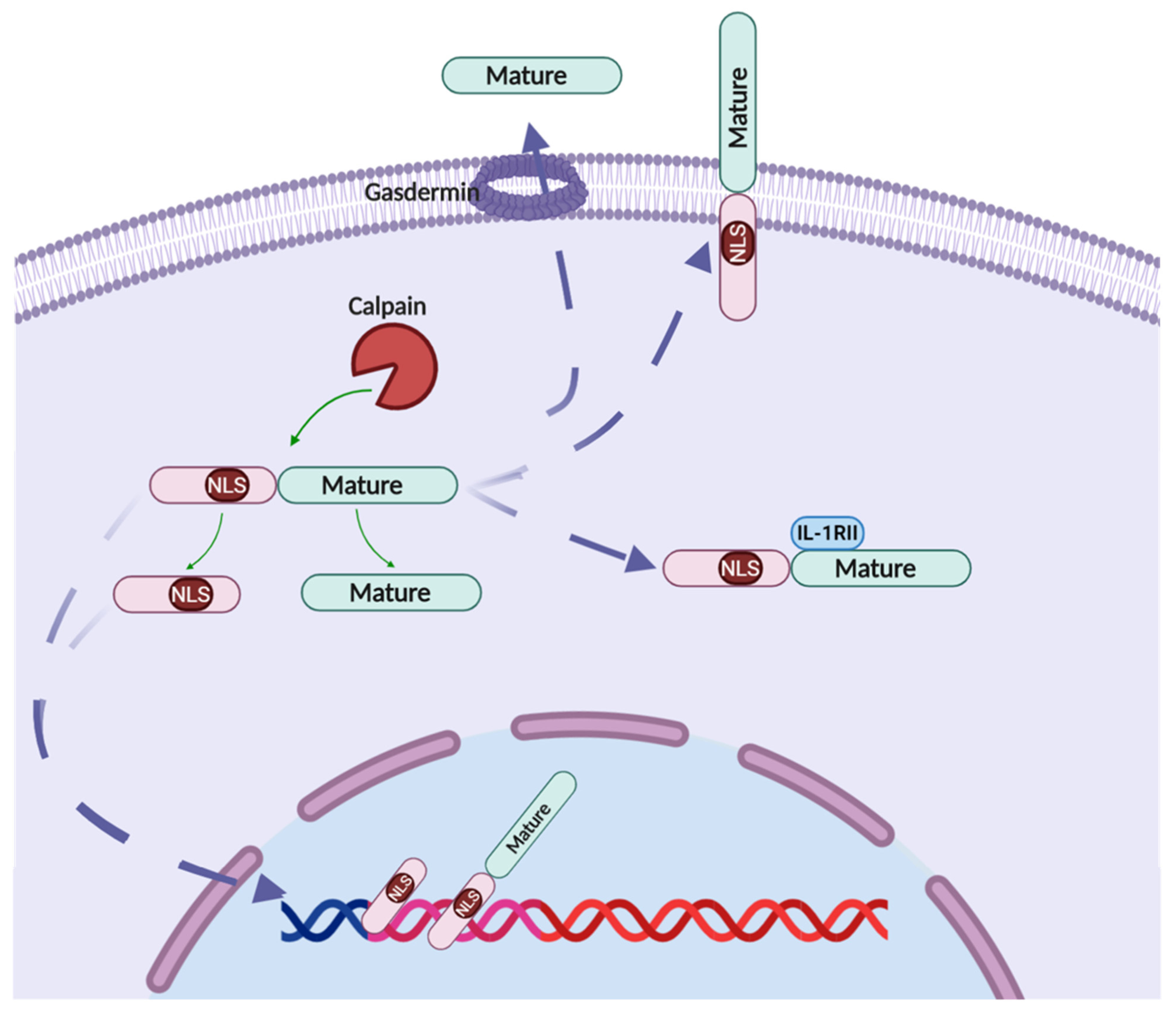
2.2. IL-1α Processing

IL-1α is translated into a 31-kDa precursor form (pro-IL-1α). The pro-IL-1α contains a nuclear localization signal (NLS) site which leads the pro-IL-1α translocation into the nucleus. IL-1α also contains several post-translational modification sites: (1) pro-IL-1α can be phosphorylated at Ser90; (2) pro-IL-1α can be myristoylated at Lys82; and (3) pro-IL-1α can be acetylated at Lys82 [10,11,12,13]. So far, the mechanisms and functions of these post-translational modifications are not well understood. The pro-IL-1α could also be cleaved by several protease to a 17-kDa mature form and a 16-kDa N terminal form named as propiece [14]. The mature form of IL-1α has higher biology activity compared to the pro-IL-1α as its binding affinity to its receptor is several folds higher than the pro-IL-1α [15]. So far, elastase, cathepsin G, proteinase-3, calpain, granzyme B, mast cell chymase, caspase-5, caspase-11 and thrombin have been identified as the pro-IL-1α cleavage proteases [16] (Figure 1).

2.2.1. Elastase, Cathepsin G and Proteinase-3

Neutrophils are the most abundant circulating leucocytes and play a vital role in clearing pathogens through phagocytosis and destruction of microorganisms via granule proteases [17]. The azurophil granule contains three major serine proteases: human leucocyte elastase, cathepsin G and proteinase-3 [18]. Elastase has long been recognized as IL-1β-processing protease and remains unclear whether it could also process IL-1α [19]. In 2011, Afonina et al. first reported that elastase could cleavage pro-IL-1α in vitro when incubating the recombinant IL-1α with elastase. The elastase cleaves IL-1α at the Ala101 site, generating a more biological functional mature form of IL-1α compared with the pro-IL-1α [20]. Recently, the other two neutrophils-derived serine proteases: cathepsin G and proteinase-3 have also been implicated to cleave pro-IL-1α under the in vitro incubation condition. Incubation of pro-IL-1α with cathepsin G and proteinase-3 in vitro generated a smaller processed form of IL-1α compared with that processed by elastase [21]. However, the cleavage site of cathepsin G and proteinase-3 are still unclear. It is of note that all these experiments were performed under in vitro cell-free conditions by incubation of recombinant pro-IL-1α with purified proteases. It is still not clear whether this cleavage occurs in physiological, pathological or both conditions. Supernatants collected from phorbol 12-myristate 13-acetate (PMA)-activated human neutrophils could sufficiently cleave pro-IL-1α, indicating the three serine proteases could process and activate IL-1α under physiological conditions, either alone or at least in combination [21].

2.2.2. Calpain

Calpains are a family of calcium-dependent cysteine proteases, ubiquitously expressed in many types of mammalian cells. Calpains can be directly activated by Ca2+ and act as primary second messengers in cellular signal transduction, being involved in many aspects of cellular bioactivity, including cell proliferation, differentiation and programmed cell death [22,23]. In the early 1990s, two independent research groups reported pro-IL-1α served as a substrate for calpain [24,25]. Calpain could cleave human pro-IL-1α between Phe118 and Leu119, whereas murine IL-1α is cleaved between Arg114 and Ser115 [16]. The calpain processed mature form of IL-1α showed several fold increase at inducing IL-2 release from EL4 cells and IL-6 release from vascular smooth muscle cells (VSMCs) compared with the pro-IL-1α, suggesting that the calpain cleaved mature IL-1α has higher bioactivity compared to its pro-form. The possible reason underlying the different bioactivity of mature IL-1α and pro-IL-1α might be due to the different binding affinity to IL-1 receptor, the calpain cleaved mature IL-1α has about 40-fold higher affinity for its receptor compared with the pro-IL-1α [26].

2.2.3. Granzyme B

Granzymes are a family of conserved serine proteases which are well known as cell death–inducing enzymes, mainly stored in NK cells and cytotoxic T cells. Human genes encode 5 granzymes (A, B, H, K, M), while mouse genes encode 11 (A, B, C, D, E, F, G, K, L, M, N) [27]. Granzyme B is one of the most abundant granzymes and has been studied extensively since its discovery in the 1980s [19,28,29,30]. The most recognized role of granzyme B is to induce the death of pathogens infected cells and cancerous cells, thus eliminating the pathogens and preventing cancer initiation and development [31]. In 2011, Afonina et al. first found that IL-1α was the substrate for granzyme B, while IL-1β was not. Granzyme B could cleave IL-1α at Asp103, a conserved motif among mammals [20]. It is worth noting that although perforin is essential for the cytotoxicity function of granzyme B, more and more evidences have shown that besides NK and T cells, non-cytotoxicity cells such as keratinocytes, mast cells and breast cancer cells also express granzyme B while no perforin is expressed [32], suggesting granzyme B alone can function as an extracellular protease to cleave pro-IL-1α released from the dead cells to mature IL-1α, thus boosting the inflammatory responses [33].

2.2.4. Chymase

Chymase is a mast cell specific protease that belongs to the family of serine proteases and is involved in many pathological conditions [34]. Chymase was first explored as the protease against IL-1β in 1990s [35]. In 2011, Afonina et al. started to investigate the possibility of whether chymase could also cleave IL-1α and found that chymase cleaved pro-IL-1α at Phe116. Chymase is largely released during degranulation of mast cells, easily to meet and cleave the pro-IL-1α leaked from dead cells to enhance the bioactivity of IL-1α [36,37].

2.2.5. Caspase-5 and Caspase-11

Caspases are a family of cysteine proteases and are inseparably linked to programmed cell death. Hundreds to thousands of potential caspase substrates have been identified in multiple biological processes, such as cell apoptosis, differentiation, tissue repair as well as tumorigenesis [38]. In 2019, Kimberley et al. found that murine caspase-11 and its human orthologue caspase-5 served as IL-1α protease [39]. During noncanonical inflammasome activation, murine caspase-11 cleaves pro-IL-1α at Asp106 site, resulting in increased IL-1α activity. Human caspase-5 shared the same cleavage site with granzyme B at Asp103. Both caspase-5 and caspase-11 are required for the senescence-associated secretory phenotype to release IL-1α during cell aging, thus targeting caspase-5 or caspase-11 might be a potential therapeutic strategy to reduce senescence-associated inflammation.

2.2.6. Thrombin

Thrombin, also known as coagulation factor II, is a serine protease that plays a vital role in maintaining blood coagulation and regulating homeostasis [40]. Various studies suggest thrombin is also involved in cell signaling and inflammation. In the early 21st century, Antonella et al. first reported the relationship between thrombin and IL-1. They found that thrombin enhanced the release of both IL-1α and IL-1β in phytohemagglutin treated human peripheral blood mononuclear cells (PBMCs). Further studies showed that thrombin increased IL-1 production at both mRNA and protein level in PBMCs. Seventeen years later, Laura et al. explored the potential proteolytic role of thrombin in processing pro-IL-1α. They found that mammalian pro-IL-1α contains a highly conserved proline-rich sequence(PRS) motif which can be cleaved by thrombin to a fragment of ~18-kDa (p18). The p18 fragment had similar activity compared with calpain cleaved p17 fragment [41]. On one hand, the cleaved p18 initiates and boosts the local immune responses; on the other hand, it feeds back to hemostasis by boosting thrombopoiesis. This finding has filled a scientific gap of how coagulation is directly linked to innate immune response.

2.3. IL-1α Signaling

IL-1 signaling pathway is triggered after binding of IL-1α to the IL-1 receptor type I (IL-1RI). IL-1RI is found on the surface of various types of cells and, both IL-1α and IL-1β act on this same receptor. Its structure consists of intracellular domain; known as the Toll-like/IL-1R (TIR), which has similar homology with other members of IL-1R and TLR families, as well as three extracellular immunoglobulin domains [42]. Binding of IL-1α to its receptor leads to dimerization of the receptor at the TIR domain and recruitment of IL-1R accessory protein, IL-1RAcP [43]. This complex then recruits other adaptor protein, myeloid differentiation factor 88 (MyD88) and IL-1R-associated kinase (IRAK). Formation of this signaling module: IL-1α, IL-1R1, IL-RAcP, MyD88 and IRAK4, is essential for subsequent downstream signaling cascade. Mice lacking the IRAK4 gene were shown to be incapable of eliciting IL-1 signaling response whereby productions of tumor necrosis factor-α (TNF-α) and IL-6 were completely abolished in the knockout mice [44]. In addition, binding of nuclear factor kappa-B (NF-κB) to DNA motif as well as activity of stress kinases such as c-Jun N-terminal kinases (JNK) and p38 mitogen activated protein kinases (MAPK) were reduced in IRAK4−/− macrophages cells after stimulation with lipopolysaccharides (LPS), suggesting that IRAK4 is a key molecule needed for IL-1 signaling [44,45]. Furthermore, serum of mice with MyD88 deficiency have significantly lower levels of TNF-α and IL-6 as compared to the wild-type mice after being intravenously injected with recombinant IL-1β [46]. Therefore, formation of MyD88 and IRAK4 complex is a critical mediator in IL-1 signaling cascades. Assembly of this signaling module results in phosphorylation of IRAK4 which in turn phosphorylates IRAK1 and IRAK2 [45,47]. Tumor necrosis factor–associated factor (TRAF) 6 is then recruited and associates with the complex. IRAK1-IRAK2-TRAF6 complex dissociates and migrates to the plasma membrane to associate with transforming growth factor-β-activated kinase (TAK1) and its binding partners; TAK1-binding proteins TAB1 and TAB2. TAK1-TAB1-TAB2-TRAF6 complex then translocates to the cytoplasm where TAK1 is phosphorylated, hence dissociating itself from the complex [48].
After dissociation from the complex, TAK1 has the ability to initiate two main signaling pathways. The first is the activation of NF-κB pathway. The core IKK complex is made up of IKKα and IKKβ together with the regulatory subunit NF-κB essential modulator (NEMO). In this case, NEMO binds to the polyubiquitin chain of IRAK1 while TAK1 binds to either TAB2 or TAB3. Inhibitor of NF-κB kinase subunit beta (IKKβ) can be activated by TAK1 which in turn phosphorylates IκB on specific serine residues, promoting the K48-linked polyubiquitination [47,49]. This results in degradation of IκB by proteasome, allowing the release of NF-κB (p50 and p65), its translocation into the nucleus and expression of important proinflammatory cytokines genes such as IL-6, TNF-α and cyclooxygenase 2 [47,48]. Another pathway is the MAPK pathway. This involves the activation of p38 MAPK, JNK and extracellular signal-regulated kinases (ERK) through interaction of TAK1 with the MKKs. The p38 MAPK and JNK are the two pathways that are activated during stress conditions that are mainly linked to apoptosis while ERK is associated with cell survival [50]. p38 MAPK and JNK are considered to have tumor suppressive roles while ERK is more towards tumor promoting. For instance, this activation of stress induced MAPK could exert its tumor suppressive role through increased production of proapoptotic proteins, Bax and Bim, and decreased production of anti-apoptotic proteins, Bcl2, hence promoting apoptosis [48].
Like other signaling pathways, IL-1 signaling cascade is under strict regulation. For example, the presence of IL-1RII acts as a decoy receptor which could bind to pro-IL1α, thus preventing it from being cleaved by proteases such as calpain [26,43]. IL-1Ra is a receptor antagonist that binds to IL-1R1, hence inhibiting its activation [51]. Another regulatory pathway is through binding of IL-1R1 to the adaptor toll-interacting protein (TOLLIP), inhibiting IRAK1 thus resulting in internalization of IL-1R1 to endosomes for degradation [52]. Several negative feedback mechanisms are also present such as the production of IκBα preventing NF-κB signaling and TAB1 phosphorylation by p38 MAPK inactivating TAK1 activity [47,53] (Figure 2).

2.4. IL-1α Cellular Localization

2.4.1. Nucleus Localization

Pro-IL-1α contains a NLS (KVLKKRRL) located at the N terminal, in front of all the known proteases cleavage sites [54,55]. Both pro-IL-1α and the cleaved N-terminal fragment can translocate into the nucleus in epithelial cells, myeloid cells, fibroblasts and keratinocytes [55]; while in VSMCs, the NLS site of pro-IL-1α is shielded by IL-1RII, preventing IL-1α nucleus translocation and retaining the IL-1α in the cytosol [26]. The underlying mechanisms on how IL-1α is translocated into the nucleus are still unclear. Cohen et al. reported that acetylation at Lys82 helped the nucleus translocation of pro-IL-1α in mouse macrophage cell line Raw 264.7 cells under genotoxic stress condition [13]. Under steady state, pro-IL-1α nuclear translocation is regulated by HS-1-associated protein X-1 (HAX-1). HAX-1 is a ubiquitously expressed protein and predominantly located at mitochondrial and endoplasmic reticulum membrane as well the nuclear envelope [56]. HAX-1 could bind to pro- and N-terminal IL-1α at three potential sites and facilitate their nuclear translocation [57,58]. Depletion of HAX-1 expression using siRNA abolished the nucleus translocation of IL-1α, demonstrating the requirement of HAX-1 for IL-1α nucleus translocation.
The proteins and chromatin interacting with nucleus localized IL-1α are not well studied. In vitro overexpression systems revealed that N-terminal IL-1α physically interacts with mammalian histone acetyltransferases Gcn5, PCAF and p300, and with adaptor protein Ada3 [45]. Confocal microscopy also showed that endogenous pro-IL-1α colocalized with histone H3 in LPS-stimulated macrophages [42]. We previously reported N-terminal IL-1α could bind to the promoter of specific protein 1 (SP1) and promoted the growth of human T-cell acute lymphoblastic leukemia cell line [43]. Yeast two-hybrid interaction analysis showed that N-terminal IL-1α interacted with five proteins ASF/SF2, CC1.4, hnRNP A/B, SMA5 and prp8, which are all involved in RNA processing, thus promoting various malignant cells apoptosis [44].

2.4.2. Membrane IL-1α

In 1985, Evelyn A et al. found fixed macrophage monolayers and isolated membranes from unfixed macrophages promoted T cell proliferation, indicating a membrane bound form of IL-1α existed [46]. This finding was later confirmed by many studies that the pro-IL-1α was anchored on the cell membrane and functioned through juxtracrine and paracrine manner [9,47,48]. It is still not clear how the cytosol pro-IL-1α translocated onto the membrane. D.T. Brody et al. proved that membrane-bound pro-IL-1α is glycosylated, indicating post-translational modification is required for pro-IL-1α membrane localization and lectin is involved in the binding [59]. We previously reported that in tumor cells, mutation of both NLS and calpain cleavage site led to increased membrane localization of pro-IL-1α, suggesting the NLS signal and protease cleavage reduced pro-IL-1α membrane translocation [60]. Furthermore, membrane-bound pro-IL-1α was also found to be cleaved by extracellular protease into mature form and function through the IL-1R signaling [61].

2.4.3. Cytosolic IL-1α

IL-1α protein was translated in the cytosol and then translocated into the nucleus or membrane. However, confocal images also showed that IL-1α was located in the cytosol. IL1-RII acts as a bona fide decoy receptor of IL-1 and serves as a potential mechanism to retain pro-IL-1α in the cytosol [62,63]. IL1-RII is highly conserved and functions to dampen inflammation caused by IL-1 [64]. Furthermore, IL1-RII binds to IL-1α with a high affinity (Kd ≈ 10−8 M) but binds IL-1Ra with a much lower affinity which allows IL-1RII to effectively inhibit the functions of IL-1α [65]. IL-1RII in its full length can act as a membrane bound decoy receptor and prevents any downstream signaling upon binding to IL-1α which would otherwise normally initiate a pro-inflammatory response [66]. It can also be proteolytically processed by metalloproteinase, a disintegrin and metalloprotease 17 (ADAM17), and eventually be released in a soluble form (sIL-1RII) that stays in the cytosol of the cell [58,67]. sIL-1RII had been shown to interact with IL-1α, specifically in the pro-IL-1α form, and that the binding of sIL-1RII to pro-IL-1α prevents the processing and cleavage of pro-IL-1α into its mature/secreted form by several enzymes such as calpain and granzyme B [20], thereby dampening IL-1α activity. This process could be reversed by caspase 1, which is able to cleave intracellular sIL-1RII thus preventing it from binding to pro-IL-1α [26]. The activity of IL-1RII to reduce IL-1-induced inflammation had been exhibited in several different pathological conditions in vivo. In a model of arthritis in rabbits, intravenous introduction of soluble IL-1RII was shown to reduce arthritis symptoms such as lessening of joint swelling [68]. In a model of heart transplant in mice, introduction of IL-1RII was shown to reduce inflammation by observing a decreased in infiltrating macrophages and CD4+ T cells and also lowering of pro-inflammatory cytokines such as TNF-α and TGF-β [69]. Thus, IL-1RII, being able to mitigate IL-1α signaling via multiple complimentary pathways, makes it a robust anti-inflammatory agent.

2.4.4. Secreted IL-1α

IL-1α lacks the signal peptide, therefore it is secreted through unconventional pathways. For a long time, the secretion of IL-1α was considered as passive release due to the loss of cell membrane integrity, such as necrosis or physical damage [9]. However, accumulating evidences suggest that IL-1α may be secreted through multiple regulatory pathways. In 1995, Keisuke found monocytes derived from the interleukin-1β converting enzyme (caspase-1) KO mice did not export either IL-1β or IL-1α upon LPS stimulation, indicating caspase-1 involved in IL-1α secretion [41]. Moreover, caspase-1-dependent inflammasomes-mediated IL-1β maturation and release also induce the IL-1α release in macrophages [14,70]. Though caspase-1 is required for IL-1α secretion, the export route of IL-1α from cytosol to extracellular space is still unclear. One hypothesis is IL-1α hijacked IL-1β for section, as IL-1β–deficient bone marrow derived dendritic cells (BMDCs) were unable to secrete IL-1α and in vitro overexpression experiments proved an interaction between IL-1α and IL-1β [54]. Another hypothesis is that IL-1α secretion is dependent on Cu2+ binding protein known as S100A13 [71]. Indeed, a study performed by Mandinova et al. showed that in human U937 cells, S100A13 is associated intracellularly with IL-1α in the context of IL-1α exportation when the cells underwent heat-stress [72]. Therefore, the exportation of IL-1α might have some similarities to that of relatively well-studied fibroblast growth factor 1 (FGF1), whose exportation has been shown to be associated with S100A13 [71]. Interestingly, IL-1 prototypes and FGF gene family exhibit high levels of structural homology [73]. Exportation of IL-1α, similar to FGF1, might occur via a Cu2+ -dependent formation of multiprotein complexes involving S100A13 which are translocated across the membrane as a molten globule [74].
Pyroptosis, a newly identified gasdermin-mediated programmed cell death, is found to be involved in IL-1β secretion in inflammatory cells, such as macrophages and neutrophils [13,57,58]. Pyroptosis is mainly trigged by danger signals and activates the caspases. Activated caspase-1 and caspase-11/4/5 cleave gasdermin D (GSDMD) in inflammatory cells, while caspase-3 cleaves gasdermin E (GSDME) in tumor cells [58], to release the N terminal domain of gasdermins that inserts into the plasma membrane to form pores that allow intracellular molecules to pass through. Though most of the pyroptosis studies have been focused on the inflammasome formation and IL-1β secretion, a few pieces of evidence suggest that IL-1α may also take advantage of gasdermin pores to be secreted out. Samantha et al. recently found that in a murine Toxoplasma gondii infection model, microglia, not peripheral myeloid cells, were the main source of IL-1α. The section of IL-1α in microglia was diminished in the GSDMD or caspase-1/11 deficient mice, suggesting GSDMD serves as the export route for IL-1α [75].
Recently, a universal unconventional protein secretion (UPS) route for leaderless protein was discovered. The chaperone HSP90A binds and unfolds leaderless proteins, including mature IL-1β, IL-1α, IL-33, Tau, et cetera. This HSP90A-bound cargo then interacts with the TMED10, resulting in the oligomerization of TMED10 to form a protein channel that translocates the leaderless protein into the lumen of the ER-Golgi intermediate compartment (ERGIC) for subsequent secretion out of the cell via a vesicular intermediate [76].
To our knowledge, IL-1α is the only cytokine that locates in nuclear, cytosol, plasma membrane as well as being secreted. The underlying mechanisms that determine and regulate the IL-1α subcellular localization remain a major question in this field [77] (Figure 3).

3. IL-1α in Cancer Development

Inflammation is closely related to carcinogenesis [78]. As an important pro-inflammatory cytokine, the role of IL-1α in cancer growth has been studied in various cancer types, and the results suggested IL-1α is a dual function cytokine which exerts pro- and anti-tumor function in different cancers. Here, we summarized the recent progress for the role of IL-1α in multiple cancer types and the possible mechanisms of how IL-1α promoted or inhibited tumor growth (Table 1).

3.1. Breast Cancer

In a study with MCF-7 breast cancer cells overexpressing mature IL-1α, when transplanted into nude mice, the MCF-7 cells swiftly formed fast growing estrogen-dependent tumors [49]. However, an earlier study by another group showed that IL-1α was able to inhibit MCF-7 breast cancer cell growth at the G0/G1 phase of the cell cycle [50]. The conflicting results of these studies could be due to their different experimental systems as Sgagias et al., exposed the MCF-7 cells to exogenous IL-1α in an in vitro setting, while Kumar et al. overexpressed IL-1α in the MCF-7 cells and studied the effects in vivo. While IL-1α expression alone in MCF-7 breast cancer cells was not able to induce metastasis, a study on breast tumor-derived IL-1α resulted in metastatic spread by inducing the expression of thymic stromal lympopoietin (TSLP) from tumor-infiltrating myeloid cells [80]. TSLP is a critical tumor survival factor required for metastasis and it promotes survival through induction of Bcl-2 expression [80]. This study introduced a novel IL-1α-TSLP-mediated crosstalk between breast tumor cells and tumor-infiltrating myeloid cells regulating metastatic breast cancer.
HER2 is overexpressed in 25% of all breast cancers, and has been correlated with poor prognosis, high relapse rate, and aggressive symptoms [51]. Hence, a study looked into the potential relationship between HER2 expression, inflammation and cancer stem cell (CSC) expansion [52]. Here, they found that HER2 induces expression of IL-1α and IL-6 in a positive feed-forward activation loop. In turn, activation of NF-kB and signal transducer and activator of transcription 3 (STAT3) were increased, leading to CSC generation and maintenance. Blockade or absence of IL-1α signaling also led to delayed tumorigenesis, reduced CSC population in tumors, reduced inflammation and improved chemotherapeutic efficacy. Another study also showed that high expression of IL-1α in breast cancer biopsies was associated with tumor dedifferentiation and more malignant phenotypic behavior [53], hence supporting the previous study of IL-1α’s role in tumorigenesis and potential CSC generation and maintenance. Conversely, a study on mammary tumor cell proliferation in MMTV-PyMT breast cancer cell showed that IL-1α dependent IL-1R signaling could suppress tumor growth and curb pulmonary metastasis [59].

3.2. Pancreatic Cancer

IL-1α expression by metastatic pancreas cell lines was able to induce hepatocyte growth factor (HGF) secretion by fibroblast [60]. HGF will then enhance pancreatic cancer invasion, proliferation and angiogenesis. Similarly, IL-1α secretion by MiaPaCa-2 cells causes NF-kB to be constitutively activated, resulting in NF-kB downstream genes that are involved in metastatic cascade and angiogenesis to be expressed [61]. In another study, coculture of cancer associated fibroblasts (CAFs) with pancreatic ductal adenocarcinoma cells (PDAC) resulted in upregulation of IL-1α and other inflammatory factors, creating an inflammatory tumor microenvironment suitable for tumor survival and progression [77]. Moreover, IL-1α was found to be the key in the formation and maintenance of this inflammatory tumor microenvironment as blockage of IL-1α alone significantly ablated the production of other inflammatory factors. Hence, in the context of pancreatic cancer, IL-1α expression in the presence of tumor-associated fibroblasts seemed to synergistically promote tumor growth and progression.

3.3. Leukemia

When acute lymphocytic leukemia (ALL) cells were stimulated with mature IL-1α, they proliferated in a dose-dependent manner. Analysis of patient samples also saw spontaneous expression of IL-1α in ALL cells that usually do not express IL-1α [71]. Thus, this strongly suggests that IL-1α could act as an autocrine growth factor in ALL cell proliferation. Overexpression of the propiece form of IL-1α increased the growth of ALL cells through the activation of NF-kB and SP1. In addition, the propiece-expressing ALL cells also had reduced apoptosis and were more resistant to low serum concentration and cisplatin treatment [43]. Interestingly, different forms of IL-1α in these studies resulted in the same effect of increased ALL proliferation. It is possible that both forms activated similar pathways, as the mature IL-1α used by Mori et al. could have bound to IL-1R1 on ALL cells and signaled to increase the expression of NF-kB; while the propiece IL-1α used by Zhang et al. directly activated expression of NF-kB in the nucleus of ALL cells.
When adult T-cell leukemia (ATL) cells from patients were cultured with recombinant IL-1α, it also proliferated in a dose dependent manner [86]. In another study looking at ATL cells from patients, some were able to release biologically active IL-1α protein [87]. The results of these two studies are similar to that in ALL cells [71]. Hence, supporting the idea that IL-1α could act via an autocrine mechanism to stimulate growth and also play a role in the pathological changes associated with ATL. However, conversely when malignant T lymphoma cells were stimulated in vitro to transiently expressed IL-1α, they showed antitumor and immunotherapeutic effects in vivo [88].

3.4. Ovarian Cancer

It has been found that a missense single nucleotide polymorphism (SNP) in IL-1α leads to the full length intracellular IL-1α to be more readily cleaved by inflammatory proteases [101]. In a very large sample size of nearly 40,000 participants, this A114S SNP in IL-1α (rs17561), was also found to be associated with a higher risk of ovarian cancer [85]. Thus, this suggests that increased IL-1α secretion could result in increased tumorigenicity and metastasis in ovarian cancer.

3.5. Head and Neck Squamous Carcinoma

Head and neck squamous carcinoma (HNSCC) patients with distant metastasis had higher IL-1α expression compared to those without metastasis [81]. Patients with higher IL-1α HNSCC were also found to have significantly lower five-year distant metastasis-free survival compared to those with low IL-1α HNSCC. Hence, IL-1α expression could be a prognostic marker of distant metastasis in HNSCC patients. Conversely, when used as a treatment, IL-1α in combination with existing EGFR inhibitor therapy, cetuximab, was found to effectively induce T cell dependent anti-tumor immune response [82]. Patients with higher baseline IL-1α expression and treated with cetuximab was also associated with more favorable progression free survival. This could potentially point towards the use of IL-1α as an immunotherapy option for EGFR-positive HNSCC.

3.6. Liver Cancer

The risk of hepatocellular carcinoma (HCC) is known to increase with chronic liver injury and inflammation. A study using a model of diethylnitrosamine (DEN)-induced liver carcinogenesis in mice found that IL-1α released from hepatocyte necrosis resulted in inflammation and a compensatory proliferative response to regenerate the liver, thus contributing to the development of HCC [84]. Inhibition of IL-1R1- or IL-1α-mediated signaling pathways could prevent HCC development. Thus, IL-1α is a key driver of HCC development in this mouse liver injury model. Meanwhile, another study looking at the function of the membrane IL-1α in murine HCC models found potential inhibitory functions on tumor development via immune cell activation [60]. In the mice models, membrane IL-1α was able to activate anti-tumor immunity via promoting CD8+ T- and NK-cells functions against the malignant cells.

3.7. Lung Cancer

Highly metastatic human lung cancer cell line was found to express higher levels of IL-1α, compared to their lower metastatic counterparts, and was able to form tumors with enhanced angiogenesis and lymphangiogenesis [79]. The tumors were also found to be extensively infiltrated by M2-type macrophages. Since these effects could be suppressed by IL-1R antagonist, the activation of angiogenesis and lymphangiogenesis seems to be IL-1α driven.

3.8. Fibrosarcoma

In a 3-MCA-induced fibrosarcoma cell line, IL-1α expression was required for tumor development when implanted into mice [89]. The authors also found that IL-1α played an important role in controlling immune-surveillance of the developing tumor [89]. Conversely, when studying the cytosolic full length and membrane form of IL-1α on fibrosarcoma, many other studies show a suppressive effect on tumor growth. In an oncogene-transformed NIH/3Y3-derived cell line overexpressing cytosolic or membrane-bound forms of IL-1α, IL-1α exerts adjuvant-like effects, increasing the immunogenicity of tumor-cell antigens and regression of the tumor [90]. Another group investigated the mechanism of this tumor suppressive effect and found that the IL-1α activated the development of tumor cell-specific cytotoxic T lymphocytes (CTL). These CTLs were important for tumor eradication, and the authors also found that fibrosarcomas that do not produce cytosolic or membrane-bound IL-1α can be induced to do so via treatment with cytokines or LPS. After induction, the tumor started regressing and mice were also able to develop immunological memory that protected them from re-challenge [91]. Another more recent study on cytosolic or membrane-bound IL-1α in fibrosarcoma also supported the above claims. The authors found that at the injection sites of IL-1α positive fibrosarcoma cells, CD 8+ T-cells, NK-cells and macrophages accumulated there [92]. The tumor regressed and was replaced by fibrotic scar tissue. It was hypothesized that the membrane-associated IL-1α on the fibrosarcomas may be able to interact with immune effector cells bearing IL-1R, thus activating them and inducing the anti-tumor activities. Overall, cytosolic or membrane-bound IL-1α in fibrosarcoma seems to induce anti-tumor activity via activation of immune cells.

3.9. Gastric Cancer

Similar to HNSCC, there is also a correlation between distant liver metastasis in gastric carcinoma patients and higher levels of IL-1α expression [83]. In patient samples, differentiated tumors were more likely to stain positive for IL-1α compared to undifferentiated tumors. In addition, there is a significantly higher incidence of liver recurrence in patients with tumors expressing IL-1α compared to those without, hence, again showing a correlation between IL-1α expression in tumor and metastasis.
In other studies of human gastric cancers, it was found that expression of mature IL-1α was significantly correlated with the liver metastatic potential and angiogenesis of gastric cancer [93]. In coculture of IL-1α-expressing gastric cancer cells and human umbilical vein endothelial cells (HUVEC), HUVEC strongly enhanced proliferation and tube formation which could be inhibited by blockade of IL-1α, thus suggesting IL-1α being the key mediator in the proliferation and angiogenesis. In another study, Sakamoto et al. found that mice treated with a DNA damaging agent, N-methyl-N-nitrosourea (MNU), increased the expression of IL-1α in gastric epithelial cells (GECs), and was positively correlated with cancer progression. Additionally, they showed that the IL-1α upregulation was via IKKβ signaling, and that it conferred the pre-malignant cells anti-apoptotic and cell proliferation effects, which then eventually led to dysplasia [95]. Hence, IL-1α is a key mediator in the proliferation and tumor progression in gastric cancers.
In another study, Shibata et al. found that IKKβ conditional knockout mice showed similar results when exposed to stress such as ionizing irradiation or Helicobacter felis infection [94]. However, in this model, IL-1α was released from the GECs due to necrosis, as opposed to being secreted out by GECs as shown in the previous study. The release of IL-1α resulted in the recruitment of inflammatory cells that accelerated the progression to gastric pre-neoplasia. Thus, given all the evidence in gastric cancer, IL-1α seems to play a promoting role in gastric cancer progression.

3.10. Prostate Cancer

Analysis of samples from patients with prostate cancer showed that IL-1α expression in malignant cells is associated with the increased progression of the disease, as well as in correlation with prostate specific antigen (PSA) levels in the serum [96]. Cytokine profiling identified IL-1α and IL-6 originating from prostate epithelial cells to act synergistically in driving PSA expression. The authors also hypothesized that IL-1α might drive PSA expression via NF-kB activation.

3.11. Oral Squamous Cell Carcinoma

Oral squamous cell carcinoma (OSCC) cells and CAFs were found to interact together to establish an inflammatory tumor microenvironment that contributes towards carcinogenesis [97]. OSCC cells can release IL-1α that stimulates CAF proliferation, concomitantly CAF increases the secretion of CCL7, CXCL1, and IL-8 cytokines that help facilitate cancer progression and invasion.

3.12. Cervical Cancer

In human papilloma virus (HPV)-induced cervical cancer cells, IL-1α was found to act as an adjuvant by activating dendritic cells for anti-tumor effects [102]. Hence, the authors used viral dsRNA (polyIC) to induce RIPK3-dependent necroptosis in cervical cancer cells, resulting in the release of intracellular IL-1α, which could then activate dendritic cells to produce IL-12 and mount an anti-tumor response.

3.13. Skin Cancer

To investigate the role of IL-1α and inflammation in cutaneous cancer progression, FVB/N transgenic mice were generated overexpressing the mature IL-1α in the epidermis. Interestingly, the authors found that the mice were resistant to carcinomas that required evolution from prior papillomas, but carcinomas still developed de novo at an accelerated rate compared to the controls [98]. In another study, A375 human melanoma cells were transfected to overexpress and secrete mature IL-1α constitutively, then they were intravenously injected into mice. This resulted in potent inflammation and induced adhesion molecule expression on lung endothelial cells (EC), enhancing their adhesiveness to tumor cells, thus resulting in increased tumor-cell retention in the lungs [99].

3.14. Kidney Cancer

Glomerular mesangial cells, a model perivascular myofibroblast cell that is able to synthesize and process IL-1α, was transduced to overexpress the propiece form of IL-1α. This resulted in the malignant transformation of the cells into a spindle cell-type tumor [75].

4. Further Perspectives

IL-1α is a unique cytokine, unlike its homologue IL-1β, both the pro- and mature forms are biologically functional. Besides, the propiece IL-1α has also been implicated as a functional regulator in cell growth and immune responses. Moreover, IL-1α could (1) translocate into the nucleus to regulate downstream gene expressions independent of the IL-1R signaling, (2) anchor to the cell membrane to activate IL-1/IL-1R signaling, (3) secrete out of the cell and activate the IL-1/IL1R signaling, and (4) stay in the cytosol with unknown function. It is of great importance to understand the mechanisms and functions of these different IL-1α isoforms in different inflammatory diseases as well as cancers. This multifaceted aspects of IL-1α functions implicated that it is critical to target IL-1α isoforms precisely for immunotherapy, though anakinra (Kineret; Amgen/Biovitrum), the IL-1R antagonist protein has already been approved by the FDA and has shown promising results for treating patients with rheumatoid arthritis and auto-immune diseases [103,104]. Besides, anakinra blocks both IL-1α/IL-1R and IL-1β/IL-1R signaling, which may have differential or opposite functions in certain disease conditions. Moreover, IL-1α could exert its function in an IL-1R independent manner which is not affected by anakinra. Thus, it is crucial to reveal the detailed roles of IL-1α isoforms in different cancer types as well as the mechanisms of their processing and regulation in order to discover new drug candidates that can target IL-1α functions more precisely for future immunotherapy.

Author Contributions

Y.M. and H.L. conceived the theme of this manuscript; J.W.C., Z.B.H., L.C.Y.C. and Y.M. wrote the manuscript; Y.M. and H.L. revised the manuscript. All authors have read and agreed to the published version of the manuscript.

Funding

This work has been supported by the MOE Tier 2 grants from the Ministry of Education of Singapore (MOE2017-T2-2-099).

Conflicts of Interest

The authors declare no conflict of interest.

References

  1. Menkin, V. Biology of inflammation; chemical mediators and cellular injury. Science 1956, 123, 527–534. [Google Scholar] [CrossRef]
  2. Atkins, E. Pathogenesis of fever. Physiol. Rev. 1960, 40, 580–646. [Google Scholar] [CrossRef] [PubMed]
  3. Dinarello, C.A. The interleukin-1 family: 10 years of discovery. FASEB J. Off. Public Fed. Am. Soc. Exp. Biol. 1994, 8, 1314–1325. [Google Scholar] [CrossRef] [Green Version]
  4. Dinarello, C.A.; Goldin, N.P.; Wolff, S.M. Demonstration and characterization of two distinct human leukocytic pyrogens. J. Exp. Med. 1974, 139, 1369–1381. [Google Scholar] [CrossRef] [PubMed]
  5. Auron, P.E.; Webb, A.C.; Rosenwasser, L.J.; Mucci, S.F.; Rich, A.; Wolff, S.M.; Dinarello, C.A. Nucleotide sequence of human monocyte interleukin 1 precursor cDNA. Proc. Natl. Acad. Sci. USA 1984, 81, 7907–7911. [Google Scholar] [CrossRef] [PubMed] [Green Version]
  6. Lomedico, P.T.; Gubler, U.; Hellmann, C.P.; Dukovich, M.; Giri, J.G.; Pan, Y.C.; Collier, K.; Semionow, R.; Chua, A.O.; Mizel, S.B. Cloning and expression of murine interleukin-1 cDNA in Escherichia coli. Nature 1984, 312, 458–462. [Google Scholar] [CrossRef]
  7. March, C.J.; Mosley, B.; Larsen, A.; Cerretti, D.P.; Braedt, G.; Price, V.; Gillis, S.; Henney, C.S.; Kronheim, S.R.; Grabstein, K.; et al. Cloning, sequence and expression of two distinct human interleukin-1 complementary DNAs. Nature 1985, 315, 641–647. [Google Scholar] [CrossRef]
  8. Voronov, E.; Dotan, S.; Krelin, Y.; Song, X.; Elkabets, M.; Carmi, Y.; Rider, P.; Idan, C.; Romzova, M.; Kaplanov, I.; et al. Unique Versus Redundant Functions of IL-1alpha and IL-1beta in the Tumor Microenvironment. Front. Immunol. 2013, 4, 177. [Google Scholar] [CrossRef]
  9. Di Paolo, N.C.; Shayakhmetov, D.M. Interleukin 1alpha and the inflammatory process. Nat. Immunol. 2016, 17, 906–913. [Google Scholar] [CrossRef] [Green Version]
  10. Beuscher, H.U.; Nickells, M.W.; Colten, H.R. The precursor of interleukin-1 alpha is phosphorylated at residue serine 90. J. Biol. Chem. 1988, 263, 4023–4028. [Google Scholar] [CrossRef]
  11. Kobayashi, Y.; Appella, E.; Yamada, M.; Copeland, T.D.; Oppenheim, J.J.; Matsushima, K. Phosphorylation of intracellular precursors of human IL-1. J. Immunol. 1988, 140, 2279–2287. [Google Scholar] [PubMed]
  12. Stevenson, F.T.; Bursten, S.L.; Fanton, C.; Locksley, R.M.; Lovett, D.H. The 31-kDa precursor of interleukin 1 alpha is myristoylated on specific lysines within the 16-kDa N-terminal propiece. Proc. Natl. Acad. Sci. USA 1993, 90, 7245–7249. [Google Scholar] [CrossRef] [Green Version]
  13. Cohen, I.; Rider, P.; Vornov, E.; Tomas, M.; Tudor, C.; Wegner, M.; Brondani, L.; Freudenberg, M.; Mittler, G.; Ferrando-May, E.; et al. IL-1alpha is a DNA damage sensor linking genotoxic stress signaling to sterile inflammation and innate immunity. Sci. Rep. 2015, 5, 14756. [Google Scholar] [CrossRef]
  14. Gross, O.; Yazdi, A.S.; Thomas, C.J.; Masin, M.; Heinz, L.X.; Guarda, G.; Quadroni, M.; Drexler, S.K.; Tschopp, J. Inflammasome activators induce interleukin-1alpha secretion via distinct pathways with differential requirement for the protease function of caspase-1. Immunity 2012, 36, 388–400. [Google Scholar] [CrossRef] [Green Version]
  15. Mosley, B.; Urdal, D.L.; Prickett, K.S.; Larsen, A.; Cosman, D.; Conlon, P.J.; Gillis, S.; Dower, S.K. The interleukin-1 receptor binds the human interleukin-1 alpha precursor but not the interleukin-1 beta precursor. J. Biol. Chem. 1987, 262, 2941–2944. [Google Scholar] [CrossRef]
  16. Afonina, I.S.; Muller, C.; Martin, S.J.; Beyaert, R. Proteolytic Processing of Interleukin-1 Family Cytokines: Variations on a Common Theme. Immunity 2015, 42, 991–1004. [Google Scholar] [CrossRef] [PubMed] [Green Version]
  17. Nemeth, T.; Sperandio, M.; Mocsai, A. Neutrophils as emerging therapeutic targets. Nat. Rev. Drug Discov. 2020, 19, 253–275. [Google Scholar] [CrossRef] [PubMed]
  18. Faurschou, M.; Borregaard, N. Neutrophil granules and secretory vesicles in inflammation. Microbes Infect. 2003, 5, 1317–1327. [Google Scholar] [CrossRef] [PubMed]
  19. Black, R.A.; Kronheim, S.R.; Cantrell, M.; Deeley, M.C.; March, C.J.; Prickett, K.S.; Wignall, J.; Conlon, P.J.; Cosman, D.; Hopp, T.P.; et al. Generation of biologically active interleukin-1 beta by proteolytic cleavage of the inactive precursor. J. Biol. Chem. 1988, 263, 9437–9442. [Google Scholar] [CrossRef]
  20. Afonina, I.S.; Tynan, G.A.; Logue, S.E.; Cullen, S.P.; Bots, M.; Luthi, A.U.; Reeves, E.P.; McElvaney, N.G.; Medema, J.P.; Lavelle, E.C.; et al. Granzyme B-dependent proteolysis acts as a switch to enhance the proinflammatory activity of IL-1alpha. Mol. Cell 2011, 44, 265–278. [Google Scholar] [CrossRef] [Green Version]
  21. Clancy, D.M.; Sullivan, G.P.; Moran, H.B.T.; Henry, C.M.; Reeves, E.P.; McElvaney, N.G.; Lavelle, E.C.; Martin, S.J. Extracellular Neutrophil Proteases Are Efficient Regulators of IL-1, IL-33, and IL-36 Cytokine Activity but Poor Effectors of Microbial Killing. Cell Rep. 2018, 22, 2937–2950. [Google Scholar] [CrossRef] [PubMed] [Green Version]
  22. Ono, Y.; Sorimachi, H. Calpains: An elaborate proteolytic system. Biochim. Biophys. Acta 2012, 1824, 224–236. [Google Scholar] [CrossRef] [PubMed] [Green Version]
  23. Ono, Y.; Saido, T.C.; Sorimachi, H. Calpain research for drug discovery: Challenges and potential. Nat. Rev. Drug Discov. 2016, 15, 854–876. [Google Scholar] [CrossRef]
  24. Kobayashi, Y.; Yamamoto, K.; Saido, T.; Kawasaki, H.; Oppenheim, J.J.; Matsushima, K. Identification of calcium-activated neutral protease as a processing enzyme of human interleukin 1 alpha. Proc. Natl. Acad. Sci. USA 1990, 87, 5548–5552. [Google Scholar] [CrossRef] [PubMed] [Green Version]
  25. Carruth, L.M.; Demczuk, S.; Mizel, S.B. Involvement of a calpain-like protease in the processing of the murine interleukin 1 alpha precursor. J. Biol. Chem. 1991, 266, 12162–12167. [Google Scholar] [CrossRef]
  26. Zheng, Y.; Humphry, M.; Maguire, J.J.; Bennett, M.R.; Clarke, M.C. Intracellular interleukin-1 receptor 2 binding prevents cleavage and activity of interleukin-1alpha, controlling necrosis-induced sterile inflammation. Immunity 2013, 38, 285–295. [Google Scholar] [CrossRef] [Green Version]
  27. Chowdhury, D.; Lieberman, J. Death by a thousand cuts: Granzyme pathways of programmed cell death. Annu. Rev. Immunol. 2008, 26, 389–420. [Google Scholar] [CrossRef] [PubMed] [Green Version]
  28. Pasternack, M.S.; Eisen, H.N. A novel serine esterase expressed by cytotoxic T lymphocytes. Nature 1985, 314, 743–745. [Google Scholar] [CrossRef] [PubMed]
  29. Brunet, J.F.; Dosseto, M.; Denizot, F.; Mattei, M.G.; Clark, W.R.; Haqqi, T.M.; Ferrier, P.; Nabholz, M.; Schmitt-Verhulst, A.M.; Luciani, M.F.; et al. The inducible cytotoxic T-lymphocyte-associated gene transcript CTLA-1 sequence and gene localization to mouse chromosome 14. Nature 1986, 322, 268–271. [Google Scholar] [CrossRef] [PubMed]
  30. Bleackley, R.C.; Duggan, B.; Ehrman, N.; Lobe, C.G. Isolation of two cDNA sequences which encode cytotoxic cell proteases. FEBS Lett. 1988, 234, 153–159. [Google Scholar] [CrossRef] [Green Version]
  31. Rousalova, I.; Krepela, E. Granzyme B-induced apoptosis in cancer cells and its regulation (review). Int. J. Oncol. 2010, 37, 1361–1378. [Google Scholar] [CrossRef] [PubMed] [Green Version]
  32. Afonina, I.S.; Cullen, S.P.; Martin, S.J. Cytotoxic and non-cytotoxic roles of the CTL/NK protease granzyme B. Immunol. Rev. 2010, 235, 105–116. [Google Scholar] [CrossRef] [PubMed]
  33. Cohen, I.; Rider, P.; Carmi, Y.; Braiman, A.; Dotan, S.; White, M.R.; Voronov, E.; Martin, M.U.; Dinarello, C.A.; Apte, R.N. Differential release of chromatin-bound IL-1alpha discriminates between necrotic and apoptotic cell death by the ability to induce sterile inflammation. Proc. Natl. Acad. Sci. USA 2010, 107, 2574–2579. [Google Scholar] [CrossRef] [PubMed] [Green Version]
  34. Caughey, G.H. Mast cell tryptases and chymases in inflammation and host defense. Immunol. Rev. 2007, 217, 141–154. [Google Scholar] [CrossRef] [PubMed]
  35. Mizutani, H.; Schechter, N.; Lazarus, G.; Black, R.A.; Kupper, T.S. Rapid and specific conversion of precursor interleukin 1 beta (IL-1 beta) to an active IL-1 species by human mast cell chymase. J. Exp. Med. 1991, 174, 821–825. [Google Scholar] [CrossRef] [Green Version]
  36. Chen, C.J.; Kono, H.; Golenbock, D.; Reed, G.; Akira, S.; Rock, K.L. Identification of a key pathway required for the sterile inflammatory response triggered by dying cells. Nat. Med. 2007, 13, 851–856. [Google Scholar] [CrossRef]
  37. Kono, H.; Karmarkar, D.; Iwakura, Y.; Rock, K.L. Identification of the cellular sensor that stimulates the inflammatory response to sterile cell death. J. Immunol. 2010, 184, 4470–4478. [Google Scholar] [CrossRef] [Green Version]
  38. Shalini, S.; Dorstyn, L.; Dawar, S.; Kumar, S. Old, new and emerging functions of caspases. Cell Death Differ. 2015, 22, 526–539. [Google Scholar] [CrossRef] [Green Version]
  39. Wiggins, K.A.; Parry, A.J.; Cassidy, L.D.; Humphry, M.; Webster, S.J.; Goodall, J.C.; Narita, M.; Clarke, M.C.H. IL-1alpha cleavage by inflammatory caspases of the noncanonical inflammasome controls the senescence-associated secretory phenotype. Aging Cell 2019, 18, e12946. [Google Scholar] [CrossRef] [Green Version]
  40. Crawley, J.T.; Zanardelli, S.; Chion, C.K.; Lane, D.A. The central role of thrombin in hemostasis. J. Thromb. Haemost. 2007, 5 (Suppl. 1), 95–101. [Google Scholar] [CrossRef]
  41. Burzynski, L.C.; Humphry, M.; Pyrillou, K.; Wiggins, K.A.; Chan, J.N.E.; Figg, N.; Kitt, L.L.; Summers, C.; Tatham, K.C.; Martin, P.B.; et al. The Coagulation and Immune Systems Are Directly Linked through the Activation of Interleukin-1alpha by Thrombin. Immunity 2019, 50, 1033–1042 e1036. [Google Scholar] [CrossRef] [PubMed] [Green Version]
  42. Lamacchia, C.; Rodriguez, E.; Palmer, G.; Gabay, C. Endogenous IL-1alpha is a chromatin-associated protein in mouse macrophages. Cytokine 2013, 63, 135–144. [Google Scholar] [CrossRef] [PubMed]
  43. Zhang, Y.; Yu, X.; Lin, D.; Lei, L.; Hu, B.; Cao, F.; Mei, Y.; Wu, D.; Liu, H. Propiece IL-1alpha facilitates the growth of acute T-lymphocytic leukemia cells through the activation of NF-kappaB and SP1. Oncotarget 2017, 8, 15677–15688. [Google Scholar] [CrossRef] [PubMed] [Green Version]
  44. Pollock, A.S.; Turck, J.; Lovett, D.H. The prodomain of interleukin 1alpha interacts with elements of the RNA processing apparatus and induces apoptosis in malignant cells. FASEB J. Off. Public Fed. Am. Soc. Exp. Biol. 2003, 17, 203–213. [Google Scholar] [CrossRef]
  45. Buryskova, M.; Pospisek, M.; Grothey, A.; Simmet, T.; Burysek, L. Intracellular interleukin-1alpha functionally interacts with histone acetyltransferase complexes. J. Biol. Chem. 2004, 279, 4017–4026. [Google Scholar] [CrossRef] [Green Version]
  46. Kurt-Jones, E.A.; Beller, D.I.; Mizel, S.B.; Unanue, E.R. Identification of a membrane-associated interleukin 1 in macrophages. Proc. Natl. Acad. Sci. USA 1985, 82, 1204–1208. [Google Scholar] [CrossRef] [Green Version]
  47. Beuscher, H.U.; Colten, H.R. Structure and function of membrane IL-1. Mol. Immunol. 1988, 25, 1189–1199. [Google Scholar] [CrossRef]
  48. Sasu, S.; Cooper, A.L.; Beasley, D. Juxtacrine effects of IL-1 alpha precursor promote iNOS expression in vascular smooth muscle cells. Am. J. Physiol. Heart Circ. Physiol. 2001, 280, H1615–H1623. [Google Scholar] [CrossRef]
  49. Kumar, S.; Kishimoto, H.; Chua, H.L.; Badve, S.; Miller, K.D.; Bigsby, R.M.; Nakshatri, H. Interleukin-1 alpha promotes tumor growth and cachexia in MCF-7 xenograft model of breast cancer. Am. J. Pathol. 2003, 163, 2531–2541. [Google Scholar] [CrossRef]
  50. Sgagias, M.K.; Kasid, A.; Danforth, D.N., Jr. Interleukin-1 alpha and tumor necrosis factor-alpha (TNF alpha) inhibit growth and induce TNF messenger RNA in MCF-7 human breast cancer cells. Mol. Endocrinol. 1991, 5, 1740–1747. [Google Scholar] [CrossRef] [Green Version]
  51. Slamon, D.J.; Clark, G.M. In Reply: Amplification c-erbB-2 and Aggressive Human Breast Tumors? Science 1988, 240, 1796–1798. [Google Scholar] [CrossRef] [PubMed] [Green Version]
  52. Liu, S.; Lee, J.S.; Jie, C.; Park, M.H.; Iwakura, Y.; Patel, Y.; Soni, M.; Reisman, D.; Chen, H. HER2 Overexpression Triggers an IL1alpha Proinflammatory Circuit to Drive Tumorigenesis and Promote Chemotherapy Resistance. Cancer Res. 2018, 78, 2040–2051. [Google Scholar] [CrossRef] [PubMed] [Green Version]
  53. Singer, C.F.; Hudelist, G.; Gschwantler-Kaulich, D.; Fink-Retter, A.; Mueller, R.; Walter, I.; Czerwenka, K.; Kubista, E. Interleukin-1alpha protein secretion in breast cancer is associated with poor differentiation and estrogen receptor alpha negativity. Int. J. Gynecol. Cancer Off. J. Int. Gynecol. Cancer Soc. 2006, 16 (Suppl. 2), 556–559. [Google Scholar] [CrossRef]
  54. Wessendorf, J.H.; Garfinkel, S.; Zhan, X.; Brown, S.; Maciag, T. Identification of a nuclear localization sequence within the structure of the human interleukin-1 alpha precursor. J. Biol. Chem. 1993, 268, 22100–22104. [Google Scholar] [PubMed]
  55. Rider, P.; Carmi, Y.; Voronov, E.; Apte, R.N. Interleukin-1alpha. Semin. Immunol. 2013, 25, 430–438. [Google Scholar] [CrossRef] [PubMed]
  56. Fadeel, B.; Grzybowska, E. HAX-1: A multifunctional protein with emerging roles in human disease. Biochim. Biophys. Acta 2009, 1790, 1139–1148. [Google Scholar] [CrossRef]
  57. Yin, H.; Morioka, H.; Towle, C.A.; Vidal, M.; Watanabe, T.; Weissbach, L. Evidence that HAX-1 is an interleukin-1 alpha N-terminal binding protein. Cytokine 2001, 15, 122–137. [Google Scholar] [CrossRef]
  58. Kawaguchi, Y.; Nishimagi, E.; Tochimoto, A.; Kawamoto, M.; Katsumata, Y.; Soejima, M.; Kanno, T.; Kamatani, N.; Hara, M. Intracellular IL-1alpha-binding proteins contribute to biological functions of endogenous IL-1alpha in systemic sclerosis fibroblasts. Proc. Natl. Acad. Sci. USA 2006, 103, 14501–14506. [Google Scholar] [CrossRef] [Green Version]
  59. Dagenais, M.; Dupaul-Chicoine, J.; Douglas, T.; Champagne, C.; Morizot, A.; Saleh, M. The Interleukin (IL)-1R1 pathway is a critical negative regulator of PyMT-mediated mammary tumorigenesis and pulmonary metastasis. Oncoimmunology 2017, 6, e1287247. [Google Scholar] [CrossRef] [Green Version]
  60. Xu, D.; Matsuo, Y.; Ma, J.; Koide, S.; Ochi, N.; Yasuda, A.; Funahashi, H.; Okada, Y.; Takeyama, H. Cancer cell-derived IL-1alpha promotes HGF secretion by stromal cells and enhances metastatic potential in pancreatic cancer cells. J. Surg. Oncol. 2010, 102, 469–477. [Google Scholar] [CrossRef]
  61. Melisi, D.; Niu, J.; Chang, Z.; Xia, Q.; Peng, B.; Ishiyama, S.; Evans, D.B.; Chiao, P.J. Secreted interleukin-1alpha induces a metastatic phenotype in pancreatic cancer by sustaining a constitutive activation of nuclear factor-kappaB. Mol. Cancer Res. 2009, 7, 624–633. [Google Scholar] [CrossRef] [PubMed] [Green Version]
  62. Mantovani, A.; Locati, M.; Vecchi, A.; Sozzani, S.; Allavena, P. Decoy receptors: A strategy to regulate inflammatory cytokines and chemokines. Trends Immunol. 2001, 22, 328–336. [Google Scholar] [CrossRef]
  63. Colotta, F.; Dower, S.K.; Sims, J.E.; Mantovani, A. The type II ‘decoy’ receptor: A novel regulatory pathway for interleukin 1. Immunol. Today 1994, 15, 562–566. [Google Scholar] [CrossRef]
  64. Morrison, R.N.; Young, N.D.; Nowak, B.F. Description of an Atlantic salmon (Salmo salar L.) type II interleukin-1 receptor cDNA and analysis of interleukin-1 receptor expression in amoebic gill disease-affected fish. Fish Shellfish Immunol. 2012, 32, 1185–1190. [Google Scholar] [CrossRef]
  65. Symons, J.A.; Young, P.R.; Duff, G.W. Soluble type II interleukin 1 (IL-1) receptor binds and blocks processing of IL-1 beta precursor and loses affinity for IL-1 receptor antagonist. Proc. Natl. Acad. Sci. USA 1995, 92, 1714–1718. [Google Scholar] [CrossRef] [Green Version]
  66. Orlando, S.; Sironi, M.; Bianchi, G.; Drummond, A.H.; Boraschi, D.; Yabes, D.; Mantovani, A. Role of Metalloproteases in the Release of the IL-1 type II Decoy Receptor. J. Biol. Chem. 1997, 272, 31764–31769. [Google Scholar] [CrossRef] [PubMed] [Green Version]
  67. Lorenzen, I.; Lokau, J.; Düsterhöft, S.; Trad, A.; Garbers, C.; Scheller, J.; Rose-John, S.; Grötzinger, J. The membrane-proximal domain of A Disintegrin and Metalloprotease 17 (ADAM17) is responsible for recognition of the interleukin-6 receptor and interleukin-1 receptor II. FEBS Lett. 2012, 586, 1093–1100. [Google Scholar] [CrossRef] [Green Version]
  68. Dawson, J.; Engelhardt, P.; Kastelic, T.; Cheneval, D.; Mackenzie, A.; Ramage, P. Effects of soluble interleukin-1 type II receptor on rabbit antigen-induced arthritis: Clinical, biochemical and histological assessment. Rheumatology 1999, 38, 401–406. [Google Scholar] [CrossRef] [Green Version]
  69. Simeoni, E.; Dudler, J.; Fleury, S.; Li, J.; Pagnotta, M.; Pascual, M.; von Segesser, L.K.; Vassalli, G. Gene transfer of a soluble IL-1 type 2 receptor-Ig fusion protein improves cardiac allograft survival in rats. Eur. J. Cardio-Thorac. Surg. Off. J. Eur. Assoc. Cardio-Thorac. Surg. 2007, 31, 222–228. [Google Scholar] [CrossRef] [Green Version]
  70. Petzold, T.; Massberg, S. Thrombin: A Gas Pedal Driving Innate Immunity. Immunity 2019, 50, 1024–1026. [Google Scholar] [CrossRef] [Green Version]
  71. Mori, N.; Shirakawa, F.; Murakami, S.; Oda, S.; Eto, S. Interleukin-1 alpha as an autocrine growth factor for acute lymphoblastic leukaemia cells. Br. J. Haematol. 1994, 86, 386–388. [Google Scholar] [CrossRef] [PubMed]
  72. Mandinova, A. S100A13 mediates the copper-dependent stress-induced release of IL-1alpha from both human U937 and murine NIH 3T3 cells. J. Cell Sci. 2003, 116, 2687–2696. [Google Scholar] [CrossRef] [PubMed] [Green Version]
  73. Zhang, J.D.; Cousens, L.S.; Barr, P.J.; Sprang, S.R. Three-dimensional structure of human basic fibroblast growth factor, a structural homolog of interleukin 1 beta. Proc. Natl. Acad. Sci. USA 1991, 88, 3446–3450. [Google Scholar] [CrossRef] [Green Version]
  74. Prudovsky, I. The non-classical export routes: FGF1 and IL-1 point the way. J. Cell Sci. 2003, 116, 4871–4881. [Google Scholar] [CrossRef] [PubMed] [Green Version]
  75. Stevenson, F.T.; Turck, J.; Locksley, R.M.; Lovett, D.H. The N-terminal propiece of interleukin 1 alpha is a transforming nuclear oncoprotein. Proc Natl Acad Sci USA 1997, 94, 508–513. [Google Scholar] [CrossRef] [Green Version]
  76. Zhang, M.; Liu, L.; Lin, X.; Wang, Y.; Li, Y.; Guo, Q.; Li, S.; Sun, Y.; Tao, X.; Zhang, D.; et al. A Translocation Pathway for Vesicle-Mediated Unconventional Protein Secretion. Cell 2020, 181, 637–652 e615. [Google Scholar] [CrossRef]
  77. Tjomsland, V.; Spangeus, A.; Valila, J.; Sandstrom, P.; Borch, K.; Druid, H.; Falkmer, S.; Falkmer, U.; Messmer, D.; Larsson, M. Interleukin 1alpha sustains the expression of inflammatory factors in human pancreatic cancer microenvironment by targeting cancer-associated fibroblasts. Neoplasia 2011, 13, 664–675. [Google Scholar] [CrossRef] [Green Version]
  78. Colotta, F.; Allavena, P.; Sica, A.; Garlanda, C.; Mantovani, A. Cancer-related inflammation, the seventh hallmark of cancer: Links to genetic instability. Carcinogenesis 2009, 30, 1073–1081. [Google Scholar] [CrossRef] [Green Version]
  79. Watari, K.; Shibata, T.; Kawahara, A.; Sata, K.; Nabeshima, H.; Shinoda, A.; Abe, H.; Azuma, K.; Murakami, Y.; Izumi, H.; et al. Tumor-derived interleukin-1 promotes lymphangiogenesis and lymph node metastasis through M2-type macrophages. PLoS ONE 2014, 9, e99568. [Google Scholar] [CrossRef]
  80. Kuan, E.L.; Ziegler, S.F. A tumor-myeloid cell axis, mediated via the cytokines IL-1alpha and TSLP, promotes the progression of breast cancer. Nat. Immunol. 2018, 19, 366–374. [Google Scholar] [CrossRef]
  81. Leon, X.; Bothe, C.; Garcia, J.; Parreno, M.; Alcolea, S.; Quer, M.; Vila, L.; Camacho, M. Expression of IL-1alpha correlates with distant metastasis in patients with head and neck squamous cell carcinoma. Oncotarget 2015, 6, 37398–37409. [Google Scholar] [CrossRef] [PubMed] [Green Version]
  82. Espinosa-Cotton, M.; Rodman Iii, S.N.; Ross, K.A.; Jensen, I.J.; Sangodeyi-Miller, K.; McLaren, A.J.; Dahl, R.A.; Gibson-Corley, K.N.; Koch, A.T.; Fu, Y.X.; et al. Interleukin-1 alpha increases anti-tumor efficacy of cetuximab in head and neck squamous cell carcinoma. J. Immunother. Cancer 2019, 7, 79. [Google Scholar] [CrossRef] [PubMed] [Green Version]
  83. Tomimatsu, S.; Ichikura, T.; Mochizuki, H. Significant correlation between expression of interleukin-1alpha and liver metastasis in gastric carcinoma. Cancer 2001, 91, 1272–1276. [Google Scholar] [CrossRef]
  84. Sakurai, T.; He, G.; Matsuzawa, A.; Yu, G.Y.; Maeda, S.; Hardiman, G.; Karin, M. Hepatocyte necrosis induced by oxidative stress and IL-1 alpha release mediate carcinogen-induced compensatory proliferation and liver tumorigenesis. Cancer Cell 2008, 14, 156–165. [Google Scholar] [CrossRef] [PubMed] [Green Version]
  85. Charbonneau, B.; Block, M.S.; Bamlet, W.R.; Vierkant, R.A.; Kalli, K.R.; Fogarty, Z.; Rider, D.N.; Sellers, T.A.; Tworoger, S.S.; Poole, E.; et al. Risk of ovarian cancer and the NF-kappaB pathway: Genetic association with IL1A and TNFSF10. Cancer Res. 2014, 74, 852–861. [Google Scholar] [CrossRef] [Green Version]
  86. Shirakawa, F.; Tanaka, Y.; Oda, S.; Eto, S.; Yamashita, U. Autocrine stimulation of interleukin 1 alpha in the growth of adult human T-cell leukemia cells. Cancer Res. 1989, 49, 1143–1147. [Google Scholar]
  87. Wano, Y.; Hattori, T.; Matsuoka, M.; Takatsuki, K.; Chua, A.O.; Gubler, U.; Greene, W.C. Interleukin 1 gene expression in adult T cell leukemia. J. Clin. Investig. 1987, 80, 911–916. [Google Scholar] [CrossRef]
  88. Voronov, E.; Weinstein, Y.; Benharroch, D.; Cagnano, E.; Ofir, R.; Dobkin, M.; White, R.M.; Zoller, M.; Barak, V.; Segal, S.; et al. Antitumor and immunotherapeutic effects of activated invasive T lymphoma cells that display short-term interleukin 1alpha expression. Cancer Res. 1999, 59, 1029–1035. [Google Scholar]
  89. Elkabets, M.; Krelin, Y.; Dotan, S.; Cerwenka, A.; Porgador, A.; Lichtenstein, R.G.; White, M.R.; Zoller, M.; Iwakura, Y.; Dinarello, C.A.; et al. Host-derived interleukin-1alpha is important in determining the immunogenicity of 3-methylcholantrene tumor cells. J. Immunol. 2009, 182, 4874–4881. [Google Scholar] [CrossRef] [Green Version]
  90. Douvdevani, A.; Huleihel, M.; Zoller, M.; Segal, S.; Apte, R.N. Reduced tumorigenicity of fibrosarcomas which constitutively generate IL-1 alpha either spontaneously or following IL-1 alpha gene transfer. Int. J. Cancer 1992, 51, 822–830. [Google Scholar] [CrossRef]
  91. Apte, R.N.; Douvdevani, A.; Zoller, M.; White, R.M.; Dvorkin, T.; Shimoni, N.; Huleihel, M.; Fima, E.; Hacham, M.; Voronov, E.; et al. Involvement of immune responses in the eradication of IL-1 alpha gene-transduced tumour cells: Mechanisms of tumour rejection and immunotherapeutical implications. Folia Biol. 1994, 40, 1–18. [Google Scholar]
  92. Dvorkin, T.; Song, X.; Argov, S.; White, R.M.; Zoller, M.; Segal, S.; Dinarello, C.A.; Voronov, E.; Apte, R.N. Immune phenomena involved in the in vivo regression of fibrosarcoma cells expressing cell-associated IL-1alpha. J. Leukoc. Biol. 2006, 80, 96–106. [Google Scholar] [CrossRef] [PubMed]
  93. Ma, J.; Sawai, H.; Matsuo, Y.; Ochi, N.; Yasuda, A.; Takahashi, H.; Wakasugi, T.; Funahashi, H.; Sato, M.; Okada, Y.; et al. Interleukin-1alpha enhances angiogenesis and is associated with liver metastatic potential in human gastric cancer cell lines. J. Surg. Res. 2008, 148, 197–204. [Google Scholar] [CrossRef] [PubMed]
  94. Shibata, W.; Takaishi, S.; Muthupalani, S.; Pritchard, D.M.; Whary, M.T.; Rogers, A.B.; Fox, J.G.; Betz, K.S.; Kaestner, K.H.; Karin, M.; et al. Conditional deletion of IkappaB-kinase-beta accelerates helicobacter-dependent gastric apoptosis, proliferation, and preneoplasia. Gastroenterology 2010, 138, 1022–1034 e1021-1010. [Google Scholar] [CrossRef] [PubMed]
  95. Sakamoto, K.; Hikiba, Y.; Nakagawa, H.; Hayakawa, Y.; Yanai, A.; Akanuma, M.; Ogura, K.; Hirata, Y.; Kaestner, K.H.; Omata, M.; et al. Inhibitor of kappaB kinase beta regulates gastric carcinogenesis via interleukin-1alpha expression. Gastroenterology 2010, 139, 226–238 e226. [Google Scholar] [CrossRef] [Green Version]
  96. Jemaa, A.B.; Bouraoui, Y.; Rais, N.B.; Nouira, Y.; Oueslati, R. Cytokine profiling identifies an interaction of IL-6 and IL-1alpha to drive PSMA-PSA prostate clones. Immunobiology 2016, 221, 1424–1431. [Google Scholar] [CrossRef]
  97. Bae, J.Y.; Kim, E.K.; Yang, D.H.; Zhang, X.; Park, Y.J.; Lee, D.Y.; Che, C.M.; Kim, J. Reciprocal interaction between carcinoma-associated fibroblasts and squamous carcinoma cells through interleukin-1alpha induces cancer progression. Neoplasia 2014, 16, 928–938. [Google Scholar] [CrossRef] [Green Version]
  98. Murphy, J.E.; Morales, R.E.; Scott, J.; Kupper, T.S. IL-1 alpha, innate immunity, and skin carcinogenesis: The effect of constitutive expression of IL-1 alpha in epidermis on chemical carcinogenesis. J. Immunol. 2003, 170, 5697–5703. [Google Scholar] [CrossRef] [Green Version]
  99. Chirivi, R.G.; Chiodoni, C.; Musiani, P.; Garofalo, A.; Bernasconi, S.; Colombo, M.P.; Giavazzi, R. IL-1alpha gene-transfected human melanoma cells increase tumor-cell adhesion to endothelial cells and their retention in the lung of nude mice. Int. J. Cancer 1996, 67, 856–863. [Google Scholar] [CrossRef]
  100. Tian, T.; Lofftus, S.; Pan, Y.; Stingley, C.A.; King, S.L.; Zhao, J.; Pan, T.Y.; Lock, R.; Marglous, J.W.; Liu, K.; et al. IL1alpha Antagonizes IL1beta and Promotes Adaptive Immune Rejection of Malignant Tumors. Cancer Immunol. Res. 2020, 8, 660–671. [Google Scholar] [CrossRef] [Green Version]
  101. Kawaguchi, Y.; Tochimoto, A.; Hara, M.; Kawamoto, M.; Sugiura, T.; Saito, S.; Kamatani, N. Contribution of single nucleotide polymorphisms of the IL1A gene to the cleavage of precursor IL-1alpha and its transcription activity. Immunogenetics 2007, 59, 441–448. [Google Scholar] [CrossRef] [PubMed]
  102. Schmidt, S.V.; Seibert, S.; Walch-Ruckheim, B.; Vicinus, B.; Kamionka, E.M.; Pahne-Zeppenfeld, J.; Solomayer, E.F.; Kim, Y.J.; Bohle, R.M.; Smola, S. Correction: RIPK3 expression in cervical cancer cells is required for PolyIC-induced necroptosis, IL-1alpha release, and efficient paracrine dendritic cell activation. Oncotarget 2019, 10, 4503–4504. [Google Scholar] [CrossRef] [PubMed]
  103. Dinarello, C.A.; Simon, A.; van der Meer, J.W. Treating inflammation by blocking interleukin-1 in a broad spectrum of diseases. Nat. Rev. Drug Discov. 2012, 11, 633–652. [Google Scholar] [CrossRef] [PubMed] [Green Version]
  104. Dinarello, C.A.; van der Meer, J.W. Treating inflammation by blocking interleukin-1 in humans. Semin. Immunol. 2013, 25, 469–484. [Google Scholar] [CrossRef] [PubMed] [Green Version]
Figure 1. Overview of IL-1α structure. Human IL-1α consists of 271 amino acids including an nuclear localization signal (NLS) domain. IL-1α can be cleaved by various proteases, such as elastase, granzyme B, caspase-5, thrombin, chymase and calpain. IL-1α also undergoes post-translational modification, including acetylation, myristoylation as well as phosphorylation.
Cells 10 00092 g001
Figure 2. Overview of IL-1α signaling pathway. The binding of IL-1α to IL-1RI leads to dimerization of the receptor at the TIR domain and recruitment of IL-1RAcP. This IL-1α/IL-1RI/IL-1RAcP complex then recruits adaptor proteins MyD88 and IRAK for subsequent downstream signaling cascade. IRAK will phosphorylate IRAK1 and IRAK2 and recruit TRAF6 to form the IRAK1-IRAK2-TRAF6 complex; subsequently TRAF6 will associate with TAK1-TAB1-TAB2 to phosphorylate TAK1. Phosphorylated TAK1 will dissociate from the TRAF6-TAK1-TAB1-TAB2 complex to initiate the NF-κB and MAPK signaling pathways to regulate the downstream proinflammatory cytokines expressions, such as IL-6, TNF-α and cyclooxygenase 2. Abbreviations: IL-1RI, IL-1 receptor I; IL-1RAcP, interleukin-1 receptor accessory protein; MyD88, myeloid differentiation factor 88; IRAK, IL-1R-associated kinase; TRAF6, tumor necrosis factor–associated factor 6; TAK1, transforming growth factor-β-activated kinase 1; NF- κB, nuclear factor kappa-B; MAPK, Mitogen-activated protein kinase.
Cells 10 00092 g002
Figure 3. Overview of IL-1α cellular localization. IL-1α is translated as pro-peptide precursors form. (1) Pro-IL-1α contains a nuclear localization sequence (KVLKKRRL) in the N-terminal region which leads pro-IL-1α and propiece IL-1α translocation into the nucleus; (2) Pro-IL-1α can be cleaved by several proteases, such as calpain, into mature form. Mature IL-1α can be secreted out of cell possibly through gasdermin-formed pores; (3) Pro-IL-1α can be myristoylated and anchored to the cell membrane to form the membrane-bound IL-1α; (4) IL-1RII binds to Pro-IL-1α, shielding the NLS and proteases cleaved sits, thus retaining the Pro-IL-1α in the cytosol.
Cells 10 00092 g003
Table 1. Summary of IL-1α function in tumor development.
Tumor TypeTumorigenesisMechanism of ActionIL-1α FormSource of IL-1αStudy TypeReference
Lung CancerPromoteActivates angiogenesis and lymphangiogenesisMatureEndogenousIn vitro and in vivo[79]
Breast CancerPromoteIncreases cell proliferationMatureTransduced In vitro and in vivo[49]
Breast CancerPromoteInduces TSLP expression from tumor-infiltrating myeloid cells to increase cancer survival and metastasis spreadMatureEndogenousIn vitro, in vivo, and patient data[80]
Breast CancerPromoteIncreases activation of NF-kB and STAT3 to generate and maintain cancer stem cells.MatureEndogenous and ExogenousIn vitro, in vivo, and patient data[52]
Breast CancerPromoteAssociated with dedifferentiation and malignancyMatureEndogenousIn vitro and patient data[53]
Breast CancerSuppressInhibits cell growth at G0/G1 phaseMatureExogenousIn vitro[50]
Breast CancerSuppressSuppresses cell proliferation through IL-1R signalingMatureEndogenous In vitro and in vivo[59]
Head and Neck Squamous CarcinomaPromoteAssociated with distant metastasis in patientsMatureEndogenousPatient data[81]
Head and Neck Squamous CarcinomaSuppressActivates T-cell dependent anti-tumor responseMatureExogenousIn vitro, in vivo, and patient data[82]
Liver CancerPromoteActivates inflammation and compensatory proliferation in liverMatureEndogenousIn vitro and in vivo[83]
Liver CancerSuppressPromotes T- and NK-cell activationMembraneTransducedIn vitro and in vivo[84]
Pancreatic CancerPromotePromotes HGF secretion by fibroblasts to promote cancer invasion, proliferation, and angiogenesisMatureEndogenous and ExogenousIn vitro[60]
Pancreatic CancerPromoteConstitutively activates NF-kB to induce metastatic behaviorMatureTransducedIn vitro and in vivo[61]
Pancreatic CancerPromoteSustains expression of inflammatory factors in tumor microenvironment beneficial for tumor survivalMatureEndogenousIn vitro and patient data[77]
Ovarian CancerPromoteIL-1α SNP (rs17561) associated with increase risk, possibly due to it being more readily cleaved to form mature IL-1αMatureEndogenousPatient data[85]
Acute Lymphocytic LeukemiaPromoteFacilitates growth of T-ALL cells through activation of NF-kB and SP1N-TerminalTransducedIn vitro and in vivo[43]
Acute Lymphocytic LeukemiaPromoteInduces ALL cell growth in an IL-1α dose dependent mannerMatureEndogenous and ExogenousIn vitro and patient data[71]
Adult T-cell LeukemiaPromoteInduces ATL cell growth in an IL-1α dose dependent manner, possibly via autocrine mechanismMatureExogenousIn vitro and patient data[86]
Adult T-cell LeukemiaPromoteAssociated with ATL patient samples and cell linesMatureEndogenousIn vitro and patient data[87]
Adult T-cell LeukemiaSuppressStimulates anti-tumor immune responsesMatureEndogenousIn vitro and in vivo[88]
FibrosarcomaPromoteInvolved in controlling the immune-surveillance of developing tumorMatureEndogenousIn vitro and in vivo[89]
FibrosarcomaSuppressReduces tumorigenicity, increases immunogenicity, and regression of tumorCytosolic Full Length and MembraneEndogenous and TransducedIn vitro and in vivo[90]
FibrosarcomaSuppressIncreases immunogenicity, induces regression of tumor and development of systemic immunityCytosolic Full Length and MembraneEndogenous and TransducedIn vitro and in vivo[91]
FibrosarcomaSuppressStimulates anti-tumor immune responses and regression of tumorCytosolic Full Length and MembraneTransducedIn vitro and in vivo[92]
Gastric CancerPromoteIncreases metastasis and tumor differentiationMatureEndogenousPatient data[83]
Gastric CancerPromoteAssociated with enhance angiogenesis and metastasisMatureEndogenousIn vitro[93]
Gastric CancerPromoteAssociated with rapid progression to gastric pre-neoplasiaFull Length and MatureEndogenousIn vitro and in vivo[94]
Gastric CancerPromotePositively correlated with carcinogenesisMatureEndogenousIn vitro and in vivo[95]
Prostate CancerPromoteCorrelated to increased serum PSA levels and progression of diseaseMatureEndogenousPatient data[96]
Oral Squamous Cell CarcinomaPromoteStimulates CAF proliferation and cytokine (CCL7, CXCL1, IL-8) secretion to promote OSCC cancer progressionMatureEndogenousIn vitro, in vivo, and patient data[97]
Cervical CancerSuppressActivates dendritic cells to produce IL-12 for anti-tumor responseMatureEndogenousIn vitro and patient data[43]
Skin CancerPromote and SuppressSuppresses carcinoma formation from prior papilloma.MatureTransducedIn vitro and in vivo[98]
Promotes carcinoma formation not from prior papilloma
Skin CancerPromoteInduces adhesion molecules on endothelial cells, increasing tumor retention. As well as inducing potent inflammation, enhancing metastasis.MatureTransducedIn vitro and in vivo[99]
Kidney CancerPromoteInduces malignant transformationN-TerminalTransducedIn vitro and in vivo[75]
Multiple Cancers (T cell lymphoma, melanoma, lung carcinoma)SuppressInhibits tumor growth by enhancing T-cell mediated antitumor immunityMatureEndogenousIn vitro and in vivo[100]
Multiple Cancers (Various malignant human tumor cell lines)SuppressInduces cell apoptosis, possibly involving RNA processing apparatusN-TerminalTransducedIn vitro[44]
Abbreviations: IL-1α, interleukin-1α; TSLP, thymic stromal lymphopoietin; NF- κB, nuclear factor kappa-B; STAT3, signal transducer and activator of transcription 3; HGF, hepatocyte growth factor; PSA, prostate specific antigen; CAF, cancer associated fibroblasts; CCL7, C-C motif chemokine ligand 7; CXCL1, C-X-C motif chemokine ligand 1; IL-8 interleukin -8; IL-12, interleukin-12.
Publisher’s Note: MDPI stays neutral with regard to jurisdictional claims in published maps and institutional affiliations.

