Review

Role of Cardiac Macrophages on Cardiac Inflammation, Fibrosis and Tissue Repair

by
William P. Lafuse
1,
Daniel J. Wozniak
1,2 and
Murugesan V. S. Rajaram
1,*
1
Department of Microbial Infection and Immunity, College of Medicine, Ohio State University, Columbus, OH 43210, USA
2
Department of Microbiology, Ohio State University, Columbus, OH 43210, USA
*
Author to whom correspondence should be addressed.
Cells 2021, 10(1), 51; https://doi.org/10.3390/cells10010051
Submission received: 10 November 2020 / Revised: 22 December 2020 / Accepted: 28 December 2020 / Published: 31 December 2020
(This article belongs to the Special Issue Macrophages in Tissue Repair, Regeneration, and Fibrosis)

Abstract

:
The immune system plays a pivotal role in the initiation, development and resolution of inflammation following insult or damage to organs. The heart is a vital organ which supplies nutrients and oxygen to all parts of the body. Heart failure (HF) has been conventionally described as a disease associated with cardiac tissue damage caused by systemic inflammation, arrhythmia and conduction defects. Cardiac inflammation and subsequent tissue damage is orchestrated by the infiltration and activation of various immune cells including neutrophils, monocytes, macrophages, eosinophils, mast cells, natural killer cells, and T and B cells into the myocardium. After tissue injury, monocytes and tissue-resident macrophages undergo marked phenotypic and functional changes, and function as key regulators of tissue repair, regeneration and fibrosis. Disturbance in resident macrophage functions such as uncontrolled production of inflammatory cytokines, growth factors and inefficient generation of an anti-inflammatory response or unsuccessful communication between macrophages and epithelial and endothelial cells and fibroblasts can lead to aberrant repair, persistent injury, and HF. Therefore, in this review, we discuss the role of cardiac macrophages on cardiac inflammation, tissue repair, regeneration and fibrosis.

Graphical Abstract

1. Introduction

Macrophages are the central regulator of immune systems, able to activate and proliferate lymphocytes to generate innate and adaptive immune response [1]. The cardiovascular system consists of the heart, blood vessels, and blood, which transports nutrients, oxygen, and hormones to cells throughout the body and removes metabolic wastes. Heart failure (HF) is the clinical indication of various forms of cardiovascular diseases (CVDs) that impact the function of the heart. CVD includes atherosclerosis, ischemic heart disease, cerebrovascular disease, hemorrhagic stroke, hypertensive heart disease, cardiomyopathy, myocarditis, atrial fibrillation, aortic aneurysm, peripheral vascular disease, and endocarditis [2]. According to 2016 mortality data, the World Health Organization reports that CVD caused more than 17.9 million deaths worldwide, which is more than cancer and chronic lower respiratory disease combined [3]. Generally, regeneration of adult heart tissue after injury is poor. Therefore, understanding how cardiac tissue is injured and how cardiac tissue regenerates or resolves the damage is of prime importance to universal health.
Like other organs, the heart is composed of a heterogeneous population of cells including cardiomyocytes, fibroblasts, pericytes, smooth muscle cells, endothelial cells, and many types of immune cells (Figure 1) [4,5]. The immune system provides a protective inflammatory response necessary for host defense from infections and also plays a critical role in resolving local damage caused by sterile inflammation in the heart. Macrophages are the central regulator of immune systems [1], and the primary immune cells that reside in the heart tissue during steady state [5,6]. A small number of monocytes and a sparse population of dendritic cells are also present. Mast cells, regulatory T and B cells are also found in resting cardiac tissue, which helps to initiate the early immune response [5]. Generally, neutrophils are not present in healthy heart tissue [5], but they are recruited upon tissue injury or infection. These cardiac immune cells are activated by either sterile inflammation or microbial infections which leads to the production of inflammatory cytokines and recruitment of diverse leukocyte populations into inflamed heart tissue. Intracellular signaling and cross talk between embryonic tissue-resident cardiac macrophages (CMs) and non-embryonic CMs are critical in the generation, propagation and development of cardiac inflammation, tissue remodeling and repair [5]. Herein, we review the current knowledge of the contribution of tissue-resident cardiac macrophages on cardiac inflammation, fibrosis, and tissue repair.

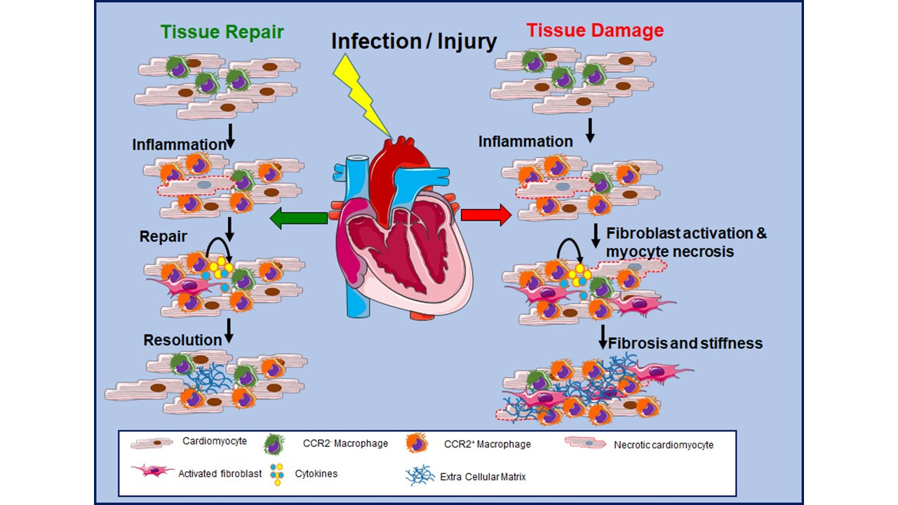
2. Cardiac Immune Cells

The heart is immunologically active even during steady state and contains all major leukocyte populations, either residing in the heart or waiting to infiltrate into the heart. There are approximately 103 leukocytes/mg of tissue in an adult mouse heart [7] (Table 1).
The majority of leukocytes present in the resting heart are F4/80+CD11b+ macrophages [5], with small populations of dendritic cells, B cells, regulatory T (Tregs) cells, and innate lymphoid cells [9,11,12,13]. Upon ischemic or non-ischemic cardiac injury, necrotic cell death leads to the activation of tissue-resident immune and non-immune cells. The danger-associated molecular patterns (DAMPs) released from necrotic cells are the key trigger for immune cell activation, which results in the release of pro-inflammatory cytokines and chemokines that are responsible for the recruitment of inflammatory leukocytes from the blood. Following the initial inflammatory phase, the expansion of neutrophil and macrophage populations facilitates phagocytosis and clearance of the dead cells, and release of cytokines and growth factors. This leads to the initiation of the healing process via the activation of myofibroblast proliferation and neovascularization of the injured myocardium [14].
Monocytes are another vital part of the innate immune system during both the initial insult and the chronic phase of cardiac injury. In steady-state conditions, the heart contains few monocytes. In humans, there are three distinct monocytic subsets which have been identified based on the expression of CD14 and CD16, namely classical (CD14++ CD16); intermediate (CD14++ CD16+) and non- classical (CD14+ CD16++) monocytes [15]. In contrast, murine monocytes have been classified into two subsets based on their expression of Ly6C; classical Ly6Chigh, CCR2high and chemokine (C-X3-C motif) receptor-1 (CX3CR1)low and non-classical Ly6Clow CCR2highCX3CR1high monocytes. The classical monocytes are recruited and accumulate at inflammatory sites, including during acute myocardial infarction (MI). Conversely, non-classical monocytes patrol the endothelium to maintain homeostasis [16]. HF in humans has also been associated with peripheral monocytosis [17] and exaggerated inflammation with distinct monocyte profiles [16,18]. In response to cardiac stress, Ly6Chigh monocytes are rapidly recruited to the mouse heart, either following ischemic injury or hypertensive stress; in contrast Ly6Clow monocytes do not get recruited into the myocardium during the early inflammatory stage but appear in the heart during the later reparative stage [6,19,20,21]. The antigen presenting dendritic cells (DCs) are dispersed throughout the heart [22], which links the innate immunity to adaptive immunity. At steady state, DCs function in maintaining\peripheral tolerance [23,24] by presenting self-antigens to Tregs [25]. The heart contains two conventional DC subsets CD103+ CD11b (cDC1) and CD103CD11b+ (cDC2) [12,26,27]. Both cDC subsets are of hematopoietic origin and dependent on Flt3L for development [12] and these DCs differ from CMs by lack of expression of CD64 and myeloid-epithelial-reproductive tyrosine kinase (MERTK) [12]. Both cardiac DCs express the chemokine receptor CCR2 and Ccr2-deficient mice result in reduced numbers of cDCs in the myocardium of the heart, indicating that CCR2 is a key regulator of the migration of pre-cDCs into the heart [12]. Recently, it has been demonstrated that cardiac-resident MHCII+ cells process and present myosin heavy chain alpha-derived peptides under steady-state heart function and have the capability to prime T cells ex vivo [28,29]. In addition, previous studies demonstrated that CD4 T cell-deficient mice, and the transgenic mice that express a single T cell receptor gene to an irrelevant peptide, show impaired wound healing and increased expansion of monocyte populations after ischemic injury, suggesting that CD4 T cells have protective functions during cardiac injury [30]. Interestingly, these cardiac CD4+ T cells are activated by presentation of self-antigens via MHCII expressing cells in the heart. This leads to immunosuppressive responses in the myocardium, suggesting that most CD4+ T cells in the heart are regulatory T cells (Tregs) that promote myocardial recovery through an IL-10-dependent pathway [31,32]. Additionally, the recruitment and proliferation of regulatory T cells (Foxp3+CD4+) in the heart improves myocardial wound healing after MI by modulating monocyte/macrophage differentiation [10]. Another study by Yang et al. reports that the Rag-1 knock out mice (do not have mature T and B cells) develop smaller infarcts than control C57BL/6 mice, and transfer CD4+T cells into the RAG-deficient mice increased the infract size and healing [33]. Interestingly, recent studies revealed that transverse aortic constriction can induce T cell responses, and the recruitment of CD4+ T cells contributes to the pathogenesis of HF [34,35].

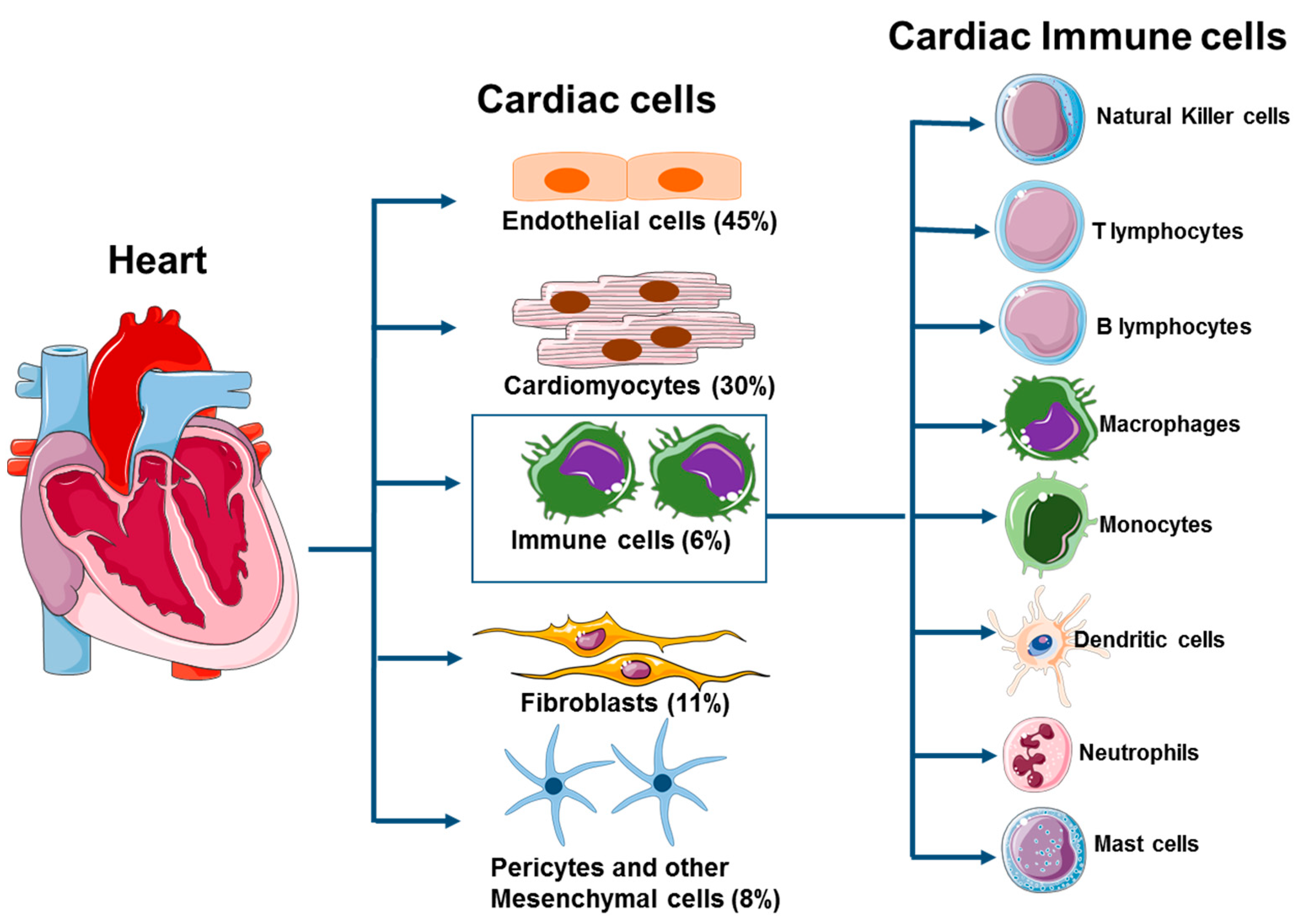
3. Cardiac-Resident Macrophages

Macrophages are specialized cells of the immune system that recognize, phagocytose and destroy apoptotic ells and pathogens and generate innate immune response. The major challenge in the field of cardiac macrophages is the discrepancy among the macrophage phenotypes and their markers identified and reported by various groups. In this review, we consolidated the CM phenotypes and common markers reported in the literature for the CM classification and function. These macrophages are dispersed throughout the heart as spindle-like cells, acting as sentinel macrophages for pathogens that may enter the myocardium. In the past few decades, it has been universally accepted that bone marrow-derived hematopoietic stems cells differentiate into circulating monocytes which enter into various tissues and differentiate into tissue-resident macrophages [36,37]. However, in recent years, the understanding of the origin of tissue-resident macrophages has been drastically revised with the recognition that resident macrophage populations are established during embryo development and maintain their numbers through self-renewal properties, rather than through infiltration of blood monocytes [6,19,38,39,40,41]. The heart contains macrophage subsets with distinct functions and origins. Using flow cytometry, linage tracing, and parabiosis studies, subsets of chemokine (C-C motif) receptor-2 (CCR2) and CCR2+ CMs have been defined [19,41,42,43,44]. (Table 2) CCR2 CMs are derived from the embryonic yolk sac and maintained without monocyte recruitment.
In contrast, CCR2+ cardiac macrophages are maintained by monocyte recruitment. These studies also show that the CCR2and CCRR2+ CM differ in function, with the CCR2CMs being involved in maintenance of homeostasis and the CCR2+ being pro-inflammatory. Furthermore, studies by Epelman et al. [6] characterized the CMs into three subsets defined by the difference in expression levels of MHC class II, CCR2 and CD11c (Table 2). The predominant cardiac macrophages were two CCR2 populations (MHCIIhigh CD11clow and MHCIIlow CD11clow). These two subsets were primarily derived from yolk sac progenitors and renewed through in situ proliferation. The third cardiac macrophage subset was CCR2+, MHCIIhigh and CD11chigh were derived and slowly replenished from circulating monocytes and these CMs were distinguished from dendritic cells by expression of macrophage markers CD64 and MerTK [47] (Figure 2).
The transcript profile analysis of the two CCR2 macrophage populations (MHCIIhigh and MHCIIlow) and the CCR2+ MHCIIhigh populations indicated overlapping and non-overlapping functions. The CCR2+ macrophages were enriched for inflammatory genes and pathways, including genes involved in the NLPR3 inflammasome and IL-1β production. Consequently, this suggests that these macrophages are inflammatory in nature. The MHCIIhigh subsets were enriched for genes involved in antigen presentation to T cells, suggesting involvement in immune surveillance. The uptake of apoptotic/necrotic cells by CCR2 MHCIIlow suggests these macrophages function in homeostasis during steady state by removing dead cells without inducing an immune response [48]. Moreover, single-Cell RNA sequencing by Dick et al. also identified four different cardiac macrophage clusters at steady state, each with unique functions [46] (Table 2). One macrophage cluster identified as CCR2 TIMD4+ LYVE1+ MHCIIlow was maintained independent of blood monocytes and corresponds to the CCR2 MHCIIlow subset [6]. This cluster of macrophages expressed the genes involved in homeostasis and regeneration, including phosphatidylserine receptor Tmid4, Lye1 and growth factor Ifg1, which is also involved in efferocytosis of dead cells [49], vascular homeostasis [50] and angiogenesis [43], respectively. The second cardiac macrophage clusters were CCR2MHChigh TIMD4 LYVE1 and were partially replaced by blood monocytes in long-term parabiosis. One of these clusters expressed high levels of genes involved in antigen presentation and thus correlates with the CCR2 MHChigh macrophage subset [6]. The third cluster, termed the Isg cluster, was enriched in genes stimulated by interferon such as Irf7, Isg20, and Ifit1. The fourth cluster contained CCR2+ macrophages, which were fully replaced by blood monocytes in long-term parabiosis. Studies of the human heart have described analogous resident CCR2and CCR2+ cardiac macrophages [44,46] as functionally distinct macrophage subsets that exist in the human heart at steady state. Cardiac macrophages are also abundant in the atrioventricular node (AV), which provide an electrical connection between the atria and ventricles while also facilitating an electrical conduction by coupling to beating CMs via connexin-43-containg gap junctions [46].

4. Macrophage Role in Cardiac Inflammation and Cardiac Dysfunction

Myocarditis is defined as an inflammation of the myocardium in the presence of necrosis or degeneration of cardiomyocytes and other cell types. Myocarditis accounts for about one in nine cases of HF and remains one of the top reasons for heart transplantation worldwide, since the lack of specific treatments [51]. The most common causes of myocarditis are infections of various pathogens including parasite Trypanosoma cruzi in South America, or viruses, such as enterovirus, parvovirus B19, adenovirus and hepatitis C virus, which are the major causes in North America [52,53,54]. Recently, we and others have shown that bacterial infection also induces myocardial inflammation and cardiac dysfunction [55,56,57,58,59,60]. In addition, non-infectious agents, such as allergic reactions to drugs and chemicals, can induce cardiomyocyte apoptosis and the release of DAMPs, which activate the innate immune response leading to sterile inflammation and myocarditis [53].
Viral infections are the most common cause of myocarditis, with periodicity to this prevalence in the population and possibly related to herd immunity. The pathophysiology of viral myocarditis involves both a direct virus-mediated response and indirect immune-mediated injury of the cardiac tissue and subsequent dysfunction. The innate immune response is essential for the elimination of virus and the restoration of the normal tissue homeostasis. Nonetheless, activation of the immune system also leads to myocardial inflammation, necrosis, release of DAMPs, and ventricular dysfunction, which occurs directly through the killing of virus-infected cardiac cells, and also through an increased production of inflammatory cytokines which subsequently affect CM function [54].
The process of virus-induced myocarditis includes three stages, an acute viremia stage, followed by sub-acute infiltrating stage, and a chronic stage [61]. In the acute stage, a virus enters into cardiomyocytes or other cells (endothelial cells, fibroblasts and immune cells) through specific surface receptors, i.e., the coxsackievirus binds to coxsackievirus and adenovirus receptor (CAR) and the co-receptor, the decay accelerating factor (DAF). Viral entry activates the inflammatory signaling cascade and produces various pro-inflammatory cytokines (IL-1, TNF, IFN-α, IFN-β) and chemokines, resulting in recruitment of immune cells into the tissue and an aggravation of the inflammatory process [61]. During the infiltration stage, the migrated immune cells activate B cells to produce neutralizing antibodies and promote viral clearance [62]. Finally, in the chronic stage, once the virus is cleared completely, apoptotic cell debris is removed by macrophages and the tissue restoration process is initiated and leads to cardiac fibrosis (discussed below). However, in some cases the viral RNA and proteins can persist in the heart and cause chronic inflammation, which triggers extensive fibrosis and ventricular dysfunction and can eventually develop into dilated cardiomyopathy [61]. The infiltrating activated immune cells into the myocardium increase the expression of matrix metalloproteinase (MMP)-9, which in turn exacerbates the immune cell recruitment and refuels the inflammatory process [63]. Several human studies have demonstrated an increase in the activity and expression of MMP-2 and MMP-9 in virus-induced dilated cardiomyopathy patients [64]. Since MMPs play prominent role in the inflammation and remodeling processes of the myocardium, great interest arises concerning the development of pharmacological inhibitors of MMPs that might be useful for the treatment of viral myocarditis and dilated cardiomyopathy.
The coronavirus infectious disease 2019 (COVID19) is caused by the severe acute respiratory syndrome coronavirus 2 (SARS-CoV2), a single-stranded RNA virus of the Coronaviridae family. Initially described as a respiratory syndrome [65], clinical studies indicate involvement of multiple organ systems in COVID19, including the heart [65,66,67]. COVID19 patients frequently show significant myocardial damage as evidenced by elevated troponin-I levels, a biomarker of cardiac damage [68,69,70]. COVID19 patients may also present other cardiovascular complications, such as myocarditis, acute myocardial infarction, heart failure, arrhythmias, and venous thromboembolic events [69,71,72], and almost half of mildly ill COVID19 patients have abnormalities in heart function [73,74,75,76]. Together these clinical studies suggest that SARS-CoV2 can directly infect cells in the heart. Indeed, in situ hybridization of cardiac tissue from autopsy cases detected SARS-CoV-2 virus in heart interstitial cells [76]. However, this study did not identify the interstitial cells that were infected with the virus. Interestingly, this study found that the presence of virus in the heart was not associated with increased infiltration of mononuclear cells into the myocardium. Post-mortem studies of a child with COVID19-related multisystem inflammatory syndrome found myocarditis characterized by inflammatory cell infiltration associated with foci of cardiomyocyte necrosis. Electron microscopy detected viral particles present in several cell types including cardiomyocytes, endothelial cells, macrophages, neutrophils, and fibroblasts. The authors of this study proposed that infection of cardiomyocytes leads to cardiomyocyte necrosis and a local inflammation response, resulting in spread of the virus to other cell types [77].
Other studies demonstrated that SARS-CoV2 can enter and replicate within human-induced pluripotent stem cell (hiPSC)-derived cardiomyocytes and induce cytotoxic effects that abolish cardiomyocyte beating [78,79,80]. SARS-CoV-2 enters cells by binding of the viral Spike protein (S) to the SARS-CoV2 receptor ACE2 and S priming by the serine protease TMPRSS2 [81]. Studies by Perez-Bermejo et al. [80] examined the expression of ACE2 and TMPRSS2 in hiPSC-derived cardiomyocytes. ACE2 was expressed by the hiPSC-derived cardiomyocytes but not TMPRSS2. They showed that SARS-CoV2 employs the ACE2 receptor to enter cardiomyocytes and utilizes the cathepsin-L endolysomal pathway to prime the S protein. Infection of human-iPSC-derived cardiomyocytes with SARS-CoV2 caused sarcomere fragmentation and transcriptomic changes in the cardiomyocytes. Although these studies indicate that SARS-CoV2 can enter and replicate within cardiomyocytes, the infectivity of resident cardiac macrophages is largely unknown. This is important in that resident cardiac macrophages are in close proximity to the cardiomyocytes and have cell–cell contact [82,83]. The influence of SARS-CoV2-induced hypoxia on cardiomyocytes, the systemic cytokine storm induced by the virus, and psychological stress likely also has significant impact on cardiac damage in COVID19 patients.
Chagas disease, caused by the protozoan Trypanosoma cruzi, is very prevalent in South and Central America [84]. Chagas disease consists of an acute phase and chronic phase. The acute phase of infection causes inflammation at the site of infection, lymphadenopathy, and hepatosplenomegaly. In the acute phase of the infection, the parasite load dictates the magnitude of the inflammatory response and cardiac tissue damage by the host immune response and the parasite itself [85,86,87]. During the severe acute phase, patients develop acute myocarditis, pericardial effusion and meningoencephalitis, which increases the mortality rate [88]. The acute phase usually resolves spontaneously over time. However, if the infection is untreated a chronic phase develops. More than 30–40% of chronically infected patients develop cardiac dysfunction, which mainly affects the conduction system and myocardium [89,90,91]. Severe manifestation leads to sinus node dysfunction, bradycardia, high-degree atrioventricular block, ventricular tachycardia and dilated cardiomyopathy with congestive HF [92]. Several experimental models of the T. cruzi infection suggest that CD4+ and CD8+ T cells-mediated Th-1 cytokine (IFN-γ, TNF-α, IL-12 and IL-18) response is important to control the parasites, while an uncontrolled production of these cytokines promotes myocarditis by inducing apoptosis of the cardiomyocytes and other immune cells [93,94,95]. Notably, T. cruzi infection in the heart enhances the production of IFN-γ, which increases T cell migration and the establishment of myocarditis by promoting the expression of chemokines (CCL5, CCl2, CXCL10 and CXCL9) and chemokine receptors, intercellular adhesion molecules and the vascular cell adhesion molecule [96,97]. In addition, chemokines play a critical role in elimination of parasites through the induction of nitric oxide production by T. cruzi-infected cardiomyocytes and macrophages, and thus contributing to the pathogenesis of Chagas cardiomyopathy [98].
Bacterial myocarditis is an uncommon cause of infectious myocarditis, mainly due to overwhelming sepsis or as part of a specific bacterial syndrome. The most common bacterial cause of myocarditis is Staphylococcus aureus, which causes myocardial abscesses and bacterial endocarditis [99,100]. In recent years, infection of Gram-positive bacteria enterococci species are responsible for 8–17% of infective endocarditis cases [101,102,103]. The infective endocarditis caused by enterococci spp. is becoming more prevalent in elderly patients with degenerative heart valve disease, prosthetic heart valves, and a higher incidence of enterococci bloodstream infections originating from the gastrointestinal or urogenital tracts [102,104,105,106]. The other bacteria that cause acute myocardial infarction and severe HF are Streptococcus pneumoniae. Brown et al. [55], demonstrated that S. pneumoniae disseminates into the bloodstream and translocate into the myocardium, where the bacteria cause microlesions. These microlesions are found in both left and right ventricles and adjacent to blood vessels and leads to the development of abnormal electrophysiology. Bacterial translocation is facilitated by pneumococcal adhesion molecules CbpA binding to the host ligands, laminin receptor and platelet activating factor receptor [55,56]. Additionally, this study revealed that the bacterial toxin pneumolysin binds to eukaryotic cell membrane, oligomerizes, and forms lytic pores. The formation of lytic pores can lead to lysis of the target cell and have a disruptive effect on cell signaling, as well as altering the entry of Ca2+ or causing loss of small molecules such as ATP [55,107]. The cardiomyocyte contractile function is dependent on calcium release; the release of pneumolysin dysregulates the calcium influx into the cardiomyocytes and thereby induces contractile dysfunction. On the other hand, pneumolysin can directly act on cardiomyocytes and other immune cells in the myocardium to induce apoptosis, which does not depend on IL-1β-mediated pyroptosis [55]. The necrotic cell death induced by pneumolysin typically elicits a strong inflammatory response due to the release of DAMPs, which recruit activated immune cells leading to the induction of cardiac fibrosis.
Other bacterial infections that lead to myocardial inflammation and cardiac dysfunction are Corynebacterium diphtheriae, Francisella tularensis sp novicida, and Mycobacterium avium species. Diphtheria toxin produced by C. diphtheriae inhibits elongation factor-2 activity in protein synthesis and causes DNA fragmentation and cytolysis, resulting in both local and systemic manifestations [108]. This leads to myocarditis, neuritis and focal necrosis in various organs. Diphtheria toxin-mediated myocarditis is mainly due to damage to muscle fibers, which progresses to myolysis with fibrosis and resulting in permanent cardiac damage, without the involvement of immune cells [109,110]. Our previous studies demonstrated that Ft. novicida infection disseminates to heart tissue, causing myocardial lesions via inducing cardiac inflammation, cardiomyocyte apoptosis, and immune cell recruitment into the heat. Due to increased myocardial cell apoptosis and clearance of dead cells, cardiac fibrosis is induced and causes severe damage to cardiac electrical conduction [60]. Furthermore, we demonstrated that the M. avium infection in aged mice enhances the recruitment of CD45+ leukocytes into the heart and increased expression of inflammatory cytokines, which results in the induction of cardiac fibrosis and cardiac hypertrophy [59]. In addition, many bacterial, fungal and viral infections cause inflammation in the endocardium of the heart [111]. Several studies demonstrated that both infectious pathogens and non-infectious agents can cause acute pericarditis, an inflammation in the pericardium of the heart [112].
There are multiple causes for myocardial infarction that lead to heart failure, including the extensive area of myocardium damage and excessive inflammatory response that induces reactive fibrosis and remodeling of the ventricular wall outside the infarction [113]. This reactive fibrosis is associated with cardiomyocyte hypertrophy to compensate for increased workload by an increase in size. Excessive secretion of pro-fibrotic factors during myocardial infarction can also lead to leakage into surrounding areas of the myocardium resulting in proliferation of local fibroblasts and collagen deposition. Incomplete and delayed resolution of the inflammatory phase of myocardial infarction may also lead to late phase remodeling and heart failure [114].

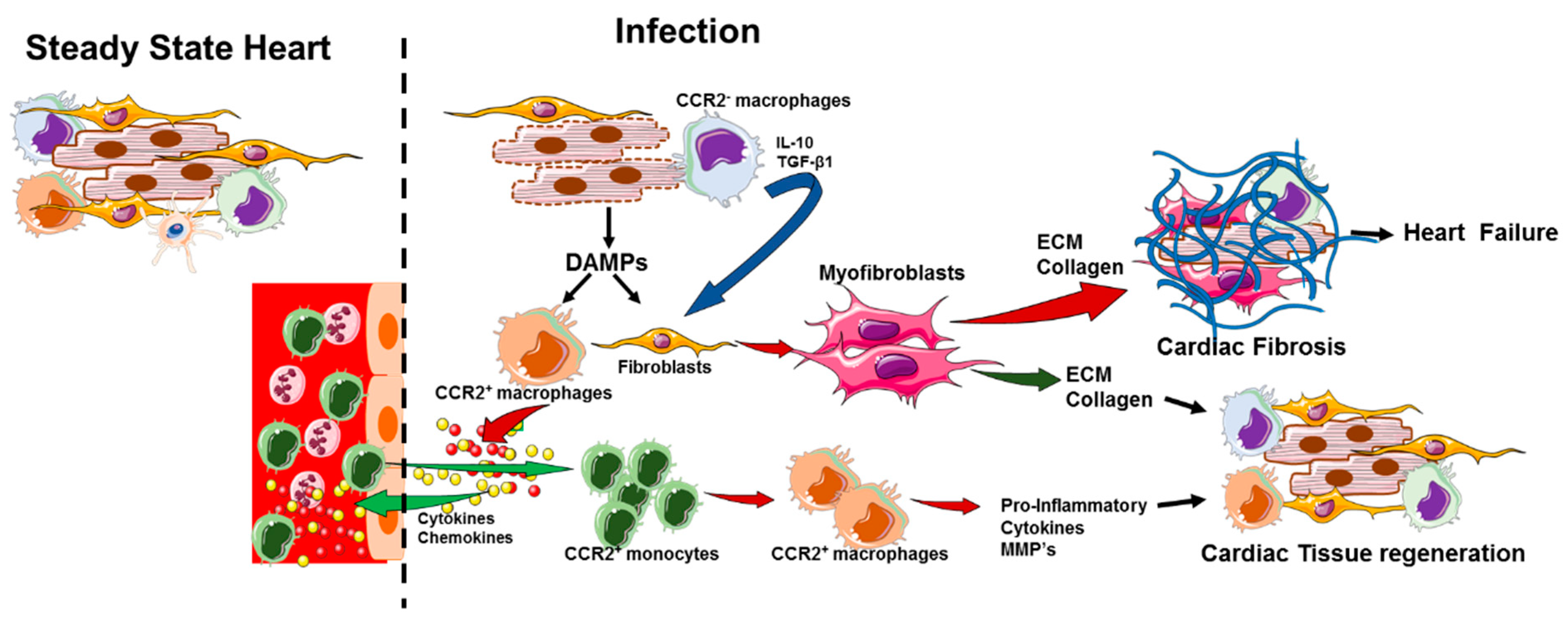
5. Activation of Cardiac Macrophages in Diseased Heart

Macrophages comprise the innate and adaptive immune system with a major role in immune system defense, inflammation and tissue restoration. Cardiac tissue contains large numbers of resident macrophages, further increased by infiltration of circulating monocytes during injury [6]. These resident macrophages are activated by the recognition of pathogen/damage-associated molecular patterns (PAMPs/DAMPs) by a number of pattern recognition receptors (PRRs) [115]. For example, lipopolysaccharides (LPS) from Gram-negative bacteria, lipoteichoic acid from Gram-positive bacteria, zymosan from fungi, lipoarabinomannan from Mycobacteria, bacterial flagellin and toxins, viral and bacterial CpG DNA and single-stranded RNA from viruses are sensed by various PRRs [116]. The PRRs are divided into two groups based on their subcellular localization: toll-like receptors (TLRs) and C-type lectin receptors are present on plasma membranes or endosomes and the second class of PRRs, including retinoic acid inducible gene –I- like receptors (RLRs), nucleotide binding and oligomerization domain (NOD)-like receptors and absent-in-melanoma-2 (AIM2) receptors, are localized in intracellular compartments [117,118]. In the heart, both PAMPs and DAMPs are detected by membrane PRRs and mediate the signaling cascades that activate nuclear factor-kB (NF-κB), activation protein-1 (AP1), and interferon regulatory factors (IRFs) transcription factors, that in turn enhance the expression of genes that encode pro-inflammatory cytokines and interferon in the heart [119]. The activation of intracellular PRRs in the heart leads to distinct pro-inflammatory signaling complexes called inflammasomes, which convert pro-caspase-1 into the catalytically active protease that is responsible for the production of interleukin-1β (IL-1β) and IL-18, subsequently triggering cardiac inflammation [120].
TLRs 1–10 are expressed in the human heart and particularly TLR2 and TLR4 are expressed abundantly [121], though the expression of TLRs have not been identified in human myocytes. Frantz et al., demonstrated that TLRs (2, 3, 4 and 6) are expressed in the myocytes of neonatal rats [122]. Although regulation of TLR expression in the heart has not been studied fully, TLR4 expression seems to be upregulated in the failing human heart and on circulating monocytes at the time of myocardial infarction [123,124]. Studies also show that loss of TLR2 in hematopoietic cells provide protection during ischemic injury [125].
The heart can be injured through a variety of pathophysiological processes, which can be grouped broadly into ischemic and non-ischemic etiologies—acute ischemic injury is the best studied model of cardiac injury and repair. Following injury, necrotic cell death leads to the activation of leukocyte populations in the cardiac microenvironment, which initiates an inflammatory response characterized by the production of pro-inflammatory cytokines and chemokines by resident immune and non-immune cells that are responsible for the recruitment of leukocytes into the area of injury. DAMPs which include adenosine triphosphate (ATP), several members of heat shock proteins (HSPs), S100A8/9, high mobility group protein B1(HMGB1), hyaluronic acid, fibrinogen, fibronectin, β-defensin, neutrophil elastase, and histones released from dying cells activate the resident macrophages, which leads to the production of inflammatory cytokines and causes sterile inflammation in the heart [115,126]. The sterile inflammation caused by non-infectious DAMPs can cause myocarditis in susceptible individuals. The sterile inflammation can be induced following cardiac injury stimuli such as myocardial infarction, cardiac surgery, allergic reactions to drugs or chemicals or excessive stress, which can be reproduced in the laboratory by exposure to myosin [127]. In addition to PAMPs and DAMPs, the cytokines released from inflammatory cells in the myocardium activate the resident macrophages, and play a critical role in macrophage polarization. For example, clearance of dead cells from the infarcted heart enhances the production of IL-10 and TGF-β, which in turn promote the anti-inflammatory M2 phenotype of CMs. In addition, secreted pro-inflammatory cytokines act on macrophages and promote the inflammatory phenotype [128].

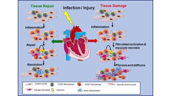
6. Role of Cardiac-Resident Macrophages in Tissue Repair during Cardiac Injury

Myocardial infarction results from blockage of coronary arteries that supply oxygen to the heart. Ischemic injury occurs in the region of the heart that is normally oxygenated by the blocked artery. Compared to adult mice, neonatal mice have striking ability to repair cardiac injury by regenerating the myocardium [129,130]. Studies by Lavine et al. [42] showed that the neonatal mice heart contains one macrophage population (MHCIIlow CCR2) and one monocyte (MHCIIlow CCR2+) subset, in contrast the adult heart contains two different resident CMs subsets (MHCIIlow CCR and MHCII high CCR2). Upon cardiac injury in neonatal mice, an embryonic-derived resident CCR2 MHCIIlow macrophage population expanded and did not recruit CCR2+ monocytes. In contrast, cardiac injury in the adult heart recruited monocytes and MHC-IIhigh CCR2+ monocyte-derived macrophages. CCR2 macrophages isolated from neonatal hearts were reparative with the ability to stimulate cardiomyocyte proliferation and angiogenesis, and minimal ability to induce an inflammatory cytokine response. Whereas CCR2+ macrophages isolated from adult hearts produced a strong inflammatory cytokine response to LPS and were unable to stimulate cardiomyocyte proliferation and angiogenesis. Thus, these studies indicate that resident CCR2 MHCIIlow macrophages are key mediators of cardiac repair in the neonatal heart.
At steady state, the adult mouse heart also contains embryonic-derived resident CCR2 MHCIIlow macrophage with reparative properties similar to the neonatal macrophages [42]. However, CCR2 resident macrophages in adult mice are rapidly lost within infracted myocardium, being reduced by 60% at day 2 after infarction [131]. The loss of resident CMs is largely due to cell death with a significant increase in dead macrophages being observed within 2 h of infarction [132]. The resident CCR2 macrophages are replaced by an influx of inflammatory CCR2+ monocytes and monocyte-derived macrophages into the infarcted tissue.
Necrotic cardiomyocytes and macrophages release DAMPs that trigger a rapid inflammatory response in the injured myocardium. The interaction of DAMPS with Toll-like receptors activates inflammatory signaling pathways leading to the production of various chemokines: CCL chemokines (CCL-2, CCL5 and CCL-7) to attract leukocytes and mast cells, and CXC chemokines (CXCL1, 2, and 8) to attract neutrophils [133,134]. A cascade of pro-inflammatory cytokines such as TNF-α, IL-1β, IL-6 and IL-18 further amplify the inflammatory response by activating the resident and invading leukocytes [115] to clear the cellular debris and dead cells [134,135]. CMs can also detect extracellular DNA released from dying cells by a cGAS-STING/IRF3 pathway that results in a Type I interferon response [136]. Release of type I interferons can then induce interferon-stimulated gene expression by autocrine and paracrine activation which can amplify the inflammatory response in the infracted heart. Granulocyte-macrophage colony-stimulating factor (GM-CSF) is also produced by cardiac fibroblasts after myocardial infarction and can act locally to promote recruitment of monocytes and distally to induce production of Ly6Chigh monocytes in the bone marrow [137].
CCL2 is essential for migration of inflammatory CCR2+ monocytes into the infracted tissue. CCL2−/−mice have reduced monocyte infiltration, lower interstitial fibrosis, and attenuated ventricular dysfunction in response to myocardial ischemia [21]. In response to myocardial injury in mice, cardiac-resident CCR2+ macrophages promote monocyte recruitment through a MyD88-dependent production of CCL2, while resident CCR2 macrophages inhibit monocyte recruitment [45]. Further, this study showed that CCR2+ resident macrophages are inflammatory, but production of inflammatory cytokines and chemokines is less than recruited CCR2+ macrophages. However, resident CCR2+ macrophages compared to recruited CCR2+macrophages differentially express type I IFN-stimulated genes in response to myocardial injury, which suggests that CCR2+-resident macrophages are responsive to type I interferon produced during myocardial infarction.
Circulating monocytes in the mouse consist of two subsets, classical Ly6Chigh monocytes which are recruited to sites of inflammation and non-classical Ly6Clow which patrol the luminal surface of the vascular endothelial cells [138]. Ly6Chigh and Ly6Clow monocyte accumulation in the infracted heart define two distinct sequential phases of monocyte recruitment [8,20] (Table 1). In the initial inflammatory phase 1, Ly6Chigh monocytes rapidly accumulate in the heart after ischemic injury, reaching a peak at 3 days post infarction and then declining. These monocytes express CCR2 and migrate into injured myocardium in response to the chemokine CCL2 and differentiate into the recruited CCR2+ macrophages. These macrophages support inflammation by secreting pro-inflammatory cytokines and chemokines and by secreting matrix metalloproteinases that break down the extracellular matrix in the injured myocardium. These recruited macrophages are phagocytic and remove debris and dying cardiomyocytes, a process that is dependent on the expression of MERKT [139]. Thus, depletion of circulating monocytes with clodronate-loaded liposomes during phase I resulted in larger areas of debris and necrotic tissue, which indicates that influx of Ly6Chigh monocytes is required for removal of debris and necrotic cells [8].
In the second phase, Ly6Clow monocytes/macrophages accumulate in the injured myocardium, with a peak at day 7 post infection (Table 1) and then declining [8,20]. Even though total numbers of Ly6Clow macrophages decline after 7 days, 75% of the macrophages are Ly6Clow through day 16 [8]. These Ly6Clow macrophages are reparative and non-inflammatory. They promote myofibroblast accumulation, angiogenesis and deposition of collagen. Two pathways have been described for the accumulation of Ly6Clow macrophages in infracted myocardium. The first pathway involves the migration of circulating Ly6Clow monocytes into the myocardium via expression of the chemokine receptor CX3CR1, which interacts with the chemokine fractalkine [8]. In this regard, depletion of circulating monocytes during phase II resulted in decreased collagen deposition and reduced numbers of smooth muscle cells. The second pathway is the differentiation of Ly6Chigh monocytes in the myocardium into proliferating Ly6Clow macrophages, which was shown to be dependent on the induction of the orphan nuclear receptor Nr4a1 [20]. In the absence of Nr4a1, Ly6Chigh monocytes expressed high levels of CCR2, increased mobilization into the myocardium, and differentiated into highly inflammatory macrophages. Further, in the absence of Nr4a1, LV function was impaired and cardiac healing was reduced with increased myocardial scar size and reduced collagen density.
Recent single-cell RNA seq analysis of recruited macrophages in the infarcted myocardium indicate that recruited macrophages are highly plastic and differentiate into multiple subsets with distinct phenotypes [45,46]. Studies by Dick et al. [46] examined recruited macrophages at day 11 after ischemic injury. At this time point the recruited monocytes have differentiated into macrophages. They found three different subsets of resident macrophages populations (described above in Cardiac-Resident Macrophages Section 3 and listed in Table 2) in the steady state and the infarcted heart tissue. In addition, they observed seven macrophage clusters unique to infarcted tissue. These unique clusters represent 64% of the macrophages present in the infracted tissue. Thus, the recruited macrophages differentiate into macrophage subsets with unique transcriptional signatures that are different from the resident macrophage populations. These unique clusters are enriched in both inflammatory pathways and reparative pathways. Single RNA sequencing studies by Bajpai et al. [45] also found seven macrophage clusters after cardiac injury. While the mechanisms involved in the differentiation of recruited monocytes that result in the macrophages clusters are unknown, Bajpai et al. found that depletion of tissue-resident CCR2+ macrophages before cardiac injury not only reduced monocyte recruitment and but also decreased a type I Interferon biased cluster. In contrast, depletion of tissue-resident CCR2 macrophages increased numbers of Arg1 and CxCL1 clusters. This suggests that cardiac-resident macrophages are important drivers of the differentiation of recruited monocytes [45].
Although recruited macrophages present during the inflammatory phase have been described as M1 macrophages and macrophages present during the reparative phase as M2 macrophages, the transcriptional heterogeneity in recruited macrophages present in infarcted tissues does not easily fit into the MI/M2 paradigm. The M1/M2 nomenclature is derived from studies of in vitro polarization of mouse macrophages with classically activated LPS+IFN-γ (M1) or alternatively activated IL-4 (M2), each with a distinct transcriptome response. See Jablonski et al. [140] for transcriptomes (GSE69607) of classically activated and alternatively activated bone marrow-derived macrophages. Further, research indicated that M1 macrophages have a pro-inflammatory phenotype with anti-pathogen activity while M2 macrophages promote anti-inflammatory and tissue repair responses [141]. Recently Orecchioni et al. [142] found that most surface markers identified on in vitro generated macrophages do not translate to the in vivo situation in classically activated mice. Thus, we prefer that macrophage subsets be classified based on their function and transcriptional profile as suggested by Nahrendorf and Swirski [143]. In this regard, in Table 2 we listed CMs present in the heart during steady state and myocardial infraction based on transcriptional profile and function.

7. Role of Macrophages in Cardiac Remodeling in Hypertension and Diabetic Cardiomyopathy

Diabetes and hypertension, which often co-exist, are predominant risk factors for cardiovascular diseases such as MI and HF, and are characterized by chronic, low grade inflammation, which promotes adverse cardiac remodeling. The inflammatory response is a critical mechanism by which the heart responds to injury and develops adaptive remodeling [144]. However, uncontrolled inflammation can weaken the adaptive response and promote cardiac injury. Both diabetes and hypertension can lead to chronic inflammation and activation of macrophages toward an inflammatory phenotype. Although macrophages play a key role in cardiac remodeling, dysregulation of macrophages between pro-inflammatory and anti-inflammatory phenotypes promotes excessive inflammation and cardiac injury [145]. Cardiac metabolic reprogramming has been implicated in cardiac adaptation to injury [146,147,148], though the healthy heart relies mainly on mitochondrial oxidative phosphorylation metabolism of fatty acid for its energy demands. In contrast, during decompensated HF, the heart relies predominantly on glycolysis [147,149], in which HF leads to the activation of hypoxia-induced factor 1 alpha (HIF-1α) transcription factor which induces transcription of a glycolytic and proinflammatory gene profile [150,151]. HIF1-α can also be stimulated by nonhypoxic mechanisms associated with obesity and hypertension, such as inflammatory cytokines, hyperglycemia, saturated fatty acid activation of TLR4, and oxidized low-density lipoprotein [152,153,154,155]. In addition, diabetic patients are prone to bacterial infections and impaired wound healing, which may be due to the impairments in pro-reparative/anti-inflammatory macrophage functions and increases in a pro-inflammatory macrophage phenotype through enhanced expression of long-chain acyl-CoA synthetase [156,157,158]. Macrophages are also susceptible to protein glycation and formation of advanced glycation end products (ACEs) due to excess glucose levels, which activates the NF-κB pathway and induces production of inflammatory cytokines [159,160]. Additionally, macrophages from patients with coronary artery disease, a common comorbidity of obesity–hypertension, display significantly increased IL-1β and TNF-α expression compared to patients with inflammatory vascular diseases [161]. Therefore, during hyperglycemia, macrophages appear to upregulate glucose uptake and utilization and subsequently increase inflammatory cytokine production, which leads to activation of an inflammatory phenotype [161]. In addition, free fatty acids, lipid mediators and adipokines can promote the inflammatory phenotype [162,163,164].
The source of cardiac macrophages plays a key role in regulation of cardiac remodeling during obesity and hypertension. Generally, the healthy heart contains a low number of M2-like macrophages [45,165,166] which are often considered protective. Recent studies have demonstrated that these M2-like macrophages (embryonic in origin) are lost with aging and replaced with CCR2+ monocyte-derived macrophages, even in the absence of injury [45]. In a pressure overload mouse model, resident M2-like macrophages initially promote myocardial adaptive remodeling to mechanical stress. However infiltrating CCR2+ monocytes promote maladaptive remodeling during the transition to decompensated HF [167]. The epicardial adipose tissue (EAT) is a rich source of macrophages, neutrophils, and lymphocytes, which may protect the heart from infections [168]. The myokines released from heart muscle during exercise act directly on EAT macrophages to promote an M2-like phenotype and provide protective effects [169]. However, during cardiac injury EAT can be a major source of inflammatory macrophages, due to hypoxia-induced activation of macrophages in EAT which can easily infiltrate into the myocardium [170,171]. In mice, EAT is a major contributor to inflammation during cardiac injury after MI [170], and in MI patients, increased EAT thickness is associated with visceral obesity and cardiac fibrosis [172]. In addition, necrotic myocytes during severe cardiac injury can recruit and activate macrophages via the release of DAMPs [166,173]. The damaged mitochondria in myocytes (a hallmark of HF) can also release DAMPs that enhance the release of proinflammatory chemokines and cytokines [174]. The other major activator of cardiac inflammation during hypertension is endothelial cell injury, due to pressure-induced shear stress and mechanical stretch, which lead to increased reactive oxygen species (ROS) production and impairment of NO signaling, which may attract nearby macrophages [175].
Although the importance of macrophages in cardiac remodeling in myocardial infarction has been studied extensively [46,165,176,177], the role of macrophage metabolism has not been investigated thoroughly. In a mouse model of MI, glycolytic genes, such as GAPDH, are strongly upregulated in macrophages found in the infarcted region at day 1 of MI. In contrast, mitochondrial genes, such as succinate dehydrogenase, are increased at day 3 after MI [173], which suggests that the cardiac microenvironment is hypoxic at day 1 but becomes reoxygenated at day 3 due to vasculogenesis [173,178]. In a rat model of MI, inhibition of glycolysis decreased cardiac macrophage glycolysis and inflammation, and improved LV function [179]. Mechanistically, the efferocytosis of necrotic cells increases intracellular fatty acid supply, fuels mitochondrial fatty acid oxidation and polarizes the macrophage M2-like phenotype [180]. In mice with hypertension and diastolic dysfunction, M2-like polarization contributes to cardiac fibrosis, though this depends initially on inflammatory macrophage infiltration and expansion [181]. Furthermore, in the mouse heart as discussed earlier in this review, resident macrophages, which are negative for CCR2, can be further divided into two groups based on the expression of MHCII [6]. Similarly, CCR2+ cardiac monocyte-derived macrophages can be derived from infiltrating Ly6Chi (M1-like) or Ly6Clow (M2-like) monocytes [8,167], which suggests that the metabolic profiles may differ not just based on the M1 and M2 paradigm but also on resident macrophages versus infiltrating monocytes and their subsets. Still the immunometabolics field is new and largely based on M1/M2 phenotypes, and in the future research will focus on understanding the metabolic processes of the cardiac macrophage subsets in the remodeling heart.

8. Role of Macrophages on Mitochondrial Homeostasis and Collagen Secretion

The majority of the tissues in the body contain tissue-resident macrophages, which are extremely heterogeneous and perform tissue-specific functions—this heterogeneity is dependent on the microenvironment in each tissue. Though macrophages are considered a key cell to mount innate and adaptive immune functions [182], they also mediate tissue-specific functions unrelated to immunity [183]. For example, in the heart, tissue-resident macrophages prevent fibrosis [184], facilitate electrical activity in the atrioventricular node [82] and are involved in healing of injured areas [46,185]. Furthermore, macrophages of unknown function are populated in the myocardium of healthy heart, that yet to be identified. Cardiomycytes are highly specialized cells in the heart with a unique function. Cardiomyocytes are large in size and contain a large number of mitochondria and sarcomeres, which occupy most of the cell volume and deal with the intense metabolic energy and mechanical demands of the heart. Additionally, cardiomyocytes are long lived and rarely renew; they are subjected to the intense mechanical stress and metabolic demands of the beating heart, which requires homeostatic mechanisms to eliminate dysfunctional mitochondria in order to maintain the healthy state of cardiomyocytes. These nonfunctional mitochondria are removed by a process called mitophagy and this preserves the crucial functions of cardiomyocytes [186]. Since cardiomyocytes are subject to extreme energetic demand and the mitochondria produce high energy ATP and reactive oxygen species, it is expected that mitochondria will become damaged and the damaged mitochondria will be degraded via autophagy machinery. Recently, Nicolas-Avila et al. demonstrated a novel non-canonical elimination of dysfunctional mitochondria and other organelles from healthy cardiomyocytes. These authors showed that dysfunctional mitochondria and other cargo are expelled in dedicated membranous particles called exophores. The exophores expose phoshotidylserine on the membrane, which is recognized by the MerTK phagocytic receptor expressed on CMs and the internalized exophores are degraded by phogosome maturation. This intracellular exchange occurs in the heart during homeostasis. In addition, depletion of CMs or deletion of the phagocytic receptor MerTK results in defective elimination of the exophores and mitochondria accumulation in the heart extracellular space, which leads to inflammasome activation, blockage of cardiomyocyte autophagy, accumulation of anomalous mitochondria in cardiomyocytes, metabolic alterations, and ventricular dysfunction [187]. Thus, cardiac macrophages are key for maintaining mitochondrial homeostasis in the healthy heart.
As discussed above, macrophages are highly plastic and functionally diverse during steady state and following injury [188]. Similarly, in the heart tissue, resident macrophages have been implicated in many disease conditions (discussed above). After myocardiac infarction or infection, a large number of immune cells are infiltrated into the heart to remove dying tissue, scavenge pathogens and promote healing. However, in some circumstances, immune cells can cause irreversible damage, contributing to HF. During the initial stage of injury, CMs release pro-inflammatory cytokines to create an inflammatory state, and later transition into a reparative M2-like state, which produce cytokines, chemokines and growth factors (TFG-β and PDGF) that activate fibroblasts to become myofibroblasts, which deposit collagen to form scars. Additionally, CMs produce metalloproteinases and regulate extracellular matrix (ECM) accumulation at the site of injury, which contributes to scar tissue formation [189]. Thus, transition of a tissue repair phase to a regenerative phase following heart injury requires an orchestrated and balanced immune response to fine-tune the interplay between a pro-fibrotic and a pro-regenerative environment. The macrophages are essential to both repair by scar formation and tissue regeneration. However, factors that drive CMs towards either regenerative or pro-fibrotic phenotype are largely unknown [190,191,192,193]. Recently, Simoes et al. [194] demonstrated that the CMs directly contribute to collagen secretion and scar formation in a Zebrafish model and a mouse model. Transcriptomic analysis revealed that the expression of collagen-associated genes (col4a3bpa, col7a1l) was upregulated upon injury in the zebrafish model. In the neonatal mouse model, the authors harvested CMs from P1 and P7 infarcted heart at day 7 and compared gene expression; they reported that several ECM genes Col8a1, Col5a2, Col6a2, Fbn, Postn and Bgn are upregulated, accompanied by evidence of collagen-1+ fibrils in the infarct region. Thus, these findings indicate the CMs are also essential for cardiac tissue repair and regeneration. However, the functional relevance of macrophage-derived collagen production in the context of cardiovascular outcomes remains to be investigated in the future.

9. Cardiac-Resident Macrophages and Cardiac Fibrosis

Cardiac fibrosis is an inevitable consequence of chronic insult to the myocardium, characterized by net accumulation of extracellular matrix (ECM) proteins in the cardiac interstitium which leads to wall thickening, systolic and diastolic dysfunction, and impaired overall heart performance. In the healthy heart, the ECM provides a scaffold for cardiac cells and in this manner ensures the structural integrity and function of the heart [189]. In addition, it has been demonstrated that each cardiomyocyte is surrounded by ECM, which ensures the transmission of electrical conduction and contractile force from a single cardiomyocyte into the whole organ, and also ECM function as a reservoir for various latent growth factors [133]. Myocardial injury can be caused by either sterile inflammation or non-sterile inflammation (infection). Moreover, terminating the inflammation requires an anti-inflammatory response, which leads to the initiation of pro-fibrotic signaling [195]. On the other hand, pathophysiological stimuli, for instance pressure overload, volume overload, metabolic dysfunction, and biological aging process may cause interstitial and perivascular fibrosis in the absence of myocardial inflammation.
Both human patients and experimental models of heart disease indicate that the extent of fibrotic remodeling is closely associated with adverse outcome. During the initial inflammatory phase, described above, debris and dead cardiomyocytes are cleared by resident embryonic and recruited macrophages and the extracellular matrix is degraded by secreted matrix metalloproteinases. Once sufficient clearance of necrotic cells and apoptotic immune cells is achieved, a proliferative phase is activated to resolve the damage. During this phase, the fibroblasts trans-differentiate into secretory and contractile cells, called myofibroblasts. This is a key cellular process that results in secretion of an abundant collagen and nonstructural matrix proteins to compensate for the cellular loss in many different conditions associated with HF [133,135]. The proliferative phase is orchestrated by several anti-inflammatory and pro-fibrotic mediators produced by both immune and non-immune cells in the injured myocardium. For example, apoptotic neutrophils release lactoferrin and annexin A1 which inhibit further leukocyte invasion [134], efferocytosis of dead cells stimulates macrophages to produce anti-inflammatory TGF-β1, IL-10, lipoxins and resolvins [134,196,197], regulatory T cell activation leads to additional secretion of TGF-β1 and IL-10 [134], and the secretion of MMPs by immune cells [198] function together and resolve the inflammatory damage. MMPs initiate remodeling by degrading ECM and act to limit leukocyte invasion by cleaving chemokines [199]. TGF-β1 is a well characterized isoform in the cardiac tissue [4], which in the healthy heart is bound to the ECM as an inactive latent complex [200,201]. Cleavage by plasmin and MMP-2 and MMP-9 releases the active form of TGF-β1 [200,202]. Active TGF-β1 binds to its type II receptor (TGFβRII) on the cell surface of fibroblasts and initiates the Smad3- signaling pathways and enhances the expression of a smooth muscle actin (x-SMA) expression and promotes myofibroblast trans-differentiation in the presence of fibronectin domain ED-A [133,203]. These myofibroblasts accumulate in the myocardium in a wide range of pathologic conditions, including myocardial infarction, myocarditis, cardiac pressure or volume overload and alcoholic cardiomyopathy [204,205,206,207]. The activated myofibroblasts secrete elevated levels of collagens and other ECM proteins to maintain the structural integrity and prevent wall rupture and myocardial dysfunction [208]. The excessive deposition of ECM proteins and collagens leads to ventricular stiffness, which can result in contractile dysfunction [209,210], and increase the risk of arrhythmogenesis and mortality [211,212] (Figure 3). Furthermore, myocardial inflammation and fibrosis within the perivascular regions may decrease the oxygen and nutrient availability to cardiac tissue and increase the pathological remodeling response [213]. Following the proliferative stage and establishment of collagen and ECM-based matrix at the infarct site, a maturation stage occurs in which a scar is formed with a cross-linked extracellular matrix. The growth factors and matrix cellular proteins that support the survival and activity of myofibroblasts are depleted, and the majority of the myofibroblasts undergo apoptosis [203,214]. Vascular cells also die, and temporary microvasculature disintegrates. In addition, Lavine et al. demonstrated that tissue-resident macrophages induce cardiomyocyte proliferation and blood vessel development after cardiac injury [42]. Consequently, monocyte recruitment to the injured adult heart is suppressed, and the embryonic macrophage population is largely preserved, resulting in reduced inflammation and accelerated repair. Therefore, cardiac macrophages differentiated from recruited bone marrow-derived monocytes exhibit tissue destructive activity, whereas the embryonically derived tissue-resident macrophage population facilitates the resolution of inflammation and instructs tissue repair in the heart (Figure 3).

10. Future Direction of Cardiac Macrophages

Although scientists have identified new phenotypes and functions of cardiac macrophages in the context of MI and HF, their role in cardiac inflammation, tissue regeneration, remodeling and fibrosis is still developing and there remains several important avenues of research for the future. First, although the roles of macrophages in regulating cardiac inflammation, fibrosis, tissue repair and regeneration in different experimental rodent models may overlay, the source and phenotypes of resident macrophage are not completely understood. Recent studies demonstrated that healthy human myocardium is populated with CCR2 macrophages and maintained through local proliferation, while CCR2+ macrophages are derived from monocytes and local proliferation [173]. However, the localization and function of these macrophages in the heart is distinct: CCR2 macrophages (M2- like) function as anti-inflammatory, helping to control the inflammation while the CCR2+ macrophages (M1-like) initiate the inflammation, which can lead to HF. However, we still lack a clear understanding of whether macrophage function is a cause or consequence in human disease development, and even less is known about the role of these cells after chronic HF has been established. Animal models have provided significant advancement in understanding of the complexity and plasticity underlying resident and recruited cardiac macrophage biology in both steady-state and chronic HF and have identified the importance of macrophages in HF pathogenesis. However, it remains unclear whether these findings will mimic human diseases. Second, the lack of knowledge defining the crosstalk between cardiac-resident macrophages and other cardiac-resident cells such as fibroblasts and cardiomyocytes, and the influence of comorbidities and risk factors on macrophage heterogeneity and function limits our understanding of the role of macrophages in chronic HF, and the development of new therapeutic strategies. Third, macrophage polarization is closely tied to changes in glycolytic (M1-like) and oxidative phosphorylation (M2-like). Macrophage metabolic shifts are not only required for energy demands but also for regulation of pro- and anti-inflammatory processes. Macrophage metabolic shifts are tightly regulated by several intracellular signaling pathways, in which NF-κB, HIF-1α, PDK1 and PPAR-γ play an important role in reprogramming the macrophages [145]. However, investigation pertaining to cardiac macrophage metabolism is still young and requires thorough investigations. Thus, understanding cardiac macrophage metabolism will provide insight into its roles and the mechanisms associated with the remodeling heart and heart failure. Additionally, the thorough investigation of macrophage metabolism will yield unique opportunities to treat HF by targeting macrophage metabolism in the heart without affecting cardiomyocyte metabolism.

11. Conclusions

Over the past few years, significant advancements were made to characterize the cardiac immune cells and their role in steady heart function, which will allow for the development of new regenerative targets for HF. In mammalian hearts, the resident cardiac fibroblasts and immune cells, specifically macrophages, regulate cardiomyocyte function in health and disease conditions. The regenerative capacity of cardiomyocytes is very limited in adult hearts and the rate of cardiomyocyte proliferation cannot support the need for new myocytes upon cardiac injury, particularly with infections. However, the resident macrophage populations recognize the danger signal from cardiomyocytes, recruit immune cells and exert detrimental inflammation to clear the infection. At the same time, the immune response activates the cardiac fibroblasts to promote cardiac fibrosis, and cardiac hypertrophy. This leads to further recruitment and enhancement of inflammation, which creates an environment hostile to regeneration and tissue repair. Cardiac fibrosis is beneficial for cardiac tissue repair upon injury. However, uncontrolled tissue damage and continuous activation of pro-inflammatory signaling leads to unregulated extra cellular matrix production, and causes defects in the cardiac electrical conduction system and HF. Despite the incredible research efforts and progress in new drug discoveries that have been made to treat the cardiac dysfunctions during infections or sterile inflammation, the human heart remains unprotected after injury. Therefore, a better understanding of immune cell phenotypes, function, and cardiac macrophage plasticity during cardiac tissue damage and repair could lead to the development of new therapies that improve the outcome of patients with severe myocardial damage caused by either sterile or infectious inflammation.

Author Contributions

W.P.L. and M.V.S.R. wrote the review article and D.J.W. edited the manuscript. All authors have read and agreed to the published version of the manuscript.

Funding

This work was supported by the National Institutes for Health (NIH); R01AI146252 and R21AI146690 (to M.V.S.R) and R01AI134895 and R01AI143916 (DJW).

Data Availability Statement

Authors do not include any large data sets in this manuscript.

Acknowledgments

The authors take the opportunity to thank Ohio State University Comprehensive Cancer Center Shared Resources, Flow cytometry core, College of Medicine.

Conflicts of Interest

The authors declare no conflict of interest.

Abbreviations Used in This Article

HFHeart failure
CVDCardiovascular diseases
CMCardiac macrophages
MIMyocardial Infarction
DAMPsDanger-Associated Molecular Patterns
PAMPsPathogen-Associated Molecular Patterns
TregT regulatory cell
cDCCardiac Dendritic cell
NabsNatural antibodies
CARCoxsackievirus and Adenovirus Receptor
DAFDecay Accelerating Factor
MMPMatrix metalloproteinase
HMGB1High mobility group box-1
MERTKMyeloid-epithelial-reproductive tyrosine kinase
EATepicardial adipose tissue

References

  1. Gordon, S.; Martinez-Pomares, L. Physiological roles of macrophages. Pflugers Arch. 2017, 469, 365–374. [Google Scholar] [CrossRef] [PubMed] [Green Version]
  2. Roth, G.A.; Johnson, C.; Abajobir, A.; Abd-Allah, F.; Abera, S.F.; Abyu, G.; Ahmed, M.; Aksut, B.; Alam, T.; Alam, K.; et al. Global, Regional, and National Burden of Cardiovascular Diseases for 10 Causes, 1990 to 2015. J. Am. Coll. Cardiol. 2017, 70, 1–25. [Google Scholar] [CrossRef]
  3. Benjamin, E.J.; Muntner, P.; Alonso, A.; Bittencourt, M.S.; Callaway, C.W.; Carson, A.P.; Chamberlain, A.M.; Chang, A.R.; Cheng, S.; Das, S.R.; et al. Heart Disease and Stroke Statistics-2019 Update: A Report from the American Heart Association. Circulation 2019, 139, e56–e528. [Google Scholar] [CrossRef] [PubMed]
  4. Frangogiannis, N.G. Cardiac fibrosis: Cell biological mechanisms, molecular pathways and therapeutic opportunities. Mol. Asp. Med. 2019, 65, 70–99. [Google Scholar] [CrossRef] [PubMed]
  5. Hulsmans, M.; Sam, F.; Nahrendorf, M. Monocyte and macrophage contributions to cardiac remodeling. J. Mol. Cell. Cardiol. 2016, 93, 149–155. [Google Scholar] [CrossRef] [PubMed] [Green Version]
  6. Epelman, S.; Lavine, K.J.; Beaudin, A.E.; Sojka, D.K.; Carrero, J.A.; Calderon, B.; Brija, T.; Gautier, E.L.; Ivanov, S.; Satpathy, A.T.; et al. Embryonic and adult-derived resident cardiac macrophages are maintained through distinct mechanisms at steady state and during inflammation. Immunity 2014, 40, 91–104. [Google Scholar] [CrossRef] [Green Version]
  7. Ramos, G.C.; van den Berg, A.; Nunes-Silva, V.; Weirather, J.; Peters, L.; Burkard, M.; Friedrich, M.; Pinnecker, J.; Abesser, M.; Heinze, K.G.; et al. Myocardial aging as a T-cell-mediated phenomenon. Proc. Natl. Acad. Sci. USA 2017, 114, E2420–E2429. [Google Scholar] [CrossRef] [Green Version]
  8. Nahrendorf, M.; Swirski, F.K.; Aikawa, E.; Stangenberg, L.; Wurdinger, T.; Figueiredo, J.L.; Libby, P.; Weissleder, R.; Pittet, M.J. The healing myocardium sequentially mobilizes two monocyte subsets with divergent and complementary functions. J. Exp. Med. 2007, 204, 3037–3047. [Google Scholar] [CrossRef] [Green Version]
  9. Zouggari, Y.; Ait-Oufella, H.; Bonnin, P.; Simon, T.; Sage, A.P.; Guerin, C.; Vilar, J.; Caligiuri, G.; Tsiantoulas, D.; Laurans, L.; et al. B lymphocytes trigger monocyte mobilization and impair heart function after acute myocardial infarction. Nat. Med. 2013, 19, 1273–1280. [Google Scholar] [CrossRef]
  10. Weirather, J.; Hofmann, U.D.; Beyersdorf, N.; Ramos, G.C.; Vogel, B.; Frey, A.; Ertl, G.; Kerkau, T.; Frantz, S. Foxp3+ CD4+ T cells improve healing after myocardial infarction by modulating monocyte/macrophage differentiation. Circ. Res. 2014, 115, 55–67. [Google Scholar] [CrossRef]
  11. Saxena, A.; Dobaczewski, M.; Rai, V.; Haque, Z.; Chen, W.; Li, N.; Frangogiannis, N.G. Regulatory T cells are recruited in the infarcted mouse myocardium and may modulate fibroblast phenotype and function. Am. J. Physiol. Heart Circ. Physiol. 2014, 307, H1233–H1242. [Google Scholar] [CrossRef] [PubMed]
  12. Clemente-Casares, X.; Hosseinzadeh, S.; Barbu, I.; Dick, S.A.; Macklin, J.A.; Wang, Y.; Momen, A.; Kantores, C.; Aronoff, L.; Farno, M.; et al. A CD103(+) Conventional Dendritic Cell Surveillance System Prevents Development of Overt Heart Failure during Subclinical Viral Myocarditis. Immunity 2017, 47, 974–989. [Google Scholar] [CrossRef] [PubMed] [Green Version]
  13. Bracamonte-Baran, W.; Chen, G.; Hou, X.; Talor, M.V.; Choi, H.S.; Davogustto, G.; Taegtmeyer, H.; Sung, J.; Hackam, D.J.; Nauen, D.; et al. Non-cytotoxic Cardiac Innate Lymphoid Cells Are a Resident and Quiescent Type 2-Commited Population. Front. Immunol. 2019, 10, 634. [Google Scholar] [CrossRef] [PubMed] [Green Version]
  14. Wagner, M.J.; Khan, M.; Mohsin, S. Healing the Broken Heart; The Immunomodulatory Effects of Stem Cell Therapy. Front. Immunol. 2020, 11, 639. [Google Scholar] [CrossRef] [PubMed] [Green Version]
  15. Ziegler-Heitbrock, L.; Ancuta, P.; Crowe, S.; Dalod, M.; Grau, V.; Hart, D.N.; Leenen, P.J.; Liu, Y.J.; MacPherson, G.; Randolph, G.J.; et al. Nomenclature of monocytes and dendritic cells in blood. Blood 2010, 116, e74–e80. [Google Scholar] [CrossRef] [PubMed]
  16. Geissmann, F.; Jung, S.; Littman, D.R. Blood monocytes consist of two principal subsets with distinct migratory properties. Immunity 2003, 19, 71–82. [Google Scholar] [CrossRef] [Green Version]
  17. Maekawa, Y.; Anzai, T.; Yoshikawa, T.; Asakura, Y.; Takahashi, T.; Ishikawa, S.; Mitamura, H.; Ogawa, S. Prognostic significance of peripheral monocytosis after reperfused acute myocardial infarction:a possible role for left ventricular remodeling. J. Am. Coll. Cardiol. 2002, 39, 241–246. [Google Scholar] [CrossRef] [Green Version]
  18. Glezeva, N.; Voon, V.; Watson, C.; Horgan, S.; McDonald, K.; Ledwidge, M.; Baugh, J. Exaggerated inflammation and monocytosis associate with diastolic dysfunction in heart failure with preserved ejection fraction: Evidence of M2 macrophage activation in disease pathogenesis. J. Card. Fail. 2015, 21, 167–177. [Google Scholar] [CrossRef]
  19. Hashimoto, D.; Chow, A.; Noizat, C.; Teo, P.; Beasley, M.B.; Leboeuf, M.; Becker, C.D.; See, P.; Price, J.; Lucas, D.; et al. Tissue-resident macrophages self-maintain locally throughout adult life with minimal contribution from circulating monocytes. Immunity 2013, 38, 792–804. [Google Scholar] [CrossRef] [Green Version]
  20. Hilgendorf, I.; Gerhardt, L.M.; Tan, T.C.; Winter, C.; Holderried, T.A.; Chousterman, B.G.; Iwamoto, Y.; Liao, R.; Zirlik, A.; Scherer-Crosbie, M.; et al. Ly-6Chigh monocytes depend on Nr4a1 to balance both inflammatory and reparative phases in the infarcted myocardium. Circ. Res. 2014, 114, 1611–1622. [Google Scholar] [CrossRef] [Green Version]
  21. Frangogiannis, N.G.; Dewald, O.; Xia, Y.; Ren, G.; Haudek, S.; Leucker, T.; Kraemer, D.; Taffet, G.; Rollins, B.J.; Entman, M.L. Critical role of monocyte chemoattractant protein-1/CC chemokine ligand 2 in the pathogenesis of ischemic cardiomyopathy. Circulation 2007, 115, 584–592. [Google Scholar] [CrossRef] [PubMed]
  22. Hart, D.N.; Fabre, J.W. Demonstration and characterization of Ia-positive dendritic cells in the interstitial connective tissues of rat heart and other tissues, but not brain. J. Exp. Med. 1981, 154, 347–361. [Google Scholar] [CrossRef] [Green Version]
  23. Van der Borght, K.; Lambrecht, B.N. Heart macrophages and dendritic cells in sickness and in health: A tale of a complicated marriage. Cell. Immunol. 2018, 330, 105–113. [Google Scholar] [CrossRef] [PubMed]
  24. Di Leo, A.; Risi, E.; Biganzoli, L. No pain, no gain... What we can learn from a trial reporting negative results. Ann. Oncol. 2017, 28, 678–680. [Google Scholar] [CrossRef] [PubMed]
  25. Ganguly, D.; Haak, S.; Sisirak, V.; Reizis, B. The role of dendritic cells in autoimmunity. Nat. Rev. Immunol. 2013, 13, 566–577. [Google Scholar] [CrossRef] [Green Version]
  26. Van der Borght, K.; Scott, C.L.; Nindl, V.; Bouche, A.; Martens, L.; Sichien, D.; Van Moorleghem, J.; Vanheerswynghels, M.; De Prijck, S.; Saeys, Y.; et al. Myocardial Infarction Primes Autoreactive T Cells through Activation of Dendritic Cells. Cell Rep. 2017, 18, 3005–3017. [Google Scholar] [CrossRef] [Green Version]
  27. Guilliams, M.; Ginhoux, F.; Jakubzick, C.; Naik, S.H.; Onai, N.; Schraml, B.U.; Segura, E.; Tussiwand, R.; Yona, S. Dendritic cells, monocytes and macrophages: A unified nomenclature based on ontogeny. Nat. Rev. Immunol. 2014, 14, 571–578. [Google Scholar] [CrossRef]
  28. Smith, S.C.; Allen, P.M. Expression of myosin-class II major histocompatibility complexes in the normal myocardium occurs before induction of autoimmune myocarditis. Proc. Natl. Acad. Sci. USA 1992, 89, 9131–9135. [Google Scholar] [CrossRef] [Green Version]
  29. Donermeyer, D.L.; Beisel, K.W.; Allen, P.M.; Smith, S.C. Myocarditis-inducing epitope of myosin binds constitutively and stably to I-Ak on antigen-presenting cells in the heart. J. Exp. Med. 1995, 182, 1291–1300. [Google Scholar] [CrossRef] [Green Version]
  30. Hofmann, U.; Beyersdorf, N.; Weirather, J.; Podolskaya, A.; Bauersachs, J.; Ertl, G.; Kerkau, T.; Frantz, S. Activation of CD4+ T lymphocytes improves wound healing and survival after experimental myocardial infarction in mice. Circulation 2012, 125, 1652–1663. [Google Scholar] [CrossRef] [Green Version]
  31. Dobaczewski, M.; Xia, Y.; Bujak, M.; Gonzalez-Quesada, C.; Frangogiannis, N.G. CCR5 signaling suppresses inflammation and reduces adverse remodeling of the infarcted heart, mediating recruitment of regulatory T cells. Am. J. Pathol. 2010, 176, 2177–2187. [Google Scholar] [CrossRef] [PubMed]
  32. Matsumoto, K.; Ogawa, M.; Suzuki, J.; Hirata, Y.; Nagai, R.; Isobe, M. Regulatory T lymphocytes attenuate myocardial infarction-induced ventricular remodeling in mice. Int. Heart J. 2011, 52, 382–387. [Google Scholar] [CrossRef] [PubMed] [Green Version]
  33. Yang, Z.; Day, Y.J.; Toufektsian, M.C.; Xu, Y.; Ramos, S.I.; Marshall, M.A.; French, B.A.; Linden, J. Myocardial infarct-sparing effect of adenosine A2A receptor activation is due to its action on CD4+ T lymphocytes. Circulation 2006, 114, 2056–2064. [Google Scholar] [CrossRef] [PubMed] [Green Version]
  34. Laroumanie, F.; Douin-Echinard, V.; Pozzo, J.; Lairez, O.; Tortosa, F.; Vinel, C.; Delage, C.; Calise, D.; Dutaur, M.; Parini, A.; et al. CD4+ T cells promote the transition from hypertrophy to heart failure during chronic pressure overload. Circulation 2014, 129, 2111–2124. [Google Scholar] [CrossRef] [PubMed] [Green Version]
  35. Nevers, T.; Salvador, A.M.; Grodecki-Pena, A.; Knapp, A.; Velazquez, F.; Aronovitz, M.; Kapur, N.K.; Karas, R.H.; Blanton, R.M.; Alcaide, P. Left Ventricular T-Cell Recruitment Contributes to the Pathogenesis of Heart Failure. Circ. Heart Fail. 2015, 8, 776–787. [Google Scholar] [CrossRef] [PubMed] [Green Version]
  36. van Furth, R.; Cohn, Z.A. The origin and kinetics of mononuclear phagocytes. J. Exp. Med. 1968, 128, 415–435. [Google Scholar] [CrossRef]
  37. Sawyer, R.T.; Strausbauch, P.H.; Volkman, A. Resident macrophage proliferation in mice depleted of blood monocytes by strontium-89. Lab. Investig. 1982, 46, 165–170. [Google Scholar]
  38. Ginhoux, F.; Greter, M.; Leboeuf, M.; Nandi, S.; See, P.; Gokhan, S.; Mehler, M.F.; Conway, S.J.; Ng, L.G.; Stanley, E.R.; et al. Fate mapping analysis reveals that adult microglia derive from primitive macrophages. Science 2010, 330, 841–845. [Google Scholar] [CrossRef] [Green Version]
  39. Schulz, C.; Gomez Perdiguero, E.; Chorro, L.; Szabo-Rogers, H.; Cagnard, N.; Kierdorf, K.; Prinz, M.; Wu, B.; Jacobsen, S.E.; Pollard, J.W.; et al. A lineage of myeloid cells independent of Myb and hematopoietic stem cells. Science 2012, 336, 86–90. [Google Scholar] [CrossRef] [Green Version]
  40. Yona, S.; Kim, K.W.; Wolf, Y.; Mildner, A.; Varol, D.; Breker, M.; Strauss-Ayali, D.; Viukov, S.; Guilliams, M.; Misharin, A.; et al. Fate mapping reveals origins and dynamics of monocytes and tissue macrophages under homeostasis. Immunity 2013, 38, 79–91. [Google Scholar] [CrossRef] [Green Version]
  41. Jakubzick, C.; Gautier, E.L.; Gibbings, S.L.; Sojka, D.K.; Schlitzer, A.; Johnson, T.E.; Ivanov, S.; Duan, Q.; Bala, S.; Condon, T.; et al. Minimal differentiation of classical monocytes as they survey steady-state tissues and transport antigen to lymph nodes. Immunity 2013, 39, 599–610. [Google Scholar] [CrossRef] [PubMed] [Green Version]
  42. Lavine, K.J.; Epelman, S.; Uchida, K.; Weber, K.J.; Nichols, C.G.; Schilling, J.D.; Ornitz, D.M.; Randolph, G.J.; Mann, D.L. Distinct macrophage lineages contribute to disparate patterns of cardiac recovery and remodeling in the neonatal and adult heart. Proc. Natl. Acad. Sci. USA 2014, 111, 16029–16034. [Google Scholar] [CrossRef] [PubMed] [Green Version]
  43. Leid, J.; Carrelha, J.; Boukarabila, H.; Epelman, S.; Jacobsen, S.E.; Lavine, K.J. Primitive Embryonic Macrophages are Required for Coronary Development and Maturation. Circ. Res. 2016, 118, 1498–1511. [Google Scholar] [CrossRef] [PubMed]
  44. Bajpai, G.; Schneider, C.; Wong, N.; Bredemeyer, A.; Hulsmans, M.; Nahrendorf, M.; Epelman, S.; Kreisel, D.; Liu, Y.; Itoh, A.; et al. The human heart contains distinct macrophage subsets with divergent origins and functions. Nat. Med. 2018, 24, 1234–1245. [Google Scholar] [CrossRef]
  45. Bajpai, G.; Bredemeyer, A.; Li, W.; Zaitsev, K.; Koenig, A.L.; Lokshina, I.; Mohan, J.; Ivey, B.; Hsiao, H.M.; Weinheimer, C.; et al. Tissue Resident CCR2- and CCR2+ Cardiac Macrophages Differentially Orchestrate Monocyte Recruitment and Fate Specification Following Myocardial Injury. Circ. Res. 2019, 124, 263–278. [Google Scholar] [CrossRef]
  46. Dick, S.A.; Macklin, J.A.; Nejat, S.; Momen, A.; Clemente-Casares, X.; Althagafi, M.G.; Chen, J.; Kantores, C.; Hosseinzadeh, S.; Aronoff, L.; et al. Self-renewing resident cardiac macrophages limit adverse remodeling following myocardial infarction. Nat. Immunol. 2019, 20, 29–39. [Google Scholar] [CrossRef]
  47. Gautier, E.L.; Shay, T.; Miller, J.; Greter, M.; Jakubzick, C.; Ivanov, S.; Helft, J.; Chow, A.; Elpek, K.G.; Gordonov, S.; et al. Gene-expression profiles and transcriptional regulatory pathways that underlie the identity and diversity of mouse tissue macrophages. Nat. Immunol. 2012, 13, 1118–1128. [Google Scholar] [CrossRef] [Green Version]
  48. Epelman, S.; Liu, P.P.; Mann, D.L. Role of innate and adaptive immune mechanisms in cardiac injury and repair. Nat. Rev. Immunol. 2015, 15, 117–129. [Google Scholar] [CrossRef]
  49. Miyanishi, M.; Tada, K.; Koike, M.; Uchiyama, Y.; Kitamura, T.; Nagata, S. Identification of Tim4 as a phosphatidylserine receptor. Nature 2007, 450, 435–439. [Google Scholar] [CrossRef] [Green Version]
  50. Lim, H.Y.; Lim, S.Y.; Tan, C.K.; Thiam, C.H.; Goh, C.C.; Carbajo, D.; Chew, S.H.S.; See, P.; Chakarov, S.; Wang, X.N.; et al. Hyaluronan Receptor LYVE-1-Expressing Macrophages Maintain Arterial Tone through Hyaluronan-Mediated Regulation of Smooth Muscle Cell Collagen. Immunity 2018, 49, 326–341.e7. [Google Scholar] [CrossRef] [Green Version]
  51. Sagar, S.; Liu, P.P.; Cooper, L.T., Jr. Myocarditis. Lancet 2012, 379, 738–747. [Google Scholar] [CrossRef] [Green Version]
  52. Coura, J.R.; Borges-Pereira, J. Chagas disease: 100 years after its discovery. A systemic review. Acta Trop. 2010, 115, 5–13. [Google Scholar] [CrossRef] [PubMed]
  53. Kindermann, I.; Barth, C.; Mahfoud, F.; Ukena, C.; Lenski, M.; Yilmaz, A.; Klingel, K.; Kandolf, R.; Sechtem, U.; Cooper, L.T.; et al. Update on myocarditis. J. Am. Coll. Cardiol. 2012, 59, 779–792. [Google Scholar] [CrossRef] [PubMed]
  54. Westermann, D.; Savvatis, K.; Schultheiss, H.P.; Tschope, C. Immunomodulation and matrix metalloproteinases in viral myocarditis. J. Mol. Cell. Cardiol. 2010, 48, 468–473. [Google Scholar] [CrossRef] [PubMed]
  55. Brown, A.O.; Mann, B.; Gao, G.; Hankins, J.S.; Humann, J.; Giardina, J.; Faverio, P.; Restrepo, M.I.; Halade, G.V.; Mortensen, E.M.; et al. Streptococcus pneumoniae translocates into the myocardium and forms unique microlesions that disrupt cardiac function. PLoS Pathog. 2014, 10, e1004383. [Google Scholar] [CrossRef] [PubMed]
  56. Brown, A.O.; Orihuela, C.J. Visualization of Streptococcus pneumoniae within Cardiac Microlesions and Subsequent Cardiac Remodeling. J. Vis. Exp. 2015. [Google Scholar] [CrossRef] [PubMed]
  57. Gilley, R.P.; Gonzalez-Juarbe, N.; Shenoy, A.T.; Reyes, L.F.; Dube, P.H.; Restrepo, M.I.; Orihuela, C.J. Infiltrated Macrophages Die of Pneumolysin-Mediated Necroptosis following Pneumococcal Myocardial Invasion. Infect. Immun. 2016, 84, 1457–1469. [Google Scholar] [CrossRef] [Green Version]
  58. Alhamdi, Y.; Neill, D.R.; Abrams, S.T.; Malak, H.A.; Yahya, R.; Barrett-Jolley, R.; Wang, G.; Kadioglu, A.; Toh, C.H. Circulating Pneumolysin Is a Potent Inducer of Cardiac Injury during Pneumococcal Infection. PLoS Pathog. 2015, 11, e1004836. [Google Scholar] [CrossRef] [Green Version]
  59. Headley, C.A.; Gerberick, A.; Mehta, S.; Wu, Q.; Yu, L.; Fadda, P.; Khan, M.; Ganesan, L.P.; Turner, J.; Rajaram, M.V.S. Nontuberculous mycobacterium M. avium infection predisposes aged mice to cardiac abnormalities and inflammation. Aging Cell 2019, 18, e12926. [Google Scholar] [CrossRef] [Green Version]
  60. Makara, M.A.; Hoang, K.V.; Ganesan, L.P.; Crouser, E.D.; Gunn, J.S.; Turner, J.; Schlesinger, L.S.; Mohler, P.J.; Rajaram, M.V. Cardiac Electrical and Structural Changes During Bacterial Infection: An Instructive Model to Study Cardiac Dysfunction in Sepsis. J. Am. Heart Assoc. 2016, 5. [Google Scholar] [CrossRef] [Green Version]
  61. Esfandiarei, M.; McManus, B.M. Molecular biology and pathogenesis of viral myocarditis. Annu. Rev. Pathol. 2008, 3, 127–155. [Google Scholar] [CrossRef] [PubMed]
  62. Mena, I.; Perry, C.M.; Harkins, S.; Rodriguez, F.; Gebhard, J.; Whitton, J.L. The role of B lymphocytes in coxsackievirus B3 infection. Am. J. Pathol. 1999, 155, 1205–1215. [Google Scholar] [CrossRef] [Green Version]
  63. Cheung, C.; Luo, H.; Yanagawa, B.; Leong, H.S.; Samarasekera, D.; Lai, J.C.; Suarez, A.; Zhang, J.; McManus, B.M. Matrix metalloproteinases and tissue inhibitors of metalloproteinases in coxsackievirus-induced myocarditis. Cardiovasc. Pathol. 2006, 15, 63–74. [Google Scholar] [CrossRef] [PubMed]
  64. Spinale, F.G.; Coker, M.L.; Heung, L.J.; Bond, B.R.; Gunasinghe, H.R.; Etoh, T.; Goldberg, A.T.; Zellner, J.L.; Crumbley, A.J. A matrix metalloproteinase induction/activation system exists in the human left ventricular myocardium and is upregulated in heart failure. Circulation 2000, 102, 1944–1949. [Google Scholar] [CrossRef] [Green Version]
  65. Wang, D.; Hu, B.; Hu, C.; Zhu, F.; Liu, X.; Zhang, J.; Wang, B.; Xiang, H.; Cheng, Z.; Xiong, Y.; et al. Clinical Characteristics of 138 Hospitalized Patients With 2019 Novel Coronavirus-Infected Pneumonia in Wuhan, China. Jama 2020, 323, 1061–1069. [Google Scholar] [CrossRef]
  66. Chen, N.; Zhou, M.; Dong, X.; Qu, J.; Gong, F.; Han, Y.; Qiu, Y.; Wang, J.; Liu, Y.; Wei, Y.; et al. Epidemiological and clinical characteristics of 99 cases of 2019 novel coronavirus pneumonia in Wuhan, China: A descriptive study. Lancet 2020, 395, 507–513. [Google Scholar] [CrossRef] [Green Version]
  67. Ranard, L.S.; Fried, J.A.; Abdalla, M.; Anstey, D.E.; Givens, R.C.; Kumaraiah, D.; Kodali, S.K.; Takeda, K.; Karmpaliotis, D.; Rabbani, L.E.; et al. Approach to Acute Cardiovascular Complications in COVID-19 Infection. Circ. Heart Fail. 2020, 13, e007220. [Google Scholar] [CrossRef]
  68. Shi, S.; Qin, M.; Shen, B.; Cai, Y.; Liu, T.; Yang, F.; Gong, W.; Liu, X.; Liang, J.; Zhao, Q.; et al. Association of Cardiac Injury with Mortality in Hospitalized Patients With COVID-19 in Wuhan, China. JAMA Cardiol. 2020, 5, 802–810. [Google Scholar] [CrossRef] [Green Version]
  69. Nishiga, M.; Wang, D.W.; Han, Y.; Lewis, D.B.; Wu, J.C. COVID-19 and cardiovascular disease: From basic mechanisms to clinical perspectives. Nat. Rev. Cardiol. 2020, 17, 543–558. [Google Scholar] [CrossRef]
  70. Guo, T.; Fan, Y.; Chen, M.; Wu, X.; Zhang, L.; He, T.; Wang, H.; Wan, J.; Wang, X.; Lu, Z. Cardiovascular Implications of Fatal Outcomes of Patients with Coronavirus Disease 2019 (COVID-19). JAMA Cardiol. 2020, 5, 811–818. [Google Scholar] [CrossRef] [Green Version]
  71. Long, B.; Brady, W.J.; Koyfman, A.; Gottlieb, M. Cardiovascular complications in COVID-19. Am. J. Emerg. Med. 2020, 38, 1504–1507. [Google Scholar] [CrossRef] [PubMed]
  72. Huang, L.; Zhao, P.; Tang, D.; Zhu, T.; Han, R.; Zhan, C.; Liu, W.; Zeng, H.; Tao, Q.; Xia, L. Cardiac Involvement in Patients Recovered From COVID-2019 Identified Using Magnetic Resonance Imaging. JACC Cardiovasc. Imaging 2020, 13, 2330–2339. [Google Scholar] [CrossRef] [PubMed]
  73. Zeng, J.H.; Liu, Y.X.; Yuan, J.; Wang, F.X.; Wu, W.B.; Li, J.X.; Wang, L.F.; Gao, H.; Wang, Y.; Dong, C.F.; et al. First case of COVID-19 complicated with fulminant myocarditis: A case report and insights. Infection 2020, 48, 773–777. [Google Scholar] [CrossRef] [PubMed] [Green Version]
  74. Li, Y.; Li, H.; Li, M.; Zhang, L.; Xie, M. The prevalence, risk factors and outcome of cardiac dysfunction in hospitalized patients with COVID-19. Intensive Care Med. 2020, 46, 2096–2098. [Google Scholar] [CrossRef] [PubMed]
  75. Xu, H.; Hou, K.; Xu, R.; Li, Z.; Fu, H.; Wen, L.; Xie, L.; Liu, H.; Selvanayagam, J.B.; Zhang, N.; et al. Clinical Characteristics and Risk Factors of Cardiac Involvement in COVID-19. J. Am. Heart Assoc. 2020, 9, e016807. [Google Scholar] [CrossRef] [PubMed]
  76. Lindner, D.; Fitzek, A.; Bräuninger, H.; Aleshcheva, G.; Edler, C.; Meissner, K.; Scherschel, K.; Kirchhof, P.; Escher, F.; Schultheiss, H.P.; et al. Association of Cardiac Infection With SARS-CoV-2 in Confirmed COVID-19 Autopsy Cases. JAMA Cardiol. 2020, 5, 1281–1285. [Google Scholar] [CrossRef]
  77. Dolhnikoff, M.; Ferreira Ferranti, J.; de Almeida Monteiro, R.A.; Duarte-Neto, A.N.; Soares Gomes-Gouvêa, M.; Viu Degaspare, N.; Figueiredo Delgado, A.; Montanari Fiorita, C.; Nunes Leal, G.; Rodrigues, R.M.; et al. SARS-CoV-2 in cardiac tissue of a child with COVID-19-related multisystem inflammatory syndrome. Lancet Child Adolesc. Health 2020, 4, 790–794. [Google Scholar] [CrossRef]
  78. Moriyama, N.; Lehtola, H.; Miyashita, H.; Piuhola, J.; Niemelä, M.; Laine, M. Hemodynamic comparison of transcatheter aortic valve replacement with the SAPIEN 3 Ultra versus SAPIEN 3: The HomoSAPIEN registry. Catheter. Cardiovasc. Interv. 2020. [Google Scholar] [CrossRef]
  79. Sharma, A.; Garcia, G.; Arumugaswami, V.; Svendsen, C.N. Human iPSC-Derived Cardiomyocytes are Susceptible to SARS-CoV-2 Infection. bioRxiv 2020. [Google Scholar] [CrossRef] [Green Version]
  80. Pérez-Bermejo, J.A.; Kang, S.; Rockwood, S.J.; Simoneau, C.R.; Joy, D.A.; Ramadoss, G.N.; Silva, A.C.; Flanigan, W.R.; Li, H.; Nakamura, K.; et al. SARS-CoV-2 infection of human iPSC-derived cardiac cells predicts novel cytopathic features in hearts of COVID-19 patients. bioRxiv 2020. [Google Scholar] [CrossRef]
  81. Hoffmann, M.; Kleine-Weber, H.; Schroeder, S.; Krüger, N.; Herrler, T.; Erichsen, S.; Schiergens, T.S.; Herrler, G.; Wu, N.H.; Nitsche, A.; et al. SARS-CoV-2 Cell Entry Depends on ACE2 and TMPRSS2 and Is Blocked by a Clinically Proven Protease Inhibitor. Cell 2020, 181, 271–280.e8. [Google Scholar] [CrossRef] [PubMed]
  82. Hulsmans, M.; Clauss, S.; Xiao, L.; Aguirre, A.D.; King, K.R.; Hanley, A.; Hucker, W.J.; Wülfers, E.M.; Seemann, G.; Courties, G.; et al. Macrophages Facilitate Electrical Conduction in the Heart. Cell 2017, 169, 510–522.e520. [Google Scholar] [CrossRef] [PubMed] [Green Version]
  83. Munshi, N.V. Resident Macrophages: Near and Dear to Your Heart. Cell 2017, 169, 376–377. [Google Scholar] [CrossRef] [Green Version]
  84. Esper, L.; Talvani, A.; Pimentel, P.; Teixeira, M.M.; Machado, F.S. Molecular mechanisms of myocarditis caused by Trypanosoma cruzi. Curr. Opin Infect. Dis. 2015, 28, 246–252. [Google Scholar] [CrossRef] [PubMed]
  85. Hidron, A.; Vogenthaler, N.; Santos-Preciado, J.I.; Rodriguez-Morales, A.J.; Franco-Paredes, C.; Rassi, A., Jr. Cardiac involvement with parasitic infections. Clin. Microbiol. Rev. 2010, 23, 324–349. [Google Scholar] [CrossRef] [PubMed] [Green Version]
  86. Rassi, A., Jr.; Rassi, A.; Marin-Neto, J.A. Chagas disease. Lancet 2010, 375, 1388–1402. [Google Scholar] [CrossRef]
  87. Andrade, Z.A. Immunopathology of Chagas disease. Mem. Inst. Oswaldo Cruz 1999, 94 (Suppl. 1), 71–80. [Google Scholar] [CrossRef] [Green Version]
  88. Dias, E.; Laranja, F.S.; Miranda, A.; Nobrega, G. Chagas’ disease; a clinical, epidemiologic, and pathologic study. Circulation 1956, 14, 1035–1060. [Google Scholar] [CrossRef] [Green Version]
  89. Viotti, R.J.; Vigliano, C.; Laucella, S.; Lococo, B.; Petti, M.; Bertocchi, G.; Ruiz Vera, B.; Armenti, H. Value of echocardiography for diagnosis and prognosis of chronic Chagas disease cardiomyopathy without heart failure. Heart 2004, 90, 655–660. [Google Scholar] [CrossRef] [Green Version]
  90. Acquatella, H. Echocardiography in Chagas heart disease. Circulation 2007, 115, 1124–1131. [Google Scholar] [CrossRef]
  91. Salvador, F.; Trevino, B.; Sulleiro, E.; Pou, D.; Sanchez-Montalva, A.; Cabezos, J.; Soriano, A.; Serre, N.; Gomez, I.P.J.; Pahissa, A.; et al. Trypanosoma cruzi infection in a non-endemic country: Epidemiological and clinical profile. Clin. Microbiol. Infect. 2014, 20, 706–712. [Google Scholar] [CrossRef] [PubMed] [Green Version]
  92. Hagar, J.M.; Rahimtoola, S.H. Chagas’ heart disease. Curr. Probl. Cardiol. 1995, 20, 825–924. [Google Scholar] [PubMed]
  93. Esper, L.; Utsch, L.; Soriani, F.M.; Brant, F.; Esteves Arantes, R.M.; Campos, C.F.; Pinho, V.; Souza, D.G.; Teixeira, M.M.; Tanowitz, H.B.; et al. Regulatory effects of IL-18 on cytokine profiles and development of myocarditis during Trypanosoma cruzi infection. Microbes Infect. 2014, 16, 481–490. [Google Scholar] [CrossRef] [PubMed]
  94. Abrahamsohn, I.A.; Coffman, R.L. Trypanosoma cruzi: IL-10, TNF, IFN-gamma, and IL-12 regulate innate and acquired immunity to infection. Exp. Parasitol. 1996, 84, 231–244. [Google Scholar] [CrossRef] [PubMed]
  95. Martins, G.A.; Vieira, L.Q.; Cunha, F.Q.; Silva, J.S. Gamma interferon modulates CD95 (Fas) and CD95 ligand (Fas-L) expression and nitric oxide-induced apoptosis during the acute phase of Trypanosoma cruzi infection: A possible role in immune response control. Infect. Immun. 1999, 67, 3864–3871. [Google Scholar] [CrossRef] [Green Version]
  96. Talvani, A.; Ribeiro, C.S.; Aliberti, J.C.; Michailowsky, V.; Santos, P.V.; Murta, S.M.; Romanha, A.J.; Almeida, I.C.; Farber, J.; Lannes-Vieira, J.; et al. Kinetics of cytokine gene expression in experimental chagasic cardiomyopathy: Tissue parasitism and endogenous IFN-gamma as important determinants of chemokine mRNA expression during infection with Trypanosoma cruzi. Microbes Infect. 2000, 2, 851–866. [Google Scholar] [CrossRef]
  97. Machado, F.S.; Koyama, N.S.; Carregaro, V.; Ferreira, B.R.; Milanezi, C.M.; Teixeira, M.M.; Rossi, M.A.; Silva, J.S. CCR5 plays a critical role in the development of myocarditis and host protection in mice infected with Trypanosoma cruzi. J. Infect. Dis. 2005, 191, 627–636. [Google Scholar] [CrossRef] [Green Version]
  98. Machado, F.S.; Martins, G.A.; Aliberti, J.C.; Mestriner, F.L.; Cunha, F.Q.; Silva, J.S. Trypanosoma cruzi-infected cardiomyocytes produce chemokines and cytokines that trigger potent nitric oxide-dependent trypanocidal activity. Circulation 2000, 102, 3003–3008. [Google Scholar] [CrossRef] [Green Version]
  99. Wasi, F.; Shuter, J. Primary bacterial infection of the myocardium. Front. Biosci. 2003, 8, s228–s231. [Google Scholar] [CrossRef] [Green Version]
  100. Cheng, A.G.; Kim, H.K.; Burts, M.L.; Krausz, T.; Schneewind, O.; Missiakas, D.M. Genetic requirements for Staphylococcus aureus abscess formation and persistence in host tissues. FASEB J. 2009, 23, 3393–3404. [Google Scholar] [CrossRef] [Green Version]
  101. Martínez-Marcos, F.J.; Lomas-Cabezas, J.M.; Hidalgo-Tenorio, C.; de la Torre-Lima, J.; Plata-Ciézar, A.; Reguera-Iglesias, J.M.; Ruiz-Morales, J.; Márquez-Solero, M.; Gálvez-Acebal, J.; de Alarcón-González, A. [Enterococcal endocarditis: A multicenter study of 76 cases]. Enferm. Infecc. Microbiol. Clin. 2009, 27, 571–579. [Google Scholar] [CrossRef] [PubMed]
  102. Sy, R.W.; Kritharides, L. Health care exposure and age in infective endocarditis: Results of a contemporary population-based profile of 1536 patients in Australia. Eur. Heart J. 2010, 31, 1890–1897. [Google Scholar] [CrossRef] [PubMed] [Green Version]
  103. McDonald, J.R.; Olaison, L.; Anderson, D.J.; Hoen, B.; Miro, J.M.; Eykyn, S.; Abrutyn, E.; Fowler, V.G., Jr.; Habib, G.; Selton-Suty, C.; et al. Enterococcal endocarditis: 107 cases from the international collaboration on endocarditis merged database. Am. J. Med. 2005, 118, 759–766. [Google Scholar] [CrossRef] [PubMed]
  104. Moreillon, P.; Que, Y.A. Infective endocarditis. Lancet 2004, 363, 139–149. [Google Scholar] [CrossRef]
  105. Rice, L.B.; Calderwood, S.B.; Eliopoulos, G.M.; Farber, B.F.; Karchmer, A.W. Enterococcal endocarditis: A comparison of prosthetic and native valve disease. Rev. Infect. Dis. 1991, 13, 1–7. [Google Scholar] [CrossRef]
  106. Di Salvo, G.; Thuny, F.; Rosenberg, V.; Pergola, V.; Belliard, O.; Derumeaux, G.; Cohen, A.; Iarussi, D.; Giorgi, R.; Casalta, J.P.; et al. Endocarditis in the elderly: Clinical, echocardiographic, and prognostic features. Eur. Heart J. 2003, 24, 1576–1583. [Google Scholar] [CrossRef] [Green Version]
  107. Braun, J.S.; Sublett, J.E.; Freyer, D.; Mitchell, T.J.; Cleveland, J.L.; Tuomanen, E.I.; Weber, J.R. Pneumococcal pneumolysin and H(2)O(2) mediate brain cell apoptosis during meningitis. J. Clin. Investig. 2002, 109, 19–27. [Google Scholar] [CrossRef]
  108. Collier, R.J. Diphtheria toxin: Mode of action and structure. Bacteriol. Rev. 1975, 39, 54–85. [Google Scholar] [CrossRef]
  109. Hadfield, T.L.; McEvoy, P.; Polotsky, Y.; Tzinserling, V.A.; Yakovlev, A.A. The pathology of diphtheria. J. Infect. Dis. 2000, 181 (Suppl. 1), S116–S120. [Google Scholar] [CrossRef]
  110. Lakkireddy, D.R.; Kondur, A.K.; Chediak, E.J.; Nair, C.K.; Khan, I.A. Cardiac troponin I release in non-ischemic reversible myocardial injury from acute diphtheric myocarditis. Int. J. Cardiol. 2005, 98, 351–354. [Google Scholar] [CrossRef]
  111. Cahill, T.J.; Prendergast, B.D. Infective endocarditis. Lancet 2016, 387, 882–893. [Google Scholar] [CrossRef] [Green Version]
  112. Doctor, N.S.; Shah, A.B.; Coplan, N.; Kronzon, I. Acute Pericarditis. Prog. Cardiovasc. Dis. 2017, 59, 349–359. [Google Scholar] [CrossRef]
  113. Talman, V.; Ruskoaho, H. Cardiac fibrosis in myocardial infarction-from repair and remodeling to regeneration. Cell Tissue Res. 2016, 365, 563–581. [Google Scholar] [CrossRef] [Green Version]
  114. Tan, Y.; Li, X.; Prabhu, S.D.; Brittian, K.R.; Chen, Q.; Yin, X.; McClain, C.J.; Zhou, Z.; Cai, L. Angiotensin II plays a critical role in alcohol-induced cardiac nitrative damage, cell death, remodeling, and cardiomyopathy in a protein kinase C/nicotinamide adenine dinucleotide phosphate oxidase-dependent manner. J. Am. Coll. Cardiol. 2012, 59, 1477–1486. [Google Scholar] [CrossRef] [Green Version]
  115. Prabhu, S.D.; Frangogiannis, N.G. The Biological Basis for Cardiac Repair After Myocardial Infarction: From Inflammation to Fibrosis. Circ. Res. 2016, 119, 91–112. [Google Scholar] [CrossRef]
  116. Zhang, X.; Mosser, D.M. Macrophage activation by endogenous danger signals. J. Pathol. 2008, 214, 161–178. [Google Scholar] [CrossRef]
  117. Mukhopadhyay, S.; Plüddemann, A.; Gordon, S. Macrophage pattern recognition receptors in immunity, homeostasis and self tolerance. Adv. Exp. Med. Biol. 2009, 653, 1–14. [Google Scholar] [CrossRef]
  118. Rathinam, V.A.; Jiang, Z.; Waggoner, S.N.; Sharma, S.; Cole, L.E.; Waggoner, L.; Vanaja, S.K.; Monks, B.G.; Ganesan, S.; Latz, E.; et al. The AIM2 inflammasome is essential for host defense against cytosolic bacteria and DNA viruses. Nat. Immunol. 2010, 11, 395–402. [Google Scholar] [CrossRef] [Green Version]
  119. Mann, D.L.; Topkara, V.K.; Evans, S.; Barger, P.M. Innate immunity in the adult mammalian heart: For whom the cell tolls. Trans. Am. Clin. Clim. Assoc. 2010, 121, 34–50; discussion 50–51. [Google Scholar]
  120. Mezzaroma, E.; Toldo, S.; Farkas, D.; Seropian, I.M.; Van Tassell, B.W.; Salloum, F.N.; Kannan, H.R.; Menna, A.C.; Voelkel, N.F.; Abbate, A. The inflammasome promotes adverse cardiac remodeling following acute myocardial infarction in the mouse. Proc. Natl. Acad. Sci. USA 2011, 108, 19725–19730. [Google Scholar] [CrossRef] [Green Version]
  121. Nishimura, M.; Naito, S. Tissue-specific mRNA expression profiles of human toll-like receptors and related genes. Biol. Pharm. Bull. 2005, 28, 886–892. [Google Scholar] [CrossRef] [PubMed] [Green Version]
  122. Frantz, S.; Kelly, R.A.; Bourcier, T. Role of TLR-2 in the activation of nuclear factor kappaB by oxidative stress in cardiac myocytes. J. Biol. Chem 2001, 276, 5197–5203. [Google Scholar] [CrossRef] [PubMed] [Green Version]
  123. Frantz, S.; Kobzik, L.; Kim, Y.D.; Fukazawa, R.; Medzhitov, R.; Lee, R.T.; Kelly, R.A. Toll4 (TLR4) expression in cardiac myocytes in normal and failing myocardium. J. Clin. Investig. 1999, 104, 271–280. [Google Scholar] [CrossRef] [PubMed] [Green Version]
  124. Kashiwagi, M.; Imanishi, T.; Ozaki, Y.; Satogami, K.; Masuno, T.; Wada, T.; Nakatani, Y.; Ishibashi, K.; Komukai, K.; Tanimoto, T.; et al. Differential expression of Toll-like receptor 4 and human monocyte subsets in acute myocardial infarction. Atherosclerosis 2012, 221, 249–253. [Google Scholar] [CrossRef] [PubMed]
  125. Arslan, F.; Smeets, M.B.; O’Neill, L.A.; Keogh, B.; McGuirk, P.; Timmers, L.; Tersteeg, C.; Hoefer, I.E.; Doevendans, P.A.; Pasterkamp, G.; et al. Myocardial ischemia/reperfusion injury is mediated by leukocytic toll-like receptor-2 and reduced by systemic administration of a novel anti-toll-like receptor-2 antibody. Circulation 2010, 121, 80–90. [Google Scholar] [CrossRef] [PubMed] [Green Version]
  126. Gong, T.; Liu, L.; Jiang, W.; Zhou, R. DAMP-sensing receptors in sterile inflammation and inflammatory diseases. Nat. Rev. Immunol. 2020, 20, 95–112. [Google Scholar] [CrossRef]
  127. Neu, N.; Rose, N.R.; Beisel, K.W.; Herskowitz, A.; Gurri-Glass, G.; Craig, S.W. Cardiac myosin induces myocarditis in genetically predisposed mice. J. Immunol. 1987, 139, 3630–3636. [Google Scholar]
  128. Martinez, F.O.; Gordon, S. The M1 and M2 paradigm of macrophage activation: Time for reassessment. F1000Prime Rep. 2014, 6, 13. [Google Scholar] [CrossRef] [Green Version]
  129. Porrello, E.R.; Mahmoud, A.I.; Simpson, E.; Hill, J.A.; Richardson, J.A.; Olson, E.N.; Sadek, H.A. Transient regenerative potential of the neonatal mouse heart. Science 2011, 331, 1078–1080. [Google Scholar] [CrossRef] [Green Version]
  130. Haubner, B.J.; Adamowicz-Brice, M.; Khadayate, S.; Tiefenthaler, V.; Metzler, B.; Aitman, T.; Penninger, J.M. Complete cardiac regeneration in a mouse model of myocardial infarction. Aging (Albany NY) 2012, 4, 966–977. [Google Scholar] [CrossRef] [Green Version]
  131. Dick, S.A.; Zaman, R.; Epelman, S. Using High-Dimensional Approaches to Probe Monocytes and Macrophages in Cardiovascular Disease. Front. Immunol. 2019, 10, 2146. [Google Scholar] [CrossRef] [PubMed] [Green Version]
  132. Heidt, T.; Courties, G.; Dutta, P.; Sager, H.B.; Sebas, M.; Iwamoto, Y.; Sun, Y.; Da Silva, N.; Panizzi, P.; van der Laan, A.M.; et al. Differential contribution of monocytes to heart macrophages in steady-state and after myocardial infarction. Circ. Res. 2014, 115, 284–295. [Google Scholar] [CrossRef] [PubMed] [Green Version]
  133. Kong, P.; Christia, P.; Frangogiannis, N.G. The pathogenesis of cardiac fibrosis. Cell Mol. Life Sci. 2014, 71, 549–574. [Google Scholar] [CrossRef] [PubMed] [Green Version]
  134. Frangogiannis, N.G. Regulation of the inflammatory response in cardiac repair. Circ. Res. 2012, 110, 159–173. [Google Scholar] [CrossRef] [PubMed]
  135. Murphy, A.M.; Wong, A.L.; Bezuhly, M. Modulation of angiotensin II signaling in the prevention of fibrosis. Fibrogenesis Tissue Repair 2015, 8, 7. [Google Scholar] [CrossRef] [PubMed] [Green Version]
  136. King, K.R.; Aguirre, A.D.; Ye, Y.X.; Sun, Y.; Roh, J.D.; Ng, R.P., Jr.; Kohler, R.H.; Arlauckas, S.P.; Iwamoto, Y.; Savol, A.; et al. IRF3 and type I interferons fuel a fatal response to myocardial infarction. Nat. Med. 2017, 23, 1481–1487. [Google Scholar] [CrossRef]
  137. Anzai, A.; Choi, J.L.; He, S.; Fenn, A.M.; Nairz, M.; Rattik, S.; McAlpine, C.S.; Mindur, J.E.; Chan, C.T.; Iwamoto, Y.; et al. The infarcted myocardium solicits GM-CSF for the detrimental oversupply of inflammatory leukocytes. J. Exp. Med. 2017, 214, 3293–3310. [Google Scholar] [CrossRef] [Green Version]
  138. Mitchell, A.J.; Roediger, B.; Weninger, W. Monocyte homeostasis and the plasticity of inflammatory monocytes. Cell. Immunol. 2014, 291, 22–31. [Google Scholar] [CrossRef]
  139. Wan, E.; Yeap, X.Y.; Dehn, S.; Terry, R.; Novak, M.; Zhang, S.; Iwata, S.; Han, X.; Homma, S.; Drosatos, K.; et al. Enhanced efferocytosis of apoptotic cardiomyocytes through myeloid-epithelial-reproductive tyrosine kinase links acute inflammation resolution to cardiac repair after infarction. Circ. Res. 2013, 113, 1004–1012. [Google Scholar] [CrossRef]
  140. Jablonski, K.A.; Amici, S.A.; Webb, L.M.; Ruiz-Rosado Jde, D.; Popovich, P.G.; Partida-Sanchez, S.; Guerau-de-Arellano, M. Novel Markers to Delineate Murine M1 and M2 Macrophages. PLoS ONE 2015, 10, e0145342. [Google Scholar] [CrossRef] [Green Version]
  141. Italiani, P.; Boraschi, D. From Monocytes to M1/M2 Macrophages: Phenotypical vs. Functional Differentiation. Front. Immunol. 2014, 5, 514. [Google Scholar] [CrossRef] [PubMed] [Green Version]
  142. Orecchioni, M.; Ghosheh, Y.; Pramod, A.B.; Ley, K. Macrophage Polarization: Different Gene Signatures in M1(LPS+) vs. Classically and M2(LPS-) vs. Alternatively Activated Macrophages. Front. Immunol. 2019, 10, 1084. [Google Scholar] [CrossRef] [PubMed]
  143. Nahrendorf, M.; Swirski, F.K. Abandoning M1/M2 for a Network Model of Macrophage Function. Circ. Res. 2016, 119, 414–417. [Google Scholar] [CrossRef] [PubMed] [Green Version]
  144. DeBerge, M.; Shah, S.J.; Wilsbacher, L.; Thorp, E.B. Macrophages in Heart Failure with Reduced versus Preserved Ejection Fraction. Trends Mol. Med. 2019, 25, 328–340. [Google Scholar] [CrossRef]
  145. Mouton, A.J.; Li, X.; Hall, M.E.; Hall, J.E. Obesity, Hypertension, and Cardiac Dysfunction: Novel Roles of Immunometabolism in Macrophage Activation and Inflammation. Circ. Res. 2020, 126, 789–806. [Google Scholar] [CrossRef]
  146. Lopatin, Y. Metabolic Therapy in Heart Failure. Card. Fail. Rev. 2015, 1, 112–117. [Google Scholar] [CrossRef] [Green Version]
  147. Berthiaume, J.M.; Young, M.E.; Chen, X.; McElfresh, T.A.; Yu, X.; Chandler, M.P. Normalizing the metabolic phenotype after myocardial infarction: Impact of subchronic high fat feeding. J. Mol. Cell. Cardiol. 2012, 53, 125–133. [Google Scholar] [CrossRef] [Green Version]
  148. Cavalera, M.; Wang, J.; Frangogiannis, N.G. Obesity, metabolic dysfunction, and cardiac fibrosis: Pathophysiological pathways, molecular mechanisms, and therapeutic opportunities. Transl. Res. 2014, 164, 323–335. [Google Scholar] [CrossRef] [Green Version]
  149. Dodd, M.S.; Ball, D.R.; Schroeder, M.A.; Le Page, L.M.; Atherton, H.J.; Heather, L.C.; Seymour, A.M.; Ashrafian, H.; Watkins, H.; Clarke, K.; et al. In vivo alterations in cardiac metabolism and function in the spontaneously hypertensive rat heart. Cardiovasc. Res. 2012, 95, 69–76. [Google Scholar] [CrossRef] [Green Version]
  150. Lauterbach, M.A.; Wunderlich, F.T. Macrophage function in obesity-induced inflammation and insulin resistance. Pflugers Arch. 2017, 469, 385–396. [Google Scholar] [CrossRef] [Green Version]
  151. Boutens, L.; Hooiveld, G.J.; Dhingra, S.; Cramer, R.A.; Netea, M.G.; Stienstra, R. Unique metabolic activation of adipose tissue macrophages in obesity promotes inflammatory responses. Diabetologia 2018, 61, 942–953. [Google Scholar] [CrossRef] [Green Version]
  152. Groh, L.; Keating, S.T.; Joosten, L.A.B.; Netea, M.G.; Riksen, N.P. Monocyte and macrophage immunometabolism in atherosclerosis. Semin ImmunoPathol. 2018, 40, 203–214. [Google Scholar] [CrossRef] [PubMed]
  153. Lancaster, G.I.; Langley, K.G.; Berglund, N.A.; Kammoun, H.L.; Reibe, S.; Estevez, E.; Weir, J.; Mellett, N.A.; Pernes, G.; Conway, J.R.W.; et al. Evidence that TLR4 Is Not a Receptor for Saturated Fatty Acids but Mediates Lipid-Induced Inflammation by Reprogramming Macrophage Metabolism. Cell Metab. 2018, 27, 1096–1110.e1095. [Google Scholar] [CrossRef] [PubMed] [Green Version]
  154. Tannahill, G.M.; Curtis, A.M.; Adamik, J.; Palsson-McDermott, E.M.; McGettrick, A.F.; Goel, G.; Frezza, C.; Bernard, N.J.; Kelly, B.; Foley, N.H.; et al. Succinate is an inflammatory signal that induces IL-1β through HIF-1α. Nature 2013, 496, 238–242. [Google Scholar] [CrossRef] [PubMed]
  155. Wu, H.M.; Zhang, L.F.; Ding, P.S.; Liu, Y.J.; Wu, X.; Zhou, J.N. Microglial activation mediates host neuronal survival induced by neural stem cells. J. Cell. Mol. Med. 2014, 18, 1300–1312. [Google Scholar] [CrossRef]
  156. Wicks, K.; Torbica, T.; Mace, K.A. Myeloid cell dysfunction and the pathogenesis of the diabetic chronic wound. Semin Immunol. 2014, 26, 341–353. [Google Scholar] [CrossRef]
  157. Kanter, J.E.; Kramer, F.; Barnhart, S.; Averill, M.M.; Vivekanandan-Giri, A.; Vickery, T.; Li, L.O.; Becker, L.; Yuan, W.; Chait, A.; et al. Diabetes promotes an inflammatory macrophage phenotype and atherosclerosis through acyl-CoA synthetase 1. Proc. Natl. Acad. Sci. USA 2012, 109, E715–E724. [Google Scholar] [CrossRef] [Green Version]
  158. Pavlou, S.; Lindsay, J.; Ingram, R.; Xu, H.; Chen, M. Sustained high glucose exposure sensitizes macrophage responses to cytokine stimuli but reduces their phagocytic activity. BMC Immunol. 2018, 19, 24. [Google Scholar] [CrossRef] [Green Version]
  159. Jin, X.; Yao, T.; Zhou, Z.; Zhu, J.; Zhang, S.; Hu, W.; Shen, C. Advanced Glycation End Products Enhance Macrophages Polarization into M1 Phenotype through Activating RAGE/NF-κB Pathway. BioMed Res. Int. 2015, 2015, 732450. [Google Scholar] [CrossRef] [Green Version]
  160. Mishra, P.K.; Ying, W.; Nandi, S.S.; Bandyopadhyay, G.K.; Patel, K.K.; Mahata, S.K. Diabetic Cardiomyopathy: An Immunometabolic Perspective. Front. Endocrinol. 2017, 8, 72. [Google Scholar] [CrossRef] [Green Version]
  161. Watanabe, R.; Hilhorst, M.; Zhang, H.; Zeisbrich, M.; Berry, G.J.; Wallis, B.B.; Harrison, D.G.; Giacomini, J.C.; Goronzy, J.J.; Weyand, C.M. Glucose metabolism controls disease-specific signatures of macrophage effector functions. JCI Insight 2018, 3. [Google Scholar] [CrossRef] [PubMed] [Green Version]
  162. Francisco, V.; Pino, J.; Campos-Cabaleiro, V.; Ruiz-Fernández, C.; Mera, A.; Gonzalez-Gay, M.A.; Gómez, R.; Gualillo, O. Obesity, Fat Mass and Immune System: Role for Leptin. Front. Physiol. 2018, 9, 640. [Google Scholar] [CrossRef] [PubMed] [Green Version]
  163. Kain, V.; Ingle, K.A.; Kachman, M.; Baum, H.; Shanmugam, G.; Rajasekaran, N.S.; Young, M.E.; Halade, G.V. Excess ω-6 fatty acids influx in aging drives metabolic dysregulation, electrocardiographic alterations, and low-grade chronic inflammation. Am. J. Physiol. Heart Circ. Physiol. 2018, 314, H160–h169. [Google Scholar] [CrossRef]
  164. Namgaladze, D.; Brüne, B. Macrophage fatty acid oxidation and its roles in macrophage polarization and fatty acid-induced inflammation. Biochim. Biophys. Acta 2016, 1861, 1796–1807. [Google Scholar] [CrossRef]
  165. Ben-Mordechai, T.; Holbova, R.; Landa-Rouben, N.; Harel-Adar, T.; Feinberg, M.S.; Abd Elrahman, I.; Blum, G.; Epstein, F.H.; Silman, Z.; Cohen, S.; et al. Macrophage subpopulations are essential for infarct repair with and without stem cell therapy. J. Am. Coll. Cardiol. 2013, 62, 1890–1901. [Google Scholar] [CrossRef] [PubMed] [Green Version]
  166. Ma, Y.; Mouton, A.J.; Lindsey, M.L. Cardiac macrophage biology in the steady-state heart, the aging heart, and following myocardial infarction. Transl. Res. 2018, 191, 15–28. [Google Scholar] [CrossRef] [Green Version]
  167. Liao, X.; Shen, Y.; Zhang, R.; Sugi, K.; Vasudevan, N.T.; Alaiti, M.A.; Sweet, D.R.; Zhou, L.; Qing, Y.; Gerson, S.L.; et al. Distinct roles of resident and nonresident macrophages in nonischemic cardiomyopathy. Proc. Natl. Acad. Sci. USA 2018, 115, E4661–e4669. [Google Scholar] [CrossRef] [Green Version]
  168. Horckmans, M.; Bianchini, M.; Santovito, D.; Megens, R.T.A.; Springael, J.Y.; Negri, I.; Vacca, M.; Di Eusanio, M.; Moschetta, A.; Weber, C.; et al. Pericardial Adipose Tissue Regulates Granulopoiesis, Fibrosis, and Cardiac Function After Myocardial Infarction. Circulation 2018, 137, 948–960. [Google Scholar] [CrossRef]
  169. Aldiss, P.; Davies, G.; Woods, R.; Budge, H.; Sacks, H.S.; Symonds, M.E. ‘Browning’ the cardiac and peri-vascular adipose tissues to modulate cardiovascular risk. Int. J. Cardiol. 2017, 228, 265–274. [Google Scholar] [CrossRef] [Green Version]
  170. Vianello, E.; Marrocco-Trischitta Massimiliano, M.; Dozio, E.; Bandera, F.; Tacchini, L.; Canciani, E.; Dellavia, C.; Schmitz, G.; Lorenzo, M.; Corsi Romanelli Massimiliano, M. Correlational study on altered epicardial adipose tissue as a stratification risk factor for valve disease progression through IL-13 signaling. J. Mol. Cell. Cardiol. 2019, 132, 210–218. [Google Scholar] [CrossRef]
  171. Guzzardi, M.A.; Iozzo, P. Brain functional imaging in obese and diabetic patients. Acta Diabetol. 2019, 56, 135–144. [Google Scholar] [CrossRef] [PubMed]
  172. Gruzdeva, O.; Uchasova, E.; Dyleva, Y.; Borodkina, D.; Akbasheva, O.; Belik, E.; Karetnikova, V.; Brel, N.; Kokov, A.; Kashtalap, V.; et al. Relationships between epicardial adipose tissue thickness and adipo-fibrokine indicator profiles post-myocardial infarction. Cardiovasc. Diabetol. 2018, 17, 40. [Google Scholar] [CrossRef] [PubMed] [Green Version]
  173. Mouton, A.J.; DeLeon-Pennell, K.Y.; Rivera Gonzalez, O.J.; Flynn, E.R.; Freeman, T.C.; Saucerman, J.J.; Garrett, M.R.; Ma, Y.; Harmancey, R.; Lindsey, M.L. Mapping macrophage polarization over the myocardial infarction time continuum. Basic Res. Cardiol. 2018, 113, 26. [Google Scholar] [CrossRef] [Green Version]
  174. Zhang, Q.; Raoof, M.; Chen, Y.; Sumi, Y.; Sursal, T.; Junger, W.; Brohi, K.; Itagaki, K.; Hauser, C.J. Circulating mitochondrial DAMPs cause inflammatory responses to injury. Nature 2010, 464, 104–107. [Google Scholar] [CrossRef] [PubMed] [Green Version]
  175. Savoia, C.; Sada, L.; Zezza, L.; Pucci, L.; Lauri, F.M.; Befani, A.; Alonzo, A.; Volpe, M. Vascular inflammation and endothelial dysfunction in experimental hypertension. Int. J. Hypertens. 2011, 2011, 281240. [Google Scholar] [CrossRef]
  176. Andreadou, I.; Cabrera-Fuentes, H.A.; Devaux, Y.; Frangogiannis, N.G.; Frantz, S.; Guzik, T.; Liehn, E.A.; Gomes, C.P.C.; Schulz, R.; Hausenloy, D.J. Immune cells as targets for cardioprotection: New players and novel therapeutic opportunities. Cardiovasc. Res. 2019, 115, 1117–1130. [Google Scholar] [CrossRef] [Green Version]
  177. Chen, B.; Frangogiannis, N.G. The Role of Macrophages in Nonischemic Heart Failure. JACC Basic Transl. Sci. 2018, 3, 245–248. [Google Scholar] [CrossRef] [PubMed]
  178. Mouton, A.J.; Ma, Y.; Rivera Gonzalez, O.J.; Daseke, M.J., 2nd; Flynn, E.R.; Freeman, T.C.; Garrett, M.R.; DeLeon-Pennell, K.Y.; Lindsey, M.L. Fibroblast polarization over the myocardial infarction time continuum shifts roles from inflammation to angiogenesis. Basic Res. Cardiol. 2019, 114, 6. [Google Scholar] [CrossRef] [Green Version]
  179. Lewis, A.J.M.; Miller, J.J.; Lau, A.Z.; Curtis, M.K.; Rider, O.J.; Choudhury, R.P.; Neubauer, S.; Cunningham, C.H.; Carr, C.A.; Tyler, D.J. Noninvasive Immunometabolic Cardiac Inflammation Imaging Using Hyperpolarized Magnetic Resonance. Circ. Res. 2018, 122, 1084–1093. [Google Scholar] [CrossRef] [Green Version]
  180. Zhang, S.; Weinberg, S.; DeBerge, M.; Gainullina, A.; Schipma, M.; Kinchen, J.M.; Ben-Sahra, I.; Gius, D.R.; Yvan-Charvet, L.; Chandel, N.S.; et al. Efferocytosis Fuels Requirements of Fatty Acid Oxidation and the Electron Transport Chain to Polarize Macrophages for Tissue Repair. Cell Metab. 2019, 29, 443–456.e445. [Google Scholar] [CrossRef] [Green Version]
  181. Hulsmans, M.; Sager, H.B.; Roh, J.D.; Valero-Muñoz, M.; Houstis, N.E.; Iwamoto, Y.; Sun, Y.; Wilson, R.M.; Wojtkiewicz, G.; Tricot, B.; et al. Cardiac macrophages promote diastolic dysfunction. J. Exp. Med. 2018, 215, 423–440. [Google Scholar] [CrossRef] [PubMed]
  182. Gordon, S. Phagocytosis: An Immunobiologic Process. Immunity 2016, 44, 463–475. [Google Scholar] [CrossRef] [PubMed] [Green Version]
  183. Davies, L.C.; Jenkins, S.J.; Allen, J.E.; Taylor, P.R. Tissue-resident macrophages. Nat. Immunol. 2013, 14, 986–995. [Google Scholar] [CrossRef] [PubMed]
  184. Chakarov, S.; Lim, H.Y.; Tan, L.; Lim, S.Y.; See, P.; Lum, J.; Zhang, X.M.; Foo, S.; Nakamizo, S.; Duan, K.; et al. Two distinct interstitial macrophage populations coexist across tissues in specific subtissular niches. Science 2019, 363. [Google Scholar] [CrossRef]
  185. Nahrendorf, M.; Swirski, F.K. Monocyte and macrophage heterogeneity in the heart. Circ. Res. 2013, 112, 1624–1633. [Google Scholar] [CrossRef]
  186. Tong, M.; Sadoshima, J. Mitochondrial autophagy in cardiomyopathy. Curr. Opin. Genet. Dev. 2016, 38, 8–15. [Google Scholar] [CrossRef] [Green Version]
  187. Nicolás-Ávila, J.A.; Lechuga-Vieco, A.V.; Esteban-Martínez, L.; Sánchez-Díaz, M.; Díaz-García, E.; Santiago, D.J.; Rubio-Ponce, A.; Li, J.L.; Balachander, A.; Quintana, J.A.; et al. A Network of Macrophages Supports Mitochondrial Homeostasis in the Heart. Cell 2020, 183, 94–109.e123. [Google Scholar] [CrossRef]
  188. Gordon, S.; Taylor, P.R. Monocyte and macrophage heterogeneity. Nat. Rev. Immunol. 2005, 5, 953–964. [Google Scholar] [CrossRef]
  189. Fan, D.; Takawale, A.; Lee, J.; Kassiri, Z. Cardiac fibroblasts, fibrosis and extracellular matrix remodeling in heart disease. Fibrogenesis Tissue Repair 2012, 5, 15. [Google Scholar] [CrossRef] [Green Version]
  190. Aurora, A.B.; Porrello, E.R.; Tan, W.; Mahmoud, A.I.; Hill, J.A.; Bassel-Duby, R.; Sadek, H.A.; Olson, E.N. Macrophages are required for neonatal heart regeneration. J. Clin. Investig. 2014, 124, 1382–1392. [Google Scholar] [CrossRef] [Green Version]
  191. Godwin, J.W.; Pinto, A.R.; Rosenthal, N.A. Macrophages are required for adult salamander limb regeneration. Proc. Natl. Acad. Sci. USA 2013, 110, 9415–9420. [Google Scholar] [CrossRef] [PubMed] [Green Version]
  192. Lai, S.L.; Marín-Juez, R.; Moura, P.L.; Kuenne, C.; Lai, J.K.H.; Tsedeke, A.T.; Guenther, S.; Looso, M.; Stainier, D.Y. Reciprocal analyses in zebrafish and medaka reveal that harnessing the immune response promotes cardiac regeneration. Elife 2017, 6. [Google Scholar] [CrossRef] [PubMed]
  193. Porrello, E.R.; Mahmoud, A.I.; Simpson, E.; Johnson, B.A.; Grinsfelder, D.; Canseco, D.; Mammen, P.P.; Rothermel, B.A.; Olson, E.N.; Sadek, H.A. Regulation of neonatal and adult mammalian heart regeneration by the miR-15 family. Proc. Natl. Acad. Sci. USA 2013, 110, 187–192. [Google Scholar] [CrossRef] [PubMed] [Green Version]
  194. Simões, F.C.; Cahill, T.J.; Kenyon, A.; Gavriouchkina, D.; Vieira, J.M.; Sun, X.; Pezzolla, D.; Ravaud, C.; Masmanian, E.; Weinberger, M.; et al. Macrophages directly contribute collagen to scar formation during zebrafish heart regeneration and mouse heart repair. Nat. Commun. 2020, 11, 600. [Google Scholar] [CrossRef] [Green Version]
  195. Frangogiannis, N.G. The immune system and cardiac repair. Pharmacol. Res. 2008, 58, 88–111. [Google Scholar] [CrossRef] [Green Version]
  196. Huynh, M.L.; Fadok, V.A.; Henson, P.M. Phosphatidylserine-dependent ingestion of apoptotic cells promotes TGF-beta1 secretion and the resolution of inflammation. J. Clin. Investig. 2002, 109, 41–50. [Google Scholar] [CrossRef]
  197. Soehnlein, O.; Lindbom, L. Phagocyte partnership during the onset and resolution of inflammation. Nat. Rev. Immunol. 2010, 10, 427–439. [Google Scholar] [CrossRef]
  198. DeLeon-Pennell, K.Y.; Meschiari, C.A.; Jung, M.; Lindsey, M.L. Matrix Metalloproteinases in Myocardial Infarction and Heart Failure. Prog. Mol. Biol. Transl. Sci. 2017, 147, 75–100. [Google Scholar] [CrossRef] [Green Version]
  199. Westermann, D.; Savvatis, K.; Lindner, D.; Zietsch, C.; Becher, P.M.; Hammer, E.; Heimesaat, M.M.; Bereswill, S.; Volker, U.; Escher, F.; et al. Reduced degradation of the chemokine MCP-3 by matrix metalloproteinase-2 exacerbates myocardial inflammation in experimental viral cardiomyopathy. Circulation 2011, 124, 2082–2093. [Google Scholar] [CrossRef]
  200. Rifkin, D.B.; Mazzieri, R.; Munger, J.S.; Noguera, I.; Sung, J. Proteolytic control of growth factor availability. APMIS 1999, 107, 80–85. [Google Scholar] [CrossRef]
  201. Annes, J.P.; Munger, J.S.; Rifkin, D.B. Making sense of latent TGFbeta activation. J. Cell Sci. 2003, 116, 217–224. [Google Scholar] [CrossRef] [PubMed] [Green Version]
  202. Alger, R.; Berkowitz, L.L.; Bergeron, B.; Buskett, D. Computer networking in an ambulatory health care setting. J. Med. Pr. Manag. 1999, 14, 303–307. [Google Scholar]
  203. Serini, G.; Bochaton-Piallat, M.L.; Ropraz, P.; Geinoz, A.; Borsi, L.; Zardi, L.; Gabbiani, G. The fibronectin domain ED-A is crucial for myofibroblastic phenotype induction by transforming growth factor-beta1. J. Cell Biol. 1998, 142, 873–881. [Google Scholar] [CrossRef] [PubMed] [Green Version]
  204. Willems, I.E.; Havenith, M.G.; De Mey, J.G.; Daemen, M.J. The alpha-smooth muscle actin-positive cells in healing human myocardial scars. Am. J. Pathol. 1994, 145, 868–875. [Google Scholar]
  205. Blyszczuk, P.; Muller-Edenborn, B.; Valenta, T.; Osto, E.; Stellato, M.; Behnke, S.; Glatz, K.; Basler, K.; Luscher, T.F.; Distler, O.; et al. Transforming growth factor-beta-dependent Wnt secretion controls myofibroblast formation and myocardial fibrosis progression in experimental autoimmune myocarditis. Eur. Heart J. 2017, 38, 1413–1425. [Google Scholar] [CrossRef] [Green Version]
  206. Leslie, K.O.; Taatjes, D.J.; Schwarz, J.; vonTurkovich, M.; Low, R.B. Cardiac myofibroblasts express alpha smooth muscle actin during right ventricular pressure overload in the rabbit. Am. J. Pathol. 1991, 139, 207–216. [Google Scholar]
  207. Law, B.A.; Levick, S.P.; Carver, W.E. Alterations in cardiac structure and function in a murine model of chronic alcohol consumption. Microsc. Microanal. 2012, 18, 453–461. [Google Scholar] [CrossRef]
  208. Travers, J.G.; Kamal, F.A.; Robbins, J.; Yutzey, K.E.; Blaxall, B.C. Cardiac Fibrosis: The Fibroblast Awakens. Circ. Res. 2016, 118, 1021–1040. [Google Scholar] [CrossRef] [Green Version]
  209. Chaturvedi, R.R.; Herron, T.; Simmons, R.; Shore, D.; Kumar, P.; Sethia, B.; Chua, F.; Vassiliadis, E.; Kentish, J.C. Passive stiffness of myocardium from congenital heart disease and implications for diastole. Circulation 2010, 121, 979–988. [Google Scholar] [CrossRef] [Green Version]
  210. Espira, L.; Czubryt, M.P. Emerging concepts in cardiac matrix biology. Can. J. Physiol. Pharmacol. 2009, 87, 996–1008. [Google Scholar] [CrossRef]
  211. de Bakker, J.M.; van Capelle, F.J.; Janse, M.J.; Tasseron, S.; Vermeulen, J.T.; de Jonge, N.; Lahpor, J.R. Fractionated electrograms in dilated cardiomyopathy: Origin and relation to abnormal conduction. J. Am. Coll. Cardiol. 1996, 27, 1071–1078. [Google Scholar] [CrossRef] [Green Version]
  212. Spach, M.S.; Boineau, J.P. Microfibrosis produces electrical load variations due to loss of side-to-side cell connections: A major mechanism of structural heart disease arrhythmias. Pacing Clin. Electrophysiol. 1997, 20, 397–413. [Google Scholar] [CrossRef] [PubMed]
  213. Kai, H.; Mori, T.; Tokuda, K.; Takayama, N.; Tahara, N.; Takemiya, K.; Kudo, H.; Sugi, Y.; Fukui, D.; Yasukawa, H.; et al. Pressure overload-induced transient oxidative stress mediates perivascular inflammation and cardiac fibrosis through angiotensin II. Hypertens. Res. 2006, 29, 711–718. [Google Scholar] [CrossRef] [PubMed] [Green Version]
  214. van den Borne, S.W.; Diez, J.; Blankesteijn, W.M.; Verjans, J.; Hofstra, L.; Narula, J. Myocardial remodeling after infarction: The role of myofibroblasts. Nat. Rev. Cardiol. 2010, 7, 30–37. [Google Scholar] [CrossRef] [PubMed]
Figure 1. Cellular components of steady-state heart. Heart tissue consists of endothelial cells, myocytes, fibroblasts, pericytes, mesenchymal cells, and various types of immune cells. The macrophages are the major immune cell population in the resting heart and are found in the interstitium and around endothelial cells. Inflammatory monocytes and neutrophils are not present in myocardial tissue but can be observed upon tissue damage. Mast cells, dendritic cells, B cells, NK cells and regulatory T cells are found sporadically in cardiac tissue.
Cells 10 00051 g001
Figure 2. Function of CCR2+ and CCR2 cardiac-resident macrophages in the resting heart. An adult heart contains CX3CR1+, CD64+, CCR2+ and CCR2-resident macrophages. The CCR2-resident macrophages enter the heart during embryonic development and possess the capability to self-proliferate. These macrophages are further divided into two groups based on MHC II expression. The CCR2 MHC IIhigh macrophages perform antigen presentation, whereas the CCR2 MHC IIlow CD11clow macrophage plays an important role in clearance of apoptotic cells and express anti-inflammatory mediators. The CCR2+ MHC IIhigh CD11chigh originate from bone marrow progenitors and perform immune surveillance functions and protect the heart from infection.
Cells 10 00051 g002
Figure 3. Cardiac immune response during infection and tissue repair. During early infection, cardiomyocytes undergo necrosis and release endogenous danger-associated molecular patterns (DAMPs), which activate resident mast cells and CCR2+ macrophages to release pro-inflammatory cytokines, such as TNFα and IL-1β, and chemokines, which activate endothelial cell dilation and recruit activated CCR2+ monocytes and neutrophils. The CCR2+ monocytes differentiate into macrophages, which together with neutrophils create an inflammatory environment and clear the invaded pathogens. IL-10 and TGFβ1 are released by the action of DAMPs directly on fibroblasts or from CCR2-macrophages that have efferocytosed necrotic cardiomycytes. IL-10 and TGF-β1 then activates fibroblasts to transdifferentiate into myofibroblasts. The myofibroblasts produce extracellular matrix (ECM) and collagen, fill in the gap, and regenerate cardiac tissue. However, increased cardiomyocyte death and cardiac inflammation leads to enhanced myofibroblast activation and uncontrolled production of ECM and collagen which accumulate in the injured heart tissue and cause cardiac fibrosis.
Cells 10 00051 g003
Table 1. Cardiac immune cells, markers and cell numbers in healthy and ischemic heart.
CD45 + CellsMarkersSteady State #/mgDay 1–3 MI #/mg7 Day MI #/mgReferences
Macrophage and DCCD11b+ F4/80+ MHC II high/lowCD64+ Ly6G56710002000[7,8]
Total MonocytesCD11b+ F4/80 CD64 Ly6G8050,00040,000unpublished data, [8]
Classical monocytesCCR2high Ly6Chigh CD11clow CX3CR1low2537,50010,000unpublished data, [8]
Non-Classical monocytesCCR2high Ly6Clow CD11chighCX3CR1high5012,50030,000unpublished data, [8]
T CellsCD11b Ly6G CD3ε+41210110[7,9]
T RegsCD4+ CD25+ Foxp3+6090175[10]
B CellsCD11b Ly6G B220+160800100[9]
NeutrophilsCD11b+ Ly6G+18100,0005000[7,8,9]
Table 2. Cardiac macrophage phenotypes, markers, origin and function.
Cardiac Macrophage PopulationsMarkersOriginTranscriptional Analysis (Cluster Defining Genes)Function
Leid et al., and Bajpai et al., [43,45]
C-C chemokine receptor type 2 (CCR2)CD64+, MERTK+, CX3CR1highEmbryonicCx3cr1, Lyve, Emr1, CD207, Ccl12, Igf1, Pdgfc, Hbegf, Cyr61Pro-angiogenic and myogenic, coronary development, and remodeling
CCR2+CD64+, MERTK+, CX3CR1intermMonocyteLy6C, CXCR2, Sel1, Irf5, Nr4a1Inflammatory, Type I Interferon response
Epelman et al., [6]
CCR2 MHCIIhighCD64+, MERTK+, CX3CR1high, CD206intermEmbryonicMHCII genes, Cd74, Slc11a1, March1, Fcgr2b, Fcgr3, Stab1, Ccl12Antigen Presentation, Inflammatory
CCR2 MHCIIlowCD64+, MERTK+, CX3CR1interm, CD206highEmbryonicFcna, Lyve1, Igf1, Slco2b1, Vsig4, C1qb, Tnk2, Cd164, Snx8, Rab34Homeostasis, Clearance of necrotic and apoptotic cells
R3 CCR2+ MHCIIhighCD64+, MERTK+, CX3CR1high, CD206highMonocyteMHCII genes, Cd74, Nlrp3, Nod2, Ptgs2, IL1b, Il18, Mefv, Tnsf14, Rgs1Antigen Presentation, Pro-inflammatory, IL-1β secretion
Dick et al., [46] Steady-State Populations
Timid4 Cluster (Equivalent to CCR2 MHCIIlow)CCR2, TIMD4+, LYVE1+, Igf1, MHCIIlowEmbryonicTimd4, Lyve1, Folr1, Mrc1, Nrp1, Igfbp4, Wwp1, Igf1, Fxyd2, Maf, Gas6Homeostasis, regenerative functions (endocytosis, lysosome function)
MHCII Cluster (Equivalent to CCR2 MHCIIhigh)CCR2, TIMD4, LYVE1, MHCIIhighEmbryonic and MonocyteMHCII genes, Cd74, Cx3cr1, Axl, Rgs1, Ccl4. Cxcl2, CCl3, Tmsb10, HexbAntigen Presentation, Chemokine Response
Isg ClusterCCR2+, TIMD4, LYVE1, Irf7, Isg20, Ifit1, MHCIIlowMonocyteIrf7, Isg20, Ifit1, C1qa, Stat1, Gbp2, Isg15, Mx1, Xaf1, Ifit2, Ifit3, Oas3, Usp18Inflammatory, Type I Interferon Response
CCR2 Cluster (Equivalent to CCR2+ MHCIIhigh)CCR2+, TIMD4, LYVE1, MHCIIhighMonocyteMHCII genes, Ccr2, Msrb1, Gstp1, Anxa2, Cox5a, Mif, Lgals3, Rac2, Clec12aAntigen Presentation, Pro-inflammatory, IL-12 and IFN-γ responses
Dick et al. [46] Populations after MI
HIf1α ClusterHif1αMonocyticIl1b, S100a11, Cdk2ap2, Msrb1, Adssl1, Tmsb10, Hif1a, Cyp4f18, Plbd1Response to Hypoxia
cDC2 ClusterCD209aMonocyticIfitm3, CD209a Samsn1, Tmsb10, Rasgrp2, Nr4a1, Il17ra, Gngt2, Adgre5, Samhd1Conventional DC cluster
Cluster 6Ifitm3,MonocyticVegfa, Arg1, Slc7a11, F10, Timp1, Ass1, Cxcl3, Fn1, Clec4eExtracellular Matrix interactions
Cluster 8Cd72, IL1bMonocyticCd72, F11r, Sh2d1b1, Napsa, Tmem119, Il1bOsteoclast Differentiation, Chemokine signaling
Cluster 9Fcrls, Ccl8MonocyticFcrls, Ccl8PI-3K signaling, ECM-receptor interactions
Cluster 10Clec4d, Lrg1MonocyticSaa3, Lrg1, Prtn3, Arg1, Fn1, Gda, C4b, Clec4d, Thbs1, Wfdc17, Fam20cDegradative pathways
Cluster 11Cd63, Cd9MonocyticGpnmb, Syngr1, Ctsd, Lgals3, Trem2, Ms4a7, Cd63, Fabp5, Clec4d, Cd9 etc.Lysosome and Glycosaminoglycan degradation, Glutathione metabolism
Publisher’s Note: MDPI stays neutral with regard to jurisdictional claims in published maps and institutional affiliations.

