Review

PE_PGRS33, an Important Virulence Factor of Mycobacterium tuberculosis and Potential Target of Host Humoral Immune Response

1
Institute of Biostructures and Bioimaging, IBB, CNR, 80134 Naples, Italy
2
Dipartimento di Scienze di Laboratorio e Infettivologiche, Fondazione Policlinico Universitario “A. Gemelli”, IRCCS, 00168 Rome, Italy
3
Dipartimento di Scienze biotecnologiche di base, cliniche intensivologiche e perioperatorie—Sezione di Microbiologia, Università Cattolica del Sacro Cuore, 00168 Rome, Italy
4
Mater Olbia Hospital, 07026 Olbia, Italy
*
Author to whom correspondence should be addressed.
Cells 2021, 10(1), 161; https://doi.org/10.3390/cells10010161
Submission received: 17 November 2020 / Revised: 4 January 2021 / Accepted: 12 January 2021 / Published: 15 January 2021
(This article belongs to the Special Issue Molecular Immunology in Bacterial Vaccine Discovery)

Abstract

:
PE_PGRS proteins are surface antigens of Mycobacterium tuberculosis (Mtb) and a few other pathogenic mycobacteria. The PE_PGRS33 protein is among the most studied PE_PGRSs. It is known that the PE domain of PE_PGRS33 is required for the protein translocation through the mycobacterial cell wall, where the PGRS domain remains available for interaction with host receptors. Interaction with Toll like receptor 2 (TLR2) promotes secretion of inflammatory chemokines and cytokines, which are key in the immunopathogenesis of tuberculosis (TB). In this review, we briefly address some key challenges in the development of a TB vaccine and attempt to provide a rationale for the development of new vaccines aimed at fostering a humoral response against Mtb. Using PE_PGRS33 as a model for a surface-exposed antigen, we exploit the availability of current structural data using homology modeling to gather insights on the PGRS domain features. Our study suggests that the PGRS domain of PE_PGRS33 exposes four PGII sandwiches on the outer surface, which, we propose, are directly involved through their loops in the interactions with the host receptors and, as such, are promising targets for a vaccination strategy aimed at inducing a humoral response.

1. Introduction

Tuberculosis (TB) is still the world’s leading infectious cause of death, according to the World Health Organization (WHO) [1,2]. Its etiological agent, Mycobacterium tuberculosis (Mtb), kills approximately two million people every year and latently infects one third of the world’s population. Latency is one of the most remarkable features of TB infection, where Mtb establishes a dynamic equilibrium with the host immune system that lasts for lifetime, with no signs or symptoms of disease [3,4]. It is assumed that during latency, Mtb persists in host tissues mostly in a dormant state. Resuscitation from dormancy, which is orchestrated by a set of cell wall hydrolases [5,6,7,8,9], is a regular event in the homeostasis of Mtb infection that continuously replenishes the bulk of replicating bacilli after their elimination by the host immune response [10,11]. TB reactivation occurs when the equilibrium between Mtb and the host immune response is broken in favor of bacterial replication and tissue damage.
Active TB disease is curable with long-lasting multidrug therapeutic regimens, but the emergence of drug-resistant TB represents a major obstacle to future TB care, with important economic and social consequences [12]. Ambitious goals for better controlling the global TB epidemic can be met with the development of new drug treatments, improved diagnostics, and most importantly the availability of a new and more effective vaccine [1,2]. At present, and 100 years after its introduction, Mycobacterium bovis Bacille Calmette and Guérin (BCG) is the only vaccine available for TB control [13,14], though its protective activity in preventing TB in adults is variable, incomplete, and overall insufficient [15,16,17]. There is an urgent need for a new and more effective vaccine, yet poor understanding of the complex relationship between Mtb and the human immune system, paired with the lack of immunological correlates of protection, makes this endeavor challenging [18]. In this review, we provide a panoramic of the current status of vaccine development against TB and highlight the potential role of PE_PGRS proteins, a class of surface proteins with interesting immunomodulatory properties, as vaccine antigens. Given the lack of structural information on PE_PGRS proteins and the importance of structural data to understand protein function, we fill this experimental gap using homology modeling. Using PE_PGRS33 (Rv1818c) as a prototype, this review provides a basis for the unraveling of the functional properties of PE_PGRS proteins as immune modulators.

2. Vaccines in TB: Current Status

In the last two decades, significant progress has been made in the search for a better vaccine against TB [19,20,21,22,23,24]. Table 1 provides an overview of the main TB vaccine candidates currently in clinical development and reports their composition. TB vaccine candidates can be grouped into two main categories: whole cell-derived vaccines and subunit vaccines. The first group can be subcategorized into live attenuated mycobacteria and killed or fractionated whole mycobacteria. The second one can be broken down into protein subunit vaccines and recombinant viral-vectored vaccines (Table 1).
The main advantage of a whole cell-derived vaccine is the presence of different antigenic components and the possibility to stimulate a broader and diverse immune response than subunit vaccines without the addition of adjuvants, since the mycobacterial cell wall serves as a potent activator of innate immune response (Table 1). Conversely, subunit vaccines or recombinant viral or bacterial vectored vaccines target one or few antigens, selected because of their immunogenicity, such as Ag85A, Ag85B, ESAT-6, CFP-10, and PPE18 [18,25]. All the new vaccines that already entered clinical trials and most of those in the preclinical stage aim at inducing a Th1-type cell response against one or more Mtb antigens, to promote early and rapid recruitment of “protective” T cells secreting the Interferon-γ (IFN-γ) at the site of Mtb infection [18,26]. However, the results obtained with the phase IIb clinical trial testing a prime-boost strategy with BCG and MVA85A highlighted the complexity and challenges associated with this strategy [27]. Mounting evidence collected in many experimental and clinical studies indicates that Th1 responses are necessary but not sufficient to mediate protection against Mtb and that other functions of the immune responses, perhaps including the somehow neglected humoral arm, are involved in the process. Moreover, certain types of cell-mediated immunity against Mtb are associated with the immunopathology rather than protection, and identifying the T cell immune phenotypes involved, or their Mtb antigen targets, is a still unsolved task [28,29,30,31]. This is of paramount importance considering that the immunodominant antigens are highly conserved and result in purifying selection in Mtb, suggesting that triggering powerful T cell responses in the human host is part of the Mtb survival strategy [32,33]. In a granuloma, the T cell responses against immunodominant Mtb antigens may promote T cell exhaustion, which impairs host immunity while favoring Mtb replication and tissue damage [34]. Designing a new vaccine against TB requires a fine understanding of these immunological processes since eliciting powerful T cell responses against strongly immunogenic and highly expressed and conserved Mtb antigens may be potentially deleterious [26].
The role of the humoral response has been questioned for a long time, though recent studies have brought new and compelling evidence for the role of antibodies in Mtb infections. BCG vaccination is known to induce antibodies, primarily against capsular polysaccharides as arabinomannan and α-glucans [48]. IgG directed against arabinomannan obtained from asymptomatic but not diseased patients is protective in relevant experimental models [49], and immunization with an arabinomannan–protein conjugative vaccine elicits levels of protection in animal experiments [50]. Hence, surface antigens may be the target of a potentially protective immune response against TB mediated by antibodies, with capsular antigens being important in this process [51]. Antibodies obtained from latent TB subjects are more effective in inhibiting Mtb growth compared to antibodies obtained from active TB patients, thanks to the ability of the former to boost macrophages to kill intracellular Mtb [52]. A group of household contacts of TB patients who did not convert to TST or IGRA (termed “resisters”) had antibodies against secreted Mtb antigens as Ag85A and ESAT-6/CFP10, suggesting a potential role of the humoral response in “resisting” infection or at least in preventing the development of overt disease [53]. Hence, mounting evidence supports the role of antibodies in protection against TB, although the functional differences in their efficacy observed between groups of different Mtb infected subjects requires further scrutiny [54]. Current knowledge indicates that the classical targets of a protective humoral response are surface or secreted antigens that exert a functional role in microbial pathogenesis.
Designing new vaccines against TB aimed at eliciting protective antibodies requires a better understanding of the mycobacterial surface antigens, with emphasis on the protein antigens that localize on the outer surface and may be available in the mycobacterial capsule. Among these antigens are proteins belonging to the PE and PPE families, which are unique for virulent mycobacterial species. Some of these proteins have been shown to interact with host components and, as such, are implicated in TB pathogenesis. In this review, we build on structural and functional data to argue that a protein belonging to the PE_PGRS subfamily, PE_PGRS33, may serve as a target for a protective humoral response against Mtb, raising the possibility for new vaccination strategies against TB that target unique surface antigens.

3. The PE_PGRS Family of Surface Proteins

Nearly 10% of the Mtb genome coding potential is occupied by unique and peculiar pe and ppe genes, which code for the corresponding protein families [55,56]. PE and PPE protein families take their names from conserved motifs in their N-terminal regions [57]: specifically in residues 7–8 (proline–glutamic acid) for PE and 7–9 (proline–proline–glutamic acid) for PPE [56]. From almost 100 known representatives of the PE protein family [58], we can distinguish three subfamilies, including (i) the PE-only subfamily (< 100 aa in length), which is typically associated with a PPE protein to form a heterodimer; (ii) the PE_unique subfamily, which presents unique amino acid sequences of various length at the protein C-terminal side [59], such as the LipY triacylglycerol lipase, embedding an α/β hydrolase responsible for the hydrolysis of intracellular and extracellular triacylglycerol (TAG); (iii) the PE_PGRS subfamily, which contains numerous glycine-rich sequences (GGA-GGX repeats) [60,61]. Over 60% of the known PE proteins belong to the PE_PGRS subfamily [58].
PE_PGRS localize on the mycobacterial surface [60,62,63,64], with its transport through the mycobacterial membrane carried out by the ESX5 system, a type VII secretion system specialized in the secretion of mycobacterial proteins [57,65,66,67]. Exact roles of PE_PGRS remain unknown, but they have been shown to play a role in Mtb virulence, in particular in the chronic/persistent phase of the infection [68]. During this phase, PE_PGRS proteins accumulate in the necrotic and caseous granulomas [69,70], where they can promote inflammation by directly interacting with Toll-like receptors (TLRs) through the PGRS domain [71,72]. Specifically, it has been shown that the PGRS domain of PE_PGRS5 (Rv0297) leads to cell death via TLR-4 dependent endoplasmic reticulum (ER) stress [73]. Similarly, PE_PGRS33 [71,74], PE_PGRS11, and PE_PGRS17 [75] are able to interact with TLR-2, whereas PE_PGRS11 and PE_PGRS17 affect the maturation of human dendritic cells (DCs) and their capacity to activate robust proliferation and production of IFN-γ and IL-5 in CD4+ T cells [75]. PE_PGRS proteins and, in general, PE and PPE proteins are processed and presented by DCs to elicit robust T cell responses, with the MHC-I and MHC-II epitopes detected in the highly conserved PE and PPE N-terminal domains, while T cell epitopes are poorly represented in the PGRS domain [61]. Strong T cell responses are directed against the epitopes in the PE domains during Mtb infection [76].
Antibodies against PE_PGRS proteins and specifically targeting their PGRS domain have been detected in Mtb-infected animals and TB patients [77,78,79]. It remains to be determined what are the functional roles of these antibodies and whether these can interfere with the PE_PGRS–host cell receptor interaction. Opsonization may promote Mtb killing by activated macrophages; in this case, antibodies may exert their activity since the early steps of Mtb infection. Neutralization of surface antigens may be of relevance also during the late stage of TB pathogenesis, by blocking the interaction of PE_PGRSs with TLRs or other host receptors, thereby reducing the inflammation and tissue damage that is the hallmark of TB disease [80]. Unfortunately, suitable experimental models are needed to properly assess the protective mechanisms of antibodies targeting PE_PGRSs.

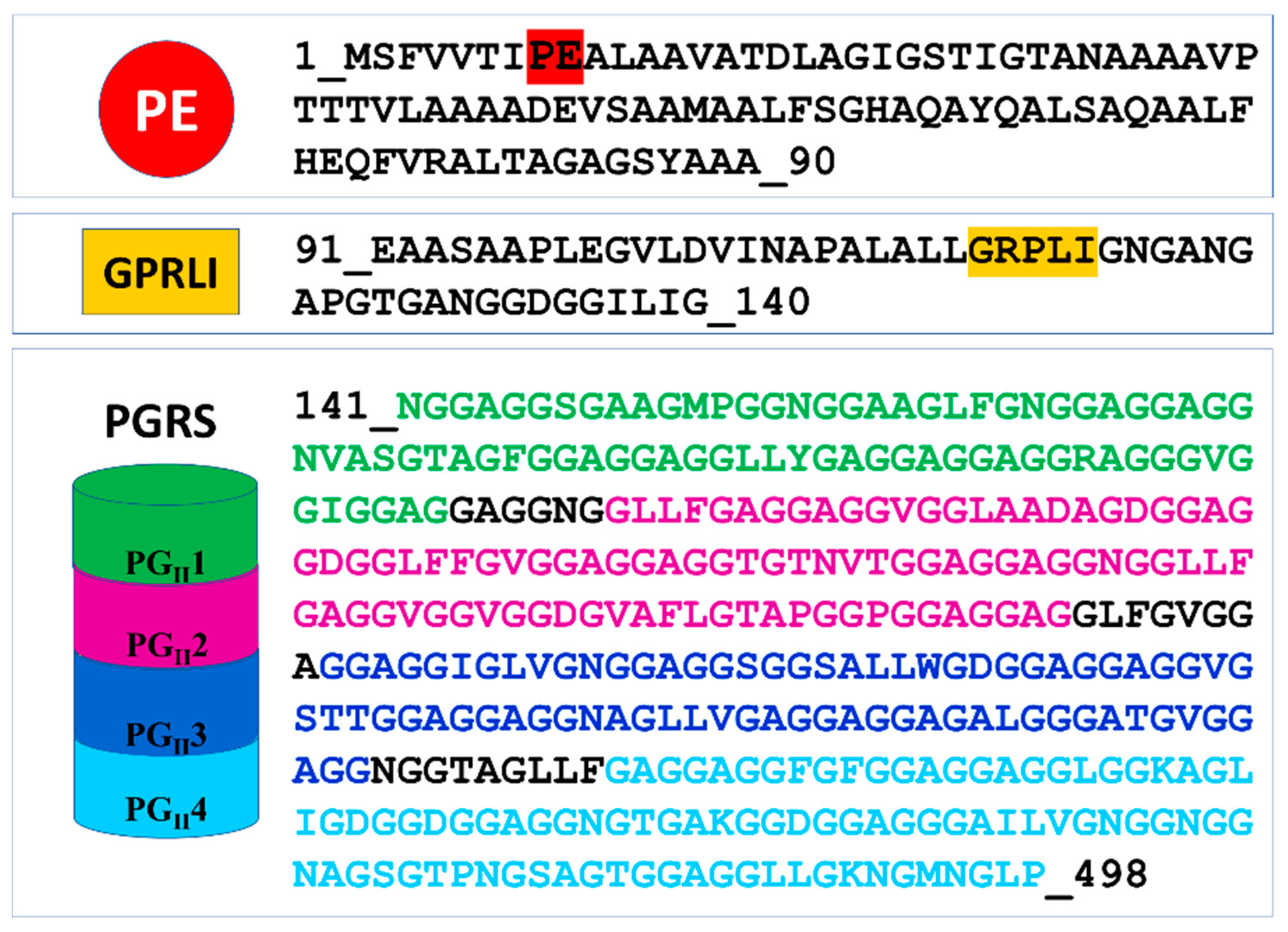
4. Structural Features of PE_PGRS33, the PE_PGRS Prototype
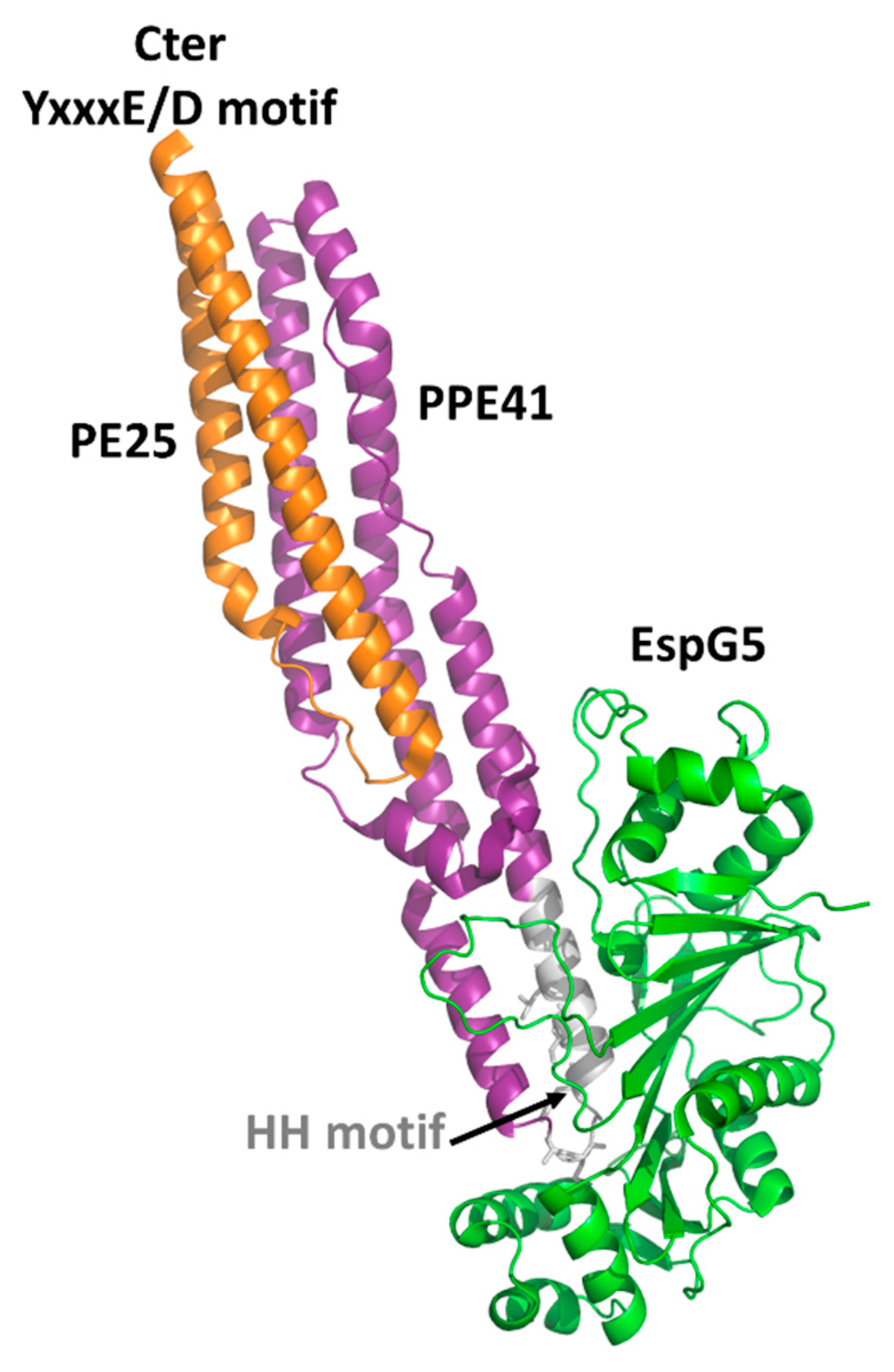
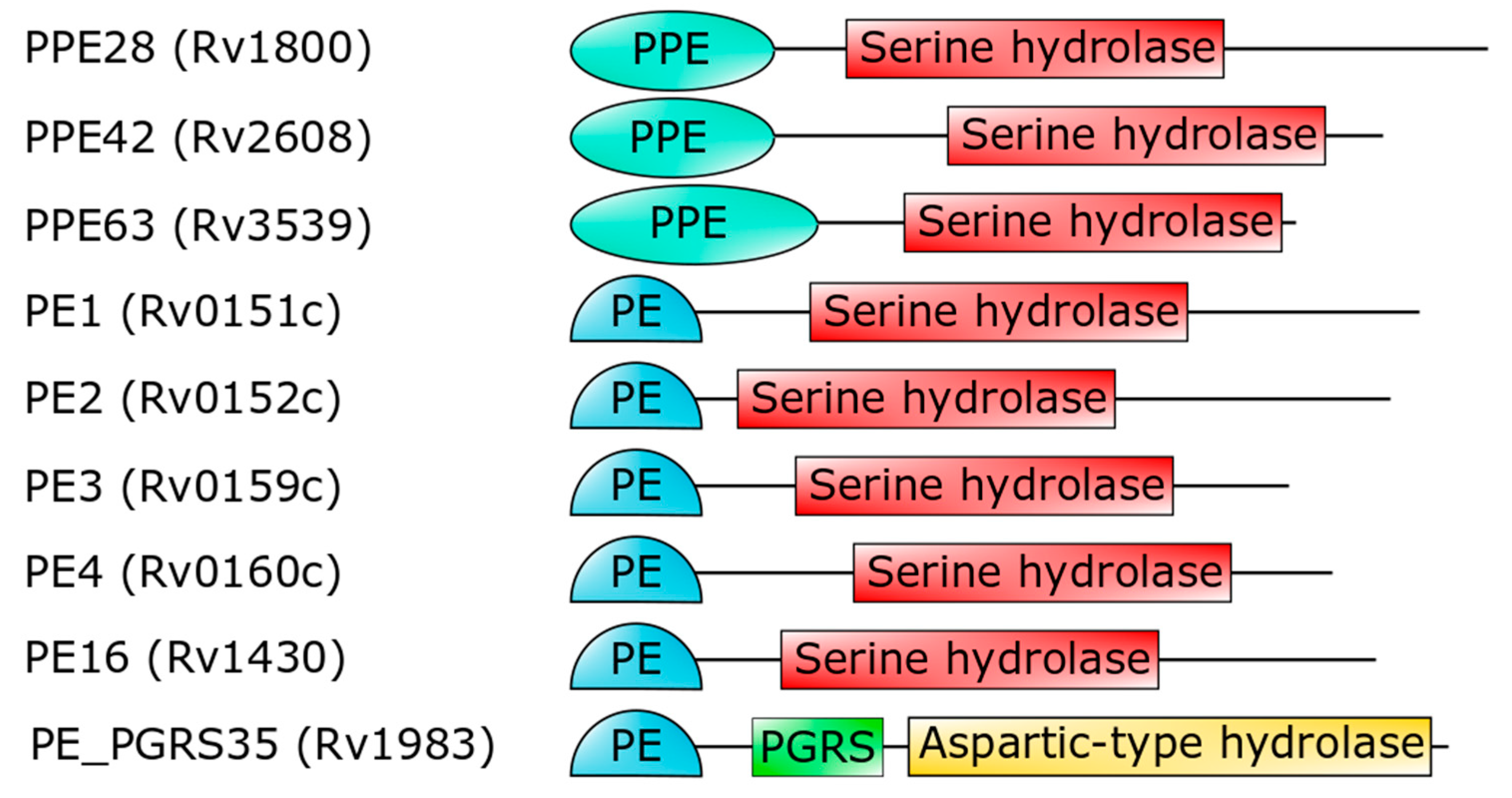
Knowing the three-dimensional structure of an antigen provides important insights into the understanding of the molecular nature of host–pathogen interactions and of the key epitopes that may serve as a target for the host antibody response. Although structural data on PE_PGRS proteins are not available, insightful information can be obtained by modeling techniques, learning from homologous proteins. Among PE_PGRS proteins, PE_PGRS33 is one of the best studied for its interaction with the immune system and has been considered a model for PE_PGRSs. As such, its putative role as a vaccine candidate is worth being investigated [81]. PE_PGRS33 is a large protein of 498 residues, with a modular architecture. A search in the PFAM database only identifies a PE domain at the N-terminal region of the protein, a small domain (residues 1–93) that is distributed nearly exclusively in actinobacteria, with only one exception for Tulasnella calospora, a genus of patch-forming fungi in the Tulasnellaceae family. A conserved linker GRLPI domain (l-GRPLI), likely acting as a transmembrane anchor, connects the PE domain to a distinctive region, not predicted by PFAM, and characterized by multiple repeats containing the GGA-GGX motif interspersed with unique sequences, and commonly denoted as PGRS domain (Figure 1).

4.1. The PE Domain

The PE domain takes its name from the conserved Pro–Glu (PE) amino acids at its N-terminus (residues 7–8) [55] Figure 1). This domain is responsible for PE_PGRS33 translocation via ESX5 and cell wall localization, with a significant role of 30 amino acids on its N-terminus [82]. A search in the PFAM database shows that the PE family is a member of clan EsxAB (CL0352), which also includes the more common PPE family and the WXG100 family, including the well-known antigen ESAT-6 (6 kDa early secreted antigenic target) and CFP-10 (10 kDa culture filtrate protein) in Mtb or EsxA (ESAT-6-like extracellularly secreted protein A) and EsxB in Staphylococcus aureus.
Crystal structures have been reported for the two PE domains PE8 and PE25, in both cases forming a heterocomplex with PPE partners (Table 2). In all structures, PE and PPE interact via a hydrophobic interface forming a four-helix bundle formed by two α-helices from the PE and two α-helices from the PPE module (Figure 2). The crystal structure of the ESX-5-secreted PE25–PPE41 heterodimer in complex with the ESX-5-encoded cytoplasmic chaperone EspG5 shows that EspG5 binds to a highly conserved hydrophobic chaperone-binding sequence on PPE, named as the hh motif [83] (Figure 2).
By binding PPE, EspG5 protects the aggregation-prone hh motif on PPE proteins and keeps the dimers in a secretion-competent state. Consistently, point mutations of this conserved hh motif affect protein secretion [83]. Both in the cases of PE25–PPE41 and PE8–PPE15, the binding of EspG5 chaperone does not cause conformational changes in the heterodimers. The two ternary complexes present highly similar structures. A superposition of their structures using DALI produces root mean square deviations (rmsd), computed on the backbone atoms of PE, PPE, and EspG5 chains, of 1.1 Å, 2.1 Å, and 0.5 Å, respectively. Importantly, EspG5 binds the PE–PPE dimers at a location that does not interfere with the signature ESX secretory motif YxxxD/E at the C-terminal side of PE proteins [87] (Figure 2).
As mentioned above, no structural information on the PE domains from PE_PGRS proteins is hitherto known. Neither it is known whether PE_PGRS proteins strictly require a PPE-like domain, as in the case of PE proteins in Table 2. Here, we fill this structural gap by adopting homology modeling. The best template was identified by HHPRED as the PE domain the ESX-5-secreted PE8 (PDB code 5xsf, sequence identity 45.8%) and the homology model built using MODELLER [88,89,90,91]. As a result, the homology model of the PE domain of PE_PGRS33 shows that all hydrophobic/aromatic residues are located on one side of the molecule (Figure 3). This feature, also observed for the PE domains of PE/PPE complexes, suggests that either the PE domain of PE_PGRS33 forms homodimers or it is prone to interact with another protein to form a heterodimer. It is hitherto not clear whether PE_PGRS proteins require a protein partner [59,86], as in the case of PE/PPE proteins. Indeed, pe_pgrs genes are expressed as single operons. Also, the PE-unique LipY protein does not require a partner to be secreted [92]. These findings suggest that PE_PGRSs can be stable on their own, albeit being endowed with prone-to-interact PE domains for their functions.
Importantly, the PE domain of PE_PGRS33 is required for the protein translocation through the mycobacterial cell wall [63,82,88,90,91]. Once exerted this role, the PE domain is cleaved from the rest of the molecule, leaving the functional PGRS domain floating on the mycomembrane [59]. Therefore, it is tempting to surmise that some hydrolases may recognize PE_PGRS33 through its PE domain. Mtb encodes for a number of PE- and PPE-containing serine α/β hydrolases, which are possible candidates as PE_PGRS33 hydrolases [93] (Figure 4). In addition to these, a PE_PGRS aspartic-type endopeptidase, denoted as PecA in M. marinum and PE_PGRS35 in Mtb, is known to cleave the lipase LipY [92]. More investigations are needed to verify which hydrolase is responsible for PE cleavage of PE_PGRS33 and if more hydrolases cleave specific PE_PGRS proteins.

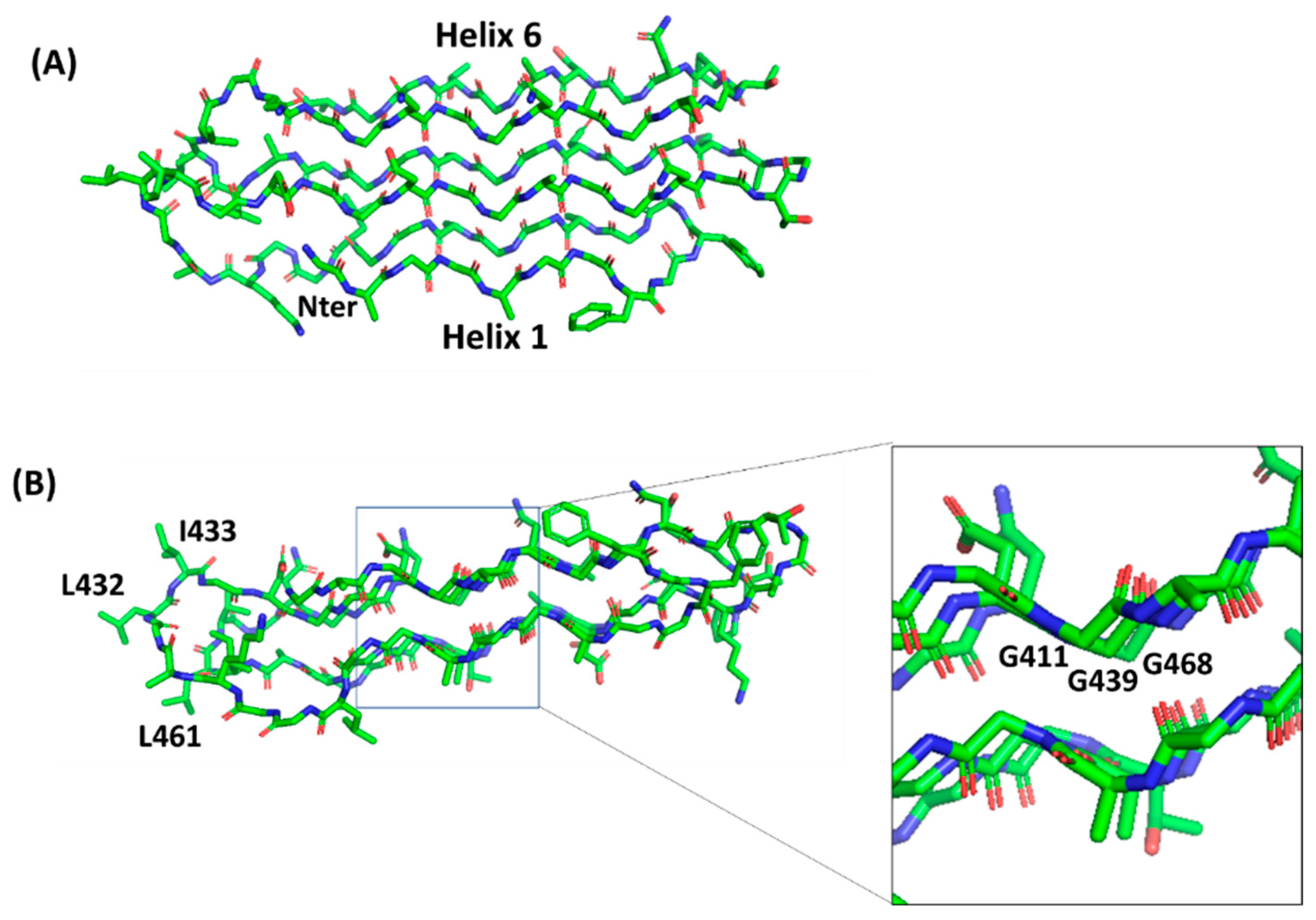
4.2. PGRS Domain Contains Multiple PGII Modules

In PE_PGRS proteins, the PGRS domain can vary in size from tens to almost 1800 amino acid residues. Its main feature is the presence of multiple repeats containing the GGA-GGX motif interspersed with unique sequences [94]. It has been shown that PGRS domains are available on the mycobacterial surface and can directly interact with host components, as TLR2 receptors [60,74,95]. To date, the structure of the PGRS domain remains unknown and should be implemented in experimental data.
The lack of structural data on PGRS domains makes the understanding of the role of these domains a hard task. However, a high sequence identity of the C-terminal part of the PGRS domain of PE_PGRS33 exists with the PGII domain of snow flea antifreeze protein sfAFP from Hypogastrura harveyi (sequence identity 60% with residues 406–486). Therefore, we performed homology modeling based on the target–template alignment using ProMod3 and the structure of sfAFP as a template (PDB code 2pne). The alignment of the sequence of this C-terminal PGII module against the entire PGRS region identifies further three modules with the same pattern and sequence identities ranging between 63.0% and 53.9% (Figure S1). This analysis shows that the PGRS domain of PE_PGRS33 is formed by four PGII domains, denoted here as PGII1, PGII2, PGII3, and PGII4, all with similar structural features.
Polyglycine conformations, such as PGII, are the most flexible ones because the lack of side chains in glycine removes steric hindrances. Consequently, extended regions of the Ramachandran plot are allowed for glycine residues, which can virtually assume any ψ angle [96]. Typical of PGII conformation, each glycine-rich triplet folds into a left-handed, elongated helix with a pitch (rise per turn) of 9.2 Å. This conformation resembles that observed for the polyproline type II (PPII) helices found in collagen [97,98]. In the PGII sandwich, six antiparallel PGII helices are stacked in two antiparallel groups, with three to four triplets spanning the PGII domain length (Figure 5). The organization of the PGRS region in PGII domains explains the high abundance of glycine residues in these domains. Glycine residues are always pointing inwards, in positions where only glycine could be sterically allowed (Figure 5).
A comparison with the structure of the PE_PGRS33 PGII domains with that of the antifreeze protein sfAFP highlights different surface characteristics, albeit presenting the same fold, likely due to completely different functions of PGII domains in the two proteins (Figure 6). In PE_PGRS33, hydrophobic residues of PGII domains are located mainly on loop regions (Figure 5B and Figure 6A), likely accounting for a role of these residues in host recognition. Consistently, as will be shown later, removal of consecutive residues belonging to PGII domains of PE_PGRS33 (alleles from 48 to 52, Table 3) results in weaker immunostimulatory activity, in terms of reduced TNF-α [72,74]. By contrast, hydrophobic residues are mostly located on one side of the PGII structure of sfAFP (Figure 6B) [99]. The accumulation of hydrophobic residues on one side of the sandwich builds a module with one hydrophilic face and one hydrophobic face, and the flat hydrophobic face is supposed to interact tightly with the highly ordered water molecules found at the surface of an ice crystal [99]. In this respect, the flatness that characterizes this domain is functional for its tight association with the ice surface [99]. Interestingly, a PGII sandwich was also observed in the Salmonella bacteriophage S16 long tail fiber. In this case, this PGII sandwich domain plays a role in the interactions of the phage with its host, with its exposed (hydrophobic) loops being determinants of host binding. Therefore, similar to PGII of PE_PGRS33, the glycine-rich core of the PGII sandwich of S16 long tail fiber exposes hypervariable β-turn loops that determine receptor specificity (Figure 6C).
In contrast to the antifreeze protein sfAFP, it is likely that in the case of both PE_PGRS33 and the bacteriophage S16 long tail fiber, the flatness of the PGII sandwich is useful to allow recognition loops to be closely spaced. In both cases, these loops evolve rapidly, as confirmed by their hypervariable nature, in a similar manner as observed for the three hypervariable complementarity-determining regions (CDRs) of immunoglobulins [100,101]. As in the case of S16 tail fiber, the PGII loops of PE_PGRS33 are likely exposed to the host and, as such, the principal targets of antibodies.

5. PE_PGRS33 as a Promising Target of the Humoral Response

PE_PGRS33 promotes cell death and increases mycobacterial survival in macrophages, as demonstrated by heterologous expression in Mycobacterium smegmatis (Ms), in a process mainly governed by its PGRS domain [74,102,103]. Being localized on the outer surface of the Mtb cell wall, PE_PGRS33 is in a position that results in the exposure to the milieu and in the capacity to interact with the host [95] (Figure 7A). Consistently, PE_PGRS33 was shown to specifically interact with TLR-2 [71,74], though it remains to be determined whether TLR2 requires heterodimerization with TLR1 or TLR6 to properly bind PE_PGRS33, and the role of coreceptors as CD14 or CD36 as well.
The binding of PE_PGRS33 with TLR-2 on macrophages can activate two different intracellular pathways. Activation of the Myd88 pathway triggers the expression of genes coding pro-inflammatory chemokines and cytokines in an NFkB-dependent mechanism. Secretion of tumor necrosis factor-α (TNF-α) promotes cell necrosis and inflammation, which increases the survival of Mtb inside the host [74] (Figure 7B). Activation of the PI3K pathway triggers the inside-out signaling pathway, which enhances Mtb internalization in host cells while dampening macrophage antimicrobial responses [71]. Interestingly, a polyclonal antiserum raised against native PE_PGRS33 was shown to inhibit Mtb entry into macrophages, without affecting the entry of the Mtb Δpe_pgrs33 strain [80]. This same Δpe_pgrs33 Mtb strain has impaired capacity to enter macrophages, likely due to the lack of interaction with TLR-2 [71,74]. Mice immunized with recombinant PE_PGRS33 were able to combat recombinant M. smegmatis overexpressing PE_PGRS33 in vivo [80]. Hence, PE_PGRS33/TLR2 interaction promotes inflammation and tissue damage while favoring Mtb replication in the host lesions, raising PE_PGRS33 as an important Mtb virulence factor [72]. This experimental evidence provides functional clues to the hypothesis that PE_PGRS33 may serve as a vaccine antigen candidate against TB [81,104,105]. Antibodies binding PE_PGRS33 on the Mtb surface may neutralize the binding with TLR2, turning off a pathogenetic pathway that promotes TB disease. PE_PGRS33-specific antibodies may also opsonize Mtb, prompting a more efficient uptake and killing by activated macrophages.
Mtb-infected and BCG-vaccinated subjects make antibodies against PE_PGRS33, with the key epitopes located mainly in the PGRS domain [104]. Most of the experimental evidence gathered so far has relied on the use of denatured recombinant PE_PGRS33 [80], making it difficult to identify the relevant epitopes responsible for the interaction with host receptors and potential targets of a protective humoral response.
Conversely, as outlined in this review, structural features of the PGRS portion of PE_PGRS33 well account for the role of this protein as a target of antibodies. The organization of PGRS in PGII sandwich modules crossing the external membrane allows the protein to efficiently expose loops for epitope recognition (Figure 7). The identification of these epitopes and the fine characterization at the atomic level of the interaction between TLR2 and PE_PGRS33 would be very useful to tailor effective immunization strategies. Altogether, these studies highlight the potential use of PE_PGRS33 as a target of a neutralizing humoral response against TB [80].

6. PGRS PGII Sandwich Structure Tolerates Polymorphism

Despite early evidence indicating PE_PGRSs as variable surface proteins responsible for Mtb antigenic variability and immune evasion [55,62], more recent reports show that most pe_pgrs genes are highly conserved and result in purifying selection [61]. The most relevant polymorphisms in pe_pgrs33 are indels of variable size occurring in the PGRS domain. Table 3 summarizes the most frequent alleles identified in large collections of Mtb clinical strains. These pe_pgrs33 alleles are conserved in the Mtb phylogeographic lineages and clades, indicating a clustering of specific alleles during Mtb evolution [72], while no clustering has been observed in drug-resistant strains [68]. The insertions detected in these alleles usually involve DNA sequences coding three amino acids, two triplets (GGX GGX) and up to 11 amino acids for allele 52. Deletions are more frequently detected and usually consist of regions coding one or more GGX triplets and may involve sequences coding up to 30, 48, or 90 amino acids. The functional consequences of some of these polymorphisms have been investigated in in vitro and in vivo models of Mtb infection. Some pe_pgrs33 alleles with large deletions appear to be associated with noncavitary TB or extrapulmonary TB [74,106]. Others have different immunoregulatory properties [74] or differential ability to bind TLR2, which may also affect the entry of Mtb in macrophages [72]. Although the impact of pe_pgrs33 polymorphisms on TB pathogenesis requires further scrutiny, it transpires that large indels do not lead to a complete loss of protein function [72].
The observation that even the pe_pgrs33 alleles showing large deletions are clustered with certain Mtb clades, coupled with the fact that Mtb transmission occurs only through the aerogenic route from patients with active pulmonary TB, indicates that these polymorphic alleles do not significantly impact Mtb fitness. This is apparently in contrast with the finding that pe_pgrs33 shows a substitution rate ratio (dN/dS) of 0.7, suggesting that a biological pressure acts on Mtb to prevent mutations that may impair PE_PGRS33 protein function [72]. We propose that the organization of the PGRS domain in PGII sandwich modules, consisting of tightly packed helices, each made of three or four turns with three residues per turn (the GGX triplets), provides a plastic structure that can tolerate large indels while maintaining proper localization of the unique amino acids that are found in the loops between the helices. PGII sandwiches are typically stabilized by hydrogen bonds involving backbone atoms of glycine residues (N—H-O and C—H-O). Therefore, glycine residues are the sole residues that cannot mutate, as their mutation to any other residues would strongly affect the structural integrity of the PGII sandwich. On the other hand, mutations of non-glycine residues are predicted to hardly contribute to structural stability, as they point toward the external part of the molecule and are not involved in stabilizing intramolecular interactions. Similarly, insertions or deletions of entire triplets, as observed in most frequent alleles (Table 3), are likely to poorly affect the PGII sandwich structural features. Therefore, the polymorphisms observed in the PGRS domain find their rationale in the structural features of PGII sandwich domains. However, while keeping the PGII structural features, the deletion of entire PGII helices in PGII sandwiches 1, 2, 3, and 4 may result in weaker immunostimulatory activity (reduced TNF-α) as observed in alleles 48–52 (Table 3), suggesting the presence of key protein domains with functional properties in these deleted regions, e.g., a domain that affects interaction with TLR2.
In summary, the observations made and results obtained so far suggest that (a) the PGRS domains of PE_PGRS33 expose four PGII sandwiches on the outer surface; (b) these PGII sandwiches are directly involved through their loops in the interactions with host receptors as TLR2; (c) the sequences encoding these regions result in purifying selection in Mtb, indicating a key role of PGII domains in Mtb survival and TB transmission; and (d) insertions and deletions in the region encoding the PGII domains may have an impact on the immunomodulating activity of PE_PGRS33, as assessed in in vitro and in vivo models. Hence, a strategy aimed at blocking or interfering with the PE_PGRS33–host cell receptor interaction might be useful to block the key steps of TB pathogenesis. As such, a vaccine formulation aimed at eliciting antibodies targeting the surface-exposed loops of PGII domains of PE_PGRS33 may serve to curb inflammatory processes and prevent or reduce the risk of a drift toward necrosis and mounting tissue immunopathology, which is the hallmark of active TB disease.
Table 3. Most frequent alleles identified in large collections of Mtb clinical strains. PGII modules of PE_PGRS33 are defined as indicated in Figure S1.
Allele *Polymorphism *Amino Acid VariationPosition (aa)PGIIClade/
Lineage
Functional NotesClinical Notes
allele 1
(allele 11) [72]
Wild type---4
LAM/
Haarlem
Associated with cavitary TB
allele 26
(allele 3) [72]
S4
D14
I4
−28 Gly X
+1 Gly Ala
239
338–498
413
3, 4, 51
EAI
Impaired entry in macrophages compared to wt allele.
Hypervirulent in mice
Associated with noncavitary TB
allele 45 1
(allele 5) [72]
S22 a
S4
I4
Gly → Ser
-
+1 Gly Ala
233
239
413
2, 4Animal lineage-
allele 461
(allele 6) [72]
D2
S4
I4
−4 Gly X
717
+1 Gly Ala
140–163
239
413
1, 41
EAI
-
allele 471
(allele 18) [72]
S4
D19 b
I4
-
−3 Gly X
+1 Gly Ala
239
257–270
413
2, 43
Delhi/CAS
-
allele 481
(ΔG184-G213) [74]
D20 c−30 aa184–2131, 2-Weaker immunostimulatory activity (reduced TNF-α)-
allele 491
(ΔL237-G327) [74]
D21 d−91 aa237–3272, 3--
allele 501
(ΔL372-A403) [74]
D22 e−32 aa372–4034--
allele 511
(ΔG196-D243) [74]
D22 f−48 aa196–2431, 2--
allele 521
(SA440) [74]
ID2 g−36 aa
+11 aa
236–271
272
2--
allele 22
(SA455) [74]
I1
S4
I4
+2 Gly Ala Gly
-
+1 Gly Ala
199
239
413
1, 4- Associated with cavitary TB
allele 40D8−47 aa213–2602--Associated with non-cavitary TB
allele 531
(33260) [71]
D23 h−238 aa260–4982, 3, 4, 5-Sufficient to complement the Mtb Δ33 mutant in the in vitro macrophage invasion assay-
allele 541
(Group 2) [106]
S4
I6 i
-
+ Ala Gly
4084--Associated with extrapulmonary TB
* According to allele sequential number and polymorphism description of Talarico et al. [107]. 1not described in [107]. In column 2, letters from a to i indicate: a, nsSNP, position 697; b, deletion of 42bp, position 772–813; c, deletion of 90bp, position 552–639; d, deletion of 273bp, position 711–981; e, deletion of 96bp, position 1116–1209; f, deletion of 144bp, position 588–729; g, deletion of 107bp, position 710–816 and insertion of 32bp in position 817; h, deletion of 714bp, position 780–1494; i, insertion of 9bp, position 1224.

7. Concluding Remarks

Vaccine development against TB has been dominated by strategies aimed at the elicitation of robust T cell responses, while antibody-based strategies have been neglected due also to the limited number and poor knowledge of Mtb surface antigens that are the natural targets of protective humoral responses. In this work, we provide a brief summary of the main challenges associated with the development of a new vaccine against TB and highlight the importance of a structure/function view to investigate the vaccine potential of a unique class of Mtb surface antigens, the PE_PGRS proteins. Among these, PE_PGRS33 can be considered a prototype protein of the family for the availability of experimental data implicating the PGRS domain in the interaction with host receptors. A vaccination strategy aimed at eliciting a protective humoral response shall ideally target secreted or surface-exposed antigens and/or virulence factors that play a key role in microbial pathogenesis. PE_PGRS33 is such a candidate, although the lack of structural data on the peculiar PGRS domain has so far hampered a fine understanding of the proper localization of this protein on the mycomembrane. For years, the glycine-rich PGRS domain has been thought to be highly flexible or unfolded, due to the conformational properties of glycine residues. Using homology modeling, we show that the PGRS portion of PE_PGRS33 is well structured and contains four compact subdomains, named as PGII sandwiches, each consisting of six left-handed PP-II helices stacked in two antiparallel groups. These sandwich domains were previously observed in antifreeze proteins, a class of macromolecules critical to the existence of life at sub-zero temperatures, and in Salmonella phage S16 adhesin. Similar to S16 adhesin, the flat structure of the PGII sandwich domains of PE_PGRS33 may be functional to projecting surface-exposed loops on one side of the molecule, like antigenic loops. Overall, the structural considerations collected here for PGRS domains also allow for the interpretation of the typical polymorphism observed for PGRS domains, since the PGII sandwich domain is able to tolerate modifications, insertions, and deletions, without destabilization. A peculiarity of PGII sandwiches is that they are stabilized solely by backbone hydrogen bonds (N—H-O and C—H-O) involving glycine residues. This feature, with glycine residues pointing inside the fold, where no other residues would fit, explains the high frequency of glycine residues in PGRS domains.
Therefore, the unique sequences in the PGRS domain coding the surface-exposed loops are likely to be responsible for the interaction with host receptors (e.g., TLR2 for PE_PGRS33) and the ideal targets for antibodies with protective potential.
In conclusion, the data examined here provide the structural basis for both the antigenic properties of PGRS domains and of their typical polymorphism. Targeting the surface-exposed loops emerging from the PGII sandwich domains with a humoral response is an out-of-the-box approach to develop new vaccines against TB, taking into account the exposure of multiple copies of PGII sandwiches on the mycobacterial outer membrane.
One important aspect to address concerns the identification of an effective vaccination strategy that can elicit strong, robust, and specific antibody responses against the surface-exposed loops. Inclusion of recombinant PE_PGRS33 purified under native conditions in already available vaccine platforms, such as BCG or new vaccines against TB, may be a strategy that can be useful to pursue [80]. Immunization with BCG strains overexpressing PE_PGRS33 or with extracellular membrane vesicles [108,109] enriched in PE_PGRS33 may serve to elicit antibody responses against protein domains presenting the appropriate structure to elicit functionally active antibodies capable of neutralizing PE_PGRS33 and/or of opsonizing Mtb. The fact that Mtb expresses more than 50 PE_PGRS proteins on the mycobacterial surface, with structural features similar to those of PE_PGRS33, and that these proteins play unique roles in the Mtb infection process and immunopathogenesis of TB, underscores the potential impact that a vaccination strategy targeting PE_PGRS proteins may have in the control of TB.

Supplementary Materials

The following are available online at https://www.mdpi.com/2073-4409/10/1/161/s1: Figure S1: Alignment of the sequence of the C-terminal region of the PGRS domain of PE_PGRS33, PGII4, against the entire PGRS region and identification of four PGII sandwich domains.

Author Contributions

Conceptualization, R.B. and G.D.; methodology and software, E.K., F.S., and R.B; formal analysis, E.K., F.S., and F.D.M.; data curation, E.K, F.S., F.D.M., G.D., and R.B.; writing—original draft preparation, E.K. and R.B.; writing—review and editing, E.K, F.S., F.D.M., G.D., and R.B.; supervision, R.B.; funding acquisition, R.B. All authors have read and agreed to the published version of the manuscript.

Funding

This work was supported by the Marie Skłodowska-Curie Action BactiVax, GA number 860325.

Acknowledgments

We would like to acknowledge the help of our technical staff, including Maurizio Amendola and Luca De Luca, and of Luciana Herda for managing BactiVax.

Conflicts of Interest

The authors declare no conflict of interest. The funders had no role in the design of the study; in the collection, analyses, or interpretation of data; in the writing of the manuscript, or in the decision to publish the results.

Abbreviations

TBTuberculosis
WHOWorld Health Organization
MtbMycobacterium tuberculosis
BCGBacille Calmette and Guérin
DCDendritic cell
IFNInterferon
TLRToll-like receptor
PDBProtein Data Bank
MsMycobacterium smegmatis
CDRComplementarity-determining region
EREndoplasmic reticulum
rmsdroot mean square deviations
ILInterleukin
TNF-αTumor necrosis factor–α

References

  1. Harding, E. WHO global progress report on tuberculosis elimination. Lancet Respir. Med. 2020, 8, 19. [Google Scholar] [CrossRef]
  2. World Health Organization. The End TB Strategy. Available online: https://www.who.int/tb/strategy/en/ (accessed on 14 January 2021).
  3. Barry, C.E., 3rd; Boshoff, H.I.; Dartois, V.; Dick, T.; Ehrt, S.; Flynn, J.; Schnappinger, D.; Wilkinson, R.J.; Young, D. The spectrum of latent tuberculosis: Rethinking the biology and intervention strategies. Nat. Rev. Microbiol. 2009, 7, 845–855. [Google Scholar] [CrossRef] [PubMed]
  4. Delogu, G.; Sali, M.; Fadda, G. The biology of mycobacterium tuberculosis infection. Mediterr. J. Hematol. Infect. Dis. 2013, 5, e2013070. [Google Scholar] [CrossRef] [PubMed] [Green Version]
  5. Nikitushkin, V.D.; Demina, G.R.; Shleeva, M.O.; Guryanova, S.V.; Ruggiero, A.; Berisio, R.; Kaprelyants, A.S. A product of RpfB and RipA joint enzymatic action promotes the resuscitation of dormant mycobacteria. Febs. J. 2015, 282, 2500–2511. [Google Scholar] [CrossRef]
  6. Ruggiero, A.; Squeglia, F.; Romano, M.; Vitagliano, L.; De Simone, A.; Berisio, R. The structure of Resuscitation promoting factor B from M. tuberculosis reveals unexpected ubiquitin-like domains. Biochim. Biophys. Acta 2016, 1860, 445–451. [Google Scholar] [CrossRef]
  7. Squeglia, F.; Romano, M.; Ruggiero, A.; Vitagliano, L.; De Simone, A.; Berisio, R. Carbohydrate recognition by RpfB from Mycobacterium tuberculosis unveiled by crystallographic and molecular dynamics analyses. Biophys. J. 2013, 104, 2530–2539. [Google Scholar] [CrossRef] [Green Version]
  8. Squeglia, F.; Ruggiero, A.; Berisio, R. Chemistry of Peptidoglycan in Mycobacterium tuberculosis Life Cycle: An off-the-wall Balance of Synthesis and Degradation. Chemistry 2018, 24, 2533–2546. [Google Scholar] [CrossRef]
  9. Squeglia, F.; Ruggiero, A.; Berisio, R. Exit from mycobacterial dormancy: A structural perspective. Curr. Med. Chem. 2015, 22, 1698–1709. [Google Scholar] [CrossRef]
  10. Gengenbacher, M.; Kaufmann, S.H. Mycobacterium tuberculosis: Success through dormancy. Fems. Microbiol. Rev. 2012, 36, 514–532. [Google Scholar] [CrossRef] [Green Version]
  11. Orme, I.M.; Basaraba, R.J. The formation of the granuloma in tuberculosis infection. Semin. Immunol. 2014, 26, 601–609. [Google Scholar] [CrossRef]
  12. Dheda, K.; Gumbo, T.; Maartens, G.; Dooley, K.E.; Murray, M.; Furin, J.; Nardell, E.A.; Warren, R.M.; Lancet Respiratory Medicine Drug-Resistant Tuberculosis Commission Group. The Lancet Respiratory Medicine Commission: 2019 update: Epidemiology, pathogenesis, transmission, diagnosis, and management of multidrug-resistant and incurable tuberculosis. Lancet Respir. Med. 2019, 7, 820–826. [Google Scholar] [CrossRef]
  13. Tran, V.; Liu, J.; Behr, M.A. BCG Vaccines. Microbiol. Spectr. 2014, 2. [Google Scholar] [CrossRef] [Green Version]
  14. Mangtani, P.; Abubakar, I.; Ariti, C.; Beynon, R.; Pimpin, L.; Fine, P.E.M.; Rodrigues, L.C.; Smith, P.G.; Lipman, M.; Whiting, P.F.; et al. Protection by BCG Vaccine Against Tuberculosis: A Systematic Review of Randomized Controlled Trials. Clin. Infect. Dis. 2014, 58, 470–480. [Google Scholar] [CrossRef] [PubMed] [Green Version]
  15. Harris, R.C.; Dodd, P.J.; White, R.G. The potential impact of BCG vaccine supply shortages on global paediatric tuberculosis mortality. BMC Med. 2016, 14, 138. [Google Scholar] [CrossRef] [PubMed] [Green Version]
  16. Nguipdop-Djomo, P.; Heldal, E.; Rodrigues, L.C.; Abubakar, I.; Mangtani, P. BCG vaccination: A long-lasting protection against tuberculosis?—Authors’ reply. Lancet Infect. Dis. 2016, 16, 408–409. [Google Scholar] [CrossRef] [Green Version]
  17. Schrager, L.K.; Harris, R.C.; Vekemans, J. Research and development of new tuberculosis vaccines: A review. F1000Research 2018, 7, 1732. [Google Scholar] [CrossRef]
  18. Delogu, G.; Manganelli, R.; Brennan, M.J. Critical research concepts in tuberculosis vaccine development. Clin. Microbiol. Infect. 2014, 20, 59–65. [Google Scholar] [CrossRef] [Green Version]
  19. Schrager, L.K.; Vekemens, J.; Drager, N.; Lewinsohn, D.M.; Olesen, O.F. The status of tuberculosis vaccine development. Lancet Infect. Dis. 2020, 20, e28–e37. [Google Scholar] [CrossRef]
  20. Mendez-Samperio, P. Global Efforts in the Development of Vaccines for Tuberculosis: Requirements for Improved Vaccines Against Mycobacterium tuberculosis. Scand. J. Immunol. 2016, 84, 204–210. [Google Scholar] [CrossRef]
  21. Gong, W.; Liang, Y.; Wu, X. The current status, challenges, and future developments of new tuberculosis vaccines. Hum. Vaccin. Immunother. 2018, 14, 1697–1716. [Google Scholar] [CrossRef] [Green Version]
  22. Kaufmann, S.H.E.; Weiner, J.; von Reyn, C.F. Novel approaches to tuberculosis vaccine development. Int. J. Infect. Dis. 2017, 56, 263–267. [Google Scholar] [CrossRef] [PubMed] [Green Version]
  23. Brazier, B.; McShane, H. Towards new TB vaccines. Semin. Immunopathol. 2020, 42, 315–331. [Google Scholar] [CrossRef] [PubMed] [Green Version]
  24. Soundarya, J.S.V.; Ranganathan, U.D.; Tripathy, S.P. Current trends in tuberculosis vaccine. Med. J. Armed Forces India 2019, 75, 18–24. [Google Scholar] [CrossRef] [PubMed]
  25. Mendez-Samperio, P. Development of tuberculosis vaccines in clinical trials: Current status. Scand. J. Immunol. 2018, 88, e12710. [Google Scholar] [CrossRef] [PubMed] [Green Version]
  26. Delogu, G.; Provvedi, R.; Sali, M.; Manganelli, R. Mycobacterium tuberculosis virulence: Insights and impact on vaccine development. Future Microbiol. 2015, 10, 1177–1194. [Google Scholar] [CrossRef]
  27. Tameris, M.D.; Hatherill, M.; Landry, B.S.; Scriba, T.J.; Snowden, M.A.; Lockhart, S.; Shea, J.E.; McClain, J.B.; Hussey, G.D.; Hanekom, W.A.; et al. Safety and efficacy of MVA85A, a new tuberculosis vaccine, in infants previously vaccinated with BCG: A randomised, placebo-controlled phase 2b trial. Lancet 2013, 381, 1021–1028. [Google Scholar] [CrossRef] [Green Version]
  28. Boer, M.C.; van Meijgaarden, K.E.; Goletti, D.; Vanini, V.; Prins, C.; Ottenhoff, T.H.; Joosten, S.A. KLRG1 and PD-1 expression are increased on T-cells following tuberculosis-treatment and identify cells with different proliferative capacities in BCG-vaccinated adults. Tuberc. (Edinb) 2016, 97, 163–171. [Google Scholar] [CrossRef]
  29. Fletcher, H.A.; Schrager, L. TB vaccine development and the End TB Strategy: Importance and current status. Trans. R Soc. Trop. Med. Hyg. 2016, 110, 212–218. [Google Scholar] [CrossRef] [Green Version]
  30. Lindenstrom, T.; Knudsen, N.P.; Agger, E.M.; Andersen, P. Control of chronic mycobacterium tuberculosis infection by CD4 KLRG1- IL-2-secreting central memory cells. J. Immunol. 2013, 190, 6311–6319. [Google Scholar] [CrossRef] [Green Version]
  31. Andersen, P.; Scriba, T.J. Moving tuberculosis vaccines from theory to practice. Nat. Rev. Immunol. 2019, 19, 550–562. [Google Scholar] [CrossRef]
  32. Comas, I.; Chakravartti, J.; Small, P.M.; Galagan, J.; Niemann, S.; Kremer, K.; Ernst, J.D.; Gagneux, S. Human T cell epitopes of Mycobacterium tuberculosis are evolutionarily hyperconserved. Nat. Genet. 2010, 42, 498–503. [Google Scholar] [CrossRef] [PubMed] [Green Version]
  33. Coscolla, M.; Copin, R.; Sutherland, J.; Gehre, F.; de Jong, B.; Owolabi, O.; Mbayo, G.; Giardina, F.; Ernst, J.D.; Gagneux, S.M. tuberculosis T Cell Epitope Analysis Reveals Paucity of Antigenic Variation and Identifies Rare Variable TB Antigens. Cell Host Microbe 2015, 18, 538–548. [Google Scholar] [CrossRef] [PubMed] [Green Version]
  34. Moguche, A.O.; Musvosvi, M.; Penn-Nicholson, A.; Plumlee, C.R.; Mearns, H.; Geldenhuys, H.; Smit, E.; Abrahams, D.; Rozot, V.; Dintwe, O.; et al. Antigen Availability Shapes T Cell Differentiation and Function during Tuberculosis. Cell Host Microbe 2017, 21, 695–706. [Google Scholar] [CrossRef] [PubMed] [Green Version]
  35. Nieuwenhuizen, N.E.; Kulkarni, P.S.; Shaligram, U.; Cotton, M.F.; Rentsch, C.A.; Eisele, B.; Grode, L.; Kaufmann, S.H.E. The Recombinant Bacille Calmette-Guerin Vaccine VPM1002: Ready for Clinical Efficacy Testing. Front. Immunol. 2017, 8, 1147. [Google Scholar] [CrossRef] [PubMed]
  36. Gonzalo-Asensio, J.; Marinova, D.; Martin, C.; Aguilo, N. MTBVAC: Attenuating the Human Pathogen of Tuberculosis (TB) Toward a Promising Vaccine against the TB Epidemic. Front. Immunol. 2017, 8, 1803. [Google Scholar] [CrossRef] [PubMed] [Green Version]
  37. Yang, X.Y.; Chen, Q.F.; Cui, X.H.; Yu, Y.; Li, Y.P. Mycobacterium vaccae vaccine to prevent tuberculosis in high risk people: A meta-analysis. J. Infect. 2010, 60, 320–330. [Google Scholar] [CrossRef] [PubMed]
  38. Yang, X.Y.; Chen, Q.F.; Li, Y.P.; Wu, S.M. Mycobacterium vaccae as adjuvant therapy to anti-tuberculosis chemotherapy in never-treated tuberculosis patients: A meta-analysis. PLoS ONE 2011, 6, e23826. [Google Scholar] [CrossRef] [Green Version]
  39. Saqib, M.; Khatri, R.; Singh, B.; Gupta, A.; Kumar, A.; Bhaskar, S. Mycobacterium indicus pranii as a booster vaccine enhances BCG induced immunity and confers higher protection in animal models of tuberculosis. Tuberc. (Edinb) 2016, 101, 164–173. [Google Scholar] [CrossRef] [PubMed]
  40. von Reyn, C.F.; Lahey, T.; Arbeit, R.D.; Landry, B.; Kailani, L.; Adams, L.V.; Haynes, B.C.; Mackenzie, T.; Wieland-Alter, W.; Connor, R.I.; et al. Safety and immunogenicity of an inactivated whole cell tuberculosis vaccine booster in adults primed with BCG: A randomized, controlled trial of DAR-901. PLoS ONE 2017, 12, e0175215. [Google Scholar] [CrossRef]
  41. Vilaplana, C.; Montane, E.; Pinto, S.; Barriocanal, A.M.; Domenech, G.; Torres, F.; Cardona, P.J.; Costa, J. Double-blind, randomized, placebo-controlled Phase I Clinical Trial of the therapeutical antituberculous vaccine RUTI. Vaccine 2010, 28, 1106–1116. [Google Scholar] [CrossRef]
  42. Day, C.L.; Tameris, M.; Mansoor, N.; van Rooyen, M.; de Kock, M.; Geldenhuys, H.; Erasmus, M.; Makhethe, L.; Hughes, E.J.; Gelderbloem, S.; et al. Induction and regulation of T-cell immunity by the novel tuberculosis vaccine M72/AS01 in South African adults. Am. J. Respir. Crit. Care Med. 2013, 188, 492–502. [Google Scholar] [CrossRef] [PubMed] [Green Version]
  43. Luabeya, A.K.; Kagina, B.M.; Tameris, M.D.; Geldenhuys, H.; Hoff, S.T.; Shi, Z.; Kromann, I.; Hatherill, M.; Mahomed, H.; Hanekom, W.A.; et al. First-in-human trial of the post-exposure tuberculosis vaccine H56:IC31 in Mycobacterium tuberculosis infected and non-infected healthy adults. Vaccine 2015, 33, 4130–4140. [Google Scholar] [CrossRef] [PubMed]
  44. Penn-Nicholson, A.; Tameris, M.; Smit, E.; Day, T.A.; Musvosvi, M.; Jayashankar, L.; Vergara, J.; Mabwe, S.; Bilek, N.; Geldenhuys, H.; et al. Safety and immunogenicity of the novel tuberculosis vaccine ID93 + GLA-SE in BCG-vaccinated healthy adults in South Africa: A randomised, double-blind, placebo-controlled phase 1 trial. Lancet Respir. Med. 2018, 6, 287–298. [Google Scholar] [CrossRef]
  45. Tkachuk, A.P.; Gushchin, V.A.; Potapov, V.D.; Demidenko, A.V.; Lunin, V.G.; Gintsburg, A.L. Multi-subunit BCG booster vaccine GamTBvac: Assessment of immunogenicity and protective efficacy in murine and guinea pig TB models. PLoS ONE 2017, 12, e0176784. [Google Scholar] [CrossRef] [Green Version]
  46. Nemes, E.; Hesseling, A.C.; Tameris, M.; Mauff, K.; Downing, K.; Mulenga, H.; Rose, P.; van der Zalm, M.; Mbaba, S.; Van As, D.; et al. Safety and Immunogenicity of Newborn MVA85A Vaccination and Selective, Delayed Bacille Calmette-Guerin for Infants of Human Immunodeficiency Virus-Infected Mothers: A Phase 2 Randomized, Controlled Trial. Clin. Infect. Dis. 2018, 66, 554–563. [Google Scholar] [CrossRef]
  47. Smaill, F.; Xing, Z. Human type 5 adenovirus-based tuberculosis vaccine: Is the respiratory route of delivery the future? Expert Rev. Vaccines 2014, 13, 927–930. [Google Scholar] [CrossRef] [Green Version]
  48. Chen, T.; Blanc, C.; Eder, A.Z.; Prados-Rosales, R.; Souza, A.C.; Kim, R.S.; Glatman-Freedman, A.; Joe, M.; Bai, Y.; Lowary, T.L.; et al. Association of Human Antibodies to Arabinomannan With Enhanced Mycobacterial Opsonophagocytosis and Intracellular Growth Reduction. J. Infect. Dis. 2016, 214, 300–310. [Google Scholar] [CrossRef]
  49. Chen, T.; Blanc, C.; Liu, Y.; Ishida, E.; Singer, S.; Xu, J.; Joe, M.; Jenny-Avital, E.R.; Chan, J.; Lowary, T.L.; et al. Capsular glycan recognition provides antibody-mediated immunity against tuberculosis. J. Clin. Investig. 2020, 130, 1808–1822. [Google Scholar] [CrossRef] [Green Version]
  50. Prados-Rosales, R.; Carreno, L.; Cheng, T.; Blanc, C.; Weinrick, B.; Malek, A.; Lowary, T.L.; Baena, A.; Joe, M.; Bai, Y.; et al. Enhanced control of Mycobacterium tuberculosis extrapulmonary dissemination in mice by an arabinomannan-protein conjugate vaccine. PLoS Pathog. 2017, 13, e1006250. [Google Scholar] [CrossRef]
  51. Prados-Rosales, R.; Carreno, L.J.; Weinrick, B.; Batista-Gonzalez, A.; Glatman-Freedman, A.; Xu, J.; Chan, J.; Jacobs, W.R., Jr.; Porcelli, S.A.; Casadevall, A. The Type of Growth Medium Affects the Presence of a Mycobacterial Capsule and Is Associated with Differences in Protective Efficacy of BCG Vaccination Against Mycobacterium tuberculosis. J. Infect. Dis. 2016, 214, 426–437. [Google Scholar] [CrossRef] [Green Version]
  52. Lu, L.L.; Chung, A.W.; Rosebrock, T.R.; Ghebremichael, M.; Yu, W.H.; Grace, P.S.; Schoen, M.K.; Tafesse, F.; Martin, C.; Leung, V.; et al. A Functional Role for Antibodies in Tuberculosis. Cell 2016, 167, 433–443. [Google Scholar] [CrossRef] [PubMed] [Green Version]
  53. Lu, L.L.; Smith, M.T.; Yu, K.K.Q.; Luedemann, C.; Suscovich, T.J.; Grace, P.S.; Cain, A.; Yu, W.H.; McKitrick, T.R.; Lauffenburger, D.; et al. IFN-gamma-independent immune markers of Mycobacterium tuberculosis exposure. Nat. Med. 2019, 25, 977–987. [Google Scholar] [CrossRef] [PubMed]
  54. Casadevall, A. Antibodies to Mycobacterium tuberculosis. N. Engl. J. Med. 2017, 376, 283–285. [Google Scholar] [CrossRef]
  55. Cole, S.T.; Brosch, R.; Parkhill, J.; Garnier, T.; Churcher, C.; Harris, D.; Gordon, S.V.; Eiglmeier, K.; Gas, S.; Barry, C.E., 3rd; et al. Deciphering the biology of Mycobacterium tuberculosis from the complete genome sequence. Nature 1998, 393, 537–544. [Google Scholar] [CrossRef]
  56. Mohareer, K.; Tundup, S.; Hasnain, S.E. Transcriptional regulation of Mycobacterium tuberculosis PE/PPE genes: A molecular switch to virulence? J. Mol. Microbiol. Biotechnol. 2011, 21, 97–109. [Google Scholar] [CrossRef] [PubMed]
  57. Meena, L.S. An overview to understand the role of PE_PGRS family proteins in Mycobacterium tuberculosis H37 Rv and their potential as new drug targets. Biotechnol. Appl. Biochem. 2015, 62, 145–153. [Google Scholar] [CrossRef]
  58. Vallecillo, A.J.; Espitia, C. Expression of Mycobacterium tuberculosis pe_pgrs33 is repressed during stationary phase and stress conditions, and its transcription is mediated by sigma factor A. Microb. Pathog. 2009, 46, 119–127. [Google Scholar] [CrossRef]
  59. De Maio, F.; Berisio, R.; Manganelli, R.; Delogu, G. PE_PGRS proteins of Mycobacterium tuberculosis: A specialized molecular task force at the forefront of host-pathogen interaction. Virulence 2020, 11, 898–915. [Google Scholar] [CrossRef]
  60. Brennan, M.J.; Delogu, G.; Chen, Y.; Bardarov, S.; Kriakov, J.; Alavi, M.; Jacobs, W.R., Jr. Evidence that mycobacterial PE_PGRS proteins are cell surface constituents that influence interactions with other cells. Infect. Immun. 2001, 69, 7326–7333. [Google Scholar] [CrossRef] [Green Version]
  61. Copin, R.; Coscolla, M.; Seiffert, S.N.; Bothamley, G.; Sutherland, J.; Mbayo, G.; Gagneux, S.; Ernst, J.D. Sequence diversity in the pe_pgrs genes of Mycobacterium tuberculosis is independent of human T cell recognition. MBio 2014, 5. [Google Scholar] [CrossRef] [Green Version]
  62. Banu, S.; Honore, N.; Saint-Joanis, B.; Philpott, D.; Prevost, M.C.; Cole, S.T. Are the PE-PGRS proteins of Mycobacterium tuberculosis variable surface antigens? Mol. Microbiol. 2002, 44, 9–19. [Google Scholar] [CrossRef] [PubMed]
  63. Cascioferro, A.; Delogu, G.; Colone, M.; Sali, M.; Stringaro, A.; Arancia, G.; Fadda, G.; Palu, G.; Manganelli, R. PE is a functional domain responsible for protein translocation and localization on mycobacterial cell wall. Mol. Microbiol. 2007, 66, 1536–1547. [Google Scholar] [CrossRef] [PubMed]
  64. Chatrath, S.; Gupta, V.K.; Garg, L.C. The PGRS domain is responsible for translocation of PE_PGRS30 to cell poles while the PE and the C-terminal domains localize it to the cell wall. Febs. Lett. 2014, 588, 990–994. [Google Scholar] [CrossRef] [Green Version]
  65. Shah, S.; Briken, V. Modular Organization of the ESX-5 Secretion System in Mycobacterium tuberculosis. Front. Cell Infect. Microbiol. 2016, 6, 49. [Google Scholar] [CrossRef] [PubMed] [Green Version]
  66. Abdallah, A.M.; Bestebroer, J.; Savage, N.D.; de Punder, K.; van Zon, M.; Wilson, L.; Korbee, C.J.; van der Sar, A.M.; Ottenhoff, T.H.; van der Wel, N.N.; et al. Mycobacterial secretion systems ESX-1 and ESX-5 play distinct roles in host cell death and inflammasome activation. J. Immunol. 2011, 187, 4744–4753. [Google Scholar] [CrossRef] [Green Version]
  67. Gey van Pittius, N.C.; Sampson, S.L.; Lee, H.; Kim, Y.; van Helden, P.D.; Warren, R.M. Evolution and expansion of the Mycobacterium tuberculosis PE and PPE multigene families and their association with the duplication of the ESAT-6 (esx) gene cluster regions. BMC Evol. Biol. 2006, 6, 95. [Google Scholar] [CrossRef] [Green Version]
  68. Kanji, A.; Hasan, Z.; Ali, A.; McNerney, R.; Mallard, K.; Coll, F.; Hill-Cawthorne, G.; Nair, M.; Clark, T.G.; Zaver, A.; et al. Characterization of genomic variations in SNPs of PE_PGRS genes reveals deletions and insertions in extensively drug resistant (XDR) M. tuberculosis strains from Pakistan. Int. J. Mycobacteriol. 2015, 4, 73–79. [Google Scholar] [CrossRef]
  69. Delogu, G.; Sanguinetti, M.; Pusceddu, C.; Bua, A.; Brennan, M.J.; Zanetti, S.; Fadda, G. PE_PGRS proteins are differentially expressed by Mycobacterium tuberculosis in host tissues. Microbes Infect. 2006, 8, 2061–2067. [Google Scholar] [CrossRef]
  70. Kruh, N.A.; Troudt, J.; Izzo, A.; Prenni, J.; Dobos, K.M. Portrait of a pathogen: The Mycobacterium tuberculosis proteome in vivo. PLoS ONE 2010, 5, e13938. [Google Scholar] [CrossRef] [Green Version]
  71. Palucci, I.; Camassa, S.; Cascioferro, A.; Sali, M.; Anoosheh, S.; Zumbo, A.; Minerva, M.; Iantomasi, R.; De Maio, F.; Di Sante, G.; et al. PE_PGRS33 Contributes to Mycobacterium tuberculosis Entry in Macrophages through Interaction with TLR2. PLoS ONE 2016, 11, e0150800. [Google Scholar] [CrossRef]
  72. Camassa, S.; Palucci, I.; Iantomasi, R.; Cubeddu, T.; Minerva, M.; De Maio, F.; Jouny, S.; Petruccioli, E.; Goletti, D.; Ria, F.; et al. Impact of pe_pgrs33 Gene Polymorphisms on Mycobacterium tuberculosis Infection and Pathogenesis. Front. Cell Infect. Microbiol. 2017, 7, 137. [Google Scholar] [CrossRef] [PubMed] [Green Version]
  73. Grover, S.; Sharma, T.; Singh, Y.; Kohli, S.; Manjunath, P.; Singh, A.; Semmler, T.; Wieler, L.H.; Tedin, K.; Ehtesham, N.Z.; et al. The PGRS Domain of Mycobacterium tuberculosis PE_PGRS Protein Rv0297 Is Involved in Endoplasmic Reticulum Stress-Mediated Apoptosis through Toll-Like Receptor 4. MBio 2018, 9. [Google Scholar] [CrossRef] [PubMed] [Green Version]
  74. Basu, S.; Pathak, S.K.; Banerjee, A.; Pathak, S.; Bhattacharyya, A.; Yang, Z.; Talarico, S.; Kundu, M.; Basu, J. Execution of macrophage apoptosis by PE_PGRS33 of Mycobacterium tuberculosis is mediated by Toll-like receptor 2-dependent release of tumor necrosis factor-alpha. J. Biol. Chem. 2007, 282, 1039–1050. [Google Scholar] [CrossRef] [PubMed] [Green Version]
  75. Bansal, K.; Elluru, S.R.; Narayana, Y.; Chaturvedi, R.; Patil, S.A.; Kaveri, S.V.; Bayry, J.; Balaji, K.N. PE_PGRS antigens of Mycobacterium tuberculosis induce maturation and activation of human dendritic cells. J. Immunol. 2010, 184, 3495–3504. [Google Scholar] [CrossRef] [PubMed] [Green Version]
  76. Sayes, F.; Pawlik, A.; Frigui, W.; Groschel, M.I.; Crommelynck, S.; Fayolle, C.; Cia, F.; Bancroft, G.J.; Bottai, D.; Leclerc, C.; et al. CD4+ T Cells Recognizing PE/PPE Antigens Directly or via Cross Reactivity Are Protective against Pulmonary Mycobacterium tuberculosis Infection. PLoS Pathog. 2016, 12, e1005770. [Google Scholar] [CrossRef]
  77. Espitia, C.; Laclette, J.P.; Mondragon-Palomino, M.; Amador, A.; Campuzano, J.; Martens, A.; Singh, M.; Cicero, R.; Zhang, Y.; Moreno, C. The PE-PGRS glycine-rich proteins of Mycobacterium tuberculosis: A new family of fibronectin-binding proteins? Microbiol. (Read.) 1999, 145, 3487–3495. [Google Scholar] [CrossRef] [Green Version]
  78. Koh, K.W.; Soh, S.E.; Seah, G.T. Strong antibody responses to Mycobacterium tuberculosis PE-PGRS62 protein are associated with latent and active tuberculosis. Infect. Immun. 2009, 77, 3337–3343. [Google Scholar] [CrossRef] [Green Version]
  79. Singh, K.K.; Zhang, X.; Patibandla, A.S.; Chien, P., Jr.; Laal, S. Antigens of Mycobacterium tuberculosis expressed during preclinical tuberculosis: Serological immunodominance of proteins with repetitive amino acid sequences. Infect. Immun. 2001, 69, 4185–4191. [Google Scholar] [CrossRef] [Green Version]
  80. Minerva, M.; De Maio, F.; Camassa, S.; Battah, B.; Ivana, P.; Manganelli, R.; Sanguinetti, M.; Sali, M.; Delogu, G. Evaluation of PE_PGRS33 as a potential surface target for humoral responses against Mycobacterium tuberculosis. Pathog. Dis. 2017, 75. [Google Scholar] [CrossRef]
  81. Gastelum-Avina, P.; Velazquez, C.; Espitia, C.; Lares-Villa, F.; Garibay-Escobar, A. A PE_PGRS33 protein of Mycobacterium tuberculosis: An ideal target for future tuberculosis vaccine design. Expert Rev. Vaccines 2015, 14, 699–711. [Google Scholar] [CrossRef]
  82. Cascioferro, A.; Daleke, M.H.; Ventura, M.; Dona, V.; Delogu, G.; Palu, G.; Bitter, W.; Manganelli, R. Functional dissection of the PE domain responsible for translocation of PE_PGRS33 across the mycobacterial cell wall. PLoS ONE 2011, 6, e27713. [Google Scholar] [CrossRef] [Green Version]
  83. Korotkova, N.; Freire, D.; Phan, T.H.; Ummels, R.; Creekmore, C.C.; Evans, T.J.; Wilmanns, M.; Bitter, W.; Parret, A.H.; Houben, E.N.; et al. Structure of the Mycobacterium tuberculosis type VII secretion system chaperone EspG5 in complex with PE25-PPE41 dimer. Mol. Microbiol. 2014, 94, 367–382. [Google Scholar] [CrossRef] [PubMed] [Green Version]
  84. Ekiert, D.C.; Cox, J.S. Structure of a PE-PPE-EspG complex from Mycobacterium tuberculosis reveals molecular specificity of ESX protein secretion. Proc. Natl. Acad. Sci. USA 2014, 111, 14758–14763. [Google Scholar] [CrossRef] [PubMed] [Green Version]
  85. Chen, X.; Cheng, H.F.; Zhou, J.; Chan, C.Y.; Lau, K.F.; Tsui, S.K.; Au, S.W. Structural basis of the PE-PPE protein interaction in Mycobacterium tuberculosis. J. Biol. Chem. 2017, 292, 16880–16890. [Google Scholar] [CrossRef] [PubMed] [Green Version]
  86. Strong, M.; Sawaya, M.R.; Wang, S.; Phillips, M.; Cascio, D.; Eisenberg, D. Toward the structural genomics of complexes: Crystal structure of a PE/PPE protein complex from Mycobacterium tuberculosis. Proc. Natl. Acad. Sci. USA 2006, 103, 8060–8065. [Google Scholar] [CrossRef] [Green Version]
  87. Daleke, M.H.; Ummels, R.; Bawono, P.; Heringa, J.; Vandenbroucke-Grauls, C.M.; Luirink, J.; Bitter, W. General secretion signal for the mycobacterial type VII secretion pathway. Proc. Natl. Acad. Sci. USA 2012, 109, 11342–11347. [Google Scholar] [CrossRef] [Green Version]
  88. Marti-Renom, M.A.; Stuart, A.C.; Fiser, A.; Sanchez, R.; Melo, F.; Sali, A. Comparative protein structure modeling of genes and genomes. Annu. Rev. Biophys. Biomol. Struct. 2000, 29, 291–325. [Google Scholar] [CrossRef] [Green Version]
  89. Fiser, A.; Do, R.K.; Sali, A. Modeling of loops in protein structures. Protein Sci. 2000, 9, 1753–1773. [Google Scholar] [CrossRef] [Green Version]
  90. Sali, A.; Blundell, T.L. Comparative protein modelling by satisfaction of spatial restraints. J. Mol. Biol. 1993, 234, 779–815. [Google Scholar] [CrossRef]
  91. Webb, B.; Sali, A. Comparative Protein Structure Modeling Using MODELLER. Curr. Protoc. Bioinform. 2016, 54. [Google Scholar] [CrossRef] [Green Version]
  92. Burggraaf, M.J.; Speer, A.; Meijers, A.S.; Ummels, R.; van der Sar, A.M.; Korotkov, K.V.; Bitter, W.; Kuijl, C. Type VII Secretion Substrates of Pathogenic Mycobacteria Are Processed by a Surface Protease. MBio 2019, 10. [Google Scholar] [CrossRef] [PubMed] [Green Version]
  93. Sultana, R.; Tanneeru, K.; Guruprasad, L. The PE-PPE domain in mycobacterium reveals a serine alpha/beta hydrolase fold and function: An in-silico analysis. PLoS ONE 2011, 6, e16745. [Google Scholar] [CrossRef] [PubMed] [Green Version]
  94. Brennan, M.J.; Delogu, G. The PE multigene family: A ‘molecular mantra’ for mycobacteria. Trends Microbiol. 2002, 10, 246–249. [Google Scholar] [CrossRef]
  95. Delogu, G.; Pusceddu, C.; Bua, A.; Fadda, G.; Brennan, M.J.; Zanetti, S. Rv1818c-encoded PE_PGRS protein of Mycobacterium tuberculosis is surface exposed and influences bacterial cell structure. Mol. Microbiol. 2004, 52, 725–733. [Google Scholar] [CrossRef]
  96. Bykov, S.; Asher, S. Raman studies of solution polyglycine conformations. J. Phys. Chem. B 2010, 114, 6636–6641. [Google Scholar] [CrossRef] [Green Version]
  97. Adzhubei, A.A.; Sternberg, M.J.; Makarov, A.A. Polyproline-II helix in proteins: Structure and function. J. Mol. Biol. 2013, 425, 2100–2132. [Google Scholar] [CrossRef]
  98. Warkentin, T.E.; Pai, M.; Linkins, L.A. Direct oral anticoagulants for treatment of HIT: Update of Hamilton experience and literature review. Blood 2017, 130, 1104–1113. [Google Scholar] [CrossRef] [PubMed] [Green Version]
  99. Pentelute, B.L.; Gates, Z.P.; Tereshko, V.; Dashnau, J.L.; Vanderkooi, J.M.; Kossiakoff, A.A.; Kent, S.B. X-ray structure of snow flea antifreeze protein determined by racemic crystallization of synthetic protein enantiomers. J. Am. Chem. Soc. 2008, 130, 9695–9701. [Google Scholar] [CrossRef] [PubMed] [Green Version]
  100. Swanson, N.A.; Cingolani, G. A Tail of Phage Adhesins. Structure 2018, 26, 1565–1567. [Google Scholar] [CrossRef] [PubMed] [Green Version]
  101. Dunne, M.; Denyes, J.M.; Arndt, H.; Loessner, M.J.; Leiman, P.G.; Klumpp, J. Salmonella Phage S16 Tail Fiber Adhesin Features a Rare Polyglycine Rich Domain for Host Recognition. Structure 2018, 26, 1573–1582. [Google Scholar] [CrossRef] [PubMed] [Green Version]
  102. Dheenadhayalan, V.; Delogu, G.; Brennan, M.J. Expression of the PE_PGRS 33 protein in Mycobacterium smegmatis triggers necrosis in macrophages and enhanced mycobacterial survival. Microbes Infect. 2006, 8, 262–272. [Google Scholar] [CrossRef] [PubMed]
  103. Balaji, K.N.; Goyal, G.; Narayana, Y.; Srinivas, M.; Chaturvedi, R.; Mohammad, S. Apoptosis triggered by Rv1818c, a PE family gene from Mycobacterium tuberculosis is regulated by mitochondrial intermediates in T cells. Microbes Infect. 2007, 9, 271–281. [Google Scholar] [CrossRef] [PubMed]
  104. Cohen, I.; Parada, C.; Acosta-Gio, E.; Espitia, C. The PGRS Domain from PE_PGRS33 of Mycobacterium tuberculosis is Target of Humoral Immune Response in Mice and Humans. Front. Immunol. 2014, 5, 236. [Google Scholar] [CrossRef] [PubMed]
  105. Chaitra, M.G.; Shaila, M.S.; Nayak, R. Evaluation of T-cell responses to peptides with MHC class I-binding motifs derived from PE_PGRS 33 protein of Mycobacterium tuberculosis. J. Med. Microbiol. 2007, 56, 466–474. [Google Scholar] [CrossRef] [PubMed]
  106. Wang, F.; Massire, C.; Li, H.; Cummins, L.L.; Li, F.; Jin, J.; Fan, X.; Wang, S.; Shao, L.; Zhang, S.; et al. Molecular characterization of drug-resistant Mycobacterium tuberculosis isolates circulating in China by multilocus PCR and electrospray ionization mass spectrometry. J. Clin. Microbiol. 2011, 49, 2719–2721. [Google Scholar] [CrossRef] [Green Version]
  107. Talarico, S.; Cave, M.D.; Foxman, B.; Marrs, C.F.; Zhang, L.; Bates, J.H.; Yang, Z. Association of Mycobacterium tuberculosis PE PGRS33 polymorphism with clinical and epidemiological characteristics. Tuberc. (Edinb) 2007, 87, 338–346. [Google Scholar] [CrossRef] [Green Version]
  108. Irene, C.; Fantappie, L.; Caproni, E.; Zerbini, F.; Anesi, A.; Tomasi, M.; Zanella, I.; Stupia, S.; Prete, S.; Valensin, S.; et al. Bacterial outer membrane vesicles engineered with lipidated antigens as a platform for Staphylococcus aureus vaccine. Proc. Natl. Acad. Sci. USA 2019, 116, 21780–21788. [Google Scholar] [CrossRef] [Green Version]
  109. Prados-Rosales, R.; Brown, L.; Casadevall, A.; Montalvo-Quiros, S.; Luque-Garcia, J.L. Isolation and identification of membrane vesicle-associated proteins in Gram-positive bacteria and mycobacteria. MethodsX 2014, 1, 124–129. [Google Scholar] [CrossRef]
Figure 1. Domain organization and sequence of PE_PGRS33. In the PE domain, the conserved PE motif is colored red. In the PGRS domain, sequences of the four PGII sandwich motifs are colored green, purple, blue, and cyan.
Cells 10 00161 g001
Figure 2. Cartoon representation of the crystal structure of PE25-PPE41 (orange/purple) ternary complex with the chaperone EspG5 (green). The signature ESX secretory motif YxxxE/D is located at the C-terminal side of PE25 (orange), whereas the EspG5 binding region is located on the HH motif of PPE41 (gray) [83].
Cells 10 00161 g002
Figure 3. Cartoon representations of the homology model of the PE domain of PE_PGRS33. Front and side views are reported on left and right sides, respectively. The model was computed with MODELLER using the structure of the PE25 domain from a type VII secretion system of Mycobacterium tuberculosis (Mtb) as a template (PDB core 4w4k, sequence identity 37%). Hydrophobic and aromatic residues are drawn in stick representation.
Cells 10 00161 g003
Figure 4. Domain architecture of PE- or PPE-containing Mtb proteins (strain H37Rv), which are predicted to embed a serine hydrolase domain [93]. The last hydrolase, PE_PGRS35, is the Mtb homolog of M. marinum PecA [92].
Cells 10 00161 g004
Figure 5. Stick representation of the homology model of the PGII sandwich domain PGII4, computed using the structure of sfAFP as a template (pdb code 2pne). The front view (A) shows the six PGII helices, whereas the side view (B) shows the localization of hydrophobic residues (e.g., L432, I433, L461) on the lateral loops. The inset shows glycine residues pointing inside and stabilizing the tightly packed PGII helices.
Cells 10 00161 g005
Figure 6. Surface and stick representations of PGII domains in (A) PE_PGRS33 (domain PGII4); (B) antifreeze protein sfAFP (pdb code 2pne) in two 180° views; and (C) Salmonella bacteriophage S16 long tail fiber (pdb code 6F45). In this panel, the PGII domain is located at the C-terminal side of the protein (stick and surface representation). Adjacent domains are drawn in surface and cartoon representations. In all panels, the color code used for PGII residues is red for negative, blue for positive, green for hydrophobic, and light blue for polar residues.
Cells 10 00161 g006
Figure 7. Schematic representations of (A) the PE_PGRS33 path to the mycobacterial membrane and (B) immune responses to PE_PGRS33 by the host.
Cells 10 00161 g007
Table 1. TB Vaccine candidates in clinical trials.
TB Vaccine CandidateAntigenAdjuvantClinical Phase
VPM 1002 [35]Live attenuated Mycobacterium tuberculosis-3
MTBVAC [36]Live attenuated M. tuberculosis-2a
Vaccae [37,38]Heat-killed M. vaccae-3
MIP [39]Heat-killed M. indicus pranii-3
DAR-901 [40]Heat-killed M. obuense-2b
RUTI [41]Cell wall fragments of M. tuberculosis-2a
M72/AS01 [42]Protein subunit (Rv1196 and Rv0125)AS012b
H56/IC31 [43]Protein subunit (ESAT-6, Ag85B, and Rv2660c)IC312b
ID93 + GLA-SE [44]Protein subunit (Rv2608, Rv3619, Rv3620, and Rv1813)GLA-SE2a
GamTBvac [45]Protein subunit (Ag85A and ESAT-6-CFP10)CpG ODN1
TB/Flu-04L[19]Recombinant influenza vector expressing (Ag85A and ESAT-6)Flu-04L2a
ChAdOx1 85A/MVA85 [46]Recombinant simian adenovirus expressing (Ag85A)ChAdOx1, MVA 1
Ad5Ag85A [47]Human adenovirus serotype 5 expressing (Ag85A)Ad5 1
Whole cell-derived vaccines, subunit vaccines, and viral-vectored vaccines are reported in white, light gray, and dark gray boxes, respectively.
Table 2. Structures of PE-like domains in Mycobacterium tuberculosis (Mtb).
ProteinPDB CodeChainResiduesLigandsReference
PE25-PPE41 heterodimer4w4kC7–83None[84]
4w4lA7–90EspG5
PE8-PPE15 heterodimer5xfsA7–84EspG5[85]
PE25-PPE41heterodimer4kxrA7–91EspG5[83]
PE25-PPE41 heterodimer2g38C8–83None[86]
Publisher’s Note: MDPI stays neutral with regard to jurisdictional claims in published maps and institutional affiliations.

