Article

Applications of Fungal Strains with Keratin-Degrading and Plant Growth Promoting Characteristics

Biotechnology Department, National Institute for Research & Development in Chemistry and Petrochemistry—ICECHIM, Spl. Independentei 202, 060021 Bucharest, Romania
*
Author to whom correspondence should be addressed.
Agronomy 2019, 9(9), 543; https://doi.org/10.3390/agronomy9090543
Submission received: 24 July 2019 / Revised: 9 September 2019 / Accepted: 10 September 2019 / Published: 12 September 2019
(This article belongs to the Section Soil and Plant Nutrition)

Abstract

:
Protein hydrolysates (PHs) are organic non-microbial biostimulants having beneficial effects on plants. The study was designed to assess the effects on plants by the applications of PHs obtained from Trichoderma isolates grown on keratin wastes. Trichoderma isolates were characterized for indole-3-acetic acid and siderophores production, activity of lytic enzymes, phosphorous solubilization and inhibition of pathogens growth, using qualitative specific tests. Fungal isolates were cultured on a medium with keratin wastes (wool and feathers) to obtain PHs. Fungal PHs were tested in vivo for plant biostimulant action, as follows: (i) seeds germination test; (ii) activation of plant proton pump; (iii) evaluation of effect on tomato seedling growth. PHs from T. asperellum cultured on feathers medium reached the highest values for all parameters recorded (plant height and diameter, number of leaves and branches), with the exception of those for plant biomass, which were maximum for the wool medium. The metabolites released by keratin degradation under the activity of selected T. asperellum isolate improved crop health and productivity. The use of PHs can be a reasonable solution for the environmental pollution of by-products from the food chain, as well as for the replacement of chemical fertilizers with microbial formulations to stimulate plant growth.

1. Introduction

Current research in agriculture is focused on the replacement of synthetic pesticides with biocontrol agents as fungal species, especially those of the Trichoderma genus, providing a viable alternative for a sustainable agriculture. The use of Trichoderma as a biocontrol agent can reduce the environmental pollution caused by the application of chemical fungicides. Trichoderma species exhibit activity against pathogens through the production of siderophores, several hydrolytic enzymes (glucanases, chitinases, lipases and proteases) and antibiotics [1,2,3,4,5,6]. Also, Trichoderma has the capacity to synthesize beneficial substances that enhance plant growth such as indole-3-acetic acid (IAA) or auxin analogues and contributes to the phosphate solubilization [7,8].
A modern and efficient agriculture to feed the increasing global population involves the development of plant biostimulants (PBs) and suitable application methods. Plant biostimulants are “substances or microorganisms applied to plants with the aim to enhance nutrition efficiency, abiotic stress tolerance and/or crop quality traits, regardless of its nutrients content” [9]. A recent systematic approach highlights the contribution of investigation and understanding of biostimulant mechanisms and their mode of action [10]. This work includes comprehensive reviews on economic and environmental benefits of biostimulant use, obtainment of higher yields of cultivated plants with improved quality, decreasing of nitrate level according to EU regulation, reducing energy consumption and utilizing resources more efficiently [11,12,13,14].
Organic, non-microbial plant biostimulants include humic acids, protein hydrolysates and seaweed extracts [15]. Protein hydrolysates (PHs) a mixture of polypeptides, oligopetides and aminoacids, have beneficial effects on plants, producing the increase of root and shoot size, higher productivity of biomass, improvement of nutrients uptake and efficient use of microelements. A good source of PHs can be obtained from by-products containing keratin (feathers, sheep wool) of food chain. This approach could be a reasonable solution for solving both environmental pollution and the valorization of their high protein content [16].
Taking into account these aspects, the present study has been designed to assess the effects on plants by the treatments with PHs obtained from two Trichoderma isolates grown on keratin wastes. Keratin wastes are recommended as source of PHs since they are abundant in nature and also are by-products that exist in large quantities from the agro-industry [17]. Keratin may be hydrolyzed in acid or alkaline conditions with the obtainment of a high content of free amino acids, but in the same time, some amino acids can be affected by strong chemicals, so mild methods such as enzymatic hydrolysis or cultivation of keratinolytic fungi are preferred [18].
In the first step of present study, several qualitative tests were carried out to evidence the characteristics of Trichoderma isolates which recommend them as plant growth promoting agents. The fungal isolates were characterized for IAA and siderophores production, activity of lytic enzymes, and phosphorous solubilization. Also, the ability to inhibit colony growth of selected plant pathogens was studied. Second, PHs obtained from fungal cultures on medium with keratin wastes were tested in vivo for plant biostimulant action, as follows: (i) seeds germination test; (ii) activation of plant proton pump; (iii) pot experiments on tomato seedlings. The positive results of these tests highly recommend the fungal isolate to be exploited as plant growth promoting towards tomato seedlings.

2. Materials and Methods

2.1. Microorganisms

Two fungal strains, Trichoderma asperellum and Trichoderma atroviride belonging to the Microbial Collection of ICECHIM were used in the experiments. The fungal isolates were maintained on solid PDA medium (potato, dextrose and agar) at 4 °C.

2.2. In Vitro Promoting Traits of Isolates

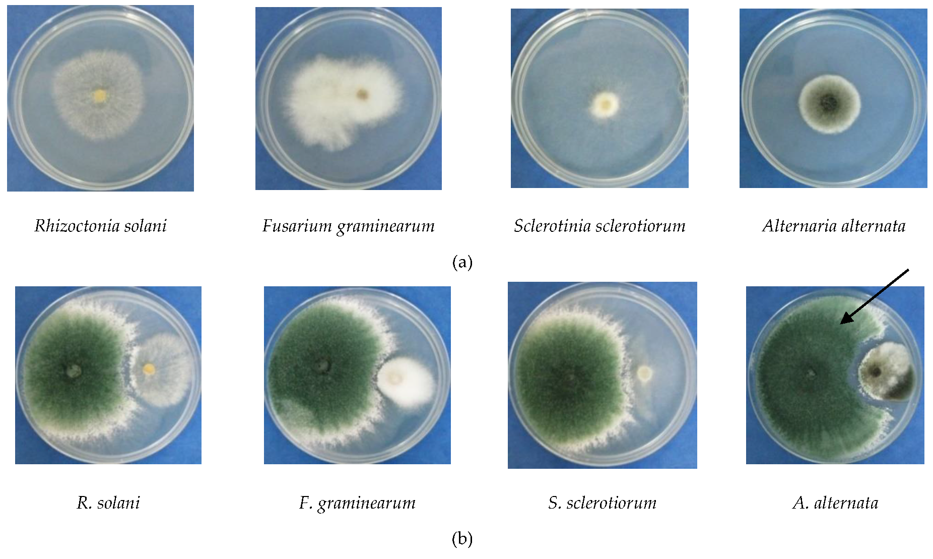
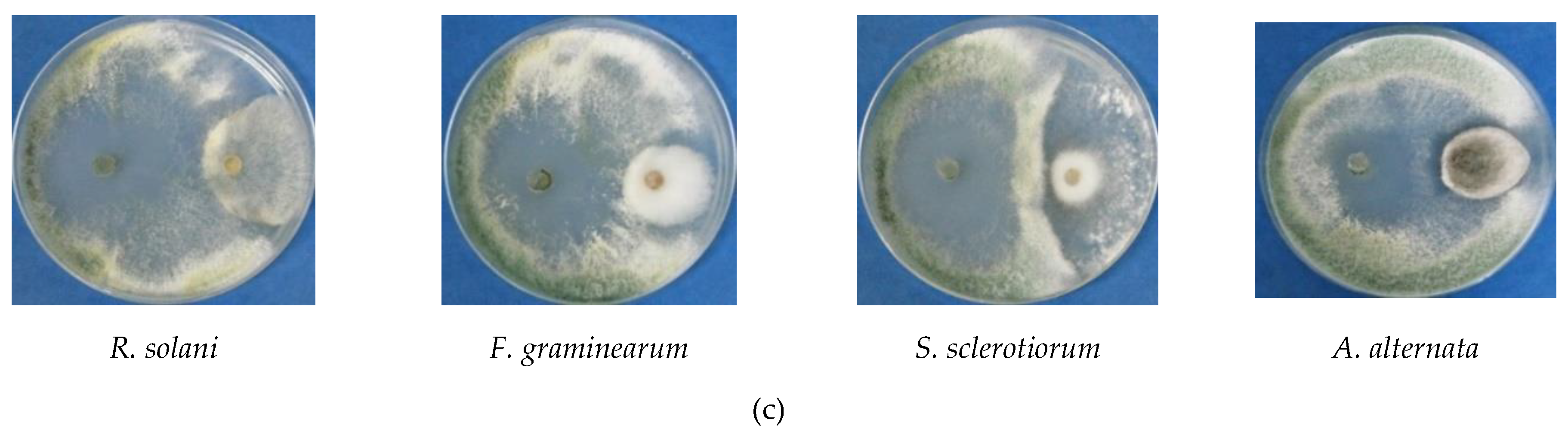
2.2.1. Antagonistic Activities Against Pathogenic Fungi

The fungal isolates were tested in vitro for their antifungal activity against Rhizoctonia solani, Fusarium graminearum and Sclerotinia sclerotiorum from Deutsche Sammlung von Mikroorganismen und Zellkulturen GmbH (DSMZ), and Alternaria alternata from the ICECHIM Collection, using dual culture method [19]. The tests were performed in Petri plates on PDA medium, where 5 mm culture disc of each fungal isolate and pathogen were inoculated. Antagonist activity was observed after incubation at 25 ± 1 °C, for 3–5 days. The value of inhibition degree was determined using the formula
I (%) = (M−P)/M × 100
where I = inhibition; M = diameter colony for pathogen control; P = colony diameter of pathogen in dual cultures in presence of antagonist. Five independent experiments were carried out.

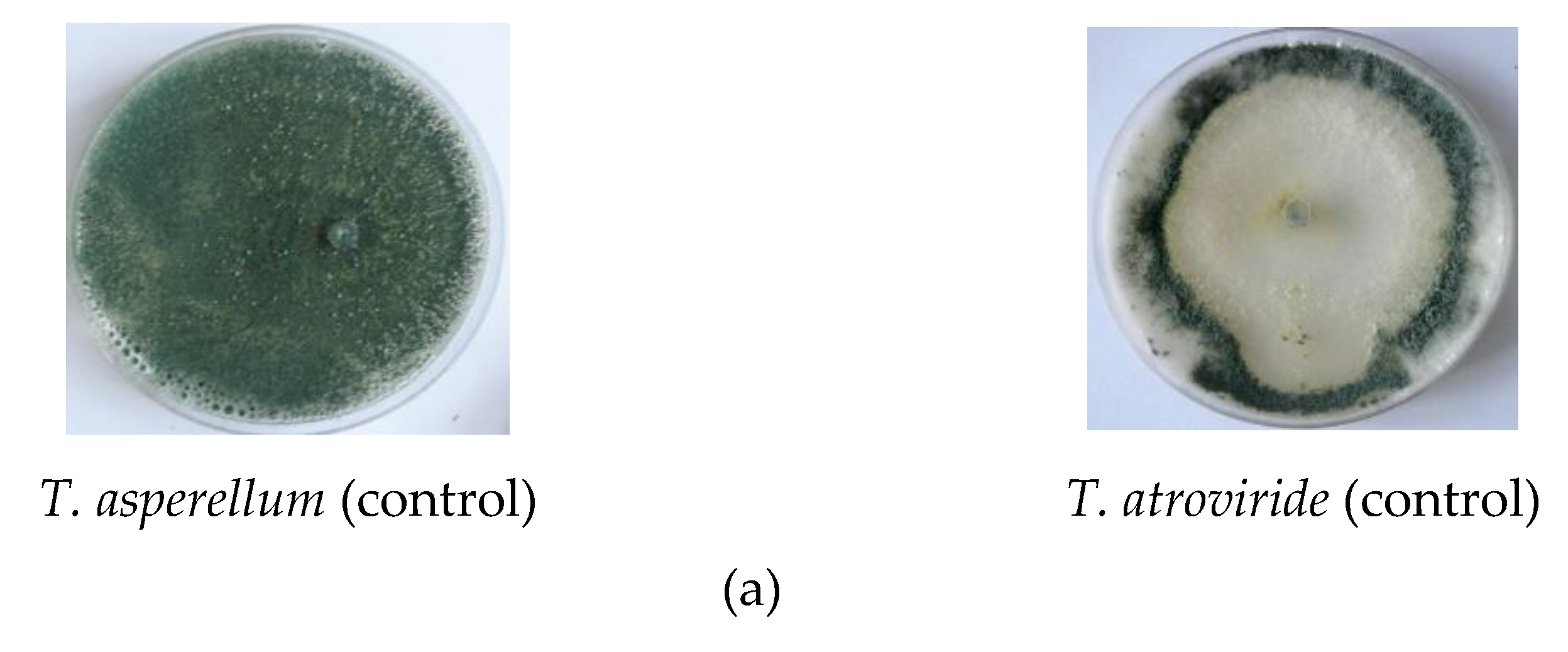
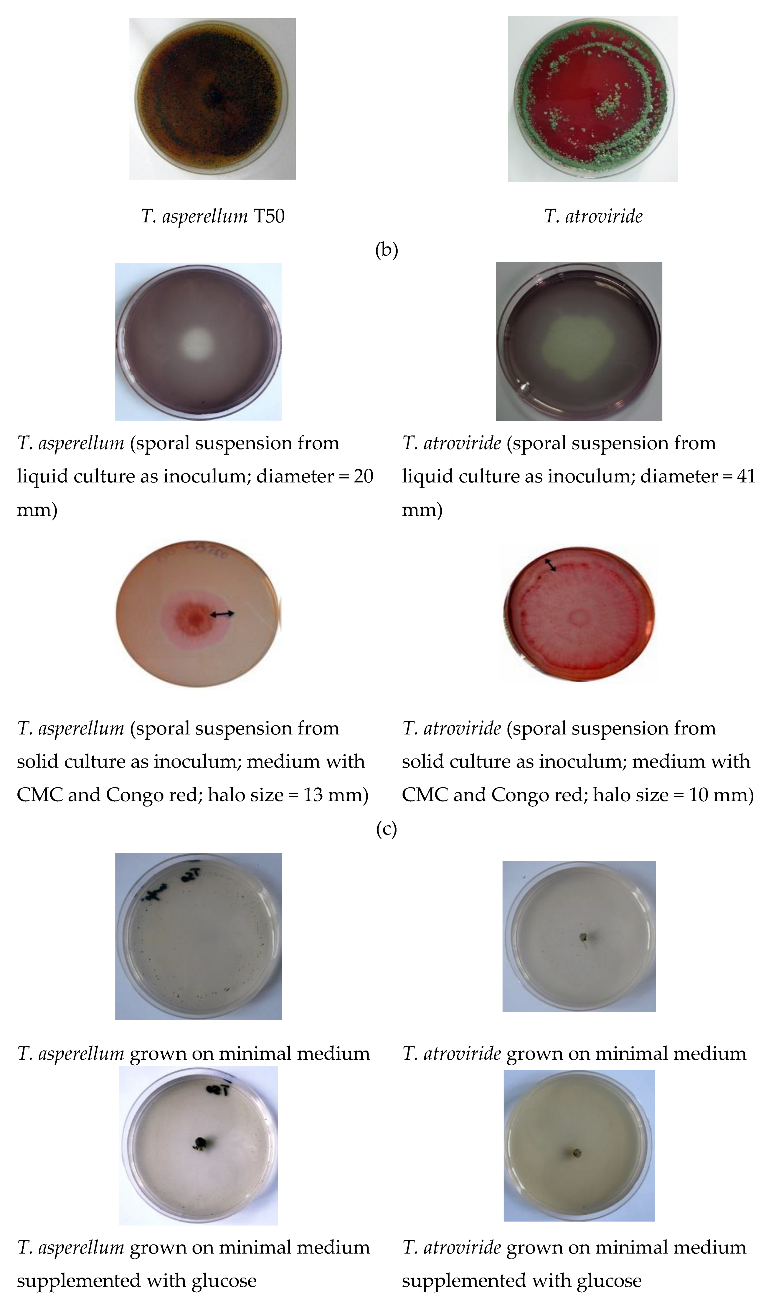
2.2.2. Enzymatic Activities

Chitinase activity was evaluated by qualitative method, using a basal medium with colloidal chitin as unique source of carbon [20]. The culture medium had the following composition (g L−1): 4.5, colloidal chitin; 0.3, MgSO4x7H2O; 3.0, (NH4)2SO4; 2.0, KH2PO4; 1.0, citric acid; 15.0, agar; 0.15, bromcresol purple; 200 µL, Tween 80. The fungal isolates were cultivated on solid PDA medium in Petri plates, for 7 days at 26 ± 2 °C. Mycelia fragments of 5 mm were incubated on chitin medium for 5–7 days, at 26 ± 2 °C. The appearance of red-violet color was considered positive result of test. The test was performed in triplicate and most representative photos are presented.
Cellulase activity was evaluated by qualitative method with carboxymethylcellulose (CMC) as cellulosic substrate [21]. The solid medium had the following composition (g L−1): 1, yeast extract; 5, CMC; 15, agar. The Petri plates were central inoculated with 20 µL liquid culture filtrate of Trichoderma and sporal suspension from Trichoderma culture on PDA medium, respectively. A 1 mL spore suspension contained approximately 1 × 10⁵ spore mL−1. Then, Petri plates were incubated at 28 ± 20 °C for 5 days. The plates were stained with Lugol solution or Congo red (0.1%), respectively, for 15 min, at 28 ± 20 °C. A positive result was considered the formation of a circular transparent zone around fungal colony which was measured. All tests were performed in triplicate.
Keratinase activity was evaluated by qualitative method with ball-milled feathers powder as enzymatic substrate [22,23]. Chicken feathers were disinfected with 70% (v/v) ethylic alcohol, rinsed with sterile distilled water, dry overnight at 50 °C, and finally, milled in ball mill Retsch (Model MM400, Haan, Germany). The fungal isolates were inoculated on agar Petri plates on the following culture media: (i) PDA common medium for fungal growth; (ii) mineral minimal medium (MM) (g L−1) containing KH2PO4 (0.1), CaCl2 (0.01); 0.1, FeSO4 7H2O (0.1), ZnSO4 7H2O (0.005), pH = 7.5; (iii) MM medium supplemented with feathers powder. Trichoderma mycelia fragments of 5 mm were incubated on agar media for 5–10 days, at 26 ± 2 °C. Fungal growth was evaluated by measurement of colony diameter, after 5 and 10 days. Macro- and microscopic observations were carried out to evaluate the process of spore formation. The test was performed in triplicate and the most relevant photos have been presented.

2.2.3. Production of Indole-3-Acetic Acid (IAA)

IAA production by fungal isolates was determined according to Gordon and Weber method (1951) [24]. The fungal isolates were grown in liquid media with following composition (g L−1): (i) PDA medium (200, potato; 20, dextrose); (ii) PDA medium supplemented with L-tryptophan (0.1 g L−1). The medium without L-tryptophan was used as control. Mycelial fragments from fresh cultures on PD agar medium were utilized as inoculant material. The inoculated flasks were incubated at 26 ± 2 °C, under orbital agitation on Heidolph Unimax 1010 (Heidolph Instruments GmBH & Co., Germany) at 130 rpm, for 8 days. The mycelium was separated by centrifugation at 4000 rpm, 15 min, at 4 °C. A total of 2 ml of each collected supernatant was treated with 2 drops of ortho-phosphoric acid. The IAA production was indicated by the appearance of pink color in test slants. Furthermore, IAA production was measured as absorbance at 530 nm wavelength using spectrometer BioMate3 (Thermo Fisher Scientific, Paisley, UK) [25]. Commercial IAA (Sigma-Aldrich) was used as calibration curve to calculate the amount of IAA produced by each isolate. Five independent experiments were carried out.

2.2.4. Hydroxyquinoline Test for Siderophores Production

The fungal isolates were inoculated on King’s B medium supplemented with 8-hydroxyquinoline, a strong chelater [26]. The composition of culture medium was (g L−1): 30, malt extract; 5, peptone; 0.05, 8-hydroxyquinoline; 15, agar. The Petri plates were central inoculated with 20 µL Trichoderma sporal suspension and then incubated at 28 ± 2 °C for 5 days. About 1 ml spore suspension contained 1 × 10⁵ spore mL−1. The positive result was obtained for those strains that grow on the medium and produced an iron chelator. The test was performed in triplicate and the most representative photos have been presented.

2.2.5. Phosphorus Solubilization

Phosphorus solubilization was determined qualitatively [27,28]. The fungal isolates were grown in the medium composed (g L−1): 0.3, MgSO4x7H2O; 0.004, MnSO4x7H2O; 0.002, FeSO4x7H2O; 20, NaCl; 0.5, yeast extract; 0.1, brom cresol purple as pH indicator; 5.0, Ca3(PO4)2; 16, agar. The pH of the culture medium was adjusted to 7.0 prior to sterilization. The Petri plates were inoculated with 5 mm mycelia fragments from fresh cultures and incubated 5 days, at 26 ± 2 °C. The plates were observed and phosphorus solubilization was confirmed by the formation of clearing zone around the colonies. The test was performed in triplicate and relevant photos have been presented.

2.2.6. Protein Hydrolysates (PHs) Preparation

PHs were prepared according to others reports [29,30]. The fungal isolates were grown on liquid medium with keratin substrate as a sole carbon and nitrogen source. The chicken feathers and sheep wool were waste products from local poultry and animal farm, respectively. The keratin substrates were disinfected with ethanol, rinsed with sterile water and dried over night at 50 °C. The basal medium for fungal growth had the following composition (g L−1): 0.5, NaH2PO4, 0.5, KH2PO4; 0.016, FeCl3; 0.1, CaCl2; 0.01, MgCl2; pH = 7. Erlenmayer flasks with basal medium and keratin substrate (1%; w/w) were inoculated with 5 ml of each pre-culture and then incubated at 26 ± 2 °C, under constant agitation (130 rpm) on Heidolph Unimax 1010, for 21 days. The culture broths were centrifuged and the free cell-supernatants as PHs were used for plant treatments.

2.2.7. Nitrogen Content of Protein Hydrolysates

The supernatants were analyzed for nitrogen content according to the Kjeldhal method [31]. Ammonia nitrogen was determined based on SR EN 15475:2009 protocol. Nitric nitrogen was determined based on nitrates reduction to ammonia using metallic iron in an acidic medium followed by ammonia determination based on EN 15558:2009 method. Total nitrogen was determined as quantitative transformation into ammonia by boiling in the presence of sulfuric acid followed by ammonia determination according to SR EN 15478:2009 method adapted in our laboratory. The quantification of ammonia was performed by titration of absorption sulfuric acid solution with a standard solution of 0.1 mol L−1 NaOH in the presence of a mixed indicator. The analytic system consisted of infrared rapid digestion equipment (Behrotest® InKjel P, Behr Labor-Technik, Dusseldorf, Germany), a process suction automatic scrubber (Behrosog S4, Behr Labor-Technik, Dusseldorf, Germany) for neutralization of H2SO4 vapors, a fully automatic steam distillation equipment (Behrotest S4® WD 40, Behr Labor-Technik, Dusseldorf, Germany). Total content of proteins was calculated by multiplying the protein nitrogen concentration by the factor of 6.25.

2.3. In Vivo Tests for Biostimulant Activity

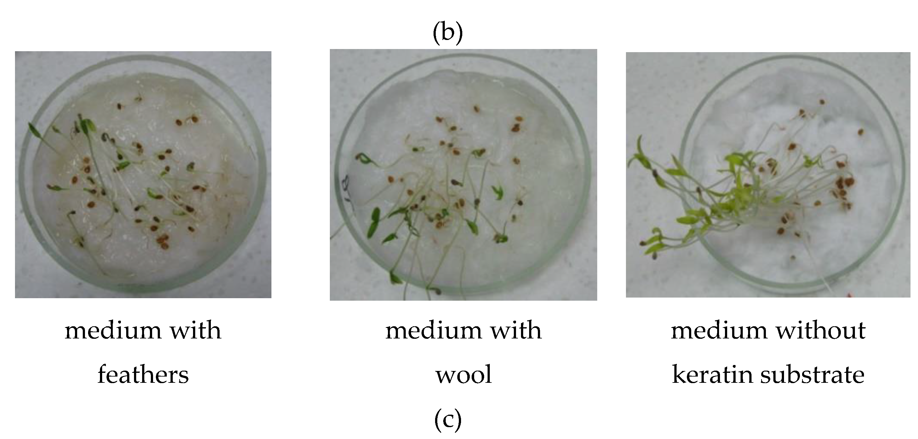
2.3.1. Seed Germination

Seeds of a tomato variety (Solanum lycopersicum) purchased from Agrosem (Tg-Mureş, Romania) were surface sterilized using commercial bleach with sodium hypochlorite (2%) for 20 min, rinsed with water, and, afterward were sown in Petri plates on wet filter paper and germinated in the dark at 25 °C. The seeds were treated with PHs from both isolates and the process was observed. Seeds treated with water were used as the control treatment.

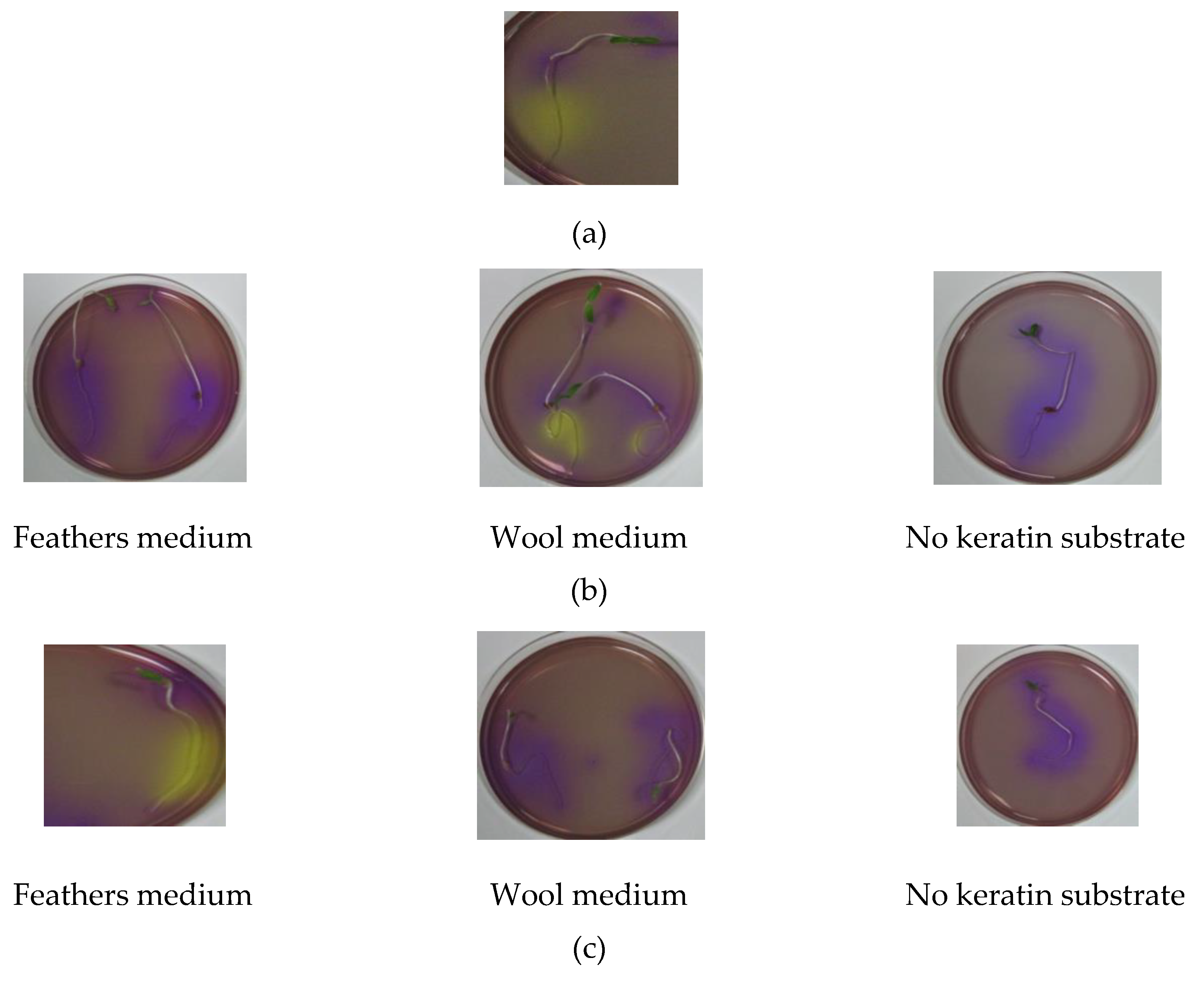
2.3.2. Plant Proton Pump

The test was carried out according to the Zandonani protocol [32]. Tomato seeds were surface sterilized with NaClO (1%; w/v) for 20 min, washed thoroughly with sterile water and placed to germinate on sterile cotton wool in Petri plates. The tomato seedlings from germinated seeds with roots of 3–4 cm long were treated for 10 and 60 min with PHs and distilled water (control), respectively. After that, pre-treated seedlings were transferred in Petri plates on agar medium supplemented with brom cresol purple and incubated for 24 h. Positive result of proton pump activation tests was considered the acidification of root zone through the appearance of yellow color.

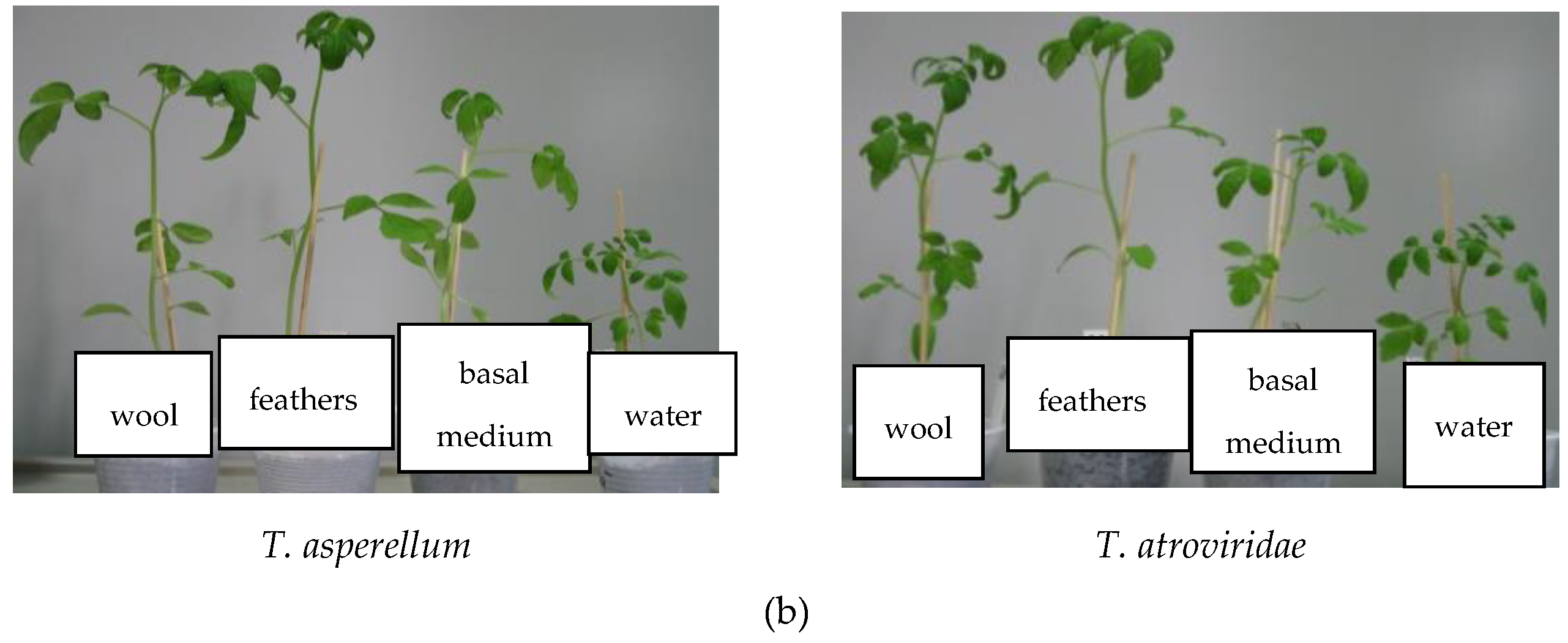
2.3.3. Test for the Capacity to Promote Plant Growth (Pot Experiment)

Tomato seeds were planted in pots and incubated in a growth chamber under controlled daylight conditions (Micro Clima Series TM, Labs Economic Lux chamber, Snijders, Netherlands). Five pots were sown for each pair of strain–keratin substrate in culture medium, and the same number of pots for control (treatment with water) and for fungal isolates cultured on basal medium without keratin substrate. Four treatments were applied at weekly intervals. Then, the plants were extracted from soil, and growth parameters (plants height, shoots length, number of branches) were compared with untreated tomato seedlings (watered only with tap water) after 21 days [33,34].

3. Results

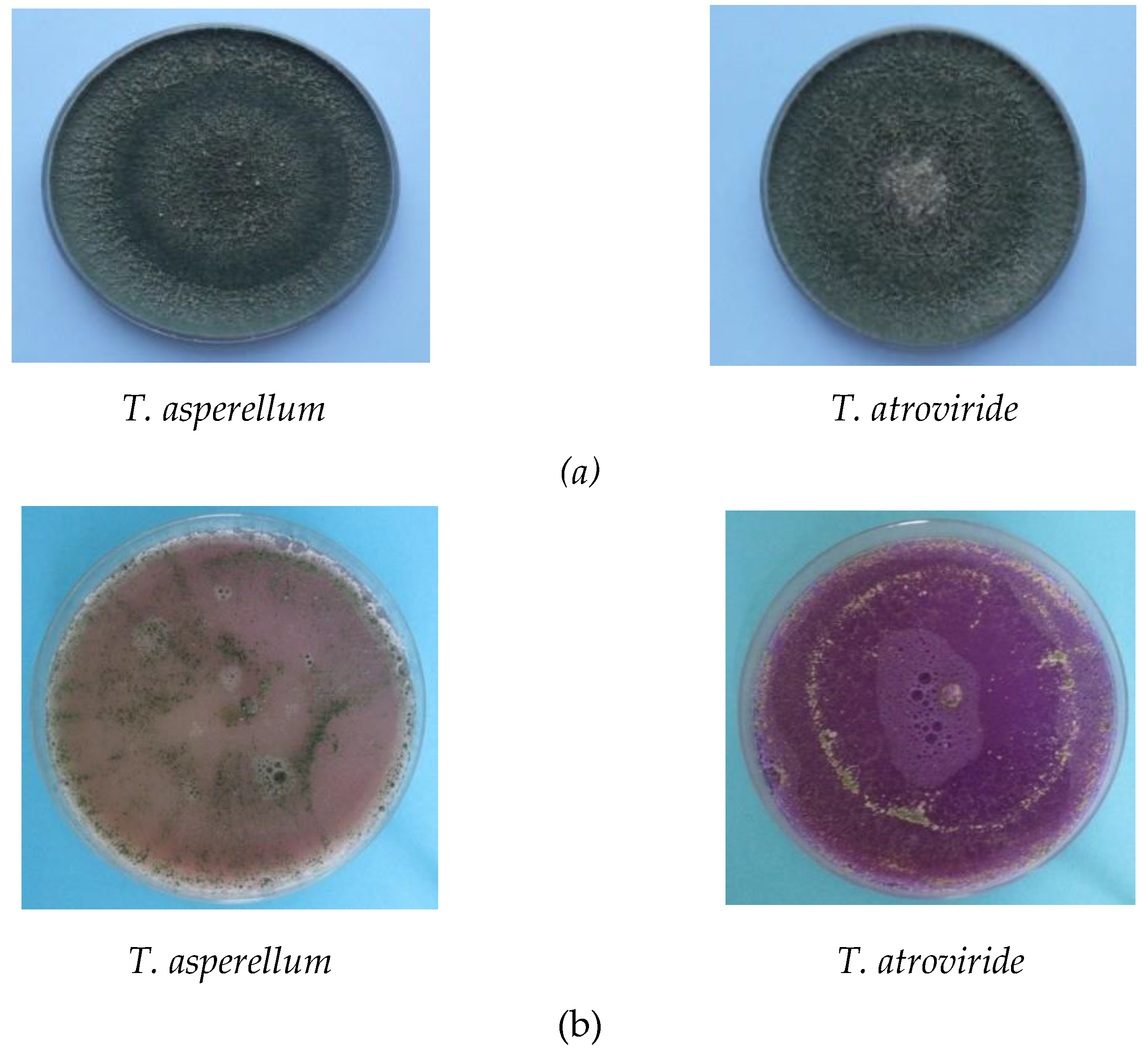
3.1. Antagonistic Effect

Trichoderma isolates were assessed for their effectiveness as biological control agents against R. solani, S. sclerotiorum, F. graminearum and A. alternata (Figure 1 and Table 1). Fungal sporulation is a complex process that usually occurs when growth rate is reduced due to some conditions that rather sustain mycelial growth. Starvation, lack of nutrients, pH, and light are major factors affecting sporulation.
In our test, T. asperellum presented a high sporulation capacity. T. asperellum mycelium had an intense green color, especially in antagonist test with A. alternata. The sporulation of T. atroviride was lower compared to T. asperellum. It can be observed that in dual test, mycelium of R. solani and A. alternata remained almost at the same size as the control, respectively. Mycelia of R. solani grew in Petri plates relatively well delimited by both Trichoderma isolates, more evident for T. asperellum. The same behavior was observed at A. alternata. For both Trichoderma isolates, F. graminearum presented a smaller mycelium compared with pathogen pure culture as control, a consequence of the inhibitory effect of Trichoderma as biocontrol agent.

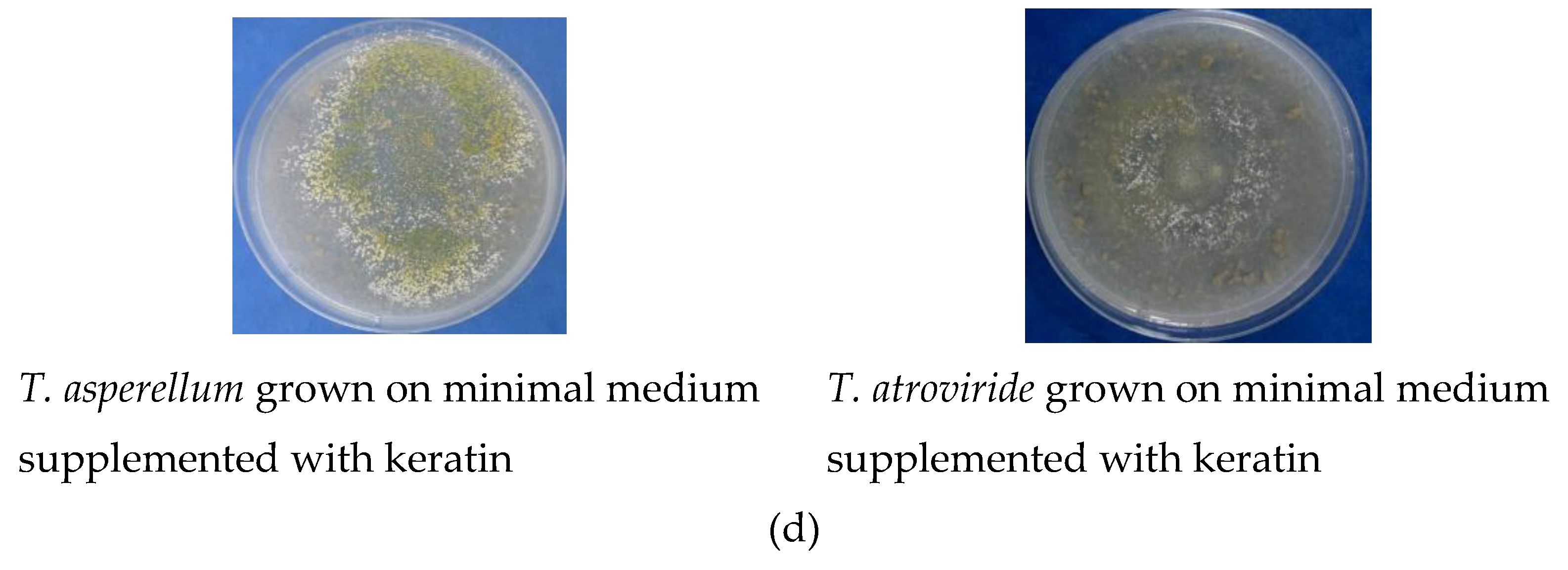
3.2. Enzymatic Activities
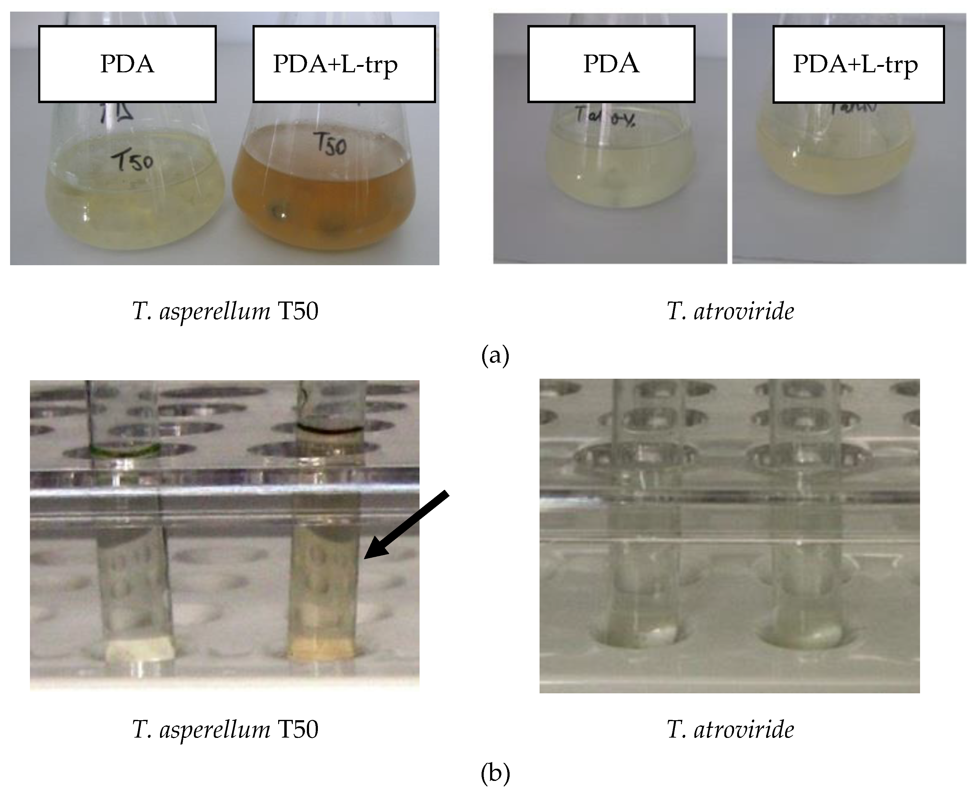
The ability of Trichoderma isolates to secrete lytic enzymes (chitinases, cellulases, keratinases) was analyzed in qualitative tests on solid specific medium. As presented in Figure 2b, both Trichoderma isolates showed chitinase activity, but the degradation system of T. atroviride exhibited higher enzymatic activity with intense red-violet color covering the whole plate, compared to T. asperellum which exhibited a medium activity with the color extended only on half plate.
Referring to cellulose activity, T. atroviride showed a higher hydrolytic activity versus the CMC substrate of 41 cm zone diameter, comparative with 20 cm zone diameter of T. asperellum (Figure 2c). Qualitative test of keratinase activity was performed through the comparative cultivation of fungal isolates on three cultures media, minimal medium, minimal medium with glucose as carbon and energy source, and on minimal medium with keratin substrate. No growth of fungal isolates was observed on minimal medium and medium supplemented with glucose (Figure 2d). The qualitative test in feather meal agar plates evidenced a negative result and no degradation halo surrounding the colony of isolates was observed.

3.3. IAA Production

In our tests, the ability for IAA production was exhibited only by T. asperellum and was evidenced by the pink color of the medium supplemented with tryptophan (Figure 3 and Table 2).
In quantitative test, the values of DO530 nm are higher for T. asperellum (0.075) comparative to T. atroviride (0.050). The values of DO530 nm for T. atroviride were almost equal to those obtained on PDA medium without L-tryptophan, 0.050 vs. 0.045. The production of IAA is a significant finding for selection of microbial biostimulants.
The fungal isolates were investigated for siderophores production with a qualitative test performed on King’s B medium, supplemented with 8-hydroxyquinoline. The results were positive for both Trichoderma isolates (Figure 4a).

3.4. Phosphate Solubilisation

The tests performed for phosphorus solubilization capacity of fungal isolates demonstrated the good capacity of T. asperellum. Figure 4b showed the changing of the medium color. T. atroviride exhibited a lower capacity to solubilize phosphorus, which is a disadvantage in its use as biostimulant microbial agent.

3.5. Nitrogen Content

An important step in the selection of microorganisms as plant growth promoting is nitrogen content of PHs. Protein hydrolysates from fungal cultures on media with keratin wastes were analyzed for ammonia nitrogen, protein nitrogen, nitric nitrogen, total protein and total nitrogen. All results are summarized in Table 3.
It can be observed that the level of ammonia nitrogen in PHs was almost similar, with the exception of hydrolysate from T. asperellum grown on wool, which contained only 9.5 mg% g, lower approximately with 40% compared to other strains. The level of protein nitrogen in HPs decreased in the following order: T. asperellum (grown on feathers) > T. asperellum 0 (grown on wool) > T. atroviridae (grown on feathers) > T. atroviride (grown on wool; under the detection limit of method). T. asperellum grown on feathers produced the highest level of total protein, a feature significant for the treatment of plants. The total nitrogen of PHs was in the following order: T. atroviridae (grown on feathers) > T. asperellum (grown on feathers) > T. atroviridae (grown on wool) > T. asperellum (grown on wool). Referring to total nitrogen content, feathers seem to be a better carbon and nitrogen source to sustain fungal growth and to ensure a higher level of nitrogen in culture broth.

3.6. Biostimulating Effect of PHs

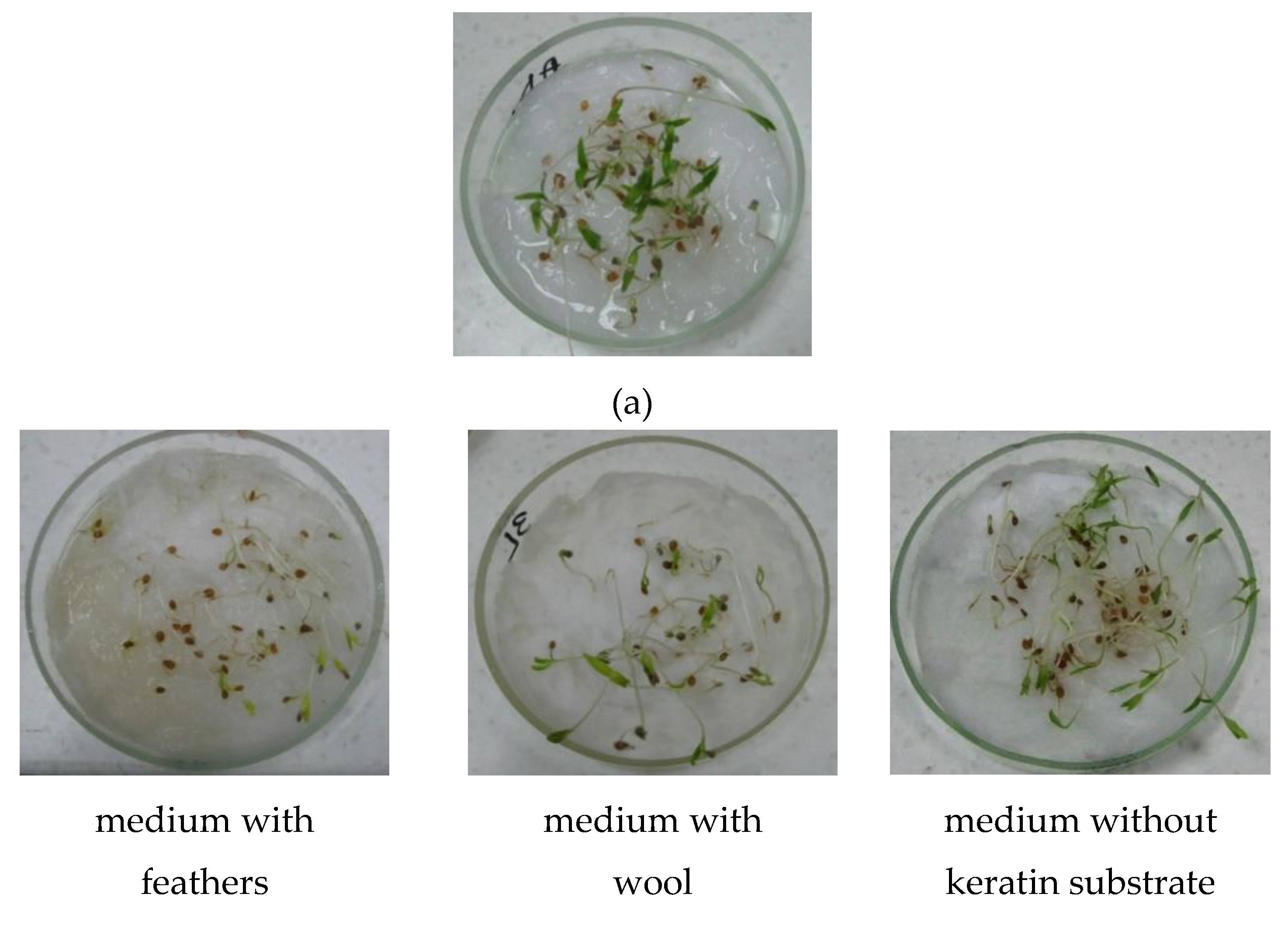
3.6.1. Seed Germination

As can be seen in Figure 5, no major differences in seed germination were observed in relation to the nature of keratin substrate from culture medium and type of microbial strain. Despite this fact, some minor differences could be highlighted in a careful analysis of germinated seeds and seedlings. Thus, the seedlings treated with PHs from culture on feathers were slightly longer compared with the others, while those treated with PHs from wool culture were slightly more vigorous than those treated with PHs from medium without keratin substrate (Figure 5).

3.6.2. Proton Pump Test

The responses related to the tests of capacity for activating the proton pump exhibited by the application of PHs are presented in Figure 6. It can be seen images of plant root acidification yellow colored, indicator of biostimulant activity on root growth.
This simple protocol with bromocresol purple offered useful data for screening biostimulant activity exhibited by protein hydrolysates from fungal strains grown on keratin substrates. No effect on tomato seedlings was visualized with PHs from Trichoderma cultures on medium without keratin, since keratin source of proteins and amino acids was absent. After 10 min of contact, PHs from T. asperellum grown on both keratin media produced a significant effect, as comparative with only medium effect reached by PHs from T. atroviride on wool. After 60 min of contact, PHs from T. asperellum grown on feathers medium activated the proton pump at the same level (“significant effect”), while the effect of PHs from wool medium decreased to “minimum effect” (Table 4).
Data obtained from experiments demonstrated the ability of PHs from T. asperellum grown on feathers medium to activate the proton pump at tomato seedlings.

3.6.3. Pot Experiments

It can be observed that after the first treatment with PHs there are no significant differences between plants aspect and size (Figure 7a). Then, after the third and especially after the fourth PHs applications (Figure 7b), the differences were evident, these plants being significantly higher, more robust with an increased number of branches compared to those treated with only water.
One week after the end of the treatments, the plants were extracted from pot soil and several plant characteristics were analyzed: height, diameter, roots length, number of branches and leaves [31,32].
Table 5 shows all measured plant growth parameters which were significantly influenced by the applications of protein hydrolysates.
PHs from T. asperellum cultured on feathers medium reached the highest values for all parameters recorded, with the exception of those for total biomass obtained in wool culture medium. The treatment with PHs from T. asperellum enhanced growth of tomato plants compared to control as evidenced by total biomass (+292% for feathers, +30% for wool), plant height (+257% for feathers, + 196% for wool), diameter (+30% for feathers, + 28% for wool), number of branches (+57% for feathers, +21% for wool) and number of leaves (+47% for feathers, +1.5% for wool). Meanwhile, the increase of parameter values for T. asperellum grown on medium without keratin was very good, but lower compared to those of strain cultured on keratin media, total biomass (+115%), diameter (+7%), number of branches (+36%). An exception was recorded at the values of leaves number, +15% compared to only 1.5% for PHs obtained from wool medium. For T. asperellum, feathers were found to be the best carbon and nitrogen source in culture medium.
The behavior of T. atroviride was completely different since most of the parameters, except plant stem diameter, have reached the maximum values in cultivation on medium without keratin. In only one case, namely the number of branches, the applications of PHs obtained from T. atroviride cultured on wool gave the same effect compared to T. asperellum (7.33 branches number/plant) (Table 5).

4. Discussion

The biocontrol activity of Trichoderma towards aggressive plant pathogens is well recognized in many reports [35,36,37]. The antagonistic properties of Trichoderma are based on the activation of multiple mechanisms, indirect or direct, each type leading to metabolites secretion. In our tests, T. asperellum showed maximum inhibition against S. sclerotiorum, over 59%, while T. atroviride was most active against F. graminearum, 61% inhibition (Figure 1 and Table 1.). The biocontrol test was not so relevant to allow a hierarchy of the strains. The biocontrol activity expressed as growth inhibition of pathogens in dual cultures was similar compared to those obtained with others Trichoderma strains. Hence, S. sclerotiorum exposed to mutants of T. harzianum after 3 days incubation in dual culture presented an inhibition over 70% for almost tested strains, with the highest value of 88% inhibition. The values for F. graminearum did not exceed 60%, the pathogen being more resistant to biocontrol agent [38]. A Trichoderma isolate showed 70% (R. solani), 68.2% (Sclerotinia rolfsii), 73.3% (M. phaseolina) and, 69.3% (Fusarium sp.) growth inhibition, respectively [39]. Redda et al. (2018) [40] studied the biocontrol activity of 380 soil isolates identified as belonging to Trichoderma genus. Out of them, isolates showed strong antagonistic activity inhibiting over 50% mycelia growth of F. oxysporum, R.s olani and B. cinerea. Wu et al. (2017) [41] reported an inhibition rate of 80.82% of new Trichoderma asperellum isolate against F. oxysporum, an aggressive pathogen infecting cucumber plants.
Dual cultures were used to study the ability of fungal isolates to secrete lytic enzymes, chitinases, cellulases and keratinase. Chitinase activity is essential for the selection of an efficient biocontrol agent since chitin is the major structural component of cell wall from pathogens. Chitinases together with β-1,3-glucanases are cell-wall-degrading enzymes involved in the initiation of mycoparasitism [42,43]. The secreted enzymes act synergistic each other, but at the same time with other metabolites. It has been reported that characteristics of chitinases are different at Trichoderma species [41].
In our experimental study, T. atroviride exhibited higher chitinase and cellulose activities compared to T. asperellum (Figure 2). Negative results were obtained for keratinase activity and no degradation halo surrounding the colony of isolates was observed in culture medium with keratin feathers. Meanwhile, the cultures of Trichoderma isolates grew very well on keratin solid medium covering the whole surface. This contradictory finding is consistent with the data from others reports. Thus, out of 69 fungal isolates which are able to grow on feather meal agar, only three produced clear zones of keratinase activity toward keratin substrate [33]. Similar results have been reported in another study of screening tests on agar plates where the strains were capable of growth without clarifying the keratin agar medium due to the lack of active extracellular enzymes [44].
Indole-3-acetic acid is the most common plant hormone belonging to auxin class that regulates various aspects of plant growth and development [7,43,44,45,46]. IAA is responsible for cell division and elongation responding to various factors as light, presence of pathogens etc. Fungi are able to use secreted IAA for interaction with plants in pathogenesis or symbiotic strategies, supporting the defense mechanism of plants [47,48,49,50,51,52]. Tryptophan is the main precursor for IAA synthesis and its presence increases the IAA production which in turn, enhances the availability of substrate for plants [50]. IAA biosynthesis of fungal species occurred near the end of the growth phase or during plant dormancy phase, so the process is quite long [50]. According to our qualitative and quantitative tests, only T. asperellum showed capacity to produce IAA (Figure 3 and Table 2). It could be possible that T. atroviride needed a longer duration of the IAA secretion process compared to T. asperellum.
Siderophores are low molecular weight secondary metabolites with a high activity of iron binding ligands. They serve as vehicles for the transport into microbial cells of ferric ion which is otherwise unavailable. The production of siderophores by Trichoderma candidates for plant disease control and growth promoting offers the possibilities to replace chemical pesticides [53,54]. In this regard, fungal Trichoderma isolates were investigated by qualitative test on King’s B medium supplemented with 8-hydroxyquinoline (Figure 4a). Both Trichoderma isolates showed positive results in test for siderophores production.
Since phosphorus is one of vital elements for growth and development of plant cultures, it is of interest to study the capacity of phosphate solubilizing expressed by fungal strains with potential as microbial biostimulant. T. asperellum exhibited a better activity to solubilize phosphorus compared to T. atroviride (Figure 4b). Normally, the lack of phosphorus is supplied by fertilizers, but meanwhile, there are beneficial microorganisms which contribute directly to the plant performance, through different mechanisms, phosphorus solubilization, phytohormone production, etc. The use of phosphate solubilizing microorganisms as biofertilizers represents a promising strategy to improve world food and at the same time conserve the environment. The beneficial microorganisms solubilize phosphorus in vitro, increasing the bioavailability of insoluble phosphorus from soil to be used by plants [55,56,57,58]. It has been reported an efficient treatment with Aspergillus niger, which stimulated significantly the growth of cabbage cultures, increasing roots and shoots length, weights of fresh and dry shoots [59]. An A. awamori strain solubilized various inorganic forms of phosphate (tri-calcium phosphate, di-calcium phosphate, ferric phosphate) in varying quantities and has improved the cultures yield. This study confirmed that phosphate solubilization occurred on different mechanism, depending on the nature of phosphate source [60].
Important data were obtained through analysis of nitrogen content. Of all experimental combinations, fungal strain—type of keratin substrate in culture medium, T. asperellum grown on feathers produced the highest level of total protein, a feature significant for the treatment of plants (Table 3). From this point of view, feathers seem to be a better carbon and nitrogen source to sustain fungal growth and to ensure a higher level of nitrogen in culture broth.
Keratin wastes as chicken feathers and wool are produced annually in a large quantity and excessive accumulation leads to environmental pollution. The nutritional minerals, keratin and amino acids contained in keratin substrate represent good substrate for the growth and development of bacteria and fungi. The first step in the assessment of biostimulating effect of protein hydrolysates from fungal strains cultured on medium with keratin wastes was the seeds germination. The images presented in Figure 5 showed that, to a certain degree, the seedlings treated with PHs from culture on feathers were longer compared with the others, while those treated with PHs from wool culture were relative more vigorous as those treated with PHs from medium without keratin substrate (Figure 5).
The second step in the evaluation of fungal isolates as plant biostimulants was the investigation of proton pump activation, which is a marker of biostimulant capacity and a responsible agent for the regulations of protons electrochemical gradient across cell membrane to uptake the nutrients. The transfer of proteins through plant cells to vacuoles, endosomal and plasma membrane is stimulated by H+ pumps [61]. Proton pump is involved in the mechanism of cells plant growth. Plant hormones are able to regulate the activity of plant proton pumps through enzymes activity involved in plant reactions versus environmental factors. The qualitative test carried out on agar medium supplemented with brom cresol purple (Table 4 and Figure 6) showed that protein hydrolysates from T. asperellum grown of feathers medium activated the proton pump at tomato seedlings. Significant effect of PHs from T. asperellum from feathers medium as proton pump activation was observed even after 60 min of contact (Figure 6b).
Another key step in the evaluation of fungal isolates as microbial biostimulants was the pot experiments performed with tomato seedlings (Figure 7). Tomato seedlings were four times treated with protein hydrolysates obtained from Trichoderma isolates cultured on media with keratin wastes and on medium without keratin substrate. Feathers seem to be a good ingredient for culture medium, releasing nutrients to stimulate plant growth. The effect of PHs applications on plants was visible, especially after fourth applications, since the fungal isolates cultured on keratin media secreted enzymes were able to disrupt chemical bonds of keratin substrate releasing nutrient stimulants for plants growth and development. The root applications of PHs can increase nitrogen assimilation through an increase of nitrate reductase and glutamine synthetase activities [62,63]. The effects of PHs applications are visible in the plant characteristics, presented in Table 5. These values confirm the increase in tomato growth resulted from the biological activity of Trichoderma isolates. Our results obtained with Trichoderma isolates are in agreement with those reported by other studies [5,8,33,64,65].

5. Conclusions

Our combined in vitro and in vivo data have proved Trichoderma asperellum isolate as a candidate to reduce the availability of keratin waste for pollution of the environment, and finally, ensure its conversion into fertilizers. The metabolites released by keratin degradation under the activity of selected Trichoderma asperellum isolate could be employed to improve the growth of plants.

Author Contributions

L.J., M.L.A., M.D. conceived and designed the experiments; M.C., I.R. and A.M.G. performed the experiments; L.J., M.L.A. and M.D. analyzed the data; L.J. and M.L.A. wrote the paper. All authors read and approved the final manuscript.

Funding

This research was funded by projects PN.16.31.01.03/2017 and PN.19.23.01.01.01/2019.

Acknowledgments

The authors thank to Ministry of Research and Innovation of Romania through Program 1- Development of the National Research and Development System, Subprogram 1.2—Institutional performance—Projects of Excellence Financing in RDI, project number PFE no 31/2018. Also, the authors want to thank to project MANU NET 33/2017 (TOX-HAZ-ASSESS).

Conflicts of Interest

The authors declare no conflict of interest.

References

  1. Howell, C.R. Mechanisms employed by Trichoderma species in the biological control of plant diseases: The history and evolution of current concepts. Plant Dis. 2003, 87, 4–10. [Google Scholar] [CrossRef] [PubMed]
  2. Harman, G.E. Overview of mechanisms and uses of Trichoderma spp. Phytopathology 2006, 96, 190–194. [Google Scholar] [CrossRef] [PubMed]
  3. Schuster, A.; Schmoll, M. Biology and biotechnology of Trichoderma. Appl. Microbiol. Biotechnol. 2010, 87, 787–799. [Google Scholar] [CrossRef] [PubMed]
  4. Heydari, A.; Pessarakli, M. A review on biological control of fungal plant pathogens using microbial antagonists. J. Biol. Sci. 2010, 10, 273–290. [Google Scholar] [CrossRef]
  5. Błaszczyk, L.; Siwulski, S.; Sobieralski, K.; Lisiecka, J.; Jędryczka, M. Trichoderma spp.—Application and prospects for use in organic farming and industry. J. Plant Prot. Res. 2014, 54, 309–317. [Google Scholar] [CrossRef]
  6. Naher, L.; Yusuf, U.K.; Ismail, A.; Hossain, K. Trichoderma spp.: A biocontrol agent for sustainable management of plant diseases. Pak. J. Bot. 2014, 46, 1489–1493. [Google Scholar]
  7. Bashan, Y.; de-Bashan, L.E. How the plant growth-promoting bacterium Azospirillum promotes plant growth—A critical assessment. In Advances in Agronomy; Sparks, D.L., Ed.; Elsevier Academic Press: Amsterdam, The Netherlands, 2010; pp. 77–136. [Google Scholar]
  8. Saba, H.; Vibhash, D.; Manisha, M.; Prashant, K.; Farhan, H.; Tauseef, A. Trichoderma–A promising plant growth stimulator and biocontrol agent. Mycosphere 2012, 3, 524–531. [Google Scholar] [CrossRef]
  9. DuJardin, P. Plant biostimulants: Definition, concept, main categories and regulation. Sci. Hort. 2015, 196, 3–14. [Google Scholar] [CrossRef] [Green Version]
  10. Povero, G.; Mejia, J.F.; Di Tommaso, D.; Piaggesi, A.; Warrior, P. A systematic approach to discover and characterize natural plant biostimulants. Front Plant Sci. 2016, 7. [Google Scholar] [CrossRef]
  11. Bulgari, R.; Cocetta, G.; Trivellini, A.; Vernieri, P.; Ferrante, A. Biostimulants and crop responses: A review. Biol. Agric. Hortic. 2015, 31, 1–17. [Google Scholar] [CrossRef]
  12. Bulgari, R.; Morgutti, S.; Cocetta, G.; Negrini, N.; Farris, S.; Calcante, A.; Spinardi, A.; Ferrari, E.; Mignani, I.; Oberti, R.; et al. Evaluation of borage extracts as potential biostimulant using a phenomic, agronomic, physiological, and biochemical approach. Front Plant Sci. 2017, 8. [Google Scholar] [CrossRef] [PubMed]
  13. Yakhin, O.I.; Lubyanov, A.A.; Yakhin, I.A.; Brown, P.H. Biostimulants in plant science: A global perspective. Front Plant Sci. 2017, 7, 2049. [Google Scholar] [CrossRef] [PubMed]
  14. Rouphael, Y.; Colla, G. Synergistic biostimulatory action: Designing the next generation of plant biostimulants for sustainable agriculture. Front Plant Sci. 2018, 9. [Google Scholar] [CrossRef] [PubMed]
  15. European Commission. Proposal for a Regulation Laying Down Rules on the Making Available on the Market of CE Marked Fertilizing Products and Amending Regulations (EC)1069/2009 and (EC)1107/2009.COM; Brussels European Commission: Brussels, Belgium, 2016; p. 157. [Google Scholar]
  16. Colla, G.; Nardi, S.; Cardarelli, M.; Ertani, A.; Lucini, L.; Canaguier, R.; Rouphael, Y. Protein hydrolysates as biostimulants in horticulture. Sci. Hort. 2015, 196, 28–38. [Google Scholar] [CrossRef]
  17. Kornillowicz-Kowalska, T.; Bohacz, J. Biodegradation of keratin waste: Theory and practical aspects. Waste Manag. 2011, 3, 1689–1701. [Google Scholar] [CrossRef] [PubMed]
  18. Sinkiewicz, I.; Sliwinska, A.; Staroszczyk, H.; Kołodziejska, I. Alternative methods of preparation of soluble keratin from chicken feathers. Waste Biomass Valor. 2017, 8, 1043–1048. [Google Scholar] [CrossRef]
  19. Begum, M.F.; Rahman, M.A.; Alam, F.M. Biological control of alternaria fruit rot of chili by Trichoderma species under field conditions. Mycobiology 2010, 38, 113–117. [Google Scholar] [CrossRef] [PubMed]
  20. Agrawal, T.; Kotasthane, A.S. Chitinolytic assay of indigenous Trichoderma isolates collected from different geographical locations of Chhattisgarh in Central India. SpringerPlus 2012, 1, 73. [Google Scholar] [CrossRef] [PubMed]
  21. Yoon, J.H.; Park, J.E.; Dong, Y.S.; Hong, S.B.; Ko, S.J.; Kim, S.H. Comparison of dyes for easy detection of extracellular cellulases in fungi. Mycobiology 2007, 35, 21–24. [Google Scholar] [CrossRef] [PubMed]
  22. Lateef, A.; Adelere, A.I.; Gueguim-Kana, E.B. Bacillus safensis LAU 13: A new source of keratinase and its multi-functional biocatalytic applications. Biotechnol. Biotechnol. Equip. 2015, 29, 54–63. [Google Scholar] [CrossRef] [PubMed]
  23. Bhange, K.; Chaturvedi, V.; Bhatt, R. Ameliorating effects of chicken feathers in plant growth promotion activity by a keratinolytic strain of Bacillus subtilis PF1. Bioresour Bioprocess 2016, 3, 13. [Google Scholar] [CrossRef]
  24. Gordon, S.A.; Weber, R.P. Colorimetric estimation of indole acetic acid. Plant Physiol. 1951, 26, 192–195. [Google Scholar] [CrossRef] [PubMed]
  25. Gutierrez, C.K.; Matsuy, G.Y.; Lincoln, D.E.; Lovel, C.R. Production of the phytohormone indole-3 acetic acid by the estuarine spesies of the genus Vibrio. Appl Environ Microbiol. 2009, 75, 2253–2258. [Google Scholar] [CrossRef] [PubMed]
  26. De Brito, A.M.; Gagne, S.; Antoun, H. Effect of compost on rhizosphere microflora of the tomato and on the incidence of plant growth-promoting rhizobacteria. Appl Environ Microbiol. 1995, 61, 194–199. [Google Scholar] [PubMed]
  27. Carvajal, L.H.; Bissett, J.; Orduz, S. Growth stimulation in bean (Phaseolus vulgaris L.) by Trichoderma. Biol Control 2009, 51, 409–416. [Google Scholar] [CrossRef]
  28. Vazquez, P.; Holgui, G.; Puente, M.E.; Lopez-Cortes, A.; Bashan, Y. Phosphate-solubilizing microorganisms associated with the rhizosphere of mangroves in a semiarid coastal lagoon. Biol. Fertil. Soils 2000, 30, 460–468. [Google Scholar] [CrossRef]
  29. Paul, T.; Halder, S.K.; Das, A.; Bera, S.; Maity, C.; Mandal, A.; Das, P.S.; Pradeep, K.; Das, M.; Pati, B.R.; et al. Exploitation of chicken feather waste as a plant growth promoting agent using keratinase producing novel isolate Paenibacillus woosongensis TKB2. Biocatal. Agric. Biotechnol. 2013, 2, 50–57. [Google Scholar] [CrossRef]
  30. Popko, M.; Wilk, R.; Górecka, H.; Chojnacka, K.; Henryk Górecki, H. Assessment of new NKSMg fertilizer based on protein hydrolysate of keratin in pot experiments. Pol. J. Environ. Stud. 2015, 24, 1765–1772. [Google Scholar] [CrossRef]
  31. Sáez-Plaza, P.; Michałowski, T.; Navas, M.J.; Asuero, A.G.; Wybraniec, S. An overview of the Kjeldahl method of nitrogen determination. Part I. Early history, chemistry of the procedure, and titrimetric finish. Crit. Rev. Anal. Chem. 2013, 43, 178–223. [Google Scholar] [CrossRef]
  32. Zandonadi, D.B.; Santos, M.P.; Caixeta, L.S.; Marinho, E.B.; Pereira Peres, L.E.; Façanha, A.R. Plant proton pumps as markers of biostimulant action. Sci. Agri. 2016, 73, 24–28. [Google Scholar] [CrossRef] [Green Version]
  33. Cavello, I.A.; Crespo, J.M.; García, S.S.; Zapiola, J.M.; Luna, M.F.; Cavalitto, S.F. Plant growth promotion activity of keratinolytic fungi growing on a recalcitrant waste known as (hair waste). Biotechnol. Res. Int. 2015. [Google Scholar] [CrossRef] [PubMed]
  34. Rajkumar, M.; Freitas, H. Effects of inoculation of plantgrowth promoting bacteria on Ni uptake by Indian mustard. Biores. Technol. 2008, 99, 3491–3498. [Google Scholar] [CrossRef] [PubMed]
  35. Gajera, H.; Domadiya, R.; Patel, S.; Kapopara, M.; Golakiya, B. Molecular mechanism of Trichoderma as bio-control agents against phytopathogen system–A review. Curr. Res. Microbiol. Biotechnol. 2013, 1, 133–142. [Google Scholar]
  36. Gousterova, A.; Nustorova, M.; Paskaleva, D.; Naydenov, M.; Neshev, G.; Tonkova, V.E. Assessment of feather hydrolysate from thermophilic actinomycetes for soil amendment and biological control application. Int. J. Environ. Res. 2012, 6, 467–474. [Google Scholar]
  37. Kumar, M.; Ashraf, S. Role of Trichoderma spp. as a biocontrol agent of fungal plant pathogens. In Probiotics and Plant Health; Kumar, V., Kumar, M., Sharma, S., Prasad, R., Eds.; Springer Nature Singapore Private Limited: Singapore, 2017; pp. 497–506. [Google Scholar]
  38. Abbasi, S.; Safaie, N.; Shams-Bakhsh, M.; Shahbazi, S. Biocontrol activities of gamma induced mutants of Trichoderma harzianum against some soilborne fungal pathogens and their DNA fingerprinting. Iran J. Biotechnol. 2016, 14, 260–269. [Google Scholar] [CrossRef] [PubMed]
  39. Mishra, B.K.; Mishra, R.K.; Mishra, R.C.; Tiwari, A.K.; Singh, Y.R.; Dikshit, A. Biocontrol efficacy of Trichoderma viride isolates against fungal plant pathogens causing disease in Vigna radiata L. Arch. Appl. Sci. Res. 2011, 3, 361–369. [Google Scholar]
  40. Redda, E.T.; Ma, J.; Mei, J.; Li, M.; Wu, B.; Jiang, X. Antagonistic potential of different isolates of Trichoderma against Fusarium oxysporum, Rhizoctonia solani and Botrytis cinereal. Eur. J. Exp. Biol. 2018, 8, 1–12. [Google Scholar] [CrossRef]
  41. Wu, Q.; Sun, R.; Ni, M.; Yu, J.; Li, Y.; Yu, C.; Dou, K.; Ren, J.; Chen, J. Identification of a novel fungus, Trichoderma asperellum GDFS1009, and comprehensive evaluation of its biocontrol efficacy. PLoS ONE 2017, 12, e0179957. [Google Scholar] [CrossRef] [PubMed]
  42. Ting, A.S.Y.; Chai, J.Y. Chitinase and β-1,3-glucanase activities of Trichoderma harzianum in response towards pathogenic and non-pathogenic isolates: Early indications of compatibility in consortium. Biocatal. Agric. Biotechnol. 2015, 4, 109–113. [Google Scholar] [CrossRef]
  43. Khatri, D.K.; Tiwari, D.N.; Bariya, H.S. Chitinolytic efficacy and secretion of cell wall-degrading enzymes from Trichoderma spp. in response to phytopathological fungi. J. App. Biol. Biotech. 2017, 5, 1–8. [Google Scholar] [CrossRef]
  44. Friedrich, J.; Gradis, H.; Mandin, D.; Chaumont, J.P. Screening fungi for synthesis of keratinolytic enzymes. Lett. Appl. Microbiol. 1999, 28, 127–130. [Google Scholar] [CrossRef]
  45. Bashan, Y.; Singh, M.; Levanony, H. Contribution of Azospirillum brasilense Cd to growth of tomato seedlings is not through nitrogen fixation. Can. J. Bot. 1989, 67, 2429–2434. [Google Scholar] [CrossRef]
  46. Beyeler, M.; Keel, C.; Michaux, P.; Haas, D. Enhanced production of indole-3-acetic acid by a genetically modified strain of Pseudomonas fluorescens CHA0 affects root growth of cucumber, but does not improve protection of the plant against Pythium root rot. FEMS Microbiol. Ecol. 1999, 28, 225–233. [Google Scholar] [CrossRef]
  47. Upadhyay, A.; Srivastava, S. Evaluation of multiple plant growth promoting traits of an isolate of Pseudomonas fluorescens strain Psd. Ind. J. Exp. Biol. 2010, 48, 601–609. [Google Scholar] [CrossRef]
  48. Fu, S.F.; Wei, J.Y.; Chen, H.W.; Liu, Y.T.; Lu, H.Y.; Chou, J.Y. Indole-3-acetic acid: A widespread physiological code in interactions of fungi with other organisms. Plant Signal Behav. 2015, 10, e1048052. [Google Scholar] [CrossRef] [PubMed] [Green Version]
  49. Fu, J.; Wang, S. Insights into auxin signaling in plant-pathogen interactions. Front Plant Sci. 2011, 2. [Google Scholar] [CrossRef]
  50. Wesam, I.A.S.; Ghoneem, K.M.; Rashad, Y.M.; Al-Askar, A.A. Trichoderma Harzianum WKY1: An indole acetic acid producer for growth improvement and anthracnose disease control in sorghum. Biocontrol Sci. Technol. 2017, 27, 654–676. [Google Scholar] [CrossRef]
  51. Numponsak, T.; Kumla, J.; Suwannarach, N.; Matsui, K.; Lumyong, S. Biosynthetic pathway and optimal conditions for the production of indole-3-acetic acid by an endophytic fungus, Colletotrichum fructicola CMU-A109. PLoS ONE 2018, 13, e0205070. [Google Scholar] [CrossRef] [PubMed]
  52. Abri, T.K.; Sengin, E.L.; Sjahrir, R. Production of Indole Acetic Acid (IAA) hormone from fungal isolates collected from rhizosphere of aromatic rice in Tana Toraja. Int. J. Curr. Res. Biosc. Plant Biol. 2015, 2, 198–201. [Google Scholar]
  53. Srivastava, M.P.; Tiwari, R.; Sharma, N. Effect of different cultural variables on siderophores produced by Trichoderma spp. Int. J. Adv. Res. 2013, 1, 1–6. [Google Scholar]
  54. Banerjee, A.; Bareh, D.A.; Joshi, S.R. Native microorganisms as potent bioinoculants for plant growth promotion in shifting agriculture (Jhum) systems. J. Soil Sci. Plant Nutr. 2017, 17, 127–140. [Google Scholar] [CrossRef] [Green Version]
  55. Srinivasan, R.; Yandigeri, M.S.; Kashyap, S.; Alagawadi, A.R. Effect of salt on survival and P-solubilization potential of phosphate solubilizing microorganisms from salt affected soils. Saudi J. Biol Sci. 2012, 19, 427–434. [Google Scholar] [CrossRef] [PubMed] [Green Version]
  56. Sharma, S.B.; Sayyed, R.Z.; Trivedi, M.H.; Gobi, T.A. Phosphate solubilizing microbes: Sustainable approach for managing phosphorus deficiency in agricultural soils. SpringerPlus 2013, 2, 587–600. [Google Scholar] [CrossRef] [PubMed]
  57. Alori, E.T.; Glick, B.R.; Babalola, O.O. Microbial phosphorus solubilization and its potential for use in sustainable agriculture. Front Microbiol. 2017, 8. [Google Scholar] [CrossRef] [PubMed]
  58. Bargaz, A.; Lyamlouli, K.; Chtouki, M.; Zeroual, Y.; Dhiba, D. Soil Microbial resources for improving fertilizers efficiency in an integrated plant nutrient management system. Front Microbiol. 2018, 9. [Google Scholar] [CrossRef] [PubMed]
  59. Wang, H.; Liu, S.; Zhal, L.; Zhang, J.; Ren, T.; Fan, B.Q.; Liu, H.B. Preparation and utilization of phosphate biofertilizers using agricultural waste. J. Integr. Agric. 2015, 14, 158–167. [Google Scholar] [CrossRef]
  60. Jain, R.; Sharma, V.; Saxena, J. Solubilization of inorganic phosphates by Aspergillus awamori S19 isolated from rhizosphere soil of a semi-arid region. Ann. Microbiol. 2011, 62, 725–735. [Google Scholar] [CrossRef]
  61. Gaxiola, R.A.; Palmgren, M.G.; Schumacher, K. Plant proton pumps. FEBS Lett. 2007, 581, 2204–2214. [Google Scholar] [CrossRef] [PubMed] [Green Version]
  62. Ertani, A.; Cavani, L.; Pizzeghello, D.; Brandellero, E.; Altissimo, A.; Ciavatta, C.; Nardi, S. Biostimulant activities of two protein hydrolysates on the growth and nitrogen metabolism in maize seedlings. J. Plant Nutr. Soil Sci. 2009, 172, 237–244. [Google Scholar] [CrossRef]
  63. Sestili, F.; Rouphael, Y.; Cardarelli, M.; Pucci, A.; Bonini, P.; Canaguier, R.; Colla, G. Protein hydrolysate stimulates growth in tomato coupled with N-dependent gene expression involved in N assimilation. Front Plant Sci. 2018, 9. [Google Scholar] [CrossRef]
  64. Kim, J.M.; Choi, Y.M.; Suh, H.J. Preparation of feather digests as fertilizer with Bacillus pumilis KHS-1. J. Microbiol. Biotechnol. 2005, 15, 472–476. [Google Scholar]
  65. Verma, J.P.; Yadav, J.; Tiwari, K.N.; Jaiswal, D.K. Evaluation of plant growth promoting activities of microbial strainsand their effect on growth and yield of chickpea (Cicer arietinum L.) in India. Soil Biol. Biochem. 2014, 70, 33–37. [Google Scholar] [CrossRef]
Figure 1. Antagonism by Trichoderma isolates against fungal phytopathogens. Dual culture of Trichoderma spp. and pathogens. (a) pathogen pure cultures—control. (b) T. asperellum versus pathogens (black arrow-sporulation). (c) T. atroviridae versus pathogens.
Agronomy 09 00543 g001aAgronomy 09 00543 g001b
Figure 2. Enzymatic activities of Trichoderma isolates expressed on agar media supplemented with specific substrate. (a) Trichoderma pure cultures on PDA medium. (b) Colored zone generated by Trichoderma isolates for chitinase activity on medium supplemented with colloidal chitin. (c) Cellulase activity expressed on medium supplemented with carboxymethylcellulose CMC. (d) Keratinase activity expressed on specific medium.
Agronomy 09 00543 g002aAgronomy 09 00543 g002bAgronomy 09 00543 g002c
Figure 3. Qualitative test for IAA production by Trichoderma isolates. Pictures showed the changing color as a marker of IAA production. (a) PDA medium with or without L-tryptophan (L-trp). (b) visualization of pink color appearance (arrow) after adding ortho-phosphoric acid.
Agronomy 09 00543 g003
Figure 4. Qualitative tests for Trichoderma isolates traits. (a) qualitative test for siderophores production on 8-hydroxychinoline medium by Trichoderma isolates. (b) qualitative test for phosphate solubilization by Trichoderma isolates. Picture showed the color difference between tested fungal isolates.
Agronomy 09 00543 g004
Figure 5. Germination of tomato seeds under treatment with protein hydrolysates PHs from Trichoderma isolates grown on different culture media. (a) application of water (control). (b) application of PHs from T. asperellum. (c) application of PHs from T. atroviride.
Agronomy 09 00543 g005aAgronomy 09 00543 g005b
Figure 6. Proton pump test applied to tomato seedlings treated with PHs from Trichoderma isolates compared. (a) Tomato seedling treated with tap water (control). (b) Treatment with PHs from T. atroviride grown on different media. (c) Treatment with PHs from T. asperellum grown on different media. Pictures show root acidification activity of 10 days old seedlings treated with fungal PHs for 60 min. The yellow color indicates acidic pH, purple color denotes neutral to alkaline pH.
Agronomy 09 00543 g006
Figure 7. Effect of PHs treatment on the growth of tomato seedlings. (a) tomato seedlings after the first treatment with PHs. (b) tomato seedlings after the four treatment with PHs.
Agronomy 09 00543 g007aAgronomy 09 00543 g007b
Table 1. Inhibition of pathogen growth in presence of antagonists.
AntagonistPathogenColony Diameter of Pathogen (cm)Inhibition
(%)
In Presence of AntagonistControl
T. asperellumR. solani3.35 ± 0.025.00 ± 0.1532.92
S. sclerotiorum.3.05 ± 0.117.50 ± 0.2159.32
F. graminearum3.00 ± 0.126.40 ± 0.1953.13
A. alternata2.80 ± 0.074.40 ± 0.2136.42
T. atroviridaeR. solani3.10 ± 0.085.00 ± 0.1438.02
S. sclerotiorum.3.35 ± 0.067.50 ± 0.3555.33
F. graminearum2.50 ± 0.056.40 ± 0.1860.93
A. alternata2.80 ± 0.084.40 ± 0.2836.47a
Values are the average of five independent experiments ± standard deviations. In terms of mycelia growth inhibition, the activity against pathogens decreased in the following order: F. graminearum (T. atroviride more active compared to T. asperellum > R. solani (T atroviride more active) > S. sclerotinum (inhibition percentage by T. asperellum higher with only 7% compared to T. atroviride). The lower inhibition values were recorded for Alternaria alternata which exhibited resistance to antifungal activity of both Trichoderma antagonists.
Table 2. Production of IAA by fungal isolates on culture media.
StrainDO530 nm
(PDA Medium)
DO530 nm
(PDA with L-Tryptophan)
T. asperellum T500.044 ± 0.0020.075 ± 0.003
T. atroviride0.045 ± 0.0010.050 ± 0.001
Control (culture medium)0.041 ± 0.0020.042 ± 0.001
Values are the average of five independent experiments ± standard deviations.
Table 3. Nitrogen content of protein hydrolysates.
StrainKeratinic SubstrateNitrogen Content (mg % g)
Ammonia NitrogenProtein NitrogenNitric NitrogenTotal Protein *Total Nitrogen
Trichoderma atroviridaeFeathers15.51.41.68.7518.5
Trichoderma atroviridaeWool16.5<0.3 **<0.3 **-16.5
Trichoderma asperellumFeathers15.03.0Not detected18.818.0
Trichoderma asperellumWool9.52.30.614.412.4
* total protein content was calculated by multiplying the protein nitrogen by 6.25; ** calculation limit of the method.
Table 4. Apparent activation of proton pump of tomato seedlings based on the bromocresol assay.
Strain10 min Exposure60 min Exposure
Control
(Medium without Keratin)
FeathersWoolControl
(Medium without Keratin)
FeathersWool
T. atroviride-+++--++
T. asperellum-++++++-++++
+ minimal effect; ++ medium effect; +++ significant effect; k—keratin substrate.
Table 5. Effect of fungal PHs on growth parameters after harvest tomato plants under pots experiment (n = 5).
HPs from
Trichoderma Isolates
Total Biomass
(g)
Plant Height
(cm)
Stem Diameter (mm)Branches Number /PlantLeaves Number/Plant
T. atroviridae on medium without keratin1.31 ± 0.0125.33 ± 0.082.55 ± 0.087.33 ± 0.0233.00 ± 1.00
T. atroviridae grown on feathers medium0.88 ± 0.0117.33 ± 0.232.60 ± 0.065.66 ± 0.0625.66 ± 3.36
T. atroviridae grown on wool medium1.00 ± 0.0323.5 ± 0.062.67 ± 0.027.33 ± 0.0527.66 ± 1.34
T. asperellum on medium without keratin1.32 ± 0.0117.66 ± 0.062.44 ± 0.056.33 ± 0.0225.60 ± 2.19
T. asperellum grown on feathers medium2.08 ± 0.0529.16 ± 0.192.96 ± 0.077.33 ± 0.0735.40 ± 1.14
T. asperellum grown on wool medium2.12 ± 0.0824.16 ± 0.172.92 ± 0.085.66 ± 0.1324.40 ± 1.14
Control (only water)0.53 ± 0.028.16 ± 0.182.27 ± 0.134.66 ± 0.0624.00 ± 1.58
Values are the average of five independent experiments ± standard deviations.

