Article

Different LED Light Intensities and 6-Benzyladenine Concentrations in Relation to Shoot Development, Leaf Architecture, and Photosynthetic Pigments of Gerbera jamesonii Bolus In Vitro

1
Department of Ornamental Plants, University of Agriculture in Krakow, 31-425 Kraków, Poland
2
Department of Vegetable and Medicinal Plants, University of Agriculture in Krakow, 31-425 Kraków, Poland
3
PXM Company, 32-003 Podłęże, Poland
*
Author to whom correspondence should be addressed.
Agronomy 2019, 9(7), 358; https://doi.org/10.3390/agronomy9070358
Submission received: 28 May 2019 / Revised: 5 July 2019 / Accepted: 5 July 2019 / Published: 7 July 2019

Abstract

:
A mixture of red and blue light-emitting diodes (LEDs; at a ratio of 7:3, respectively) were used to analyze the effects of different photosynthetic photon flux densities (PPFDs) (40, 80, and 120 µmol m−2 s−1 hereafter known as LED 40, 80, and 120, respectively) on the micropropagation of Gerbera jamesonii Bolus shoots. The experiment also examined the effect of 6-benzyladenine (BA) in 1, 2.5, and 5 µM concentrations in the media. Biometrical observations and analyses of leaf morphometry and photosynthetic pigment content were conducted. Shoot multiplication increased with an increasing BA concentration. A PPFD of 80 µmol m−2 s−1 and 5 µM BA is suggested as efficient for shoot propagation and economically viable. LED 120 increased the leaf blade area and its width, and circularity and elongation ratios. The intensity of light did not affect the fresh weight, which increased at higher BA concentrations (2.5 and 5 μM). The dry weight content decreased with increasing cytokinin concentration; the greatest content was observed on media with 1 µM BA under PPFD 120 µmol m−2 s−1. LED 80 increased the photosynthetic pigments content in the leaves in comparison to the standard intensity of LED 40. Increased BA concentration raises the content of chlorophyll a.

1. Introduction

Light is essential for plant growth and development. It regulates photosynthesis, morphogenesis, metabolism, gene expression, and other physiological processes. The effects of light are caused by the total amount of energy that reaches a plant and how much light is utilized by it [1]. In plant production, in addition to light quality and photoperiod, light intensity is also very important, and each plant species requires its optimal photosynthetic photon flux density (PPFD) to be determined [2,3].
The level of light intensity in in vitro cultures is lower than in greenhouse cultivation; for gerbera, the photosynthetic photon flux (PPF) is high—up to 200 µmol m−2 s−1 [4]. In in vitro culture conditions, low light levels are a sufficient informational signal to determine morphogenesis because of the presence of sugar in the medium as a source of energy. Moreover, energy consumption is lower, and plants in closed vessels do not overheat [1]. The PPFD for plants in vitro varies [5,6], and the range for herbaceous plants is from 7 to 120 µmol m−2 s−1, but for most species the optimal level is at 30–40 µmol m−2 s−1.
Light intensity affects plant growth and shoot development [7,8]. It alters both the physiology and morphology which is associated with the interaction of gene expression and the environmental stimuli. Light is captured by photoreceptors, and the following chromoproteins are involved in photomorphogenetic processes: phytochromes, cryptochromes, phototropins, ZEITLUPE receptors, and the little-known ultraviolet (UV) receptors [9,10].
In vitro plant developmental processes are influenced by the quality of light, and a red and blue light mixture is the most appropriate [11,12,13]. The growth of plants is also influenced by the interaction between the quality and quantities of light and the level of endogenous plant hormones [14]. It has been reported that plants can regulate their physiological responses by dynamic changes in the concentration of phytohormones as a way of counteracting light stress [2]. Exogenous plant growth regulators added to the medium act as secondary messengers in light signal transduction processes [14]. Many aspects of the relationship between light and hormones in in vitro cultures have not been explained [15]. Thus far, published results mainly concern interactions between light quality and the composition and level of phytohormones in plants [16,17]. The relationship between light intensity and exogenous growth regulators in the medium has been much less explored.
Currently, light-emitting diodes (LEDs) are often used for in vitro cultures because of the many associated advantages when compared to previously used lamps [18,19]. The most important advantage is the ability to set a selected, narrow spectrum of light that matches the absorbance of plant photoreceptors [3,20]. The most important light parameter for plants cultured in vitro is photosynthetic photon flux density in the range of photosynthetically active radiation (PAR). Light intensity can be adjusted in LED systems and set at varying (and where necessary high) PPFD levels [21,22]. LEDs do not overheat plants due to the low radiant heat output [23]; they emit light directionally, concentrating all photons towards the plants and not dispersing it in all directions. LEDs also have a long lifetime [5,13,18,19] and are more economical to use.
The aim of our research was to examine how the increase of light intensity over the standard 40 µmol m−2 s−1 would affect the growth and development of gerbera. In our study, LEDs were used for the first time to determine the effect of the increased intensity of red and blue light mixtures (at a ratio of 7:3, respectively) on the development of axillary shoots in vitro. This species originates from South Africa and Asia [24,25], where the light intensity is high. Our hypothesis is that an increased PPFD in vitro would raises the efficiency of gerbera shoot multiplication when light-emitting diode system is applied. This is based on the fact that even in open field cultivation, where full sunlight is up to 10000 lux (around 200 µmol m−2 s−1) or more, the light intensity has an influence on gerbera growth and later on flower characteristic [26]. LEDs have low power consumption, and therefore they will not significantly increase costs. The usage of specific light intensities emitted by LEDs along with cytokinin in the medium could be an effective tool for modifying the potency of shoot multiplication, varying the plant morphometry and photosynthetic pigment content. The effects of different 6-benzyladenine (BA) levels in the medium were also analyzed. Furthermore, the energy consumption of the LEDs was evaluated. It was crucial to adjust the intensity of light to the plant demand and to obtain the required photomorphogenetic effects with economical energy consumption.

2. Materials and Methods

2.1. Plant Material and Experiments Design

Axillary shoots of Gerbera jamesonii ‘Big Apple’ were collected from plantlets multiplied in vitro on Murashige and Skoog [27] medium (MS) supplemented with 30 g·dm−3 sucrose, 0.5 µM 1-naphthaleneacetic acid (NAA) and 5 µM 6-benzyladenine (BA) (Duchefa Biochemie, Haarlem, the Netherlands), 0.5% BioAgar (Biocorp, Warsaw, Poland), pH 5.7. The same basal medium was used in the experiments, and three different concentrations of BA were tested: 1, 2.5, and 5 µM. Axillary shoots with their leaves cut off were used as explants. The cultures were maintained for 6 weeks in a growth chamber, with 16/8 h photoperiod (day/night), temperature of 23/21 ± 1 °C (day/night) and 80% relative humidity.
Light-emitting diodes were used as a source of light, and the spectrum consisted of combined red (670 nm) and blue (430 nm) wavelengths at the 7:3 ratio, respectively. The effect of three different photosynthetic photon flux density (PPFD) levels was studied: 40, 80, and 120 µmol m−2 s−1 (here after known as LED 40, LED 80, and LED 120, respectively). The light parameters were adjusted using an LI-250A light meter with a Q 50604 sensor (LI-COR, Lincoln, NE, USA) and a BTS256 spectrometer (Gigahertz-Optik, Turkenfeld, Germany). Fluorescent lamps (Philips TK-D 36W/54) at 40 µmol m−2 s−1 PPFD served as control (Fl 40). Tested light spectra are shown in Figure 1.
The experiment was conducted in 12 combinations, each with seven recipients and five explants in each recipient (35 explants in one combination). Four light treatments (LED 40, LED 80, LED 120, and Fl 40) and three concentrations of BA (1, 2.5, 5 µM) were tested. The biometrical observations and analysis of samples were conducted after six weeks of shoot multiplication.
The shoot multiplication rate (number of new shoots per explant), number of leaves, height of plantlets, and fresh weight of plantlets were measured. To determine dry weight, axillary shoot samples (1000 mg) were oven-dried at 65 °C in a laboratory air sterilizer (Sanyo Electric Co MOV-112S, Osaka, Japan) until a constant weight was reached.
A spectrophotometric method was applied to measure the level of photosynthetic pigments. Cut leaf samples (200 mg) were subjected to the extraction procedure and dissolved in 80% acetone. Solutions were filtered until fully transparent. The absorbance was measured using a UV/VIS Helios Alpha spectrophotometer (Unicam Ltd., Cambridge, UK). The content of chlorophyll a, b, and carotenoids was measured at the following wavelength maxima (Amax): chlorophyll a—663.2 nm, chlorophyll b—646.8 nm, total carotenoids—470 nm. Measurements were calculated with the following formulas: chlorophyll a (ca) (µg/ml) = 12.25 A663.2—2.79 A646.8; chlorophyll b (cb) (µg/ml) = 21.50 A646.8—5.10 A663.2; carotenoids (µg/ml) = (1000 A470—1.82 ca—85.02 cb)/198 (where A is the absorption level) [28].
The developed leaves were selected for leaf morphometry analyses. The leaves were visualized via digital camera in WinDIAS Leaf Image Analysis System (Delta-T Devices Ltd., Cambridge, UK). Leaf blade area, perimeter, length, width, and petiole length were measured. Circularity, elongation, and shape ratios were calculated. The following software calculations were used: Circularity is the square root of the ratio of the actual area of the object to the area of a circle with the same circumscribed diameter (C = (A × AP−1)1/2, where A is the actual area of the object and AP is the area of a circle with a diameter equal to the circumscribed diameter or length of the object; elongation is the ratio of the length (l) and width (w) (E = w × l−1); shape ratio is the ratio of the actual perimeter to that of a circle with the same area (S = P × PC−1), where P is the perimeter of the object and PC is the perimeter of a circle with the same area as the object; PC is calculated as follows: PC = 2(∏ × A)1/2.

2.2. Statistical Analysis

Data were analyzed statistically using Statistica version 13 (TIBCO Software Inc., Palo Alto, CA, USA). The effects of the treatments were tested for significance using analysis of variance (ANOVA). The effects of light intensity, BA content, and the interactions between them were evaluated at two levels of significance: p ≤0.05 and p ≤0.01. The Duncan post hoc multiple range test was used to separate significantly different means and to provide homogeneous groups for the means (at p ≤0.05).
Stepwise multiple regression with backward elimination was applied to investigate the importance of some measured plant parameters in predicting the number of shoots of multiplied plantlets. Ten variables were selected to perform regression analysis. These initial variables were as follows: fresh weight of one shoot, height of the plantlet, number of leaves per shoot, leaf parameters (blade area, perimeter, length, width), and plant pigments: chlorophyll a, chlorophyll b, carotenoids. The model was evaluated by coefficient of determination R2, adjusted coefficient of determination R2adj., standard error of estimate SEE, F-value, and p-value.

2.3. Energy Consumption

Considering the economic aspect of in vitro plant production, the calculations of energy consumption by all lighting systems were performed using a DT 21 digital energy meter (DPMSolid, Kowanówko, Poland). The measurements were taken after the lighting system was set. The calculations were based on the lighting system working time and duration time of the in vitro culture.

3. Results and Discussion

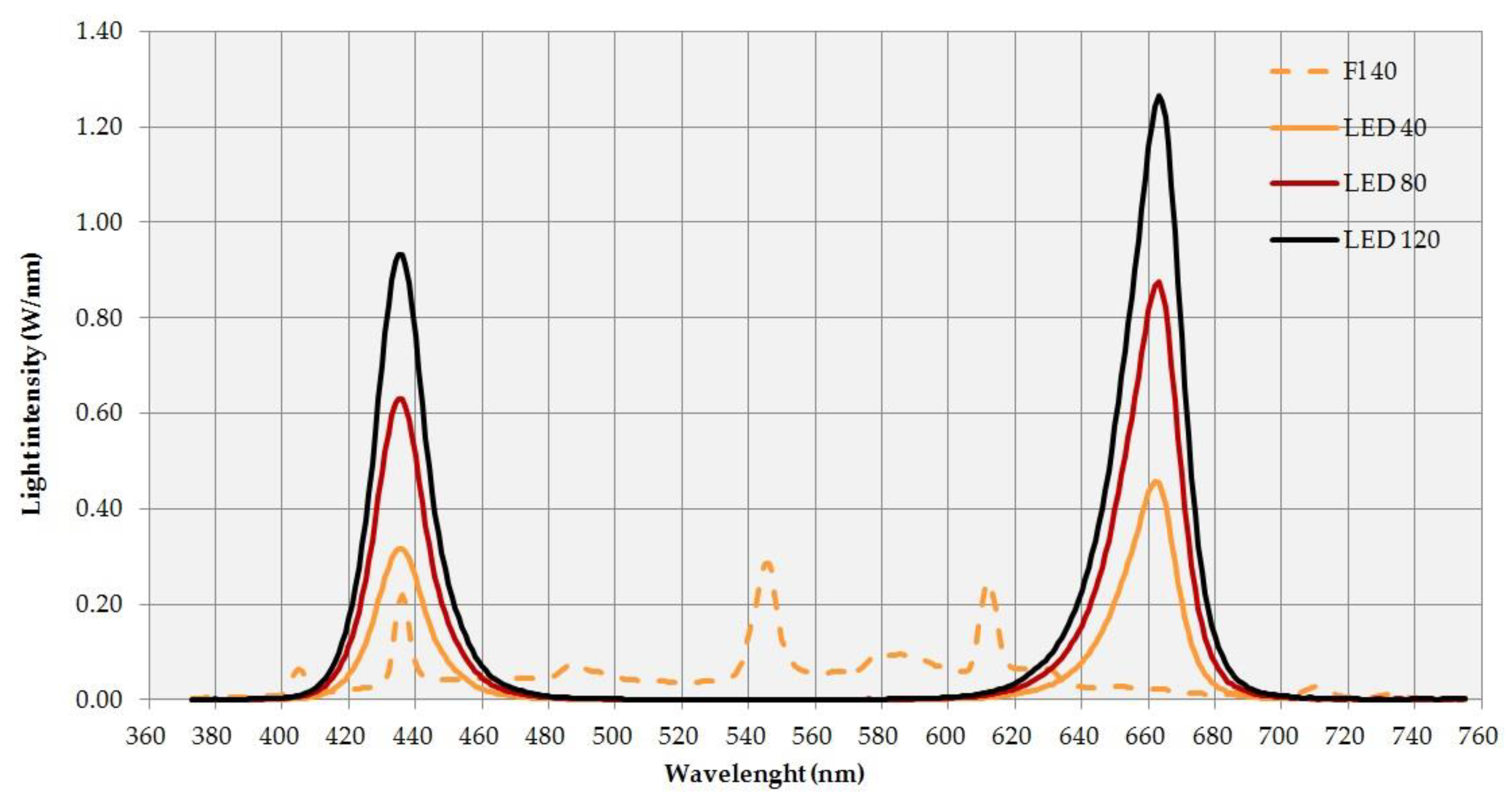
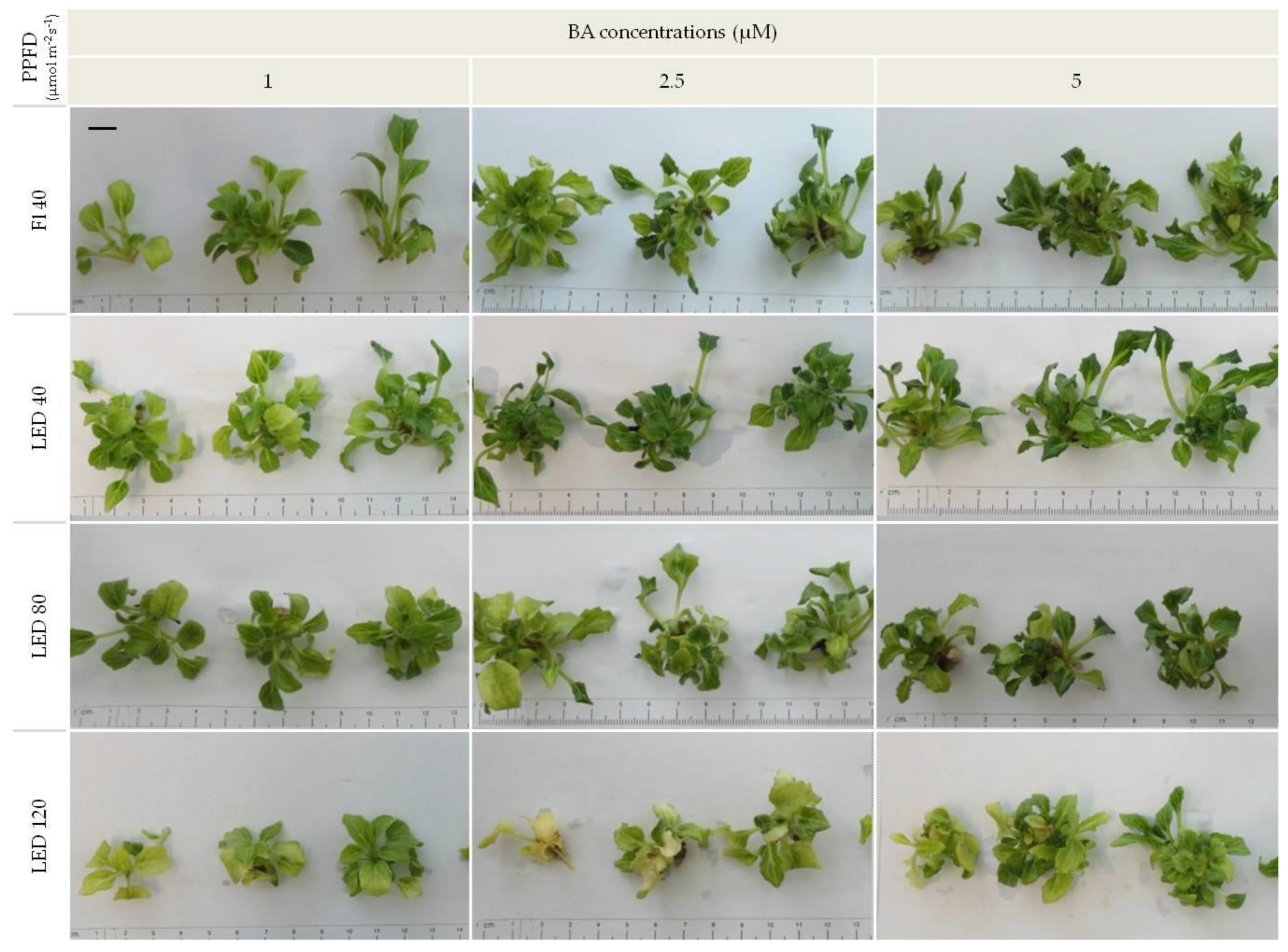
3.1. Light Intensity

Herein, for the first time, the results of experiments where various light intensities emitted by LEDs were studied. In contrast to many other studies where light quality emitted by LEDs were examined, the present research showed that light intensity is also very important for in vitro plant photomorphogenesis. It was demonstrated that these intensities and BA concentrations in the media affected the biometrical properties of plantlets, leaf morphometry, and photosynthetic pigment content during micropropagation of gerbera axillary shoots (Figure 2, Figure 3 and Figure 4, Table 1 and Table 2, Tables S1–S4). It is known that excess light intensity causes light stress in plants. Understanding the effects of these excesses still requires a lot of research. High light intensity stress leads to photodamage of the photosynthetic apparatus and degradation of photosynthetic proteins [29]. Plants respond to this stress with a reduction in chlorophyll levels [2], accumulation of anthocyanins, and a bleaching or yellowing of the leaves [30]. However, controlled increased light intensity in plants in vitro cultures can also enhance the photosynthetic rate and turn on the autotrophic system [31]. Plants can achieve external and internal photoprotection using various mechanisms. They can react to high light intensities by reducing leaf surface area, increasing leaf thickness, adjusting leaf angle or movement, through nonassimilatory electron transport, presence of antioxidants, and through the formation of photoinactivated photosystem II (PS II) centers [29].
Our earlier studies with a standard PPFD level (40 µmol m−2 s−1) confirmed that red and blue light mixture is the most appropriate light quality for gerbera. Such a mixture with the addition of 2% far-red or 20% white light in the 3500 K color temperature emitted by the light-emitting diodes (LEDs) did not improve the examined growth parameters of gerbera [32]. In the present studies, LED light with a PPFD intensity of 120 µmol m−2 s−1 reduced plantlet height. The highest shoots were observed at a PPFD of 40 μmol m−2 s−1, both for LED light (3.5 cm) and for fluorescent lamps (3.8 cm), and this influence was statistically significant in comparison to other treatments. An increase in the height of the plants due to the decrease in the availability of light is related to the apical dominance caused by the increase in auxin levels at the shoot apex. This is connected with shade avoidance syndrome and the etiolation process where other hormones like brassinosteroids and gibberellic acid also participate [33,34].
There was no stimulatory effect of increased light intensity on the shoot multiplication rate, which was 4.9 shoots, on average, per explant at the highest tested PPFD. When the medium contained 5 μM BA, this coefficient was statistically identical in this concentration to the control Fl 40 light and LED 80 (Table 1, Table S1). High light intensity (2.60, 10.10, and 17.40 MJ m−2) stimulated the growth, elongation, and multiplication of shoots as well as the number of sclerenchyma cells and cell wall thickness in Brachiria brizantha (A.Rich.) Stapf and Panicum maximum var. trichoglume (K. Schum.) C.E. Hibbard cultured in a greenhouse [35]. Ocimum gratissimum L. cultured in a herbarium showed a linear increase in shoot biomass production with increased light intensities, and a simultaneous reduction in plant height with intensities higher than 10 μmol m−2 s−1 [36]. The shoot proliferation rate also increased for Castanea sativa Mill. when the PPFD was raised from 50 to 150 μmol m−2 s−1 in in vitro cultures [37]. High light intensities (200 μmol m−2 s−1) emitted by fluorescent lamps inhibited shoot multiplication and decreased their height in in vitro cultures of Momordica grosvenori Swingle, despite the photoautotrophical system (CO2 feeding), and the best growth was observed at 100 µmol m−2 s−1 [38]. An intensity of 60 or 90 μmol m−2 s−1 caused decreased values of shoot length, bulb size, leaf area, and fresh and dry weight in Alocasia ×amasonica in in vitro cultures compared to lower tested PPFD (15 or 30 μmol m−2 s−1) [39]. The highest biometric parameters in cultures of Achillea’s lateral shoots were found at 27 μmol m−2 s−1, and only half of the explants survived at higher intensities [5]. For Gossypium hirsutum L. in vitro, the proliferation of shoots was substantially greater when the explants were cultured in low light (vs. 5 to 60 μmol m−2 s−1) [40]. A lower light intensity of visible light was more efficient for shoot growth of Vitis vinifera L. in vitro (2500 and 5000 lux) [41]. The highest multiplication rate of Plectranthus amboinicus (Lour.) Spreng. shoots in vitro was obtained with PPFD of 69 or 94 µmol m−2 s−1, and it was observed that the height of the shoots decreased with an increase in light intensity from 26 to 130 μmol m−2 s−1 [6]. In contrast, increasing the PPFD from 13 to 69 μmol m−2 s−1 in Hyptis suaveolens L. in in vitro cultures did not affect the multiplication and height of plants [42]. A study on Phoenix dactylifera L. showed that the most shoot buds formed at 1000–1500 lux, while the weakest effects were obtained in the dark and at 2000–3000 lux [43]. Increasing the PPFD to 60 and 120 μmol m−2 s−1 in in vitro ginger cultures stimulated growth, but a further increase to 180 μmol m−2 s−1 inhibited it [44]. Research conducted by Sánchez-Sánchez et al. [45] showed that optimal PPFDs for the best biometrical features of Limonium sintuatum (L.) Mill. in vitro were dependent on the variety. These results were confirmed by Kulchin et al. [46] on Stevia rebaudiana Bertoni and Solanum tuberosum L. using a LED light source and different PPFDs. Subtle differences in light intensity (96.9, 99.6, 101.2, or 102.7 µmol m−2 s−1) may cause various plant responses. High light intensity inhibits shoot proliferation in Gossypium L. in vitro and the response depends on the explant type and plant species or cultivar [40]. Natural habitat requirements affect preferences under controlled growth conditions. We observed that light intensity did not significantly increase gerbera multiplication rate. This could mean that despite some other limitations in development caused by high light intensity, light stress was not a strong inhibiter of gerbera’s ability to multiply, but also failed to improve it.
Our research did not show the effect of light intensity on the number of leaves on gerbera shoots, but light intensity affected some leaf parameters. Leaves show high phenotypic plasticity as a result of changes in light intensity [36,47]. During plant development, leaves perform critical and sensitive roles and acclimatize to various stresses as a response to environmental conditions, and these changes are easier to observe than those in stems or roots [48,49]. In our research the highest PPFD (120 μmol m−2 s−1) increased the leaf blade area and its width but decreased its length and the length of the petiole (Table 2, Table S2). It also had a statistically significant effect on the calculated leaf ratios; the highest circularity and elongation ratios were recorded for leaves under LED 120, and the shape ratio decreased with increasing light intensity. In Figure 4, we illustrated the main effects of light intensity on several ratios calculated for the leaves (Table S4). There was no statistically significant impact of BA on these parameters as well as no interaction effects. Increased leaf area was also observed in Momordica grosvenori cultured in vitro at increasing light intensity from 50 to 100 μmol m−2 s−1. However, the intensity of 200 μmol m−2 s−1 reduced it to the level observed at 25 μmol m−2 s−1 [38]. Similarly, in a study on Plectranthus amboinicus, the largest leaf area was observed at intensities in the range of 51–94 μmol m−2 s−1, while lower or higher ones resulted in its reduction [6]. The study carried out by Xiao et al. [50] showed that average leaf area of gerbera cultured in the photoautotrophic system using large vessels with forced ventilation and high light intensity (100 µmol m−2 s−1) was greater than in the photomixotrophic micropropagation system with natural ventilation and 50 µmol m−2 s−1. High light intensities in in vitro cultures can stimulate plants to form leaves with larger leaf area, making the plant seek a natural, autotrophic state of functioning. In contrast, excessively high light intensities in vivo can cause a decrease in leaf area and even destruction of the photosynthetic system. Moreover, plants have different light intensity requirements under artificial lighting than under natural light [51]. High-intensity light in greenhouse cultivations can increase the number of leaves, but an excessively high intensity reduces their number and their area [36,52]. A larger leaf area increases the photosynthetic surface and improves light absorption by the plant at low light intensities [53]. The results of our experiment showed that in high light intensity, leaves became wider, more corpulent, and their length was inhibited. Therefore, the light intensity was sufficient or the amount of light was excessive. Plants were able to compensate for this by changing the size and shape of their leaves.
The intensity of light did not affect the fresh weight or the dry weight of the gerbera plantlets (Table 1, Table S1). In contrast, an increase in dry weight was observed with an increase in PPFD from 13 to 69 µmol m−2 s−1 in Hyptis suaveolens [42]. The highest dry weight of shoots was recorded in Achillea milefolium at 27 µmol m−2 s−1 [5], while in Momordica grosvenori, the fresh weight content increased with increasing PPFD from 50 to 100 μmol m−2 s−1, and the dry weight content increased even when the PPFD increased to 200 µmol m−2 s−1 [38]. The highest leaf dry weight in Plectranthus amboinicus cultures was observed at a medium light intensity (69 µmol m−2 s−1) [6].
In our study, PPFD of 80 μmol m−2 s−1 increased the content of photosynthetic pigments in gerbera leaves (total chlorophyll content: 0.61 mg g−1 FW and total carotenoid contents: 0.17 mg g−1 fresh weight), but the level of pigments decreased with a further increase in light intensity (up to 0.35 for chlorophylls and 0.10 for carotenoids, on average) (Figure 3, Table S3). A statistically significant reduction in chlorophyll a content in P. amboinicus was observed at 69 µmol m−2 s−1 and higher PPFDs. Higher photosynthetic pigment levels could also be obtained with higher light intensities but only to a certain level [6]. Further increases in light intensity reduced the level of photosynthetic pigments in tissues and caused their degradation. This was also confirmed by a study on ginger, where the level of chlorophyll decreased with an increase in PPFD from 60 to 180 µmol m−2 s−1 [44]. This phenomenon was related to the limited possibility of excess light energy dispersion; this being absorbed by the photosynthetic apparatus without damage [54]. This leads to the interaction between light receptor pigments and oxygen, and as a consequence, may degrade these pigments [55,56]. A higher concentration of carotenoids can prevent chlorophyll photodestruction [56,57]. Changes in the content of photosynthetic pigments are the result of adapting to different light intensities [58].

3.2. Exogenous Cytokinin and Interaction with Light

Previous studies on the effect of light intensity on plant growth in in vitro cultures did not take into account different levels of growth regulators in the medium. In this experiment, we studied the influence of BA cytokinin concentration (1, 2.5, and 5 μM) on the propagation of gerbera shoots, and its increasing concentration was shown to increase the shoot multiplication rate irrespective of light intensity (Table 1, Table S1). This experiment confirmed what is generally known about the influence of BA on increasing number of shoots, but also showed that there is no interaction between light intensity and the BA content in the medium. Raising the light intensity over the standard 40 µmol m−2 s−1 maintained the effect of cytokinin. Cytokinin in the medium is necessary for gerbera multiplication, but its high concentrations (BA above 8 μM) may cause a reduction in multiplication. Different cultivars of gerbera were propagated in a light of 27 to 97.2 µmol m−2 s−1 PPFD [24,32,58]. The analysis of the other biometrical features showed that there was no interaction between light intensity and BA concentration on the number of new leaves. Light intensity affected plant height and it was statistically significant and correlated with BA content at p ≤0.05.
An increased fresh weight of plantlets was observed at higher BA concentrations (2.5 and 5 μM), and the dry weight content decreased with increasing cytokinin concentration. There is no interaction between the tested factors as regards fresh weight, but it showed a strong correlation for dry weight at p ≤0.01, where the impact of BA concentration was especially important. An increase in fresh weight is correlated with the effect of raising multiplication rate due to the raising concentration of BA in the medium. Studies have shown the tendency of BA to promote minimum dry weight. In Rosa ‘Frisco’ in vitro cultures, the highest BA concentration resulted in the lowest dry weight [59].
Univariate analysis showed that leaf blade area was the smallest at 5 μM BA. Cytokinins play an important role in the development and structural differentiation of chloroplasts. They also activate genes involved in their development in in vitro cultures [60]. This is probably why increasing the BA content in the medium resulted in higher total chlorophyll (0.48–0.61 mg g−1 FW) and carotenoid contents (0.14–0.16 mg g−1 FW) in the leaves. However, there was no interaction between the content of the growth regulator and the intensity of light on photosynthetic pigment content or on leaf morphometry parameters.

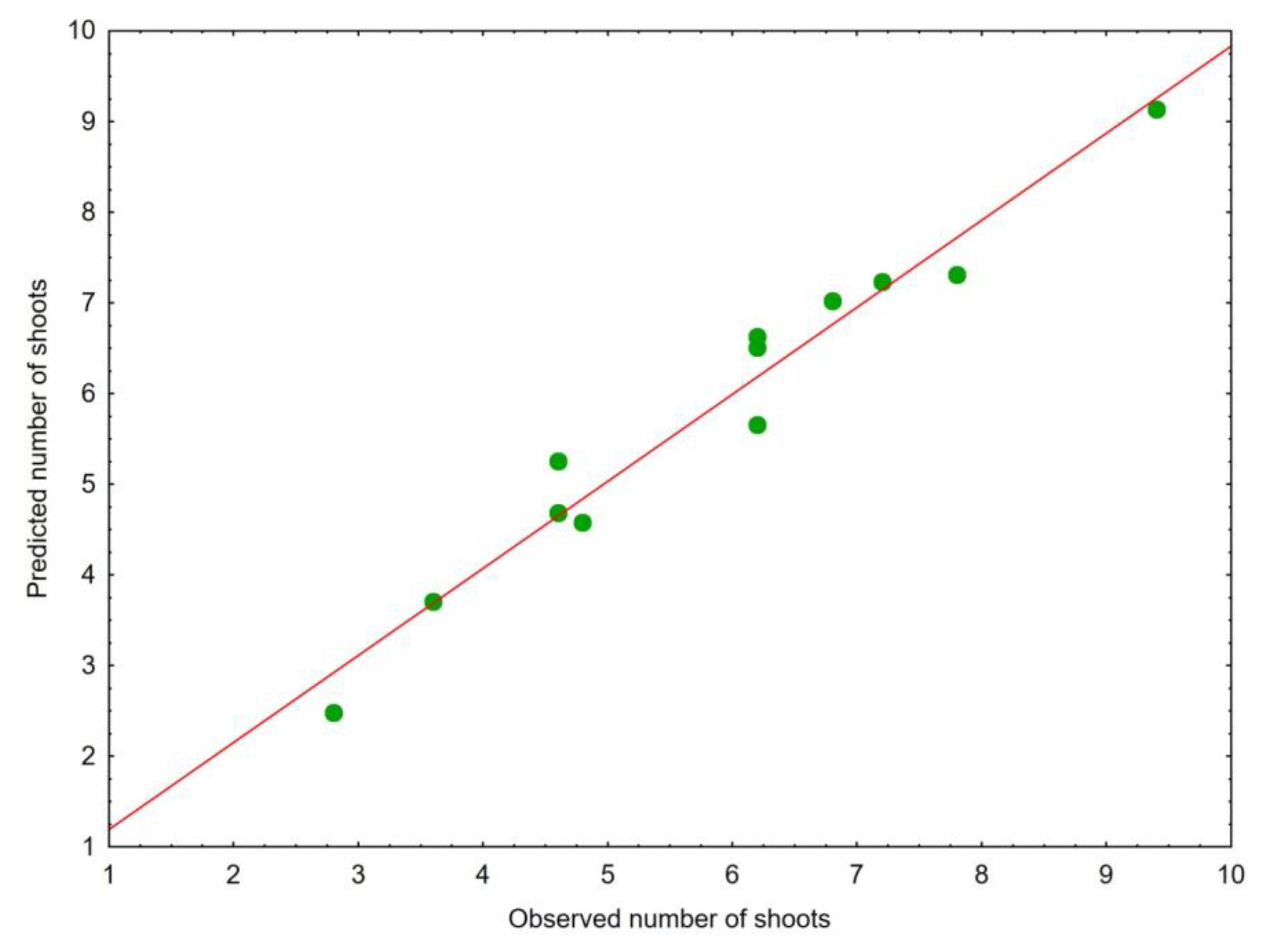
3.3. Predictive Regression Model

A predictive regression model for the number of shoots of multiplied plantlets from morphological descriptors and chlorophyll and carotenoid pigment contents was developed. The equation parameters with descriptive statistics of particular variables are given in Table 3 (Table S5), and it can be observed that six parameters remained from the initial 10 measured plant parameters. Stepwise multiple regression revealed leaf blade area and leaf blade width (R2 = 0.9773 and 0.9672, respectively) to be the most important predictors of the plantlets’ number of shoots, while chlorophyll b appeared to explain a much smaller part of the variation (R2 = 0.3730). The statistical parameters associated with the final step of the multiple regression that represents the best explanatory independent variables were as follows: coefficient of determination for the model, R2 = 0.9598; adjusted coefficient of determination, R2adj. = 0.9117; standard error of estimate (SEE) = 0.5531. The obtained regression model turned out to be significant (F = 19.9174, p = 0.0024). Estimates plotted against the observed values showed a sufficiently precise fit to predict the number of shoots of the multiplied plantlets (Figure 5, Table S6).

3.4. Energy Consumption

The cost of energy consumption, which depends on the type of lamps and the intensity of light in the growth chambers, is an important economic determinant in in vitro cultures. Lighting with standard lamps and the associated cooling energy to remove the wasted heat is the highest cost factor in a culture room. Lighting represents 65% of the entire energy consumption in a culture room when illuminating the in vitro cultures with fluorescent lamps [28]. Additionally, the power requirements of LED lamps are 10–100 times lower than those of conventional lighting sources [19]. The highest optimal PPFD for particular species will allow the best utilization of light by plants and prepare them for intense irradiance of sunlight after acclimatization. Excessively high light intensities adversely affect photomorphogenesis and cause financial losses associated with unnecessary energy consumption, for example, in the form of emitting inefficient light [61]. Each LED panel used in our research consumed the following amount of watts (W) per hour: LED 40 ⇒ 23 W; LED 80 ⇒ 43 W; LED 120 ⇒ 62 W; and control Fl 40 ⇒ 124 W. This was determined by referring the obtained results to 1 m2 of cultivation area in the growth chamber and a 42-day culture cycle where the energy consumption was: LED 40 ⇒1344 W; LED 80 ⇒ 2508 W; LED 120 ⇒ 3617 W; Fl 40 ⇒ 7233 W. This means that LEDs use five times less electrical energy compared to fluorescent lamps with the same intensity level (40 μmol m−2 s−1). At a three-fold increase in PPFD (120 μmol m−2 s−1), the power consumption was half that compared to the control fluorescent lamps. Low energy consumption was also influenced by LED spectrum (red and blue at a ratio of 7:3, respectively), because red LEDs consumed less energy and generated higher economic efficiency compared to blue LEDs at the same level of intensity [55].

4. Conclusions

  • The usage of combination of red (70%) and blue (30%) LEDs in specific light intensities (40–120 µmol m−2 s−1) and different BA concentration in the media (1–5 µM) could be an effective and economical tool for modifying the potency of gerbera shoot multiplication and for controlling plant morphometry and photosynthetic pigment content.
  • Cytokinin in the media influenced the shoot propagation of gerbera; with the increase BA concentration, the shoot multiplication rate intensified in all light intensities. A PPFD of 80 µmol m−2 s−1 and 5 µM BA is suggested as efficient for shoot propagation and economically viable simultaneously.
  • A PPFD of 120 µmol m−2 s−1 decreased plantlet height. The interaction between light intensities and BA concentration was observed in terms of the effect on plant height. With PPFD 80 µmol m−2 s−1, a decreased concentration of BA (1 µM) enhanced the plantlet height, unlike with PPFD 40 µmol m−2 s−1, where the highest plants were noted with 5 µM BA in media.
  • PPFD of 120 µmol m−2 s−1 increased some leaf parameters: blade area and width, and circularity and elongation ratios; the shape ratio decreased with an increasing light intensity.
  • The dry weight content decreased with increasing cytokinin concentration, and the largest was observed in shoots multiplicated on media with 1 µM BA under PPFD µmol 120 m−2 s−1 and under control fluorescent lamps.
  • PPFD 80 µmol m−2 s−1 increased the photosynthetic pigment content in the leaves in comparison to the standard intensity of light. Increased BA concentration raises the content of chlorophyll a.

Supplementary Materials

The following are available online at https://www.mdpi.com/2073-4395/9/7/358/s1, Table S1: Values of the significant effect for the Table 1, Table S2: Values of the significant effect for the Table 2, Table S3: Descriptive statistics for Figure 3, Table S4: One-way analysis of variance for Figure 4, Table S5: Regression model calculations, Table S6: Regression model data for Figure 5.

Author Contributions

Conceptualization, B.P.; Formal analysis, M.C. and A.K.; Investigation, M.C.; Methodology, B.P.; Resources, M.Ż.; Software, M.Ż.; Supervision, B.P.; Visualization, M.C. and A.K.; Writing—original draft, M.C. and A.K.; Writing—review and editing, B.P.

Funding

This work was supported by the Polish Ministry of Science and Higher Education. The funders had no role in study design, data collection and analysis, decision to publish, or preparation of the manuscript.

Acknowledgments

Very special thanks to the Teresa Foszczka in vitro Laboratory (Rzgów, Poland) for supporting with plant material.

Conflicts of Interest

The authors declare no conflict of interest.

References

  1. George, E.F.; Davies, W. Effects of the physical environment. In Plant propagation by Tissue Culture, 3rd ed.; George, E.F., Hall, M.A., de Klerk, G.-J., Eds.; Springer: AA Dordrecht, The Netherlands, 2008; pp. 423–464. [Google Scholar]
  2. Banerjee, A.; Roychoudhury, A. Plant responses to light stress: oxidative damages, photoprotection, and role of phytohormones. In Plant Hormones under Challenging Environmental Factors; Ahammed, G.J., Yu, J.Q., Eds.; Springer: AA Dordrecht, The Netherlands, 2016; pp. 181–214. [Google Scholar] [CrossRef]
  3. Miler, N.; Kulus, D.; Woźny, A.; Rymarz, D.; Hajzer, M.; Wierzbowski, K.; Nelke, R.; Szeffs, L. Application of wide-spectrum light-emitting diodes in micropropagation of popular ornamental plant species: a study on plant quality and cost reduction. Vitr. Cell. Dev. Biol. -Plant 2019, 55, 99–108. [Google Scholar] [CrossRef]
  4. Moe, R.; Grimstad, S.O.; Gislerød, H.R. The use of artificial light in year round production of greenhouse crops in Norway. Acta Hortic. 2006, 711, 35–42. [Google Scholar] [CrossRef]
  5. Alvarenga, I.C.A.; Pacheco, F.V.; Silva, S.T.; Bertolucci, S.K.V.; Pinto, J.E.B.P. In vitro culture of Achillea millefolium L.: Quality and intensity of light on growth and production of volatiles. Plant Cell, Tissue Organ Cult. 2015, 122, 299–308. [Google Scholar] [CrossRef]
  6. Silva, S.T.; Bertolucci, S.K.V.; da Cunha, S.H.B.; Lazzarini, L.E.S.; Tavares, M.C.; Pinto, J.E.B.P. Effect of light and natural ventilation systems on the growth parameters and carvacrol content in the in vitro cultures of Plectranthus amboinicus (Lour.) Spreng. Plant Cell, Tissue Organ Cult. 2017, 129, 501–510. [Google Scholar] [CrossRef]
  7. Soontornchainaksaeng, P.; Chaicharoen, S.; Sirijuntarut, M.; Kruatrachue, M. In vitro studies on the effect of light intensity on plant growth of Phaius tankervilliae (Banks ex L ’ Herit.) Bl. and Vanda coerulea Griff. Sci. Asia 2001, 27, 233–237. [Google Scholar] [CrossRef]
  8. Liu, C.; Guo, C.; Wang, Y.; Ouyang, F. Effect of light irradiation on hairy root growth and artemisinin biosynthesis of Artemisia annua L. Process Biochem. 2002, 38, 581–585. [Google Scholar] [CrossRef]
  9. Morelli, G.; Ruberti, I. Signaling shade avoidance responses. Driving auxin along lateral routes. Plant Physiol. 2000, 122, 621–626. [Google Scholar] [CrossRef]
  10. Folta, K.M.; Childers, K.S. Light as a growth regulator: Controlling plant biology with narrow-bandwidth solid-state lighting systems. HortScience 2008, 43, 1957–1964. [Google Scholar] [CrossRef]
  11. Moon, H.K.; Park, S.-Y.; Kim, Y.W.; Kim, C.S. Growth of Tsuru-rindo (Tripterospermum japonicum) cultured in vitro under various sources of light-emitting diode (LED) irradiation. J. Plant Biol. 2006, 49, 174–179. [Google Scholar] [CrossRef]
  12. Nhut, D.T.; Takamura, T.; Watanabe, H.; Okamoto, K.; Tanaka, M. Responses of strawberry plantlets cultured in vitro under superbright red and blue light-emitting diodes (LEDs). Plant Cell, Tissue Organ Cult. 2003, 73, 43–52. [Google Scholar] [CrossRef]
  13. Silva, M.M.A.; De Oliveira, A.L.B.; Oliveira-Filho, R.A.; Gouveia-Neto, A.S.; Camara, T.J.R.; Willadino, L.G. Effect of blue/red LED light combination on growth and morphogenesis of Saccharum officinarum plantlets in vitro. Prog. Biomed. Opt. Imaging - Proc. SPIE 2014, 89471. [Google Scholar] [CrossRef]
  14. OuYang, F.; Mao, J.-F.; Wang, J.; Zhang, S.; Li, Y. Transcriptome analysis reveals that red and blue light regulate growth and phytohormone metabolism in Norway Spruce [Picea abies (L.) Karst.]. PLOS One 2015, 10, 1–19. [Google Scholar] [CrossRef] [PubMed]
  15. Suzuki, R.M.; Kerbauy, G.B. Effects of light and ethylene on endogenous hormones and development of Catasetum fimbriatum (Orchidaceae). Brazilian J. Plant Physiol. 2006, 18, 359–365. [Google Scholar] [CrossRef]
  16. Aksenova, N.P.; Konstantinova, T.N.; Sergeeva, L.I.; Macháčková, I.; Golyanovskaya, S.A. Morphogenesis of potato plants in vitro. I. Effect of light quality and hormones. J. Plant Growth Regul. 1994, 13, 143–146. [Google Scholar] [CrossRef]
  17. Karnachuk, R.A.; Tishchenko, S.Y.; Golovatskaya, I.F. Endogenous phytohormones and regulation of morphogenesis of Arabidopsis thaliana by blue light. Russ. J. Plant Physiol. 2001, 48, 226–230. [Google Scholar] [CrossRef]
  18. Bourget, C.M. An introduction to light-emitting diodes. HortScience 2008, 43, 1944–1946. [Google Scholar] [CrossRef]
  19. Dutta Gupta, S. Light Emitiing Diodes for Agriculture-Smart Lighting, 1st ed.; Springer: Singapore, 2017. [Google Scholar] [CrossRef]
  20. Lin, K.H.; Huang, M.Y.; Huang, W.D.; Hsu, M.H.; Yang, Z.W.; Yang, C.M. The effects of red, blue, and white light-emitting diodes on the growth, development, and edible quality of hydroponically grown lettuce (Lactuca sativa L. var. capitata). Sci. Hortic. (Amsterdam) 2013, 150, 86–91. [Google Scholar] [CrossRef]
  21. Hanus-Fajerska, E.; Wojciechowska, R. Impact of Light-Emitting Diodes (LEDs) on propagation of orchids in tissue culture. In Light Emitting Diodes for Agriculture; Gupta, S.D., Ed.; Springer: Singapore, 2017; pp. 305–320. [Google Scholar]
  22. Viršilė, A.; Olle, M.; Duchovskis, P. LED Lighting in Horticulture. In Light emitiing diodes for agriculture - smart lighting, 1st ed.; Gupta, S., Ed.; Springer: Singapore, 2017; pp. 113–147. [Google Scholar] [CrossRef]
  23. Morrow, R.C. LED lighting in horticulture. HortScience 2008, 43, 1947–1950. [Google Scholar] [CrossRef]
  24. Minerva, G.; Kumar, S. Micropropagation of Gerbera (Gerbera jamesonii Bolus). In Protocols for Micropropagation of Selected Economically-Important Horticultural Plants; Lambardi, M., Ozudogru, E.A., Jain, S.M., Eds.; Springer: New York, NY, USA, 2013; pp. 305–316. [Google Scholar] [CrossRef]
  25. Sevelius, N. Photosynthetic features of three gerbera cultivars in low light. Acta Hortic. 2003, 624, 297–302. [Google Scholar] [CrossRef]
  26. Keditsu, R. Performance of open field Gerbera in resposne to planting time. African J. Agri. Res 2013, 8, 3068–3074. [Google Scholar] [CrossRef]
  27. Murashige, T.; Skoog, F. A revised medium for rapid growth and bio assays with tabacco tissue cultures. Physiol. Plant. 1962, 15, 473–497. [Google Scholar] [CrossRef]
  28. Bornwaßer, T.; Tantau, H.J. Evaluation of LED lighting systems in in vitro cultures. Acta Hortic. 2012, 956, 555–562. [Google Scholar] [CrossRef]
  29. Robinson, A.S. Plant Light Stress. Encycl. Life Sci. 2001, 1–6. [Google Scholar] [CrossRef]
  30. Singh, A.S.; Jones, A.M.P.; Shukla, M.R.; Saxena, P.K. High light intensity stress as the limiting factor in micropropagation of sugar maple (Acer saccharum Marsh.). Plant Cell. Tissue Organ Cult. 2017, 129, 209–221. [Google Scholar] [CrossRef]
  31. Wolf, S.; Kalman-Rotem, N.; Yakir, D.; Ziv, M. Autotrophic and heterotrophic carbon assimilation of in vitro grown potato (Solanum tuberosum L.) plants. J. Plant Physiol. 1998, 153, 574–580. [Google Scholar] [CrossRef]
  32. Pawłowska, B.; Żupnik, M.; Szewczyk-Taranek, B.; Cioć, M. Impact of LED light sources on morphogenesis and levels of photosynthetic pigments in Gerbera jamesonii grown in vitro. Hortic. Environ. Biotechnol. 2018, 59, 115–123. [Google Scholar] [CrossRef]
  33. Vanneste, S.; Friml, J. Auxin: A trigger for change in plant development. Cell 2009, 136, 1005–1016. [Google Scholar] [CrossRef]
  34. Symons, G.M.; Reid, J.B. Hormone levels and response during de-etiolation in pea. Planta 2003, 216, 422–431. [Google Scholar] [CrossRef]
  35. Deinum, B.; Sulastri, R.D.; Zeinab, M.H.J.; Maassen, A. Effects of light intensity on growth, anatomy and forage quality of two tropical grasses (Brachiaria brizantha and Panicum maximum var. trichoglume). Netherlands J. Agric. Sci. 1996, 44, 111–124. [Google Scholar]
  36. Fernandes, V.F.; de Almeida, L.B.; Feijó, E.V.R.; Silva, D.D.; Oliveira, R.A.; Mielke, M.S.; Costa, L.C. Light intensity on growth, leaf micromorphology and essential oil production of Ocimum gratissimum. Brazilian J. Pharmacogn. 2013, 23, 419–424. [Google Scholar] [CrossRef]
  37. Sáez, P.L.; Bravo, L.A.; Latsague, M.I.; Sánchez, M.E.; Ríos, D.G. Increased light intensity during in vitro culture improves water loss control and photosynthetic performance of Castanea sativa grown in ventilated vessels. Sci. Hortic. (Amsterdam). 2012, 138, 7–16. [Google Scholar] [CrossRef]
  38. Zhang, M.; Zhao, D.; Ma, Z.; Li, X.; Xiao, Y. Growth and photosynthetic capability of Momordica grosvenori plantlets grown photoautotrophically in response to light intensity. HortScience 2009, 44, 757–763. [Google Scholar] [CrossRef]
  39. Jo, E.A.; Tewari, R.K.; Hahn, E.J.; Paek, K.Y. Effect of photoperiod and light intensity on in vitro propagation of Alocasia amazonica. Plant Biotechnol. Rep. 2008, 2, 207–212. [Google Scholar] [CrossRef]
  40. Gupta, S.K.; Singh, P.K.; Sawant, S.V.; Chaturvedi, R.; Tuli, R. Effect of light intensity on in vitro multiple shoot induction and regeneration of cotton (Gossypium hirsutum L. cv Khandawa-2). Indian J. Exp. Biol. 2000, 38, 399–401. [Google Scholar] [PubMed]
  41. Fallah, F.; Kahrizi, D. Effect of Light spectrum and intensity on growth of grape (Vitis vinifera) under in vitro conditions. Orig. Artic. J. Appl. Biotechnol. Rep. Autumn 2016, 3, 495–499. [Google Scholar]
  42. Andrade, H.B.; Braga, A.F.; Bertolucci, S.K.V.; Hsie, B.S.; Silva, S.T.; Pinto, J.E.B.P. Effect of plant growth regulators, light intensity and LED on growth and volatile compound of Hyptis suaveolens (L.) Poit in vitro plantlets. Acta Hortic. 2017, 1155, 277–284. [Google Scholar] [CrossRef]
  43. Meziani, R.; Jaiti, F.; Mazri, M.A.; Anjarne, M.; Chitt, M.A.; El Fadile, J.; Alem, C. Effects of plant growth regulators and light intensity on the micropropagation of date palm (Phoenix dactylifera L.) cv. Mejhoul. J. Crop Sci. Biotechnol. 2015, 18, 325–331. [Google Scholar] [CrossRef]
  44. Zhou, M.; Guan, Q.; Wei, Y.; Zhang, Z. Effects of sucrose concentration and light intensity on growth and photosynthesis of ginger plantlets in vitro. Chinese J. Appl. Environ. Biol. 2008, 14, 356–361. [Google Scholar]
  45. Sánchez-Sánchez, J.; López-Orenes, A.; Ferrer, M.A.; Calderón, A.A. Subtle changes in light intensity affect in vitro responses but not ex vitro performance of Limonium sinuatum. 3 Biotech 2018, 8, 1–7. [Google Scholar] [CrossRef]
  46. Kulchin, Y.N.; Nakonechnaya, O.V.; Gafitskaya, I.V.; Grishchenko, O.V.; Epifanova, T.Y.; Orlovskaya, I.Y.; Zhuravlev, Y.N.; Subbotin, E.P. Plant morphogenesis under different light intensity. Defect Diffus. Forum 2018, 386, 201–206. [Google Scholar] [CrossRef]
  47. Valladares, F.; Niinemets, Ü. Shade tolerance, a key plant feature of complex nature and consequences. Annu. Rev. Ecol. Evol. Syst. 2008, 39, 237–257. [Google Scholar] [CrossRef]
  48. Nevo, E.; Pavlíek, T.; Beharv, A.; Bolshakova, M.A.; Martyn, G.I.; Musatenko, L.I.; Sytnikl, K.M. Drought and light anatomical adaptive leaf strategies in three woody species caused by microclimatic selection at “Evolution Canyon”, Israel. Isr. J. Plant Sci. 2000, 48, 33–46. [Google Scholar] [CrossRef]
  49. Guerfel, M.; Baccouri, O.; Boujnah, D.; Chaïbi, W.; Zarrouk, M. Impacts of water stress on gas exchange, water relations, chlorophyll content and leaf structure in the two main Tunisian olive (Olea europaea L.) cultivars. Sci. Hortic. (Amsterdam). 2009, 119, 257–263. [Google Scholar] [CrossRef]
  50. Xiao, Y.; He, L.; Liu, T.; Yang, Y. Growth promotion of gerbera plantlets in largevessels by using photoautotrophic micropropagation system with forced ventilation. Propag. Ornam. Plants 2005, 5, 179–185. [Google Scholar]
  51. Yao, X.Y.; Liu, X.Y.; Xu, Z.G.; Jiao, X.L. Effects of light intensity on leaf microstructure and growth of rape seedlings cultivated under a combination of red and blue LEDs. J. Integr. Agric. 2017, 16, 97–105. [Google Scholar] [CrossRef]
  52. Kubota, S.; Yoneda, K. Effect of light intensity preceding day-night temperatures on the sensitivity of Phalaenopsis to flower (Japanese). J. Japanese Soc. Hortic. Sci. 1993, 62, 595–600. [Google Scholar] [CrossRef]
  53. Lambers, H.; Chapin, F.S.; Pons, T.L. Plant Physiological Ecology, 2nd ed.; Springer: New York, NY, USA, 2008. [Google Scholar] [CrossRef]
  54. Takahashi, S.; Murata, N. How do environmental stresses accelerate photoinhibition? Trends Plant Sci. 2008, 13, 178–182. [Google Scholar] [CrossRef] [PubMed]
  55. Darko, E.; Heydarizadeh, P.; Schoefs, B.; Sabzalian, M.R. Photosynthesis under artificial light: The shift in primary and secondary metabolism. Philos. Trans. R. Soc. B Biol. Sci. 2014, 369, 1–7. [Google Scholar] [CrossRef]
  56. Franck, F.; Schoefs, B.; Barthélemy, X.; Myśliwa-Kurdziel, B.; Strzałka, K.; Popovic, R. Protection of native chlorophyll(ide) forms and of photosystem II against photodamage during early stages of chloroplast differentiation. Acta Physiol. Plant. 1995, 17, 123–132. [Google Scholar]
  57. Bartley, G.E.; Scolnik, P.A. Plant carotenoids: Pigments for photoprotection, visual attraction, and human health. Plant Cell 1995, 7, 1027–1038. [Google Scholar] [CrossRef]
  58. Gonçalves, J.F.; Barreto, D.C.; Santos Junior, U.M.; Fernandes, A.V.; Sampaio, P.D.; Buckeridge, M.S. Growth, photosynthesis and stress indicators in young rosewood plants (Aniba rosaeodora Ducke) under different light intensities. Brazilian J. Plant Physiol. 2005, 17, 325–334. [Google Scholar] [CrossRef]
  59. Mahmood, S.; Reza, R.; Hossain, G.; Hauser, B. Response of cytokinins on in vitro shoot multiplication of rose cv. Frisco. Res. Rev. 2017, 6–11. [Google Scholar]
  60. Dobránszki, J.; Mendler-Drienyovszki, N. Cytokinin-induced changes in the chlorophyll content and fluorescence of in vitro apple leaves. J. Plant Physiol. 2014, 171, 1472–1478. [Google Scholar] [CrossRef] [PubMed]
  61. Ruban, A.V.; Belgio, E. The relationship between maximum tolerated light intensity and photoprotective energy dissipation in the photosynthetic antenna: chloroplast gains and losses. Philos. Trans. R. Soc. B Biol. Sci. 2014, 369, 1–9. [Google Scholar] [CrossRef] [PubMed]
Figure 1. Tested light spectra during in vitro cultures of Gerbera jamesonii. Fl 40—control, fluorescent Philips TK-D 36W/54 lamps. LED 40, LED 80, LED 120—light-emitting diodes (red and blue 7:3) with photosynthetic photon flux densities (PPFDs) of 40, 80, or 120 µmol m−2 s−1, respectively. LED—light-emitting diode.
Agronomy 09 00358 g001
Figure 2. Gerbera jamesonii plantlets after six weeks of in vitro culture on Murashige and Skoog (MS) medium with various 6-benzyladenine (BA) concentrations and different photosynthetic photon flux densities (PPFDs). Fl 40—control, fluorescent Philips TK-D 36W/54 lamps. LED 40, LED 80, LED 120—combination of red (70%) and blue (30%) light emitted by light-emitting diodes with 40, 80, and 120 µmol m−2 s−1 PPFD, respectively.
Agronomy 09 00358 g002
Figure 3. Photosynthetic pigment contents in Gerbera jamesonii leaves after six weeks of in vitro culture on Murashige and Skoog (MS) medium with various 6-benzyladenine (BA) concentrations and different photosynthetic photon flux densities (PPFDs). (A) Chlorophyll a content. (B) Chlorophyll b content. (C) Carotenoid contents. (D) The ratio of chlorophyll to carotenoid contents. Fl 40—control, fluorescent Philips TK-D 36W/54 lamps. LED 40, LED 80, LED 120—combination of red (70%) and blue (30%) light emitted by light-emitting diodes with 40, 80, and 120 µmol m−2 s−1 PPFD, respectively. Error bars represent ± standard errors.
Agronomy 09 00358 g003
Figure 4. Circularity, elongation, and shape ratios after six weeks of in vitro culture on Murashige and Skoog (MS) medium averaged for various 6-benzyladenine (BA) concentrations as depending on different photosynthetic photon flux densities (PPFDs). Fl 40—control, fluorescent Philips TK-D 36W/54 lamps; LED 40, LED 80, LED 120—combination of red (70%) and blue (30%) light emitted by light-emitting diodes with 40, 80, and 120 µmol m−2 s−1 PPFD, respectively. Different letters indicate significantly different values following Duncan’s multiple range test at p ≤0.05. Error bars represent ± standard errors.
Agronomy 09 00358 g004
Figure 5. Observed number of shoots of multiplied plantlets versus predicted data generated by the developed regression model.
Agronomy 09 00358 g005
Table 1. Biometrical properties of growth and fresh and dry weight of Gerbera jamesonii after six weeks of in vitro culture on Murashige and Skoog (MS) medium with various 6-benzyladenine (BA) concentrations and different photosynthetic photon flux densities PPFDs. Significant effect: **, p ≤0.05; ***, p ≤0.01; n.s.—not significant. Fl 40—control, fluorescence Philips TK-D 36W/54 lamps. LED 40, LED 80, LED 120—combination of red (70%) and blue (30%) light emitted by light-emitting diodes with 40, 80, and 120 µmol m−2 s−1 PPFD, respectively. Means ± standard deviations within a column followed by the same letter are not significantly different according to Duncan’s multiple range test at p ≤0.05.
PPFD
(µmol m−2 s−1)
BA
(µM)
Biometric Features
Multiplication RatePlantlet Height
(cm)
Number of Leaves per ShootFresh Weight of Plantlet (g)Dry Weight (%)
Fl 4014.6 ± 0.9 a–c4.0 ± 0.9 d4.6 ± 0.9 ab0.81 ± 0.15 a–c8.91 ± 1.15 cd
2.56.8 ± 0.8 c–e3.8 ± 0.8 cd4.2 ± 0.3 a1.11 ± 0.20 b–e7.04 ± 0.07 ab
59.4 ± 3.2 e3.7 ± 0.5 cd5.1 ± 1.3 ab1.54 ± 0.33 e6.40 ± 0.15 a
LED 4014. 8 ± 2.2 a–c2.9 ± 0.8 a–c5.0 ± 0.8 ab0.70 ± 0.23 ab7.59 ± 0.67 a–c
2.56.2 ± 1.8 b–d3.6 ± 0.4 cd4.9 ± 0.3 ab1.17 ± 0.45 c–e8.29 ± 2.27 b–d
56.2 ± 1.3 b–d4.1 ± 0.5 d5.9 ± 1.4 b1.18 ± 0.27 c–e6.25 ± 0.43 a
LED 8013.6 ± 1.1 ab3.6 ± 0.4 b–d5.2 ± 0.3 ab0.88 ± 0.15 a–c7.11 ± 0.36 ab
2.56.2 ± 1.9 b–d3.0 ± 0.5 a–c5.4 ± 1.8 ab1.37 ± 0.32 de6.89 ± 0.14 ab
57.8 ± 3.5 de2.9 ± 0.7 a–c4.8 ± 0.6 ab1.26 ± 0.44 c–e6.65 ± 0.42 a
LED 12012.8 ± 1.3 a2.2 ± 0.9 a6.0 ± 1.5 b0.64 ± 0.24 a9.31 ± 0.30 d
2.54.6 ± 1.1 a–c2.7 ± 0.4 ab5.6 ± 1.0 ab0.98 ± 0.19 a–d6.81 ± 0.68 ab
57.2 ± 2.6 c–e2.6 ± 0.6 a5.4 ± 0.5 ab1.20 ± 0.53 c–e6.58 ± 0.63 a
Means for PPFD
Fl 40 6.9 ± 2.7 b3.8 ± 0.7 c4.6 ± 0.9 a1.15 ± 0.38 a7.45 ± 1.27 a
LED 40 5.7 ± 1.8 ab3.5 ± 0.7 bc5.3 ± 1.0 ab1.02 ± 0.38 a7.38 ± 1.50 a
LED 80 5.9 ± 2.9 ab3.2 ± 0.6 b5.1 ± 1.0 ab1.17 ± 0.37 a6.88 ± 0.35 a
LED 120 4.9 ± 2.5 a2.5 ± 0.6 a5.7 ± 1.0 b0.94 ± 0.40 a7.57 ± 1.40 a
Means for BA
14.0 ± 1.6 a3.2 ± 1.0 a5. 2 ± 1.0 a0.76 ± 0.20 a8.23 ± 1.12 c
2.56.0 ± 1.6 b3.3 ± 0.7 a5.0 ± 1.1 a1.15 ± 0.32 b7.26 ± 1.19 b
57.7 ± 2.8 c3.3 ± 0.8 a5.3 ± 1.0 a1.30 ± 0.40 b6.47 ± 0.41 a
Source of variation
PPFD × BAn.s.**n.s.n.s.***
PPFD n.s.***n.s.n.s.n.s.
BA ***n.s.n.s. **
Table 2. Leaf morphometry of Gerbera jamesonii after six weeks of in vitro culture on Murashige and Skoog (MS) medium with various 6-benzyladenine (BA) concentrations and different photosynthetic photon flux densities PPFDs. Significant effect: **, p ≤0.05; ***, p ≤0.01; n.s. not significant. Fl 40—control, fluorescence Philips TK-D 36W/54 lamps. LED 40, LED 80, LED 120—combination of red (70%) and blue (30%) light emitted by light-emitting diodes with 40, 80 and 120 µmol m−2 s−1 PPFD, respectively. Means ± standard deviations within a column followed by the same letter are not significantly different according to Duncan’s multiple range test at p ≤0.05.
PPFD
(µmol m−2 s−1)
BA
(µM)
Leaf bladePetiole
Area (mm2)Perimeter (mm)Length (mm)Width (mm)Length (mm)
Fl 40176.91 ± 37.50 bc71.36 ± 24.51 b30.04 ± 10.68 e6.70 ± 2.19 b–d19.64 ± 7.96 bc
2.559.36 ± 22.42 ab68.99 ± 20.81 b28.60 ± 8.83 c–e5.88 ± 1.81 ab21.95 ± 8.46 c
546. 30 ± 35.29 a55.58 ± 21.52 a22.62 ± 8.83 ab4.62 ± 3.00 a15.92 ± 7.80 b
LED 40171.33 ± 29.78 ab66.04 ± 18.62 ab26.97 ± 7.91 b–e6.75 ± 1.78 b–d17.22 ± 6.18 b
2.582.20 ± 28.96 bc67.94 ± 13.52 ab26.81 ± 5.93 b–e7.46 ± 2.07 b–d16.75 ± 5.21 b
561.98 ± 27.13 ab61.93 ± 20.06 ab23.92 ± 8.63 a–d6.19 ± 1.54 bc16.19 ± 7.57 b
LED 801132.81 ± 47.44 e74.23 ± 22.75 b29.15 ± 9.35 de9.50 ± 3.59 fg17.04 ± 5.39 b
2.598.00 ± 35.18 cd70.02 ± 14.44 b27.93 ± 6.24 b–e8.32 ± 2.44 d–f16.58 ± 4.43 b
578.32 ± 25.15 bc64.67 ± 16.78 ab25.13 ± 7.37 a–e7.63 ± 1.60 c–e16.91 ± 5.40 b
LED 1201132.64 ± 63.48 e63.44 ± 12.82 ab24.38 ± 3.94 a–d10.15 ± 3.23 g10.58 ± 2.56 a
2.5108.61 ± 44.88 ed61.14 ± 10.65 ab23.54 ± 4.25 a–c9.10 ± 2.49 e–g10.22 ± 3.20 a
581.06 ± 30.34 bc55.07 ± 10.14 a21.18 ± 4.31 a7.85 ± 1.66 c–e10.35 ± 3.46 a
Means for PPFD
Fl 40 60.86 ± 34.29 a65.31 ± 23.04 ab27.08 ± 9.87 b5.73 ± 2.50 a19.17 ± 8.32 c
LED 40 71.84 ± 29.36 a65.30 ± 17.51 ab25.90 ± 7.58 b6.80 ± 1.86 b16.72 ± 6.30 b
LED 80 103.04 ± 42.93 b69.64 ± 18.44 b27.40 ± 7.81 b8.48 ± 2.74 c16.84 ± 5.01 b
LED 120 107.44 ± 51.91 b59.88 ± 11.63 a23.04 ± 4.32 a9.03 ± 2.67 c10.38 ± 3.05 a
Means for BA
1103.43 ± 54.18 c68.77 ± 20.26 b27.63 ± 8.49 b8.27 ± 3.16 b16.00 ± 6.65 a
2.587.04 ± 38.11 b67.02 ± 15.42 b26.72 ± 6.69 b7.69 ± 2.49 b16.49 ± 6.97 a
566.92 ± 32.36 a59.31 ± 17.82 a23.21 ± 7.51 a6.57 ± 2.39 a14.84 ± 6.73 a
Source of variation
PPFD × BA n.s.n.s.n.s.n.s.n.s.
PPFD **************
BA ************n.s.
Table 3. Estimated equation parameters and statistics for stepwise (backward elimination) multiple regression performed for the multiplied plantlets’ number of shoots.
VariableEquation ParametersStandard Errorst Statistic Valuesp
Intercept27.66004.87665.67200.0024
Fresh weight of one shoot (g)55.050514.14783.89110.0115
Number of leaves per shoots−3.17920.6716−4.73400.0052
Leaf area (cm2)−0.18010.0403−4.46620.0066
Leaf perimeter (cm)−0.27350.0497−5.50590.0027
Leaf width (cm)2.05660.57463.57890.0159
Chlorophyll b (mg g-1 fresh weight)12.22954.61242.65150.0454
p—probability level; Model parameters: R2 = 0.9598; R2adj. = 0.9117; SEE = 0.5531; p = 0.0024; R2—coefficient of determination; R2adj.—adjusted coefficient of determination; SEE—standard error of the estimate.

