Review

Coordination of Cryptochrome and Phytochrome Signals in the Regulation of Plant Light Responses

1
Basic Forestry and Proteomics Research Center, Fujian Agriculture and Forestry University, Fuzhou 350002, Fujian, China
2
College of Forestry, Fujian Agriculture and Forestry University, Fuzhou 350002, Fujian, China
3
College of Life Science, Fujian Agriculture and Forestry University, Fuzhou 350002, Fujian, China
4
Department of Molecular, Cell & Developmental Biology, University of California, Los Angeles, CA 90095, USA
*
Author to whom correspondence should be addressed.
Agronomy 2017, 7(1), 25; https://doi.org/10.3390/agronomy7010025
Submission received: 30 January 2017 / Revised: 13 March 2017 / Accepted: 13 March 2017 / Published: 21 March 2017
(This article belongs to the Special Issue Impacts of Photoreceptors on Plant Growth and Development)

Abstract

:
In nature, plants integrate a wide range of light signals from solar radiation to adapt to the surrounding light environment, and these light signals also regulate a variety of important agronomic traits. Blue light-sensing cryptochrome (cry) and red/far-red light-sensing phytochrome (phy) play critical roles in regulating light-mediated physiological responses via the regulated transcriptional network. Accumulating evidence in the model plant Arabidopsis has revealed that crys and phys share two mechanistically distinct pathways to coordinately regulate transcriptional changes in response to light. First, crys and phys promote the accumulation of transcription factors that regulate photomorphogenesis, such as HY5 and HFR1, via the inactivation of the CONSTITUTIVE PHOTOMORPHOGENIC1/SUPPRESSOR OF PHYA-105 E3 ligase complex by light-dependent binding. Second, photoactive crys and phys directly interact with PHYTOCHROME INTERACTING FACTOR transcription factor family proteins to regulate transcriptional activity. The coordinated regulation of these two pathways (and others) by crys and phys allow plants to respond with plasticity to fluctuating light environments in nature.

1. Introduction

Because plant life relies on solar radiation as an energy source, plants have acquired a light-sensing mechanism to maximize the availability of light for photosynthesis [1,2]. Specifically, germination does not occur in deep subterranean darkness and occurs only when the plant senses a low amount of light under the ground at depths close to the surface of the soil. The post-germinative seedlings in the soil elongate the hypocotyl with a closed hook, and yellowish cotyledons move toward the surface of soil. Upon reaching the surface and absorbing light, seedlings start photomorphogenic development, in which hypocotyl elongation ceases, the hook and cotyledons start to open, and chloroplasts begin developing to maximize light capture and autotrophic growth. This developmental transition from a dark-grown seedling to a light-grown seedling is called de-etiolation. Even aboveground plants must sense the presence and proximity of neighboring plants and compete with them for solar energy through a response called Shade Avoidance Syndrome (SAS). Plants detect shade from neighboring plants as alterations in the light quantity and quality, and, in response, they elongate the hypocotyl, petiole, and internode to bring the photosynthetic organs to more favorable conditions for photosynthesis. Plants also perceive the day length to control the transition from the vegetative phase to reproductive phase.
As exemplified above, light as an environmental signal regulates plant development throughout the plant life cycle. The blue (B) and red (R) light regions of the light spectrum are most efficiently utilized for the photosynthesis; thus, B light-sensing cryptochrome (cry) and R light-absorbing phytochrome (phy) play major roles in regulating plant light responses, such as light-dependent seed germination, de-etiolation, SAS and photoperiodic flowering [3,4,5]. Importantly, crys or phys also mediate important agronomic traits in crop species [4,5,6]. For example, rice mutants defective in all phy proteins set very few seeds [7]. Reduced expression of CRY1a and CRY1b in barley impairs seed dormancy [8]. A decreased level of CRY2a expression accelerates senescence in soybean [9]. All these examples indicate the importance of cry and phy action in regulating the growth and quality of crops. Great progress has been made in understanding the action mechanisms of crys and phys in the model plant Arabidopsis [5,10,11]. In this article, we review the mechanisms of action of cry and phy in Arabidopsis and discuss how cry and phy coordinate to regulate light responses. The findings on the coordinated action of crys and phys in Arabidopsis will provide insights to improve the yield and quality of crop species.

2. Physiological Functions and Actions of Phytochrome

Phys are a unique type of photoreceptor that display photoreversible conformational changes between two spectrally distinct forms: R light-absorbing Pr and far-red (FR) light-absorbing Pfr. R light transforms Pr into biologically active Pfr to induce phy-mediated responses, such as seed germination, de-etiolation, etc. Conversely, FR light inactivates phys by converting Pfr back into Pr. Phy proteins are synthesized as Pr and remain in the cytosol in the dark. Upon light absorption, phy proteins become Pfr and translocate into the nucleus, where phys regulate the transcription of a number of genes that mediate plant light responses [12,13]. Interestingly, phy proteins form granular structures called photobodies (originally called nuclear speckles) in the nucleus in response to light. Although photobody formation by phy has been shown to require HEMERA protein and the photobody is a presumed site for protein degradation [14,15], the functions of this structure are not fully understood.
The Arabidopsis genome encodes five phys (phyA–phyE) [16]. Of these, phyA and phyB play the most important roles. Accordingly, these two phys are found in all angiosperm species whose genomes have been sequenced to date, including crops such as rice and soybean [7,17,18]. Analyses of phyA and phyB mutants revealed that phyA and phyB regulate related responses, such as seed germination and de-etiolation, via different modes of action [3]. phyB is a major photoreceptor that mediates low light fluence-induced R/FR-reversible seed germination and hypocotyl elongation inhibition under continuous R light [19,20], whereas phyA is the sole photoreceptor mediating notably sensitive low light fluence-induced R/FR-irreversible seed germination and hypocotyl elongation inhibition under continuous FR light [20,21,22]. Moreover, because of the partially overlapping absorption spectra of Pr and Pfr, an equilibrium is established between Pr and Pfr even under continuous monochromatic light. Importantly, FR light is capable of establishing a 1% Pfr/Ptotal ratio at a photoequilibrium state, whereas R light establishes an 80% Pfr/Ptotal ratio [19]. Therefore, both phyA-mediated responses require a low amount of Pfr (<0.1% Pfr/Ptotal for seed germination and 1% Pfr/Ptotal at photoequilibrium for FR-induced inhibition of hypocotyl elongation), suggesting that phyA is a highly sensitive phy species.
The sensitive phyA-mediated responses are presumably enabled by the high levels of phyA in etiolated seedlings [23]. However, the overexpression of phyB at a similar level to phyA was capable of complementing the phyB-mediated hypocotyl response under R light but not the phyA-mediated hypocotyl response under FR light in a phyAphyB double mutant [24,25,26], suggesting that phyA acquires specific functions necessary for sensitive responses. The nuclear localization of phyA and phyB is required for FR- and R-mediated inhibition of hypocotyl elongation, respectively [27,28,29,30]. Accordingly, a sensitive response can be observed in the nuclear accumulation of phyA but not phyB under FR [31]. This FR-induced nuclear accumulation of phyA is mediated by two cargo proteins, FAR-RED ELONGATED HYPOCOTYL1 (FHY1) and its homolog FHY1-LIKE, which specifically bind with the Pfr form of phyA but not that of phyB [30,32,33,34]. However, this sensitive nuclear translocation mechanism is not sufficient to explain the observed difference in sensitivity between phyA and phyB because nuclear-targeted phyB does not mediate de-etiolation under FR [24,28]. Therefore, phyA must have a specific unidentified mechanism by which it exerts a sensitized response in the nucleus. In contrast, upon the light exposure that transforms Pr into Pfr, the phyA-mediated response is attenuated by rapidly removing the Pfr of phyA through proteasome-mediated degradation [35,36,37], whereas phyB remains relatively stable under such conditions [38]. This desensitization mechanism is at least partially mediated by CONSTITUTIVE PHOTOMORPHOGENIC1 (COP1), which constitutes the E3 ubiquitin ligase subunit [39].
The light-labile property of phyA restricts the action of phyA to seedling development that occurs under subterranean darkness to the light transition or under dense canopies in the natural environment [3,40]. Because plant tissue absorbs R light but reflects or transmits FR light, a plant canopy provides low R/FR conditions. Under a dense canopy where the R/FR ratio is low enough to inactivate phyB and stabilize phyA, sensitive phyA functions antagonize the loss of phyB activity, thereby resulting in modest hypocotyl elongation [40]. Modest phyA-mediated hypocotyl elongation may prevent detrimental excessive elongation under such conditions in nature. The vital importance of phyA is highlighted under a dense canopy [41]. However, an intermediate R/FR ratio, which is encountered by proximal neighbor plants, degrades phyA and shifts the photoequilibrium of phyB toward reducing the ratio of active Pfr so that plants elongate the hypocotyl robustly to compete with their neighboring plants [40]. This response is known as low R/FR-induced SAS [42,43]. Low R/FR also accelerates petiole and internode elongation by inactivating phyB. In the high R/FR condition, where the plant density is low in an open habitat, phyA is degraded and phyB plays critical roles in regulating seedling development. In such conditions, phyB also regulates a wide range of responses, such as the inhibition of the petiole and internode elongation, promotion of stomatal development and determination of flowering time [3]. It should be noted here that the phyA level is not zero, and, even in such conditions, it maintains certain functions, albeit marginal compared with those of phyB.

3. Physiological Functions and Actions of Cryptochrome

Arabidopsis has two members of the cry family that have B light photoreceptor functions: cry1 and cry2 [5,44,45]. The other angiosperm species whose genomes have been sequenced to date also have at least two CRY genes each [4,46]. Both crys undergo B light-dependent phosphorylation, and the phosphorylation of the crys is closely correlated with their functions [47,48,49]. Homodimerization is required for the phosphorylation and physiological activity of the crys [50,51,52]. Recently, it was found that cry2 forms a homodimer in response to B light [53]. Thus, these results indicate that B light-dependent homodimerization is the elementary process of cry2 photoactivation. The B light inhibitors of cryptochromes (BIC) 1 and 2 were identified as negative regulators of cry1 and cry2, and they interact with cry2 to suppress the B light-dependent dimerization, phosphorylation, and physiological activities. Therefore, BIC1 and BIC2 constitute the desensitizing mechanism of both crys to sustain the homeostasis of physiologically active cry [53]. However, whether BICs inhibit cry1 dimer formation remains to be tested. In addition to this, cry2 activity is attenuated by B light-dependent rapid protein degradation, at least in part via a COP1-mediated pathway [54,55,56,57].
cry2 localizes exclusively in the nucleus [55]. Although cry1 localizes equally in the nucleus and cytoplasm [58,59], the nuclear-targeted cry1 regulates most cry1-mediated responses [58]. In addition, thousands of genes are regulated by both cry1 and cry2 in response to B light during the de-etiolation process [53,60,61,62,63]. Thus, it is generally accepted that photoactivated cry1 and cry2 transduce light signals to downstream components, primarily in the nucleus. Different from cry1 but similar to phys, photoactivated cry2 forms photobodies in the nucleus [56,64,65]. Photobody formation is closely correlated with cry2 function and degradation. However, the function and mechanism of cry2 photobody formation are not fully understood, which is similar to the case of phy photobody formation. Interestingly, cry2 photobodies partly overlap with phyB photobodies, suggesting that these photobodies are the site for cry2–phyB interactions [65].
Mutant analyses of cry1 and cry2 mutants have revealed that cry1 is the primary photoreceptor mediating the B-induced inhibition of hypocotyl elongation [44]. cry1 mediates de-etiolation in a wide range of B light intensities. However, cry2 regulates the hypocotyl response preferentially under weak B light because of the B light fluence-rate-dependent degradation [54]. However, compared with phyA, cry2 does not show a sensitive light response, although both phyA and cry2 are rapidly degraded by light exposure.
Arabidopsis is a long-day (LD) plant that flower under LD conditions. cry2 mutants exhibit a robust late-flowering phenotype only in LD conditions [45], whereas cry1 mutants do not show a late-flowering phenotype [66,67,68], indicating that cry2 is the predominant B light receptor regulating photoperiodic flowering. However, certain experiments have shown that cry1 single mutants flower late under LD conditions, although their late-flowering phenotype is not as obvious as that of cry2 mutants [69,70]. Two dominant cry1 alleles harboring either L407F or G389R flower early in LD conditions [71,72]. Moreover, compared with any of the cry1 or cry2 single mutants, the cry1cry2 double mutant flowered later than the wild type in continuous B light [73], suggesting that cry1 acts redundantly with cry2 to promote flowering in this condition. Thus, the evidence suggests that cry1 also partially functions in the regulation of photoperiodic flowering time. Therefore, cry1 and cry2 have their specific roles in the regulation of hypocotyl elongation and photoperiodic flowering, and they partially redundantly regulate these responses.
Although B light-mediated inhibition of hypocotyl elongation and photoperiodic flowering are two major phenotypes analyzed as functions of cry, the protein also mediates several other B light responses [4,5]. For example, recent analyses have highlighted the functions of cry in the control of SAS [74,75,76]. Shading by neighboring plants attenuates R light as well as B light. Simply reducing B light induces similar morphological changes to low R/FR-induced SAS, including the elongation of the hypocotyl, petiole, and internode in Arabidopsis. This low B light (LBL)-induced SAS has been shown to be regulated by crys [74,75,76]. cry1cry2 double mutant seedlings display a significantly reduced response to LBL in the regulation of hypocotyl elongation, whereas both cry1 and cry2 mutant seedlings are still responsive to LBL to a large extent, suggesting that both cry1 and cry2 regulate the effect of LBL-induced SAS on hypocotyl elongation. However, the cry1 mutant elongates the petiole irrespective of the unshaded or LBL condition, whereas the cry2 mutant remained responsive to LBL. Thus, cry1 functions become more dominant in controlling LBL-induced SAS in adult plants.

4. Signal Transduction Pathways of Cryptochromes and Phytochromes

Crys and phys mediate related responses through regulated transcription, although they work under differing wavelengths of light. Accordingly, recent analyses have revealed that crys and phys share two common signaling pathways, and they both bind with the COP1/SUPPRESSOR OF PHYA-105 (SPA) complex, which targets certain sets of transcription factors for degradation [77,78,79,80,81], and a subset of bHLH transcription factors, PHYTOCHROME INTERACTING FACTORs (PIFs) [76,82,83], to control transcription. In addition to these common signaling partners, cry2 also binds with CRYPTOCHROME-INTERACTING BASIC HELIX-LOOP-HELIX (CIB) proteins, another set of bHLH transcription factors that directly bind to the promoter of the flowering inducer gene FLOWERING LOCUS T (FT) and promote its transcription [84,85]. Phys also interact with other potential signaling partners, such as PHYTOCHROME KINASE SUBSTRATE (PKS) family proteins [86]. However, PKS family proteins appear to function more in linking phy signals to phototropic responses, which are regulated by another set of B light photoreceptors, phototropins, rather than cry-mediated responses [87,88]. Therefore, we have omitted these proteins from the discussion in this article.

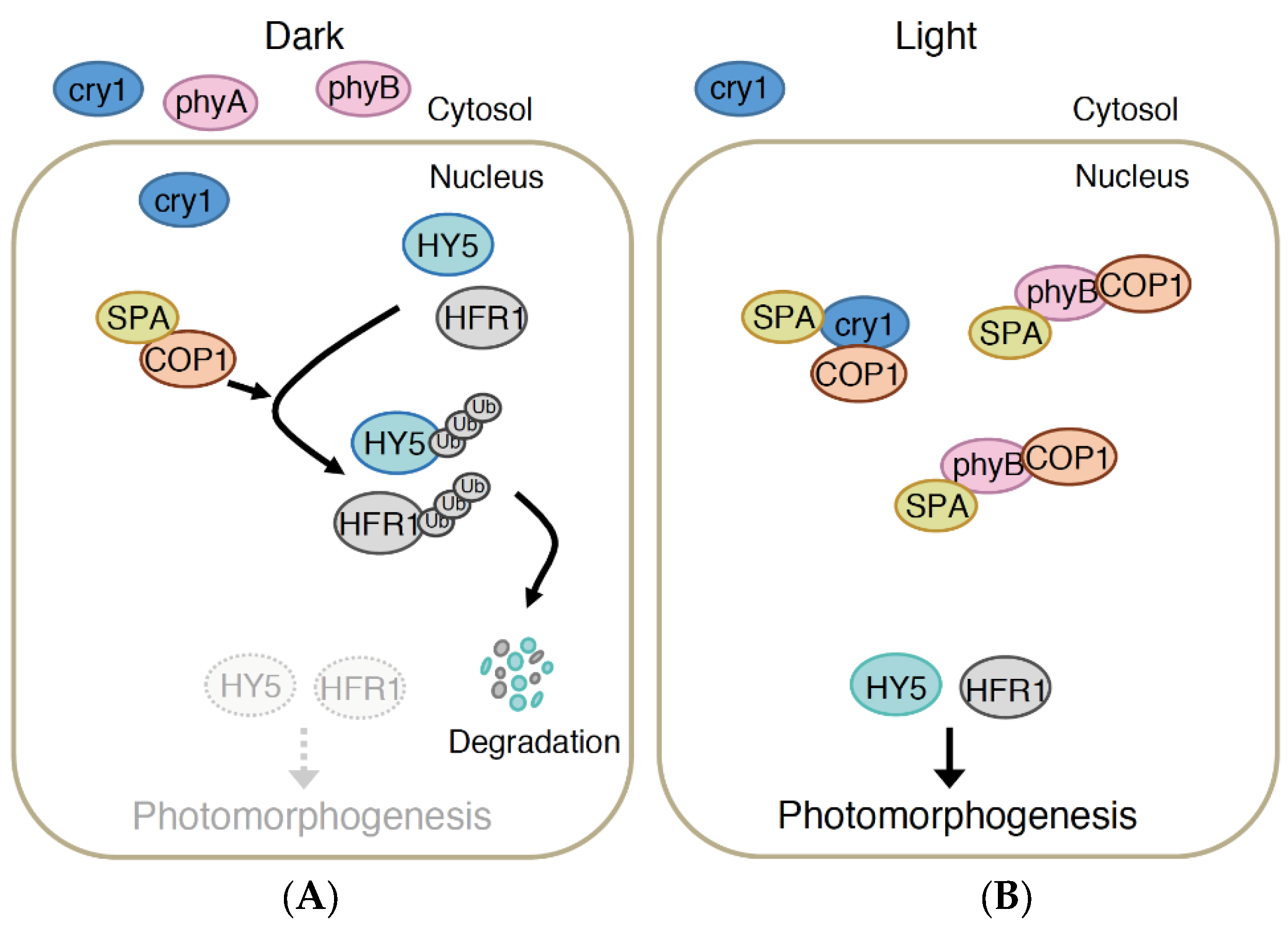
4.1. COP1/SPA Pathway
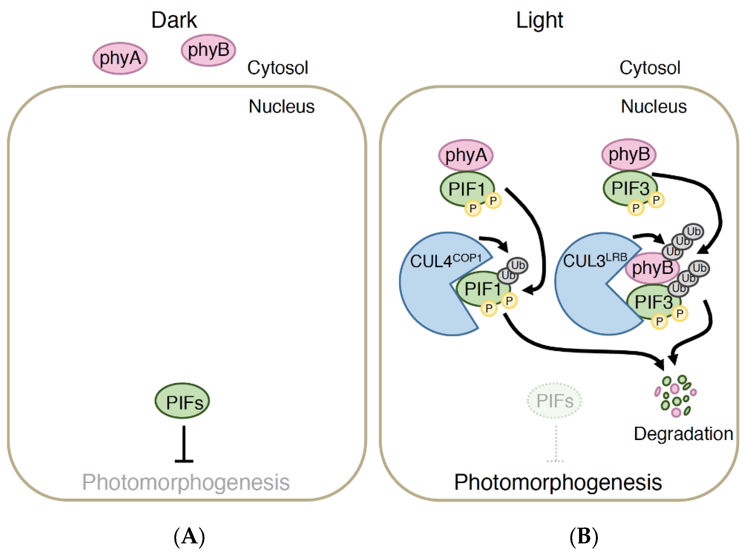
The cop1 mutant displays a constitutively photomorphogenic (cop) phenotype, in which dark-grown cop1 mutant seedlings mimic light-grown wild-type seedlings [89]. The spa1 mutant was initially identified as the suppressor mutant of phyA-105, a weak phyA allele that promotes a long hypocotyl under continuous FR light [90,91]; however, subsequent evidence showed that a higher-order combination of spa1 and mutants of other SPA genes (SPA2–4), e.g., spa1spa2spa3spa4, displayed strong cop-like phenotypes [92,93]. This genetic evidence indicates that COP1 and SPAs are the negative regulators of photomorphogenesis. Compared with the cop1 and spa mutants, a mutant of ELONGATED HYPOCOTYL 5 (HY5) was identified as a long-hypocotyl mutant under white light but not in the dark, thus indicating that the HY5 gene encodes the positive regulator of photomorphogenesis [89,94]. Indeed, HY5 has been shown to act as the bZIP transcription factor regulating the transcription of a number of light-responsive genes [95,96]. Consistent with these mutant phenotypes, COP1 and SPA proteins together with CULLIN4 (CUL4) constitute the E3 ubiquitin ligase complex, which ubiquitinates HY5 proteins for degradation through the 26S proteasome pathway to suppress photomorphogenesis in the dark [97] (Figure 1). Upon light exposure, crys and phys inactivate the COP1/SPA complex, thereby leading to the accumulation of HY5 protein in the nucleus to induce photomorphogenesis [89,94]. The COP1/SPA complex targets other positive regulators of photomorphogenesis, such as LONG HYPOCOTYL IN FAR-RED1 (HFR1), HY5 HOMOLOGUE, and LONG AFTER FAR-RED LIGHT1, and a key regulator of flowering, CONSTANS (CO), in the dark [98,99,100,101,102,103].
Cry1 and cry2 reportedly bind with SPA proteins in response to B light [77,78,79]. Moreover, phyA and phyB have been shown to interact with SPA proteins in a Pfr conformer-specific manner [80,81]. Consistent with the observations that COP1 requires a SPA1 association to exert E3 ligase activity in vitro and COP1 and SPA1 are dissociated by light exposure in plant cells [100,104], the binding of all these photoreceptors (except for cry2) to SPA proteins weakens the connection between COP1 and SPA proteins [77,78,80,81]. Hence, light-activated cry1, phyA, and phyB bind to SPAs to dissociate COP1 from SPAs, thus inhibiting COP1 activity and leading to the accumulation of HY5 and possibly other transcription factors, which promotes de-etiolation. However, B light-dependent cry2 binding to SPA proteins does not alter the affinity of COP1 for SPA proteins but strengthens the binding of cry2 to COP1 [79]. Although the mechanism remains unknown, this enhanced binding between cry2 and COP1 may attenuate COP1 activity to promote the accumulation of CO, which binds to the promoter of FT and induces its expression, thereby resulting in floral initiation. In addition to the light-dependent COP1 inactivation mechanism, these photoreceptors sequester the COP1 protein from transcription factors by depleting it from the nucleus under prolonged light exposure [105]. However, this nuclear exclusion mechanism is not fully understood.
The overlapping functions of cry1 and cry2 in regulating de-etiolation and photoperiodic flowering are at least partially explained by the COP1/SPA pathway (and PIF pathway) shared by these two B light receptors. However, the different modes of action of cry1 and cry2 to regulate COP1 activity may determine the substrate specificity of COP1 in the generation of functional differences between cry1 and cry2. The different affinities of cry1 and cry2 for SPA proteins may also account for the functional specificities because SPA proteins have diverse roles in plant development to a certain extent. However, the largest difference in signal transduction mechanisms between cry1 and cry2 is that only cry2 can bind with CIBs, which are specialized to function in photoperiodic flowering, in a B light-dependent manner [84,85]. Namely, the COP1/SPA or the PIF pathway likely represents the overlapping functions between cry1 and cry2, and the CIB pathway may confer on cry2 its more specific roles in the regulation of photoperiodic flowering [106]. However, the mechanism by which the COP1/SPA pathway and CIB pathway, which both regulate FT expression, converge downstream of cry2 is still unknown.

4.2. PIF Pathway

PIF3 was first identified as a protein that binds to phys in a Pfr-specific manner [107,108]. Of the bHLH subfamily 15, to which PIF3 belongs, 7 PIFs (PIF1, PIF3, PIF4, PIF5, PIF6, PIF7 and PIF8) have been demonstrated to bind to Pfr of phyB through the Active Phytochrome B Binding (APB) motif [83]. PIF1 and PIF3 have also been shown to bind to phyA. Accordingly, PIF1 and PIF3 possess the Active PhyA Binding (APA) motif, which is necessary for phyA binding, in addition to the APB motif and the DNA binding bHLH domain. PIF1, PIF3, PIF4 and PIF5 are phosphorylated and degraded rapidly by R light exposure [109,110,111,112,113,114]. The R light-induced rapid phosphorylation and degradation of PIFs is largely dependent on phyA and phyB. Furthermore, the pif1pif3pif4pif5 (pifQ) mutant exhibited a cop-like phenotype, albeit weak [115,116], suggesting that PIFs repress photomorphogenesis in the dark and that phyA and phyB induce photomorphogenesis by removing the PIF-imposed repression of photomorphogenesis via the rapid degradation of PIF1, PIF3, PIF4 and PIF5 in response to R light (Figure 2). However, phyB levels increase more in higher-order pif mutants under continuous R light [117,118]. The gentle degradation of phyB is dependent on binding to the APB motif of PIF3 and possibly other PIFs [119], which suggests a negative feedback circuit in which the binding of active phyB to PIFs not only promotes photomorphogenesis but also attenuates phyB signaling by reducing active phyB. The gentle degradation of phyB is partially mediated by COP1 [38]. In addition, the E3 ligase complex composed of CULLIN 3 and Light-Response Bric-a-Brac/Tramtrack/Broad (CUL3LRB) was found to recognize the phyB–PIF3 complex for its ubiquitination and proteasomal degradation under prolonged continuous R light [120] (Figure 2).
Interestingly, PIF1 is degraded under continuous FR light or by exposure to a small amount of R light [113,121]. This sensitive PIF1 degradation is mediated by phyA via binding to APA. Thus, one may expect that the binding of phyA to PIF1 is the sensitive reaction. However, the binding affinity to PIF1 in vitro does not differ between phyA and phyB [122]. Nevertheless, PIF1 has the highest affinity to phyA among all PIF family proteins tested to date. In addition, the CUL4COP1/SPA E3 ligase complex has recently been shown to promote phyA-induced rapid PIF1 degradation [123] (Figure 2). Mutations in CUL3 do not affect phyA-induced rapid PIF1 degradation, thus defining the difference between CUL3LRB-mediated, phyB-dependent PIF3 degradation under prolonged R light conditions and CUL4COP1/SPA-mediated, phyA-induced PIF1 degradation. CUL4COP1/SPA-mediated sensitive PIF1 degradation provides clues to understanding the mechanism underlying the phyA-mediated sensitive response in the nucleus. Further analysis regarding the specificity or kinetics of these E3 ligase complexes will be necessary to fully understand the different modes of action of phyA and phyB.
The phyB regulation of PIF activity is accomplished not only of R-induced rapid degradation but also of other mechanisms. Specifically, the N-terminal photosensory domain of phyB is capable of binding to PIFs and mediating de-etiolation [28,124,125,126], although it does not degrade PIF3 [127]. Instead, PIF3, which co-immunoprecipitates the RGA and PIL1 promoter DNA sequences, no longer binds with these promoter sequences under R light in transgenic plants expressing the N-terminal photosensory domain of phyB [127]. Both full-length phyB and the N-terminal photosensory domain of phyB inhibit the DNA binding of PIF1 and PIF3 under R light but not in the dark in vitro, suggesting that phyB promotes de-etiolation not only by degrading PIFs but also by sequestering PIFs from their target DNA sequences in response to R light [127]. Another example is that PIF7, which is phosphorylated but not degraded by R light, regulates de-etiolation and low R/FR-induced SAS [117,128]. However, how PIF7 activity is regulated remains unknown.
Recently, B light-dependent binding of crys to PIFs has been demonstrated [76,82]. To date, cry1 has been shown to bind to PIF3, PIF4 and PIF5 and cry2 has been shown to bind to PIF4 and PIF5, although not all combinations have been tested. These interactions regulate the B light-mediated inhibition of hypocotyl elongation, LBL-induced SAS and B light inhibition of warm temperature-induced cell growth. Similar to phys, crys are suggested to inhibit the activity of PIF4 and PIF5, although crys do not induce the degradation of PIF4 and PIF5 [76,82]. Whether crys sequester PIF4 and PIF5 from their DNA binding sites in a similar manner to phyB has not been tested. Thus, the mechanisms by which cry regulates the activity of PIF4 and PIF5 must be elucidated in the future.
Although most of the PIFs appear to have considerable functional redundancy in regulating de-etiolation, PIF family member proteins have distinct control functions in the diverse array of light-regulated physiological responses [83]. For instance, PIF1 has a dominant role in regulating seed germination [129] and PIF6 regulates seed dormancy [130]. PIF4, PIF5 and PIF7 promote SAS, with PIF4 and PIF5 acting more in LBL-induced and cry-mediated SAS and PIF7 mediating low R/FR-induced and phyB-mediated SAS [76,128]. Interestingly, phyB has recently been shown to act as a thermosensor [131,132]. Accordingly, PIF4 regulates temperature-dependent responses [133]. In addition, PIF4 is also known to regulate stomatal development [134]. However, neither the pif single nor the pifQ mutant shows a clear flowering phenotype in the normal temperature [116,135], implying that cry-PIF may not provide the mechanisms underlying the functional specificity between cry1 and cry2. In addition to the other diverse roles of PIFs, this family integrates light signals via endogenous signals, such as phytohormone signals and the circadian clock, thus highlighting the importance of PIFs in coordinating the transcriptional network to regulate plant development [83].

4.3. Interaction between the COP1/SPA Pathway and PIF Pathway

As described above, the CUL4COP1/SPA E3 ligase complex directly targets PIF1 for light-induced rapid degradation [123]. In addition, the accumulation of PIF3 proteins is considerably reduced in the cop1 mutant, even in the dark [110], suggesting that COP1 indirectly promotes the accumulation of PIF3, which likely occurs via the degradation of an as yet unknown negative regulator of PIF accumulation in the dark. Therefore, the COP1/SPA pathway directly and indirectly regulates the accumulation of PIFs in the dark or under light conditions.
HY5 and PIFs (at least PIF1 and PIF3) have been shown to physically bind to form an antagonizing module that participates in the regulation of photosynthetic gene and reactive oxygen species-responsive gene transcription [136,137]. Although HY5 and PIF3 independently mediate the hypocotyl response under continuous R or FR light, PIF3 function is dependent on HY5 in the regulation of FR light-induced anthocyanin accumulation [138]. These results indicate the complex interaction between HY5 and PIFs downstream of photoreceptors. Consistent with the observation that the function of HFR1, which promotes phy-mediated de-etiolation and SAS, depends on PIF4 and PIF5, HFR1 inhibits the activity of PIF4 and PIF5 by binding to them to form non-functional heterodimers, which no longer bind to their target DNA sequences [139,140]. The hfr1 mutant was initially identified as the long-hypocotyl mutant under continuous FR light [141]. However, subsequent reports have indicated that a phyA-independent but cry1-dependent long-hypocotyl phenotype occurs under continuous B light [142] and HFR1 regulates the transcription of more than 70% of B light- and cry1-mediated genes [143], indicating that HFR1 acts downstream of cry1 signal transduction. Moreover, at least PIF4 (and one of PIF1, PIF3 and PIF5) has been reported to regulate the cry1-mediated hypocotyl response under continuous B light [82] and that PIF4 and PIF5 regulate cry-mediated and LBL-induced SAS [74,75,76,144]. The involvement of both HFR1 and PIF4/5 in cry-mediated responses suggests that HFR1 also forms non-functional heterodimers with PIF4 and PIF5 downstream of crys. Therefore, the regulation of PIF functions by the COP1/SPA complex involves at least two separate mechanisms: One that directly or indirectly regulates PIF accumulation and the other that regulates PIF activity by regulating the accumulation of its target proteins, such as HY5 and HFR1. However, the dependency and specificity of these interactions may differ across responses or light environments.
In the regulation of de-etiolation in the dark, cop1 and pif1 have a synergistic effect. In addition, HY5 protein accumulates in pifQ, even in the dark. According to these observations, PIF1 binds with COP1 to enhance the HY5 recognition of COP1 and promote the ubiquitination of HY5 [145]. Therefore, the COP1/SPA pathway and the PIF pathway affect each other, thus representing the complexity of downstream cry and phy signal transduction.

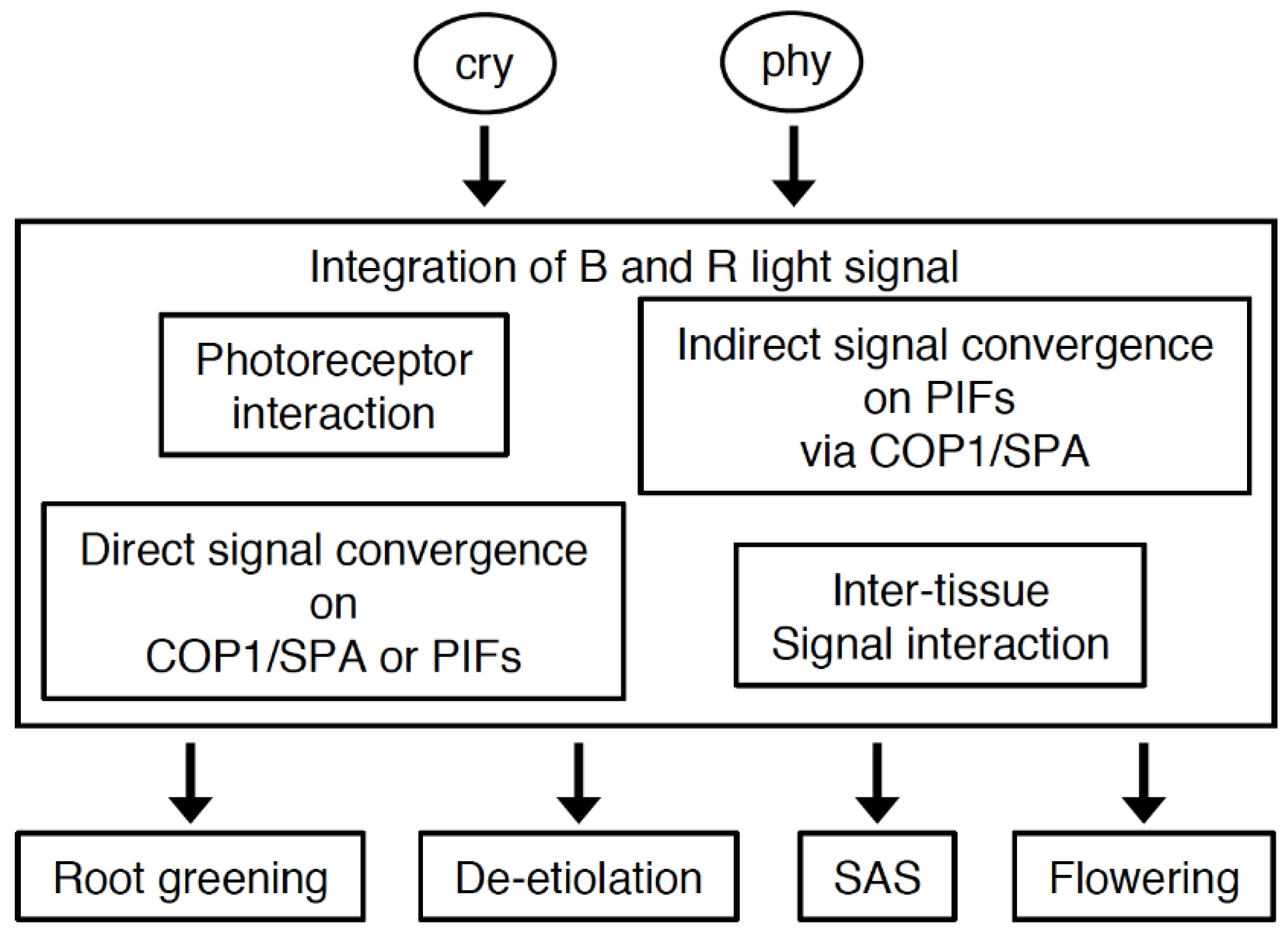
5. Coordination of Cryptochrome and Phytochrome Signal Transduction

Compared with the monochromatic light conditions in the laboratory, both crys and phys regulate related responses via shared signaling pathways under solar radiation in nature. Understanding how these photoreceptors act in natural or near-natural conditions is a challenging topic because of the complexity. Crys and phys independently incorporate B and R/FR light information, converge signals by photoreceptor interactions, and converge signals on common intermediates. Alternatively, they transduce the signals to selective intermediates and converge downstream (Figure 3). Here, we dissect the complex relations between cry and phy and examine the convergence of cry and phy signaling in the regulation of light responses.
Crys and phys converge blue and red light signals at different levels to co-regulate physiological responses, such as root greening, de-etiolation, SAS, photoperiodic flowering, etc. The co-action mechanism includes photoreceptor interactions, direct signal convergence on common intermediates, indirect signal convergence on PIFs downstream of COP1/SPA, inter-tissue signal interactions, etc., although the role of photoreceptor interactions and the mechanism responsible for the direct convergence of cry and phy signals on the COP1/SPA complex or PIFs remain elusive. The types of co-action may be different depending on the responses. Indirect signal convergence on PIFs via COP1/SPA and inter-tissue signal interaction are evident in the regulation of SAS and photoperiodic flowering, respectively.

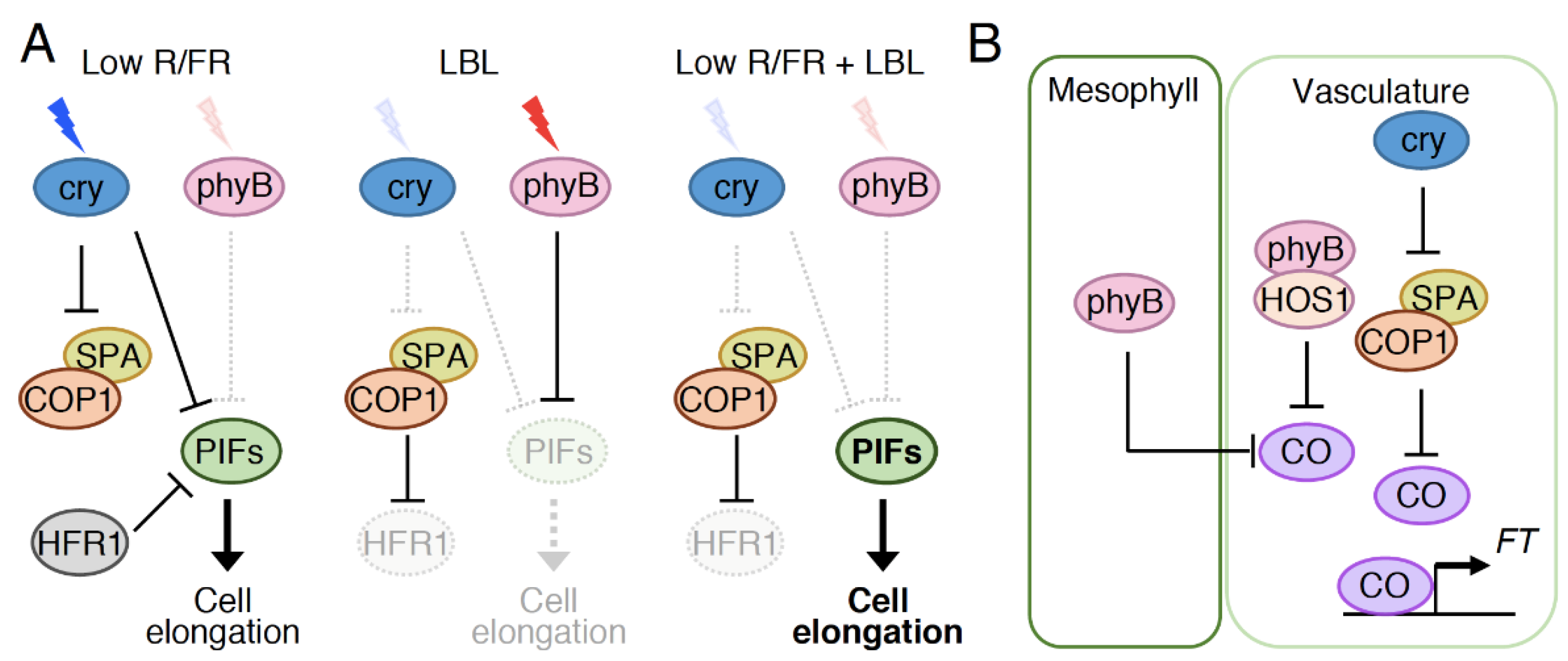
5.1. Observed Co-Action between Cryptochromes and Phytochromes in Physiological Responses

The cry1cry2 double mutant responds normally to R light [73]. Conversely, the phyAphyB double mutant and the phy mutant, which lacks all five Arabidopsis phy species, respond to B light [146,147], although their responses are impaired to a certain extent, likely because phys absorb B light. These results clearly indicate that crys and phys do not require each other to exert their substantial signaling functions. Nevertheless, classical photobiological analyses have indicated co-action between crys and phys under certain conditions [148]. Specifically, B light exposure enhanced the effect of phy-mediated R light induction of the accumulation of glyceraldehyde-3-phosphate dehydrogenase in milo seedlings. phy-mediated anthocyanin accumulation in milo required B light exposure. In addition, an analysis of Arabidopsis photoreceptor mutants has provided genetic evidence of co-action between crys and phys. The most prominent synergistic effect has been observed in root greening [149]. B light induces stronger root greening than R light. This strong effect of B light on root greening can be observed in the phyA and phyB single mutants but is almost completely defective in the cry1 mutant and the phyAphyB double mutant, indicating that cry1 and phyA or phyB promote B light-induced root greening in a coordinated manner. The synergism between cry and phy mutations can also be seen in the regulation of de-etiolation. phyB mutation diminishes the function of cry1, and cry1 mutation does so for the function of phyB in the regulation of hypocotyl response under simultaneous R and B irradiation [150]. However, these effects can be observed only under a short period of dichromatic irradiation with B and R light, not under continuous dichromatic irradiation, suggesting that cry1 and phyB coordinate the inhibition of hypocotyl elongation under suboptimal light conditions. Such synergism is not observed between cry1 and phyA. In addition, phyB and cry2 mutations displayed a synergistic effect, albeit weakly, rather than an additive effect in the inhibition of hypocotyl elongation under continuous dichromatic irradiation with B and R light [73]. Thus, phyB partially co-acts with cry1 or cry2 in the regulation of hypocotyl response. The co-action between phyB and crys can also be observed in SAS because LBL enhances the effect of low R/FR-induced, phyB-mediated SAS [144].
In contrast, antagonistic effects have been reported between cry and phy mutations. For example, the cry1 mutant has a substantially elongated hypocotyl compared with the wild type under continuous white light; however, the hypocotyl length of cry1phyA is significantly shorter than that of cry1 [151]. A similar antagonistic effect between cry1 and phyA is also observed in cotyledon expansion and chlorophyll accumulation under continuous white light [151]. The antagonistic effect between cry and phy is more evident in the regulation of photoperiodic flowering [73]. Namely, the cry2 mutant shows a late-flowering phenotype specifically under LD conditions, but the effect of cry2 mutation is considerably reduced in the phyB mutant, which flowers early regardless of day length.

5.2. Possible Roles of Photoreceptor Interactions and Direct Signal Convergence on Common Intermediates in the Co-Action between Cryptochromes and Phytochromes

Cry1 physically binds with phyA and phyB in vitro, and cry2 directly binds with phyB in plant cells [65,152]. Particularly, interactions between phyB and cry2 have been observed only in certain photobodies [65]. These photoreceptor binding interactions may represent the underlying mechanism for the physiological co-action of crys and phys. Avena phyA, which displays kinase activity, directly binds with and phosphorylates cry1 in vitro [152]. However, the phosphorylation of cry1 occurs normally in Arabidopsis phyA or other phy mutants [48], suggesting that the observed in vitro cry1 phosphorylation catalyzed by phyA exerts marginal effects on physiological responses. Hence, the effects of photoreceptor interactions on their physiological co-action remain unknown. Given that both phyB-PIF3 and cry2 undergo light-dependent phosphorylation that triggers polyubiquitination and protein degradation, it would be interesting to examine whether related protein kinases may act as the common regulators for the two distinct photoreceptors.
Shared signaling partners between crys and phys may provide clues to the molecular mechanism underlying the co-action of crys and phys. The interaction of crys with the COP1/SPA complex or PIFs does not require phys and vice versa because they bind to these signaling partners in yeast cells or in vitro systems where other photoreceptors are not observed [76,77,78,79,80,81,82,108,153]. However, the binding of cry to these signaling partners may modify their affinity for phy and vice versa, the binding of cry may change the binding of phy to these signaling partners and vice versa, and the photoreceptor interactions may accelerate or decelerate binding to signaling partners. These possibilities should be experimentally addressed in the future, and the binding sites of these photoreceptors on their signaling partners should be a focus of such investigations. Crys and phys share a binding site on SPA proteins in certain cases; however, they recognize different structures in other cases [77,78,79,80,81]. Therefore, crys and phys possibly form a large protein complex or compete for the same structure of SPAs.
Crys and phyB recognize the different structures of PIF4/5 because crys bind with PIF4/5mAPB that harbor a mutation on the APB motif, which abolishes phyB binding [76]. In addition, phyB indeed binds with PIF4/5 under LBL conditions, in which crys can interact with PIF4/5 [76], suggesting that a trimeric complex can be formed among phyB, crys, and PIF4/5. Interestingly, the expression of PIF4/5mAPB does not fully induce hypocotyl elongation in response to LBL [76], suggesting that phyB binding to PIF4/5 promotes PIF4/5 activity to enhance hypocotyl growth in response to LBL. This promotion of PIF4/5 activity by phyB under LBL conditions is in striking contrast to the function of phyB in repressing PIF4/5 activity in R light [83], implying that crys may possibly reverse the function of phyB in the trimeric cry-PIF4/5-phyB complex under these light conditions.
Accordingly, B light-induced degradation of PIFs is also of great interest [154,155]. The B light-activated phyA mediate the rapid degradation of PIF1 and PIF3, whereas phy-mediated PIF1 degradation is enhanced in a cry1cry2 double mutant under B light [154], indicating that crys antagonize phyA-mediated PIF1 degradation. Cry-phy binding may inhibit phyA to bind to PIF1, thereby protecting PIF1 proteins from degradation. Because crys bind to PIF3, PIF4 and PIF5 [76,82], crys may also interact with PIF1. Therefore, the binding of crys to PIF1 may reduce the affinity of PIF1 for phyA or impair the function of PIF1-bound phyA in a trimeric cry-PIF1–phyA complex. Moreover, cry1 and cry2 may function indirectly to antagonize phyA functions via the regulation of COP1/SPA activity because COP1 plays a role in the degradation of PIF1 [123]. Thus, the mechanism underlying the cry-mediated stabilization of PIFs, which represents the antagonism between crys and phyA, should be carefully examined in the future.

5.3. Indirect Signal Convergence Downstream of Cryptochromes and Phytochromes

Regardless of the common intermediates, cry and phy may have preferential inputs and interact downstream. A transcriptome analysis comparing seedling response to continuous R light and 3 h of B light included with the continuous R light, in which cry1 and phyB coordinate to regulate de-etiolation, revealed that only 6% of B light-regulated, cry1-mediated genes are regulated by phyB under background R light conditions [63], suggesting that cry1 and phyB regulate transcription mostly via independent pathways. In addition, the expression of certain B light-regulated, cry1-mediated genes persists in R light even in the absence of B light, and this expression contributes to promoting the co-action of R and B light effects in the de-etiolation process, suggesting that phyB incorporates the independent, cry1-mediated transcriptional network into its signaling pathway to enhance light responses. The co-action between crys and phyB in the control of de-etiolation requires SPA1, SPA4, HY5, and HYH [63].
The preferential roles of common intermediates downstream of cry and phy are more evident in the regulation of SAS. phyB and crys bind to PIFs to regulate low R/FR-mediated and LBL-mediated SAS, respectively [76,114,128]. Because low R/FR reduces the Pfr/Ptot ratio, it also impairs the binding of phyB to PIFs, thereby increasing the active PIF level to promote cell elongation. Although low R/FR increases the abundance of PIF4/5 [114], phyB favors PIF7 rather than PIF4/5 to regulate low R/FR-mediated SAS primarily by regulating the expression of auxin biosynthesis genes and auxin-responsive genes [128]. Instead, crys favor PIF4/5 to regulate LBL-induced SAS. PIF4/5 preferentially regulate the transcription of genes involved in cell wall modifications to facilitate cell elongation under LBL [76]. It should be noted here that the transcription of the cell wall modifying genes is frequently regulated by auxin and LBL-induced and cry-mediated SAS is partly dependent on auxin [75]. Thus, the mechanism how crys regulate the transcription of the cell wall modifying genes in LBL via PIF4/5 should be carefully examined in the future.
Moreover, the function of the COP1/SPA pathway that positively regulates SAS is of great interest for understanding the complex interactions between phy and cry. COP1 enhances the effects of PIF3/4/5 on SAS under shade conditions with both LBL and low R/FR ratios [156], although whether it enhances the effects of PIF7 under such conditions is currently unknown. The function of COP1 in the regulation of SAS is largely dependent on HFR1. Interestingly, COP1 decreases the stability of the HFR1 protein but increases the abundance of the HY5 protein, which likely antagonizes the function of PIFs to avoid excess elongation under shade conditions [156], although the methods by which COP1 differentially recognize HY5 and HFR1 under these conditions are currently unknown. As described above, HFR1 and PIF4/PIF5 form non-functional heterodimers and the functions of HFR1 under shade conditions are dependent on PIF4/5 [139]. Therefore, under shade conditions, the COP1/SPA complex promotes the removal of HFR1-mediated repression, which represents the mechanism by which PIF4/5 exert their functions and elongate the hypocotyl. In addition, the stepwise application of low R/FR and LBL differentiated the roles of photoreceptors and enabled the description of more specific roles [144]. Namely, LBL enhances the effect of low R/FR via PIF4/5/7 in SAS-mediated petiole and hypocotyl elongation. First, LBL enhances the low R/FR-mediated abundance of PIF5. Notably, LBL combined with low R/FR decreases HFR1 abundance, although low R/FR alone stabilizes HFR1. Therefore, LBL enhances the effect of low R/FR not only by increasing the accumulation of PIF5 but also by promoting the proteasomal degradation of HFR1, thus highlighting the role of crys in regulating COP1/SPA activity in response to shade. Taking this evidence together, shade likely activates the function of PIFs by reversing phyB and cry binding, and in addition, the inactivated crys remove the HFR1-mediated inhibition of PIF activity via the regulated COP1/SPA pathway, thereby facilitating PIF-mediated cell growth [144,156] (Figure 4). Interestingly, DELLA proteins, which are the negative regulators of gibberellin signaling and the inhibitors of PIF functions, are degraded under shade conditions [157], thus representing another mechanism underlying the regulation of PIF activity by photoreceptors.
Other types of photoreceptor signaling interactions were also found in the regulation of photoperiodic flowering. As mentioned earlier, cry2 antagonizes phyB in the regulation of photoperiodic flowering [73]. The accumulation of CO, which promotes the transcription of FT in the vascular tissue to initiate flowering, represents this antagonistic effect between cry2 and phyB [158]. Namely, cry2 stabilizes CO, whereas phyB degrades it. As described above, cry2 stabilizes CO by inactivating the COP1/SPA complex [79,98,103]. However, the phyB-mediated degradation of CO is COP1 independent [98]. PFT1, a component of phyB signal transduction, promotes earlier flowering, although it regulates CO expression rather than protein stability [159,160]. PHL, a phyB binding protein that regulates flowering time, binds to CO protein at dusk not at dawn when phyB promotes CO degradation [161]. Whether PHL affects the stability of CO is also unclear. HOS1, a phyB binding E3 ubiquitin ligase, mediates the degradation of CO in the phloem companion cells [162,163]. However, the mechanism underlying phyB-mediated CO degradation is not fully understood. Interestingly, the tissue-specific expression of photoreceptors demonstrated that phyB expressed in mesophyll cells regulates flowering time [163,164], although phyB expressed in vascular tissue partly complement the early flowering phyB mutant phenotype [163]. On the contrary, cry2 promotes photoperiodic flowering solely in vascular tissue [165], thus suggesting that cry2 and phyB antagonize each other to control photoperiodic flowering partly via inter-tissue signaling between vascular tissue and mesophyll cells. The substance of inter-tissue signaling that is generated by phyB in mesophyll cells and suppresses flowering by promoting the degradation of CO via a COP1-independent mechanism in vascular tissue should be carefully investigated to fully understand the co-action between photoreceptors. Given that both of crys and phys are expressed throughout plant body, it would be interesting to examine whether this type of non-cell-autonomous interaction among photoreceptors regulate other light responses.

6. Conclusions and Future Outlook

Crys and phys act under different colors of the light spectrum to regulate related developmental processes via two independent but shared signaling pathways. These two common signaling pathways and direct cry-phy interactions provide clues to understand the molecular mechanisms underlying the co-action of crys and phys. However, as exemplified by cry- and phy-mediated SAS, cry and phy differentially regulate light responses via the preferential selection of signaling pathways that lead to the regulation of PIF activity. The mechanisms underlying the preferential selection of signaling pathways by crys and phys regardless of the binding of both photoreceptors to common signaling intermediates must be addressed to fully understand plant light responses. Crys and phys have complicated interactions including direct binding, signal convergence on or downstream of their common intermediates, and inter-tissue signal transduction, thus providing the mechanism by which plants are capable of responding plastically to fluctuating light environments in nature. These interactions likely provide general regulation of cell growth and photoperiodic flowering in many crop species. In addition, photoreceptors regulate many important agronomic traits that are likely species specific. Therefore, understanding the cry–phy interactions in individual crop species will provide insights for improving the yields and quality of crops.

Acknowledgments

This work was supported by the Fujian–Taiwan Joint Innovative Center for Germplasm Resources and Cultivation of Crops (FJ 2011 Program, No. 2015-75, China), the Program for New Century Excellent Talents in Fujian Province University, and the National Natural Science Foundation of China 31500548 (to Bobin Liu) and 31650110478 (to Yoshito Oka).

Author Contributions

Jun Su, Bobin Liu and Jiakai Liao wrote the manuscript. Zhaohe Yang made the figures. Chentao Lin and Yoshito Oka edited the manuscript.

Conflicts of Interest

The authors declare no conflict of interest.

References

  1. Chen, M.; Chory, J.; Fankhauser, C. Light signal transduction in higher plants. Annu. Rev. Genet. 2004, 38, 87–117. [Google Scholar] [CrossRef] [PubMed]
  2. Kami, C.; Lorrain, S.; Hornitschek, P.; Fankhauser, C. Light-regulated plant growth and development. Curr. Top. Dev. Biol. 2010, 91, 29–66. [Google Scholar] [PubMed]
  3. Franklin, K.A.; Quail, P.H. Phytochrome functions in Arabidopsis development. J. Exp. Bot. 2010, 61, 11–24. [Google Scholar] [CrossRef] [PubMed]
  4. Wang, X.; Wang, Q.; Nguyen, P.; Lin, C. Cryptochrome-mediated light responses in plants. Enzymes 2014, 35, 167–189. [Google Scholar] [PubMed]
  5. Yang, Z.; Liu, B.; Su, J.; Liao, J.; Lin, C.; Oka, Y. Cryptochromes orchestrate transcription regulation of diverse blue light responses in plants. Photochem. Photobiol. 2017, 93, 112–127. [Google Scholar] [CrossRef] [PubMed]
  6. Gururani, M.A.; Ganesan, M.; Song, P.S. Photo-biotechnology as a tool to improve agronomic traits in crops. Biotechnol. Adv. 2015, 33, 53–63. [Google Scholar] [CrossRef] [PubMed]
  7. Takano, M.; Inagaki, N.; Xie, X.; Kiyota, S.; Baba-Kasai, A.; Tanabata, T.; Shinomura, T. Phytochromes are the sole photoreceptors for perceiving red/far-red light in rice. Proc. Natl. Acad. Sci. USA 2009, 106, 14705–14710. [Google Scholar] [CrossRef] [PubMed]
  8. Barrero, J.M.; Downie, A.B.; Xu, Q.; Gubler, F. A role for barley CRYPTOCHROME1 in light regulation of grain dormancy and germination. Plant Cell 2014, 26, 1094–1104. [Google Scholar] [CrossRef] [PubMed]
  9. Meng, Y.; Li, H.; Wang, Q.; Liu, B.; Lin, C. Blue light-dependent interaction between cryptochrome 2 and CIB1 regulates transcription and leaf senescence in soybean. Plant Cell 2013, 25, 4405–4420. [Google Scholar] [CrossRef] [PubMed]
  10. Menon, C.; Sheerin, D.J.; Hiltbrunner, A. SPA proteins: SPAnning the gap between visible light and gene expression. Planta 2016, 244, 297–312. [Google Scholar] [CrossRef] [PubMed]
  11. Lee, N.; Choi, G. Phytochrome-interacting factor from Arabidopsis to liverwort. Curr. Opin. Plant Biol. 2016, 35, 54–60. [Google Scholar] [CrossRef] [PubMed]
  12. Nagatani, A. Light-regulated nuclear localization of phytochromes. Curr. Opin. Plant Biol. 2004, 7, 708–711. [Google Scholar] [CrossRef] [PubMed]
  13. Yamaguchi, R.; Nakamura, M.; Mochizuki, N.; Kay, S.A.; Nagatani, A. Light-dependent translocation of a phytochrome B-GFP fusion protein to the nucleus in transgenic Arabidopsis. J. Cell Biol. 1999, 145, 437–445. [Google Scholar] [CrossRef] [PubMed]
  14. Chen, M.; Galvao, R.M.; Li, M.; Burger, B.; Bugea, J.; Bolado, J.; Chory, J. Arabidopsis HEMERA/pTAC12 initiates photomorphogenesis by phytochromes. Cell 2010, 141, 1230–1240. [Google Scholar] [CrossRef] [PubMed]
  15. Qiu, Y.; Li, M.; Pasoreck, E.K.; Long, L.; Shi, Y.; Galvao, R.M.; Chou, C.L.; Wang, H.; Sun, A.Y.; Zhang, Y.C.; et al. HEMERA couples the proteolysis and transcriptional activity of PHYTOCHROME INTERACTING FACTORs in Arabidopsis photomorphogenesis. Plant Cell 2015, 27, 1409–1427. [Google Scholar] [CrossRef] [PubMed]
  16. Clack, T.; Mathews, S.; Sharrock, R.A. The phytochrome apoprotein family in Arabidopsis is encoded by five genes: The sequences and expression of PHYD and PHYE. Plant Mol. Biol. 1994, 25, 413–427. [Google Scholar] [CrossRef] [PubMed]
  17. Wu, F.Q.; Fan, C.M.; Zhang, X.M.; Fu, Y.F. The phytochrome gene family in soybean and a dominant negative effect of a soybean PHYA transgene on endogenous Arabidopsis PHYA. Plant Cell Rep. 2013, 32, 1879–1890. [Google Scholar] [CrossRef] [PubMed]
  18. Mathews, S. Evolutionary studies illuminate the structural-functional model of plant phytochromes. Plant Cell 2010, 22, 4–16. [Google Scholar] [CrossRef] [PubMed]
  19. Mancinelli, A.L. The physiology of phytochrome action. In Photomorphogenesis in Higher Plants, 2nd ed.; Kendrick, R.E., Kronenberg, G.H.M., Eds.; Springer: Dordrecht, The Netherlands, 1994; pp. 211–269. [Google Scholar]
  20. Shinomura, T.; Nagatani, A.; Hanzawa, H.; Kubota, M.; Watanabe, M.; Furuya, M. Action spectra for phytochrome A- and B-specific photoinduction of seed germination in Arabidopsis thaliana. Proc. Natl. Acad. Sci. USA 1996, 93, 8129–8133. [Google Scholar] [CrossRef] [PubMed]
  21. Nagatani, A.; Reed, J.W.; Chory, J. Isolation and initial characterization of Arabidopsis mutants that are deficient in phytochrome A. Plant Physiol. 1993, 102, 269–277. [Google Scholar] [CrossRef] [PubMed]
  22. Parks, B.M.; Quail, P.H. hy8, a new class of Arabidopsis long hypocotyl mutants deficient in functional phytochrome A. Plant Cell 1993, 5, 39–48. [Google Scholar] [CrossRef] [PubMed]
  23. Sharrock, R.A.; Clack, T. Patterns of expression and normalized levels of the five Arabidopsis phytochromes. Plant Physiol. 2002, 130, 442–456. [Google Scholar] [CrossRef] [PubMed]
  24. Oka, Y.; Ono, Y.; Toledo-Ortiz, G.; Kokaji, K.; Matsui, M.; Mochizuki, N.; Nagatani, A. Arabidopsis phytochrome A is modularly structured to integrate the multiple features that are required for a highly sensitized phytochrome. Plant Cell 2012, 24, 2949–2962. [Google Scholar] [CrossRef] [PubMed]
  25. Wagner, D.; Fairchild, C.D.; Kuhn, R.M.; Quail, P.H. Chromophore-bearing NH2-terminal domains of phytochromes A and B determine their photosensory specificity and differential light lability. Proc. Natl. Acad. Sci. USA 1996, 93, 4011–4015. [Google Scholar] [CrossRef] [PubMed]
  26. Wagner, D.; Koloszvari, M.; Quail, P.H. Two small spatially distinct regions of phytochrome B are required for efficient signaling rates. Plant Cell 1996, 8, 859–871. [Google Scholar] [CrossRef] [PubMed]
  27. Huq, E.; Al-Sady, B.; Quail, P.H. Nuclear translocation of the photoreceptor phytochrome B is necessary for its biological function in seedling photomorphogenesis. Plant J. 2003, 35, 660–664. [Google Scholar] [CrossRef] [PubMed]
  28. Matsushita, T.; Mochizuki, N.; Nagatani, A. Dimers of the N-terminal domain of phytochrome B are functional in the nucleus. Nature 2003, 424, 571–574. [Google Scholar] [CrossRef] [PubMed]
  29. Toledo-Ortiz, G.; Kiryu, Y.; Kobayashi, J.; Oka, Y.; Kim, Y.; Nam, H.G.; Mochizuki, N.; Nagatani, A. Subcellular sites of the signal transduction and degradation of phytochrome A. Plant Cell Physiol. 2010, 51, 1648–1660. [Google Scholar] [CrossRef] [PubMed]
  30. Genoud, T.; Schweizer, F.; Tscheuschler, A.; Debrieux, D.; Casal, J.J.; Schafer, E.; Hiltbrunner, A.; Fankhauser, C. FHY1 mediates nuclear import of the light-activated phytochrome A photoreceptor. PLoS Genet. 2008, 4, e1000143. [Google Scholar] [CrossRef] [PubMed]
  31. Kircher, S.; Kozma-Bognar, L.; Kim, L.; Adam, E.; Harter, K.; Schafer, E.; Nagy, F. Light quality-dependent nuclear import of the plant photoreceptors phytochrome A and B. Plant Cell 1999, 11, 1445–1456. [Google Scholar] [CrossRef] [PubMed]
  32. Hiltbrunner, A.; Viczian, A.; Bury, E.; Tscheuschler, A.; Kircher, S.; Toth, R.; Honsberger, A.; Nagy, F.; Fankhauser, C.; Schafer, E. Nuclear accumulation of the phytochrome A photoreceptor requires FHY1. Curr. Biol. 2005, 15, 2125–2130. [Google Scholar] [CrossRef] [PubMed]
  33. Hiltbrunner, A.; Tscheuschler, A.; Viczian, A.; Kunkel, T.; Kircher, S.; Schafer, E. FHY1 and FHL act together to mediate nuclear accumulation of the phytochrome A photoreceptor. Plant Cell Physiol. 2006, 47, 1023–1034. [Google Scholar] [CrossRef] [PubMed]
  34. Rausenberger, J.; Tscheuschler, A.; Nordmeier, W.; Wust, F.; Timmer, J.; Schafer, E.; Fleck, C.; Hiltbrunner, A. Photoconversion and nuclear trafficking cycles determine phytochrome A’s response profile to far-red light. Cell 2011, 146, 813–825. [Google Scholar] [CrossRef] [PubMed]
  35. Jabben, M.; Shanklin, J.; Vierstra, R.D. Ubiquitin-phytochrome conjugates. Pool dynamics during in vivo phytochrome degradation. J. Biol. Chem. 1989, 264, 4998–5005. [Google Scholar] [PubMed]
  36. Jabben, M.; Shanklin, J.; Vierstra, R.D. Red light-induced accumulation of ubiquitin-phytochrome conjugates in both monocots and dicots. Plant Physiol. 1989, 90, 380–384. [Google Scholar] [CrossRef] [PubMed]
  37. Vierstra, R.D. Phytochrome degradation. In Photomorphogenesis in Higher Plants, 2nd ed.; Kendrick, R.E., Kronenberg, G.H.M., Eds.; Springer: Dordrecht, The Netherlands, 1994; pp. 141–162. [Google Scholar]
  38. Jang, I.C.; Henriques, R.; Seo, H.S.; Nagatani, A.; Chua, N.H. Arabidopsis PHYTOCHROME INTERACTING FACTOR proteins promote phytochrome B polyubiquitination by COP1 E3 ligase in the nucleus. Plant Cell 2010, 22, 2370–2383. [Google Scholar] [CrossRef] [PubMed]
  39. Seo, H.S.; Watanabe, E.; Tokutomi, S.; Nagatani, A.; Chua, N.H. Photoreceptor ubiquitination by COP1 E3 ligase desensitizes phytochrome A signaling. Genes Dev. 2004, 18, 617–622. [Google Scholar] [CrossRef] [PubMed]
  40. Martinez-Garcia, J.F.; Gallemi, M.; Molina-Contreras, M.J.; Llorente, B.; Bevilaqua, M.R.; Quail, P.H. The shade avoidance syndrome in Arabidopsis: The antagonistic role of phytochrome A and B differentiates vegetation proximity and canopy shade. PLoS ONE 2014, 9, e109275. [Google Scholar] [CrossRef] [PubMed]
  41. Yanovsky, M.J.; Casal, J.J.; Whitelam, G.C. Phytochrome A, phytochrome B and HY4 are involved in hypocotyl growth responses to natural radiation in Arabidopsis: Weak deetiolation of the phyA mutant under dense canopies. Plant Cell Environ. 1995, 18, 788–794. [Google Scholar] [CrossRef]
  42. Casal, J.J. Shade avoidance. Arabidopsis Book 2012, 10, e0157. [Google Scholar] [CrossRef] [PubMed]
  43. Casal, J.J. Photoreceptor signaling networks in plant responses to shade. Annu. Rev. Plant Biol. 2013, 64, 403–427. [Google Scholar] [CrossRef] [PubMed]
  44. Ahmad, M.; Cashmore, A.R. HY4 gene of A. thaliana encodes a protein with characteristics of a blue-light photoreceptor. Nature 1993, 366, 162–166. [Google Scholar] [CrossRef] [PubMed]
  45. Guo, H.; Yang, H.; Mockler, T.C.; Lin, C. Regulation of flowering time by Arabidopsis photoreceptors. Science 1998, 279, 1360–1363. [Google Scholar] [CrossRef] [PubMed]
  46. Xu, P.; Zhu, H.L.; Xu, H.B.; Zhang, Z.Z.; Zhang, C.Q.; Zhang, L.X.; Ma, Z.Q. Composition and phylogenetic analysis of wheat cryptochrome gene family. Mol. Biol. Rep. 2010, 37, 825–832. [Google Scholar] [CrossRef] [PubMed]
  47. Wang, Q.; Barshop, W.D.; Bian, M.; Vashisht, A.A.; He, R.; Yu, X.; Liu, B.; Nguyen, P.; Liu, X.; Zhao, X.; et al. The blue light-dependent phosphorylation of the CCE domain determines the photosensitivity of Arabidopsis CRY2. Mol. Plant 2015, 8, 631–643. [Google Scholar] [CrossRef] [PubMed]
  48. Shalitin, D.; Yu, X.; Maymon, M.; Mockler, T.; Lin, C. Blue light-dependent in vivo and in vitro phosphorylation of Arabidopsis cryptochrome 1. Plant Cell 2003, 15, 2421–2429. [Google Scholar] [CrossRef] [PubMed]
  49. Shalitin, D.; Yang, H.; Mockler, T.C.; Maymon, M.; Guo, H.; Whitelam, G.C.; Lin, C. Regulation of Arabidopsis cryptochrome 2 by blue-light-dependent phosphorylation. Nature 2002, 417, 763–767. [Google Scholar] [CrossRef] [PubMed]
  50. Sang, Y.; Li, Q.H.; Rubio, V.; Zhang, Y.C.; Mao, J.; Deng, X.W.; Yang, H.Q. N-terminal domain-mediated homodimerization is required for photoreceptor activity of Arabidopsis cryptochrome 1. Plant Cell 2005, 17, 1569–1584. [Google Scholar] [CrossRef] [PubMed]
  51. Yu, X.; Shalitin, D.; Liu, X.; Maymon, M.; Klejnot, J.; Yang, H.; Lopez, J.; Zhao, X.; Bendehakkalu, K.T.; Lin, C. Derepression of the NC80 motif is critical for the photoactivation of Arabidopsis CRY2. Proc. Natl. Acad. Sci. USA 2007, 104, 7289–7294. [Google Scholar] [CrossRef] [PubMed]
  52. Rosenfeldt, G.; Viana, R.M.; Mootz, H.D.; von Arnim, A.G.; Batschauer, A. Chemically induced and light-independent cryptochrome photoreceptor activation. Mol. Plant 2008, 1, 4–14. [Google Scholar] [CrossRef] [PubMed]
  53. Wang, Q.; Zuo, Z.; Wang, X.; Gu, L.; Yoshizumi, T.; Yang, Z.; Yang, L.; Liu, Q.; Liu, W.; Han, Y.J.; et al. Photoactivation and inactivation of Arabidopsis cryptochrome 2. Science 2016, 354, 343–347. [Google Scholar] [CrossRef] [PubMed]
  54. Lin, C.; Yang, H.; Guo, H.; Mockler, T.; Chen, J.; Cashmore, A.R. Enhancement of blue-light sensitivity of Arabidopsis seedlings by a blue light receptor cryptochrome 2. Proc. Natl. Acad. Sci. USA 1998, 95, 2686–2690. [Google Scholar] [CrossRef] [PubMed]
  55. Yu, X.; Klejnot, J.; Zhao, X.; Shalitin, D.; Maymon, M.; Yang, H.; Lee, J.; Liu, X.; Lopez, J.; Lin, C. Arabidopsis cryptochrome 2 completes its posttranslational life cycle in the nucleus. Plant Cell 2007, 19, 3146–3156. [Google Scholar] [CrossRef] [PubMed]
  56. Yu, X.; Sayegh, R.; Maymon, M.; Warpeha, K.; Klejnot, J.; Yang, H.; Huang, J.; Lee, J.; Kaufman, L.; Lin, C. Formation of nuclear bodies of Arabidopsis CRY2 in response to blue light is associated with its blue light-dependent degradation. Plant Cell 2009, 21, 118–130. [Google Scholar] [CrossRef] [PubMed]
  57. Weidler, G.; Zur Oven-Krockhaus, S.; Heunemann, M.; Orth, C.; Schleifenbaum, F.; Harter, K.; Hoecker, U.; Batschauer, A. Degradation of Arabidopsis CRY2 is regulated by SPA proteins and phytochrome A. Plant Cell 2012, 24, 2610–2623. [Google Scholar] [CrossRef] [PubMed]
  58. Wu, G.; Spalding, E.P. Separate functions for nuclear and cytoplasmic cryptochrome 1 during photomorphogenesis of Arabidopsis seedlings. Proc. Natl. Acad. Sci. USA 2007, 104, 18813–18818. [Google Scholar] [CrossRef] [PubMed]
  59. Cashmore, A.R.; Jarillo, J.A.; Wu, Y.J.; Liu, D. Cryptochromes: Blue light receptors for plants and animals. Science 1999, 284, 760–765. [Google Scholar] [CrossRef] [PubMed]
  60. Folta, K.M.; Pontin, M.A.; Karlin Neumann, G.; Bottini, R.; Spalding, E.P. Genomic and physiological studies of early cryptochrome 1 action demonstrate roles for auxin and gibberellin in the control of hypocotyl growth by blue light. Plant J. 2003, 36, 203–214. [Google Scholar] [CrossRef] [PubMed]
  61. Ma, L.; Li, J.; Qu, L.; Hager, J.; Chen, Z.; Zhao, H.; Deng, X.W. Light control of Arabidopsis development entails coordinated regulation of genome expression and cellular pathways. Plant Cell 2001, 13, 2589–2607. [Google Scholar] [CrossRef] [PubMed]
  62. Ohgishi, M.; Saji, K.; Okada, K.; Sakai, T. Functional analysis of each blue light receptor, cry1, cry2, phot1, and phot2, by using combinatorial multiple mutants in Arabidopsis. Proc. Natl. Acad. Sci. USA 2004, 101, 2223–2228. [Google Scholar] [CrossRef] [PubMed]
  63. Sellaro, R.; Hoecker, U.; Yanovsky, M.; Chory, J.; Casal, J.J. Synergism of red and blue light in the control of Arabidopsis gene expression and development. Curr. Biol. 2009, 19, 1216–1220. [Google Scholar] [CrossRef] [PubMed]
  64. Zuo, Z.C.; Meng, Y.Y.; Yu, X.H.; Zhang, Z.L.; Feng, D.S.; Sun, S.F.; Liu, B.; Lin, C.T. A study of the blue-light-dependent phosphorylation, degradation, and photobody formation of Arabidopsis CRY2. Mol. Plant 2012, 5, 726–733. [Google Scholar] [CrossRef] [PubMed]
  65. Más, P.; Devlin, P.F.; Panda, S.; Kay, S.A. Functional interaction of phytochrome B and cryptochrome 2. Nature 2000, 408, 207–211. [Google Scholar] [PubMed]
  66. Mockler, T.; Yang, H.; Yu, X.; Parikh, D.; Cheng, Y.C.; Dolan, S.; Lin, C. Regulation of photoperiodic flowering by Arabidopsis photoreceptors. Proc. Natl. Acad. Sci. USA 2003, 100, 2140–2145. [Google Scholar] [CrossRef] [PubMed]
  67. Goto, N.; Kumagai, T.; Koornneef, M. Flowering responses to light-breaks in photomorphogenic mutants of Arabidopsis thaliana, a long-day plant. Physiol. Plant 1991, 83, 209–215. [Google Scholar] [CrossRef]
  68. Blazquez, M.A.; Ahn, J.H.; Weigel, D. A thermosensory pathway controlling flowering time in Arabidopsis thaliana. Nat. Genet. 2003, 33, 168–171. [Google Scholar] [CrossRef] [PubMed]
  69. Mozley, D.; Thomas, B. Developmental and photobiological factors affecting photoperiodic induction in Arabidopsis thaliana heynh. Landsberg erecta. J. Exp. Bot. 1995, 46, 173–179. [Google Scholar] [CrossRef]
  70. Bagnall, D.J.; King, R.W.; Hangarter, R.P. Blue-light promotion of flowering is absent in hy4 mutants of Arabidopsis. Planta 1996, 200, 278–280. [Google Scholar] [CrossRef] [PubMed]
  71. Exner, V.; Alexandre, C.; Rosenfeldt, G.; Alfarano, P.; Nater, M.; Caflisch, A.; Gruissem, W.; Batschauer, A.; Hennig, L. A gain-of-function mutation of Arabidopsis cryptochrome 1 promotes flowering. Plant Physiol. 2010, 154, 1633–1645. [Google Scholar] [CrossRef] [PubMed] [Green Version]
  72. Gu, N.N.; Zhang, Y.C.; Yang, H.Q. Substitution of a conserved glycine in the PHR domain of Arabidopsis cryptochrome 1 confers a constitutive light response. Mol. Plant 2012, 5, 85–97. [Google Scholar] [CrossRef]
  73. Mockler, T.C.; Guo, H.; Yang, H.; Duong, H.; Lin, C. Antagonistic actions of Arabidopsis cryptochromes and phytochrome B in the regulation of floral induction. Development 1999, 126, 2073–2082. [Google Scholar] [PubMed]
  74. Keller, M.M.; Jaillais, Y.; Pedmale, U.V.; Moreno, J.E.; Chory, J.; Ballare, C.L. Cryptochrome 1 and phytochrome B control shade-avoidance responses in Arabidopsis via partially independent hormonal cascades. Plant J. 2011, 67, 195–207. [Google Scholar] [CrossRef] [PubMed]
  75. Keuskamp, D.H.; Sasidharan, R.; Vos, I.; Peeters, A.J.; Voesenek, L.A.; Pierik, R. Blue-light-mediated shade avoidance requires combined auxin and brassinosteroid action in Arabidopsis seedlings. Plant J. 2011, 67, 208–217. [Google Scholar] [CrossRef] [PubMed]
  76. Pedmale, U.V.; Huang, S.S.; Zander, M.; Cole, B.J.; Hetzel, J.; Ljung, K.; Reis, P.A.; Sridevi, P.; Nito, K.; Nery, J.R.; et al. Cryptochromes interact directly with PIFs to control plant growth in limiting blue light. Cell 2016, 164, 233–245. [Google Scholar] [CrossRef] [PubMed]
  77. Lian, H.L.; He, S.B.; Zhang, Y.C.; Zhu, D.M.; Zhang, J.Y.; Jia, K.P.; Sun, S.X.; Li, L.; Yang, H.Q. Blue-light-dependent interaction of cryptochrome 1 with SPA1 defines a dynamic signaling mechanism. Genes Dev. 2011, 25, 1023–1028. [Google Scholar] [CrossRef] [PubMed]
  78. Liu, B.; Zuo, Z.; Liu, H.; Liu, X.; Lin, C. Arabidopsis cryptochrome 1 interacts with SPA1 to suppress COP1 activity in response to blue light. Genes Dev. 2011, 25, 1029–1034. [Google Scholar] [CrossRef] [PubMed]
  79. Zuo, Z.; Liu, H.; Liu, B.; Liu, X.; Lin, C. Blue light-dependent interaction of CRY2 with SPA1 regulates COP1 activity and floral initiation in Arabidopsis. Curr. Biol. 2011, 21, 841–847. [Google Scholar] [CrossRef] [PubMed]
  80. Lu, X.D.; Zhou, C.M.; Xu, P.B.; Luo, Q.; Lian, H.L.; Yang, H.Q. Red-light-dependent interaction of phyB with SPA1 promotes COP1-SPA1 dissociation and photomorphogenic development in Arabidopsis. Mol. Plant 2015, 8, 467–478. [Google Scholar] [CrossRef] [PubMed]
  81. Sheerin, D.J.; Menon, C.; zur Oven Krockhaus, S.; Enderle, B.; Zhu, L.; Johnen, P.; Schleifenbaum, F.; Stierhof, Y.D.; Huq, E.; Hiltbrunner, A. Light-activated phytochrome A and B interact with members of the SPA family to promote photomorphogenesis in Arabidopsis by reorganizing the COP1/SPA complex. Plant Cell 2015, 27, 189–201. [Google Scholar] [CrossRef] [PubMed]
  82. Ma, D.; Li, X.; Guo, Y.; Chu, J.; Fang, S.; Yan, C.; Noel, J.P.; Liu, H. Cryptochrome 1 interacts with PIF4 to regulate high temperature-mediated hypocotyl elongation in response to blue light. Proc. Natl. Acad. Sci. USA 2016, 113, 224–229. [Google Scholar] [CrossRef] [PubMed]
  83. Leivar, P.; Quail, P.H. PIFs: Pivotal components in a cellular signaling hub. Trends Plant Sci. 2011, 16, 19–28. [Google Scholar] [CrossRef] [PubMed]
  84. Liu, H.; Yu, X.; Li, K.; Klejnot, J.; Yang, H.; Lisiero, D.; Lin, C. Photoexcited CRY2 interacts with CIB1 to regulate transcription and floral initiation in Arabidopsis. Science 2008, 322, 1535–1539. [Google Scholar] [CrossRef] [PubMed]
  85. Liu, Y.; Li, X.; Li, K.; Liu, H.; Lin, C. Multiple bHLH proteins form heterodimers to mediate CRY2-dependent regulation of flowering-time in Arabidopsis. PLoS Genet. 2013, 9, e1003861. [Google Scholar] [CrossRef] [PubMed]
  86. Fankhauser, C.; Yeh, K.C.; Lagarias, J.C.; Zhang, H.; Elich, T.D.; Chory, J. PKS1, a substrate phosphorylated by phytochrome that modulates light signaling in arabidopsis. Science 1999, 284, 1539–1541. [Google Scholar] [CrossRef] [PubMed]
  87. Lariguet, P.; Schepens, I.; Hodgson, D.; Pedmale, U.V.; Trevisan, M.; Kami, C.; de Carbonnel, M.; Alonso, J.M.; Ecker, J.R.; Liscum, E.; et al. PHYTOCHROME KINASE SUBSTRATE 1 is a phototropin 1 binding protein required for phototropism. Proc. Natl. Acad. Sci. USA 2006, 103, 10134–10139. [Google Scholar] [CrossRef] [PubMed]
  88. Demarsy, E.; Schepens, I.; Okajima, K.; Hersch, M.; Bergmann, S.; Christie, J.; Shimazaki, K.; Tokutomi, S.; Fankhauser, C. Phytochrome Kinase Substrate 4 is phosphorylated by the phototropin 1 photoreceptor. EMBO J. 2012, 31, 3457–3467. [Google Scholar] [CrossRef] [PubMed]
  89. Lau, O.S.; Deng, X.W. The photomorphogenic repressors COP1 and DET1: 20 years later. Trends Plant Sci. 2012, 17, 584–593. [Google Scholar] [CrossRef] [PubMed]
  90. Hoecker, U.; Tepperman, J.M.; Quail, P.H. SPA1, a WD-repeat protein specific to phytochrome A signal transduction. Science 1999, 284, 496–499. [Google Scholar] [CrossRef] [PubMed]
  91. Hoecker, U.; Xu, Y.; Quail, P.H. SPA1: A new genetic locus involved in phytochrome A-specific signal transduction. Plant Cell 1998, 10, 19–33. [Google Scholar] [CrossRef] [PubMed]
  92. Laubinger, S.; Fittinghoff, K.; Hoecker, U. The SPA quartet: A family of WD-repeat proteins with a central role in suppression of photomorphogenesis in Arabidopsis. Plant Cell 2004, 16, 2293–2306. [Google Scholar] [CrossRef] [PubMed]
  93. Laubinger, S.; Marchal, V.; Le Gourrierec, J.; Wenkel, S.; Adrian, J.; Jang, S.; Kulajta, C.; Braun, H.; Coupland, G.; Hoecker, U. Arabidopsis SPA proteins regulate photoperiodic flowering and interact with the floral inducer CONSTANS to regulate its stability. Development 2006, 133, 3213–3222. [Google Scholar] [CrossRef] [PubMed]
  94. Yi, C.; Deng, X.W. COP1–from plant photomorphogenesis to mammalian tumorigenesis. Trends Cell Biol. 2005, 15, 618–625. [Google Scholar] [CrossRef] [PubMed]
  95. Lee, J.; He, K.; Stolc, V.; Lee, H.; Figueroa, P.; Gao, Y.; Tongprasit, W.; Zhao, H.; Lee, I.; Deng, X.W. Analysis of transcription factor HY5 genomic binding sites revealed its hierarchical role in light regulation of development. Plant Cell 2007, 19, 731–749. [Google Scholar] [CrossRef] [PubMed]
  96. Zhang, H.; He, H.; Wang, X.; Wang, X.; Yang, X.; Li, L.; Deng, X.W. Genome-wide mapping of the HY5-mediated gene networks in Arabidopsis that involve both transcriptional and post-transcriptional regulation. Plant J. 2011, 65, 346–358. [Google Scholar] [CrossRef] [PubMed]
  97. Osterlund, M.T.; Hardtke, C.S.; Wei, N.; Deng, X.W. Targeted destabilization of HY5 during light-regulated development of Arabidopsis. Nature 2000, 405, 462–466. [Google Scholar] [PubMed]
  98. Jang, S.; Marchal, V.; Panigrahi, K.C.; Wenkel, S.; Soppe, W.; Deng, X.W.; Valverde, F.; Coupland, G. Arabidopsis COP1 shapes the temporal pattern of CO accumulation conferring a photoperiodic flowering response. EMBO J. 2008, 27, 1277–1288. [Google Scholar] [CrossRef] [PubMed]
  99. Holm, M.; Ma, L.G.; Qu, L.J.; Deng, X.W. Two interacting bZIP proteins are direct targets of COP1-mediated control of light-dependent gene expression in Arabidopsis. Genes Dev. 2002, 16, 1247–1259. [Google Scholar] [CrossRef] [PubMed]
  100. Seo, H.S.; Yang, J.Y.; Ishikawa, M.; Bolle, C.; Ballesteros, M.L.; Chua, N.H. LAF1 ubiquitination by COP1 controls photomorphogenesis and is stimulated by SPA1. Nature 2003, 423, 995–999. [Google Scholar] [CrossRef] [PubMed]
  101. Duek, P.D.; Elmer, M.V.; van Oosten, V.R.; Fankhauser, C. The degradation of HFR1, a putative bHLH class transcription factor involved in light signaling, is regulated by phosphorylation and requires COP1. Curr. Biol. 2004, 14, 2296–2301. [Google Scholar] [CrossRef] [PubMed]
  102. Yang, J.; Lin, R.; Sullivan, J.; Hoecker, U.; Liu, B.; Xu, L.; Deng, X.W.; Wang, H. Light regulates COP1-mediated degradation of HFR1, a transcription factor essential for light signaling in Arabidopsis. Plant Cell 2005, 17, 804–821. [Google Scholar] [CrossRef] [PubMed]
  103. Liu, L.J.; Zhang, Y.C.; Li, Q.H.; Sang, Y.; Mao, J.; Lian, H.L.; Wang, L.; Yang, H.Q. COP1-mediated ubiquitination of CONSTANS is implicated in cryptochrome regulation of flowering in Arabidopsis. Plant Cell 2008, 20, 292–306. [Google Scholar] [CrossRef]
  104. Saijo, Y.; Sullivan, J.A.; Wang, H.; Yang, J.; Shen, Y.; Rubio, V.; Ma, L.; Hoecker, U.; Deng, X.W. The COP1-SPA1 interaction defines a critical step in phytochrome A-mediated regulation of HY5 activity. Genes Dev. 2003, 17, 2642–2647. [Google Scholar] [CrossRef] [PubMed]
  105. Von Arnim, A.G.; Deng, X.W. Light inactivation of Arabidopsis photomorphogenic repressor COP1 involves a cell-specific regulation of its nucleocytoplasmic partitioning. Cell 1994, 79, 1035–1045. [Google Scholar] [CrossRef]
  106. Liu, B.; Yang, Z.; Gomez, A.; Liu, B.; Lin, C.; Oka, Y. Signaling mechanisms of plant cryptochromes in Arabidopsis thaliana. J. Plant Res. 2016, 129, 137–148. [Google Scholar] [CrossRef] [PubMed]
  107. Ni, M.; Tepperman, J.M.; Quail, P.H. PIF3, a phytochrome-interacting factor necessary for normal photoinduced signal transduction, is a novel basic helix-loop-helix protein. Cell 1998, 95, 657–667. [Google Scholar] [CrossRef]
  108. Ni, M.; Tepperman, J.M.; Quail, P.H. Binding of phytochrome B to its nuclear signalling partner PIF3 is reversibly induced by light. Nature 1999, 400, 781–784. [Google Scholar] [PubMed]
  109. Park, E.; Kim, J.; Lee, Y.; Shin, J.; Oh, E.; Chung, W.I.; Liu, J.R.; Choi, G. Degradation of phytochrome interacting factor 3 in phytochrome-mediated light signaling. Plant Cell Physiol. 2004, 45, 968–975. [Google Scholar] [CrossRef] [PubMed]
  110. Bauer, D.; Viczián, A.; Kircher, S.; Nobis, T.; Nitschke, R.; Kunkel, T.; Panigrahi, K.C.; Ádám, É.; Fejes, E.; Schäfer, E. Constitutive photomorphogenesis 1 and multiple photoreceptors control degradation of phytochrome interacting factor 3, a transcription factor required for light signaling in Arabidopsis. Plant Cell 2004, 16, 1433–1445. [Google Scholar] [CrossRef] [PubMed]
  111. Al-Sady, B.; Ni, W.; Kircher, S.; Schäfer, E.; Quail, P.H. Photoactivated phytochrome induces rapid PIF3 phosphorylation prior to proteasome-mediated degradation. Mol. Cell 2006, 23, 439–446. [Google Scholar] [CrossRef] [PubMed]
  112. Shen, Y.; Khanna, R.; Carle, C.M.; Quail, P.H. Phytochrome induces rapid PIF5 phosphorylation and degradation in response to red-light activation. Plant Physiol. 2007, 145, 1043–1051. [Google Scholar] [CrossRef] [PubMed]
  113. Shen, H.; Zhu, L.; Castillon, A.; Majee, M.; Downie, B.; Huq, E. Light-induced phosphorylation and degradation of the negative regulator PHYTOCHROME-INTERACTING FACTOR1 from Arabidopsis depend upon its direct physical interactions with photoactivated phytochromes. Plant Cell 2008, 20, 1586–1602. [Google Scholar] [CrossRef] [PubMed]
  114. Lorrain, S.; Allen, T.; Duek, P.D.; Whitelam, G.C.; Fankhauser, C. Phytochrome-mediated inhibition of shade avoidance involves degradation of growth-promoting bHLH transcription factors. Plant J. 2008, 53, 312–323. [Google Scholar] [CrossRef] [PubMed]
  115. Leivar, P.; Monte, E.; Oka, Y.; Liu, T.; Carle, C.; Castillon, A.; Huq, E.; Quail, P.H. Multiple phytochrome-interacting bHLH transcription factors repress premature seedling photomorphogenesis in darkness. Curr. Biol. 2008, 18, 1815–1823. [Google Scholar] [CrossRef] [PubMed]
  116. Shin, J.; Kim, K.; Kang, H.; Zulfugarov, I.S.; Bae, G.; Lee, C.H.; Lee, D.; Choi, G. Phytochromes promote seedling light responses by inhibiting four negatively-acting phytochrome-interacting factors. Proc. Natl. Acad. Sci. USA 2009, 106, 7660–7665. [Google Scholar] [CrossRef] [PubMed]
  117. Leivar, P.; Monte, E.; Al-Sady, B.; Carle, C.; Storer, A.; Alonso, J.M.; Ecker, J.R.; Quail, P.H. The Arabidopsis phytochrome-interacting factor PIF7, together with PIF3 and PIF4, regulates responses to prolonged red light by modulating phyB levels. Plant Cell 2008, 20, 337–352. [Google Scholar] [CrossRef] [PubMed]
  118. Leivar, P.; Monte, E.; Cohn, M.M.; Quail, P.H. Phytochrome signaling in green Arabidopsis seedlings: Impact assessment of a mutually negative phyB-PIF feedback loop. Mol. Plant 2012, 5, 734–749. [Google Scholar] [CrossRef] [PubMed]
  119. Al-Sady, B.; Kikis, E.A.; Monte, E.; Quail, P.H. Mechanistic duality of transcription factor function in phytochrome signaling. Proc. Natl. Acad. Sci. USA 2008, 105, 2232–2237. [Google Scholar] [CrossRef] [PubMed]
  120. Ni, W.; Xu, S.L.; Tepperman, J.M.; Stanley, D.J.; Maltby, D.A.; Gross, J.D.; Burlingame, A.L.; Wang, Z.Y.; Quail, P.H. A mutually assured destruction mechanism attenuates light signaling in Arabidopsis. Science 2014, 344, 1160–1164. [Google Scholar] [CrossRef] [PubMed]
  121. Shen, H.; Moon, J.; Huq, E. PIF1 is regulated by light-mediated degradation through the ubiquitin-26S proteasome pathway to optimize photomorphogenesis of seedlings in Arabidopsis. Plant J. 2005, 44, 1023–1035. [Google Scholar] [CrossRef] [PubMed]
  122. Huq, E.; Al-Sady, B.; Hudson, M.; Kim, C.; Apel, K.; Quail, P.H. Phytochrome-interacting factor 1 is a critical bHLH regulator of chlorophyll biosynthesis. Science 2004, 305, 1937–1941. [Google Scholar] [CrossRef] [PubMed]
  123. Zhu, L.; Bu, Q.; Xu, X.; Paik, I.; Huang, X.; Hoecker, U.; Deng, X.W.; Huq, E. CUL4 forms an E3 ligase with COP1 and SPA to promote light-induced degradation of PIF1. Nat. Commun. 2015, 6, 7245. [Google Scholar] [CrossRef] [PubMed]
  124. Kikis, E.A.; Oka, Y.; Hudson, M.E.; Nagatani, A.; Quail, P.H. Residues clustered in the light-sensing knot of phytochrome B are necessary for conformer-specific binding to signaling partner PIF3. PLoS Genet. 2009, 5, e1000352. [Google Scholar] [CrossRef] [PubMed]
  125. Oka, Y.; Matsushita, T.; Mochizuki, N.; Quail, P.H.; Nagatani, A. Mutant screen distinguishes between residues necessary for light-signal perception and signal transfer by phytochrome B. PLoS Genet. 2008, 4, e1000158. [Google Scholar] [CrossRef] [PubMed]
  126. Oka, Y.; Matsushita, T.; Mochizuki, N.; Suzuki, T.; Tokutomi, S.; Nagatani, A. Functional analysis of a 450-amino acid N-terminal fragment of phytochrome B in Arabidopsis. Plant Cell 2004, 16, 2104–2116. [Google Scholar] [CrossRef] [PubMed]
  127. Park, E.; Park, J.; Kim, J.; Nagatani, A.; Lagarias, J.C.; Choi, G. Phytochrome B inhibits binding of phytochrome-interacting factors to their target promoters. Plant J. 2012, 72, 537–546. [Google Scholar] [CrossRef] [PubMed]
  128. Li, L.; Ljung, K.; Breton, G.; Schmitz, R.J.; Pruneda-Paz, J.; Cowing-Zitron, C.; Cole, B.J.; Ivans, L.J.; Pedmale, U.V.; Jung, H.S.; et al. Linking photoreceptor excitation to changes in plant architecture. Genes Dev. 2012, 26, 785–790. [Google Scholar] [CrossRef] [PubMed]
  129. Oh, E.; Kim, J.; Park, E.; Kim, J.I.; Kang, C.; Choi, G. PIL5, a phytochrome-interacting basic helix-loop-helix protein, is a key negative regulator of seed germination in Arabidopsis thaliana. Plant Cell 2004, 16, 3045–3058. [Google Scholar] [CrossRef]
  130. Penfield, S.; Josse, E.M.; Halliday, K.J. A role for an alternative splice variant of PIF6 in the control of Arabidopsis primary seed dormancy. Plant Mol. Biol. 2010, 73, 89–95. [Google Scholar] [CrossRef] [PubMed]
  131. Legris, M.; Klose, C.; Burgie, E.S.; Rojas, C.C.; Neme, M.; Hiltbrunner, A.; Wigge, P.A.; Schafer, E.; Vierstra, R.D.; Casal, J.J. Phytochrome B integrates light and temperature signals in Arabidopsis. Science 2016, 354, 897–900. [Google Scholar] [CrossRef] [PubMed]
  132. Jung, J.H.; Domijan, M.; Klose, C.; Biswas, S.; Ezer, D.; Gao, M.; Khattak, A.K.; Box, M.S.; Charoensawan, V.; Cortijo, S.; et al. Phytochromes function as thermosensors in Arabidopsis. Science 2016, 354, 886–889. [Google Scholar] [CrossRef] [PubMed]
  133. Wigge, P.A. Ambient temperature signalling in plants. Curr. Opin. Plant Biol. 2013, 16, 661–666. [Google Scholar] [CrossRef] [PubMed]
  134. Casson, S.A.; Franklin, K.A.; Gray, J.E.; Grierson, C.S.; Whitelam, G.C.; Hetherington, A.M. Phytochrome B and PIF4 regulate stomatal development in response to light quantity. Curr. Biol. 2009, 19, 229–234. [Google Scholar] [CrossRef] [PubMed]
  135. Kumar, S.V.; Lucyshyn, D.; Jaeger, K.E.; Alós, E.; Alvey, E.; Harberd, N.P.; Wigge, P.A. Transcription factor PIF4 controls the thermosensory activation of flowering. Nature 2012, 484, 242–245. [Google Scholar] [CrossRef] [PubMed]
  136. Chen, D.; Xu, G.; Tang, W.; Jing, Y.; Ji, Q.; Fei, Z.; Lin, R. Antagonistic basic helix-loop-helix/bZIP transcription factors form transcriptional modules that integrate light and reactive oxygen species signaling in Arabidopsis. Plant Cell 2013, 25, 1657–1673. [Google Scholar] [CrossRef] [PubMed]
  137. Toledo-Ortiz, G.; Johansson, H.; Lee, K.P.; Bou-Torrent, J.; Stewart, K.; Steel, G.; Rodríguez-Concepción, M.; Halliday, K.J. The HY5-PIF regulatory module coordinates light and temperature control of photosynthetic gene transcription. PLoS Genet. 2014, 10, e1004416. [Google Scholar] [CrossRef] [PubMed] [Green Version]
  138. Shin, J.; Park, E.; Choi, G. PIF3 regulates anthocyanin biosynthesis in an HY5-dependent manner with both factors directly binding anthocyanin biosynthetic gene promoters in Arabidopsis. Plant J. 2007, 49, 981–994. [Google Scholar] [CrossRef] [PubMed]
  139. Hornitschek, P.; Lorrain, S.; Zoete, V.; Michielin, O.; Fankhauser, C. Inhibition of the shade avoidance response by formation of non-DNA binding bHLH heterodimers. EMBO J. 2009, 28, 3893–3902. [Google Scholar] [CrossRef] [PubMed]
  140. Galstyan, A.; Cifuentes-Esquivel, N.; Bou-Torrent, J.; Martinez-Garcia, J.F. The shade avoidance syndrome in Arabidopsis: A fundamental role for atypical basic helix-loop-helix proteins as transcriptional cofactors. Plant J. 2011, 66, 258–267. [Google Scholar] [CrossRef] [PubMed]
  141. Fairchild, C.D.; Schumaker, M.A.; Quail, P.H. HFR1 encodes an atypical bHLH protein that acts in phytochrome A signal transduction. Genes Dev. 2000, 14, 2377–2391. [Google Scholar] [PubMed]
  142. Duek, P.D.; Fankhauser, C. HFR1, a putative bHLH transcription factor, mediates both phytochrome A and cryptochrome signalling. Plant J. 2003, 34, 827–836. [Google Scholar] [CrossRef] [PubMed]
  143. Zhang, X.; Wu, Y.; Tobias, J.W.; Brunk, B.P.; Deitzer, G.F.; Liu, D. HFR1 is crucial for transcriptome regulation in the cryptochrome 1-mediated early response to blue light in Arabidopsis thaliana. PLoS ONE 2008, 3, e3563. [Google Scholar] [CrossRef] [PubMed]
  144. De Wit, M.; Keuskamp, D.H.; Bongers, F.J.; Hornitschek, P.; Gommers, C.M.; Reinen, E.; Martinez-Ceron, C.; Fankhauser, C.; Pierik, R. Integration of phytochrome and cryptochrome signals determines plant growth during competition for light. Curr. Biol. 2016, 26, 3320–3326. [Google Scholar] [CrossRef] [PubMed]
  145. Xu, X.; Paik, I.; Zhu, L.; Bu, Q.; Huang, X.; Deng, X.W.; Huq, E. PHYTOCHROME INTERACTING FACTOR1 Enhances the E3 ligase activity of CONSTITUTIVE PHOTOMORPHOGENIC1 to Synergistically Repress Photomorphogenesis in Arabidopsis. Plant Cell 2014, 26, 1992–2006. [Google Scholar] [CrossRef] [PubMed]
  146. Strasser, B.; Sánchez-Lamas, M.; Yanovsky, M.J.; Casal, J.J.; Cerdán, P.D. Arabidopsis thaliana life without phytochromes. Proc. Natl. Acad. Sci. USA 2010, 107, 4776–4781. [Google Scholar] [CrossRef] [PubMed]
  147. Hu, W.; Franklin, K.A.; Sharrock, R.A.; Jones, M.A.; Harmer, S.L.; Lagarias, J.C. Unanticipated regulatory roles for Arabidopsis phytochromes revealed by null mutant analysis. Proc. Natl. Acad. Sci. USA. 2013, 110, 1542–1547. [Google Scholar] [CrossRef] [PubMed] [Green Version]
  148. Mohr, H. Coaction between pigment systems. In Photomorphogenesis in Higher Plants, 2nd ed.; Kendrick, R.E., Kronenberg, G.H.M., Eds.; Springer: Dordrecht, The Netherlands, 1994; pp. 353–373. [Google Scholar]
  149. Usami, T.; Mochizuki, N.; Kondo, M.; Nishimura, M.; Nagatani, A. Cryptochromes and phytochromes synergistically regulate Arabidopsis root greening under blue light. Plant Cell Physiol. 2004, 45, 1798–1808. [Google Scholar] [CrossRef] [PubMed]
  150. Casal, J.J.; Mazzella, M.A. Conditional synergism between cryptochrome 1 and phytochrome B is shown by the analysis of phyA, phyB, and hy4 simple, double, and triple mutants in Arabidopsis. Plant Physiol. 1998, 118, 19–25. [Google Scholar] [CrossRef] [PubMed]
  151. Neff, M.M.; Chory, J. Genetic interactions between phytochrome A, phytochrome B, and cryptochrome 1 during Arabidopsis development. Plant Physiol. 1998, 118, 27–35. [Google Scholar] [CrossRef] [PubMed]
  152. Ahmad, M.; Jarillo, J.A.; Smirnova, O.; Cashmore, A.R. The CRY1 blue light photoreceptor of Arabidopsis interacts with phytochrome A in vitro. Mol. Cell 1998, 1, 939–948. [Google Scholar] [CrossRef]
  153. Khanna, R.; Huq, E.; Kikis, E.A.; Al-Sady, B.; Lanzatella, C.; Quail, P.H. A novel molecular recognition motif necessary for targeting photoactivated phytochrome signaling to specific basic helix-loop-helix transcription factors. Plant Cell 2004, 16, 3033–3044. [Google Scholar] [CrossRef] [PubMed]
  154. Castillon, A.; Shen, H.E. Blue light induces degradation of the negative regulator phytochrome interacting factor 1 to promote photomorphogenic development of Arabidopsis seedlings. Genetics 2009, 182, 161–171. [Google Scholar] [CrossRef] [PubMed]
  155. Bu, Q.; Castillon, A.; Chen, F.; Zhu, L.; Huq, E. Dimerization and blue light regulation of PIF1 interacting bHLH proteins in Arabidopsis. Plant Mol. Biol. 2011, 77, 501–511. [Google Scholar] [CrossRef] [PubMed]
  156. Pacin, M.; Semmoloni, M.; Legris, M.; Finlayson, S.A.; Casal, J.J. Convergence of CONSTITUTIVE PHOTOMORPHOGENESIS 1 and PHYTOCHROME INTERACTING FACTOR signalling during shade avoidance. New Phytol. 2016, 211, 967–979. [Google Scholar] [CrossRef] [PubMed]
  157. Djakovic-Petrovic, T.; de Wit, M.; Voesenek, L.A.; Pierik, R. DELLA protein function in growth responses to canopy signals. Plant J. 2007, 51, 117–126. [Google Scholar] [CrossRef] [PubMed]
  158. Valverde, F.; Mouradov, A.; Soppe, W.; Ravenscroft, D.; Samach, A.; Coupland, G. Photoreceptor regulation of CONSTANS protein in photoperiodic flowering. Science 2004, 303, 1003–1006. [Google Scholar] [CrossRef] [PubMed]
  159. Inigo, S.; Alvarez, M.J.; Strasser, B.; Califano, A.; Cerdan, P.D. PFT1, the MED25 subunit of the plant Mediator complex, promotes flowering through CONSTANS dependent and independent mechanisms in Arabidopsis. Plant J. 2012, 69, 601–612. [Google Scholar] [CrossRef] [PubMed]
  160. Cerdan, P.D.; Chory, J. Regulation of flowering time by light quality. Nature 2003, 423, 881–885. [Google Scholar] [CrossRef] [PubMed]
  161. Endo, M.; Tanigawa, Y.; Murakami, T.; Araki, T.; Nagatani, A. PHYTOCHROME-DEPENDENT LATE-FLOWERING accelerates flowering through physical interactions with phytochrome B and CONSTANS. Proc. Natl. Acad. Sci. USA 2013, 110, 18017–18022. [Google Scholar] [CrossRef] [PubMed]
  162. Lazaro, A.; Valverde, F.; Pineiro, M.; Jarillo, J.A. The Arabidopsis E3 ubiquitin ligase HOS1 negatively regulates CONSTANS abundance in the photoperiodic control of flowering. Plant Cell 2012, 24, 982–999. [Google Scholar] [CrossRef] [PubMed]
  163. Lazaro, A.; Mouriz, A.; Pineiro, M.; Jarillo, J.A. Red Light-Mediated Degradation of CONSTANS by the E3 Ubiquitin Ligase HOS1 Regulates Photoperiodic Flowering in Arabidopsis. Plant Cell 2015, 27, 2437–2454. [Google Scholar] [CrossRef] [PubMed]
  164. Endo, M.; Nakamura, S.; Araki, T.; Mochizuki, N.; Nagatani, A. Phytochrome B in the mesophyll delays flowering by suppressing FLOWERING LOCUS T expression in Arabidopsis vascular bundles. Plant Cell 2005, 17, 1941–1952. [Google Scholar] [CrossRef] [PubMed]
  165. Endo, M.; Mochizuki, N.; Suzuki, T.; Nagatani, A. CRYPTOCHROME2 in vascular bundles regulates flowering in Arabidopsis. Plant Cell 2007, 19, 84–93. [Google Scholar] [CrossRef] [PubMed]
Figure 1. Hypothetical model depicting the mechanism how crys and phys regulate the COP1/SPA pathway. (A) In the dark, COP1 and SPAs constitute an active E3 ligase complex that ubiquitinates the transcription factors, such as HY5 and HFR1, which are positive regulators of photomorphogenesis. Poly-ubiquitinated HY5 and HFR1 are degraded through 26S proteasome pathway. (B) Upon light exposure, active cry1, phyA and phyB bind with SPAs in the nucleus and disrupt the interaction between COP1 and SPAs. This disruption inactivates the COP1/SPA complex, and therefore, HY5 and HFR1 accumulate to promote photomorphogenesis. cry2 also binds with SPAs in response to blue light. The binding of cry2 to SPAs does not disrupt the interaction between COP1 and SPAs but strengthens the binding of cry2 to COP1. This enhanced interaction between cry2 and COP1 inactivates the activity of COP1 to promote accumulation of CO that initiates flowering. The exact mechanism by which enhanced interaction between cry2 and COP1 regulates the COP1 activity remains unknown. COP1, CONSTITUTIVE PHOTOMORPHOGENIC1; cry1, cryptochrome 1; HFR1, LONG HYPOCOTYL IN FAR-RED1; HY5, ELONGATED HYPOCOTYL 5; phyA, phytochrome A; phyB, phytochrome B; SPA, SUPPRESSOR OF PHYA-105; Ub, ubiquitin.
Agronomy 07 00025 g001
Figure 2. Hypothetical model depicting the mechanism by which phyA and phyB regulate the degradation of PIFs. (A) In the dark, the inactive phyA and phyB are localized in the cytosol. On the other hand, the nuclear localized PIFs regulate the expression of their target genes to repress photomorphogenesis. (B) In response to light, phyA and phyB translocate from the cytosol into the nucleus and bind with PIFs. The binding of phyA or phyB to PIFs initiates the phosphorylation and subsequent degradation of PIFs. phyA and phyB mediate the degradation of PIFs via distinct mechanisms. phyB bound PIF3 is recognized, ubiquitinated by CUL3LRB E3 ligase complex and degraded through 26S proteasome pathway. phyA mediates the phosphorylation of PIF1 in response to very low amount of light and the CUL4COP1 E3 ligase complex recognizes the phosphorylated PIF1 for the ubiquitination and subsequent degradation through 26S proteasome pathway. The rapid degradation of PIFs trigger photomorphogenesis by removing the PIF-imposed repression of photomorphogenesis. COP1, CONSTITUTIVE PHOTOMORPHOGENIC1; CUL, cullin; LRB, Light-Response Bric-a-Brac/Tramtrack/Broad; P, phosphate group; phyA, phytochrome A; phyB, phytochrome B; PIF, PHYTOCHROME INTERACTING FACTOR; Ub, ubiquitin.
Agronomy 07 00025 g002
Figure 3. Integration of blue and red light signals.
Agronomy 07 00025 g003
Figure 4. Indirect signal convergence downstream of crys and phyB. (A) cry- and phyB-mediated SAS. In the regulation of SAS, PIFs integrate cry and phyB signals via the direct binding of crys and phyB to PIFs and via cry-mediated inactivation of COP1/SPA. Under LBL conditions, inactivated crys relieve their direct inhibition of PIF activity and COP1/SPA activity, thereby leading to the degradation of HFR1 proteins. Pfr of phyB degrades PIF4/5 and phosphorylates and subsequently inactivates PIF7. Compared with non-shade conditions, LBL conditions induce PIF5 accumulation; however, active phyB may sequester PIFs from their target sequence under non-shade conditions. Low R/FR inactivates phyB, which promotes PIF4/5 accumulation and PIF7 dephosphorylation. In addition, crys bind to at least PIF4/5 to negatively regulate their activity and they inactivate the COP1/SPA complex, thereby promoting HFR1 accumulation to form non-functional heterodimers with PIF4/5. Therefore, in low R/FR conditions, although active PIFs accumulate, their activity is inhibited by the binding of crys and HFR1, which leads to the moderate activity of PIFs. Under low R/FR combined with LBL conditions, accumulated PIFs are released from direct inhibition of crys and HFR1 and exert full activity. Low R/FR-induced HFR1 transcription is also inhibited by LBL. Moreover, HY5 may be stabilized under low R/FR combined with LBL. HY5 accumulations presumably inhibit detrimental excessive elongation. The molecular mechanism by which crys and phyB generate these different assignments remains unknown. (B) Antagonistic effect of phyB and cry2 on the regulation of photoperiodic flowering. Cry2 promotes flowering, whereas phyB inhibits flowering. cry2 promotes CO accumulation via the inactivation of the COP1/SPA complex in vascular tissue, and CO directly regulates FT expression to induce flowering. However, phyB promotes CO degradation via a COP1-independent mechanism. phyB binds with HOS1, an E3 ubiquitin ligase, to promote CO degradation in vascular tissue. In addition, phyB decelerates flowering in mesophyll cells. Therefore, phyB is also likely to promote CO degradation via inter-tissue signaling, although this signal has not been identified.
Agronomy 07 00025 g004

