Article

Impact of the Disruption of ASN3-Encoding Asparagine Synthetase on Arabidopsis Development

1
Institut Jean-Pierre Bourgin, INRA, AgroParisTech, CNRS, Université Paris-Saclay, RD10, 78026 Versailles Cedex, France
2
The University of Tokyo, Department of Applied Biological Chemistry, Yayoi l-1-1, Bunkyo-ku, 113-8657 Tokyo, Japan
3
Osaka University, Institute for Protein Research, Suita, Osaka 565-0871, Japan
4
INRA, IJPB, UMR1318, ERL CNRS 3559, Saclay Plant Sciences, RD10, F-78026 Versailles, France
*
Author to whom correspondence should be addressed.
Agronomy 2016, 6(1), 12; https://doi.org/10.3390/agronomy6010012
Submission received: 22 October 2015 / Revised: 28 January 2016 / Accepted: 4 February 2016 / Published: 14 February 2016
(This article belongs to the Special Issue Nitrogen Transport and Assimilation in Plants)

Abstract

:
The aim of this study was to investigate the role of ASN3-encoded asparagine synthetase (AS, EC 6.3.5.4) during vegetative growth, seed development and germination of Arabidopsis thaliana. Phenotypic analysis of knockout (asn3-1) and knockdown (asn3-2) T-DNA insertion mutants for the ASN3 gene (At5g10240) demonstrated wild-type contents of asparagine synthetase protein, chlorophyll and ammonium in green leaves at 35 days after sowing. In situ hybridization localized ASN3 mRNA to phloem companion cells of vasculature. Young siliques of the asn3-1 knockout line showed a decrease in asparagine but an increase in glutamate. The seeds of asn3-1 and asn3-2 displayed a wild-type nitrogen status expressed as total nitrogen content, indicating that the repression of ASN3 expression had only a limited effect on mature seeds. An analysis of amino acid labeling of seeds imbibed with (15N) ammonium for 24 h revealed that asn3-1 seeds contained 20% less total asparagine while 15N-labeled asparagine ((2-15N)asparagine, (4-15N)asparagine and (2,4-15N)asparagine) increased by 12% compared to wild-type seeds. The data indicate a fine regulation of asparagine synthesis and hydrolysis in Arabidopsis seeds.

Graphical Abstract

1. Introduction

Higher plants take up inorganic nitrogen by absorbing nitrate and ammonium from the soil. Nitrate is reduced to ammonium by the combined action of nitrate reductase (NAD(P)H-NR, EC 1.7.1.1; EC 1.7.1.2; EC 1.7.1.3) and ferredoxin (Fd)-nitrite reductase (Fd-NiR, EC 1.7.1.4) while ammonium is also produced by photorespiration and the breakdown of nitrogenous compounds. It is then assimilated into glutamine and glutamate by glutamine synthetase (GS, EC 6.3.1.3) and glutamate synthase (GOGAT, EC 1.4.7.1 and EC 1.4.1.14) [1]. Asparagine synthetase (AS, EC 6.3.5.4) transfers the amide group of glutamine to aspartate, forming asparagine and glutamate. Asparagine, glutamine, aspartate and glutamate are important nitrogen carriers transported in the phloem; however, asparagine is a major nitrogen transporter since it contains more nitrogen per carbon (2N:4C) compared to glutamine (2N:5C), aspartate (1N:4C) and glutamate (1N:5C) [2,3].
Asparagine synthetase in Arabidopsis thaliana is encoded by three genes: ASN1 (At3g47340), ASN2 (At5g65010) and ASN3 (At5g10240) [4]. All plants appear to contain a small ASN gene family consisting of two or three members [3]. A phylogenetic analysis has grouped ASN1 in dicot-subclass I while ASN2 and ASN3 were placed in dicot-subclass II [3]. Class I ASN genes are differentially regulated, when compared to class II ASN, by light, sugars and inorganic/organic nitrogen metabolites [5,6], thus suggesting a different physiological function. Several lines of evidence indicate that ASN1-encoded asparagine synthetase plays a role in nitrogen export from source to sink organs via the phloem in the dark [7] and in the recapture of ammonium produced under biotic and abiotic stresses in Arabidopsis [7]. Loss-of-function studies of ASN2-deficient mutants provided evidence that ASN2-encoded asparagine synthetase is involved in leaf primary nitrogen assimilation during the vegetative stage [8] and in ammonium detoxification under abiotic stress [9].
On the other hand, little is known about the physiological role of ASN3-encoded asparagine synthetase. The aim of this study was to examine the impact of ASN3 disruption so as to decipher the role of ASN3-encoded asparagine synthetase during vegetative growth, seed development and germination using Arabidopsis T-DNA insertion mutants affected in ASN3 expression.

2. Materials and Methods

2.1. Isolation of Homozygous ASN3 T-DNA Insertion Lines

Seeds of T-DNA mutagenized Arabidopsis thaliana (Col-0 ecotype) for asn3-1 (SALK_053490) and asn3-2 (SALK_074279) were obtained from the Nottingham Arabidopsis Stock Centre (Nottingham, UK). Homozygous mutants were PCR-screened with gene-specific primers and a T-DNA border primer. The first PCR was carried out using the following gene-specific primers: ASN3-1: left primer (LP): 5’-GGAATTTTCCGGAGACAAAAC-3’, right primer (RP): 5’-GCAGAGTGCTGCTAGAGCAAC-3’; ASN3-2: LP: 5’-TTGCATGAACCAACCTAAACC-3’, RP: 5’-AGAAATGGGTGTTACGCAATG-3’. The PCR program consisted of one cycle at 95 °C for 3 min, 35 cycles of 94 °C for 30 s, 58 °C for 1.5 min, 72 °C for 1.5 min and one cycle at 72 °C for 10 min. The second PCR analysis used a gene-specific primer and the LBb1 border primer: 5’-GCGTGGACCGCTTGCTGCAATT-3’. Amplified fragments were visualized by ethidium bromide in agarose gels.

2.2. Plant Growth

Arabidopsis thaliana wild-type and asn3 mutants were grown in a growth chamber (16 h light at 21 °C with 150 μmol photons m−2·s−1 and 8 h dark at 17 °C) using a standard nutrient solution with 10 mM·NO3−1 as nitrogen source [10]. The complete solution contained 1.1 mM KH2PO4, 0.8 mM MgSO4, 3.9 mM KNO3, 3.1 mM Ca(NO3)2, 0.2 mM NaCl, 23 µM H3BO3, 9.0 µM MnSO4, 0.3 µM (NH4)6Mo7O24, 1.0 µM CuSO4 and 3.5 µM ZnSO4 and 10 mg·L−1 Fe-EDTA (Sequestrene). Rosette leaves and fourth siliques numbered from the top of 35-day-old plants were harvested 3 h into the light period and immediately frozen in liquid nitrogen prior to analysis.

2.3. Real-Time Quantitative RT-PCR Analysis

Leaf total RNA was extracted using the RNeasy kit (Qiagen, Courtaboeuf, France). For seeds, total RNA was isolated by vortexing seed powder with the following reagent (100 mM LiCl, 100 mM Tris, pH 8.0, 10 mM EDTA, 1% SDS and 1.5% β-mercaptoethanol) with acidic phenol, pH 4.0. After centrifugation, the supernatant was extracted by acidic phenol/chloroform 5:1 and centrifuged. An equal volume of 8 M LiCl was added to the supernatant, kept at −80 °C, and RNA was recovered by centrifugation at 14000× g for 15 min. After a DNase treatment, the synthesis of cDNA was carried out using 2 μg RNA and the Omniscript RT kit (Qiagen, Courtaboeuf, France). Quantitative real-time RT-PCR (qPCR) was carried out using Takyon™ RoxSYBR MasterMixdTT Blue according to the manufacturer’s instructions (Eurogentec, Seraing, Belgium). Amplification was carried out in a total volume of 20 μL and the following program: 40 cycles of 95 °C for 5 s, 55 °C for 15 s and 68 °C for 40 s using a BIO-RAD CFX Connect Real-Time System (Bio-Rad Laboratories, Marnes-la-Coquette, France). For isogenes, primer sets were designed using gene specific DNA regions and the sequences were as follows [5’ to 3’]: ACT2 (At3g18780): F: CTTGCACCAAGCAGCATGAA, and R: CCGATCCAGACACTGTACTTCCTT: AGT1 (At2g13360) F: CACAACGAGACCGCGACCG and R: CACCGTCCACTAGCAGCAAAG; ASN3 (At510240) F: GGTCCAAGTGTGGCATGTAGC and R: CAATGGCTGGAGTCTTCTCTGC; ASPGA1 (At5g08100) F: TTGACTGAGGCAGCGGCTTA and R: TCGCTAGCACAAGCCCTGAA; ASPGB1 (At3g16150) F: TGGTGTGTCGTGTACCGGAG and R: GCAATGAGTCCAGCGAACCC: GLN1;2 (At1g66200) F: TCTCAGACAACAGTGAAAAGATCA and R: TGTCTTGACCAGGAGCTTGAC. Melting curves were monitored to confirm amplification specificity. The results are expressed relative to Actin 2 (ACT2) as a reference gene.

2.4. In Situ Hybridization

Leaf fixation and 8-μm-section microtome preparations of Arabidopsis rosette leaves were carried out as described in [8]. Hybridization probes were prepared from cDNA strands synthesized from 2 μg total RNA using an Omniscript RT kit (Qiagen, GmbH, Germany). Sense and antisense DNA fragments were amplified by PCR using ASN3-specific primers and by introducing the T7 sequence (5’-TGTAATACGACTCACTATAGGGC-3’) at the 5’-end of both reverse and forward primers: ASN3 forward primer: 5’-TACCAGGAGGTCCAAGTGTGG-3’ and ASN3 reverse primer: 5’-CAATGGCTGGAGTCTTCTCTGC-3’. Amplified sense and antisense DNA fragments (400 ng each) were reverse-transcribed with a Promega transcription kit (Madison WI, USA) using digoxigenin (DIG)-UTP and subjected to DNase digestion. In situ hybridization was carried out as described in [8] except for (not include in the steps) the incubation of leaf sections with secondary anti-DIG antibody conjugated to alkaline phosphatase. The visualization of ASN3 RNA by alkaline phosphatase activity was carried out with sealed slides and fluorescence observed using a Leica DMR microscope (Leica Microsystems, Wetzlar, Germany).

2.5. Western Blot Analysis

Total soluble proteins were extracted by grinding in an extraction buffer consisting of 50 mM sodium phosphate buffer, pH 7.5, 5 mM EDTA and 14 mM β-mercaptoethanol. After centrifugation at 14000× g for 15 min, the supernatant was recovered. Denatured proteins were subjected to SDS-PAGE using 7% gels [11]. Proteins were transferred to a nitrocellulose membrane and probed with rabbit antibodies raised against Arabidopsis recombinant ASN2-encoded asparagine synthetase [8]. After hybridization with goat serum anti-rabbit antibodies conjugated with a peroxidase, ASN protein was detected by the peroxidase activity in the presence of 3.4 mM 4-chloro-1-naphtol and 0.01% (v/v) H2O2. Protein intensities were estimated using Multi Gauge V3.2 software (Fuji Film, Bois d’Arcy, France).

2.6. Chlorophylls and Ammonium Measurements

Total chlorophyll contents were measured by the method of Arnon [12]. Free ammonium contents were determined by a phenol hypochlorite assay [8].

2.7. Quantitative Amino Acid PROFILING

Total soluble metabolites were extracted and quantified by GC-MS according to Fiehn [13]. The GC-MS analysis was performed using an Agilent 7890A gas chromatograph coupled to an Agilent 5975C mass spectrometer as described in [8]. Peaks were identified using the AMDIS 32 software after retention index (RI) calibration with an alkane mix (C10, C12, C15, C19, C22, C28, C32, C36) injected during the course of the analysis. Statistical analyses by permutation (Student’s t-test and 1 Way-ANOVA) were performed using the MeV software [14].

2.8. 15N Labeling Analysis

The 15N labeling analysis was carried out on three sets each comprising more than 150 seeds using the same initial weight per genotype. Seeds were incubated with 2 mM [15N]ammonium (99% enrichment) (Euriso-top S.A., Saint-Aubin, France) in Petri dishes at 20 °C in darkness and harvested after 24 h. Amino acids were extracted with 50% methanol (v/v) containing 100 µM methionine sulfone as an internal control to normalize amino acid contents. After centrifugation, the supernatant was filtered and analyzed by a capillary electrophoresis system coupled to a mass spectrophotometer (CE/MS) according to Takahashi et al. [15]. For capillary electrophoresis, a fused silica capillary (0.050 mm internal diameter; GL Sciences, Torrance, CA, USA) was used with a 1 M formic acid (pH 1.5) elution solution at +24 V and at 20 °C. The sheath solution was a mixture of 0.1% (w/v) formic acid and 50% (v/v) methanol with positive-mode detection.

3. Results

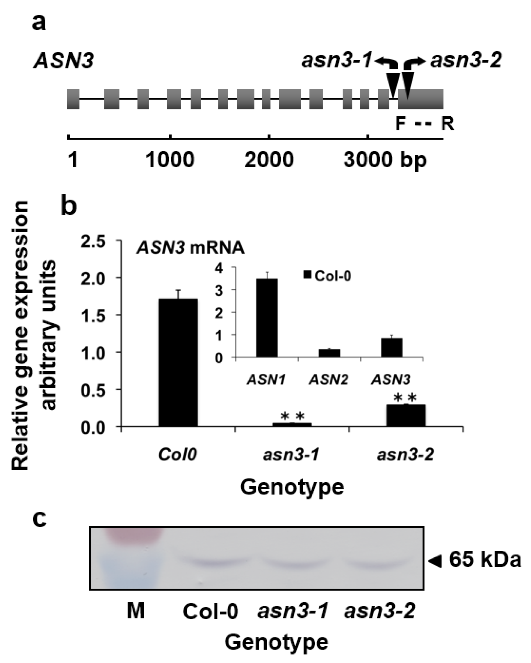
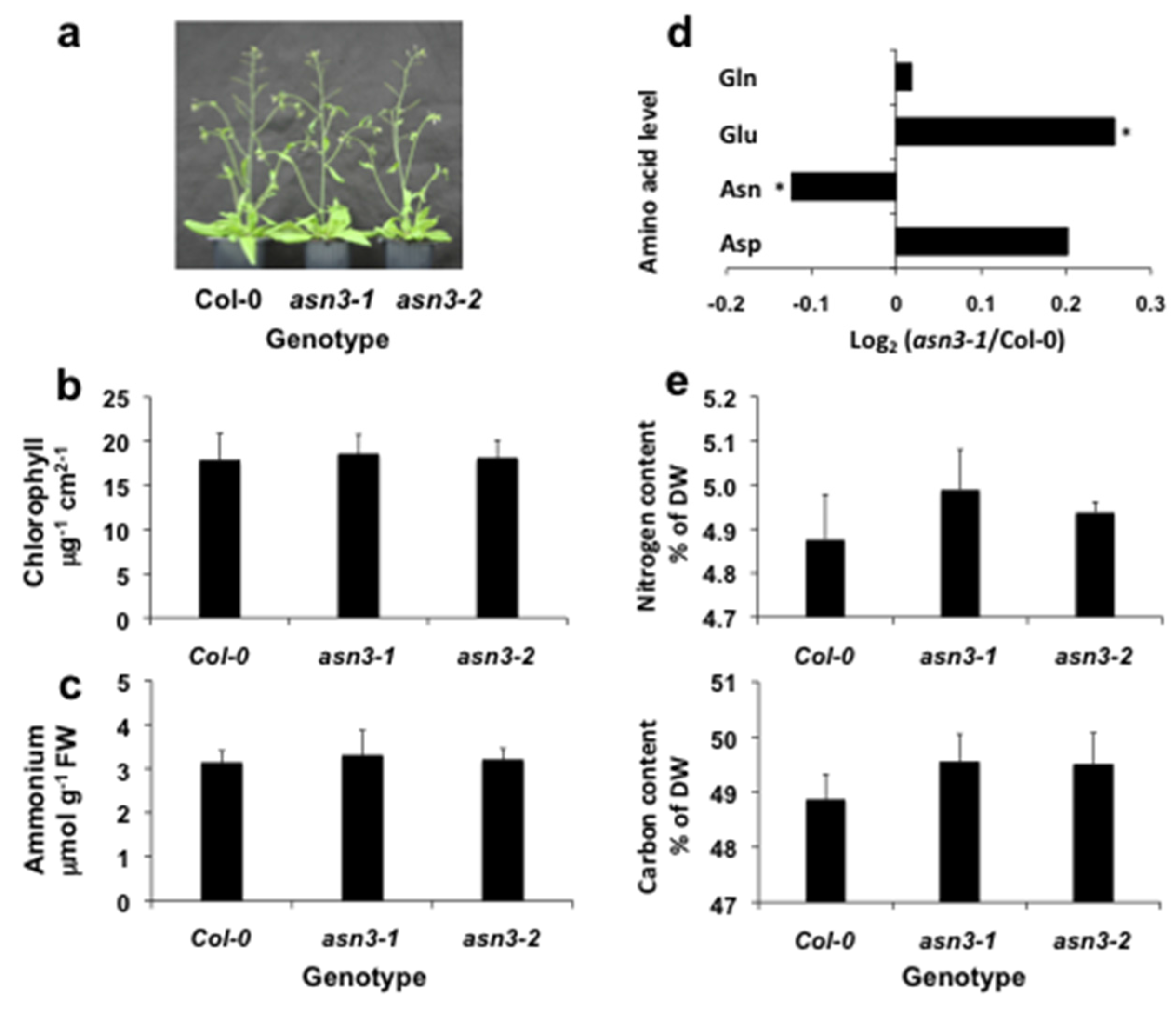
3.1. Characterization of asn3 Mutants

The Arabidopsis genome database [4] contains three functional genes for asparagine synthetase (AS): ASN1 (At3g47340), ASN2 (At5g65010) and ASN3 (At5g10240). Arabidopsis T-DNA insertion lines for the ASN3 gene were PCR-screened and mutants containing a T-DNA insertion either in intron 13 or in exon 14 were isolated. Homozygous lines were PCR-selected and named asn3-1 (SALK_053490) and asn3-2 (SALK_074279), respectively (Figure 1a).
Figure 1. Characterization of asn3-1 and asn3-2 T-DNA insertion mutants using rosette leaves from 35-day-old Arabidopsis plants. (a) Schematic presentation of the T-DNA insertion site within the ASN3 gene of the asn3-1 (intron 13) and asn3-2 (exon 14) lines; (b) ASN1 mRNA, ASN2 mRNA and ASN3 mRNA levels of wild-type (Col-0) (insert), and ASN3mRNA levels in wild-type (Col-0), asn3-1 and asn3-2 lines; (c) Western blot showing asparagine synthetase protein in wild-type (Col-0), asn3-1 and asn3-2 lines. Boxes and lines in the gene structure correspond to exons and introns, respectively. Black triangles indication T-DNA insertions (the size is not to scale). ASN3 mRNA levels were measured by qPCR relative to Actin 2 (At3g18780) and expressed as the mean ± SE of three biological replicates. F and R represent forward and reverse primers, respectively. Asterisks indicate significant differences between the Col-0 and asn3 mutants using a Student’s t-test P-values ** p < 0.01. Molecular mass markers on the Western blot membrane (M) correspond to 55 kDa and 72 kDa (Thermo Fisher Scientific Inc, Villebon-sur-Yvette, France).
Agronomy 06 00012 g001
The phenotypic analysis was carried out using rosette leaves from 35-day-old plants. A qPCR analysis showed that wild-type leaves contained ASN3 mRNA levels that were lower than ASN1 but higher than ASN2 (Figure 1b). The asn3-1 and asn3-2 lines contained 2.4% and 17% of wild-type RNA levels, respectively (Figure 1b). Asparagine synthetase protein abundance in total soluble proteins of green leaves was examined by Western blots probed with antibodies raised against Arabidopsis recombinant ASN2-encoded asparagine synthetase [8]. It was assumed that the observed 65 kDa band on the membrane contained the three asparagine synthetase isoforms encoded by ASN1, ASN2 and ASN3 with molecular masses of 65.5 kDa, 65 kDa and 65.2 kDa, respectively, and that the asparagine synthetase 2 antibody cross-reacted with epitopes of asparagine synthetase 3 since the three asparagine synthetases share a 87% to 92% amino acid similarity [3]. This indicated that rosette leaves of asn3-1 and asn3-2 lines displayed asparagine synthetase protein contents that were similar to wild-type (WT ) plants (Figure 1c).

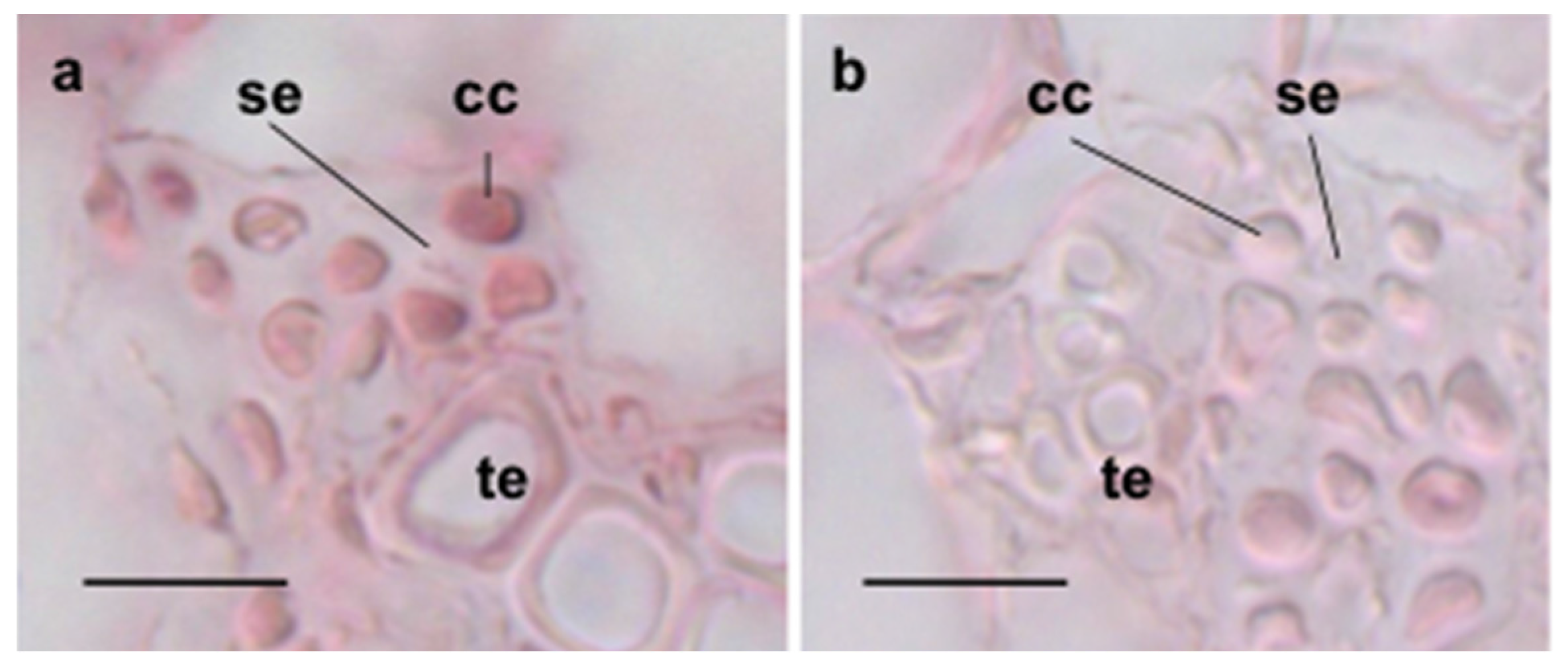
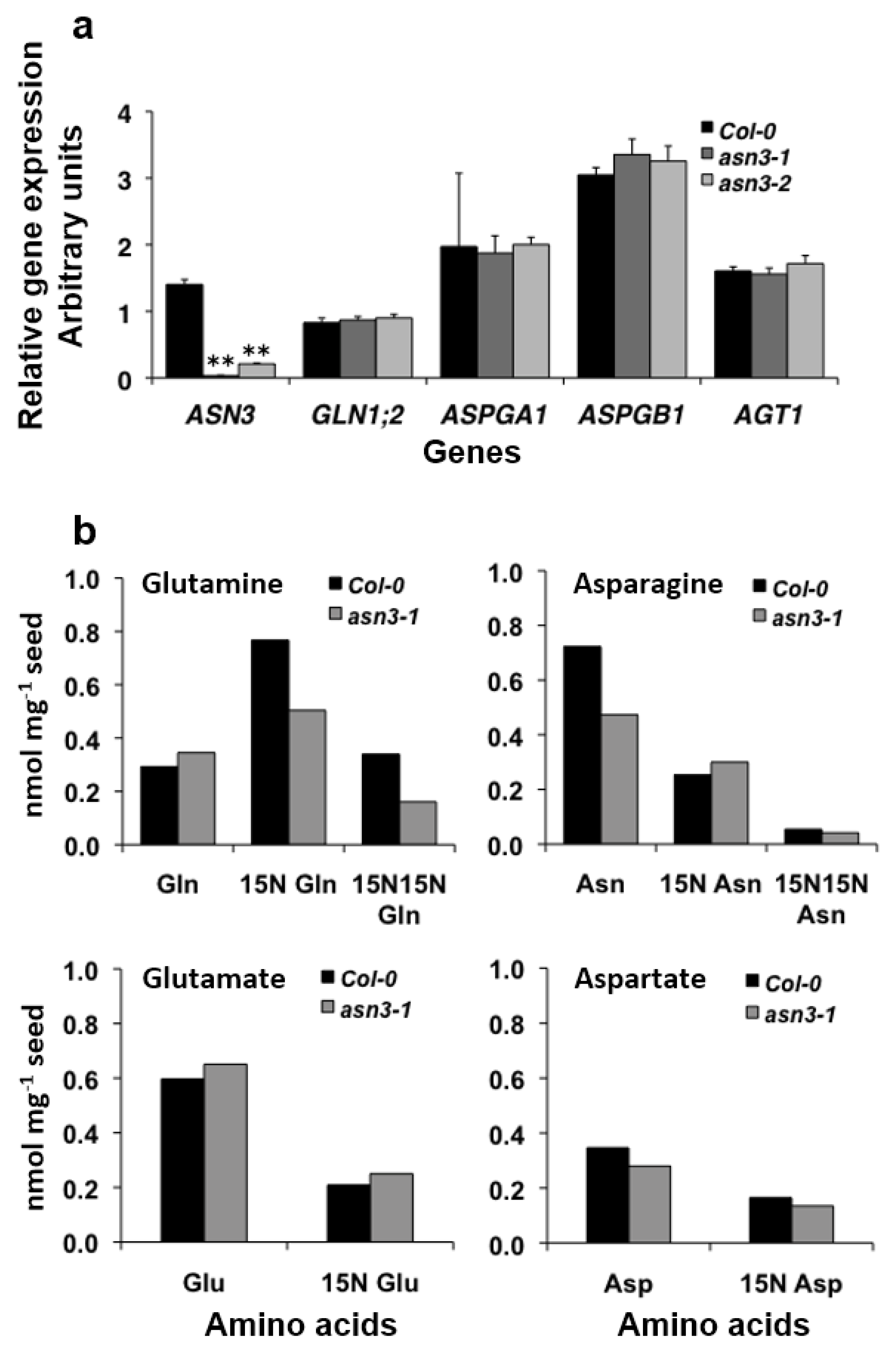
3.2. Cellular Expression of ASN3 mRNA and Phenotypic Analysis of asn3 Mutants

As our aim was to evaluate the function of ASN3-encoded asparagine synthetase in Arabidopsis development, comparative phenotypic analyses were carried out on both asn3 mutants and Col-0 plants. Because the function of asparagine synthetase is closely related to cellular localization, cell-specific expression patterns of ASN3 mRNA were first determined by in situ hybridization. Thin leaf sections were hybridized with either antisense or control sense ASN3 probes. A specific brown signal was associated with the companion cell/sieve tube element complex within the minor veins (Figure 2a).
Figure 2. In situ hybridization of ASN3 mRNA in Arabidopsis leaves with (a) antisense ASN3 mRNA and (b) control sense ASN3 mRNA. Digoxigenin (DIG)-UTP-labeled in situ hybridization signals were detected by microscopy. cc: companion cell, se: sieve element, te: tracheary element. Bar = 20 µm.
Agronomy 06 00012 g002
The specificity of the signal was controlled by hybridization of leaf sections with a sense ASN1 mRNA probe which gave no specific signal (Figure 2b).
A phenotypic analysis of the asn3-1 and asn3-2 lines was carried out 35 days after sowing at the vegetative stage and compared with the Col-0 line. No visible phenotype was detected for the asn3-1 and asn3-2 mutants (Figure 3a). Both asn3-1 and asn3-2 rosette leaves contained wild-type levels of chlorophyll (Figure 3b) and ammonium content (Figure 3c), indicating that ASN3 disruption did not cause a defective nitrogen status during vegetative growth. During seed development, leaves and stems serve as source tissues to supply nitrogen resources to developing siliques which in turn deliver nitrogen to seeds. As asparagine is one of the primary nitrogen carriers from the source to sink organs, the impact of ASN3 disruption on asparagine, glutamine, aspartate and glutamate levels was investigated in asn3-1 siliques and compared to the WT situation. Fourth siliques numbered from the top of the plants at stage 8 [16], mainly containing early- to late-heart-stage embryos, were harvested. Soluble metabolites were quantified by GC-MS, and differences were expressed as log2([amino acid]asn3-1/[amino acid]Col-0). The young siliques of the asn3-1 knockout line showed an increase in glutamine (Glnasn3-1 to GlnCol-0 ratio of 1.014), glutamate (Gluasn3-1 to GluCol-0 ratio of 1.189) and aspartate (Aspasn3-1 to AspCol-0 ratio of 1.149) and a decrease in asparagine (Asnasn3-1 to AsnCol-0 ratio of 0.902) (Figure 3d). To evaluate the effect of ASN3 disruption on nitrogen remobilization to seeds (the ultimate sink organ), the total nitrogen and carbon contents of dry seeds were determined using a micro-Carbon Nitrogen (CN) analyzer. The total nitrogen and total carbon contents of asn3-1 and asn3-2 seeds were not statistically different from those of the wild type (Figure 3e).
Figure 3. Phenotypic analysis of Arabidopsis asn3-1 and asn3-2 lines. (a) Representative visual growth phenotype; (b) chlorophyll content; and (c) ammonium content in rosette leaves of 35-day-old wild-type (Col-0), asn3-1 and asn3-2 rosettes; (d) ratios of selected amino acid contents in young siliques of Col-0 and asn3-1 expressed as Log2 [amino acid]asn3-1/[amino acid]Col-0. A positive value represents a higher metabolite content in the asn3-1 line, and a negative value corresponds to a lower metabolite content in the asn3-1 line; (e) total nitrogen and carbon contents in dry seeds of Col-0, asn3-1 and asn3-2 lines. The values represent the mean ± SE of three biological replicates. Asterisks indicate significant differences between the wild-type and transgenic lines with a Student’s t-test p values * p < 0.05.
Agronomy 06 00012 g003

3.3. 15N Labeling of Amino Acids in Germinating asn3 Seeds

Seed imbibition triggers quiescent seeds to become highly metabolic embryonic cells [17] in which nitrogen mobilization takes place for the synthesis of amino acids required for developing embryonic organs. Asparagine is one of the major free amino acids in the germinating seeds of Arabidopsis, serving to translocate nitrogen within the seed. To investigate the physiological function of ASN3-encoded asparagine synthetase in germinating seeds, expression profiles of genes involved in asparagine metabolism including ASN3, GLN1;2 (At1g66200), ASPGA1 (At5g08100), ASPGB1 (At3g16150) and AGT1 (At2g13360) were determined in 24 h–imbibed seeds.
Figure 4 shows the pathways of asparagine metabolism. GLN1;2 codes for a cytosolic GS1 that supplies glutamine for glutamine synthetase activity. ASPGA1 and ASPGB1 code for cytosolic asparaginase isoforms (ASPG, EC 3.5.1.1) and AGT1 codes for peroxisomal asparagine aminotransferase (AsnAT, EC 2.6.1.45). Asparaginase and asparagine aminotransferase release the amide group and amino group of asparagine as ammonia, respectively, and both nitrogen groups are used for subsequent amino acid synthesis [18]. Total RNA was isolated from Col-0 and asn3-1 seeds imbibed for 24 h, and mRNA levels were measured by qPCR. The level of ASN3 mRNA was reduced to 2.5% and 15% of the wild-type value in the seeds of asn3-1 and asn3-2, respectively (Figure 5a).
Figure 4. Schematic presentation of asparagine metabolism. AS: asparagine synthetase (EC 6.3.5.4), Asn: asparagine, AsnAT: asparagine aminotransferase (EC 2.6.1.45), Asp: aspartate, AspAT: aspartate aminotransferase (EC 2.6.1.1), ASPG: asparaginase (EC 3.5.1.1), Gln: glutamine, Glu: glutamate, GOGAT: glutamate synthase (Ferredoxin-GOGAT, EC 1.4.7.1 and NADH-GOGAT, EC 1.4.1.14), GS: glutamine synthetase (EC 6.3.1.2), OAA: oxaloacetate, 2-OG: 2-oxoglutarate, OSA: 2-oxosuccinamate.
Agronomy 06 00012 g004
Similar GLN1;2 and AGT1 mRNA levels were detected in wild-type and asn3 seeds. Of the two ASPG genes, ASPGB1 showed higher mRNA levels than ASPGA1 in both wild-type and asn3 seeds (Figure 5a).
To investigate the function of ASN3-encoded asparagine synthetase in the synthesis of amino acids in seeds, 15N labeling patterns of glutamine, glutamate, asparagine and aspartate were accessed by focusing on the asn3-1 knockout line. Wild-type Col-0 and asn3-1 seeds were incubated with 15[N]ammonium (99% enrichment) for 24 h in the dark at 20 °C, and the total amino acids were extracted. The labeling of glutamine, glutamate, asparagine and aspartate was analyzed using a capillary electrophoresis system coupled to a mass spectrophotometer as described in the Materials and Methods (2.8). When compared to wild-type seeds, asn3-1 seeds displayed reduced glutamine (by 30%), asparagine (20%) and aspartate (20%) contents while exhibiting increased glutamate (10%) amounts (Figure 5b; Table S1). When control Col-0 seeds were incubated with [15N]ammonium for 24 h, 15N-labeled glutamine ([5-15N]glutamine + [2-15N]glutamine and [2,5-15N]glutamine) accounted for 79% of the total glutamine (Figure 5b; Table S1). A lower percentage of labeling was detected for glutamate ([2-15N]glutamate) (26%), aspartate ([2-15N]aspartate) (32%) and asparagine ([2-15N]asparagine + [4-15N]asparagine and [2,4-15N]asparagine) (30%) (Figure 5b; Table S1), thus indicating an active GS in wild-type seeds. Interestingly, asn3-1 seeds showed a lower percentage of 15N-labeled glutamine than Col-0 seeds (66% versus 79%) (Figure 5b; Table S1) while they contained enhanced 15N-labeled asparagine (42% versus 30%) and glutamate (28% versus 26%) when compared to Col-0 seeds (Figure 5b; Table S1). Aspartate was equally labeled in both wild-type and asn3-1 lines (Figure 5b; Table S1).
Figure 5. Comparison of (a) mRNA levels of ASN3, GLN1;2, ASPGA1, ASPGB1 and AGT1 and (b) 15N labeling of glutamine, glutamate, asparagine and aspartate in Col-0 and asn3-1 seeds imbibed for 24 h. A qRT-PCR analysis was carried out to estimate mRNA levels of ASN3 (At5g10240) coding for asparagine synthetase, GLN1;2 (At1g66200) for cytosolic GS1, ASPGA1 (At5g08100) and ASPGB1 (At3g16150) for asparaginase and AGT1 (At2g13360) for asparagine aminotransferase. Transcript levels, relative to Actin 2 (At3g18780) as a reference gene, are expressed as the mean ± SE of three biological replicates. The 15N labeling analysis was carried out on three sets, each comprising more than 150 seeds using the same initial weight per genotype to determine single labeling (15N glutamine, 15N glutamate, 15N asparagine, 15N aspartate) and double labeling (15N15N glutamine, 15N15N asparagine). Asterisks indicate significant differences between wild-type and transgenic lines with Student’s t test p-values ** p < 0.01.
Agronomy 06 00012 g005

4. Discussion

Three ASN genes encode asparagine synthetase in Arabidopsis, and little is known about the role of asparagine synthetase encoded by ASN3. We assessed the physiological role of ASN3-encoded asparagine synthetase in nitrogen metabolism at three developmental stages including vegetative growth, seed maturation and seed germination. During vegetative growth, asn3-1 knockout and asn3-2 knockdown mutant leaves displayed wild-type asparagine synthetase protein, chlorophylls and ammonium contents and no visible phenotype (Figure 3). A weak effect of ASN3 disruption at this stage of development can be associated with lower ASN3 expression levels relative to ASN1 and ASN2 in vegetative leaves, as indicated by qPCR (Figure 1; [8]). Also, lower ASN3 mRNA levels have been reported by transcriptomics analysis of eight-day-old whole seedlings (expression ratio of ASN1:ASN2:ASN3 = 1.7:5.5:1), young rosette leaf n = 6 (ASN1:ASN2:ASN3 = 0.4:7:1) and seeds at the green cotyledon stage (ASN1:ASN2:ASN3 = 22:0.1:1) [19]. However, among seven organs under different developmental stages, higher ASN3 mRNA levels were reported for the seven-day-old shoot apex (ASN1:ASN2:ASN3 = 0.2:0.6:1) [19]. Despite the low mRNA level measured by qPCR, our in situ mRNA hybridization analysis localized ASN3 mRNA to companion cells that are in close vicinity to the sieve element of the leaf phloem vasculature (Figure 2), allowing a symplastic loading of asparagine into the phloem sieve element for export. As amino acids and peptides serve to translocate nitrogen through the phloem from source to sink organs, it was interesting to find that the asparagine content of asn3-1 siliques was reduced while the glutamate content was enhanced (Figure 3d). However, although siliques remobilize nitrogen to developing seeds during embryogenesis and maturation [7], the total nitrogen contents of dry seeds of Col-0, asn3-1 and asn3-2 were found to be similar (Figure 3), suggesting that ASN3 disruption did not affect seed nitrogen status.
During nitrogen mobilization in seeds imbibed for 24 h, exogenous (15N)ammonium was assimilated into amino acids in both the Col-0 and asn3-1 lines (Figure 5; Table S1). The asn3-1 seeds exhibited changes in amino acid composition and (15N)amide and (15N)amino acid labeling patterns. In particular, a 20% reduction of the total asparagine content in the asn3-1 seeds relative to the Col-0 seeds could be caused by a reduced supply of non-labeled amino acids from the seed (0.47 nmol·mg−1 seed in asn3-1 and 0.72 nmol·mg−1 seed in Col-0) due to the disruption of ASN3, while 15N-labeled asparagine ((2-15N)asparagine + (4-15N)asparagine and (2,4-15N)asparagine) was enhanced from 30% in Col-0 seeds to 42% in asn3-1 seeds (Figure 5; Table S1). The increase in the amount of labeled asparagine in germinated asn3-1 seeds, depending on its synthesis and hydrolysis, could be associated with a low availability of labeled aspartate and glutamine, precursors for asparagine synthesis. This is in agreement with the lower aspartate content and contrasting higher glutamate level in asn3-1 seeds compared to Col-0 seeds despite the reversible transamination reaction between aspartate and glutamate catalyzed by aspartate aminotransferase (EC 2.6.1.1) (Figure 5; Table S1). The asn3-1 seeds displayed a lower total content of 15N-labeled amino acids (glutamine, glutamate, asparagine and aspartate) than Col-0 seeds. This decline in asn3-1 seeds was associated with lower amounts of 15N-labeled glutamine and aspartate and with higher 15N-labeled asparagine and glutamate levels (Figure 5b; Table S1). These altered 15N labeling patterns of glutamine, asparagine, glutamate and aspartate might be correlated with a lower GS activity and increased asparagine synthetase and glutamate synthase activities in asn3-1 seeds. Since GLN1;2 expression was similar in asn3 and wild-type seeds, the reduced glutamine content could reflect an altered utilization rather than a modified synthesis. Likewise, the lower content of non-labeled asparagine could be associated with its hydrolysis by asparaginase into ammonium and aspartate, which is reversibly transaminated to glutamate, and asparagine aminotransferase that produces 2-oxosuccinamate which is converted to ammonium and oxaloacetate by ω-amidase (EC 3.5.1.3) [20]. This would produce intermediates to feed the tricarboxylic acid (TCA ) cycle (oxaloacetate, 2-oxoglutarate, and malate) and the γ-aminobutyric acid (GABA ) shunt (glutamate) for amino acid synthesis and energy generation at the expense of asparagine. It was found that germinating asn3-1 seeds expressed wild-type ASPGA1 and ASPGB1 mRNA levels (Figure 5). However, a promoter analysis of ASPGA1::GUS demonstrated an expression in seed epidermal cells that began 24 h after sowing [21], suggesting an implication of asparaginase in asparagine hydrolysis. Moreover, the imbibed asn3-1 seeds contained wild-type levels of AGT1 mRNA (Figure 5). AGT1 is the single gene encoding serine:glyoxylate aminotransferase which catalyzes transamination reactions with multiple substrates including asparagine as an amino donor [22,23,24]. Previous studies demonstrated that Arabidopsis asparagine aminotransferase acts as a serine:glyoxylate aminotransferase [24]. During photorespiration in leaves, this peroxisomal aminotransferase catalyzes the transamination of serine with glyoxylate to give glycine and hydroxypyruvate. Thus, asparagine aminotransferase might play a role in detoxifying glyoxylate which can inhibit RuBisCO activity. Both the wild-type expression level of AGT1 and its high catalytic efficiency, expressed as Vmax/Km, of asparagine aminotransferase (10.4 × 10⁻⁸ kcat mg−1·mM−1), are similar to that of ASPGB1-encoded asparaginase (9.72 × 10⁻⁸ kcat mg−1·mM−1) [24,25], thus suggesting that asparagine hydrolysis not only provides ammonium but also pre-conditions aspartate and glutamate in response to the lower energy status of the germinating seeds. Despite ASN3 disruption, increased levels of 15N-labeled asparagine in asn3-1 seeds may be due to the decreased endogenous asparagine content, suggesting that ASN3-encoded asparagine synthetase may contribute to providing at least a basal level of asparagine in germinating seeds. Indeed, our data are in agreement with a fine regulation of substrate supply to asparagine synthesis and its hydrolysis in Arabidopsis organs.

Supplementary Files

Supplementary File 1

Acknowledgments

We would like to thank Michael Hodges for language editing of the manuscript. We would also like to thank to Joël Talbotec, Philippe Marechal and Hervé Ferry for plant maintenance.

Author Contributions

The work was carried out by all authors who contributed to different extents to experiment design, analytical methods and experiments, and the writing of the manuscript.

Conflicts of Interest

The authors declare no conflict of interest.

References

  1. Coruzzi, G.M. Primary N-assimilation into amino acids in Arabidopsis. In The Arabidopsis Book; Somerville, C., Meyerrowitz, E., Eds.; American Society of Plant Biologists: Rockville, MD, USA, 2003; pp. 1–17. [Google Scholar]
  2. Lea, P.J.; Sodeck, L.; Parry, M.A.J.; Shewry, P.R.; Halford, N.G. Asparagine in plants. Ann. Appl. Biol. 2007, 150, 1–26. [Google Scholar] [CrossRef]
  3. Gaufichon, L.; Reisdorf-Cren, M.; Rothstein, S.J.; Chardon, F.; Suzuki, A. Biological functions of asparagine synthetase in plants. Plant Sci. 2010, 179, 141–153. [Google Scholar] [CrossRef]
  4. Arabidopsis Genome Initiative. Analysis of the genome sequence of the flowering plant Arabidopsis thaliana. Nature 2000, 408, 796–815. [Google Scholar]
  5. Herrera-Rodriguez, M.B.; Maldonado, J.M.; Perez-Vicente, R. Light and metabolic regulation of HAS1, HAS1.1 and HAS2, three asparagine synthetase genes in Helianthus annuus. Plant Physiol. Biochem. 2004, 42, 511–518. [Google Scholar] [CrossRef] [PubMed]
  6. Bläsing, O.E.; Gibon, Y.; Günther, M.; Höhne, M.; Morcuende, R.; Osuna, D.; Thimm, O.; Björn, U.; Scheobe, W.-R.; Stitt, M. Sugars and circadian regulation make major contributions to the global regulation of diurnal gene expression in Arabidopsis. Plant Cell 2005, 17, 3257–3281. [Google Scholar] [CrossRef] [PubMed]
  7. Lam, H.-M.; Wong, P.; Chan, H.-K.; Yam, K.-M.; Cheng, L.; Chow, C.-M.; Coruzzi, G.M. Overexpression of the ASN1 gene enhances nitrogen status in seeds of Arabidopsis. Plant Physiol. 2003, 132, 926–935. [Google Scholar] [CrossRef] [PubMed]
  8. Gaufichon, L.; Masclaux-Daubresse, C.; Tcherkez, G.; Reisedorf-Cren, M.; Sakakibara, Y.; Hase, T.; Clément, G.; Avice, J.-C.; Grandjean, O.; Marmagne, A.; et al. Arabidopsis thaliana ASN2 encoding asparagine synthetase is involved in the control of nitrogen assimilation and export during vegetative growth. Plant Cell Environ. 2013, 36, 328–342. [Google Scholar] [CrossRef] [PubMed]
  9. Wong, H.-K.; Chan, H.-K.; Coruzzi, G.M.; Lam, H.-M. Correlation of ASN2 gene expression with ammonium metabolisms in Arabidopsis. Plant Physiol. 2004, 134, 332–338. [Google Scholar] [CrossRef] [PubMed]
  10. Coïc, Y.; Lessaint, C. Comment assurer une bonne nutrition en eau et en ions minéreaux en holticulture? Holticulture Fr. 1971, 8, 11–14. [Google Scholar]
  11. Laemmli, U.K. Cleavage of structural proteins during the assembly of bacteriophage T4. Nature 1970, 227, 680–685. [Google Scholar] [CrossRef] [PubMed]
  12. Arnon, D.I. Copper enzymes in isolated chloroplasts. Polyphenol oxidase in Beta vulgaris L. Plant Physiol. 1949, 24, 1–15. [Google Scholar] [CrossRef] [PubMed]
  13. Fiehn, O. Metabolic profiling in Arabidopsis. Method Mol. Biol. 2006, 323, 439–447. [Google Scholar]
  14. Saeed, A.I.; Sharov, V.; White, J.; Li, J.; Liang, W.; Bhagabati, N.; Braisted, J.; Klapa, M.; Currier, T.; Thiagarajan, M.; et al. TM4: A free, open-source system from microarray data management and analysis. Biotechniques 2003, 34, 374–378. [Google Scholar] [PubMed]
  15. Boyes, D.C.; Zayed, A.M.; Ascenzi, R.; McCaskill, A.J.; Hoffman, N.E.; Davis, K.R. Growth stage-based phenotypic analysis of Arabidopsis: A model for high throughput functional genomics in plants. Plant Cell 2001, 13, 1499–1510. [Google Scholar] [CrossRef] [PubMed]
  16. Takahashi, H.; Hayashi, M.; Goto, F.; Sato, S.; Soga, T.; Nishioka, T.; Tomita, M.; Kawai-Yamada, M.; Uchimiya, H. Evaluation of metabolic alteration in transgenic rice overexpressing dihydroflavonol-4-reductase. Ann. Bot. 2006, 98, 819–825. [Google Scholar] [CrossRef] [PubMed]
  17. Gallardo, K.; Job, C.; Groot, S.P.C.; Puype, M.; Demol, H.; Vandekerckhove, J.; Job, D. Proteomics of Arabidopsis seed germination. A comparative study of wild-type and gibberellin-deficient seeds. Plant Physiol. 2007, 129, 823–837. [Google Scholar] [CrossRef] [PubMed]
  18. Ireland, R.J.; Lea, P.J. The enzymes of glutamine, glutamate, asparagine and aspartate metabolism. In Plant Amino Acids; Singh, B.K., Ed.; Marcel Dekker, Inc.: New York, NY, USA; Basel, Switzerland; Hong Kong, China, 1999; pp. 49–109. [Google Scholar]
  19. Schmid, M.; Davidson, T.S.; Hnz, S.; Pap, U.J.; Demar, M.; Vingron, M.; Schölkopf, B.; Weigel, D.; Lohmann, J.U. A gene expression map of Arabidopsis thaliana development. Nat. Genet. 2005, 37, 501–506. [Google Scholar] [CrossRef] [PubMed]
  20. Zhang, Q.; Marsolais, F. Identification and characterization of omega-amidase as an enzyme metabolically linked to asparagine transamination in Arabidopsis. Phytochemisty 2014, 99, 36–43. [Google Scholar] [CrossRef] [PubMed]
  21. Ivanov, A.; Kameka, A.; Pajak, A.; Bruneau, L.; Beyaert, R.; Hernández-Sebastià, C.; Marsolais, F. Arabidopsis mutants lacking asparaginases develop normally but exhibit enhanced root inhibition by exogenous asparagine. Amino Acids 2012, 42, 2307–2318. [Google Scholar] [CrossRef] [PubMed]
  22. Joy, K.W.; Prabha, C. The role of transamination in the synthesis of homoserine in peas. Plant Physiol. 1986, 82, 99–102. [Google Scholar] [CrossRef] [PubMed]
  23. Liepman, A.H.; Olsen, L.J. Peroxisomal alanine:glyoxylate aminotransferase (AGT1) is a photorespiratory enzyme with multiple substrates in Arabidopsis thaliana. Plant J. 2001, 25, 487–498. [Google Scholar] [CrossRef] [PubMed]
  24. Zhang, Q.; Lee, J.; Pandurangan, S.; Clark, M.; Pajak, A.; Marsolas, F. Characterization of Arabidopsis serine: Glyoxylate aminotransferase, AGT1, as an asparagine aminotransferase. Phytochemistry 2013, 85, 30–35. [Google Scholar] [CrossRef] [PubMed]
  25. Gabriel, M.; Telmer, P.G.; Marsolais, F. Role of asparaginase variable loop at the carboxyl terminal of the alpha subunit in the determination of substrate preference in plants. Planta 2012, 235, 1013–1022. [Google Scholar] [CrossRef] [PubMed]

