Article

Detection and Characterization of Plum Pox Virus (Potyvirus plumpoxi) Marcus Strains in Spanish Apricot and Peach Orchards Through RNA-Seq Analysis


by
Lucía Rodríguez-Robles
,
Pedro J. Martínez-García
,
Pedro Martínez-Gómez
* and
Manuel Rubio
Department of Plant Breeding, CEBAS-CSIC, Espinardo, P.O. Box 164, E-30100 Murcia, Spain
*
Author to whom correspondence should be addressed.
Agronomy 2026, 16(6), 608; https://doi.org/10.3390/agronomy16060608
Submission received: 29 January 2026 / Revised: 2 March 2026 / Accepted: 8 March 2026 / Published: 12 March 2026
(This article belongs to the Collection Crop Breeding for Stress Tolerance)

Abstract

Cultivated species of the Prunus genus are of great economic importance worldwide and can be severely affected by viral diseases that compromise both yield and fruit quality. Among the most significant is Potyvirus plumpoxi (PPV), the causal agent of sharka disease, which has a direct and severe impact on stone fruit production. In this study, high-throughput RNA sequencing was employed to detect and characterize viruses present in commercial peach and apricot orchards located in different regions of Spain. After processing five samples, a total of ten viruses were identified, with PPV being the predominant virus in all analyzed samples, specifically the Marcus strain (PPV-M), which is described as one of the most aggressive PPV strains. In addition, other viruses were detected with high sequencing depth, including Luteovirus nucipersicae (nectarine stem pitting associated virus, NSPaV) and Peach-associated luteovirus (PaLV). Single-nucleotide variation (SNV) analysis of PPV-M populations revealed specific mutations distributed across the viral genome. Furthermore, phylogenetic analyses indicated the presence of multiple infection sources of European origin. These results highlight the presence of PPV-M in Spain, providing evidence of different routes of exchange of infected plant material. These findings underscore the need to strengthen monitoring programs, certification of planting material, and phytosanitary control measures to limit the dissemination of viruses and minimize their impact on stone fruit production.

1. Introduction

The genus Prunus, belonging to the subfamily Prunoideae within the family Rosaceae, is of economic importance due to comprising numerous species that produce edible drupes [1]. It includes more than 230 species, which are mainly classified into three major subgenera (Amygdalus, Cerasus, and Prunus), in addition to the desert almond subgenus Eplectocladus [2]. In 2022, global production of stone fruit trees was estimated at approximately 50 million tons, with peaches and nectarines being the most important crops, accounting for around 25 million tons [3].
Peach (Prunus persica L. Batsch) belongs to the Rosaceae family and is among the most widely cultivated perennial fruit trees worldwide. Global production reached 27,077,873 tons in a cultivated area of 1,561,641 ha [3]. Spain is one of the most important producing countries, where peach and nectarine cultivation leads stone fruit production with over 1,300,000 tons, followed by apricots with 155,567 tons and plums with 135,749 tons [4]. Stone fruit production is severely threatened by pests and diseases, among which sharka disease, caused by the Potyvirus plumpoxi (PPV; family Potyviridae), is notably significant [5]. PPV infection has a substantial economic impact, reducing yield by up to 80% and producing unmarketable infected fruits due to their appearance, low sugar content, poor flavor, and reduced shelf life [6,7]. The expression of PPV symptoms in host plants varies in type and severity according to the viral strain, the time of infection, the cultivar, and environmental conditions [8]. The first symptoms appear on leaves, including mild light-green discoloration, chlorotic spots, bands, rings, vein clearing, yellowing, and/or leaf deformation. Infected fruits show chlorotic spots, faintly pigmented yellow rings, and often become deformed and even show brown or necrotic areas in the rings [9]. Emphasis is placed on virus epidemics in stone fruit orchards, as they can rapidly spread, leading to reductions in yield and fruit quality, as well as costs associated with tree eradication.
In Spain, sharka was detected for the first time in 1984 [10]. Among the different strains of PPV described [5], only less aggressive Dideron-type isolates (PPV-D) have widely spread; they have mainly affected apricot production but not peach or plum (Prunus salicina Lindl.) production. Marcus-type (PPV-M) isolates are more aggressive and greatly affect peach fruit production, inducing rapid epidemics in peach, apricot (Prunus armeniaca L.), and plum [9,11]. PPV-M was detected for the first time in Aragón (northern Spain) in 2004, and it was eradicated [12]. However, a few years ago, outbreaks of Marcus strains began to reappear. Catalunya was the first region to officially declare the fight against sharka Marcus in 2015 [13], followed by Aragón in 2020 [14]. However, PPV was no longer considered a quarantine organism by the European Union (Implementing Regulation EU 2019/2072).
On the other hand, mixed infections by plant viruses are common in nature, highlighting the need for comprehensive transcriptomic analyses to simultaneously detect and quantify co-infecting viruses [15]. Therefore, advanced molecular tools are essential for the detection and characterization of plant viruses. High-throughput sequencing (HTS) technology, including RNA sequencing (RNA-Seq), is a key tool for virus screening in natural habitats [16]. RNA-Seq enables the simultaneous detection of multiple viruses, including both RNA and DNA viruses, by leveraging viral reference databases such as the Database of Plant Viruses (DPV) and the National Center for Biotechnology Information (NCBI) [16,17,18]. In addition, through de novo assembly approaches, undescribed viruses can potentially be discovered [19]. In Korea, five viruses belonging to the family Betaflexiviridae, a novel virus from the family Tymoviridae, and two viroids were detected in peach using RNA-seq [20]. In the United States (USA), RNA-seq analysis of peach samples from 14 commercial orchards detected several viruses, including prunus necrotic ringspot virus (PNRSV) and the viroid peach latent mosaic viroid (PLMVd), as well as viruses detected in peach for the first time, such as tomato mosaic virus (ToMV) [21]. HTS has also been employed for virus detection in other Prunus species. Small RNA-seq of apricot varieties detected cherry virus A (ChVA) and little cherry virus 1 (LChV-1) for the first time in Hungary [22]. In Korea, hop stunt viroid (HSVd), apple chlorotic leaf spot virus (ACLSV), plum bark necrosis stem pitting-associated virus (PBNSPaV), and PNRSV were identified in plums through RNA-seq, highlighting the need to validate viral detection with RT-PCR due to cases of misidentification [23]. However, despite being one of the main Prunus-producing countries, plant virus detection in Spanish orchards using RNA-seq is scarce.
Beyond virus detection, NGS enables the characterization of viral genetic variability through the analysis of viral populations within the host, referred to as the ‘virome’ [20,24]. Additionally, it allows the inference of evolutionary origins and the reconstruction of phylogenetic relationships among viral populations [24,25,26]. These approaches provide insights into the processes by which viruses diversify, adapt, and spread [27]. In peach, 18 genomes of viruses and viroids were obtained using transcriptomes, detecting hundreds of single-nucleotide polymorphisms, which revealed genetic relationships through phylogenetic analyses [20]. Phylogenetic trees of apricot viral isolates reveal the genetic proximity between isolates from different hosts and geographic regions, providing a framework for classification and evolutionary insights [22]. In other fruit tree species, such as apple, phylogenetic analysis of viral populations obtained from transcriptome data has revealed that apple stem grooving virus (ASGV) and citrus tatter leaf virus (CTLV) are the same virus [28].
In the present study, HTS using the Illumina platform was employed to screen for plant viruses in commercial peach and apricot orchards that showed severe sharka symptoms. An RNA-Seq detection workflow, including de novo assembly, was implemented in Prunus species, enabling the simultaneous detection of multiple virus species, including the important outbreak of the aggressive PPV Marcus (PPV-M) strain in Spain, confirming the presence of Marcus isolates in some of the country’s main peach-producing areas. Additionally, variant calling was performed to identify specific population mutations of the PPV-M. These results provide new insights into the phylogeny of PPV-M strain populations and supply novel sequences for the plant virology community.

2. Materials and Methods

2.1. Plant Material and RNA Extraction

Peach leaf samples were collected from nine commercial orchards located in Aragón, Catalunya, and the Región de Murcia. In addition, an apricot leaf sample was collected from a commercial farm in Catalunya. Leaf samples showing strong symptoms of sharka disease, confirmed by the presence of sharka symptoms on the fruits, were analyzed. Leaf tissues from each sample were immersed in 30 mL of RNALater® (Sigma-Aldrich, Darmstadt, Germany) solution in Falcon tubes for two days, till our arrival at the lab, where samples were ground with liquid nitrogen using a mortar and pestle and stored at −80 °C until RNA extraction.
For total RNA extraction, 100 mg of ground leaf tissue from each sample was extracted using the NucleoSpin® RNA Plant and Fungi kit (Macherey-Nagel, Düren, Germany), following the manufacturer’s protocol. To eliminate possible DNA contamination, extracts were treated with DNase I (Invitrogen, Carlsbad, CA, USA). RNA integrity was validated by agarose gel electrophoresis, and concentration was measured using a NanoDropTM One spectrophotometer (Thermo ScientificTM, Wilmington, DE, USA). Extracts were normalized to a final volume of 40 µL for downstream analyses.
Among the ten collected samples, those that tested unequivocally positive with PPV-M-specific primers (P1-PM) [29] were selected. Five samples were ultimately chosen for RNA-seq analysis: three peaches from Aragón (ARA1, ARA2, and ARA3), one apricot from Catalunya (CAT1), and one peach from Murcia (MUR1). Samples ARA2, ARA3, and CAT1 were collected at the border between the two regions (an area of about 15 km in diameter).

2.2. Preparation of Libraries for RNA-Seq

The 15 libraries (5 samples × 3 replicates) were constructed from total RNA using the QIAseq FastSelect –rRNA Plant Kit (Qiagen sciences, Germantown, MD, USA) that removes ribosomal RNA sequences, followed by enrichment of messenger RNA. Library quality was assessed with the QIAxcel Advanced System (Sample to Insight—QIAGEN). Sequencing was performed on the Illumina NovaSeq platform (Illumina Inc., San Diego, CA, USA), generating an average of 52 million reads per sample. Paired-end sequencing was carried out with 2 × 150 bp reads (300 cycles).

2.3. RNA-Seq Bioinformatic Analyses

Raw FASTQ files were processed for adapter removal and low-quality read trimming using Trimmomatic v0.39. [ILLUMINACLIP:TruSeq3-PE-2.fa:2:30:10 HEADCROP:13 LEADING:30 TRAILING:30 SLIDINGWINDOW:4:30 MINLEN:36] [30,31]. These options removed adapter and Illumina-specific sequences (with 2 mismatches allowed in the seed, a palindrome clip threshold of 30, and a simple clip threshold of 10), cropped the first 13 bases of each read, trimmed bases from the 5′ and 3′ ends if their quality score was below 30, applied a sliding window of 4 bases with a minimum average quality of 30, and discarded reads shorter than 36 bases after trimming. Curated RNA-Seq reads were aligned to the host genomes (Prunus persica ‘Lovell’ v2 and Prunus armeniaca ‘Rojo Pasión’ v1) [32,33] using Bowtie2 [34]. Reads that mapped to the host genomes were removed to eliminate plant-derived sequences. The non-host reads were retained for downstream viral analyses. To maximize read utilization, paired-end and single-end reads were combined into a single dataset.
Assembly was performed using Trinity v2.5.1 [35] with unpaired and paired reads. Assembled contigs were mapped using BLASTn [36] against a viral genomic sequence database of Prunus retrieved from the NCBI GenBank database, comprising 1223 complete viral genomes and 5336 partial sequences. BLAST v2.17.0 results were filtered for a contig length between 500 and 14,000 nucleotides (nt), a length of alignment higher than 300 nt, and an E-value lower than 1 × 10⁻⁵ [31]. The identified viruses were extracted from the database to perform a specific alignment of the non-host reads against them using BWA [37]. Subsequently, the output SAM files were processed, evaluating coverage and depth across the alignments.
A principal component analysis (PCA) was performed based on viral read counts to explore similarities among samples and biological replicates. For each virus, the most representative GenBank accession was selected. Only viruses detected in all three replicates of at least one sample or showing a cumulative read count higher than 100 across all samples were included in the analysis (Supplementary Table S1).

2.4. RT-PCR Virus Detection Validation

The presence of the identified viruses in leaf samples was confirmed by two specific RT-PCR assays targeting PPV-D and PPV-M.
Two independent RT-PCR assays were performed to detect PPV-D and PPV-M. Each reaction was carried out in a final volume of 25 µL and consisted of 2.5 µL of GoTaq DNA polymerase buffer (1×), 1.5 µL of MgCl2 (25 mM), and 2 µL of dNTPs (10 mM). To optimize the amplification, 2 µL of Triton X-100 (4%) and 0.5 µL of formamide were added. The reactions also contained the enzymes avian myeloblastosis virus reverse transcriptase (AMV RT) 0.2 µL (5 units) and GoTaq®Flexi DNA polymerase (Promega, Madison, WI, USA) 0.2 µL (5 units), 2 µL of the universal primer P1 (10 µM), and 2 µL of the strain-specific primer (PD for PPV-D or PM for PPV-M, both 10 µM).
The primers used for PPV detection were P1 (universal) (5′-ACCGAGACCACTACACTCCC-3′) as the forward primer; PPV-M (5′-CTTCAACAACGCCTGTGCGT-3′); and PPV-D (5′-CTTCAACGACACCCGTACGG-3′) [29].
The cycling program consisted of a reverse transcription step at 42 °C for 45 min, followed by an initial denaturation at 92 °C for 2 min. Amplification was then performed for 35 cycles of 92 °C for 30 s, 60 °C for 30 s, and 72 °C for 60 s, with a final extension at 72 °C for 10 min.
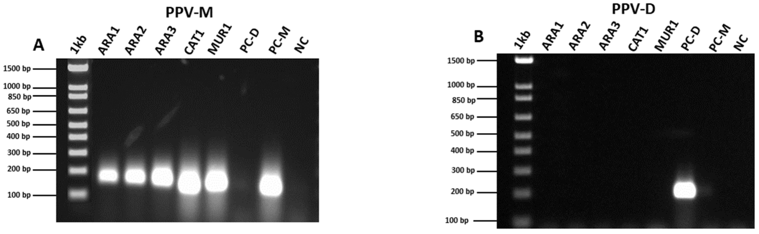
The resulting amplification products were subjected to electrophoresis on a 1.5% agarose gel in 1× TAE buffer and stained with Gel Red® (Biotium, Fremont, CA, USA) to confirm the amplification results. The expected amplicon size was 198 bp for both PPV-D and PPV-M.

2.5. Single Nucleotide Variant of Potyvirus plumpoxi (Marcus)

Sample-assembled contigs were aligned against the PPV Marcus strain (NCBI reference: AJ243957.1) [38], and contigs from the three replicates per sample were subsequently merged to obtain a representative set. Single-nucleotide variants (SNVs) were identified using SAMtools and BCFtools [39]. Specifically, the sorted BAM files were processed with SAMtools mpileup using the parameters -q 10 and -Q 20 to filter out reads with mapping quality below 10 and bases with base quality below 20, respectively, ensuring that only high-confidence alignments and bases were considered.

2.6. Phylogenetic Analyses

A consensus sequence for each merged sample was generated by incorporating the detected SNVs into the PPV Marcus reference genome. The PPV-M strain is one of the three major strains showing the broadest geographic distribution and is recognized as an evolutionarily successful recombinant strain of European origin [40]. Two phylogenetic analyses were then conducted using the five samples: (i) together with a subset of different PPV strains and (ii) together with a subset of the PPV Marcus strain from several countries (Supplementary Table S1). Only complete sequences publicly available in the NCBI database were included in these comparisons. Phylogenetic trees were constructed using the MEGA12 tool [41]. Sequences were aligned using ClustalW, and the tree was constructed by the neighbor-joining method with 1000 bootstrap replicates and maximum composite likelihood parameter distance [40].

3. Results

3.1. RNA-Seq Data Processing

An average of 52,664,838 paired-end raw reads per sample was generated (Table 1). Read curation (removal of adapter sequences, empty reads, and low-quality sequences) removed around 2 million reads per sample. After this step, 49,992,781 high-quality reads (96%) were retained and used for subsequent analyses. Reads that mapped to the host genomes were also removed, with an average of 31 million reads per sample discarded. The unmapped reads from the reference genome alignment were further analyzed using BLAST against a database generated from all known viruses affecting the genus Prunus available in NCBI.
The de novo assembly of RNA-Seq reads produced contigs with an average length of approximately 1000 nucleotides and an N50 of 1236 nucleotides, indicating a moderate level of assembly continuity. Contig lengths ranged from a minimum of ~380 nucleotides to a maximum exceeding 16,500 nucleotides, reflecting a broad size distribution of assembled sequences. The total number of assembled bases reached approximately 51 Mb per sample for subsequent analyses.

3.2. Virus Identification

Assembled contigs were used for virus identification using BLASTn against a viral database compiled from NCBI, containing all viruses reported to infect the genus Prunus (Supplementary Table S2). A total of ten distinct viruses were detected in the analyzed Prunus samples, including Potyvirus plumpoxi (PPV-M), youcai mosaic virus (YoMV), Luteovirus nucipersicae (NSPaV), peach-associated luteovirus (PaLV), Luteovirus avii (ChaLV, ChaLV), plum ourmia-like virus (POLV), Marafivirus pruni (PeVD), Marafivirus asteroides (GAMaV), grapevine red globe virus (GRGV), and peach virus T (PrVT) (Supplementary Table S3). Uniquely, PPV-M was detected in all analyzed samples, followed by NSPaV, which was consistently identified in all Huesca samples. In addition, principal component analysis (PCA) based on viral read counts showed a consistent clustering of biological replicates, supporting the robustness of virus detection and abundance estimates obtained for each sample (Supplementary Figure S1).
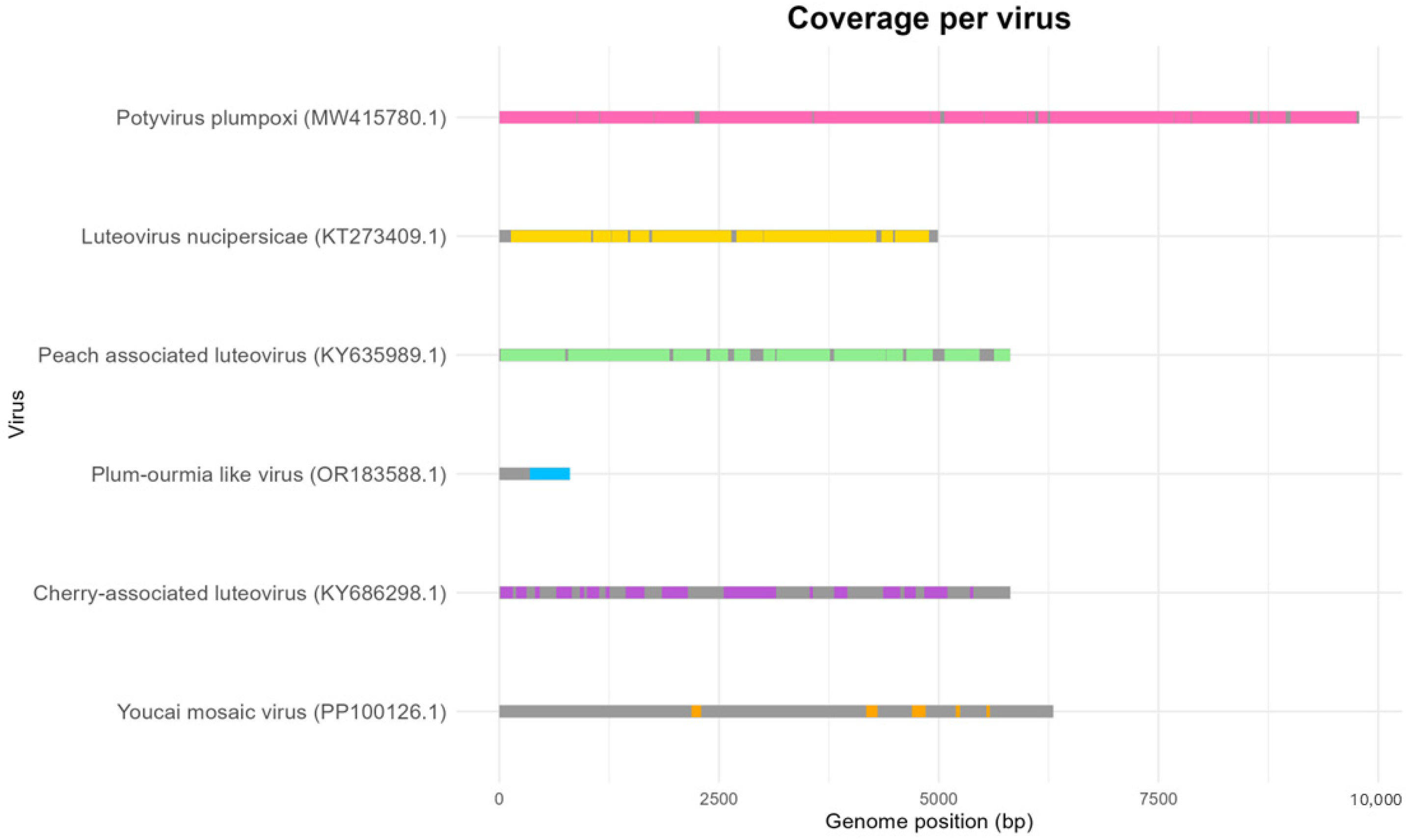
Across all analyzed samples (Table 2), PPV-M showed coverages higher than 99.5% and average depths higher than 2000×, except in the ARA2 sample (96.98% and 500×). Regarding NSPaV, coverages higher than 70% and depths exceeding 50× were observed in samples ARA2 and ARA1, reaching over 90% in ARA2. In contrast, samples CAT1 and ARA3 showed much lower values of coverage and depth (>10% and approximately 1–2×, respectively). ChaLV was mainly detected in ARA2 and ARA3, with coverages above 46% and an average depth of 70.87×. In contrast, the virus was detected to a much lesser extent in MUR1, where it appeared in only one replicate, with a coverage of 4.13% and an average depth of 1.7×. A similar pattern was observed in PaLV, which showed coverages above 89% in samples ARA2 and ARA3, with average depths higher than 90×, but MUR1 showed a much lower coverage of 2.35% and an average depth of only 1×. The remaining viruses (YoMV, GRGV, GAMaV, peach virus D, and peach virus T) were detected only residually in some samples, showing low coverage and/or sequencing depth (Figure 1).

3.3. Confirmation of Viral Infection in Plant Material by RT-PCR

RT-PCR assays for PPV-D and PPV-M were performed before RNA-seq to verify that the sampled leaves matched the expected infection status based on visual symptoms. These assays confirmed that the plants were infected with the Marcus strain (PPV-M) and not with the Dideron strain (PPV-D) (Figure 2). As a result, these RT-PCR assays confirmed several findings obtained from the downstream analyses.

3.4. Single Nucleotide Variants of Potyvirus plumpoxi (Marcus)

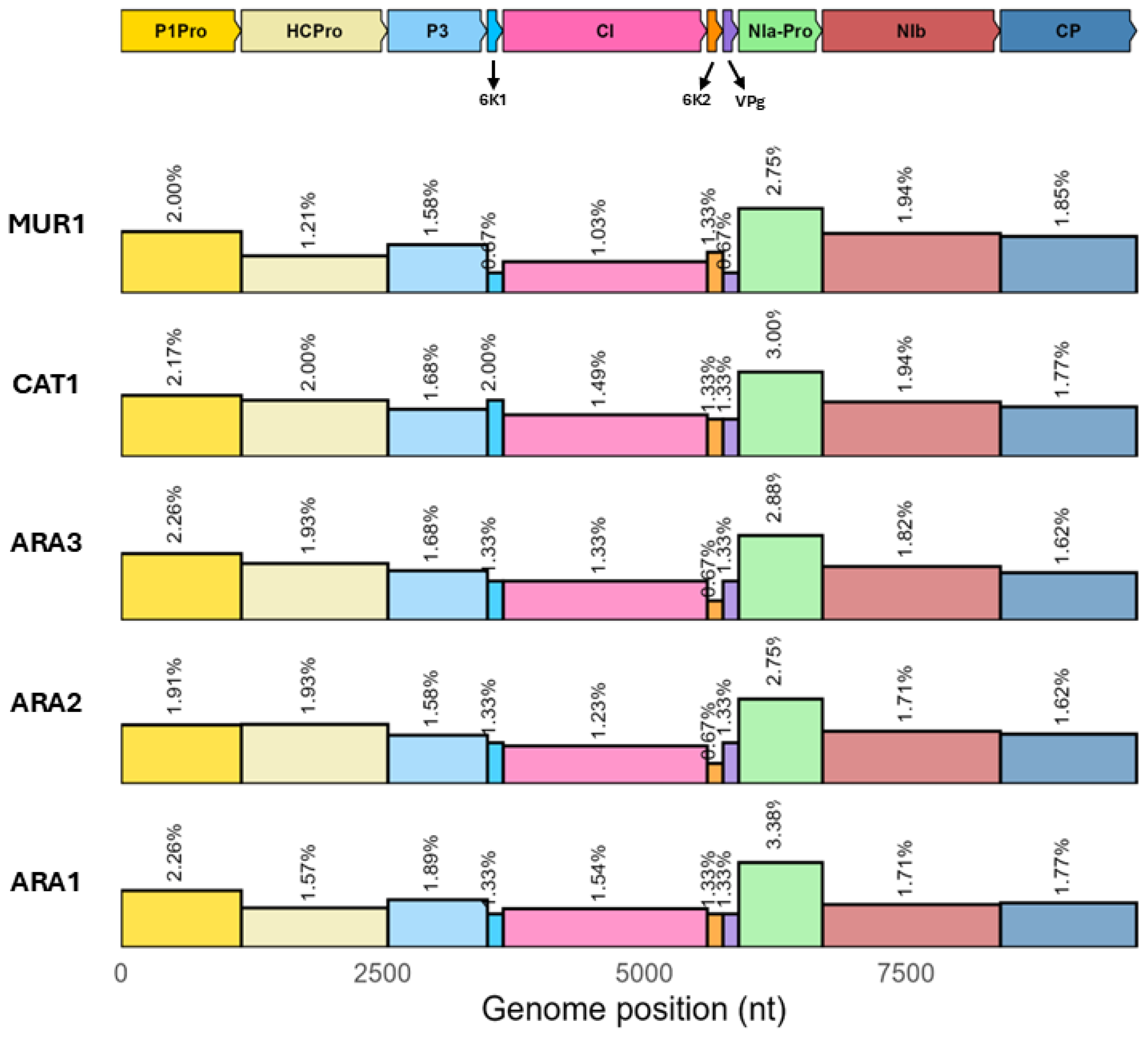
Single-nucleotide variants (SNVs) were analyzed in the studied samples against the reference Potyvirus plumpoxi Marcus strain (AJ243957.1). A total of 330 unique SNVs were detected in the studied samples (Supplementary Table S4), ranging from 158 to 186 SNVs per sample, with an average of 173.2 SNVs per sample. SNVs were mainly transitions (~85%), with an average of 38.02% A↔G and 47.60% U↔C. Among the transversions, A↔C was the most common change (average 7.14%), followed by A↔U (3.60%), G↔U (3.30%), and G↔C (0.34%) (Table 3).
Across the 9786-nucleotide genome (AJ243957.1), the overall mutation rate was 1.77 per 100 nt (1.77%). Mutation rates differed among genomic regions; however, the variability of these regions was consistent across samples (Figure 3). The most variable regions were NIa-Pro (average 2.95%), P1-Pro (2.12%), and NIb (1.82%), whereas the most conserved regions were 6K2 (1.07%), VPg (1.20%), and CI (1.32%).

3.5. Phylogenetic Analysis of PPV Sequences

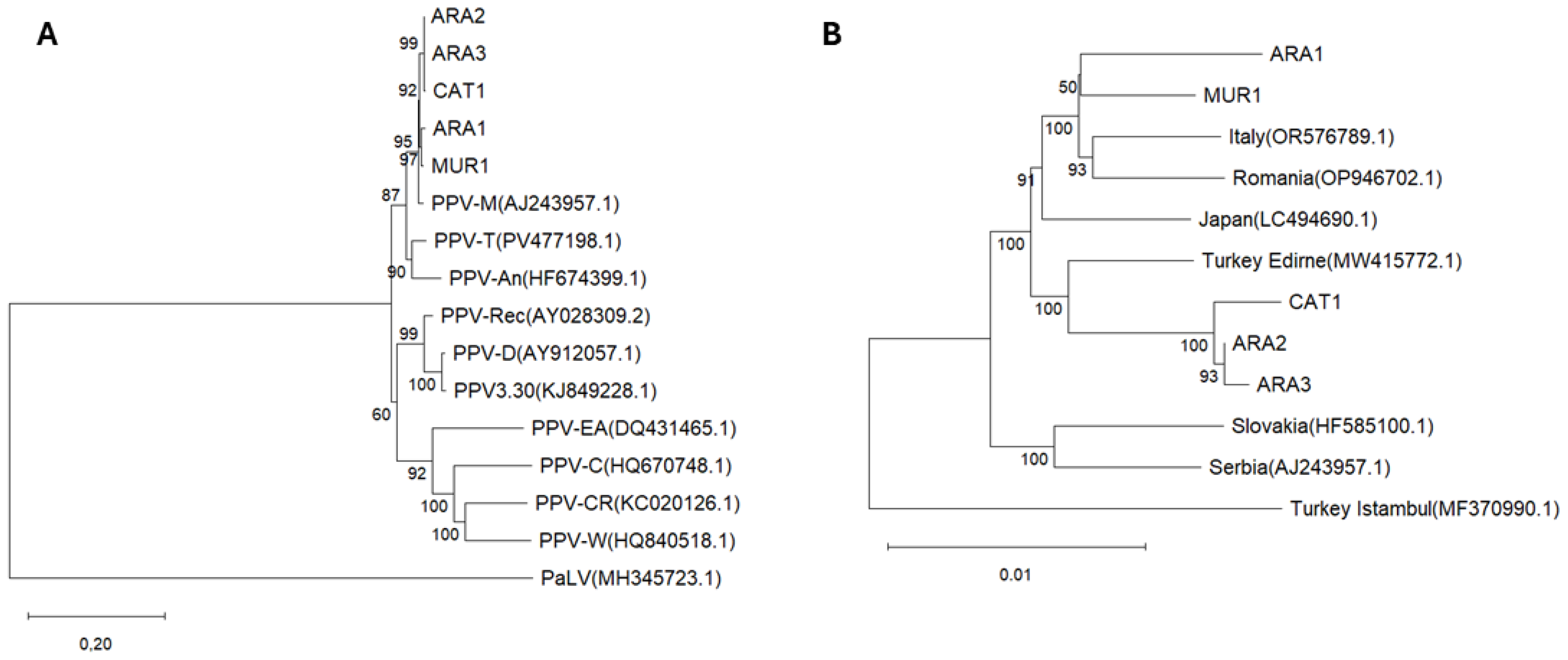
The SNVs detected were used to generate consensus sequences for the five studied samples. Two phylogenetic trees were then constructed: the first including the studied samples together with different PPV strains, and the second including PPV Marcus strains from several countries (Figure 4).
In the first tree, two clades were identified. One clade included the samples studied here, the PPV Marcus strain, PPV-T (Turkey isolate), PPV-An (Marcus ancestor), PPV-R (recombinant between Marcus and Dideron strains), PPV-D (Dideron strain), PPV-3.30 (a Dideron strain isolate), and PPV-EA (El Amar isolate). The other clade comprised PPV-C (W strain isolates from Latvia), PPV-CR (Cherry Russia), and PPV-W (BY181 isolate of the W strain). The studied samples clustered within a subclade, with ARA2, ARA3, and CAT1 forming one group and ARA1 and MUR1 forming another group, both closely related to the PPV-M strain. In the second phylogenetic tree, including PPV-M isolates, two main clades were observed: one comprising one Turkish isolate and the other including the remaining isolates. Within this second clade, two subclades were distinguished: one containing the studied samples together with the Italian, Romanian, and Japanese isolates, and the other grouping the Slovakian and Serbian isolates. The studied samples were separated within the first subclade, with ARA1 and MUR1 clustering closer to the Italian, Romanian, and Japanese isolates, while ARA2, ARA3, and CAT1 were closer to the other Turkish isolate.

4. Discussion

The screening and detection of viruses in Spanish Prunus orchards has been implemented through an RNA-seq workflow. This approach has enabled the simultaneous identification of other viruses and, to the best of the authors’ knowledge, represents the reappearance of the PPV-M strains that are currently present in Spanish Prunus orchards after the first detection and eradication in 2004 [12]. The characterization of plant viral populations provides valuable insights into the diversity and dynamics of viral infections within these crops. The sequencing reads obtained enabled not only the identification but also the molecular characterization of the PPV-M populations analyzed.
The sequencing reads obtained indicated a high sequencing depth and overall data integrity, yielding over 52 million paired-end reads, of which 95% were of high quality. These values are comparable to, or even higher than, those reported in other plant viral screening studies using the Illumina platform [20]. The reads remaining after host genome filtering are consistent with viral genomic analyses aimed at reducing background noise and improving the recovery of viral sequences [30]. The de novo assembly metrics obtained (average contig length ≈ 1050 nt; N50 = 1236 nt) demonstrated adequate assembly quality for viral genome reconstruction [43]. Although N50 values in plant virome assemblies can vary depending on viral abundance and sequencing depth [28], values around 1 kb are considered robust for the identification of complete or partial viral genomes [20,44]. In addition, the presence of contigs exceeding 16 kb indicates that the workflow was effective in recovering complete viral genomes [20,22,23,43,45].
The employed metatranscriptomic analysis revealed a predominance of PPV-M across all analyzed samples. The detection of PPV in all sample sets with high sequence depth reflects its high replicative activity and widespread distribution in Prunus hosts. Similar observations have been reported in RNA-Seq studies of susceptible apricot (Prunus armeniaca) genotypes inoculated with PPV, where a total of 18.87 million raw reads (equivalent to 5.6% of the clean reads) mapped to the PPV genome in inoculated samples [22,46]. Other viruses, such as NSPaV and PaLV, were also detected with high read depths, supporting their widespread occurrence previously reported in Prunus species [47,48,49,50]. In terms of viral diversity, the number of co-infecting viruses varied among the samples analyzed. Samples ARA1, ARA2, and ARA3 exhibited higher viral richness, each containing six viruses, whereas CAT1 and MUR1 harbored only three. This is consistent with the notion that Prunus species harbor complex viral communities [20,23]. Although some of them were detected in residual loads, a similar number of viruses were detected here when compared to previous fruit tree transcriptomic studies [20,21,43]. Notably, the majority of the isolates originated from peach samples (ARA1, ARA2, ARA3, and MUR1), while sample CAT1 corresponded to an apricot, highlighting the presence of complex viral communities across different Prunus hosts.
Single-nucleotide variations detected in PPV-M from the samples analyzed through variant calling revealed that most mutations were transitions, which is consistent with previous reports in Potyvirus species and with the biochemical principle that transitions are more likely to occur than transversions [51,52]. The mutation rate was of a similar order of magnitude to other viruses in peach, such as ACSLV and APV, although lower than the high mutation rates reported for PLMV [20]. Specific regions of the virus showed a high number of SNVs, specifically NIa-Pro and P1-Pro. High genomic variation has been reported previously in NIa-Pro in PPV, but also in more potyviruses such as sugarcane mosaic virus (SCMV) or leek yellow stripe virus (LYSV) [51]. In previous studies, NIa-Pro has been reported to exhibit more positively selected sites than expected and has been shown to cleave host plant proteins, suggesting that its variability may contribute to viral adaptation [51,53]. On the other hand, P1-Pro has a hypervariable N-terminal region and controls virus replication, host defense responses, and pathogenicity [54,55]. The high variability in this region among potyviruses has been linked to their ability to adapt to a broad spectrum of host species [56,57].
The variability of PPV samples was also characterized through phylogenetic analyses, which confirmed their expected closeness to the PPV-M reference strain. However, genetic differences between the two groups of samples studied were evident in the phylogenetic trees, separating the three samples from the Aragón–Catalunya border (CAT1, ARA2, and ARA3), the other sample (ARA1) from Central Aragón, and the one from Murcia (MUR1). Although these clustering patterns may suggest the possibility of two different sources of germplasm, such inferences should be considered tentative, given the limited number of samples analyzed and potential uneven sampling representation in public databases. Interestingly, these results could indicate two different sources of germplasm exchange, because while ARA1 and MUR1 samples cluster together with the Italian source, the other three (CAT1, ARA2, and ARA3) studied here cluster with one of the Turkish isolates from the Edirne region. This observed geographical association is consistent with phylogeographic models, suggesting that the movement of PPV-infected material proceeded from Turkey to the Balkans and Central Europe [39].
A similar exchange case was observed with Italian and Romanian strains, which was recently reported by [58], reflecting the need for stricter certification and monitoring measures for imported planting material. The close genetic relationship observed among these isolates is more consistent with recent transboundary movement of infected planting material than with long-term historical dispersal events. Therefore, the phylogenetic clustering detected here likely reflects contemporary exchange routes within European production systems, reinforcing the epidemiological relevance of the present findings in the context of the recent reemergence of PPV-M in Spanish orchards.
The virus screening approach based on RNA-seq enabled the identification of a total of 10 different viruses in Spanish Prunus orchards, including ChaLV, NSPaV, PaLV, and PPV-M. In this sense, HTS offers a powerful approach for finding organisms that we did not know were there, with the consequence of possible unknown interactions between pathogens. RT-PCR validation confirmed the accuracy of the RNA-seq results. The analyses revealed the mutation patterns present in PPV-M and clarified the phylogenetic relationships among the Spanish samples studied. The phylogenetic results highlight the need for improved control measures to prevent the spread of the disease through the exchange of plant material. The reads and sequences obtained will contribute to expanding the available viral resources for the plant virology community.
The reappearance of PPV-M in Spanish orchards represents a critical issue that must be addressed. This strain is regarded as one of the most aggressive forms of the virus, being associated with severe symptom expression on leaves and fruits and, like most of the PPV strains, with rapid dissemination by aphids. In regions where Prunus cultivation constitutes a key economic activity, such as Aragón, Catalunya, and Murcia, as well as other production areas, the establishment of PPV-M may have serious consequences on peach production, directly linked to substantial losses in yield and fruit quality. In light of the results obtained in the present study, the importance of phytosanitary control and plant certification systems for the exchange of plant material is reaffirmed, independently of the provenance.

Supplementary Materials

The following supporting information can be downloaded at https://www.mdpi.com/article/10.3390/agronomy16060608/s1. Table S1: NCBI reference; Table S2: Summary of sequencing reads processing for each sample; Table S3: Viral identification, genome coverage, and taxonomic classification for each sample; Table S4: Single-nucleotide variants detected in the Potyvirus plumpoxi genome across samples; and Figure S1: Principal component analysis (PCA).

Author Contributions

Conceptualization, L.R.-R. and M.R.; methodology, L.R.-R.; software, L.R.-R. and P.J.M.-G.; formal analysis, L.R.-R.; investigation, L.R.-R. and M.R.; writing—original draft preparation, L.R.-R.; writing—review and editing, L.R.-R. and M.R.; supervision, M.R.; funding acquisition, M.R. and P.M.-G. All authors have read and agreed to the published version of the manuscript.

Funding

This study has been supported by the projects “Resistance to Plum pox virus Marcus-type isolate (sharka) in peach” (PID2021-123764OB-I00) from the Ministry of Science and Innovation (Spain), “Stone fruit breeding for emerging challenges using new phenomic, genomic and modelling approaches” (23051/GERM/25) of the Seneca Foundation of the Region of Murcia (Spain), and PRE2022-103362 contract by the Ministry of Science and Innovation.

Data Availability Statement

The original contributions presented in this study are included in the article. Further inquiries can be directed to the corresponding authors.

Acknowledgments

We want to acknowledge J López-Alcolea and MC García-Campayo for their effort, dedication, and technical assistance.

Conflicts of Interest

The authors declare no conflicts of interest.

References

  1. Martínez-Gómez, P.; Crisosto, C.H.; Bonghi, C.; Rubio, M. New approaches to Prunus transcriptome analysis. Genetica 2011, 139, 755–769. [Google Scholar] [CrossRef] [PubMed]
  2. Rubio, C.; Martínez-Gómez, P.; Marais, A.; Sánchez-Navarro, J.A.; Pallás, V.; Candresse, T. Recent advances and prospects in Prunus virology. Ann. Appl. Biol. 2017, 171, 125–138. [Google Scholar] [CrossRef]
  3. FAOSTAT. Available online: https://www.fao.org/faostat/en/#data/QCL (accessed on 22 December 2025).
  4. MAPA. Ministerio de Agricultura Pesca y Alimentación. 2024. Available online: https://www.mapa.gob.es/dam/mapa/contenido/agricultura/temas/producciones-agricolas/frutas-y-hortalizas/frutas-y-hortalizas/informecampana2024frutadehueso.pdf (accessed on 1 January 2026).
  5. García, J.A.; Glasa, M.; Cambra, M.; Candresse, T. Plum pox virus and sharka: A model potyvirus and a major disease. Mol. Plant Pathol. 2014, 15, 226–241. [Google Scholar] [CrossRef] [PubMed]
  6. Cambra, M.; Capote, N.; Myrta, A.; Llácer, G. Plum pox virus and the estimated costs associated with sharka disease. EPPO Bull. 2006, 36, 202–204. [Google Scholar] [CrossRef]
  7. DEFRA. Department for Environment Food & Rural Affairs. Plum Pox Virus (PPV)-Department for Environment Food and Rural Affairs. 2024. Available online: https://planthealthportal.defra.gov.uk/assets/factsheets/Plum-pox-factsheet_final-v2.pdf (accessed on 1 January 2026).
  8. Rubio, M.; Rodríguez-Moreno, L.; Ballester, A.R.; Castor de Moura, M.; Bonghi, C.; Candresse, T.; Martínez-Gómez, P. Analysis of gene expression changes in peach leaves in response to Plum pox virus infection using RNA-Seq. Mol. Plant Pathol. 2015, 16, 164–176. [Google Scholar] [CrossRef] [PubMed]
  9. García, J.A.; Rodamilans, B.; Martínez-Turiño, S.; Valli, A.A.; Simón-Mateo, C.; Cambra, M. Plum pox virus: An overview of the potyvirus behind sharka, a harmful stone fruit disease. Ann. Appl. Biol. 2025, 186, 49–75. [Google Scholar] [CrossRef]
  10. Llácer, G.; Cambra, M.; Laviña, A. Detection of plum pox virus in Spain. EPPO Bull. 1985, 15, 325–329. [Google Scholar] [CrossRef]
  11. James, D.; Varga, A.; Sanderson, D. Genetic diversity of Plum pox virus: Strains, disease and related challenges for control. Can. J. Plant Pathol. 2013, 35, 431–441. [Google Scholar] [CrossRef]
  12. Cambra, M.A.; Crespo, M.T.; Gorris, M.T.; Martínez, M.C.; Olmos, A.; Capote, N.; Cambra, M. Detection and eradicacion of Plum Pox Virus Marcus type in Aragon (Spain). Acta Hort. 2004, 657, 231–235. [Google Scholar] [CrossRef]
  13. Generalitat de Catalunya. El Virus de la Sharka Plum Pox Virus (PPV); Departament d’Agricultura, Ramaderia, Pesca, Alimentació i Medi Natural: Barcelona, Spain, 2008; 18p. [Google Scholar]
  14. Gobierno de Aragón. ORDEN AGM/1048/2020, de 19 de Octubre, Por la que se Declara de Utilidad Pública la Lucha Contra el Virus de la Sharka-m y se Establecen Medidas Para su Prevención y Lucha; Boletín Oficial de Aragón: Zaragoza, Spain, 2020. [Google Scholar]
  15. Syller, J. Facilitative and antagonistic interactions between plant viruses in mixed infections. Mol. Plant Pathol. 2012, 13, 204–216. [Google Scholar] [CrossRef]
  16. Kamitani, M.; Nagano, A.J.; Honjo, M.N.; Kudoh, H. RNA-Seq reveals virus–virus and virus–plant interactions in nature. FEMS Microbiol. Ecol. 2016, 92, fiw176. [Google Scholar] [CrossRef] [PubMed]
  17. Descriptions of Plant Viruses. DPV—Database of Plant Viruses. 2025. Available online: https://dpvweb.net/ (accessed on 1 January 2026).
  18. Geer, L.Y.; Marchler-Bauer, A.; Geer, R.C.; Han, L.; He, J.; He, S.; Liu, C.; Shi, W.; Bryant, S.H. The NCBI biosystems database. Nucleic Acids Res. 2010, 38, D492–D496. [Google Scholar] [CrossRef]
  19. Kreuze, J.F.; Perez, A.; Untiveros, M.; Quispe, D.; Fuentes, S.; Barker, I.; Simon, R. Complete viral genome sequence and discovery of novel viruses by deep sequencing of small RNAs: A generic method for diagnosis, discovery and sequencing of viruses. Virology 2009, 388, 1–7. [Google Scholar] [CrossRef]
  20. Jo, Y.; Lian, S.; Chu, H.; Cho, J.K.; Yoo, S.H.; Choi, H.; Yoon, J.Y.; Choi, S.K.; Lee, B.C.; Cho, W.K. Peach RNA viromes in six different peach cultivars. Sci. Rep. 2018, 8, 1844. [Google Scholar] [CrossRef] [PubMed]
  21. Dias, N.P.; Hu, R.; Hensley, D.D.; Hansen, Z.R.; Domier, L.L.; Hajimorad, M.R. A Survey for Viruses and Viroids of Peach in Tennessee Orchards by RNA Sequencing. Plant Health Prog. 2022, 23, 265–268. [Google Scholar] [CrossRef]
  22. Baráth, D.; Jaksa-Czotter, N.; Molnár, J.; Varga, T.; Balássy, J.; Szabó, L.K.; Kirilla, Z.; Tusnády, G.E.; Preininger, É.; Várallyay, É. Small RNA NGS Revealed the Presence of Cherry Virus A and Little Cherry Virus 1 on Apricots in Hungary. Viruses 2018, 10, 318. [Google Scholar] [CrossRef]
  23. Jo, Y.; Choi, H.; Lian, S.; Cho, J.K.; Chu, H.; Cho, W.K. Identification of viruses infecting six plum cultivars in Korea by RNA-sequencing. PeerJ 2020, 8, e9588. [Google Scholar] [CrossRef]
  24. Roossinck, M.J.; Martin, D.P.; Roumagnac, P. Plant Virus Metagenomics: Advances in Virus Discovery. Phytopathology 2015, 105, 716–727. [Google Scholar] [CrossRef]
  25. Donaire, L.; Rozas, J.; Ayllón, M.A. Molecular characterization of Botrytis ourmia-like virus, a mycovirus close to the plant pathogenic genus Ourmiavirus. Virology 2016, 489, 158–164. [Google Scholar] [CrossRef]
  26. Trovão, N.S.; Baele, G.; Vrancken, B.; Bielejec, F.; Suchard, M.A.; Fargette, D.; Lemey, P. Host ecology determines the dispersal patterns of a plant virus. Virus Evol. 2015, 1, vev016. [Google Scholar] [CrossRef]
  27. Jo, Y.; Choi, H.; Kim, S.M.; Kim, S.L.; Lee, B.C.; Cho, W.K. The pepper virome: Natural co-infection of diverse viruses and their quasispecies. BMC Genom. 2017, 18, 453. [Google Scholar] [CrossRef] [PubMed]
  28. Jo, Y.; Choi, H.; Kim, S.M.; Kim, S.L.; Lee, B.C.; Cho, W.K. Integrated analyses using RNA-Seq data reveal viral genomes, single nucleotide variations, the phylogenetic relationship, and recombination for Apple stem grooving virus. BMC Genom. 2016, 17, 579. [Google Scholar] [CrossRef] [PubMed]
  29. Candresse, T.; Cambra, M.; Dallot, S.; Lanneau, M.; Asensio, M.; Gorris, M.T.; Revers, F.; Macquaire, G.; Olmos, A.; Boscia, D.; et al. Comparison of monoclonal antibodies and polymerase chain reaction assays or the typing of isolates belonging to the D and M serotypes of Plum pox potyvirus. Phytopathology 1998, 88, 198–204. [Google Scholar] [CrossRef] [PubMed]
  30. Bolger, A.M.; Lohse, M.; Usadel, B. Trimmomatic: A flexible trimmer for Illumina sequence data. Bioinformatics 2014, 30, 2114–2120. [Google Scholar] [CrossRef]
  31. Donaire, L.; Aranda, M.A. Computational pipeline for the detection of plant RNA viruses using high-throughput sequencing. Plant-Virus Interact. 2024, 2724, 1–20. [Google Scholar]
  32. Verde, I.; Jenkins, J.; Dondini, L.; Micali, S.; Pagliarani, G.; Vendramin, E.; Paris, R.; Tartarini, S.; Dettori, M.T.; Schmutz, J. The Peach v2.0 release: High-resolution linkage mapping and deep resequencing improve chromosome-scale assembly and contiguity. BMC Genom. 2017, 18, 225. [Google Scholar] [CrossRef]
  33. Campoy, J.A.; Sun, H.; Goel, M.; Jiao, W.B.; Folz-Donahue, K.; Wang, N.; Rubio, M.; Liu, C.; Kukat, C.; Ruiz, D.; et al. Gamete binning: Chromosome-level and haplotype-resolved genome assembly enabled by high-throughput single-cell sequencing of gamete genomes. Genome Biol. 2020, 21, 306. [Google Scholar] [CrossRef]
  34. Langmead, B.; Salzberg, S.L. Fast gapped-read alignment with Bowtie 2. Nat. Methods 2012, 9, 357–359. [Google Scholar] [CrossRef]
  35. Grabherr, M.G.; Haas, B.J.; Yassour, M.; Levin, J.Z.; Thompson, D.A.; Amit, I.; Adiconis, X.; Fan, L.; Raychowdhury, R.; Zeng, Q.; et al. Full-length transcriptome assembly from RNA-Seq data without a reference genome. Nat. Biotechnol. 2011, 29, 644–652. [Google Scholar] [CrossRef]
  36. Boratyn, G.M.; Camacho, C.; Cooper, P.S.; Coulouris, G.; Fong, A.; Ma, N.; Madden, T.L.; Sayers, E.W.; Tao, T.; Ye, J.; et al. BLAST: A more efficient report with usability improvements. Nucleic Acids Res. 2013, 41, W29–W33. [Google Scholar] [CrossRef]
  37. Li, H. Aligning sequence reads, clone sequences and assembly contigs with BWA-MEM. arXiv 2013, arXiv:1303.3997. [Google Scholar] [CrossRef]
  38. Sáenz, P.; Cervera, M.T.; Dallot, S.; Quiot, L.; Quiot, J.B.; Riechmann, J.L.; García, J.A. Identification of a pathogenicity determinant of Plum pox virus in the sequence encoding the C-terminal region of protein P3+6K1. J. Gen. Virol. 2000, 81, 557–566. [Google Scholar] [CrossRef] [PubMed]
  39. Danecek, P.; Bonfield, J.K.; Liddle, J.; Marshall, J.; Ohan, V.; Pollard, M.O.; Whitwham, A.; Keane, T.; McCarthy, S.A.; Davies, R.M.; et al. Twelve years of SAMtools and BCFtools. Gigascience 2021, 10, giab008. [Google Scholar] [CrossRef] [PubMed]
  40. Palmisano, F.; Kawakubo, S.; Chiumenti, M.; Leonetti, P.; Pantaleo, V.; Candresse, T.; Minafra, A. Bayesian phylogenetic and recombination analyses of plum pox virus provide a refined vision of its evolutionary history. Virol. J. 2025, 22, 319. [Google Scholar] [CrossRef]
  41. Kumar, S.; Stecher, G.; Suleski, M.; Sanderford, M.; Sharma, S.; Tamura, K. MEGA12: Molecular Evolutionary Genetic Analysis version 12 for adaptive and green computing. Mol. Biol. Evol. 2024, 41, msae263. [Google Scholar] [CrossRef]
  42. García, J.A.; Riechman, J.L.; Laín, S.; Cimon, L.; Domingez, E.; Cervera, M.T. Molecular characterization of plum pox potyvirus. EPPO Bull. 1994, 24, 543–553. [Google Scholar] [CrossRef]
  43. Wright, A.A.; Cross, A.R.; Harper, S.J. A bushel of viruses: Identification of seventeen novel putative viruses by RNA-seq in six apple trees. PLoS ONE 2020, 15, e0227669. [Google Scholar] [CrossRef]
  44. Kim, N.Y.; Lee, H.J.; Kim, H.S.; Lee, S.H.; Moon, J.S.; Jeong, R.D. Identification of Plant Viruses Infecting Pear Using RNA Sequencing. Plant Pathol. J. 2021, 37, 258–267. [Google Scholar] [CrossRef]
  45. Prajapati, M.R.; Gupta, N.; Thapa, P.; Diksha, D.; Sharma, S.K.; Baranwal, V.K. Identification and Characterization of a Novel Cytorhabdovirus Associated with Pear (P. communis ’Bartlett’) Through RNA-seq. Curr. Microbiol. 2024, 82, 39. [Google Scholar] [CrossRef]
  46. Rubio, M.; Ballester, A.R.; Olivares, P.M.; Castro de Moura, M.; Dicenta, F.; Martínez-Gómez, P. Gene Expression Analysis of Plum pox virus (Sharka) Susceptibility/Resistance in Apricot (Prunus armeniaca L.). PLoS ONE 2015, 10, e0144670. [Google Scholar] [CrossRef]
  47. Candresse, T.; Faure, C.; Theil, S.; Marais, A. First Report of Nectarine stem pitting-associated virus Infecting Prunus mume in Japan. Plant Dis. 2017, 101, 393. [Google Scholar] [CrossRef]
  48. Jo, Y.; Cho, J.K.; Choi, H.; Lian, S.; Cho, W.K. First Report of Nectarine stem pitting-associated virus and Plum bark necrosis and stem pitting-associated virus Infecting a Peach Cultivar in Korea. Plant Dis. 2017, 101, 1067. [Google Scholar] [CrossRef]
  49. Krizbai, L.; Kriston, E.; Kreuze, J.; Melika, G. Identification of Nectarine stem pitting-associated virus infecting Prunus persica in Hungary. New Dis. Rep. 2017, 35, 18. [Google Scholar] [CrossRef]
  50. Morán, F.; Olmos, A.; Malagón, J.; Candresse, T.; Ruiz-García, A.B. First Report of Peach-Associated Luteovirus (PaLV) Infecting Almond Trees (Prunus dulcis) in Spain. Plant Dis. 2025, 109, 727. [Google Scholar] [CrossRef]
  51. Nigam, D.; LaTourrette, K.; Souza, P.F.N.; Garcia-Ruiz, H. Genome-Wide Variation in Potyviruses. Front. Plant Sci. 2019, 10, 1439. [Google Scholar] [CrossRef]
  52. Hasiów-Jaroszewska, B.; Fares, M.A.; Elena, S.F. Molecular Evolution of Viral Multifunctional Proteins: The Case of Potyvirus HC-Pro. J. Mol. Evol. 2014, 78, 75–86. [Google Scholar] [CrossRef]
  53. Xiao, H.; Lord, E.; Sanfaçon, H. Proteolytic Processing of Plant Proteins by Potyvirus NIa Proteases. J. Virol. 2022, 96, e01444-21. [Google Scholar] [CrossRef]
  54. Pasin, F.; Simón-Mateo, C.; García, J.A. The Hypervariable Amino-Terminus of P1 Protease Modulates Potyviral Replication and Host Defense Responses. PLoS Pathog. 2014, 10, e1003985. [Google Scholar] [CrossRef]
  55. Revers, F.; García, J.A. Molecular Biology of Potyviruses. Adv. Virus Res. 2015, 92, 101–199. [Google Scholar]
  56. Valli, A.; López-Moya, J.J.; García, J.A. Recombination and gene duplication in the evolutionary diversification of P1 proteins in the family Potyviridae. J. Gen. Virol. 2007, 88, 1016–1028. [Google Scholar] [CrossRef]
  57. Rodamilans, B.; Casillas, A.; García, J.A. P1 of Sweet Potato Feathery Mottle Virus Shows Strong Adaptation Capacity, Replacing P1-HCPro in a Chimeric Plum Pox Virus. J. Virol. 2021, 95, e0015021. [Google Scholar] [CrossRef]
  58. Ruiz-García, A.B.; Zagrai, L.A.; Olmos, A.; Zagrai, I. Emergence of PPV-M in Romania in newly established Prunus domestica orchards: Implications for certification schemes. J. Plant Pathol. 2025, 107, 1959–1968. [Google Scholar] [CrossRef]
Figure 1. Read coverage across viral genomes identified in sample ARA2 (replicate 1). Plots represent the position and depth of RNA-seq reads mapped along the corresponding reference genome (x-axis), with distinct viral species displayed along the y-axis. For each detected virus, the reference species name, GenBank accession number, and genome length (in nucleotides) are indicated. Coverage profiles illustrate the distribution of sequencing reads across the viral genomes, highlighting differences in genome completeness and read density among the identified viruses.
Agronomy 16 00608 g001
Figure 2. Full-length agarose gels showing RT-PCR results obtained with virus-specific primers for PPV-M and PPV-D. The sample lanes across all panels (A,B) correspond sequentially to ARA1, ARA2, ARA3, CAT1, and MUR1. (A) RT-PCR amplification of PPV-M. (B) RT-PCR amplification of PPV-D. The final lanes represent the positive Dideron and Marcus controls (PC-D and PC-M) and the negative control (NC). The expected PCR product size is 198 bp.
Agronomy 16 00608 g002
Figure 3. Distribution of mutation rates (SNVs per 100 nt) across viral coding regions for each sample, based on the genomic organization of Potyvirus plumpoxi [41,42].
Agronomy 16 00608 g003
Figure 4. Phylogenetic trees were constructed using the studied samples together with (A) different PPV strains and (B) PPV Marcus isolates from several countries.
Agronomy 16 00608 g004
Table 1. Summary of read mapping results showing the average values for each analyzed sample.
SampleRaw ReadsClean Reads (%)Non-Host Reads (%)
ARA145,321,22742,816,233 (94.47)17,041,622 (38.91)
ARA256,533,06853,758,852 (95.09)35,529,621 (66.03)
ARA351,121,46848,513,776 (94.90)31,292,779 (64.97)
CAT154,563,71951,917,063 (95.15)37,208,257 (71.65)
MUR155,784,71152,957,985 (94.93)36,440,167 (68.79)
Average52,664,83849,992,781 (94.88)31,502,489 (62.07)
Table 2. Maximum viral genome coverage (%) per sample for Potyvirus plumpoxi (PPV-M), Luteovirus nucipersicae (NSPaV), peach-associated luteovirus (PaLV), youcai mosaic virus (YoMV), grapevine red globe virus (GRGV), plum ourmia-like virus (POLV), Marafivirus pruni (PeVD), Marafivirus asteroides (GAMaV), peach virus T (PrVT), and Luteovirus avii (ChaLV). Only viruses consistently detected in all technical and biological replicates of each sample are included. A dash (–) indicates that no reads were detected for the corresponding virus in that sample.
VirusSamples
ARA1ARA2ARA3CAT1MUR1
PPV-M99.60%96.68%99.55%99.67%99.55%
NSPaV76.80%92.94%-8.35%-
PaLV-90.24%89.90%--
POLV-81.84%---
ChaLV-46.26%48.34%--
YoMV-16.80%-5.85%-
GRGV6.72%----
PeVD1.66%----
GAMaV1.26%----
PrVT0.98%----
Table 3. Detected SNVs in each sample, indicating transition and transversion rates.
TransitionsTransversion
SampleSNPsA↔G (%)U↔C (%)A↔C (%)G↔C (%)A↔U (%)G↔U (%)
ARA118246.7040.116.040.002.754.40
ARA216535.7649.098.480.613.642.42
ARA317534.2949.148.570.573.434.00
CAT118636.0248.397.530.543.763.76
MUR115837.3451.275.060.004.431.90
Average17338.0247.607.140.343.603.30
Disclaimer/Publisher’s Note: The statements, opinions and data contained in all publications are solely those of the individual author(s) and contributor(s) and not of MDPI and/or the editor(s). MDPI and/or the editor(s) disclaim responsibility for any injury to people or property resulting from any ideas, methods, instructions or products referred to in the content.