Article

Phytolith Concentration and Morphological Variation in Dendrocalamus brandisii (Munro) Kurz. Leaves in Response to Sodium Silicate Foliar Application Across Vegetative Phenological Stages

1
Key Laboratory for Forest Resources Conservation and Utilization in the Southwest Mountains of China, Ministry of Education, Kunming 650224, China
2
College of Forestry, Southwest Forestry University, Kunming 650224, China
*
Author to whom correspondence should be addressed.
These authors contributed equally to this work.
Agronomy 2025, 15(9), 2138; https://doi.org/10.3390/agronomy15092138
Submission received: 3 August 2025 / Revised: 4 September 2025 / Accepted: 4 September 2025 / Published: 5 September 2025
(This article belongs to the Section Horticultural and Floricultural Crops)

Abstract

This study investigated the effects of the foliar application of sodium silicate (SS) on phytolith formations in Dendrocalamus brandisii (Munro) Kurz. leaves by analyzing the phytolith concentration, morphological parameters, and assemblage compositions across leaves of varying ages and different phenological stages. The results showed that SS significantly increased the phytolith concentration in D. brandisii leaves, showing a trend of old leaves > mature leaves > young leaves. The concentration of phytoliths was the highest at the late shooting stage (November) and the lowest at the dormancy stage (January). August (shooting stage) and May (branching and leafing stage) were the critical periods for phytolith formation and the size and morphological variation. Sodium silicate significantly increased the proportion of saddle, bilobate, and stomatal phytoliths, which might help optimize the silicified structure of leaf epidermal cells and enhance the leaf resistance and light energy utilization efficiency. The results help clarify the mechanism of phytolith formation in different phenological periods of D. brandisii and provide a theoretical basis for the efficient use of silicon fertilizers in bamboo cultivation.

1. Introduction

A phytolith is a microscopic amorphous silicon dioxide structure. Amorphous silicic acid [Si(OH)4] is absorbed by plant roots from the soil, transported to different tissues through the vascular system during transpiration, and precipitated in plant cell walls, intracellular cavities, and between cell walls [1]. Phytoliths are the primary form of silicon in higher plants [2]. Their formation significantly enhances plant mechanical stability [3,4] and improves resistance to erosion [5]. In leaves, these structures facilitate light transmission to mesophyll cells, thereby boosting photosynthetic efficiency [6]. They also mitigate the adverse impacts of biotic and abiotic stresses through morphological, physiological, and biochemical mechanisms [7]. Phytoliths are present in a variety of plants. Differences in phytolith morphology between species depend on the specific anatomy of the organs, especially the epidermis, where most of the phytoliths are formed. That is why phytoliths have taxonomic relevance. Additionally, their morphological characteristics are closely related to physiological functions, growing conditions, and the plant nutritional status. Morphological differences in phytoliths in different plant species or the same plant at different phenological stages can be significant [8]. These differences are an important manifestation of a plant’s adaptation to the environment.
As an efficient form of nutrient supplementation, foliar spraying can directly transport nutrients to plant leaves and quickly meet the needs of plant growth [9]. Studies have shown that the foliar spraying of silicon sources such as sodium silicate can promote the accumulation and distribution of silicon in plants, thereby improving physiological processes, including growth, stress resistance, and photosynthetic water use [10,11,12]. It can play a positive role in plant growth and physiological activities by affecting the accumulation and distribution of silicon [13]. Bamboos are among the most silicon-rich plants, with epidermal tissue containing up to 70% silicon [14]. Significant differences were found in the concentration and morphology of phytoliths in different tissues and organs of the same bamboo species [15]. Bamboo leaves have the largest morphological diversity of phytoliths [16]. As leaves mature, the silicon deposition and phytolith concentrations in the leaves increase [17]. The increment of silica deposits in tissues with age has been shown in many groups of plants, especially grasses. In their study on the tropical forage grass Brachiaria brizantha, de Melo et al. [18] found that the silica deposition in leaves decreases in the order of mature leaf blades > recently expanded leaf blades > non-expanded leaf blades, and leaves continue to accumulate silica throughout their entire life cycle, including after maturation. However, in Honaine’s study, it was found that silica deposition is not solely determined by age, but is rather a combined effect of age, the environment, and physiological processes (such as transpiration and senescence) [19]. Additionally, Motomura et al. found that silicification on Sasa veitchii leaves of different ages was not the same. Short cells in epidermal cells were preferentially silicified, while the number of silicified cells in other epidermal cells gradually increased with leaf aging. There was a certain difference in silicification among cells of different growth stages [20]. Li et al. conducted a continuous observation of phytoliths in Dendrocalamus ronganensis leaves for 21 months and found that the phytolith morphological variation was related to the season and leaf maturity [21]. The phytolith concentration in Dendrocalamus giganteus was highest at the late shooting stage and lowest at the branching and leafing stages. Saddle and dumbbell phytoliths were the dominant morphotypes in D. giganteus leaves [22]. Previous studies on phytoliths in Bambusoideae mainly focused on the concentration and morphology of phytoliths in a single phenological period or specific plant organs. However, there are few studies on the effects of the foliar spraying of sodium silicate on phytoliths, especially the dynamic response of bamboo plants to a silicon fertilizer application during an entire phenological cycle.
Dendrocalamus brandisii is mainly distributed in tropical and subtropical regions of China and in Myanmar, Laos, Vietnam, and Thailand [23]. Dendrocalamus brandisii features a short growth cycle and a strong regeneration capacity and is a fine substitute for wood [24]. Its shoots are edible with rich nutrients, making it an excellent dual-purpose bamboo for both shoots and culms [25]. Bamboo species have high ecological and economic value and are generally characterized by rapid growth, a large biomass, and an extremely high demand for silicon [26,27]. Phytoliths play an important role in their growth and development. Marking key stages of plant growth and metabolism, its vegetative phenological period involves a series of physiological processes such as shoot germination, leaf extension, and stem thickening [28]. There are significant differences in nutritional needs and metabolic intensity in each period. In this study, the effects of the foliar application of sodium silicate on the concentration and morphological variation in phytoliths in leaves of D. brandisii at different phenological periods were analyzed, e.g., the shooting stage, late shooting stage, dormancy stage, branching, and leafing stage. This study also aims to provide a theoretical basis for the application of silicon fertilizers in bamboo cultivation.

2. Materials and Methods

2.1. Sample Collection and Preparation

The experiment was conducted in the cultivated bamboo garden of Southwest Forestry University, Kunming, China. The local climate is described in Table 1. Six clusters of D. brandisii were selected, with each cluster containing at least three bamboo culms from each age class (1, 2, and ≥3 years). The silicon solution was prepared using sodium silicate nonahydrate (Na2SiO3·9H2O); the concentration of which was based on the effects of exogenous silicon application on D. brandisii in the previous literature [29]. According to the vegetative phenological stages during the whole year, on 1 August 2022 (shooting stage), 1 November 2022 (late shooting stage), 1 January 2023 (dormancy stage), and 1 May 2023 (branching and leafing stage), three clusters were sprayed with 2 mmol/L sodium silicate (SS) solution, and another three clumps were sprayed with distilled water as a control. One spray application was conducted from 9:00 to 11:00 a.m. on the first day. A handheld sprayer was used to evenly spray the solution on both the adaxial (upper) and abaxial (lower) surfaces of the plant leaves until fine droplets formed on the leaf surface but did not yet begin to drip down along the leaves. On the 5th, 10th, and 15th days of each phenological stage, young, mature, and old leaves were separately collected from the culms of each age class (1, 2, ≥3 years) in each cluster, with a collection amount of 30 g for each group of leaves. Three replicates were set for each stage. The old leaves were yellow-green, and the leaf tips or edges were withered, usually growing near the base of the branch tips. The mature leaves were dark green and grew in the middle of the branches. The young leaves were light-green in color, curled or not fully expanded, and growing at the top of the branches. All experimental materials were washed and placed in a 105 °C oven for dehydration and fixation and then dried to a constant mass for storage.

2.2. Experimental Methods

2.2.1. Determination of Phytolith Concentration

The phytoliths were extracted by wet oxidation [30]. The samples were cut into pieces and placed in a constant-temperature drying oven to dry to a constant mass, and 0.3 g of the sample was weighed and placed in a digestion tank, evenly spread at the bottom of the test tube, with 10 mL HNO2 and 2 mL perchloric acid for 12 h. After being fully dissolved, the solution was boiled for two hours. When the liquid was reduced to 5-7 drops, 3 mL of dilute hydrochloric acid was added and boiled in the digestion instrument for 5 min. Once cooled, the material was poured into a 15 mL test tube and transferred to a centrifuge at 3500 rpm for five minutes. This process was repeated four times, each time with clean ultrapure water, until the supernatant was completely neutral. After washing, the precipitate was dried in an oven at 65 °C to obtain dry phytoliths, and the phytolith concentration was calculated.
Phytolith concentration (%) = (m2/M2) × 100
where m2 is the weight of the extracted phytoliths (g); and M2 is the weight of the leaf specimen (g).

2.2.2. Phytolith Classification

Following the procedure outlined by Pearsall [31], a small amount of acetone was added to a centrifuge tube containing dry phytoliths and shaken well, then dropped onto a glass slide to dry, and then sealed with a neutral resin to produce a mount. Each sample included at least three slides. The proportions of each morphological type were calculated according to the morphological classification proposed by the International Nomenclature of Phytoliths ICPN 1.0 [32] and ICPN 2.0 [33]. The total number of phytolith particles identified for each species (with at least 400 particles per slide and at least 3 slides measured for each species) was used as the base. The number of particles for each morphotype was counted separately, and then the proportion of each morphotype in the species was calculated. Each morphological type was photographed, and their morphological parameters were measured under a microscope (Leica DM 1000, Tokyo, Japan) to obtain data on horizontal width (W, width) and vertical length (L, length) under a 40× objective lens, and then the phytolith size was measured following Wang and Lu [2].

2.2.3. Statistical Analysis

The means derived from the experiments were statistically analyzed by using multiple comparisons based on a one-way ANOVA and independent sample t-tests. The least significant difference method (LSD) was used to determine the level of significance. These analyses were conducted in SPSS (Statistical Package for the Social Sciences) 20.0 for Windows software (SPSS Inc., Chicago, IL, USA).

3. Results

3.1. Phytolith Concentration in D. brandisii Leaves at Different Phenological Stages

We observed that the phytolith concentration in the leaves of the control group increased from August to November and reached a maximum in November, indicating that the bamboo accumulated a large amount of silicon during the shooting period and formed a large amount of phytoliths afterward. In January, the overall phytolith concentration decreased. The phytolith concentration in the control leaves at different phenological periods was expressed as old leaves > mature leaves > young leaves (Figure 1).
With the SS treatment, the phytolith concentration in D. brandisii leaves of different ages increased. In August, the phytolith concentration in young and mature leaves increased significantly on the 15th day; however, the old leaves showed clear increases on both the 5th and 10th days. In November, the enhancement was significant in young and mature leaves on the 10th day (Figure 1). When D. brandisii entered the dormancy stage in January, the phytolith concentration further increased in mature leaves on the 5th day, but the phytolith concentration’s enhancement in young and old leaves was not recorded until the 15th day. With the SS treatment, no obvious phytolith concentration enhancement was found in young leaves in May, but an enhancement in mature and old leaves was observed on the 15th day (Figure 1).

3.2. Phytolith Morphotypes and Variation in D. brandisii Leaves Under Treatment at Different Phenological Stages

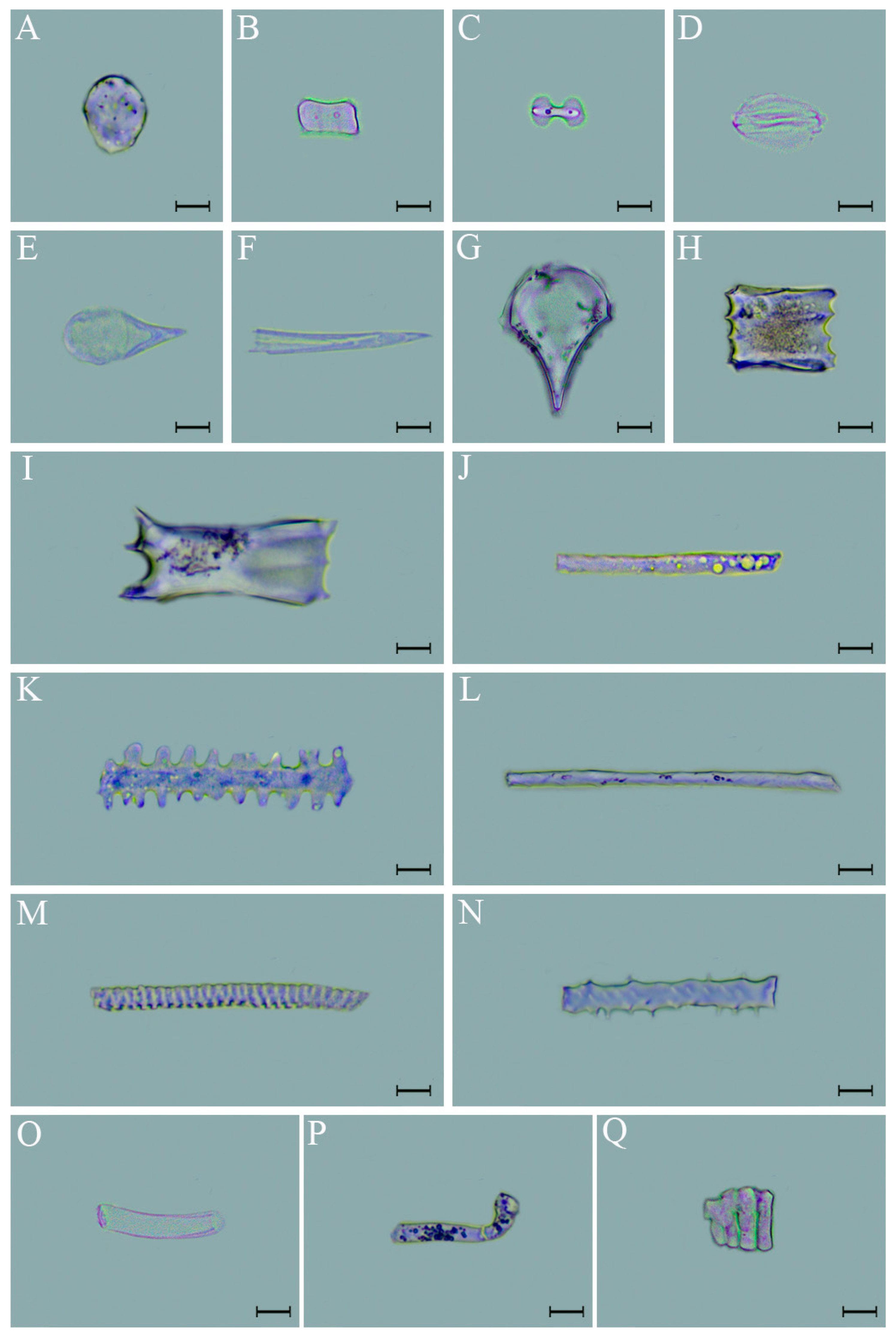
Phytoliths’ morphotypes in D. brandisii leaves at different phenological stages were identical. According to the classification system of ICPN 1.0 [32] and ICPN 2.0 [33], the phytolith morphotypes observed in D. brandisii leaves included elliptical, saddle, bilobate, stomata, acute, flabellate, blocky, elongate, and irregular classes—17 types in all (Figure 2).
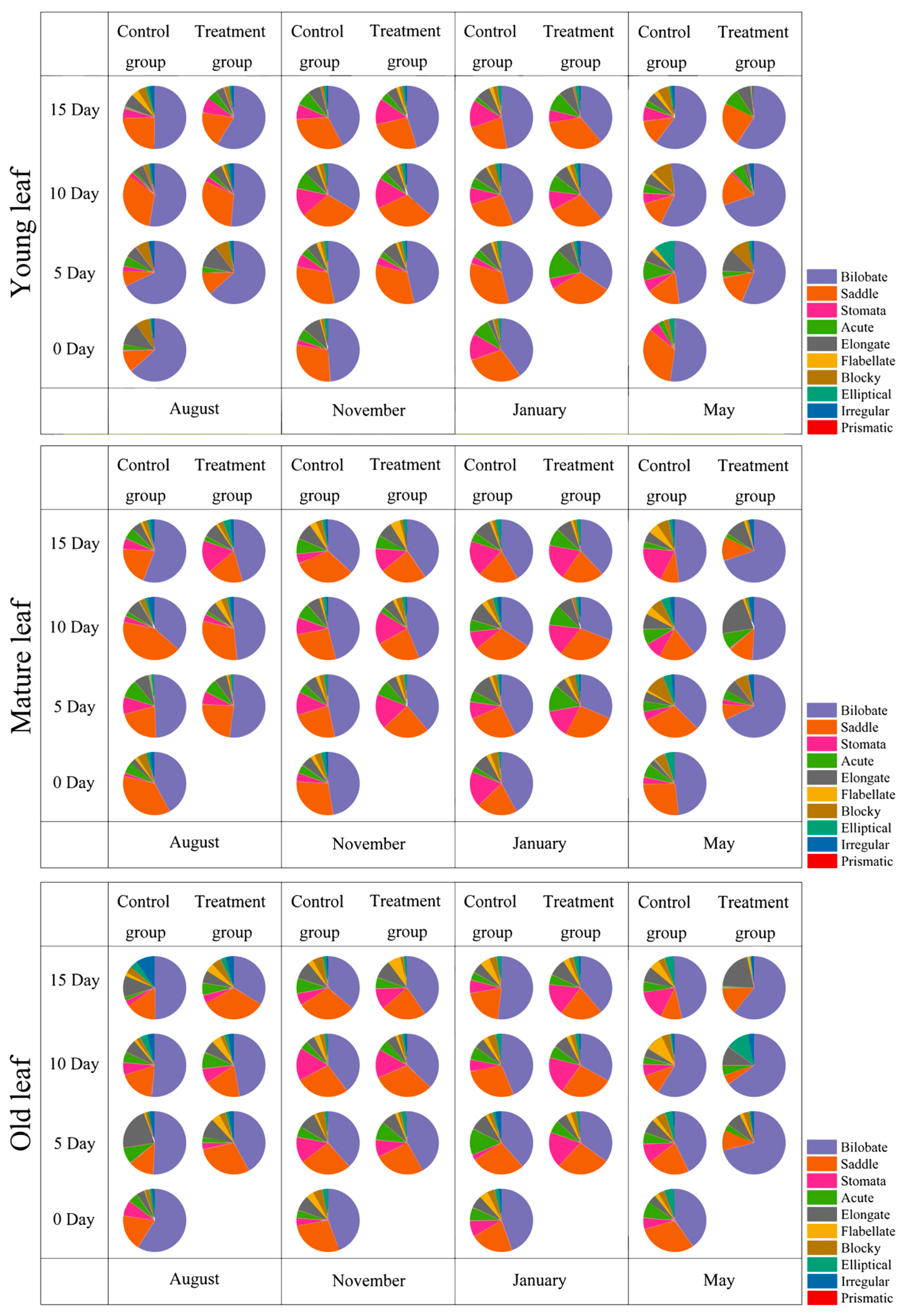
Phytolith morphotypes in D. brandisii leaves at different phenological stages were characterized by a high frequency of bilobates and saddles, accounting for more than 65%; whereas, elliptical, flabellate, and irregular morphotypes accounted for the smallest proportion, normally below 5% (Figure 3).
In the control group, proportions of different types of phytoliths in D. brandisii leaves varied at different phenological stages. In August, the proportion of stomata and acute phytoliths was relatively small (less than 5%), while the proportion of blocky and elongate phytoliths was around 10% and decreased over time (Table S1). The bilobate phytolith had the largest proportion in the young leaves, reaching the maximum value on the fifth day. The saddle phytolith had a relatively large proportion in the mature leaves and increased with time (Figure 3). In November, the proportion of blocky phytoliths was relatively small (less than 5%), while the proportion of stomata, acute, and elongate phytoliths was around 10%. The proportion of saddle and bilobate phytoliths was relatively stable, maintaining a high proportion during sampling in November (Figure 3, Table S2). In January during dormancy, the proportion of blocky phytoliths was relatively small (less than 5%) (Table S3), while the proportion of acute and elongate phytoliths was around 10%. The proportion of stomata phytoliths in young and mature leaves increased with time and reached maximum values on the 15th day. The proportion of saddle and bilobate phytoliths remained relatively stable, maintaining a high proportion throughout (Figure 3). In the branching and leafing stage in May, the proportion of stomata phytoliths was relatively small (less than 5%) (Table S4), while the proportion of acute, blocky, and elongate phytoliths was around 10%. The saddle phytoliths decreased over time, while the proportion of bilobate phytoliths remained relatively stable (around 40%) (Figure 3, Tables S1–S4).
After the SS treatment, the statistical analysis revealed that proportions of the different morphotypes in D. brandisii leaves varied by phenological stage. In August, the proportion of blocky phytoliths in young leaves significantly decreased and continued to decline with time, while elongate phytoliths significantly increased on the fifth day. The stomata phytolith in mature leaves significantly increased on the 15th day. Saddle phytoliths in the old leaves significantly increased, reaching a maximum on the 15th day (Figure 3). In November, the proportion of stomata phytoliths in mature leaves significantly increased and continued to increase over time. The acute phytoliths in old leaves significantly increased on the fifth day. No significant variations were observed in other phytolith morphotypes (Figure 3). In January, saddle phytoliths in young leaves and stomata phytoliths in mature and old leaves significantly increased and continued to increase over time. Moreover, acute phytoliths in young and mature leaves also significantly increased, while their proportion in old leaves decreased (Figure 3). In May, the proportions of bilobate phytoliths in leaves of all ages significantly increased over the controls. Elongate phytoliths in mature leaves significantly increased, while other types of phytoliths were relatively stable (Figure 3).

3.3. Phytolith Size in D. brandisii Leaves Under Treatment at Different Phenological Stages

The phytolith assemblage in D. brandisii leaves was characterized by a high frequency of bilobate, saddle, acute, flabellate, and elongate types—the sizes of which varied in leaves with the SS treatment (Figure 3; Tables S5–S9).
Observations of the control group revealed that there were differences in the size of phytoliths in various aged leaves during different phenological stages of D. brandisii. During the bamboo shooting stage in August, there were no significant differences in the length and width of saddle and bilobate phytoliths among different leaf ages (Tables S5 and S6). The length of acute phytoliths in young leaves was significantly shorter than that in old leaves (Table S7), while flabellate phytoliths in young leaves were significantly longer than those in mature leaves (Table S8). Elongate phytoliths in young leaves were significantly shorter than those in old leaves (Table S9). In November, at the late shooting stage, there were no significant differences in the length or width of saddle, bilobate, acute, flabellate, and elongate phytoliths among different aged leaves (Tables S5–S9). During dormancy in January, flabellate phytoliths were significantly longer in mature and old leaves on the 5th and 10th days than were those in young leaves (Table S8), while there were no significant differences in the length or width of other morphotypes among different aged leaves. During the branching and leafing stage in May, there were no significant differences in the length or width of saddle, bilobate, acute, and elongate phytoliths among leaves of different ages, while the length of flabellate phytoliths increased over time and were longer in old leaves than in young or mature leaves (Table S8).
After the treatment with SS, the sizes of different types of phytoliths in leaves of D. brandisii at various maturity stages during different phenological stages increased to varying degrees over time. At the bamboo shooting stage in August, the width of saddle phytoliths significantly decreased in young and mature leaves at the 5th and 10th days (Table S5), the length of acute phytoliths significantly decreased in young leaves at the 5th and 15th days (Table S7), the width of flabellate phytoliths significantly increased in young and mature leaves at the 5th and 10th days (Table S8), and the length of elongate phytoliths significantly increased in young leaves at the 10th and 15th days (Table S9). In November and January, the response of all morphotypes to the SS treatment was weak, showing no significant changes (Tables S5–S9). In the branching and leafing stage in May, the width of bilobate phytoliths significantly increased in old leaves at the 10th and 15th days (Table S6), and the length of acute phytoliths significantly decreased in young leaves at the 5th and 15th days (Table S7). The width of flabellate phytoliths significantly increased in young and mature leaves at the 5th and 10th days (Table S8), while the length of elongated phytoliths significantly increased in young leaves at the 10th and 15th days (Table S9).
Overall, during the bamboo shooting stage in August, the saddle, acute, flabellate, and elongated phytoliths were more sensitive to the SS treatment, especially in young and mature leaves, where significant changes in the phytolith width or length occurred. In November and January, the phytoliths’ variation response to the SS treatment was insignificant. During the branching and leafing stage in May, the bilobate, acute, flabellate, and elongated phytoliths were more sensitive to the SS treatment, particularly in young and old leaves. The results indicated that phytoliths in D. brandisii responded differently to the SS treatment during different phenological stages, and August and May were critical periods for the phytolith morphology and size variation.

4. Discussion

4.1. Effect of SS on Phytolith Concentration in D. brandisii Leaves at Different Phenological Stages

Phytoliths are the primary form of silicon present in plants [34]. Studies have shown that Poaceae plants have notably high phytolith concentrations, especially the bamboos [35]. Liu et al. reported that the weather conditions (temperature, precipitation) and leaf age were key factors controlling phytolith accumulation in Moso bamboo leaves [36]. The formation of phytoliths is also related to phenological and developmental changes [22]. Zhu et al. reported that the phytolith concentration was lowest in the leaves of Dendrocalamus giganteus during the period of branching and leafing and the highest during the late shooting period [22], consistent with our observations on D. brandisii. Moreover, the phytolith concentration in D. brandisii at different phenological stages showed an order of old leaves > mature leaves > young leaves. This was consistent with previous research results showing that the phytolith concentration in young tissues was lower than that of old tissues in bamboo [15,22]. This indicates that with the maturity of leaves, silicon is continuously deposited in leaves to form phytoliths, which is consistent with de Melo et al.’s [18] study on the tropical forage grass, Brachiaria brizantha.
Leaves and roots are the main organs for plants to absorb silicon. And variation in silicon absorption at different growth stages substantially affects the phytolith formation and morphology. Zhang et al. found that an appropriate exogenous silicon application promoted the absorption and utilization rate of silicon in the leaves of D. brandisii [29]. Yang et al. found that a silicon fertilizer application significantly increased the phytolith concentration in rice, enhancing the potential of phytolith-occluded carbon [37]. In our trials, after the SS application, the phytolith concentration in D. brandisii leaves increased to some extent, particularly in August when the concentration in leaves of different ages significantly increased. Silicic acid is concentrated and precipitated in leaf cells by transpiration [38]. Increases in temperature and evaporation rates in August might accelerate the deposition of phytoliths. During the branching and leafing stage in May, the phytolith concentration in young leaves is relatively low. This is because the cells of young leaves are still dividing and differentiating, and deposition sites such as silica cells have not yet fully matured, making them unable to fix large amounts of silicon [39]. In November, the phytolith concentration in old leaves was the highest. It is known that the water content in old leaves is lower than that of young and mature leaves. Due to water loss in old leaves, silicon deposition occurs passively, resulting in higher phytolith concentrations [40,41], which is consistent with a report that Brachiaria brizantha continuously accumulates silicon in its leaves during senescence due to passive water transport and transpiration [18]. However, the degree of silicification in older leaves of Bothriochloa laguroides is lower. The impact of senescence on silicon accumulation may vary with species, environmental conditions, and leaf developmental stages and does not always result in an increase in silicon accumulation [19].

4.2. Effect of SS on Phytolith Morphology in D. brandisii Leaves at Different Phenological Stages

Plant leaves are the primary sites for transpiration, respiration, and photosynthesis. Different morphotypes of phytoliths originate from various cells and have distinct functions [42]. Interestingly, phytolith morphotypes in D. brandisii leaves at different phenological stages were identical, including elliptical, saddle, bilobate, acute, flabellate, blocky, and elongate types, among which the saddle and bilobate accounted for more than 65% of the total, consistent with reports by Zhan et al. [15] and Gu et al. [43], who showed that phytolith assemblages of leaves of 26 bamboo species included a high proportion of saddle and bilobate phytoliths.
Saddle and bilobate phytoliths are formed in short cells, and previous studies showed that bilobate and saddle phytoliths were least affected by environmental factors, making them the most stable phytolith morphotypes; their morphological parameters (size and proportion) can be used to help classify grass species [14,44,45]. In our study, the size of saddle and bilobate phytoliths showed an insignificant variation after the SS treatment at different phenological stages, confirming their stability, and, thus, their parameters may have taxonomic value. In May and August, with the SS treatment, it was observed that the proportion of bilobate phytoliths in D. brandisii leaves of different ages significantly increased. It was also found in corn leaves that, with the rising temperature, the proportion of bilobate phytoliths gradually increased [45]. When D. brandisii entered the dormancy stage in January, the proportion of saddle phytoliths significantly increased. A statistical analysis of phytoliths in Phragmites australis in Northeast China revealed that the proportion of saddle phytoliths accounted for 77%, which is higher than that in other regions, suggesting that low temperatures might favor the formation of saddle phytoliths [46].
Acute phytoliths are shaped like needles or hooks and mainly form in trichomes. Their presence in leaves can increase mechanical defense and increase the discomfort of herbivores during feeding [43,47]. In November and January, after the SS treatment, the proportion of acute phytoliths in D. brandisii mature leaves increased, suggesting that the SS application significantly enhanced the physical barrier of mature leaves, which may imply a stronger ability to resist biotic and abiotic stress.
Bulliform cells are often the main deposition sites of silicon in plant leaves, and this phenomenon is particularly prominent in the leaves of Brachiaria brizantha, where silicon is specifically deposited on the cell walls of its bulliform cells [18]. Bulliform cells are related to plant transpiration. Honaine et al. [19] demonstrated that transpiration may be the main factor determining the proportion and distribution of silicified cells in leaves, with a particularly significant impact on bulliform cells. Blocky and flabellate phytoliths mainly formed in tubular bulliform cells [43]. After the SS application, the proportion and size of flabellate phytoliths in young and mature leaves significantly increased in August and May, indicating that SS might enhance the transpiration of young and mature leaves, which could then promote an increase in the proportion and size of flabellate phytoliths. Tang et al. also found that after the artificial fertilization of rice, the sizes of flabellate phytoliths were significantly larger than those without fertilization [48].
Stomata phytoliths mainly originate from the epidermal cells surrounding the stomatal complex (subsidiary cells or adjacent cells) [49], optimizing stomatal function by enhancing the mechanical strength of the cell wall [50]. The silicification of stomata is usually associated with transpiration [51]. In August, November, and January, after the SS application, the proportion of stomata phytoliths in mature leaves significantly increased, indicating that the SS application might improve leaf stomatal function, enhance the mechanical strength of the surrounding stomatal structures, and reduce water loss. However, silicification is not always beneficial. It can also occur as a consequence of senescence or under conditions of a sufficient silicon supply, leading to the occlusion and immobilization of guard cells. When silicon is in excess, the silicification process may involve cells that do not necessarily need to form silica deposits, resulting in the impairment of their functions [51].
Elongate phytoliths are formed in long cells, which protect the leaf vascular tissue, significantly improving plant photosynthesis, mechanical strength, and physical properties [43]. After applying SS to D. brandisii in August and May, the proportion of elongate phytoliths in mature leaves increased significantly, and their length also increased significantly, indicating that SS promoted the development of elongate phytoliths, enhancing the leaves’ mechanical strength and support capacity and improving the photosynthetic capacity.

4.3. The Physiological and Ecological Significance of the Effects of SS on the Differences in Phytoliths in D. brandisii Leaves at Different Phenological Stages

It is generally believed that both genetic and environmental factors affect the morphology and size of phytoliths. In addition to being controlled by genetic factors [52], leaf maturity [53] and hydrothermal conditions [54] will affect the morphology and size of phytoliths. Our study found that bilobate, saddle, acute, flabellate, and elongate phytoliths were sensitive to the SS treatment at different phenological stages, and the width or length of phytoliths changed significantly. Studies have shown that silicon fertilizer treatments increased the length of epidermal cells (long cells, short cells, and motor cells) at the seedling stage of rice, and silicon promoted rice growth by increasing the ductility of cell walls [55]. The application of silicon fertilizer could promote the division and expansion of epidermal cells in rice leaves, resulting in a significant increase in both their number and size [56]. Silicon is deposited in the cell walls of leaf epidermal cells, enhancing the degree of silicification, with silicified cells being clearly visible on the leaf surface [57]. Furthermore, Hayasaka et al. [58] confirmed that silicon deposition thickens the rice leaf veins, thereby strengthening the physical barrier and inhibiting pathogen penetration. Fertilization at the same time can significantly increase the leaf thickness, length, width, and area of rice [59,60], so the enlarged leaves and enlarged epidermal cells after fertilization promote the development of larger phytoliths. Our study shows that fertilization is also a significant factor affecting the size of phytoliths, consistent with an earlier report by Tang et al. [48]. At the same time, different phenological periods have different responses to SS treatments. May and August are key periods for the changes in the phytolith morphology and size, and these changes have important physiological and ecological ramifications. May is the leafing and branching stage; cell division and differentiation are active. The demand for silicon is large. Silicon accumulates in leaf epidermal cells and vascular bundle sheaths, not only enhancing the mechanical strength of cell walls but also forming silicified structures around vascular bundles [57,58]. These silicified structures support the material supply required for the growth of new organs and build a basic defense barrier for young tissues to resist pests and wind and rain stress [61,62]. At the shooting stage in August, the SS treatment significantly increased the size of saddle and acute phytoliths, enhanced the rigidity of cell walls, and likely improved the ability of leaves to resist transpiration and high temperatures. These differential responses can provide a basis for formulating precise silicon fertilizer application strategies for bamboo.

5. Conclusions

This study demonstrated that SS had a significant impact on the phytolith concentration and morphological parameters in D. brandisii leaves at different phenological stages. SS significantly increased the phytolith concentration in D. brandisii leaves, showing a trend of old leaves > mature leaves > young leaves. Phytoliths’ morphotypes in D. brandisii leaves at different phenological stages were identical. The saddle and bilobate accounted for over 65% of the total phytoliths. August and May were critical periods for phytolith formation and variation in the phytolith morphology and size. The SS treatment significantly increased the proportion of saddle, bilobate, and stomatal phytoliths and may help optimize the silicified structure of leaf epidermal cells and enhance leaf resistance and light utilization. Our observations help elucidate the mechanisms of phytolith formation in different phenological periods of D. brandisii and can serve as a theoretical basis for the efficient use of silicon fertilizers in bamboo cultivation.

Supplementary Materials

The following supporting information can be downloaded at https://www.mdpi.com/article/10.3390/agronomy15092138/s1: Table S1. Proportion of different phytolith morphotypes in D. brandisii leaves under SS treatment in August (%); Table S2. Proportion of different phytolith morphotypes in D. brandisii leaves under SS treatment in November (%); Table S3. Proportion of different phytolith morphotypes in D. brandisii leaves under SS treatment in January (%); Table S4. Proportion of different phytolith morphotypes in D. brandisii leaves under SS treatment in May (%); Table S5. The sizes of saddle phytoliths in D. brandisii leaves under SS treatment at different phenological stages (μm); Table S6. The sizes of bilobate phytolith in D. brandisii leaves under SS treatment at different phenological stages (μm); Table S7. The sizes of acute phytoliths in D. brandisii leaves under SS treatment at different phenological stages (μm); Table S8. The sizes of flabellate phytoliths in D. brandisii leaves under SS treatment at different phenological stages (μm); and Table S9. The sizes of elongate phytoliths in D. brandisii leaves under SS treatment at different phenological stages (μm).

Author Contributions

Conceptualization, Y.Y., L.H., and L.Y.; methodology, Y.Y.; software, Y.Y. and L.H.; validation, C.W.; formal analysis, Y.Y.; investigation, Y.Y.; resources, H.Z.; data curation, Y.Y., L.H., and L.Y.; writing—original draft preparation, Y.Y.; writing—review and editing, Y.Y. and H.Z.; visualization, Y.Y. and L.H.; supervision, C.W., S.W., and M.L.; project administration, H.Z. and S.W.; funding acquisition, H.Z. All authors have read and agreed to the published version of the manuscript.

Funding

This study was jointly funded by National Natural Science Foundation of China (NO. 32160415), Opening Foundation of Forestry First Discipline Degree program and Key Laboratory of Southwest Forestry University (NO: LXXK-2025D17), and Xingdian Talent Support Plan Project (2022).

Data Availability Statement

All data generated or analyzed during this study are included in the article.

Acknowledgments

We are very grateful to every scientific researcher who supports and helps us in our work.

Conflicts of Interest

The authors declare no conflicts of interest.

References

  1. An, X.; Xie, B. Phytoliths from woody plants: A review. Diversity 2022, 14, 339. [Google Scholar] [CrossRef]
  2. Wang, Y.; Lu, H. Phytolith Study and Its Application; China Ocean Press: Beijing, China, 1993. [Google Scholar]
  3. Ma, J.F.; Yamaji, N. Functions and transport of silicon in plants. Cell. Mol. Life Sci. 2008, 65, 3049–3057. [Google Scholar] [CrossRef] [PubMed]
  4. Miyake, Y.; Takahashi, E. Effect of silicon on the growth of solution cultured cucumber plant. Soil Sci. Plant Nutr. 1983, 29, 71–83. [Google Scholar] [CrossRef]
  5. Massey, F.P.; Hartley, S.E. Physical defences wear you down: Progressive and irreversible impacts of silica on insect herbivores. J. Anim. Ecol. 2009, 78, 281–291. [Google Scholar] [CrossRef] [PubMed]
  6. Sato, K.; Yamauchi, A.; Ozaki, N.; Ishigure, T.; Oaki, Y.; Imai, H. Optical properties of biosilicas in rice plants. RSC Adv. 2016, 6, 109168–109173. [Google Scholar] [CrossRef]
  7. Mandlik, R.; Thakral, V.; Raturi, G.; Shinde, S.; Nikolić, M.; Tripathi, D.K.; Sonah, H.; Deshmukh, R. Significance of silicon uptake, transport, and deposition in plants. J. Exp. Bot. 2020, 71, 6703–6718. [Google Scholar] [CrossRef]
  8. Wang, Y.; Lu, H. The Study of Phytolith and Its Application; Haiyang Press: Beijing, China, 2008. [Google Scholar]
  9. Niu, J.; Liu, C.; Huang, M.; Liu, K.; Yan, D. Effects of foliar fertilization: A review of current status and future perspectives. J. Soil Sci. Plant Nutr. 2021, 21, 104–118. [Google Scholar] [CrossRef]
  10. Laane, H.M. The effects of foliar sprays with different silicon compounds. Plants 2018, 7, 45. [Google Scholar] [CrossRef] [PubMed]
  11. Tayade, R.; Ghimire, A.; Khan, W.; Lay, L.; Attipoe, J.Q.; Kim, Y. Silicon as a smart fertilizer for sustainability and crop improvement. Biomolecules 2022, 12, 1027. [Google Scholar] [CrossRef]
  12. Dos Santos, S.M.M.; de Mello Prado, R.; de Souza Júnior, J.P.; Teixeira, G.C.M.; dos Santos Duarte, J.C.; de Medeiros, R.L.S. Silicon supplied via foliar application and root to attenuate potassium deficiency in common bean plants. Sci. Rep. 2021, 11, 19690. [Google Scholar] [CrossRef]
  13. Zaman, Q.U.; Rashid, M.; Nawaz, R.; Hussain, A.; Ashraf, K.; Latif, M.; Heile, A.O.; Mehmood, F.; Salahuddin, S.; Chen, Y. Silicon fertilization: A step towards cadmium-free fragrant rice. Plants 2021, 10, 2440. [Google Scholar] [CrossRef]
  14. Jones, L.H.; Milne, A.A.; Sanders, J.V. Tabashir: An opal of plant origin. Science 1966, 151, 464–466. [Google Scholar] [CrossRef]
  15. Zhan, H.; Li, J.; Niu, Z.; Li, M.; Wang, C.; Wang, S. Silicon variation and phytolith morphology in different organs of Dendrocalamus brandisii (Munro) Kurz (Bambusoideae). Braz. J. Bot. 2019, 42, 529–541. [Google Scholar] [CrossRef]
  16. Gu, Y.; Yan, H.; Zheng, M.; Liu, H.; Ji, Y.; Zhang, Y. Phytolith records from 15 continuously growing Bambusa emeiensis leaves and its climatic significance. Rev. Palaeobot. Palynol. 2022, 300, 104620. [Google Scholar] [CrossRef]
  17. Katz, O. Silica phytoliths in angiosperms: Phylogeny and early evolutionary history. New Phytol. 2015, 208, 642–646. [Google Scholar] [CrossRef]
  18. de Melo, S.P.; Monteiro, F.A.; De Bona, F.D. Silicon distribution and accumulation in shoot tissue of the tropical forage grass Brachiaria brizantha. Plant Soil. 2010, 336, 241–249. [Google Scholar] [CrossRef]
  19. Fernández Honaine, M.; Osterrieth, M.L. Silicification of the adaxial epidermis of leaves of a panicoid grass in relation to leaf position and section and environmental conditions. Plant Biol. 2012, 14, 596–604. [Google Scholar] [CrossRef]
  20. Motomura, H.; Fujii, T.; Suzuki, M. Silica deposition in relation to ageing of leaf tissues in Sasa veitchii (Carriere) Rehder (Poaceae: Bambusoideae). Ann. Bot. 2004, 93, 235–248. [Google Scholar] [CrossRef] [PubMed]
  21. Li, R.; Fan, J.; Carter, J.; Jiang, N.; Gu, Y. Monthly variations of phytoliths in the leaves of the bamboo Dendrocalamus ronganensis (Poaceae: Bambusoideae). Rev. Palaeobot. Palynol. 2017, 246, 62–69. [Google Scholar] [CrossRef]
  22. Zhu, F.; Niu, Z.; Li, J.; Yu, L.; Wang, S.; Wang, C.; Zhan, H. Variation of the phytolith content and morphology of Dendrocalamus giganteus at different phenological periods. J. Southwest For. Univ. 2022, 42, 71–77. [Google Scholar]
  23. Li, D.; Wang, Z.; Zhu, Z.; Xia, N.; Jia, L.; Guo, Z.; Yang, G.; Stapleton, C. Bambuseae. In Flora of China; Wu, Z., Raven, P., Hong, D., Eds.; Science Press: Beijing, China, 2006. [Google Scholar]
  24. Huang, L.; Yang, Y.; Zhu, F.; Chang, J.; Wang, S.; Wang, C.; Li, M.; Zhan, H. Foliar application of sodium gluconate enhances photosynthetic efficiency and photoassimilate accumulation in Dendrocalamus brandisii across vegetative phenological stages. Ind. Crops Prod. 2025, 230, 121030. [Google Scholar] [CrossRef]
  25. Peng, X.; Liu, J.; Tang, N.; Deng, J.; Liu, C.; Kan, H.; Zhao, P.; Zhang, X.; Shi, Z.; Liu, Y. Sequential extraction, structural characterization, and antioxidant activity of polysaccharides from Dendrocalamus brandisii bamboo shoot shell. Food Chem. X 2023, 17, 100621. [Google Scholar] [CrossRef] [PubMed]
  26. Emamverdian, A.; Ding, Y.; Ranaei, F.; Ahmad, Z. Application of bamboo plants in nine aspects. Sci. World J. 2020, 2020, 7284203. [Google Scholar] [CrossRef]
  27. Umemura, M.; Takenaka, C. Biological cycle of silicon in moso bamboo (Phyllostachys pubescens) forests in central Japan. Ecol. Res. 2014, 29, 501–510. [Google Scholar] [CrossRef]
  28. Franklin, D.C. Vegetative phenology and growth of a facultatively deciduous bamboo in a monsoonal climate. Biotropica 2005, 37, 343–350. [Google Scholar] [CrossRef]
  29. Zhang, L.; Zhan, H.; Niu, Z.; Wang, C.; Wang, S. Effects of exogenous silicon on silicon, chlorophyll, and soluble sugar contents in seedling leaf blades of Dendrocalamus brandisii. J. Plant Nutr. Fert. 2016, 45, 101–106. [Google Scholar]
  30. Parr, J.F.; Dolic, V.; Lancaster, G.; Boyd, W.E. A microwave digestion method for the extraction of phytoliths from herbarium specimens. Rev. Palaeobot. Palynol. 2001, 116, 203–212. [Google Scholar] [CrossRef]
  31. Pearsall, D.M. Paleoethnobotany: A Handbook of Procedures, 3rd ed.; Routledge: New York, NY, USA, 2015. [Google Scholar]
  32. Madella, M.; Alexandre, A.; Ball, T. International code for phytolith nomenclature 1.0. Ann. Bot. 2005, 96, 253–260. [Google Scholar] [CrossRef]
  33. Neumann, K.; Stromberg, C.; Ball, T.; Albert, R.; Vrydaghs, L.; Cummings, L.S. International code for phytolith nomenclature (ICPN) 2.0. Ann. Bot. 2019, 124, 189–199. [Google Scholar] [CrossRef]
  34. Chen, I.; Li, K.T.; Tsang, C.H. Silicified bulliform cells of Poaceae: Morphological characteristics that distinguish subfamilies. Bot. Stud. 2020, 61, 5. [Google Scholar] [CrossRef]
  35. Hodson, M.J.; White, P.J.; Mead, A.; Broadley, M.R. Phylogenetic variation in the silicon composition of plants. Ann. Bot. 2005, 96, 1027–1046. [Google Scholar] [CrossRef] [PubMed]
  36. Liu, L.; Chang, S.; Huang, C.; Yu, X.; Zhi, Y.; Jiang, P. Controls for phytolith accumulation in Moso bamboo leaves across China. Sci. Total Environ. 2023, 893, 164886. [Google Scholar] [CrossRef]
  37. Yang, X.; Ni, Y.; Li, Z.; Yue, K.; Wang, J.; Li, Z.; Yang, X.; Song, Z. Silicon in paddy fields: Benefits for rice production and the potential of rice phytoliths for biogeochemical carbon sequestration. Sci. Total Environ. 2024, 929, 172497. [Google Scholar] [CrossRef] [PubMed]
  38. Rotmuenwai, N.; Aryuyo, K.; Kruethaworn, N.; Wattananit, W.; Yookongkaew, N. Exploring silica accumulation in bamboo leaves: A study on phytolith morphology and epidermal patterning in the tropical giant bamboo Dendrocalamus copelandii. Ann. Bot. 2025, 135, 757–768. [Google Scholar] [CrossRef]
  39. Dai, C.; Xu, R.; Yu, L.; Zhu, F.; Li, M.; Li, J.; Wang, S.; Wang, C.; Zhan, H. Silicon uptake and phytolith morphology in Dendrocalamus brandisii seedling leaf from different rearing methods. Forests 2023, 14, 1877. [Google Scholar] [CrossRef]
  40. Li, R.; Chen, X.; Wen, M.; Vachula, R.; Tan, S.; Dong, H.; Zhou, L.; Gu, Z.; Xu, M. Phytolith-occluded carbon in leaves of Dendrocalamus ronganensis influenced by drought during growing season. Physiol. Plant. 2022, 174, e13748. [Google Scholar] [CrossRef] [PubMed]
  41. Matichenkov, V.; Bocharnikova, E. The relationship between silicon and soil physical and chemical properties. Stud. Plant Sci. 2001, 8, 209–219. [Google Scholar]
  42. Strömberg, C.A.; Di Stilio, V.S.; Song, Z. Functions of phytoliths in vascular plants: An evolutionary perspective. Funct. Ecol. 2016, 30, 1286–1297. [Google Scholar] [CrossRef]
  43. Gu, Y.; Liu, H.; Wang, H.; Li, R.; Yu, J. Phytoliths as a method of identification for three genera of woody bamboos (Bambusoideae) in tropical southwest China. J. Archaeol. Sci. 2016, 68, 46–53. [Google Scholar] [CrossRef]
  44. Gu, Y.; Zhao, Z.; Pearsall, D.M. Phytolith morphology research on wild and domesticated rice species in East Asia. Quat. Int. 2013, 287, 141–148. [Google Scholar] [CrossRef]
  45. Li, R.; Tan, S.; Qin, X.; Ma, J.; Chen, G.; Liu, P. Study on the phytolith variation in maize during its growing season. Acta Micropalaeontol. Sin. 2016, 33, 170–179. [Google Scholar]
  46. Liu, H.; Jie, D.; Liu, L.; Guo, M.; Li, N. Change characters of Phragmites australis phytolith in Northeast China. Chin. J. Plant Ecol. 2013, 37, 861–871. [Google Scholar] [CrossRef]
  47. Zhang, X.; Gélin, U.; Spicer, R.A.; Wu, F.; Farnsworth, A.; Chen, P.; Rio, C.D.; Li, S.; Liu, J.; Huang, J.; et al. Rapid Eocene diversification of spiny plants in subtropical woodlands of central Tibet. Nat. Commun. 2022, 13, 3787. [Google Scholar] [CrossRef] [PubMed]
  48. Tang, X.; Xie, J.; Liu, Z.; Sun, G.; Yuan, F.; Lü, H. Effect of artificial fertilization on morphological development of rice bulliform phytoliths and its significance. Quat. Sci. 2022, 42, 1792–1805. [Google Scholar]
  49. Piperno, D.R. Phytoliths: A comprehensive guide for archaeologists and paleoecologists. Rowman Altamira: Lanham, MD, USA, 2006. [Google Scholar]
  50. Kumar, S.; Elbaum, R. Interplay between silica deposition and viability during the life span of sorghum silica cells. New Phytol. 2018, 217, 1137–1145. [Google Scholar] [CrossRef]
  51. De Tombeur, F.; Raven, J.A.; Toussaint, A.; Lambers, H.; Cooke, J.; Hartley, S.E.; Johnson, S.N.; Coq, S.; Katz, O.; Schaller, O.; et al. Why do plants silicify? Trends Ecol. Evol. 2023, 38, 275–288. [Google Scholar] [CrossRef]
  52. Tang, X.; Lu, H.; Cao, Z.; Xie, J. Morphological characteristics of homozygous wild rice phytoliths and their significance in the study of rice origins. Sci. China Earth Sci. 2022, 65, 107–117. [Google Scholar] [CrossRef]
  53. Zheng, Y.; Dong, Y.; Matsui, A.; Udatsu, T.; Fujiwara, H. Molecular genetic basis of determining subspecies of ancient rice using the shape of phytoliths. J. Archaeol. Sci. 2023, 30, 1215–1221. [Google Scholar] [CrossRef]
  54. Wang, C.; Lu, H.; Zhang, J.; Mao, L.; Ge, Y. Bulliform phytolith size of rice and its correlation with hydrothermal environment: A preliminary morphological study on species in Southern China. Front. Plant Sci. 2019, 10, 1037. [Google Scholar] [CrossRef]
  55. Hossain, M.T.; Mori, R.; Soga, K.; Wakabayashi, K.; Kamisaka, S.; Fujii, S.; Yamamoto, R.; Hoson, T. Growth promotion and an increase in cell wall extensibility by silicon in rice and some other Poaceae seedlings. J. Plant Res. 2002, 115, 23–27. [Google Scholar] [CrossRef] [PubMed]
  56. Shui, M.; Chen, D.; Qin, S.; Jiang, S. The silicification of young tissues of rice and relationship with its resistance to blast of rice. J. Plant Nutr. Fert. 1999, 4, 352–358. [Google Scholar]
  57. Cai, K.; Gao, D.; Luo, S.; Zeng, R.; Yang, J.; Zhu, X. Physiological and cytological mechanisms of silicon-induced resistance in rice against blast disease. Physiol. Plant. 2008, 134, 324–333. [Google Scholar] [CrossRef] [PubMed]
  58. Hayasaka, T.; Fujii, H.; Ishiguro, K. The role of silicon in preventing appressorial penetration by the rice blast fungus. Phytopathology 2008, 98, 1038–1044. [Google Scholar] [CrossRef] [PubMed]
  59. Gong, J.; Zhang, H.; Li, J.; Dai, Q.; Huo, Z.; Xu, K.; Li, D.; Hua, Z.; Sha, A.; Zhou, Y.; et al. Progress in research on pat terns and systematic theories of super-high-yielding cultivation in rice. Chin. J. Rice Sci. 2010, 24, 417–424. [Google Scholar]
  60. Lin, Z.; Zhan, X.; Cheng, S.; Cao, L. Progress on mapping and cloning genes for leaf architecture in rice (Oryza sativa. L). J. Nucl. Agric. Sci. 2013, 27, 1662–1669. [Google Scholar]
  61. Ranganathan, S.; Suvarchala, V.; Rajesh, Y.B.R.D.; Prasad, M.S.; Padmakumari, A.P.; Voleti, S.R. Effects of silicon sources on its deposition, chlorophyll content, and disease and pest resistance in rice. Biol. Plant. 2006, 50, 713–716. [Google Scholar] [CrossRef]
  62. Yamaji, N.; Sakurai, G.; Mitani-Ueno, N.; Ma, J.F. Orchestration of three transporters and distinct vascular structures in node for intervascular transfer of silicon in rice. Proc. Natl. Acad. Sci. USA 2015, 112, 11401–11406. [Google Scholar] [CrossRef]
Figure 1. Phytolith concentration in D. brandisii leaves at different phenological stages (%). Note: Data are presented as mean ± standard deviation. Different lowercase letters above trend lines denote the significant differences between leaves at different stages in the control group at p < 0.05 according to LSD. Different uppercase letters above trend lines denote significant differences between leaves at different stages in the experimental group at p < 0.05 according to LSD. * Denotes significant differences at p < 0.05 and ** at p < 0.01 between treatments on the same day.
Agronomy 15 02138 g001
Figure 2. Phytolith morphotypes in D. brandisii leaves at different phenological stages. (A) Elliptical. (B) Saddle. (C) Bilobate. (D) Stomata. (E,F) Acute: (E) acute and (F) extended acute. (G) Flabellate. (H,I) Blocky: (H) Blocky. (I) Rectangular. (JN) Elongate: (J) Elongate scrobiculate; (K) Elongate columnar; (L) Elongate granulate; (M) Tracheary annulate; and (N) Elongate dentate. (OQ) Irregular: (O) Arcuate; (P) Uncinate; and (Q) Prismatic.
Agronomy 15 02138 g002
Figure 3. Proportions of different phytolith morphotypes in D. brandisii leaves by treatment at different phenological stages.
Agronomy 15 02138 g003
Table 1. The climate of Kunming City, Yunnan Province, China.
AugustNovemberJanuaryMay
Average high temperature (°C)25181424
Average low temperature (°C)169415
Average temperature (°C)20151017
Precipitation (mm)235.737.337.2131.4
Average humidity (%)78.37251.769
The data were downloaded from the official website of the World Weather Online. https://www.worldweatheronline.com/kunming-weather-history/yunnan/cn.aspx (accessed on 2 August 2025).
Disclaimer/Publisher’s Note: The statements, opinions and data contained in all publications are solely those of the individual author(s) and contributor(s) and not of MDPI and/or the editor(s). MDPI and/or the editor(s) disclaim responsibility for any injury to people or property resulting from any ideas, methods, instructions or products referred to in the content.