Article

Early Detection of Wheat Fusarium Head Blight During the Incubation Period Using FTIR-PAS

1
College of Agricultural Engineering, Jiangsu University, Zhenjiang 212013, China
2
State Key Laboratory of Wheat Improvement, Jinan 250100, China
3
The Key Laboratory for Crop Production and Smart Agriculture of Yunnan Province, Kunming 650201, China
4
The Key Laboratory of Spectroscopy Sensing, Ministry of Agriculture and Rural Affairs, Hangzhou 310058, China
5
Nanjing Institute of Environmental Science, Ministry of Ecological Environment, Nanjing 210042, China
*
Author to whom correspondence should be addressed.
Agronomy 2025, 15(9), 2100; https://doi.org/10.3390/agronomy15092100
Submission received: 15 July 2025 / Revised: 26 August 2025 / Accepted: 26 August 2025 / Published: 30 August 2025
(This article belongs to the Section Precision and Digital Agriculture)

Abstract

The apparent normalcy of wheat during the incubation period of Fusarium head blight (FHB) makes early diagnosis challenging. This study employed Fourier transform infrared photoacoustic spectroscopy (FTIR-PAS) to conduct layer-by-layer scanning of wheat leaves during the disease outbreak stage and performed a differential spectral analysis. Spectral information was collected from five sites (D0~D4) on diseased leaves at reducing distances from the lesion caused by the Fusarium graminearum pathogen. The results revealed that the disease caused an increase in spectral similarity between deeper and shallower layers. The spectra of leaves, after removing the D0 background, showed a correlation of 83.5% to that of the pathogen, and the similarity increased at sites closer to the lesion, suggesting that the original spectra captured a large amount of hidden information related to the pathogen. With the threshold for the absorption intensity ratio of R1650/1050 for background-subtracted spectra set at 0.5, the optimal overall accuracy and F1-score were 86.0% and 0.89 for diagnosing outbreak-stage samples, respectively, while for incubation-period samples, they were 82.5% and 0.83. These results elucidate the mechanism of using FTIR-PAS to diagnose FHB during its incubation period, providing a theoretical and technical foundation for detecting disease information in other crops.

1. Introduction

Crop information perception is a crucial link in modern agricultural production. The accuracy of perception determines the effectiveness of subsequent decisions, such as intelligent control and precise input. However, research on the mechanisms of agronomic information perception is still insufficient, and there remain many scenarios where perception accuracy is low and difficult to meet production requirements [1,2]. Intelligent diagnosis of crop diseases is one of the most prominent issues, particularly during the incubation-period stage [3]. The incubation period refers to the stage from pathogen invasion to symptom appearance. For instance, wheat, one of the most important food crops with high economic value in the world, is seriously threatened by Fusarium head blight (FHB) disease in terms of production safety. The incubation period for FHB is approximately 1–2 weeks [4,5], followed by the symptomatic period. This period is a critical window for disease control, offering cost-reducing and efficiency-improving control effects. Control measures applied during the symptomatic period often result in 20–50% yield reduction or even total crop failure, while blind pesticide application easily causes resource waste and environmental pollution [6]. However, during the incubation period, crops primarily respond to pathogen invasion through microscopic metabolic changes while appearing normal in the macroscopic phenotype. This makes it difficult to be detected with the naked eye, becoming a major limiting factor for conducting early diagnosis of crop diseases.
The current research on intelligent perception methods for wheat FHB information mainly focuses on two aspects: firstly, for the symptomatic period, deep learning-based machine vision technologies are used to non-destructively detect the severity of crop diseases through remote sensing imaging techniques like multispectral imaging [7] and hyperspectral imaging by unmanned aerial vehicle (UAV) [8,9], and remote sensing by satellites [10]; secondly, traditional machine learning-based multi-source data fusion technologies are employed, monitoring the severity of crop diseases with conventional spectral scanning [11] or the influencing external factors including meteorological conditions using sensors and weather stations to establish a predictive model for the possible occurrence of diseases [12]. However, these conventional disease perception technologies still have unresolved problems: they can only determine disease severity during the symptomatic period temporally, are unable to conduct diagnosis targeting the asymptomatic incubation period, or the research subjects are not directly related to the crop, instead using indirect factors such as meteorological indicators or pathogen spores to predict disease occurrence. These technologies are more inclined toward application in macro-scale scenarios and are difficult to physically penetrate samples to scan internal microscopic metabolism information for diagnosis [13,14]. Therefore, against the background where diagnostic accuracy for diseases cannot meet production demands, exploring rapid perception technologies at the microscopic scale complementary to the macroscopic scale for crop diseases is of significant importance.
Among numerous rapid analysis technologies, infrared spectroscopy (IR) has gained widespread application due to its prominent advantages of low cost, short cycle (a single detection can be controlled within 1 min), and no need for complex pre-treatments [15,16]. There are various ways with different perception principles for infrared spectroscopy detection. Among them, photoacoustic spectroscopy (PAS) originates from the photoacoustic effect generated when a sample absorbs light radiation energy and undergoes non-radiative relaxation in the form of thermal energy. Thermal energy can diffuse to different depths, leading to its outstanding advantage of in situ layer-by-layer scanning, meaning the obtained spectra can reflect more information at different physical depths of the detection point than the general spectral techniques [6,16]. Furthermore, for biological samples, the spectrum is one manifestation of metabolic patterns; its profile is mainly influenced by the types and concentrations of metabolites and their interactions [16]. The obtained information, when combined with chemometric analysis methods and machine learning algorithms, can be used to qualitatively or quantitatively analyze the metabolic phenotype of samples at the microscopic level, thereby reflecting various physiological states of crops. Additionally, mid-infrared spectroscopy (MIR; 2.5–25 μm) exhibits richer light absorption signals than near-infrared spectroscopy (NIR; 0.78–2.5 μm), providing more detailed structural and metabolic information about plants. Consequently, Fourier transform infrared photoacoustic spectroscopy (FTIR-PAS) in the MIR region has been widely applied to various agricultural information detection needs [17]. For example, qualitative detections using FTIR-PAS include sensing rice quality and its response to the environment, combined with a principal component analysis (PCA) [18]; distinguishing pesticide residue categories on plant surfaces [15]; and analyzing the composition of composite materials as sensors [19]. Quantitative perceptions include characterizing the content of plant nitrogen nutrition and its two-dimensional distribution on leaves using multivariate calibration methods like partial least squares (PLS) [20,21], detecting available phosphorus concentration in plant substrates [17], and probing the abundance of biomacromolecules like protein within plants [18,20,22].
Actually, in the field of precision medicine, the spectroscopic technique has developed into a novel omics technology capable of high-throughput characterization of metabolites in biological materials, measuring the biomarkers that are specially induced by diseases within target organisms to diagnose their occurrence, including cancer and heart disease and so on [23]. Furthermore, existing research has demonstrated that using FTIR-PAS combined with a probabilistic neural network (PNN) in the mid-infrared band (2.5~25 μm) can identify the severity level of rice blast occurrence in the symptomatic period. It can also distinguish asymptomatic leaves from mildly infected samples with high accuracy, achieving overall accuracy rates of 90% and 82% for greenhouse and field samples, respectively [6]. Thus, FTIR-PAS coupled with analytical algorithms has shown its potential capacity to diagnose plant disease by perceiving microscopic metabolic information variation.
Based on the above content, this study explores the use of FTIR-PAS technology to detect the spectral change characteristics of wheat leaves infected by FHB and analyze the underlying metabolic or structural change mechanisms, employing analytical methods, such as a differential spectroscopy analysis, to mine characteristic diagnostic indicators. Then, applying the indicators during the incubation period to prove the feasibility of FTIR-PAS for obtaining information on FHB disease in the incubation stage, providing a theoretical basis and technical support for the early detection of disease information in other crops.

2. Materials and Methods

2.1. FHB Pathogen Culture and Wheat Infection

An activated Fusarium graminearum pathogen culture was used to prepare a spore suspension at a concentration of 1 × 10⁵ spores/mL [24]. Gelatin (0.2% wt/vol) was added to enhance the suspension’s surface activity. Flag leaves of wheat plants (Triticum aestivum ‘Sumai 188’) were selected for inoculation. These leaves were at the growth stage most sensitive to FHB, the anthesis initiation stage (characterized by approximately 50% of main spikes flowering). Inoculation primarily employed a wound spray method: multiple wounds were created at the tip of each flag leaf using a pin, and then the spore suspension was sprayed onto the wounded areas, applying approximately 5 mL per leaf. After inoculation, the plants were maintained under greenhouse conditions with a temperature cycle of 25 °C, 20,000 Lux light intensity for 14 h at 20 °C, 0 Lux for 10 h, and a relative humidity of 80–90% for 1–2 weeks; disease development was monitored periodically. Samples exhibiting lesions were collected as the symptomatic period group. Samples collected before lesion appearance (approximately 5–14 days post-infection) that subsequently developed symptoms after about 2 weeks were designated incubation-period samples.

2.2. Leaf Sampling Point Division

Figure 1 shows the division of sampling points on leaves from the symptomatic period, incubation period, and healthy controls. For diseased samples, position D4 was located at the leaf tip. The area immediately adjacent to the lesion was designated D3, followed by positions spaced approximately 5 cm apart, designated D2 and D1. The position immediately adjacent to D1 was designated D0, serving as the primary background subtraction region for the leaf during subsequent data processing. The sampling points for healthy samples were similar: H1, H2, H3, and H4 were located at positions corresponding to D1-D4 in diseased samples, spaced approximately 5 cm apart. The position immediately adjacent to H1 was designated H0 and served as the primary leaf background region. For incubation-period samples and their corresponding healthy controls, the widest part in the middle of the leaf was designated the background. Other sampling positions were randomly selected in proximity to this background region. All sampling points were circular regions approximately 2 cm in diameter. There were 20 biological duplicates for each sampling point randomly picked on the leaves from 20 different plants.

2.3. Spectral Acquisition Parameters and Procedure

Fourier transform infrared photoacoustic spectroscopy (FTIR-PAS) measurements were performed on all samples using an FTIR spectrometer (Nicolet 6700, Thermo Fisher Scientific Inc., Waltham, Massachusetts, USA) equipped with an MTEC 300 photoacoustic accessory (USA). Leaf disc samples were placed in sample cells measuring 10 mm in diameter and 3 mm in height and purged with dry helium gas at a flow rate of 10 mL·min−1 for 12 s to eliminate interference from CO2 and H2O. The spectral scanning range covered the mid-infrared region from 800 to 2000 cm−1 at a resolution of 4 cm−1, averaging 64 scans per spectrum. A depth profiling analysis used the three scanning mirror velocities that were selected by prior works [15,16]: 0.3, 0.4, and 0.6 cm·s−1. Each leaf disc sample was measured 10 times; the average spectrum was used as the representative spectrum for that sample. All spectra were corrected using a carbon black standard spectrum as the background. The diffusion depth (μ) of the photoacoustic signal was calculated according to Equation (1):
μ = D / ( π f )
where μ is the thermal diffusion length, characterizing the scanning depth of the spectrometer. D is the thermal diffusivity of the leaf, with a value of approximately 10−4 cm2·s−1 for plant materials, similar to that of polymers [25]; f represents the modulation frequency (Hz), calculated as the product of the spectral wavenumber and the moving mirror velocity. Using Equation (1), the theoretical scanning depths were calculated at wavenumbers of 800, 1050, 1650, and 2000 cm−1 for mirror velocities of 0.3, 0.4, and 0.6 cm·s−1. These calculated diffusion depths at the specified wavenumbers and velocities are shown in Table 1.

2.4. Spectral Data Preprocessing

All data processing was performed in Matlab 2020b. The specific procedure is as follows: Firstly, spectral smoothing and noise reduction were applied using wavelet transform filtering [26], with the cutoff frequency of the Butterworth filter set to 0.03. The built-in ‘mean’ function was used to calculate the average spectral absorption intensity from multiple scans as the final spectral data. A principal component analysis (PCA) was performed on the spectral data using the ‘pca’ function. Score plots for the first N principal components accounting for a cumulative explained variance exceeding 80% were generated to observe data distribution [27].

2.5. Background Spectrum Removal

The background spectra were subtracted from each corresponding target spectrum using a scaling factor α (representing the approximation: background signal in the sample ≈ α × reference background signal). This was achieved in two steps. Firstly, the optimal background scaling factor α was optimized, as shown in Equation (2):
min α i i n d y i α r i 2    
where ind represents the wavenumber interval 800~2000 cm−1; yi is the absorbance of the target sample spectrum at wavenumber i; and ri is the absorbance of the background spectrum at wavenumber i. The α value minimizing the sum of squared residuals in Equation (2) is the optimal scaling factor. α was constrained to the interval [0.5, 1.5] to prevent extreme values resulting from noise or interference. Then, the background spectrum was removed from the target spectrum using Equation (3):
Y i = y i α r i
where i is the wavenumber variable (800~2000 cm−1); yi is the absorbance of the target sample spectrum at wavenumber i; ri is the absorbance of the background spectrum at wavenumber i; and Yi is the absorbance of the target sample after background removal at wavenumber i.
After background removal, the first-order polynomial baseline fit was applied to the spectral signal versus the corresponding wavenumber (800~2000 cm−1) using the built-in ‘polyfit’ function to obtain the fitting coefficients P. The baseline level calculated using P across the 800~2000 cm−1 range was then subtracted from the target spectrum to eliminate baseline drift [27].

2.6. Data Similarity and Distance Analysis

The similarities between spectral curves were evaluated using the Pearson correlation coefficient (R), calculated as follows:
R = i = 1 n x i x ¯ y i y ¯ i = 1 n x i x ¯ 2 i = 1 n y i y ¯ 2
where xi, yi are the intensity values of the two spectra at the i-th band, and x ¯ , y ¯ are the mean values of the two spectra.
The distance (DFro) between two matrices of data was represented by the Frobenius norm of the matrix difference, essentially the generalized Euclidean distance by treating the matrices as vectors, as follows:
D F r o = i = 1 m j = 1 n a i j b i j 2
where aij, bij represent the corresponding values in the i-th row and j-th column of numerical matrices A and B, respectively.

2.7. Diagnostic Performance Evaluation Metrics

Four types of metrics were used to evaluate the diagnostic effectiveness for the disease: precision, recall, total accuracy, and F1-score [28]. The calculation methods for each metric are shown below:
Precision = TP/(TP + FP)
Recall = TP/(TP + FN)
Total Accuracy = (TP + TN)/(TP + TN + FP + FN)
F1-score = 2 × Precision × Recall/(Precision + Recall)
where TP and FN represent the number of diseased samples diagnosed as diseased (true positive) and healthy (false negative), respectively. Similarly, TN and FP represent the number of healthy samples diagnosed as healthy (true negative) and diseased (false positive), respectively. The F1-score is a comprehensive evaluation metric that balances the diagnostic evaluation for both healthy and diseased samples. A better diagnostic method generally has an F1-score value greater than 0.5; values closer to 1 indicate better diagnostic performance [29].

3. Results and Discussion

3.1. Analysis of Leaf Spectral Features and Functional Group Assignments at Different Scanning Depths

Based on the single-leaf position division scheme shown in Figure 1, this study employed FTIR-PAS technology to conduct depth-resolved spectral detection across the wavenumber range of 800–2000 cm−1 at different positions on healthy leaves (H1–H3) and diseased leaves (D1–D3) (Figure 2). The spectral analysis revealed three relatively distinct characteristic absorption peaks in both leaf types, centered at approximately 1050 cm−1, 1550 cm−1, and 1650 cm−1. By comparing with the existing literature [6,20,21,25], we systematically assigned these characteristic peaks to specific functional groups: the strong absorption peak at 1050 cm−1 primarily originates from C–O bond stretching vibrations, and the peaks at 1650 cm−1 and 1550 cm−1 correspond to the amide I band (mainly C=O stretching vibration) and amide II band (coupling of N–H bending vibration and C–N stretching vibration), respectively. Typically, the signal intensity of the amide I band is significantly higher than that of the amide II band.
The leaf epidermis, as the interface directly contacting the external environment, deserves particular attention due to its structural characteristics [30]. This hydrophobic film (thickness: 0.1–10 μm) consists mainly of the waxy layer and cuticle, and its chemical constituents are dominated by aliphatic compounds (e.g., acids, alcohols, aldehydes, and esters) and hydrocarbons, along with trace amounts of sugars and phenolics [15]. Calculations based on the theoretical model for PAS detection depth (Equation (1)) indicated that, under the experimental conditions (moving mirror velocities of 0.3, 0.4, and 0.6 cm·s−1), the maximum detection depths within the 800–2000 cm−1 wavenumber range were 10.9 μm, 9.5 μm, and 7.8 μm, respectively (Table 1). These results suggest that the observed spectral signals likely contain contributions from both the epidermal layer and the cellular layer. Accordingly, we established material origin assignments for functional groups at different detection depths, as shown in Table 2 [31,32,33,34].
Notably, the characteristic amide bond peaks at 1650 cm−1 and 1550 cm−1 primarily originate from protein components [22]. Since proteins are mainly distributed within cells and are essentially absent in the epidermis [35,36], changes in the intensity of these characteristic peaks can serve as key indicators for determining the primary source of spectral information: stronger amide signals indicate that the spectrum primarily reflects cellular layer information, while weaker signals suggest that dominant contributions come from the epidermal layer. Furthermore, the material origins of other functional groups also differ significantly with depth. Overall, the cellular layer exhibits more complex spectral features due to its richer biomolecular content, whereas the epidermis shows more straightforward spectral assignments due to its relatively simpler composition. This discovery provides an important theoretical basis for subsequent studies using FTIR-PAS technology to investigate changes in the tissue structure of wheat leaves before and after disease onset.

3.2. Spectral Feature Changes at Different Sampling Positions

Through a comparative analysis of FTIR-PAS spectra from healthy and diseased leaves, this study found that the three characteristic absorption peaks (1050, 1550, and 1650 cm−1) were present at all detection positions (Figure 2). All three characteristic peaks at 1050 cm−1, 1550 cm−1, and 1650 cm−1 exhibited clear disease-associated responses. In healthy leaves (Figure 2A–C), spectral features showed typical depth-dependent changes: at a moving mirror velocity of 0.3 cm·s−1, the amide bond absorption peak intensities at all three positions were significantly higher. As the moving mirror velocity increased to 0.4 cm·s−1 and 0.6 cm·s−1, the peak intensities decreased synchronously (consistent behavior was observed at positions H1–H3). This phenomenon has corroborated that the spectral signal contains contributions from both the epidermal and cellular layers: the greater detection depth (7.3–10.9 μm) at 0.3 cm·s−1 enables penetration through the epidermis to acquire cellular layer information, while the shallower detection depth (5.2–7.8 μm) at 0.6 cm·s−1 primarily reflects the epidermal layer characteristics. Notably, spectral features at a 0.4 cm·s−1 moving mirror velocity (6.3–9.5 μm) in healthy leaves resembled those at 0.6 cm·s−1, indicating its detection range remained largely confined to the epidermis.
However, diseased leaves exhibited a significantly different pattern: under the 0.3 cm·s−1 mirror velocity condition, the spectral features at the three detection sites (Figure 2D–F) were similar to those of healthy leaves. When the moving mirror velocity increased to 0.4 cm·s−1, however, the absorption peaks’ intensity increased abnormally, causing the spectral characteristics to resemble those at 0.3 cm·s−1 rather than 0.6 cm·s−1. This was consistent across positions D1–D3. As illustrated in Figure 3, this key finding indicates that although the theoretical scanning depth for 0.4 cm·s−1 remains 6.3–9.5 μm, its primary spectral signal no longer originates solely from the epidermis but includes significant contributions from the cellular layer. Therefore, the disease substantially altered the effective detection layer at 0.4 cm·s−1 moving mirror velocity. Based on this, the study infers that disease-induced thinning of the epidermal layer is the primary reason for this phenomenon; diseased leaves likely possess a thinner epidermis compared to healthy samples. A further analysis revealed a gradient change in the detection results at a 0.6 cm·s−1 mirror velocity across different positions on the diseased leaf: from D1 to D3 positions, the amide bond absorption peak intensity progressively increased, gradually approaching the spectral features observed at 0.3 cm·s−1 and 0.4 cm·s−1. This gradient pattern further supports the above inference and reveals that the disease-induced reduction in epidermal thickness is position-dependent, negatively correlating with distance from the lesion center (i.e., thinning is more pronounced closer to the lesion center).
The epidermis serves as the plant’s primary barrier against adverse environmental assaults [37,38]. Pathogens employ various mechanisms to damage it for successful invasion; consequently, disease-induced epidermal thinning is a common phenomenon [39,40]. During leaf invasion, fungal spores form infection hyphae that physically penetrate and damage the epidermis. Simultaneously, they secrete enzymes like cutinase onto the leaf surface, causing enzymatic degradation of the epidermis, or stimulate plant cells to accumulate reactive oxygen species (ROSs), leading to chemical oxidation of cellular structures [6]. Additionally, pathogens can suppress epidermal synthesis through biological means such as regulating gene expression [41]. FHB, a fungal disease caused by Fusarium graminearum, similarly involves physical and chemical destruction of the epidermis or inhibition of its biosynthesis, thereby reducing its thickness. Furthermore, pathogen distribution is typically denser near lesions on diseased leaves, exerting a more pronounced effect on the epidermis and resulting in greater thickness reduction closer to the lesion. These results demonstrate, for the first time using FTIR-PAS technology, the progressive thinning of the epidermis in regions adjacent to FHB lesions during disease development and that the degree of thinning negatively correlates with distance from the lesion. They also establish the feasibility of using FTIR-PAS technology for diagnosing FHB occurrence.

3.3. Analysis of Leaf Spectral Features After Background Spectrum Subtraction

The above results indicate that the degree of disease-induced epidermal thickness reduction may be related to the pathogen and its abundance. If pathogen information could be captured by FTIR-PAS, it could enable efficient FHB diagnosis. However, information from pathogen mycelia is masked by leaf signals and is difficult to discern directly in spectra from diseased leaves. To isolate pathogen information from the leaf spectrum, this study employed a background subtraction method: using the spectrum from position D1 as the background and subtracting it from the spectra of positions D1–D4 on the same leaf. The results are shown in Figure 4A. As expected, the spectrum at D1 was completely subtracted, yielding a flat baseline. Significant absorption peaks remained in the spectra of D2–D4, primarily centered near 1050 cm−1, 1550 cm−1, and 1650 cm−1. The peak intensity at 1050 cm−1 remained relatively stable across positions. Notably, the absorption peak intensity near 1650 cm−1 increased progressively closer to position D4. Similarly, the peak at 1550 cm−1 became more pronounced, with its shape increasingly resembling the pathogen spectrum (Figure 4C). In contrast, the background subtraction of healthy leaves (using H1 as the background; Figure 4B) resulted in only baseline noise levels at 1650 cm−1 for positions H1–H3, showing a clear distinction from diseased samples. This confirms that the 1650 cm−1 peak in diseased leaves is the most sensitive indicator of pathogen dynamics.
Combined with previous findings suggesting variable epidermal thickness across diseased leaf positions due to pathogen load, we propose a dual-factor mechanism: pathogen proliferation directly contributes to the characteristic peak signal and alters photoacoustic signal penetration depth by degrading the epidermis, creating a signal amplification effect. This hypothesis is supported by comparing data before and after spectral subtraction. Among 60 diseased samples (20 each from D2–D4), the background subtraction increased the signal-to-noise ratio (SNR) of the 1650 cm−1 peak by an average factor of 6.5 ± 2.3. Furthermore, the average Pearson correlation coefficient between the subtracted spectra (800–2000 cm−1 range) and the standard pathogen spectrum reached 83.5% ± 7.1% (the maximum value of p < 0.05).
To visually compare spectral differences across positions D1–D4 in diseased leaves after background removal, a principal component analysis (PCA) was performed on 80 samples from these positions (20 samples per position, D0 background subtracted). The distribution of principal components is shown in Figure 4C. The first two principal components, PC1 and PC2, explained 87.25% and 6.31% of the variance, respectively. Their cumulative variance (93.56%) exceeded 80%, so the PC1 and PC2 scores were used to visualize spectral information distribution. Samples from positions D1 and D2 clustered closely (Frobenius norm distance ≈ 50), while D3 and D4 samples were more similar (distance ≈ 52). The distances between D1 and D3 (≈96) and D1 and D4 (≈92) were larger. The distances between D2 and D3 (≈99) and D2 and D4 (≈101) exhibited similar patterns of separation. Compared with the spectral distribution of the original spectra (without background removal), the spectral distribution regions of the four positions D1–D4 exhibited greater overlap after the principal component analysis (PCA) (Figure 4D). D1 and D2, being farther from the lesion, share more similar tissue structures, resulting in greater spectral overlap. Positions D3 and D4, closer to the lesion, likely contain higher pathogen loads, with D4 (the lesion site) harboring abundant pathogens and exhibiting severe epidermal damage. This makes their spectral distribution distinct from D1 and D2. The PCA results further support the plausibility of the dual-factor mechanism and demonstrate that FTIR-PAS acquired pathogen-specific signals in diseased leaves. This information provides a strong basis for subsequent FHB diagnosis.

3.4. Synergistic Diagnosis of FHB Symptomatic Period Using Dual Bands at 1650/1050 cm−1

The preceding results demonstrate that FTIR-PAS has the potential to detect spectral information from Fusarium graminearum, although this signal is masked by the leaf spectrum. However, after background removal, pathogen-specific information becomes detectable at positions such as 1050 cm−1 and 1650 cm−1. Therefore, this study further analyzed the feasibility of using absorption intensity changes at these wavenumbers for disease diagnosis. Through a differential spectral analysis of healthy leaves (H1–H4, 50 samples) and diseased leaves (D1–D4, 80 samples)—subtracting H0 and D0 backgrounds, respectively—a dual-band feature space distribution map was constructed (Figure 5A). The results show that most of the healthy samples exhibit negative differential absorbance at 1650 cm−1, while the diseased samples display significant positive shifts, enabling clear group separation. The negative absorbance in healthy samples stems from the background subtraction process: the H0 background position exhibits higher absorbance at 1650 cm−1 compared to other healthy positions, resulting in inverted signals after subtraction. In the diseased samples, the D0 background (far from the lesion) shows no such inversion due to the minimal pathogen loads mentioned earlier, confirming the critical impact of background selection on the differential spectral analysis. A further analysis also revealed a significant difference in the 1050 cm−1 absorbance between the healthy and diseased groups (p < 0.05). However, within the diseased group, the 1050 cm−1 absorbance showed a strong positive linear correlation with 1650 cm−1 absorbance (R2 = 0.81, p < 0.001). This synchronous variation suggests that 1050 cm−1 likely carries pathogen-specific signals whose intensity increases coordinately with mycelial proliferation, explaining its correlation with the 1650 cm−1 peak.
Based on the above results, a diagnostic model was further constructed using the characteristic peak intensity ratio of 1650 cm−1 to 1050 cm−1 (R1650/1050). As shown in Figure 5B, the healthy samples exhibited a bipolar distribution of ratios (−2.12 to 2.56), primarily attributed to positional fluctuations in the background subtraction signal at 1650 cm−1 during H0 background subtraction, with 54.0% (27/50) of ratios concentrated within the interquartile range (Q1 = −0.39 to Q3 = 0.33). The diseased samples (D1–D4) showed distinct spatial gradients in R1650/1050: D1 (0.53 ± 0.20) < D2 (1.09 ± 0.19) < D3 (1.66 ± 1.02) < D4 (1.98 ± 1.44), demonstrating a strong negative correlation with distance from the lesion. Due to additive effects of pathogen-specific signals, 73.8% (59/80) of the diseased samples clustered within the Q1–Q3 range (0.5–1.5), forming a pronounced diagnostic window. Validation was performed on 130 samples (50 healthy, 80 diseased) using diagnostic thresholds for the absolute value of R1650/1050: 0.4, 0.5, and 0.6 (Table 3), revealing optimal performance at a threshold of 0.5: the overall accuracy reached 86% (misdiagnosis rate of 0.14), with its F1-score (0.89) significantly exceeding those at other thresholds (0.84 at 0.4 threshold; 0.78 at 0.6 threshold). A further analysis of the misdiagnosed samples indicated that increasing the threshold (0.4→0.6) reduced the false positive rate (misdiagnosed healthy samples decreased from 16 to 8) but caused a sharp increase in false negatives (misdiagnosed diseased samples rose from 10 to 23), while lowering the threshold produced the opposite trend. The 0.5 threshold achieved optimal balance between precision (88%) and recall (90%).

3.5. Synergistic Diagnosis of FHB Asymptomatic Period Using Dual Bands at 1650/1050 cm−1

The results above demonstrate that after background subtraction of the wheat leaf spectra during the disease onset period, the absolute value of R1650/1050 increases with the occurrence of wheat FHB, making it a viable diagnostic indicator. To verify whether this indicator functions during the incubation period of wheat FHB, this study collected spectral data from 20 incubation-period samples (approximately 5–14 days post-infection) and 20 healthy samples at the corresponding growth stage. After removing leaf background (at the widest part of the leaf), changes in R1650/1050 were analyzed, as shown in Figure 6. Plotting the absorbance intensity values at 1650 cm−1 and 1050 cm−1 for both the healthy and incubation-period groups revealed that their distribution regions were difficult to distinguish (Figure 6A), differing from the clear separation observed between the diseased period groups (Figure 5A). Furthermore, negative absorbance values also appeared at 1650 cm−1 in the spectra of the incubation-period samples. This is likely because wheat leaves in the incubation period lack visible lesions, and the position used for background subtraction may harbor a higher concentration of pathogens, leading to stronger absorption intensity at 1650 cm−1 and, thus, negative values after background subtraction. An analysis of the R1650/1050 values (Figure 6B) showed that 4 healthy samples (4 false positives) had absolute values ≥ 0.5, resulting in 80% accuracy (16/20) for healthy samples. Among the incubation-period samples, 3 had absolute values < 0.5, yielding an accuracy of 85% (17/20).
A total of 21 samples were diagnosed as disease-positive, with 17 being true positives and 4 being false positives, yielding a true positive rate of 81%. A total of 19 samples were diagnosed as negative, with 16 being true negatives and 3 being false negatives, yielding a true negative rate of 84%. Using 0.5 as the diagnostic threshold, the overall accuracy was 82.5% (33/40) with an F1-score of 0.83, indicating good overall performance. However, as shown in Table 2, when using 0.5 as the threshold during the symptomatic period, the precision for the diseased samples was approximately 88%, which is significantly higher than the precision of 81% (17/21) for the incubation-period samples. This lower precision may be because samples in the incubation period are in the early stage of infection, where the pathogen’s impact on the leaves is less pronounced than in the symptomatic period, making the hidden disease information within the leaves harder to detect by FTIR-PAS, thus reducing diagnostic precision for the incubation-period samples.

3.6. Progressiveness Analysis of the Diagnostic Method

Compared to other traditional technologies based on image-sensing, multi-spectral, and general spectral detection (Table 4), this study demonstrates significant advantages in the detection stage and interpretability while maintaining comparable FHB diagnostic accuracy. Currently, most diagnostic periods target the symptomatic stage and require machine learning algorithms to interpret disease information [42]. For example, in the mid-infrared band, a classification model created with a linear discriminant analysis (LDA) achieved an overall diagnostic accuracy of 93% for post-harvest wheat kernels [43]; a multi-spectral model using vegetation indices as an input, aided by k-nearest neighbor (kNN), support vector machine (SVM), or XGBoost, achieved 84.64–85.02% accuracy [7]; and an RGB-YOLOv4 wheat ear FHB detection model based on RGB image data achieved a recognition rate of 93.69% [44]. Furthermore, research on early diagnosis specifically targeting the FHB incubation period is limited. As adjacent asymptomatic wheat grains on symptomatic plants were considered as asymptomatic infections, a near-infrared spectroscopy (NIR) model built with SVM obtained an accuracy of 73.33% for asymptomatic and healthy wheat grains [4]; models established with multiple feature fusion and modeling methods yielded an accuracy ranging from 75.11 to 90.95% [5]. But, in reality, the prediction models of the asymptomatic samples mentioned above were not for the incubation period. Most of all, the reported diagnostic techniques similarly require combination with various machine learning algorithms to interpret and diagnose disease information, resulting in poor interpretability.
Due to differences in data sources and formats, data analysis methods from other studies are not fully applicable here. The infrared spectral data used in this study originate from the structural characteristics of each sample, and changes of the sample structure/composition are directly reflected in the spectral information, which facilitates the potential for using FTIR-PAS technology to detect specific structural changes and pathogen presence during disease onset. The R1650/1050 indicator represents the strength of this signal for disease diagnosis and remains applicable during the incubation period. Using this indicator, the diagnostic accuracy reached 86% for symptomatic disease occurrence and 82% overall accuracy during the incubation period. Notably, the choice of the background spectra (D0, H0, and the middle of the incubation leaf) removed from the original spectra may affect the diagnosis results. But, according to Figure 6, taking different positions from the lesion as the background had affected the positive or negative ratio of R1650/1050. Therefore, theoretically, the diagnosis result would not change significantly. This is because in practical applications, multiple different points on the same leaf will be detected to obtain more accurate results. This study demonstrated characteristic change patterns in wheat leaves during FHB onset and their direct spectral manifestation (R1650/1050), yielding stronger interpretability. Therefore, compared to other technologies, applying these results requires no complex machine-learning models; diagnosis is made directly based on the value of the diagnostic indicator R1650/1050 in the spectral information.

4. Conclusions

This study uses FTIR-PAS to scan spectral characteristics at different positions and depths of healthy and symptomatic wheat leaves. Comparing spectral curve changes revealed that the shallow-layer spectra of diseased leaves resembled those of deeper-layer spectra, indicating disease-induced thinning of the leaf epidermis, with thinning degree positively correlating with pathogen quantity. Consequently, background-subtracted spectral information showed high similarity to Fusarium graminearum spectra, increasing near lesions. The R1650/1050 value serves as an indicator of the degree of this similarity for FHB diagnosis. Using a 0.5 threshold, the diagnostic accuracy reached 86% for the symptomatic samples and 82% for the incubation-period samples. This demonstrates that FTIR-PAS enables rapid, high-accuracy incubation-period FHB diagnosis while providing a reference framework for the early diagnosis of other crop diseases. Subsequent research could explore establishing multi-indicator diagnostic systems in complex environments and apply the proposed technical proposal to FHB and other crop-disease detection to advance modern agriculture.

Author Contributions

Conceptualization, G.L.; methodology, G.L. and J.L.; software, G.L. and J.L.; formal analysis, D.S.; investigation, G.L. and J.L.; resources, D.S.; data curation, D.S.; writing—original draft preparation, G.L.; writing—review and editing, F.L., H.M., and W.S.; project administration, W.S.; funding acquisition, W.S. All authors have read and agreed to the published version of the manuscript.

Funding

This research was supported by State Key Laboratory of Wheat Improvement (KFKT202507), the Key Laboratory of Modern Agricultural Equipment and Technology (Jiangsu University), Ministry of Education (Project No. MAET202320), a grant from the Key Laboratory for Crop Production and Smart Agriculture of Yunnan Province (Project No. 2024ZHNY03), the Key Laboratory of Spectroscopy Sensing, Ministry of Agriculture and Rural Affairs, P.R. China (Project No. 2024ZJUGP002), the National Key R&D Program of China (2022ZD0115801), and the Innovation and Entrepreneurship Program of Jiangsu Province (JSSCBS20210952).

Data Availability Statement

The data used and presented in this paper are available upon request from the corresponding authors.

Conflicts of Interest

The authors declare no conflicts of interest.

Abbreviations

The following abbreviations are used in this manuscript:
NIRSNear-Infrared Spectroscopy
MIRSMid-Infrared Spectroscopy
FTIR-PASFourier Transform Infrared Photoacoustic Spectroscopy
FHBFusarium Head Blight
PLSPartial Least Squares
PCAPrincipal Component Analysis
PNNProbabilistic Neural Network

References

  1. Peng, Y.; Zhao, S.; Liu, J. Fused-deep-features based grape leaf disease diagnosis. Agronomy 2021, 11, 2234. [Google Scholar] [CrossRef]
  2. Zhao, S.; Peng, Y.; Liu, J.; Wu, S. Tomato leaf disease diagnosis based on improved convolution neural network by attention module. Agriculture 2021, 11, 651. [Google Scholar] [CrossRef]
  3. Wang, S.; Li, T.; Wang, Y.; Chen, L.; Jiang, F.; Zhang, X.; Wei, M.; Chen, S.; Xu, L.; Yang, N. MOS sensor array based on multi-modal data weighted composite membership optimization for rice blast detection in symptomless stage. Comput. Electron. Agric. 2025, 232, 110153. [Google Scholar] [CrossRef]
  4. Ba, W.; Jin, X.; Lu, J.; Rao, Y.; Zhang, T.; Zhang, X.; Zhou, J.; Li, S. Research on predicting early Fusarium head blight with asymptomatic wheat grains by micro-near infrared spectrometer. Spectrochim. Acta Part A Mol. Biomol. Spectrosc. 2023, 287, 122047. [Google Scholar] [CrossRef]
  5. Xiong, J.; Gu, S.; Rao, Y.; Zhang, X.; Wu, Y.; Lu, J.; Jin, X. An innovative fusion method with micro-vision and spectrum of wheat for detecting asymptomatic Fusarium head blight. J. Food Compos. Anal. 2025, 140, 107258. [Google Scholar] [CrossRef]
  6. Lv, G.; Du, C.; Ma, F.; Shen, Y.; Zhou, J. Responses of leaf cuticles to rice blast: Detection and identification using depth-profiling fourier transform mid-infrared photoacoustic Spectroscopy. Plant Dis. 2020, 104, 847–852. [Google Scholar]
  7. Gao, C.; Ji, X.; He, Q.; Gong, Z.; Sun, H.; Wen, T.; Guo, W. Monitoring of wheat fusarium head blight on spectral and textural analysis of UAV multispectral imagery. Agriculture 2023, 13, 293. [Google Scholar] [CrossRef]
  8. Liu, L.; Dong, Y.; Huang, W.; Du, X.; Ma, H. Monitoring wheat fusarium head blight using unmanned aerial vehicle hyperspectral imagery. Remote Sens. 2020, 12, 3811. [Google Scholar] [CrossRef]
  9. Bao, W.; Huang, C.; Hu, G.; Su, B.; Yang, X. Detection of Fusarium head blight in wheat using UAV remote sensing based on parallel channel space attention. Comput. Electron. Agric. 2024, 217, 108630. [Google Scholar] [CrossRef]
  10. Sheng, Q.; Ma, H.; Zhang, J.; Gui, Z.; Huang, W.; Chen, D.; Wang, B. Coupling Multi-Source Satellite Remote Sensing and Meteorological Data to Discriminate Yellow Rust and Fusarium Head Blight in Winter Wheat. Phyton 2025, 94, 421. [Google Scholar] [CrossRef]
  11. Peiris, K.; Pumphrey, M.; Dong, Y.; Maghirang, E.; Berzonsky, W.; Dowell, F. Near-infrared spectroscopic method for identification of fusarium head blight damage and prediction of deoxynivalenol in single wheat kernels. Cereal Chem. 2010, 87, 511–517. [Google Scholar] [CrossRef]
  12. Matengu, T.T.; Bullock, P.R.; Mkhabela, M.S.; Zvomuya, F.; Henriquez, M.A.; Ojo, E.R.; Fernando, W.D. Weather-based models for forecasting Fusarium head blight risks in wheat and barley: A review. Plant Pathol. 2024, 73, 492–505. [Google Scholar] [CrossRef]
  13. Terentev, A.; Dolzhenko, V.; Fedotov, A.; Eremenko, D. Current state of hyperspectral remote sensing for early plant disease detection: A review. Sensors 2022, 22, 757. [Google Scholar] [CrossRef]
  14. Khaled, A.Y.; Abd Aziz, S.; Bejo, S.K.; Nawi, N.M.; Seman, I.A.; Onwude, D.I. Early detection of diseases in plant tissue using spectroscopy–applications and limitations. Appl. Spectrosc. Rev. 2018, 53, 36–64. [Google Scholar] [CrossRef]
  15. Lv, G.; Du, C.; Ma, F.; Shen, Y.; Zhou, J. Rapid and nondestructive detection of pesticide residues by depth-profiling Fourier transform infrared photoacoustic spectroscopy. ACS Omega 2018, 3, 3548–3553. [Google Scholar] [CrossRef] [PubMed]
  16. Lv, G.; Du, C.; Ma, F.; Shen, Y.; Zhou, J. In situ detection of rice leaf cuticle responses to nitrogen supplies by depth-profiling Fourier transform photoacoustic spectroscopy. Spectrochim. Acta Part A Mol. Biomol. Spectrosc. 2020, 228, 117759. [Google Scholar] [CrossRef]
  17. Huang, J.; Glæsner, N.; Triolo, J.M.; Bekiaris, G.; Bruun, S.; Liu, F. Application of Fourier transform mid-infrared photoacoustic spectroscopy for rapid assessment of phosphorus availability in digestates and digestate-amended soils. Sci. Total Environ. 2022, 832, 155040. [Google Scholar] [CrossRef] [PubMed]
  18. Wei, L.; Ma, F.; Du, C. Application of FTIR-PAS in rapid assessment of rice quality under climate change conditions. Foods 2021, 10, 159. [Google Scholar] [CrossRef]
  19. Suresh, R.; Álvarez, Á.; Sandoval, C.; Ramírez, E.; Santander, P.; Mangalaraja, R.; Yáñez, J. Fe2O3/NiO nanocomposites: Synthesis, characterization and roxarsone sensing by Fourier transform infrared photoacoustic spectroscopy. New J. Chem. 2023, 47, 12806–12815. [Google Scholar] [CrossRef]
  20. Li, C.; Du, C.; Zeng, Y.; Ma, F.; Shen, Y.; Xing, Z.; Zhou, J. Two-dimensional visualization of nitrogen distribution in leaves of Chinese cabbage (Brassica rapa subsp. chinensis) by the Fourier transform infrared photoacoustic spectroscopy technique. J. Agric. Food Chem. 2016, 64, 7696–7701. [Google Scholar] [CrossRef] [PubMed]
  21. Wu, K.; Du, C.; Ma, F.; Shen, Y.; Liang, D.; Zhou, J. Rapid diagnosis of nitrogen status in rice based on Fourier transform infrared photoacoustic spectroscopy (FTIR-PAS). Plant Methods 2019, 15, 94. [Google Scholar] [CrossRef] [PubMed]
  22. Lin, X.; Liu, R.; Wang, S.; Yang, J.; Tao, N.; Wang, X.; Zhou, Q.; Xu, C. Direct identification and quantitation of protein peptide powders based on multi-molecular infrared spectroscopy and multivariate data fusion. J. Agric. Food Chem. 2023, 71, 10819–10829. [Google Scholar] [CrossRef]
  23. Cutshaw, G.; Uthaman, S.; Hassan, N.; Kothadiya, S.; Wen, X.; Bardhan, R. The emerging role of Raman spectroscopy as an omics approach for metabolic profiling and biomarker detection toward precision medicine. Chem. Rev. 2023, 123, 8297–8346. [Google Scholar] [CrossRef]
  24. Kheiri, A.; Moosawi Jorf, S.A.; Malihipour, A. Infection process and wheat response to Fusarium head blight caused by Fusarium graminearum. Eur. J. Plant Pathol. 2019, 153, 489–502. [Google Scholar] [CrossRef]
  25. Du, C.; Zhou, J.; Liu, J. Identification of Chinese medicinal fungus Cordyceps sinensis by depth-profiling mid-infrared photoacoustic spectroscopy. Spectrochim. Acta Part A Mol. Biomol. Spectrosc. 2017, 173, 489–494. [Google Scholar] [CrossRef] [PubMed]
  26. Zhou, X.; Sun, J.; Tian, Y.; Wu, X.; Dai, C.; Li, B. Spectral classification of lettuce cadmium stress based on information fusion and VISSA-GOA-SVM algorithm. J. Food Process Eng. 2019, 42, e13085. [Google Scholar] [CrossRef]
  27. Li, Y.; Pan, T.; Li, H.; Chen, S. Non-invasive quality analysis of thawed tuna using near infrared spectroscopy with baseline correction. J. Food Process Eng. 2020, 43, e13445. [Google Scholar] [CrossRef]
  28. Liu, J.; Abbas, I.; Noor, R.S. Development of deep learning-based variable rate agrochemical spraying system for targeted weeds control in strawberry crop. Agronomy 2021, 11, 1480. [Google Scholar] [CrossRef]
  29. Tao, T.; Wei, X. STBNA-YOLOv5: An Improved YOLOv5 Network for Weed Detection in Rapeseed Field. Agriculture 2024, 15, 22. [Google Scholar] [CrossRef]
  30. Chang, Z.; Zhao, M.; Qin, B.; Hong, L. Polar-localized OsLTPG22 regulates rice leaf cuticle deposition and drought response. Plant Stress 2024, 14, 100586. [Google Scholar] [CrossRef]
  31. Farber, C.; Li, J.; Hager, E.; Chemelewski, R.; Mullet, J.; Rogachev, A.Y.; Kurouski, D. Complementarity of raman and infrared spectroscopy for structural characterization of plant epicuticular waxes. Acs Omega 2019, 4, 3700–3707. [Google Scholar] [CrossRef]
  32. Farber, C.; Wang, R.; Chemelewski, R.; Mullet, J.; Kurouski, D. Nanoscale structural organization of plant epicuticular wax probed by atomic force microscope infrared spectroscopy. Anal. Chem. 2019, 91, 2472–2479. [Google Scholar] [CrossRef] [PubMed]
  33. Hu, X.; Shi, J.; Zhang, F.; Zou, X.; Holmes, M.; Zhang, W.; Huang, X.; Cui, X.; Xue, J. Determination of retrogradation degree in starch by mid-infrared and Raman spectroscopy during storage. Food Anal. Methods 2017, 10, 3694–3705. [Google Scholar] [CrossRef]
  34. Wu, X.; Zhou, J.; Wu, B.; Sun, J.; Dai, C. Identification of tea varieties by mid-infrared diffuse reflectance spectroscopy coupled with a possibilistic fuzzy c-means clustering with a fuzzy covariance matrix. J. Food Process Eng. 2019, 42, e13298. [Google Scholar] [CrossRef]
  35. Lara, I.; Belge, B.; Goulao, L.F. A focus on the biosynthesis and composition of cuticle in fruits. J. Agric. Food Chem. 2015, 63, 4005–4019. [Google Scholar] [CrossRef]
  36. Ge, S.; Qin, K.; Ding, S.; Yang, J.; Jiang, L.; Qin, Y.; Wang, R. Gas chromatography–mass spectrometry metabolite analysis combined with transcriptomic and proteomic provide new insights into revealing cuticle formation during pepper development. J. Agric. Food Chem. 2022, 70, 12383–12397. [Google Scholar] [CrossRef]
  37. Wang, Y.-K.; Li, Y.-L.; Fu, Z.-L.; Huang, Q.; Yue, X.-G.; Wang, Y.; Zhu, K.-M.; Wang, Z.; Ge, Y.-S.; Wang, Z.-H. Transcriptome analysis of Brassica napus wax-deficient mutant revealed the dynamic regulation of leaf wax biosynthesis is associated with Basic pentacysteine 6. Int. J. Agric. Biol. 2019, 21, 1228–1234. [Google Scholar]
  38. Liu, H.; Han, X.; Fadiji, T.; Li, Z.; Ni, J. Prediction of the cracking susceptibility of tomato pericarp: Three-point bending simulation using an extended finite element method. Postharvest Biol. Technol. 2022, 187, 111876. [Google Scholar] [CrossRef]
  39. Huang, H.; He, X.; Sun, Q.; Liu, G.; Tang, Y.; Sun, J. Differential changes in cuticular wax affect the susceptibility to fruit decay in pitaya after harvest: A cultivar comparative study. Postharvest Biol. Technol. 2024, 210, 112751. [Google Scholar] [CrossRef]
  40. Reshma, T.; Balan, S.; Dileep, C. First report of rice grain discolouration and leaf blight caused by Pantoea ananatis in the Kuttanad agro-ecosystem, Kerala, India. Can. J. Plant Pathol. 2023, 45, 30–34. [Google Scholar] [CrossRef]
  41. Munzert, K.S.; Engelsdorf, T. Plant cell wall structure and dynamics in plant–pathogen interactions and pathogen defence. J. Exp. Bot. 2025, 76, 228–242. [Google Scholar] [CrossRef] [PubMed]
  42. Zhu, W.; Feng, Z.; Dai, S.; Zhang, P.; Wei, X. Using UAV multispectral remote sensing with appropriate spatial resolution and machine learning to monitor wheat scab. Agriculture 2022, 12, 1785. [Google Scholar] [CrossRef]
  43. Almoujahed, M.B.; Rangarajan, A.K.; Whetton, R.L.; Vincke, D.; Eylenbosch, D.; Vermeulen, P.; Mouazen, A.M. Non-destructive detection of fusarium head blight in wheat kernels and flour using visible near-infrared and mid-infrared spectroscopy. Chemom. Intell. Lab. Syst. 2024, 245, 105050. [Google Scholar] [CrossRef]
  44. Hong, Q.; Jiang, L.; Zhang, Z.; Ji, S.; Gu, C.; Mao, W.; Li, W.; Liu, T.; Li, B.; Tan, C. A lightweight model for wheat ear fusarium head blight detection based on RGB images. Remote Sens. 2022, 14, 3481. [Google Scholar] [CrossRef]
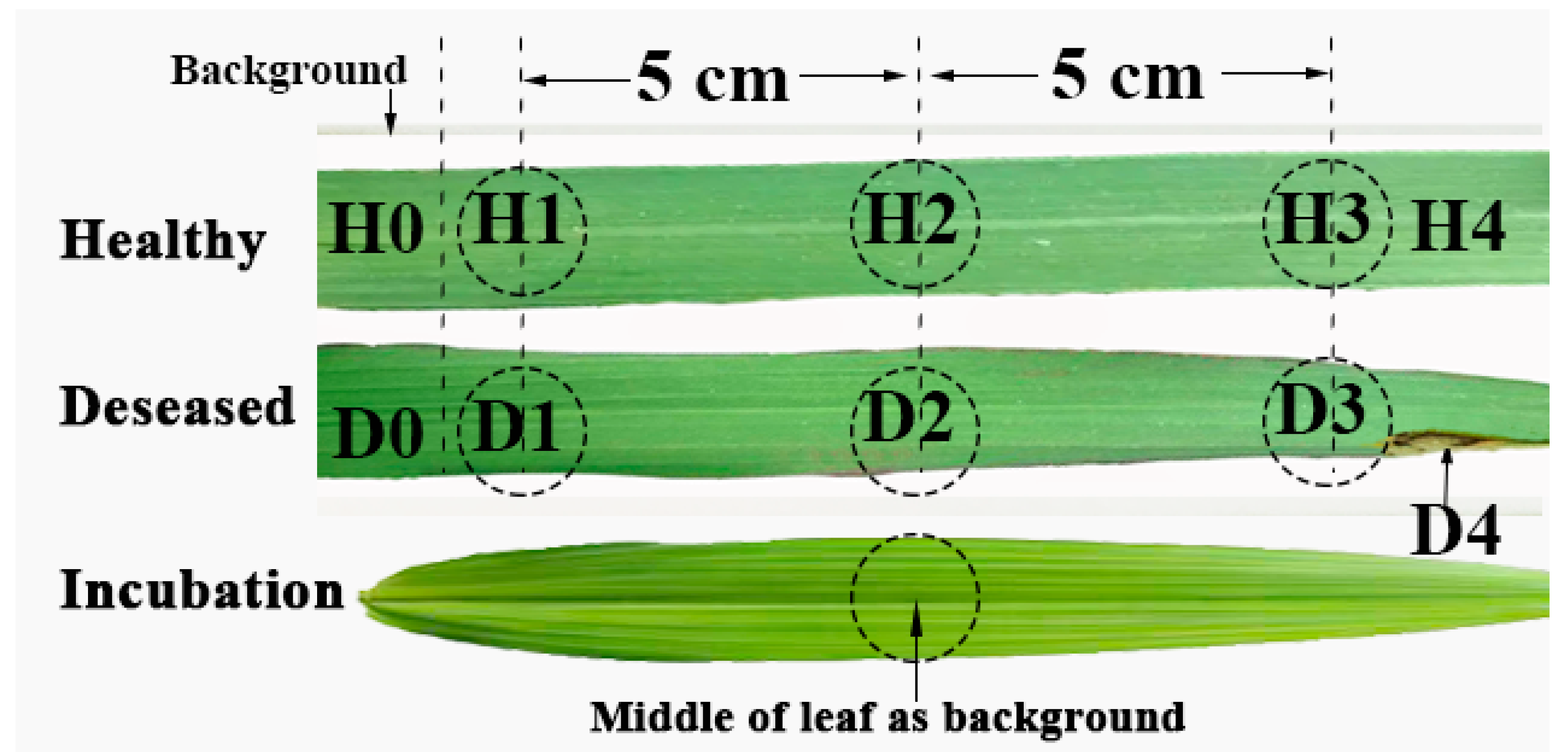
Figure 1. Schematic diagram of spectral collection sites for various leaf samples.
Agronomy 15 02100 g001
Figure 2. The spectra of healthy and diseased samples in 800~2000 cm−1 with 3 moving mirror velocities. (A), (B), and (C) as H1, H2, and H3; and (D), (E), and (F) as D1, D2, and D3, respectively.
Agronomy 15 02100 g002
Figure 3. Schematic diagram of scanning layers in healthy and diseased samples by moving mirror velocities. The red, green and blue colors represent the scanning layers with the moving mirror velocities of 0.6, 0.4, and 0.3 cm/s, respectively.
Agronomy 15 02100 g003
Figure 4. Comparison of FHB spectrum with background removal spectra of diseased (A) and healthy (B) samples with D1 and H1 as background, respectively. Principal components distribution of background removal (C, D0 as background) and original spectra (D) of diseased samples.
Agronomy 15 02100 g004
Figure 5. Original absorbance values (A) and ratio (B) distribution at 1650 and 1050 cm−1 of healthy (H0 background-removed) and diseased (D0 background-removed) samples.
Agronomy 15 02100 g005
Figure 6. Original absorbance values (A) and ratio (B) distribution at 1650 and 1050 cm−1 of background-removed spectra from healthy and FHB asymptomatic samples.
Agronomy 15 02100 g006
Table 1. Theoretical scanning depth (µm) of FTIR-PAS at different wavenumbers under three different moving mirror velocities.
Wavenumber (cm−1)Diffusion Length (µm) with Moving Mirror Velocities (cm·s−1)
0.30.40.6
20007.36.35.2
16508.06.95.7
105010.08.77.1
80010.99.57.8
Table 2. Functional group sources and substance assignments of spectral absorption peaks at different scanning levels.
Wavenumber
(cm−1)
Cellular LayerCuticle Layer
VibrationAttributionVibrationAttribution
1650Amide IProteins,
Nucleosides
1550Amide IIProteins,
Nucleosides
ν(C=C)Unsaturated aliphatics
1050ν(C-O)Saccharides, Aliphaticsν(C-O)Alcohol
δ(N-H)Proteins,
Nucleosides
Notes: ν denotes stretching vibration, and δ denotes bending vibration. The symbol ‘—’ means that the vibration cannot be detected commonly at that specific wavenumber range.
Table 3. Evaluation of diagnostic performance in the wheat FHB symptomatic period of R1650/1050 under different thresholds.
Evaluation IndicatorsSample SizeMisdiagnosis Details
0.40.50.6
Healthy set5016108
Diseased set8010823
Total amount130261830
Proportion0.200.140.24
F1-Score0.840.890.78
Note: 0.4, 0.5, and 0.6 are the absolute values of the R1650/1050 indicators.
Table 4. Detection stage and accuracy of different methods in wheat FHB perception.
MethodsInformation InterpretationOnset StageAccuracy
MIR spectroscopyLDASymptomatic 93%
Multi-spectralkNN, SVM, XGBoost Symptomatic 84.64–85.02%
RGB imageRGB-YOLOv4Symptomatic 93.69%
NIR spectroscopySVMAsymptomatic (organ)73.33%
Micro-vision and spectrumMultiple feature fusion and modelingAsymptomatic (organ)75.11–90.95%
FTIR-PAS
(proposed method)
Ratio of absorption intensity Symptomatic86%
Asymptomatic82%
Disclaimer/Publisher’s Note: The statements, opinions and data contained in all publications are solely those of the individual author(s) and contributor(s) and not of MDPI and/or the editor(s). MDPI and/or the editor(s) disclaim responsibility for any injury to people or property resulting from any ideas, methods, instructions or products referred to in the content.