Article

Metabolomic Profiling of Desiccation Response in Recalcitrant Quercus acutissima Seeds

1
College of Forestry and Grassland, Nanjing Forestry University, Nanjing 210037, China
2
Co-Innovation Center for Sustainable Forestry in Southern China, Southern Tree Inspection Center National Forestry Administration, 159 Longpan Road, Xuanwu District, Nanjing 210037, China
3
Shandong Provincial Center of Forest and Grass Germplasm Resources, No. 2011 Gangjiu Road, Licheng District, Jinan 250102, China
*
Authors to whom correspondence should be addressed.
These authors contributed equally to this work.
Agronomy 2025, 15(7), 1738; https://doi.org/10.3390/agronomy15071738
Submission received: 18 June 2025 / Revised: 13 July 2025 / Accepted: 17 July 2025 / Published: 18 July 2025
(This article belongs to the Section Plant-Crop Biology and Biochemistry)

Abstract

Quercus acutissima seeds exhibit high desiccation sensitivity, posing significant challenges for long-term preservation. This study investigates the physiological and metabolic responses of soluble osmoprotectants—particularly soluble proteins and proline—during the desiccation process. Seeds were sampled at three critical moisture content levels: 38.8%, 26.8%, and 14.8%, corresponding to approximately 99%, 52%, and 0% germination, respectively. We measured germination ability, soluble protein content, and proline accumulation, and we performed untargeted metabolomic profiling using LC-MS. Soluble protein levels increased early but declined later during desiccation, while proline levels continuously increased for sustained osmotic adjustment. Metabolomics analysis identified a total of 2802 metabolites, with phenylpropanoids and polyketides (31.12%) and lipids and lipid-like molecules (29.05%) being the most abundant. Kyoto Encyclopedia of Genes and Genomes pathway enrichment analysis showed that differentially expressed metabolites were mainly enriched in key pathways such as amino acid metabolism, energy metabolism, and nitrogen metabolism. Notably, most amino acids decreased in content, except for proline, which showed an increasing trend. Tricarboxylic acid cycle intermediates, especially citric acid and isocitric acid, showed significantly decreased levels, indicating energy metabolism imbalance due to uncoordinated consumption without effective replenishment. The reductions in key amino acids such as glutamic acid and aspartic acid further reflected metabolic network disruption. In summary, Q. acutissima seeds fail to establish an effective desiccation tolerance mechanism. The loss of soluble protein-based protection, limited capacity for proline-mediated osmotic regulation, and widespread metabolic disruption collectively lead to irreversible cellular damage. These findings highlight the inherent metabolic vulnerabilities of recalcitrant seeds and suggest potential preservation strategies, such as supplementing critical metabolites (e.g., TCA intermediates) during storage to delay metabolic collapse and mitigate desiccation-induced damage.

1. Introduction

Recalcitrant seeds are characterized by their lack of complete desiccation during maturation, high moisture content (MC) at harvest, and extreme sensitivity to desiccation and low temperatures [1,2]. Unlike orthodox seeds, recalcitrant seeds cannot tolerate significant moisture reduction, and even slight desiccation can cause fatal damage [1]. These seeds typically have a large size, thin seed coats, contain large amounts of free water, are metabolically active, do not enter dormancy after separation from the parent plant, and have a much shorter lifespan than orthodox seeds [3,4].
Due to their high moisture content, recalcitrant seeds can neither be dried nor frozen during storage, since ice crystals formed within cells cause irreversible damage, ultimately leading to the loss of viability [5]. Studies have shown that approximately 8% of angiosperm species produce desiccation-sensitive seeds, primarily distributed in tropical and temperate regions [6]. These biological characteristics pose severe challenges for long-term preservation: conventional seed bank conditions of low temperature and dryness are unsuitable, and once the MC falls below a critical threshold (typically around 25–35%), seed viability rapidly deteriorates [7]. Additionally, these seeds generally lack post-maturation dormancy mechanisms, often germinating immediately after harvest, while parent trees exhibit long fruiting cycles and low fruiting rates, further limiting large-scale reproduction and long-term preservation of species [8]. Therefore, the high MC and desiccation sensitivity of recalcitrant seeds challenge traditional germplasm preservation, prompting the scientific community to conduct in-depth research on their desiccation tolerance mechanisms [5,9].
From a physiological and biochemical perspective, recalcitrant seeds lack effective protective mechanisms against desiccation. Under dehydration stress, orthodox seeds typically accumulate osmoprotectants such as proline and sugars, as well as stress-responsive proteins like LEA proteins, to stabilize cellular structures and resist environmental stress [1,3]. For example, proline is a well-known osmoprotective amino acid that alleviates water stress by regulating osmotic potential, scavenging reactive oxygen species, and stabilizing proteins and membranes [10]. Similarly, soluble proteins (e.g., LEA proteins) can replace water and maintain cellular integrity during dehydration, serving as key factors in desiccation tolerance of orthodox seeds [11]. Previous studies have shown that recalcitrant seeds do not significantly accumulate these osmoprotectants and antioxidants during desiccation, which may be a major reason for their sensitivity to desiccation [11,12,13,14].
In recent years, with the advancement of high-throughput omics technologies, metabolomics has been widely applied in studies of plant responses to environmental stress, particularly in uncovering the metabolic regulatory mechanisms under drought or desiccation conditions [15]. By comprehensively profiling small-molecule changes during seed drying, metabolomics can reveal critical processes such as disruption of metabolic homeostasis, energy imbalance, and deficiencies in osmotic adjustment [16]. Several studies have analyzed metabolic changes in recalcitrant seeds. A metabolomic study of Garcinia mangostana showed significant changes in sugars, organic acids, amino acids, and phenylpropanoids during seed development. Fluctuations in TCA-related and phenylpropanoid metabolites reflected shifts in metabolic regulation and stress tolerance across maturation stages [17]. Szuba et al. [18] found that during sub-zero storage of Quercus robur seeds, sugars, organic acids, and amino acids accumulated significantly, but these changes were insufficient to prevent cellular damage, highlighting a limited desiccation survival mechanism. Moreover, recalcitrant seeds with different levels of desiccation sensitivity exhibit distinct metabolic response strategies, suggesting that osmotic regulation and metabolic stability are key determinants of desiccation tolerance [19].
Quercus acutissima Carruth is a temperate deciduous broadleaf tree native to East Asia, widely distributed in southeastern China, with significant ecological and economic value [20]. However, as typical recalcitrant seeds, Q. acutissima seeds are extremely sensitive to desiccation, posing major challenges for species preservation. In forestry practice, oak recalcitrant seeds can usually only be sown immediately after collection, limiting the time window for seedling cultivation [21]; for germplasm resource banks, ultra-dry and low-temperature frozen storage of Q. acutissima seeds is virtually impossible [22]. Additionally, Q. acutissima is widely used in ecological restoration, with both natural regeneration and artificial facilitation depending on seed viability. As climate change leads to increased extreme drought events, the risk of desiccation stress for Q. acutissima seeds in the wild is also increasing [23]. Meanwhile, as a typical desiccation-sensitive model among temperate deciduous tree species, research on Q. acutissima seeds will expand our understanding of the biological characteristics of recalcitrant seeds from different climate zones.
Based on the above background, this study focuses on the osmotic regulation and metabolic responses of Q. acutissima seeds during desiccation. By integrating physiological measurements with untargeted LC-MS metabolomics, we systematically investigated changes in germination capacity, soluble protein content, and proline levels across a gradient of moisture contents. Key metabolic pathways and differential metabolites were identified to uncover the underlying metabolic basis of their desiccation sensitivity. This study provides new insights into the metabolic vulnerability of recalcitrant seeds and offers a theoretical foundation for future germplasm conservation strategies.

2. Materials and Methods

2.1. Experimental Materials

Q. acutissima seeds used in this study were from the same batch as those used in our previous nuclear magnetic resonance study [24]. The Q. acutissima seeds were collected on 16 November 2022, from the Nanjing Forestry University Experimental Forest (119°12′58″ E, 32°7′26″ N). After collection, the cupules were removed, and the seeds were subjected to water selection to remove floating seeds. Seeds that sank to the bottom underwent visual inspection to discard those with insect damage, discoloration or rot, morphological abnormalities, or incomplete development. Seeds with intact appearance, consistent maturity, uniform brown color, and normal size were selected as experimental materials. After thorough washing with distilled water, the seeds were air-dried at room temperature (20 ± 2 °C) for approximately 0.5–1 h to remove surface moisture and were immediately used for the experiments. The initial moisture content was then determined as detailed in Section 2.3.

2.2. Desiccation Design

Q. acutissima seed desiccation was conducted using the silica gel drying method [25]. The experiment was performed at room temperature (20 °C) with a relative humidity of 50% to 60%. Using the initial MC of 38.8% as the control, nine MC gradients were established. Based on preliminary germination tests, critical viability transitions in Q. acutissima seeds occurred within narrow moisture ranges; thus, 3% intervals were adopted to accurately identify these transition points. The nine MC gradients were (38.8%, 35.8%, 32.8%, 29.8%, 26.8%, 23.8%, 20.8%, 17.8%, and 14.8%). Each MC level included three biological replicates, with each replicate containing 300 seeds placed in large self-sealing bags containing silica gel (seed to silica gel mass ratio approximately 1:3), ensuring complete seed immersion in silica gel. During desiccation, the silica gel was promptly replaced when its color changed from deep blue to pink.
To monitor changes in seed MC, 30 seeds were randomly selected from each self-sealing bag as monitoring samples, placed separately in marked small mesh bags, and weighed to determine the initial fresh weight (W1) prior to drying. These seeds were dried together with the other seeds, and their mass (W2) was periodically monitored. The relative moisture content (MC) was calculated using the following formula: RMC (%) = (W2 − W1 × (1 − G/100))/W2 × 100%, where G represents the initial MC (38.8%). To verify the accuracy of the weighing method, random samples were taken from non-monitored seeds at each sampling time point for direct MC measurement using the oven-drying method. The results indicated that the deviation between the two methods was less than ±1%, confirming the reliability of the weighing method. Therefore, all moisture content values in this study are expressed as MC. When the target MC was reached, random samples were taken from each treatment for subsequent experimental measurements.

2.3. Determination of Seed MC

The initial seed MC was determined using constant low-temperature oven drying method following the International Seed Testing Association [26] procedures. Thirty intact seeds were randomly selected, cut into 3–5 mm thin slices, and 5–8 g of this material was evenly placed in pre-dried, pre-weighed covered aluminum boxes. After recording the total weight of the samples and aluminum boxes (M2, representing the weight of the sample box, lid, and seed sample before drying in grams), they were placed in a constant temperature oven at 103 °C for 17 h (starting when the oven reached the target temperature). After drying, the samples were cooled in a desiccator for 30 min, the aluminum box lids were replaced, and the samples were weighed (M3, representing the weight of the sample box, lid, and seed sample after drying in grams). The dry weight of the sample box and lid (M1, in grams) was also recorded. Seed MC was calculated according to the following formula: Seed MC (%) = [(M2 − M3)/(M2 − M1)] × 100%.

2.4. Determination of Seed Germination Percentage, Proline Content, and Soluble Protein Content

2.4.1. Seed Germination Percentage (GP)

Three sets of 50 seeds each were selected. Seed coats were removed, and 1/2 of the cotyledons were cut off. Germination boxes were disinfected with 75% alcohol and filled with a sand bed. The seeds were then incubated at 25 °C under constant temperature and light conditions. Germination was defined as radicle protrusion of at least 2 mm [27], and the germination period was 28 days. Non-germinating seeds were examined for morphological changes to distinguish between seed death and dormancy. Seed GP (%) = (∑Gt)/N × 100%, where Gt is the number of seeds germinating daily during the germination test period, and N is the total number of seeds.

2.4.2. Soluble Protein Content

At each MC level, 30 seeds were randomly selected (3 replicates), immediately pulverized in a pre-cooled liquid nitrogen grinder, and homogenized. The samples were placed in frozen centrifuge tubes for determination of proline content and soluble protein content.
Soluble protein (SP) content was determined using the Coomassie Brilliant Blue method [28]. A 0.1 g seed powder sample (converted to dry weight m according to the seed MC at each stage) was homogenized with 5 mL distilled water (Vt), and the supernatant was collected after centrifugation. A total of 1 mL of supernatant (Vs) was mixed with 5 mL Coomassie Brilliant Blue G-250 reagent, and after complete reaction, the absorbance was measured at a wavelength of 595 nm. A standard curve was constructed using bovine serum albumin (BSA) as the standard, and the protein concentration (C) of the samples was calculated according to the standard curve. Soluble protein content was calculated using the following formula: SP content (mg·g−1 DW) = C × Vt/(m × Vs).

2.4.3. Proline Content

Proline (Pro) content was determined according to the method of Kaur et al. [29]. A series of standard Pro solutions with concentrations of 0–10 μg·mL−1 were prepared to construct a standard curve. A 0.1 g seed powder sample (converted to dry weight m according to the seed MC at each stage) was extracted with 6 mL extraction solution (V). Two mL of standard solution or sample extract (V1) was placed in a 20 mL centrifuge tube, and the following were added sequentially: 2 mL ice acetic acid, 4 mL 2.5% acidic ninhydrin reagent, and 2 mL 3% salicylaldehyde solution. The tubes were placed in a boiling water bath for color development, naturally cooled to room temperature, and then 4 mL toluene was added. After thorough shaking and extraction, the upper colored solution was taken for absorbance measurement at 520 nm. Pro content (C) was determined according to the standard curve, and the sample Pro content was calculated using the following formula: Pro content (% DW) = C × V/(V1 × m) × 100%.

2.5. Metabolome Sample Preparation

Seeds at three critical desiccation sensitivity points were selected based on germination data: (1) initial moisture content (38.8%, 99% GP, IM) representing maximum viability, (2) semi-lethal moisture content (26.8%, 52% GP, SLM) representing the critical transition point, and (3) lethal moisture content (14.8%, 0% GP, LM) representing complete viability loss. Thirty Q. acutissima seeds were collected at each critical moisture content level (with 6 biological replicates per treatment), immediately flash-frozen in liquid nitrogen, and ground.
Exactly 80 ± 0.05 mg of each sample was weighed, and 20 μL of internal standard (L-2-chlorophenylalanine, 0.06 mg/mL methanol solution) and 1 mL methanol–water mixture (V/V = 7:3) were added [30]. After pre-cooling at −20 °C for 2 min, samples were ground using a high-speed automatic sample grinder (Wonbio-E) with two small steel beads (60 Hz, 2 min), followed by ultrasonic extraction in an ice-water bath for 30 min, and then left to stand overnight at −20 °C. After centrifugation (13,000 rpm, 4 °C, 10 min), 150 μL of supernatant was filtered through a 0.22 μm organic phase filter membrane, and the filtrate was transferred to a chromatographic sample vial and stored at −80 °C for analysis. Quality Control (QC) samples were prepared by pooling equal volumes of all sample extracts. All extraction reagents used in the experiment were pre-cooled at −20 °C.

2.6. Liquid Chromatography and Mass Spectrometry Analysis Conditions

Analysis was performed using an ACQUITY UPLC I-Class plus ultra-high performance liquid chromatography system (Waters Corporation, Milford, CT, USA) coupled with a QE plus high-resolution mass spectrometer (Waters Corporation, Milford, CT, USA) [31]. Sample separation was achieved using an ACQUITY UPLC HSS T3 chromatography column (Thermo Fisher Scientific, Waltham, MA, USA) (100 mm × 2.1 mm, 1.8 μm) with a column temperature of 45 °C. Mobile phase A was 0.1% formic acid aqueous solution, and mobile phase B was 0.1% formic acid acetonitrile solution, with a flow rate maintained at 0.35 mL/min and a sample injection volume of 2 μL. Gradient elution conditions are shown in Table 1.
Mass spectrometry analysis was performed using an ESI ion source, with sample signal acquisition in both positive and negative ion modes. The operational parameters of the mass spectrometer are shown in Supplementary Table S1.

2.7. Data Preprocessing and Metabolite Identification

The raw data were processed using Progenesis QI v2.3 software (Nonlinear Dynamics, Newcastle, UK), including baseline filtering, peak detection, integration, retention time correction, peak alignment, and normalization [32]. Parameters were set as follows: precursor tolerance 5 ppm, product tolerance 10 ppm, and product ion threshold 5%. Compound identification was based on exact mass, secondary fragments, and isotope distribution, combined with the PMDB database. During data screening, ion peaks with missing values exceeding 50% within groups were eliminated and remaining zero values were imputed as half of the minimum detected value. Metabolite identification employed a 60-point scoring system based on accurate mass matching (20 points), MS/MS fragmentation (20 points), and isotope distribution (20 points). Finally, data from positive and negative ionization modes were combined into a single matrix for subsequent analysis.
In cases where multiple metabolites exhibited similar or overlapping retention times, annotation was determined based on a combination of orthogonal information. The use of a high-resolution mass spectrometer (QE Plus) coupled with data-dependent acquisition (DDA) mode enabled the sequential isolation and fragmentation of multiple co-eluting precursor ions within the same chromatographic window, generating distinct MS/MS spectra for structural discrimination. The MS/MS fragmentation spectra of several representative metabolites, including L-asparagine, L-aspartic acid, L-glutamic acid, and L-proline, were validated by comparison with those of authentic standards, with the corresponding spectra shown in Supplementary Figure S1 and the major fragment ions listed in Supplementary Table S2 to further support the reliability of their structural annotation.

2.8. Statistical Analysis, Differential Metabolite Screening and Metabolic Pathway Enrichment Analysis

Multivariate statistical analyses were conducted through Principal Component Analysis (PCA), Partial Least Squares Discriminant Analysis (PLS-DA), and Orthogonal Partial Least Squares Discriminant Analysis (OPLS-DA). PCA was used to observe sample distribution and evaluate analytical stability, while PLS-DA and OPLS-DA were used to identify differences between seed samples with different MC to screen for Differentially Expressed Metabolites (DEMs). To verify model reliability, seven-fold cross-validation and 200 response permutation tests were implemented. Univariate T-tests and fold change analyses were performed simultaneously, and volcano plots were used to visualize p-values and fold changes. DEMs were selected based on variable importance in projection (VIP) scores > 1 from the first principal component of the OPLS-DA model, and t-test p-values < 0.05.
Metabolic pathway enrichment analysis of DEMs was performed based on the KEGG database. Hypergeometric tests were used to assess the significance of metabolic pathway enrichment (p-value ≤ 0.05), with the enrichment factor (number of significantly different metabolites/total number of metabolites in the pathway) reflecting the degree of enrichment. The KEGG pathway mapper function was utilized to display differential metabolic pathways.
The Shapiro–Wilk test was used to assess the normality of physiological indicator data distribution. Data analysis was performed using SPSS 26.0 statistical software (IBM, Armonk, NY, USA), primarily including one-way analysis of variance and Duncan’s multiple comparison test. Experimental results were visualized through graphs created with Origin 2023 (OriginLab, Northampton, MA, USA). Data are presented as mean ± standard deviation (Mean ± SD) with three biological replicates per group, and statistical significance was set at p < 0.05.

3. Results

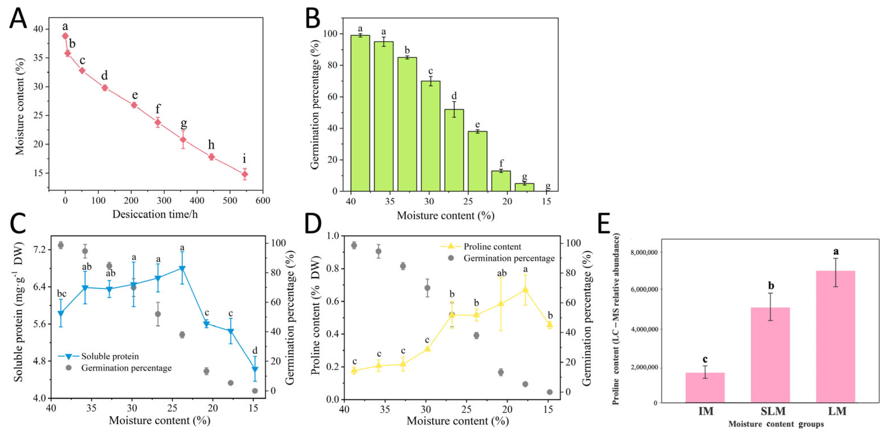
3.1. Effects of Desiccation on Seed MC and GP

Q. acutissima seeds initially had a moisture content (MC) of 38.8% and a GP of 99%. All subsequent moisture content values during desiccation are expressed as MC. During desiccation, seed MC exhibited a decreasing trend that was initially rapid and then slowed (Figure 1A). In the first 51 h of drying, MC decreased rapidly, after which the rate of reduction became more gradual. After 545 h of desiccation, the MC decreased to 14.8%. During desiccation, seed GP declined significantly with decreasing MC (Figure 1B). When MC decreased to 35.8%, the GP was 95%, which was not significantly different from the GP at the initial MC of 38.8% (99%); when MC further decreased to 32.8%, GP significantly decreased to 85%; when MC decreased to 26.8%, GP further significantly decreased to 52%. At this point, nearly 50% of the seeds had lost viability, which can be considered the semi-lethal moisture content (SLM). When MC decreased to 14.8%, the GP was 0%, indicating that this MC represented the lethal moisture content (LM) for the seeds. Morphological examination confirmed that non-germinating seeds exhibited an obvious deterioration signs. The embryonic axes and cotyledons turned dark brown or even black, with tissues becoming shriveled and hardened. Lower moisture contents resulted in more severe deterioration. These morphological changes, combined with germination failure, confirmed seed death rather than dormancy (detailed morphological observations and images shown in Chen and Shen [33]).

3.2. Effects of Desiccation on Seed Osmotic Regulation

SP content exhibited a trend of initial increase followed by decrease during desiccation (Figure 1C). During the early stages of desiccation (MC 38.8% to 29.8%), SP content significantly increased from an initial value of 5.84 mg·g−1 to 6.45 mg·g−1, subsequently reaching its highest value of 6.59 mg·g−1 at the SLM, representing a 12.8% increase over the initial value. This upward trend contrasted with the significant decrease in seed GP. However, when MC further decreased, SP content began to show a declining trend. During the deep desiccation stage (MC below 20.8%), SP content decreased sharply, ultimately declining to 5.90 mg·g−1 at the LM.
Proline, an important osmotic regulator, was present at relatively low levels in fresh seeds (0.17%) but showed significant changes under desiccation stress (Figure 1D). During the initial stages of desiccation (MC 38.8% to 29.8%), Pro content increased gradually, and at the SLM, it significantly increased to 0.52%. When MC decreased to 17.8%, Pro reached its highest value (0.67%), at which point the GP had dropped to 5%. At the LM, Pro content decreased to 0.46%. LC-MS metabolomics analysis further confirmed the trend of proline accumulation during seed desiccation (Figure 1E). Consistent with spectrophotometric results, the relative abundance of proline showed a significant increase from IM to LM groups, validating the consistency across different analytical methods.

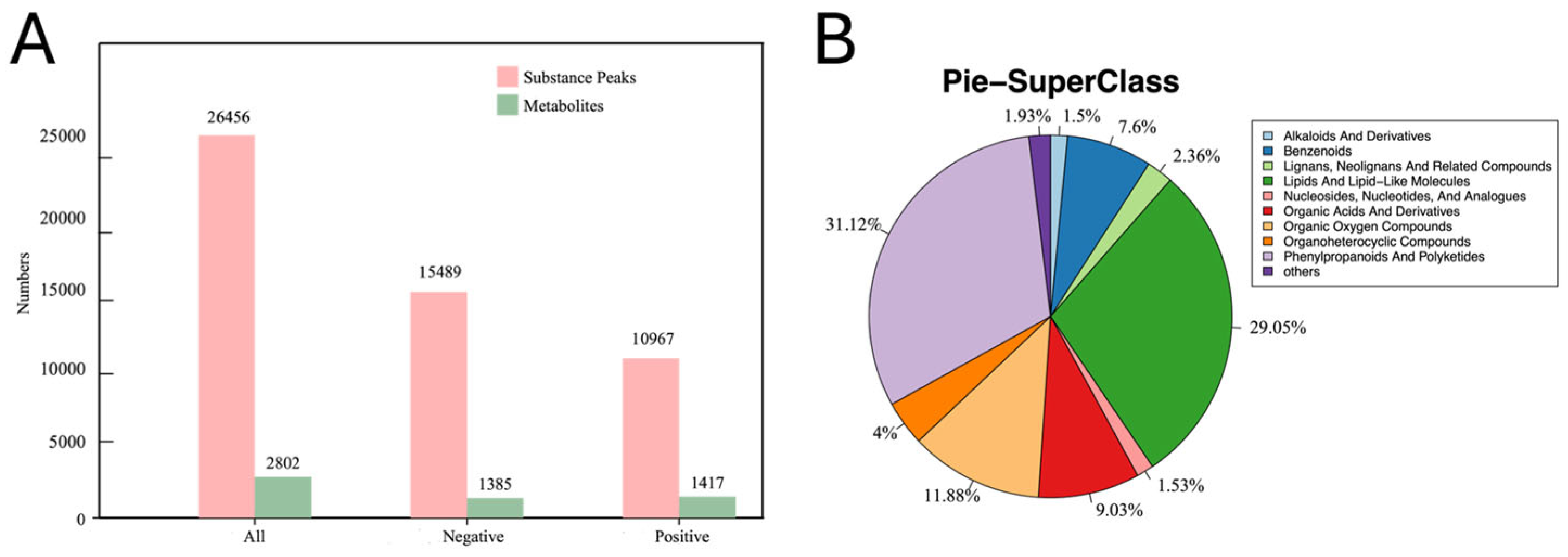
3.3. Metabolite Analysis of Q. acutissima Seeds During Desiccation

Based on GP data, Q. acutissima seeds were selected at three critical desiccation sensitivity points: initial moisture content (IM), semi-lethal moisture content (SLM), and lethal moisture content (LM) for metabolomic analysis (6 biological replicates, 18 samples total). A total of 26,456 metabolite peaks were detected through qualitative and quantitative analysis, including 15,489 negative ions and 10,967 positive ions. After quality control filtering, 2802 metabolites were obtained, including 1385 negative ions and 1417 positive ions (Figure 2A). Representative base peak chromatograms for positive and negative ionization modes are shown in Figure S2 and Figure S3, respectively, and the complete metabolite data matrix is provided in Table S3. As shown in Figure 2B, these metabolites can be classified into 10 major categories. Phenylpropanoids and polyketides were the most abundant, with 872 types accounting for 31.12% of the total. This was followed by lipids and lipid-like molecules (814 types, 29.05%), organic oxygen compounds (333 types, 11.88%), organic acids and derivatives (253 types, 9.03%), and benzenoids (213 types, 7.60%). The remaining categories included lignans and neolignans, 66 types (2.36%); alkaloids and derivatives, 42 types (1.50%); nucleosides and nucleotides, 43 types (1.53%); heterocyclic compounds, 112 types (4.00%); and other compounds, 54 types (1.93%).

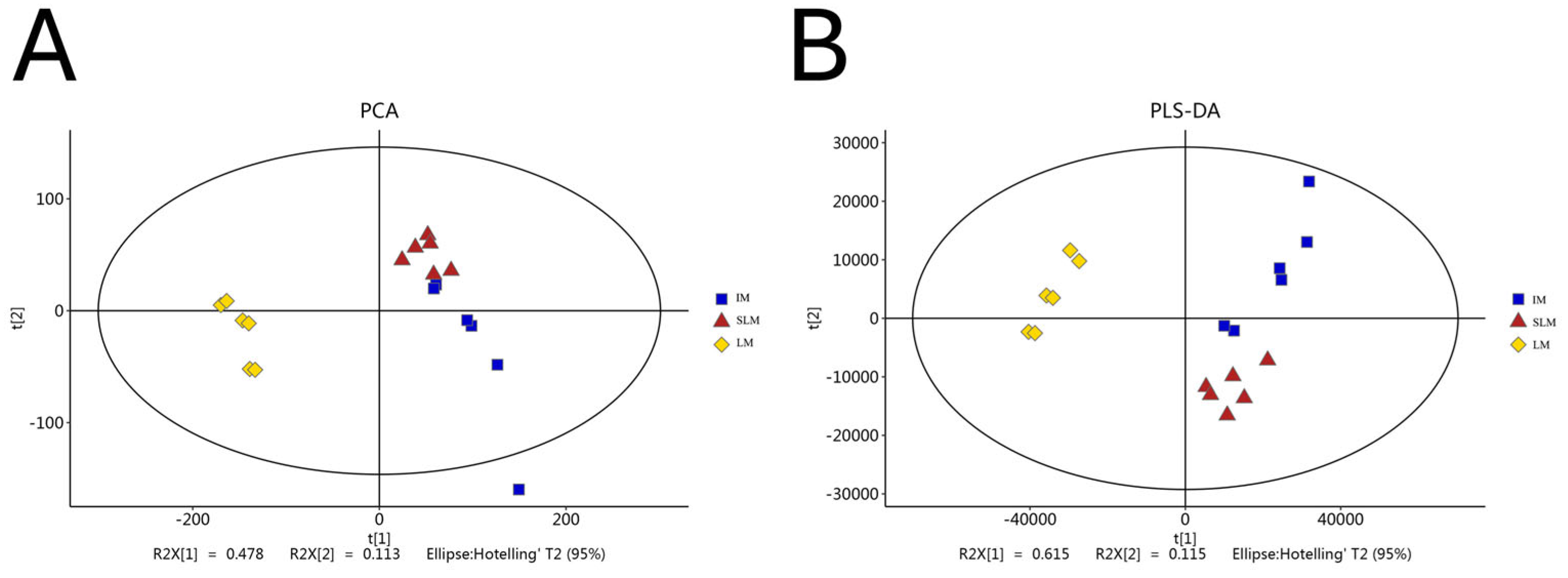
3.4. Multivariate Statistical Analysis of Metabolic Profiles During Desiccation in Q. acutissima Seeds

Multivariate statistical analysis revealed that the PCA score plot showed distinct metabolic profiles across the three moisture content groups, with PC1 and PC2 explaining 47.8% and 11.3% of the total variance, respectively (Figure 3A). Compared to PCA, PLS-DA analysis demonstrated enhanced separation between groups, with PC1 and PC2 explaining 61.5% and 11.5% of the variance, respectively (Figure 3B). To further validate group differences, pairwise OPLS-DA analyses were performed for all comparisons (Figure S4). The OPLS-DA score plots showed clear separation between SLM vs. IM (R2X = 0.582, R2Y = 0.916), LM vs. IM (R2X = 0.581, R2Y = 0.912), and LM vs. SLM (R2X = 0.617, R2Y = 0.716). All multivariate statistical analyses demonstrated tight clustering of biological replicates within each group, confirming significant metabolic differences between different moisture content levels with good experimental reproducibility.

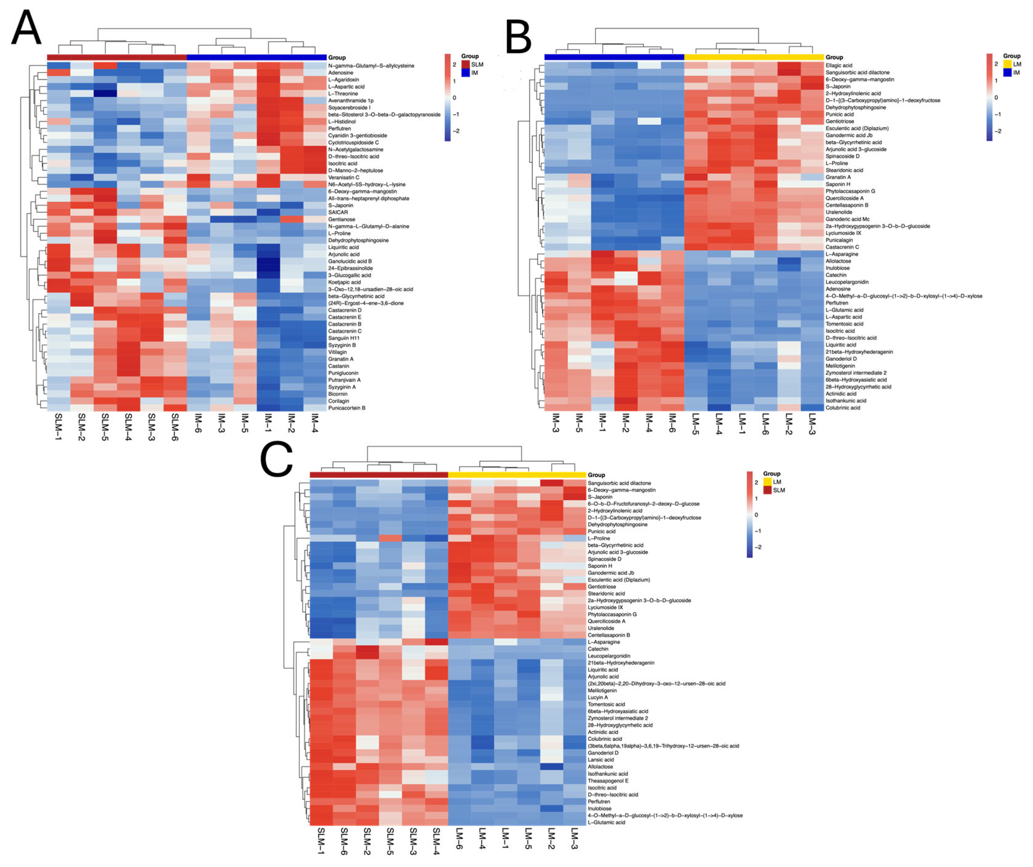
3.5. Differentially Expressed Metabolites (DEMs) Screening and Analysis

Utilizing VIP > 1 and t-test p < 0.05 as screening criteria, three comparison groups (SLM vs. IM, LM vs. IM, and LM vs. SLM) were established for DEMs analysis. The results revealed 103 DEMs (37 with decreased levels and 66 with increased levels) in the SLM vs. IM group, 268 DEMs (124 with decreased levels and 144 with increased levels) in the LM vs. IM group, and 248 DEMs (123 with decreased levels and 125 with increased levels) in the LM vs. SLM group. Among these, the LM vs. SLM, SLM vs. IM, and LM vs. IM groups specifically showed increased levels in 15, 37, and 20 DEMs, and specifically showed decreased levels in 26, 15, and 19 DEMs, respectively. Detailed information on all differentially expressed metabolites is provided in Table S4. Across the three groups, 15 commonly increased DEMs and 14 commonly decreased DEMs were identified (Figure S5). The commonly increased DEMs across the three comparisons primarily comprised various terpenoid compounds of the Castacrenin series, beta-Glycyrrhetinic acid, glycosides such as Corchoroside B and S-Japonin, the osmotic regulating substance Pro, and membrane stability-related compounds such as Dehydrophytosphingosine (Figure 4). These metabolites with increased levels represented potential protective responses during desiccation stress. The commonly decreased DEMs primarily comprised energy metabolism-related compounds such as Isocitric acid and L-Threonine, amino acids such as L-Aspartic acid and L-Histidinol, nucleosides such as Adenosine, flavonoid compounds such as Cyanidin 3-gentioboside, sterol compounds such as beta-Sitosterol 3-O-beta-D-galactopyranoside, and ceramide compounds such as Soyacerebroside I.

3.6. DEMs Clustering Analysis

To visualize the relationships among samples and differentially expressed metabolites, hierarchical clustering analysis was performed on the top 50 significant DEMs in each comparison group (Figure 5). The results demonstrated that Q. acutissima seeds exhibited significantly distinct metabolic characteristics at different desiccation stages (IM, SLM, and LM), with clear clustering of samples within each treatment group, indicating good experimental reproducibility.
In the SLM vs. IM comparison, the principal DEMs comprised amino acid compounds such as aspartic acid, threonine, and Pro; tannin compounds such as Castanin and Punicacortein B; steroid compounds such as 24-Epibrassinolide; and organic acid compounds (isocitric acid). In the LM vs. IM comparison, the DEMs primarily comprised phenolic acid compounds such as Ellagic acid; amino acid compounds such as asparagine, glutamic acid, and aspartic acid; flavonoid compounds such as Catechin; and organic acid compounds such as isocitric acid. In the LM vs. SLM comparison, the DEMs primarily involved fatty acid compounds such as 2-Hydroxylinolenic acid; amino acid compounds such as Pro, asparagine, and glutamic acid; and sugar compounds such as 6-O-b-D-Fructofuranosyl-2-deoxy-D-glucose.

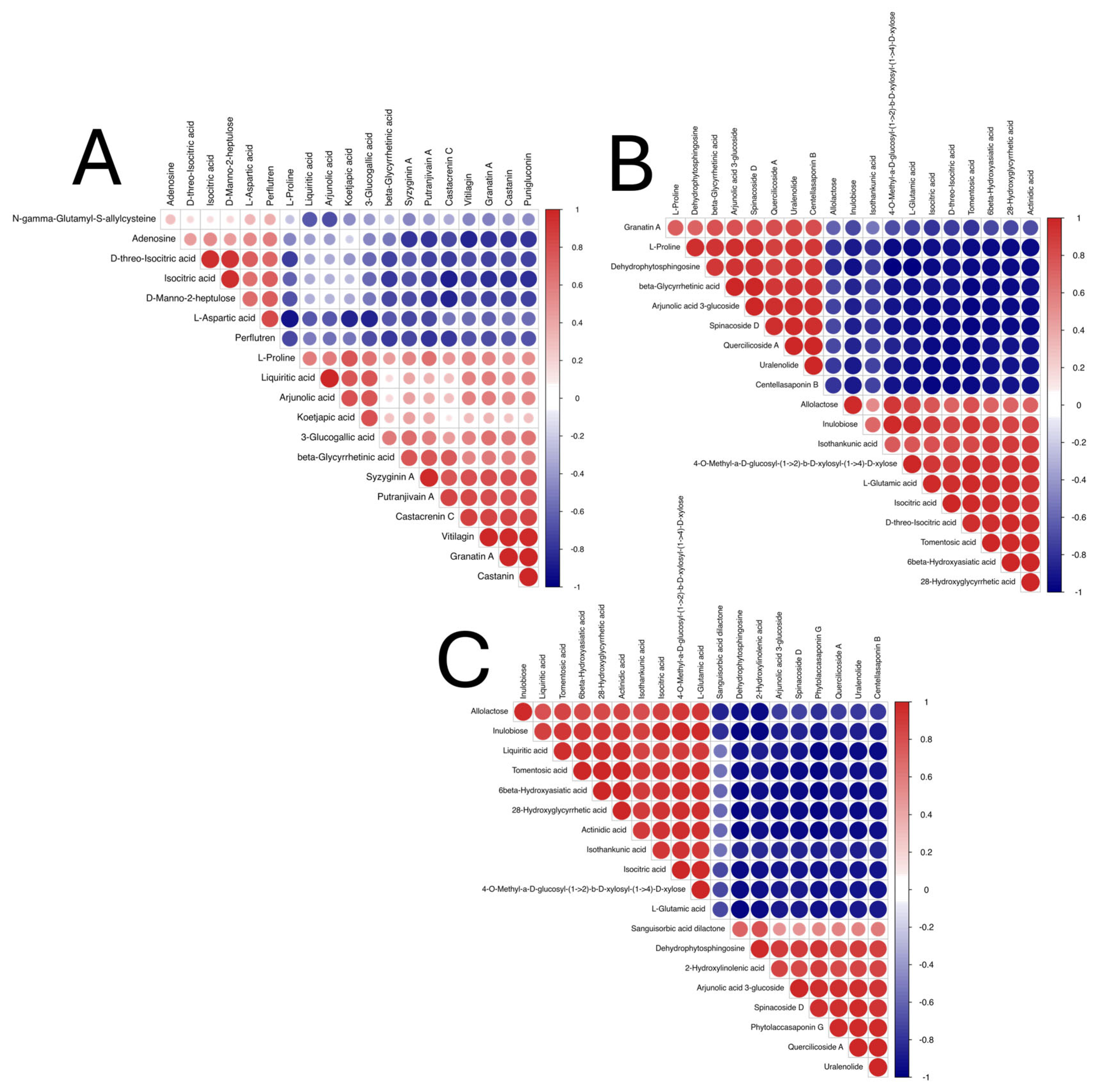
3.7. DEMs Correlation Analysis

Correlation analysis revealed complex interaction relationships among DEMs during desiccation of Q. acutissima seeds (Figure 6). In the SLM vs. IM comparison group, energy metabolism-related metabolites such as adenosine and D-threo-Isocitric acid exhibited significant negative correlations with defense-related metabolites such as Puniglucoin and Castanin. In the LM vs. IM comparison group, osmotic regulation-related metabolites such as Pro and membrane stability-related metabolites such as dehydrophytosphingosine formed a significant positive correlation cluster but showed significant negative correlations with TCA cycle-related substances such as isocitric acid and D-threo-Isocitric acid. In the LM vs. SLM comparison group, sugar metabolites (inulin biose) exhibited negative correlations with membrane lipid metabolites (dehydrophytosphingosine).

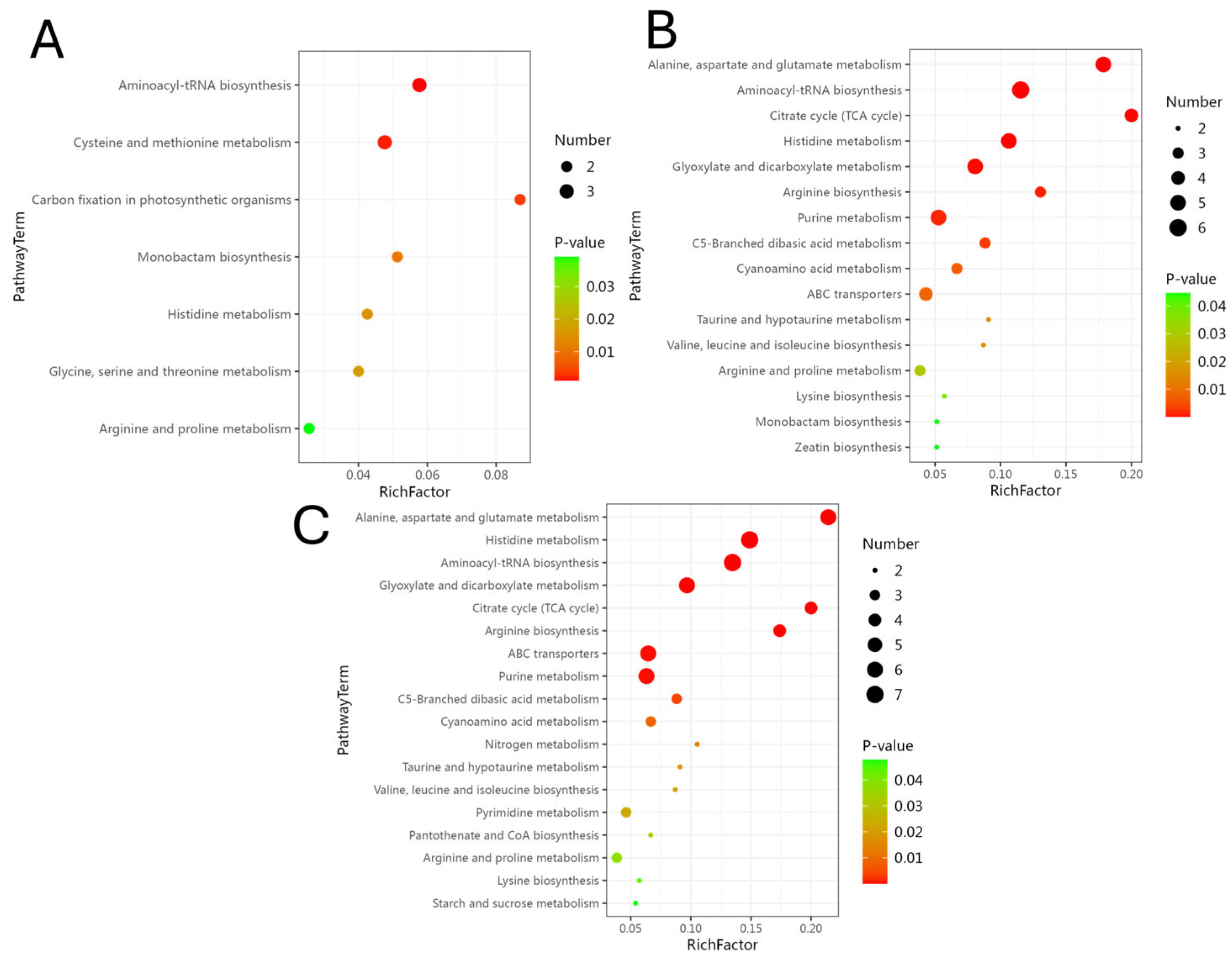
3.8. KEGG Pathway Enrichment Analysis of DEMs

KEGG pathway enrichment analysis (p < 0.05) demonstrated that DEMs were primarily associated with pathways related to amino acid metabolism and biosynthesis, energy metabolism, and nitrogen metabolism (Figure 7). In the SLM vs. IM comparison, significantly enriched pathways included Aminoacyl-tRNA biosynthesis, Cysteine and methionine metabolism, Carbon fixation in photosynthetic organisms, Histidine metabolism, Glycine, serine and threonine metabolism, and Arginine and Pro metabolism. In the LM vs. IM comparison, significantly enriched pathways comprised Alanine, aspartate and glutamate metabolism, Aminoacyl-tRNA biosynthesis, Citrate cycle, Histidine metabolism, ABC transporters, and Arginine and Pro metabolism. In the LM vs. SLM comparison, enriched metabolic pathways included Alanine, aspartate and glutamate metabolism, Histidine metabolism, Aminoacyl-tRNA biosynthesis, Citrate cycle, ABC transporters, Nitrogen metabolism, Arginine and Pro metabolism, and Starch and sucrose metabolism.

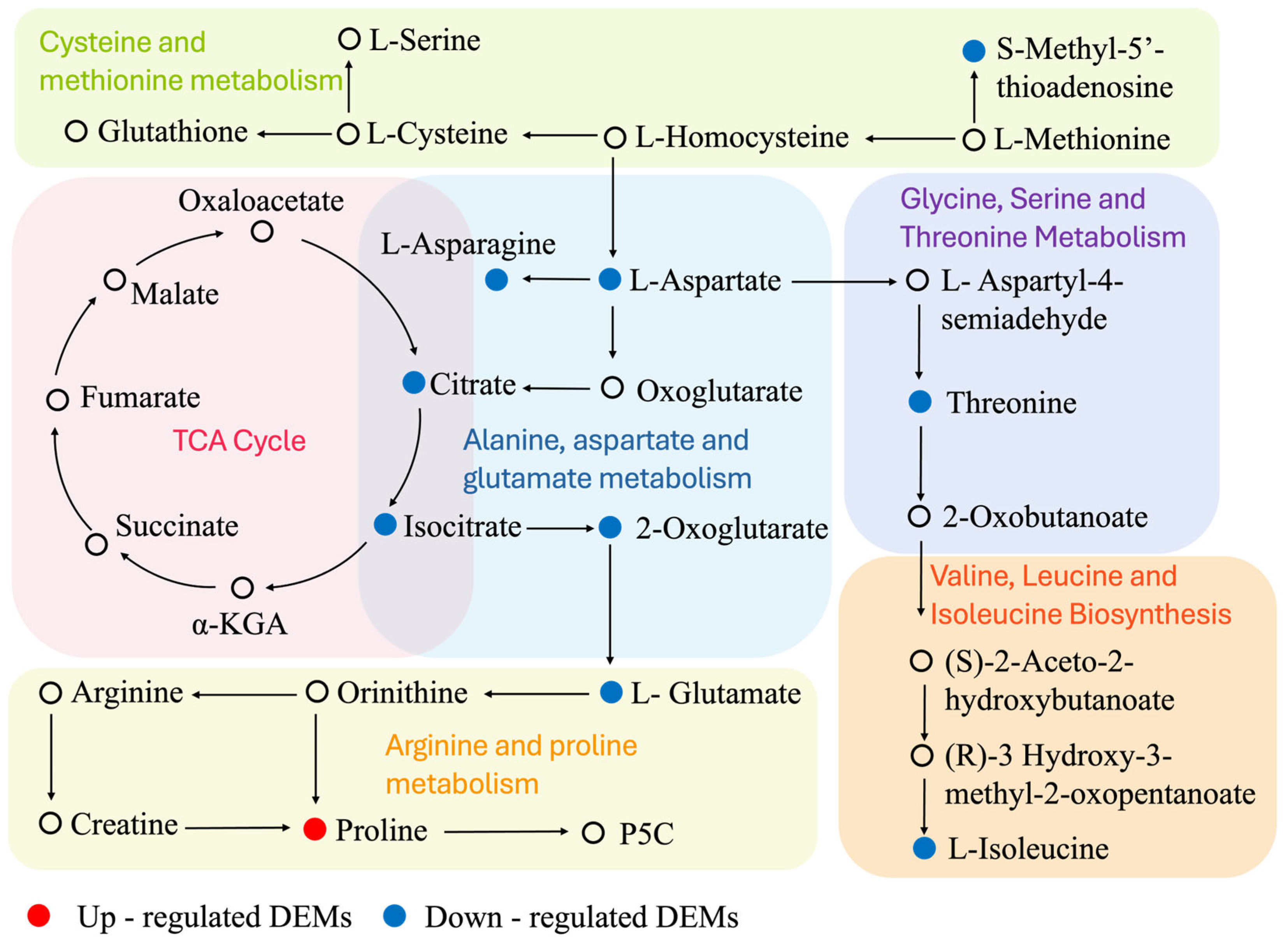
3.9. DEMs Associated with Amino Acid and Energy Metabolism Changes

Based on enrichment analysis, DEMs from key amino acid metabolism pathways including cysteine and methionine metabolism (map00270), alanine, aspartate and glutamate metabolism (map00250), glycine, serine and threonine metabolism (map00260), branched-chain amino acid biosynthesis (map00290), and arginine and proline metabolism (map00330) pathways, as well as the energy metabolism tricarboxylic acid cycle (map00020) pathway, were screened and analyzed. This study summarized a pathway interaction network related to amino acid metabolism and energy metabolism changes, identifying 10 key DEMs (Figure 8).
In the TCA cycle pathway, isocitric acid showed decreased levels in all three comparison groups, while citrate was significantly decreased in both LM vs. IM and LM vs. SLM comparisons. In the alanine, aspartate, and glutamate metabolism pathway, S-Methyl-5′-thioadenosine was significantly decreased in both LM vs. IM and SLM vs. IM comparisons. In the aspartate family metabolic pathway, L-aspartic acid showed decreased levels in all three comparison groups, while L-asparagine and 2-Oxoglutarate were significantly decreased in both LM vs. IM and LM vs. SLM comparisons. Related to this, threonine also exhibited significantly decreased levels in all three comparison groups. In the branched-chain amino acid biosynthesis pathway, L-isoleucine was significantly decreased in the LM vs. IM comparison. In the arginine and proline metabolism pathway, Pro showed increased levels in all three comparison groups, while glutamic acid levels were significantly reduced in both LM vs. IM and LM vs. SLM comparisons.

4. Discussion

4.1. Critical Moisture Thresholds and Osmotic Regulation Response

Multiple studies on Quercus seeds have demonstrated that their semi-lethal MC varies widely, ranging from 20% to 40%, with complete loss of viability typically occurring when MC falls below 10–20% [34]. Q. acutissima seeds exhibit a sharp loss of viability as their internal MC decreases. Fresh seeds possess high GP, but even mild drying leads to significant viability reduction. The semi-lethal MC of seeds is approximately 26.8% (at which GP is 52%), and no seeds survive when dehydrated to approximately 14.8% MC. This observation has practical significance: Q. acutissima seeds must be stored or processed at moisture levels close to their original state, as drying beyond critical thresholds results in rapid viability loss.
The accumulation of osmotic regulating substances plays a crucial role in maintaining cellular turgor during early stages of seed desiccation [35]. Various proteins and carbohydrates protect cell membranes against severe desiccation damage [36]. For instance, in the hypocotyl of Butia capitata, abundant embryonic storage materials, particularly proteins and starch, enable it to maintain viability even when water potential drops to -2 MPa [37]. Similarly, Q. acutissima seeds exhibited significant increases in soluble protein content during early desiccation, potentially reducing the risk of cellular damage [38]. This protective response aligns with observations in partially dehydrated Trichilia dregeana seeds, where upregulation of protein synthesis may contribute to physical and biochemical damage repair [39]. However, as desiccation intensified, this protective mechanism gradually failed, as evidenced by the decline in soluble protein content in Q. acutissima seeds. When MC fell below a critical threshold, protein denaturation or enhanced proteolytic enzyme activity may have caused protein content reduction, while desiccation-induced physiological metabolic inhibition further limited protein synthesis [39].
Proline, as an important osmotic protectant, accumulated continuously during Q. acutissima seed desiccation, suggesting its potential role in stress mitigation [40]. However, under severe desiccation conditions, this increase appeared insufficient to fully counteract the effects of desiccation. Research indicates that seeds with different sensitivities to desiccation exhibit variations in osmotic regulation capacity. Studies have shown that in orthodox Acer platanoide and recalcitrant A. pseudoplatanus seeds, Pro content in recalcitrant sycamore seeds shows significant positive correlation with environmental temperature, while orthodox seeds lack this correlation [10]. Similarly, studies on Madhuca latifolia and Camellia sinensis seeds found that P5CS activity or gene expression levels decreased correspondingly with declining seed MC [14,41]. These findings highlight the diverse adaptive strategies of recalcitrant seeds in response to water stress.

4.2. Metabolic Disturbances During Desiccation

Non-targeted metabolomic analysis provided a comprehensive view of biochemical changes associated with seed desiccation. A total of 2802 metabolites were identified, with phenylpropanoids and polyketides (31.12%) and lipids and lipid-like molecules (29.05%) as the main components. As Q. acutissima seed MC decreased, the number of DEMs gradually increased. Fifteen metabolites were significantly increased across all three comparison groups (primarily including terpenoids, glycosides, and osmotic regulators, potentially representing active defense mechanisms of seeds against desiccation stress), while 14 metabolites were significantly decreased (mainly involving energy metabolism and membrane lipid components, indicating progressive depletion of cellular energy reserves and compromised membrane integrity).
Q. acutissima seeds exhibited characteristics of cellular energy depletion and accumulation of membrane lipid peroxidation during desiccation, potentially forming a detrimental feedback loop that further exacerbated the loss of cellular viability. Declining energy metabolism and membrane system damage have also been observed in the aging process of Brassica pekinensis seeds [42], suggesting potential interactions between energy metabolism and membrane system stability, the dynamic regulation of which is crucial for seed viability. Further analysis revealed that the main DEMs at different desiccation stages included amino acids, tannins, steroids, and organic acids. Somi and Marques [43] found significant changes in amino acids, organic acids, sugars, and other compatible solutes in their study of Araucaria angustifolia seed embryos during desiccation/rehydration, indicating the critical roles of these metabolites in desiccation response. Correlation analysis revealed complex interaction patterns within the metabolomic network. Energy metabolism-related metabolites exhibited significant antagonistic relationships with defense metabolites, while osmotic regulation and membrane stability-related metabolites demonstrated synergistic changes. Meanwhile, the observed negative correlation between lipid metabolites and energy metabolites suggested that membrane damage during desiccation was closely linked to imbalances in energy metabolism.
Comparing metabolome changes during desiccation of Eugenia astringens and E. uniflora seeds, researchers found that E. astringens seeds accumulated higher levels of metabolites related to osmotic regulation (such as monosaccharides and sugar alcohols), potentially minimizing desiccation-induced damage through regulation of osmotic pressure and energy metabolism [18]. In contrast, E. uniflora seeds contained higher levels of amino acids and organic acids but failed to effectively mitigate water loss and associated damage [18]. This difference suggests that the manner of metabolic network adjustment during desiccation may be one of the key factors causing species differences in desiccation tolerance.

4.3. Amino Acid Metabolism Reorganization

KEGG pathway enrichment analysis showed that DEMs were mainly enriched in key pathways such as amino acid metabolism, energy metabolism, and nitrogen metabolism. Amino acids not only play important roles in nitrogen transport in plants but also regulate plant responses to the environment [44]. Many amino acids serve as precursors of osmotic regulators and function in protecting membrane structures [45]. Protein degradation and amino acid accumulation are commonly observed during desiccation in desiccation-tolerant species such as Selaginella tamariscina and Sporobolus stapfianus [46]. Research indicates that the survival of Butia eriospatha embryos at low moisture levels is also closely related to increases in stress-related amino acids [47].
Q. acutissima seeds exhibited significant changes in multiple amino acid metabolic pathways during desiccation. Glutamic acid, a precursor for various amino acids, decreased significantly. This contrasted sharply with desiccation-tolerant species such as Selaginella lepidophylla and Sporobolus stapfianus, which accumulate glutamic acid, glutamine, and other compounds as osmotic protectants during desiccation [48,49]. Studies suggest that glutamic acid reduction not only affects amino acid biosynthesis but may also reduce cell membrane stability by influencing membrane phospholipid synthesis or membrane protein interactions [50]. Asparagine, aspartic acid, and glutamine, as major nitrogen transport carriers, participate in the biosynthesis of other amino acids, nucleic acids, and nitrogen-containing compounds [51]. Among these, aspartic acid serves as a precursor for various metabolites and a biomarker for drought response [52]. The significant decreased levels of aspartic acid family metabolites in Q. acutissima seeds indicated that desiccation impaired the basic nitrogen metabolism and transport capacity of seeds, further exacerbating metabolic network disruption.
Notably, among all detected DEMs, only Pro showed an upregulation trend. Although Pro plays important roles in plant stress resistance as an osmotic protectant and intracellular signal [39], its sustained accumulation in Q. acutissima seeds failed to prevent viability loss. This suggests that, while some osmotic adjustment mechanisms are activated, they are insufficient to counteract the overall damage caused by desiccation. Unlike orthodox seeds, which typically coordinate proline accumulation with enhanced antioxidant activity and energy regulation to form a comprehensive defense system [53], Q. acutissima lacks such integration, highlighting its metabolic vulnerability. Most amino acids decreased during desiccation, and only proline increased, a pattern also observed in recalcitrant Eugenia seeds, suggesting that insufficient amino acid accumulation may be a key factor contributing to desiccation sensitivity [18,54].

4.4. Energy Metabolism Imbalance

In desiccation-tolerant seeds, the desiccation phase is accompanied by reduced respiratory activity [55] and decreased TCA cycle intermediates [56], indicating these seeds can undergo adaptive metabolic downregulation. Stavrinides et al. [57] found in coffee seed studies that orthodox seeds demonstrate better metabolic downregulation capacity, and this coordinated metabolic inhibition helps prevent oxidative stress and toxic metabolite accumulation.
However, desiccation-sensitive seeds lack a desiccation phase during maturation and, therefore, maintain consistently high metabolic activity [1]. When such seeds are forced to dehydrate, as observed in Q. acutissima seeds, they cannot appropriately moderate their metabolism. As seeds dried, TCA cycle intermediates (especially citric acid and isocitric acid) show significantly decreased levels, indicating they were rapidly consumed without effective replenishment. This uncoordinated metabolic slowdown is detrimental—when metabolism continues uncontrollably in drying cells, it leads to redox imbalance and excessive production of ROS [58]. Wang et al. [59] also found through transcriptome analysis that most TCA cycle-related genes were downregulated during desiccation of Panax notoginseng seeds. This lack of coordinated metabolic inhibition is thought to be a primary contributor to lipid peroxidation and membrane deterioration in recalcitrant seeds, ultimately leading to seed death due to excessive accumulation of reactive oxygen species causing oxidative stress. Therefore, compared to species with desiccation tolerance, Q. acutissima seeds lack effective metabolic regulation to sustain essential cellular functions.
Addressing the osmotic regulation disorder and energy metabolism collapse during desiccation of Q. acutissima seeds, we recommend the following preservation strategy: through supplementation of TCA cycle intermediates (such as citric acid and isocitric acid) to maintain energy metabolism balance and enhance antioxidant capacity, thereby delaying viability loss of recalcitrant seeds during preservation.

5. Conclusions

This study combined germination assays, biochemical analyses, and metabolomic approaches to elucidate the mechanisms underlying desiccation sensitivity in Q. acutissima seeds. Our findings revealed that Q. acutissima seeds cannot survive below a critical MC threshold (approximately 25–30%), with complete mortality occurring at approximately 14.8% seed moisture. Desiccation induced only limited protective responses, particularly the moderate accumulation of Pro and soluble proteins during early moisture loss, which ultimately proved insufficient to prevent cellular damage. Non-targeted metabolomics revealed extensive metabolic collapse during desiccation. Correlation analysis demonstrated negative correlations between lipid metabolites and energy metabolites, suggesting that membrane damage during desiccation is closely linked to energy metabolism disruption, forming a vicious cycle that exacerbates loss of cellular viability. During seed desiccation, important amino acids such as glutamic acid and aspartic acid were significantly decreased, while Pro showed increased levels, affecting osmotic regulation and nitrogen metabolism. Citric acid and isocitric acid in the TCA cycle were significantly decreased, reflecting rapid consumption of energy intermediates and ultimately leading to collapse of cellular energy supply systems. The metabolic vulnerability of Q. acutissima seeds suggests that preservation strategies should incorporate supplementation of TCA cycle intermediates to prevent collapse of the metabolic network and delay viability loss during storage.

Supplementary Materials

The following supporting information can be downloaded at: https://www.mdpi.com/article/10.3390/agronomy15071738/s1, Figure S1. Representative MS/MS fragmentation spectra of four key metabolites (L-Asparagine, L-Aspartic acid, L-Glutamic acid, and L-Proline); Figure S2. Representative base peak chromatograms of Q. acutissima seed samples in positive ionization mode. Each chromatogram represents an individual biological replicate across the three moisture content groups (IM, SLM, and LM); Figure S3. Representative base peak chromatograms of Q. acutissima seed samples in negative ionization mode. Each chromatogram represents an individual biological replicate across the three moisture content groups (IM, SLM, and LM); Figure S4. OPLS-DA score plots for pairwise comparisons of Q. acutissima seeds during desiccation. (a) SLM vs IM, (b) LM vs IM, (c) LM vs SLM; Figure S5. Venn diagrams of DEMs in Q. acutissima seeds during desiccation. (a) Upregulated DEMs across different moisture content comparisons; (b) Downregulated DEMs across different moisture content comparisons; Table S1. Mass spectrometer parameters; Table S2. Fragment ion information for key annotated metabolites; Table S3. Metabolite data matrix of Q. acutissima seeds during desiccation; Table S4. Detailed information of differentially expressed metabolites in Q. acutissima seeds.

Author Contributions

H.C. and Y.S. conceptualized the study; H.C. and F.S. performed the formal analysis and investigation; H.C., F.S. and B.T. conducted data curation and visualization; H.C. wrote the original draft; F.S., B.T., Y.L. and Y.S. participated in writing—review and editing; Y.L. provided resources and acquired funding; Y.S. supervised the project and administered the study. All authors have read and agreed to the published version of the manuscript.

Funding

This research was funded by Key R&D Program of Shandong Province “Accurate identification and innovative utilization of germplasm resources of Quercus acutissima and Catalpa bungei“ (2024LZGC00301) and Innovation and popularization of forest technology in Jiangsu Province “long-term scientific research basefor the in vitro conservation of ray native tree germplasm resourcesin Jiangsu Province” (LYKJ[2021]03).

Data Availability Statement

The data that support the findings of this study are available from the corresponding author upon reasonable request.

Acknowledgments

We would like to thank the Nanjing Forestry University Experimental Forest, Jiangsu, China, for providing Q. acutissima seeds. We are grateful to Zhujing Pan for her assistance with experimental sampling.

Conflicts of Interest

The authors declare no conflicts of interest.

References

  1. Berjak, P.; Pammenter, N.W. From Avicennia to Zizania: Seed recalcitrance in perspective. Ann. Bot. 2008, 101, 213–228. [Google Scholar] [CrossRef]
  2. Roberts, E.H. Predicting the storage life of seeds. Seed Sci. Technol. 1973, 1, 499–514. [Google Scholar]
  3. Walters, C. Orthodoxy, recalcitrance and in-between: Describing variation in seed storage characteristics using threshold responses to water loss. Planta 2015, 242, 397–406. [Google Scholar] [CrossRef]
  4. Gleiser, G.; Picher, M.C.; Veintimilla, P.; Martinez, J.; Verdu, M. Seed dormancy in relation to seed storage behaviour in Acer. Bot. J. Linn. Soc. 2004, 145, 203–208. [Google Scholar] [CrossRef]
  5. Berjak, P.; Pammenter, N.W. Implications of the lack of desiccation tolerance in recalcitrant seeds. Front. Plant Sci. 2013, 4, 478. [Google Scholar] [CrossRef]
  6. Wyse, S.V.; Dickie, J.B. Taxonomic affinity, habitat and seed mass strongly predict seed desiccation response: A boosted regression trees analysis based on 17 539 species. Ann. Bot. 2018, 121, 71–83. [Google Scholar] [CrossRef] [PubMed]
  7. Walters, C.; Berjak, P.; Pammenter, N.; Kennedy, K.; Raven, P. Preservation of recalcitrant seeds. Science 2013, 339, 915–916. [Google Scholar] [CrossRef]
  8. Ganatsas, P.; Tsakaldimi, M.A. Comparative study of desiccation responses of seeds of three drought-resistant mediterranean oaks. Forest Ecol. Manag. 2013, 305, 189–194. [Google Scholar] [CrossRef]
  9. Li, D.-Z.; Pritchard, H.W. The science and economics of ex situ plant conservation. Trends Plant Sci. 2009, 14, 614–621. [Google Scholar] [CrossRef]
  10. Kijowska-Oberc, J.; Staszak, A.M.; Wawrzyniak, M.K.; Ratajczak, E. Changes in proline levels during seed development of orthodox and recalcitrant seeds of genus Acer in a climate change scenario. Forests 2020, 11, 1362. [Google Scholar] [CrossRef]
  11. Delahaie, J.; Hundertmark, M.; Bove, J.; Leprince, O.; Rogniaux, H.; Buitink, J. LEA polypeptide profiling of recalcitrant and orthodox legume seeds reveals ABI3-regulated LEA protein abundance linked to desiccation tolerance. J. Plant Physiol. 2013, 64, 4559–4573. [Google Scholar] [CrossRef]
  12. Chen, Q.; Yang, L.; Ahmad, P.; Wan, X.; Hu, X. Proteomic profiling and redox status alteration of recalcitrant tea (Camellia sinensis) seed in response to desiccation. Planta 2011, 233, 583–592. [Google Scholar] [CrossRef]
  13. Lima Lawrance, S.; Sunil Kesava Deth, G.; Ajith Kumar, K.G.; Raju, P.; John, S.; Nair, S.P. Status of moisture and sugar during embryogenesis and embryo desiccation in the desiccation-intolerant seeds of Humboldtia vahliana Wight. Russ. J. Plant Physiol. 2022, 69, 124. [Google Scholar] [CrossRef]
  14. Chandra, J.; Dubey, M.; Varghese, B.; Sershen; Keshavkant, S. Towards understanding the basis of desiccation-induced oxidative stress in recalcitrant seeds: The case of Madhuca latifolia Roxb. S. Afr. J. Bot. 2021, 142, 100–105. [Google Scholar] [CrossRef]
  15. Lah, N.H.C.; El Enshasy, H.A.; Mediani, A.; Azizan, K.A.; Aizat, W.M.; Tan, J.K.; Afzan, A.; Noor, N.M.; Rohani, E.R. An insight into the behaviour of recalcitrant seeds by understanding their molecular changes upon desiccation and low temperature. Agronomy 2023, 13, 2099. [Google Scholar] [CrossRef]
  16. Fukushima, A.; Kusano, M.; Mejia, R.F.; Iwasa, M.; Kobayashi, M.; Hayashi, N.; Watanabe-Takahashi, A.; Narisawa, T.; Tohge, T.; Hur, M.; et al. Metabolomic characterization of knockout mutants in Arabidopsis: Development of a metabolite profiling database for knockout mutants in Arabidopsis. Plant Physiol. 2014, 165, 948–961. [Google Scholar] [CrossRef]
  17. Mazlan, O.; Aizat, W.M.; Baharum, S.N.; Azizan, K.A.; Noor, N.M. Metabolomics analysis of developing Garcinia mangostana seed reveals modulated levels of sugars, organic acids and phenylpropanoid compounds. Sci. Hortic. 2018, 233, 323–330. [Google Scholar] [CrossRef]
  18. Szuba, A.; Kalemba, E.M.; Wawrzyniak, M.K.; Suszka, J.; Chmielarz, P. Deterioration in the quality of recalcitrant Quercus robur seeds during six months of storage at subzero temperatures: Ineffective activation of prosurvival mechanisms and evidence of freezing stress from an untargeted metabolomic study. Metabolites 2022, 12, 756. [Google Scholar] [CrossRef]
  19. Rodrigues, G.A.G.; Mauve, C.; Gakiere, B.; Bailly, C.; Steiner, N. The metabolic profiles of Eugenia astringens and E. uniflora (Myrtaceae) sensitive seeds affect desiccation. Physiol. Plant. 2024, 176, e14220. [Google Scholar] [CrossRef]
  20. Li, X.; Li, Y.; Zang, M.; Li, M.; Fang, Y. Complete chloroplast genome sequence and phylogenetic analysis of Quercus acutissima. Int. J. Mol. Sci. 2018, 19, 2443. [Google Scholar] [CrossRef]
  21. Negi, M.; Rawal, R.S. Desiccation response of seeds of Himalayan oak, Quercus floribunda Lindl. Ex A. Camus. Natl. Acad. Sci. Lett. 2019, 42, 291–294. [Google Scholar] [CrossRef]
  22. Ballesteros, D.; Pritchard, H.W. The cryobiotechnology of oaks: An integration of approaches for the long-term ex situ conservation of Quercus species. Forests 2020, 11, 1281. [Google Scholar] [CrossRef]
  23. Xia, K.; Daws, M.; Peng, L.-L. Climate drives patterns of seed traits in Quercus species across China. New Phytol. 2022, 234, 1629–1638. [Google Scholar] [CrossRef] [PubMed]
  24. Chen, H.; Shen, Y. Investigation of water distribution and mobility dynamics in recalcitrant Quercus acutissima seeds during desiccation using magnetic resonance methods. Forests 2023, 14, 738. [Google Scholar] [CrossRef]
  25. Li, D.; Li, Y.; Qian, J.; Liu, X.; Xu, H.; Zhang, G.; Ren, J.; Wang, L.; Zhang, L.; Yu, H. Comparative transcriptome analysis revealed candidate genes potentially related to desiccation sensitivity of recalcitrant Quercus variabilis seeds. Front. Plant Sci. 2021, 12, 717563. [Google Scholar] [CrossRef]
  26. International Seed Testing Association (ISTA). Chapter 9—Determination of Moisture Content. In International Rules for Seed Testing 2021 Edition; ISTA: Bassersdorf, Switzerland, 2020. [Google Scholar]
  27. Xia, K.; Daws, M.I.; Hay, F.R.; Chen, W.-Y.; Zhou, Z.-K.; Pritchard, H.W. A comparative study of desiccation responses of seeds of Asian evergreen oaks, Quercus subgenus Cyclobalanopsis and Quercus subgenus Quercus. S. Afr. J. Bot. 2012, 78, 47–54. [Google Scholar] [CrossRef]
  28. Wen, B. Cytological and physiological changes related to cryotolerance in recalcitrant Livistona chinensis embryos during seed development. Protoplasma 2011, 248, 483–491. [Google Scholar] [CrossRef]
  29. Kaur, G.; Kumar, S.; Thakur, P.; Malik, J.A.; Bhandhari, K.; Sharma, K.D.; Nayyar, H. Involvement of proline in response of chickpea (Cicer arietinum L.) to chilling stress at reproductive stage. Sci. Hortic. 2011, 128, 174–181. [Google Scholar] [CrossRef]
  30. Cao, M.; Liu, Y.; Jiang, W.; Meng, X.; Zhang, W.; Chen, W.; Peng, D.; Xing, S. UPLC/MS-based untargeted metabolomics reveals the changes of metabolites profile of Salvia miltiorrhiza Bunge during sweating processing. Sci. Rep. 2020, 10, 19524. [Google Scholar] [CrossRef]
  31. Rischer, H.; Orešič, M.; Seppänen-Laakso, T.; Katajamaa, M.; Lammertyn, F.; Ardiles-Diaz, W.; Van Montagu, M.C.E.; Inzé, D.; Oksman-Caldentey, K.-M.; Goossens, A. Gene-to-metabolite networks for terpenoid indole alkaloid biosynthesis in Catharanthus roseus cells. Proc. Natl. Acad. Sci. USA 2006, 103, 5614–5619. [Google Scholar] [CrossRef]
  32. Xie, C.; Yan, S.; Zhang, Z.; Gong, W.; Zhu, Z.; Zhou, Y.; Yan, L.; Hu, Z.; Ai, L.; Peng, Y. Mapping the metabolic signatures of fermentation broth, mycelium, fruiting body and spores powder from Ganoderma lucidum by untargeted metabolomics. LWT 2020, 129, 109494. [Google Scholar] [CrossRef]
  33. Chen, H.; Shen, Y. Comparative proteomic analysis of desiccation responses in recalcitrant Quercus acutissima seeds. Plant Mol. Biol. 2025, 115, 68. [Google Scholar] [CrossRef]
  34. Chen, H.; Liu, J.; Shen, Y. Comparison of seed desiccation sensitivity between Quercus chenii and Q. acutissima. Trees 2025, 39, 28. [Google Scholar] [CrossRef]
  35. Farrant, J.M.; Moore, J.P. Programming desiccation-tolerance: From plants to seeds to resurrection plants. Curr. Opin. Plant Biol. 2011, 14, 340–345. [Google Scholar] [CrossRef] [PubMed]
  36. Marques, A.; Buijs, G.; Ligterink, W.; Hilhorst, H. Evolutionary ecophysiology of seed desiccation sensitivity. Funct. Plant Biol. 2018, 45, 1083–1095. [Google Scholar] [CrossRef] [PubMed]
  37. Goncalves, B.G.; Ribeiro, L.M.; Dias, D.S.; Mazzottini-dos-Santos, H.C.; Santos Martins, C.d.P.; Nascimento Lopes, P.S.; Mercadante-Simoes, M.O. Embryo responses to extreme water events provide insights into the behavior of Butia capitata (Arecaceae) seed banks during hydration cycles. Environ. Exp. Bot. 2020, 169, 103904. [Google Scholar] [CrossRef]
  38. Fàbregas, N.; Fernie, A.R. The metabolic response to drought. J. Exp. Bot. 2019, 70, 1077–1085. [Google Scholar] [CrossRef]
  39. Moothoo-Padayachie, A.; Varghese, B.; Pammenter, N.W.; Govender, P.; Sershen. A comparison of partial desiccation and hydrated storage-induced changes in viability, reactive oxygen species production, and glutathione metabolism in two contrasting recalcitrant-seeded species. Acta Physiol. Plant. 2018, 40, 14. [Google Scholar] [CrossRef]
  40. Chandrakar, V.; Dubey, A.; Keshavkant, S. Modulation of arsenic-induced oxidative stress and protein metabolism by diphenyleneiodonium, 24-epibrassinolide and proline in Glycine max L. Acta Bot. Croat. 2018, 77, 51–61. [Google Scholar] [CrossRef]
  41. Jin, X.; Liu, D.; Ma, L.; Gong, Z.; Cao, D.; Liu, Y.; Li, Y.; Jiang, C. Transcriptome and expression profiling analysis of recalcitrant tea (Camellia sinensis L.) seeds sensitive to desiccation. Int. J. Genomics 2018, 2018, e5963797. [Google Scholar] [CrossRef]
  42. Lv, H.-X.; Xu, H.; Yang, K.; Yan, M. Comparative metabolomic analyses reveal metabolites associated with seed deterioration in Chinese cabbage. Sci. Hortic. 2024, 331, 113170. [Google Scholar] [CrossRef]
  43. Somi, C. Induction of Desiccation Tolerance in Araucaria angustifolia Seeds. Master’s Thesis, Wageningen University, Wageningen, The Netherlands, 2015. [Google Scholar]
  44. Azevedo, R.A.; Lancien, M.; Lea, P.J. The aspartic acid metabolic pathway, an exciting and essential pathway in plants. Amino Acids 2006, 30, 143–162. [Google Scholar] [CrossRef]
  45. Suprasanna, P.; Nikalje, G.C.; Rai, A.N. Osmolyte accumulation and implications in plant abiotic stress tolerance. In Osmolytes and Plants Acclimation to Changing Environment: Emerging Omics Technologies; Iqbal, N., Nazar, R., Khan, N.A., Eds.; Springer: New Delhi, India, 2016; pp. 1–12. ISBN 978-81-322-2616-1. [Google Scholar]
  46. Dinakar, C.; Bartels, D. Desiccation tolerance in resurrection plants: New insights from transcriptome, proteome and metabolome analysis. Front. Plant Sci. 2013, 4, 482. [Google Scholar] [CrossRef]
  47. Goeten, D.; Elias, R.A.; Polesi, L.G.; Walters, C.; Guerra, M.P.; Steiner, N. Effect of water content and biochemical cell state on the germination percentage of cryopreserved Butia eriospatha embryos (Arecaceae). Plant Cell Tissue Organ Cult. 2023, 152, 339–356. [Google Scholar] [CrossRef]
  48. Yobi, A.; Wone, B.W.M.; Xu, W.; Alexander, D.C.; Guo, L.; Ryals, J.A.; Oliver, M.J.; Cushman, J.C. Metabolomic profiling in Selaginella lepidophylla at various hydration states provides new insights into the mechanistic basis of desiccation tolerance. Mol. Plant 2013, 6, 369–385. [Google Scholar] [CrossRef]
  49. Martinelli, T. In situ localization of glucose and sucrose in dehydrating leaves of Sporobolus stapfianus. J. Plant Physiol. 2008, 165, 580–587. [Google Scholar] [CrossRef]
  50. Oliver, M.J.; Guo, L.; Alexander, D.C.; Ryals, J.A.; Wone, B.W.M.; Cushman, J.C. A sister group contrast using untargeted global metabolomic analysis delineates the biochemical regulation underlying desiccation tolerance in Sporobolus stapfianus. Plant Cell 2011, 23, 1231–1248. [Google Scholar] [CrossRef]
  51. Oliveira, I.C.; Brenner, E.; Chiu, J.; Hsieh, M.-H.; Kouranov, A.; Lam, H.-M.; Shin, M.J.; Coruzzi, G. Metabolite and light regulation of metabolism in plants: Lessons from the study of a single biochemical pathway. Braz. J. Med. Biol. Res. 2001, 34, 567–575. [Google Scholar] [CrossRef]
  52. Chen, H.; Li, Y.; Ma, X.; Guo, L.; He, Y.; Ren, Z.; Kuang, Z.; Zhang, X.; Zhang, Z. Analysis of potential strategies for cadmium stress tolerance revealed by transcriptome analysis of upland cotton. Sci. Rep. 2019, 9, 86. [Google Scholar] [CrossRef]
  53. Hoekstra, F.A.; Golovina, E.A.; Buitink, J. Mechanisms of plant desiccation tolerance. Trends Plant Sci. 2001, 6, 431–438. [Google Scholar] [CrossRef]
  54. Zandalinas, S.I.; Mittler, R.; Balfagón, D.; Arbona, V.; Gómez-Cadenas, A. Plant adaptations to the combination of drought and high temperatures. Physiol. Plant. 2018, 162, 2–12. [Google Scholar] [CrossRef] [PubMed]
  55. Bragante, R.B.; Hell, A.F.; Silva, J.P.N.; Centeno, D.d.C.; Figueiredo-Ribeiro, R.d.C.L.; Barbedo, C.J. Physiological and metabolic responses of immature and mature seeds of Libidibia ferrea ((Mart. Ex Tul.) L.P. Queiroz) under contrasting storage temperatures. Braz. J. Bot. 2018, 41, 43–55. [Google Scholar] [CrossRef]
  56. Wang, L.; Fu, J.; Li, M.; Fragner, L.; Weckwerth, W.; Yang, P. Metabolomic and proteomic profiles reveal the dynamics of primary metabolism during seed development of lotus (Nelumbo nucifera). Front. Plant Sci. 2016, 7, 750. [Google Scholar] [CrossRef] [PubMed]
  57. Stavrinides, A.K.; Dussert, S.; Combes, M.-C.; Fock-Bastide, I.; Severac, D.; Minier, J.; Bastos-Siqueira, A.; Demolombe, V.; Hem, S.; Lashermes, P.; et al. Seed comparative genomics in three coffee species identify desiccation tolerance mechanisms in intermediate seeds. J. Exp. Bot. 2020, 71, 1418–1433. [Google Scholar] [CrossRef]
  58. Fuchs, H.; Plitta-Michalak, B.P.; Małecka, A.; Ciszewska, L.; Sikorski, Ł.; Staszak, A.M.; Michalak, M.; Ratajczak, E. The chances in the redox priming of nondormant recalcitrant seeds by spermidine. Tree Physiol. 2023, 43, 1142–1158. [Google Scholar] [CrossRef]
  59. Wang, Y.; Feng, Y.; Yan, M.; Pu, X.; Lu, D.; Yuan, H.; Wu, C. Transcriptome analyses reveal the mechanism of changes in the sugar constituents of jujube fruits under saline–alkali stress. Agronomy 2023, 13, 2243. [Google Scholar] [CrossRef]
Figure 1. Changes in MC, GP, and osmotic regulation during desiccation of Q. acutissima seeds. (A) Changes in seed MC during desiccation. (B) Changes in seed GP during desiccation. (C) Changes in soluble protein content during desiccation. (D) Changes in Pro content during desiccation. (E) Proline content determined by LC-MS. Note: Different lowercase letters in the same column indicate significant differences at the 0.05 level. IM, SLM, and LM represent initial moisture (38.8%, 99% GP), semi-lethal moisture (26.8%, 52% GP), and lethal moisture (14.8%, 0% GP), respectively.
Agronomy 15 01738 g001
Figure 2. Metabolite analysis in Q. acutissima seeds during desiccation. (A) Statistical distribution of identified metabolite peaks. (B) Pie chart of metabolite classification at superclass level.
Agronomy 15 01738 g002
Figure 3. Multivariate analysis of Q. acutissima seed metabolomes. (A) PCA and (B) PLS-DA score plots showing separation between different moisture content groups.
Agronomy 15 01738 g003
Figure 4. Heatmap of commonly increased (red) and decreased (blue) DEMs among three comparison groups during desiccation.
Agronomy 15 01738 g004
Figure 5. Heatmap analysis of DEMs in Q. acutissima seeds during desiccation. (A) SLM vs. IM; (B) LM vs. IM; (C) LM vs. SLM.
Agronomy 15 01738 g005
Figure 6. Correlation analysis of DEMs in Q. acutissima seeds during desiccation. (A) SLM vs. IM; (B) LM vs. IM; (C) LM vs. SLM.
Agronomy 15 01738 g006
Figure 7. KEGG pathway enrichment analysis of DEMs during desiccation. (A) SLM vs. IM; (B) LM vs. IM; (C) LM vs. SLM.
Agronomy 15 01738 g007
Figure 8. KEGG pathway interaction network of DEMs related to amino acid and energy metabolism during desiccation.
Agronomy 15 01738 g008
Table 1. LC-MS elution gradient.
Elution Time (min)Mobile Phase A Ratio (%)Mobile Phase B Ratio (%)
0955
2955
57030
85050
102080
140100
150100
15.1955
16955
Disclaimer/Publisher’s Note: The statements, opinions and data contained in all publications are solely those of the individual author(s) and contributor(s) and not of MDPI and/or the editor(s). MDPI and/or the editor(s) disclaim responsibility for any injury to people or property resulting from any ideas, methods, instructions or products referred to in the content.

