Article

Heavy Metal Immobilization by Phosphate-Solubilizing Fungus and Phosphogypsum Under the Co-Existence of Pb(II) and Cd(II)

1
Department of Chemical Engineering, Tsinghua University, Beijing 100084, China
2
College of Resources and Environmental Sciences, Nanjing Agricultural University, Nanjing 210095, China
3
Guangdong Provincial Key Laboratory of Environmental Pollution Control and Remediation Technology, Sun Yat-sen University, Guangzhou 510006, China
4
School of Earth Sciences and Resources, China University of Geosciences, Beijing 100083, China
*
Author to whom correspondence should be addressed.
These authors contributed equally to this work.
Agronomy 2025, 15(7), 1632; https://doi.org/10.3390/agronomy15071632
Submission received: 21 May 2025 / Revised: 28 June 2025 / Accepted: 2 July 2025 / Published: 4 July 2025

Abstract

Globally, phosphogypsum (PG) is the primary by-product of the phosphorus industry. Aspergillus niger (A. niger), one of the most powerful types of phosphate-solubilizing fungi (PSF), can secrete organic acids to dissolve the phosphates in PG. This study investigated heavy metal (HM) remediation by PG and A. niger under the co-existence of Pb and Cd. It demonstrated that 1 mmol/L Pb2+ stimulated the bioactivity of A. niger during incubation, based on the CO2 emission rate. PG successfully functioned as P source for the fungus, and promoted the growth of the fungal cells. Meanwhile, it also provided sulfates to immobilize Pb in the solution. The subsequently generated anglesite was confirmed using SEM imaging. The immobilization rate of Pb reached over 95%. Under co-existence, Pb2+ and 0.01 mmol/L Cd2+ maximized the stimulating effect of A. niger. However, the biotoxicity of Pb2+ and elevated Cd2+ (0.1 mmol/L) counterbalanced the stimulating effect. Finally, 1 mmol/L Cd2+ dramatically reduced the fungal activity. In addition, organic matters from the debris of A. niger could still bind Pb2+ and Cd2+ according to the significantly lowered water-soluble Pb and Cd concentrations. In all treatments with the addition of Cd2+, the relatively high biotoxicity of Cd2+ induced A. niger to absorb more Pb2+ to minimize the sorption of Cd2+ based on the XRD results. The functional group analysis of ATR-IR also confirmed the phenomenon. This pathway maintained the stability of Pb2+ immobilization using the fungus and PG. This study, hence, shed light on the application of A. niger and solid waste PG to remediate the pollution of Pb and Cd.

1. Introduction

Lead (Pb) is a persistent, bio-accumulative, and toxic heavy metal (HM) in the environment [1,2]. It might cause cardiovascular disease and mild mental retardation to humans [3]. Pb will bind to carboxyl and amino groups on the microbial cell walls, thus disrupting the integrity of the cell wall [4]. Although Cd is not as abundant as Pb on Earth, it usually exhibits a higher environmental toxicity [5,6]. It can disrupt microbial cellular mechanisms, such as damaging mitochondrial function [7]. In addition, Cd2+ can replace the functional cations in microbial cells, thus disrupting the normal physiological metabolism of microorganisms [8]. Pb and Cd pollution is particularly serious in agricultural soil. Approximately 82.8% of the contaminated agricultural soils were contaminated by inorganic pollutants, among which Pb and Cd accounted for 1.5% and 7.0%, respectively [9,10,11]. Approximately 200,000 square kilometers of rice fields have been contaminated by Cd [12].
Cd contamination is often associated with Pb contamination [13,14]. Pb and Cd can also block functional groups of biologically important molecules and denature/inactivate enzymes in fungi [15]. The adsorption of Pb and Cd by organisms showed significant differences. For example, when Pb and Cd were jointly exposed to the soil planted with Solanum nigrum L., the uptake of Cd was significantly reduced [16]. Additionally, co-exposure of Pb and Cd weakened toxicity with respect to single Pb or Cd exposure [17]. This is because Pb and Cd co-exposure induced antagonistic interactions at potential binding sites of plant roots [18]. Similarly, the biotoxicity of Pb and Cd together towards zebrafish larvae was significantly less than that of single Pb or Cd [19]. Furthermore, co-exposure can induce novel physiological effects that were not manifested under single-element pollution [20,21]. For example, it disrupts the mRNA expression rhythm of the melatonin-rate-limiting enzyme in zebrafish larvae [22].
The remediation of HM contamination includes physical remediation, chemical remediation, and bioremediation. Physical remediation often disrupts native ecosystems and requires extended remediation periods [23]. Chemical remediation is prone to cause secondary environmental pollution [24]. Bioremediation utilizes environmentally friendly and cost-effective techniques [25]. The introduction of Bacillus coagulans and olivine strengthened the immobilization of Pb and Cd within the sediment matrix [26]. The maximum tolerance of the fungus Aspergillus niger (A. niger) to Pb2+ was over 1500 mg/L [15], while the tolerance to Cd2+ was as low as <100 mg/L [27,28]. An appropriate concentration of HMs can induce excitability of microorganisms within a short time [29]. For example, the bioactivity of A. niger was stimulated in a short time at Pb of <1000 mg/L [15,30]. The strong reproductive capacity of microorganisms such as fungi and bacteria ensures the sustainability of the HM bioremediation process, which is an advantage that physical and chemical bioremediation do not possess [31].
Phosphogypsum (PG) is the by-product of the wet chemical attack process of phosphate ores, most of which is geological apatite [32,33]. As a solid waste, PG has been recycled and used as an alternative for natural gypsum [34]. Beyond its conventional uses, PG contains activated P that is able to immobilize HMs [35]. Meanwhile, A. niger can produce low molecular weight organic acids (LMWOAs) to improve the solubility of phosphate. It has been proposed that the combination of A. niger and PG improved the immobilization efficiency of Pb [36]. Furthermore, our previous studies demonstrated that Cd2+ removal by A. niger improved with the co-existence of Pb2+ [30]. Therefore, it is meaningful to explore the effect of PG addition to the remediation of Pb and Cd co-contamination by A. niger.
The aim of this study was to investigate the bioactivity of the phosphate-solubilizing fungus A. niger under the coexistence of Pb and Cd. Further, the immobilization of HMs was also studied.

2. Materials and Methods

2.1. PG Preparation and A. niger Activation

The phosphogypsum (PG) was collected from Yichang, Hubei Province. The accession number of A. niger is M 2023240 in China Center for Type Culture Collection (CCTCC). The information about A. niger (Nanjing, China) in this experiment can be found in a previous study [30]. The fungi stored in the −80 °C refrigerator were activated before the experiment. The spore suspension was defrosted at 28 °C, and then inoculated on Potato Dextrose Agar (PDA) medium in a clean bench. Intensive spores formed after 5 days of extended incubation at 28 °C. The surface of the medium was washed with sterile water in a clean bench and the spores were carefully scraped off with a sterile inoculation ring. Finally, three layers of sterile gauze were used to filter the spore suspensions to remove the broken mycelium fragments.

2.2. A. niger Incubation

The modified Pikovskaya (PVK) medium used in this experiment contained 10.0 g Dextrose, 0.03 g FeSO4·7H2O, 0.03 g MgSO4·7H2O, 0.3 g NaCl, 0.3 g KCl, 0.5 g (NH4)2SO4, 0.03 g MnSO4·H2O, and 1000 mL ultrapure water. No P source was added to the PVK.
Before incubation, 50 mL PVK medium was added to 150 mL serum bottle and sterilized at 121 °C for 20 min. Then, 1.0 mL suspension of A. niger spores and 1.0 g PG were added to each serum bottle [36]. Cd2+ and Pb2+ cations were added by Cd (NO3)2 and Pb (NO3)2 powder (Xilong Scientific Ltd., Shantou, China). Nitrates were used in the incubation to avoid the influence of sulfate and chlorides [37]. Five treatments were performed in this study, i.e., Control (no HMs), Pbs (sole 1 mmol/L Pb2+), Pb-CdL (1 mmol/L Pb2+ + 0.01 mmol/L Cd2+), Pb-CdM (1 mmol/L Pb2+ + 0.1 mmol/L Cd2+), and Pb-CdH (1 mmol/L Pb2+ + 1 mmol/L Cd2+). Each treatment was performed with three replicates.
The serum bottles were sealed with a parafilm (BS-QM-003, Biosharp, Hefei, China) containing a 0.22 μm air breathable membrane. Then, A. niger was incubated for six days (28 °C, 180 rpm).

2.3. Chemical Properties and Spectral Analysis

After incubation, the serum bottles were filled with air for five minutes. The CO2 emissions were measured. The medium was filtered using a 0.22 μm polyethersulfone (PES) to separate the solid phase from the liquid phase for subsequent testing. After filtration, the filtrate was obtained to measure the pH. Finally, Pb, Cd, and water-soluble P concentrations in the filtrate were measured using ICP-MS. In addition, the immobilization rates of Pb2+ and Cd2+ were calculated based on the following formula:
C T C S C T × 100 %
where the CT (mg/L) refers to the initial added Pb2+ and Cd2+ concentrations, while the CS (mg/L) represents Pb2+ and Cd2+concentrations after incubation [36].
One-way ANOVAs were used to test significant differences in CO2 emission rates; filtrate pH values; water-soluble Pb, Cd, and P concentrations; and heavy metal immobilization rates. The significance level (α) was set at 0.05. All these analyses were performed using IBM SPSS Statistics 27.
A portion of the solid phase obtained by filtration for each treatment was collected. The solid phase above was ground into a powder after drying for attenuated total reflection infrared spectroscopy (ATR-IR) and X-ray diffraction (XRD) tests.
In addition, the remaining solid phase above was used for the SEM-EDS test. Firstly, it was placed in 2.5% glutaraldehyde solution for 12 h (25 °C) to fix the microbial skeleton. Then, it was dehydrated using ethanol at concentrations of 25%, 50%, 75%, 90%, and 100%. Finally, it was dried in a freeze-dryer for 48 h, and it was glued on conductive tape for SEM-EDS analysis.

2.4. Instrumentation

The CO2 emissions of the system were measured by gas chromatography (Agilent 7890B with Agilent OpenLab CDS ChemStation Edition) [38]. Then, the pH of the filtrate was measured using Mettler pH meter (Pro-ISM-IP67). Finally, ICP-MS (NexION 5000 with Syngistix 2.5) was used for the measurement of Pb2+, Cd2+, and water-soluble P concentrations in the filtrate. The filtrates were dried in a vacuum freeze-dryer (Genscience Instrument Pro-4055, Nanjing, China).
The XRD test of the solid phase was conducted using a Bruker D8 diffractometer (Cu-Kα; 40 kV; 40 mA; scanned from 10 to 80° at a speed of 0.02°/s). The results of the XRD test were analyzed using MDI Jade 6 and Origin module. The ATR-IR test was applied on a Nicolet iS5 Fourier-transform infrared spectrometer (ThermoFisher Scientific Inc., USA) with Thermo Scientific OMNIC 9.3 software. The results of the ATR-IR test were analyzed using an Origin module to determine the functional groups in the solid phase.
The morphology of the microorganisms was observed through SEM and the elements contained in the solid phase were analyzed using EDS. The SEM image acquisition was tested using the Carl Zeiss SUPRATM 55 system. Semi-quantitative analysis was performed using Oxford Aztec X-Max 150 energy dispersive X-ray spectrometer (EDS). For SEM imaging, the operating voltage was set at 5 kV to obtain images with higher clarity, whereas for the EDS analysis, a voltage of 15 kV was applied to enhance elemental signal detection.
The solid phase of the Pb–CdH treatment was not determined by ATR-IR, XRD, and SEM-EDS, as the growth of all strains was almost fully inhibited.

3. Results

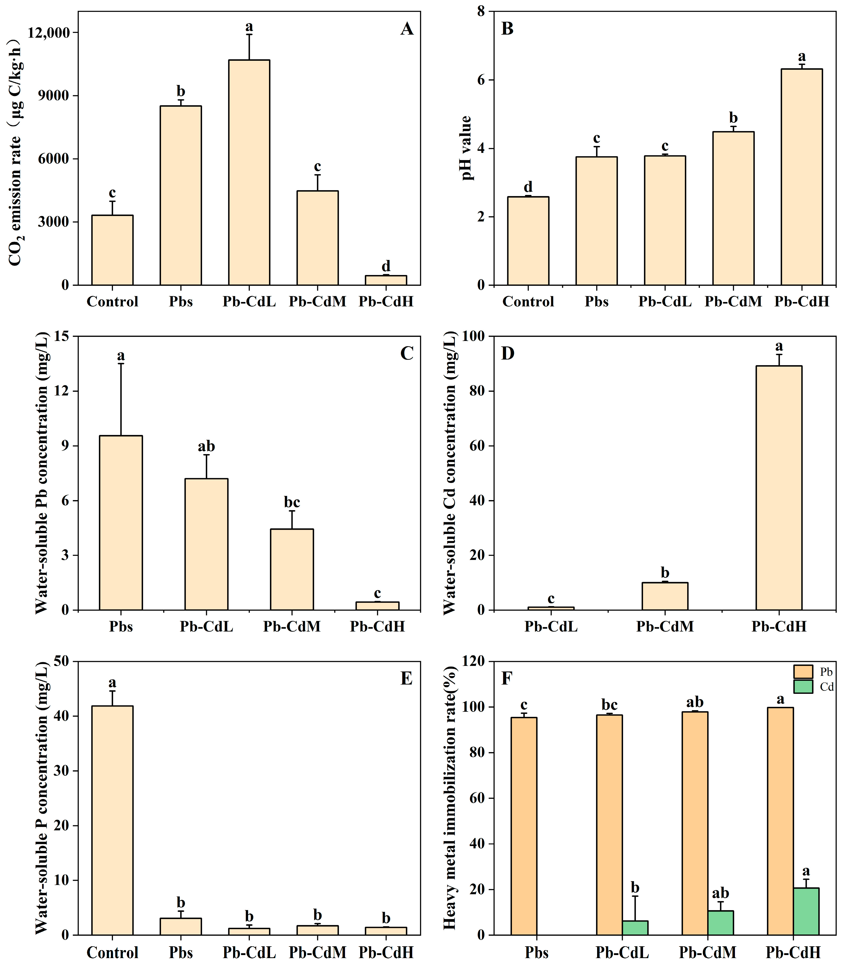
3.1. CO2 Emission Rate and pH Value

In the control treatment, CO2 emission rates were 3319 μg·C·kg·h−1 after incubation (Figure 1A). After the addition of Pb2+, the respiration intensity significantly increased to 8515 μg·C·kg·h−1 for the Pbs treatment. The respiration intensity of the Pb–CdL treatment was significantly higher than that of the Pbs treatment, i.e., 10,701 μg·C·kg·h−1 (Figure 1A). This also suggests a stimulating effect. Then, the respiration intensity was reduced to 4475 μg·C·kg·h−1 for the Pb-CdM treatment, and finally dramatically decreased to 449 μg·C·kg·h−1 for the Pb-CdH treatment.
For the treatments without Cd2+, the pH value increased from 2.58 (control treatment) to 3.75 (Pbs treatment). With the addition of Cd2+, the pH value increased from 3.78 (Pb-CdL treatment) to 4.48 (Pb–CdM treatment), and finally reached 6.32 in the Pb–CdH treatment after incubation (Figure 1B).

3.2. Pb, Cd, and P Concentrations

In the Pbs treatment, the Pb concentration was 9.55 mg/L (Figure 1C). After the addition of Cd2+, the Pb concentration decreased to 7.20 mg/L for the Pb–CdL treatment. The Pb concentration of the Pb–CdM treatment (4.44 mg/L) was lower than that of Pb–CdL treatment (Figure 1C). Then, the Pb concentration was significantly reduced to 0.45 mg/L for the Pb–CdH treatment (Figure 1C). The Cd concentration of the Pb–CdL treatment was 1.05 mg/L (Figure 1D). The value increased to 10.05 mg/L for the Pb–CdM treatment (Figure 1D). Finally, the Cd concentration increased significantly to 89.14 mg/L for the Pb–CdH treatment (Figure 1D).
The water-soluble P concentration in the control treatment was 41.86 mg/L (Figure 1E). After the addition of both Pb2+ and Cd2+, the concentration of water-soluble P in the solution decreased dramatically from 3.09 mg/L (Pbs treatment) to 1.25 mg/L (Pb–CdL treatment). Moreover, there was no significant difference in treatments with the addition of Cd, i.e., from 1.74 mg/L (Pb–CdM treatment) to 1.44 mg/L (Pb–CdH treatment) (see Figure 1E).
In the Pbs treatment, the removal rate of Pb2+ was 95.39% after incubation (Figure 1F). With the increasing concentration of Cd2+, the removal rates of Pb2+ were relatively stable, i.e., 96.52% (Pb–CdL treatment), 97.96% (Pb–CdM treatment), and 99.78% (Pb–CdH treatment). Compared with the removal rates of Pb2+, the removal of Cd2+ showed significant changes. The values increased significantly from 6.22% (Pb–CdL treatment) to 10.63% (Pb–CdM treatment). In the Pb–CdH treatment, the removal rate of Cd2+ was 20.70%, nearly twice that of Pb–CdM treatment (Figure 1F).

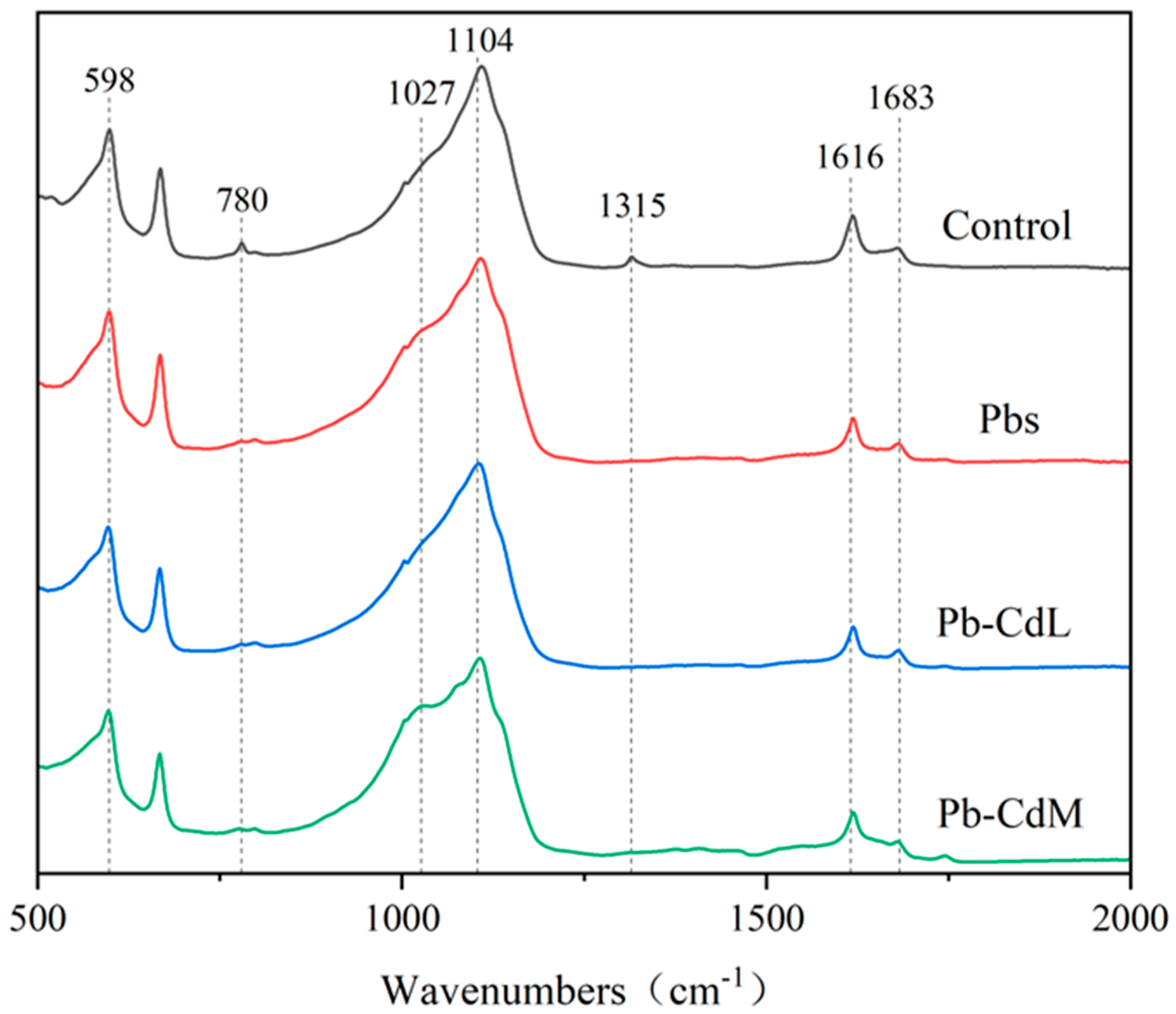
3.3. ATR-IR Analysis
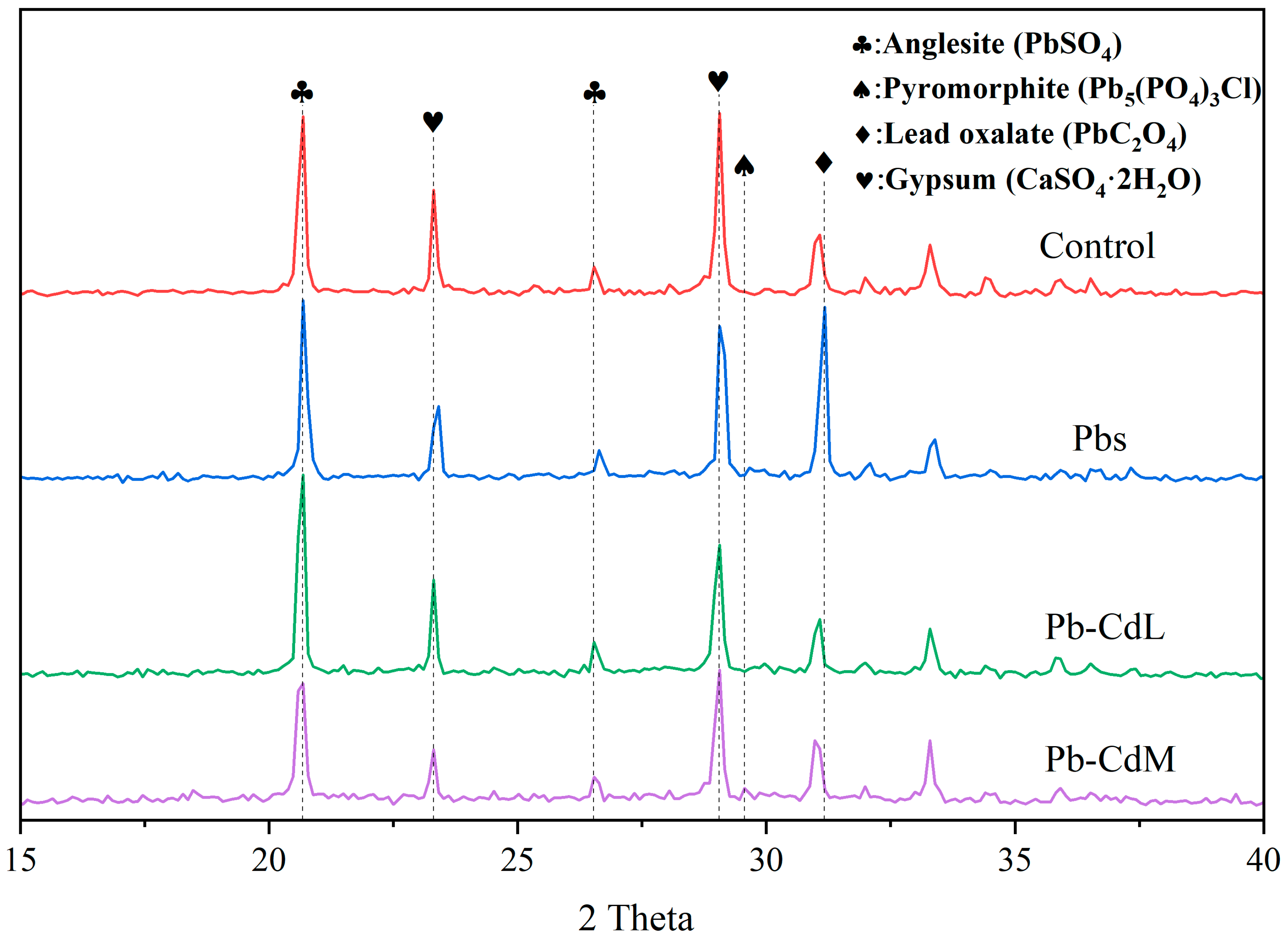
The peak at 598 cm−1 in the ATR-IR spectra was assigned to the ν4 P-O vibration (Figure 2) [39]. The peak at 1027 cm−1 was assigned to the ν1 and ν3 P-O vibration of phosphates (Figure 2) [40,41]. Meanwhile, the peak at 780 cm−1 was ascribed to the P-O vibration from HPO4. This peak disappeared in the Pbs, Pb-CdL and Pb-CdM treatment (Figure 2). The representative peaks at 1315 and 1616 cm−1 could be assigned to symmetric stretching vibrations of C-O from C2O42− and asymmetric stretching vibrations from HC2O4, which were usually associated with oxalic acid (Figure 2) [42]. The 1683 cm−1 peak represented the C=O stretching vibration (Figure 2) [43,44]. In addition, the 1104 cm−1 peak was a combination of P-O (for PO43−) and S-O (for SO42−) vibrations, which reflected both the ν3 P-O stretching vibration of phosphate and the sulfate content in PG (Figure 2) [39,45].

3.4. XRD Analysis

The peaks located at 23.31° and 29.05° were the two representative peaks of gypsum [45]. These two peaks indicated that gypsum was abundant in the solid phase regardless of the addition of HM (Figure 3). The peaks at 20.68° and 26.53° were associated with anglesite [36]. The representative peak of pyromorphite was differentiated at 29.56 (Figure 3). It appeared after the addition of 0.1 mmol/L Cd2+, whose peak intensity was relatively weak. In the Pb–CdM treatment, A. niger was stressed by Pb and Cd, which resulted in decreased oxalic acid secretion. Then, Pb2+ began to be mineralized into pyromorphite (Figure 4C,F). Moreover, the peak located at 31.17° ascribed to lead oxalate could be observed (Figure 3) [36]. The intensity of this peak in Pbs treatment was relatively strong (Figure 3).

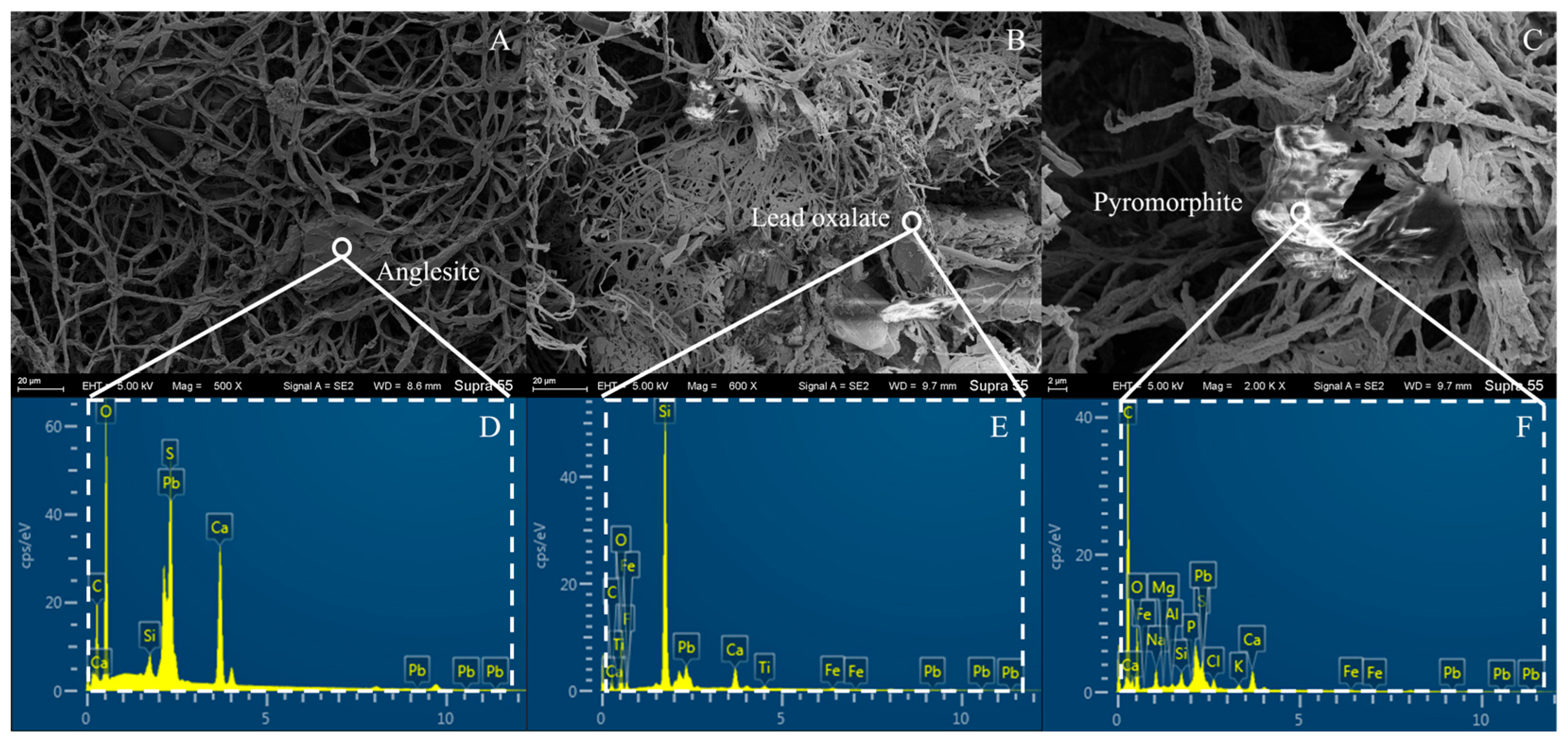
3.5. SEM-EDS Analysis

The solid phases obtained from the Pbs treatment and Pb-CdM treatment were observed using SEM (Figure 4). The SEM image and EDS result of the Pbs treatment indicated that Pb2+ replaced Ca2+ in PG and combined with SO42− to induce the mineralization of anglesite (Figure 4A,D) [36]. SEM imaging demonstrated that the hypha of A. niger adsorbed a large amount of gypsum (Figure 4A). Anglesite formed by the reaction was tightly wrapped in mycelium (Figure 4A). In addition, lead oxalate and pyromorphite were observed from the solid phase of Pb–CdM treatment (Figure 4B,C,E,F). Both of them were attached to the ends of A. niger mycelium (Figure 4A). The minerals observed above indicated that the A. niger complex PG system was capable of providing sufficient anions to ensure the immobilization of Pb2+.

4. Discussion

It was proposed that 1 mmol/L Pb2+ stimulated the bioactivity of A. niger and enhanced its metabolism during incubation (Figure 1A), which was consistent with our previous study [15]. This phenomenon was further confirmed by the significant increase in the biomass of A. niger after Pb addition (Figure 1A,E). Concurrently, water-soluble P decreased dramatically, which was primarily due to the consumption for the intense growth of fungus. Additionally, a small portion of P in PG participated in the mineralization of Pb2+ to form pyromorphite (Figure 4C,F). The toxicity of Pb2+ was relatively weak, attributed to its low charge density and mobility [46]. The low toxicity provides more chances for microorganisms to survive.
The presence of low Cd2+ (0.01 mmol/L) also had a stimulating effect on A. niger (Figure 1A). A. niger could activate antioxidative systems to reduce oxidative damage generated by Cd2+ [47]. Thus, the respiration of the fungus was significantly elevated (see Figure 1A). Therefore, the co-exposure of Pb and low-level Cd could benefit the long-term remediation of both HMs. Moreover, the immobilization rate of Pb2+ maintained >95% (Figure 1F). This can be attributed to the fact that Pb2+ was more readily adsorbed and fixed by A. niger with respect to Cd2+. From a chemical perspective, Pb2+ exhibited a higher adsorption affinity due to its higher standard electrode potential and smaller covalent radius [48,49,50]. As a result, A. niger preferentially adsorbed more Pb2+ to minimize direct contact with Cd2+. Although Cd2+ itself showed only a modest remediation effect, its presence made the immobilization process of Pb2+ in solution relatively stable (Figure 1F).
A. niger secreted a large amount of oxalic acid when growing rapidly, triggering the anions (e.g., SO42−) in PG to be released (Figure 3). Most of them combined with Pb2+ to form anglesite, resulting in more pronounced peaks for anglesite and oxalic acid with respect to Pbs treatment (Figure 3). This process further reduced the Pb concentration in solution. Compared with anglesite (Ksp = 1.82 × 10⁻⁸), cadmium sulfate is soluble, lowering its mineralization. Instead, it is highly probable that the oxalic acid secreted by A. niger combined with Cd2+ to form cadmium oxalate (Ksp = 1.51 × 10⁻⁸) (Figure 1F). In actual scenarios of soil contamination, Cd2+ typically co-exists with Pb2+, with the concentration of Pb2+ invariably exceeding that of Cd2+ [51]. The concentration of 0.01 mmol/L Cd2+ was suitable for many pollution scenarios (the background levels of Cd in rice soils are 0.37 μg/g). This indicates that the application of the A. niger complex PG system could effectively address the issue of HM contamination by Pb and Cd.
In the Pb–CdM treatment (0.1 mmol/L Cd2+), the high toxicity of Cd2+ inhibited the respiration of A. niger. This suggests the strong toxicity of Cd2+ on A. niger, which counterbalanced its stimulating effect (Figure 1B). In the absence of Cd2+, the biomass of A. niger grew significantly, accompanied by a large amount of secreted oxalic acid (Figure 1B). That led to the formation of more lead oxalate in the solution (Figure 4B,E). Under 0.1 mmol/L Cd2+, the secretion of oxalic acid declined, resulting in less free C2O42−. However, Pb2+ on the surface of A. niger occupied the potential sites for Cd2+ [30], thereby maintaining a relatively stable immobilization rate for Pb2+ (Figure 1B). The XRD results showed that only Pb–CdM treatment had the representative peak of pyromorphite (Figure 3). The A. niger complex PG system primarily induced Pb2+ in the environment mineralized into anglesite (Ksp = 1.6 × 10⁻⁸) [36]. Pb2+ was more inclined to be combined with SO42−, and then be precipitated as anglesite [39]. However, anglesite was not as stable as pyromorphite [52], which was inclined to redissolve in acidic environments [53]. In this study, the pH of the incubation system increased with increasing Cd2+ concentrations (Figure 1C,D). Therefore, the secondary release of Pb2+ was not evident (Figure 1E,F).
When the Cd concentration reached 1 mmol/L (i.e., 112.41 mg/L), Cd2+ exhibited strong toxicity to the fungal cells [31]. Our previous studies demonstrated that A. niger tolerance to Cd2+ was around 100 mg/L [28,54]. However, due to the presence of Pb2+ in the system, the antagonism between the two HMs weakened their toxicity [18,30,55]. Then, some A. niger was able to survive, which was confirmed by the consumption of P by Pb–CdH treatment (Figure 1E). Nevertheless, the metabolic process of A. niger was seriously stressed [56]. Notably, the immobilization rate of Cd2+ increased significantly from 10% to 20% (Figure 1F). Although the biological activity of the fungus was significantly reduced after six days of incubation under Cd stress, the organic matter from the debris (e.g., glucans, lipids, chitins, and chitin derivatives in the cell wall) could still bind Pb2+ and Cd2+ in a short time [57]. This suggested that even if the microorganism was not fully biologically functional, Cd2+ could still be immobilized by biological debris from A. niger. Additionally, PG itself could dissociate phosphate [58], generating cadmium phosphate (Ksp = 2.53 × 10⁻³³).
In previous studies, phosphorus-soluble bacteria (PSB) Ochrobactrum sp. J023 were combined with biochar for the remediation of Pb pollution [59]. The experiment was conducted in a Ca3(PO4)2 medium with a Pb concentration of 100 mg/L. It was reported that 71.30% Pb was immobilized by the combination of biochar and PSB. In contrast, the combination of PSF A. niger and PG demonstrated evident promotion of Pb remediation (Figure 1F). This is because fungi usually have a higher biomass than bacteria. Meanwhile, the spores of the A. niger promise its sustainable growth under stress.

5. Conclusions

The experimental results show that A. niger and PG had a better remediation on Pb pollution compared with Cd. When Cd was present in the environment, the remediation effect on Pb remained stable. The immobilization of Cd slightly increased with the elevation of Cd2+ concentration. Meanwhile, the presence of Pb2+ would stimulate the bioactivity of A. niger during incubation. In addition, a trace amount of Cd2+ had a similar effect. The combination of PSF and PG has a vitally broad prospect for remediating the environment polluted by Pb and Cd.

Author Contributions

X.L., Z.C. and H.L. wrote the manuscript. J.J., X.S., Y.C. and Z.L. (Zhengda Li) assisted in the data collection. C.L. and J.Y. performed supervision and project administration. Z.L. (Zhen Li) and L.X. conceived the idea, revised the manuscript, and led the project. All authors contributed to the article and approved the submitted version. All authors have read and agreed to the published version of the manuscript.

Funding

This work was supported by the National Key R&D Program of China (No. 2023YFC3707600) and the Research Fund Program of Guangdong Provincial Key Laboratory of Environmental Pollution Control and Remediation Technology (No. 2023B1212060016).

Data Availability Statement

The original contributions presented in the study are included in the article, further inquiries can be directed to the corresponding author.

Conflicts of Interest

The authors declare no conflicts of interest.

References

  1. Rassaei, F. Adsorption Kinetics and Isotherm Modeling of Lead in Calcareous Soils: Insights into Thermodynamics, Desorption, and Soil Properties. Commun. Soil Sci. Plant Anal. 2023, 54, 2059–2076. [Google Scholar] [CrossRef]
  2. Liu, X.; Song, Q.; Tang, Y.; Li, W.; Xu, J.; Wu, J.; Wang, F.; Brookes, P.C. Human health risk assessment of heavy metals in soil-vegetable system: A multi-medium analysis. Sci. Total Environ. 2013, 463, 530–540. [Google Scholar] [CrossRef] [PubMed]
  3. Fewtrell, L.J.; Prüss-Üstün, A.; Landrigan, P.; Ayuso-Mateos, J.L. Estimating the global burden of disease of mild mental retardation and cardiovascular diseases from environmental lead exposure. Environ. Res. 2004, 94, 120–133. [Google Scholar] [CrossRef]
  4. Newby, P.J.; Gadd, G.M. Synnema induction in penicillium-funiculosum by tributyltin compounds. Trans. Br. Mycol. Soc. 1987, 89, 381–384. [Google Scholar] [CrossRef]
  5. Deng, Z.; Cao, L.; Huang, H.; Jiang, X.; Wang, W.; Shi, Y.; Zhang, R. Characterization of Cd- and Pb-resistant fungal endophyte Mucor sp. CBRF59 isolated from rapes (Brassica chinensis) in a metal-contaminated soil. J. Hazard. Mater. 2011, 185, 717–724. [Google Scholar] [CrossRef] [PubMed]
  6. Zare, M.-R.; Amin, M.-M.; Nikaeen, M.; Bina, B.; Rahmani, A.; Hemmati-Borji, S.; Rahmani, H. Acute toxicity of Hg, Cd, and Pb towards dominant bacterial strains of sequencing batch reactor (SBR). Environ. Monit. Assess. 2015, 187, 263. [Google Scholar] [CrossRef]
  7. Genchi, G.; Sinicropi, M.S.; Lauria, G.; Carocci, A.; Catalano, A. The Effects of Cadmium Toxicity. Int. J. Environ. Res. Public Health 2020, 17, 3782. [Google Scholar] [CrossRef]
  8. Gadd, G.M. Interactions of fungi with toxic metals. New Phytol. 1993, 124, 25–60. [Google Scholar] [CrossRef]
  9. Shi, T.; Ma, J.; Zhang, Y.; Liu, C.; Hu, Y.; Gong, Y.; Wu, X.; Ju, T.; Hou, H.; Zhao, L. Status of lead accumulation in agricultural soils across China (1979–2016). Environ. Int. 2019, 129, 35–41. [Google Scholar] [CrossRef]
  10. Chen, R.S.; De Sherbinin, A.; Ye, C.; Shi, G.Q. China’s Soil Pollution: Farms on the Frontline. Science 2014, 344, 691. [Google Scholar] [CrossRef]
  11. Yang, S.; Xiao, Q.; Li, B.; Zhou, T.; Cen, Q.; Liu, Z.; Zhou, Y. Insights into remediation of cadmium and lead contaminated-soil by Fe-Mn modified biochar. J. Environ. Chem. Eng. 2024, 12, 112771. [Google Scholar] [CrossRef]
  12. Gao, Y.; Duan, Z.; Zhang, L.; Sun, D.; Li, X. The Status and Research Progress of Cadmium Pollution in Rice- (Oryza sativa L.) and Wheat- (Triticum aestivum L.) Cropping Systems in China: A Critical Review. Toxics 2022, 10, 794. [Google Scholar] [CrossRef]
  13. Deng, M.; Yang, X.; Dai, X.; Zhang, Q.; Malik, A.; Sadeghpour, A. Heavy metal pollution risk assessments and their transportation in sediment and overlay water for the typical Chinese reservoirs. Ecol. Indic. 2020, 112, 106166. [Google Scholar] [CrossRef]
  14. Hussain, B.; Lin, Q.; Hamid, Y.; Sanaullah, M.; Di, L.; Hashmi, M.L.U.R.; Khan, M.B.; He, Z.; Yang, X. Foliage application of selenium and silicon nanoparticles alleviates Cd and Pb toxicity in rice (Oryza sativa L.). Sci. Total Environ. 2020, 712, 136497. [Google Scholar] [CrossRef]
  15. Tian, D.; Jiang, Z.; Jiang, L.; Su, M.; Feng, Z.; Zhang, L.; Wang, S.; Li, Z.; Hu, S. A new insight into lead (II) tolerance of environmental fungi based on a study of Aspergillus niger and Penicillium oxalicum. Environ. Microbiol. 2019, 21, 471–479. [Google Scholar] [CrossRef]
  16. Li, X.; Cui, X.; Zhang, X.; Liu, W.; Cui, Z. Combined toxicity and detoxification of lead, cadmium and arsenic in Solanum nigrum L. J. Hazard. Mater. 2020, 389, 121874. [Google Scholar] [CrossRef]
  17. Zhao, F.J.; Lombi, E.; McGrath, S.P. Assessing the potential for zinc and cadmium phytoremediation with the hyperaccumulator Thlaspi caerulescens. Plant Soil 2003, 249, 37–43. [Google Scholar] [CrossRef]
  18. Baig, J.A.; Kazi, T.G.; Shah, A.Q.; Arain, M.B.; Afridi, H.I.; Khan, S.; Kandhro, G.A.; Soomro, A.S. Evaluating the accumulation of arsenic in maize (Zea mays L.) plants from its growing media by cloud point extraction. Food Chem. Toxicol. 2010, 48, 3051–3057. [Google Scholar] [CrossRef]
  19. Komjarova, I.; Blust, R. Multimetal interactions between Cd, Cu, Ni, Pb, and Zn uptake from water in the zebrafish Danio rerio. Environ. Sci. Technol. 2009, 43, 7225–7229. [Google Scholar] [CrossRef]
  20. Wang, G.; Fowler, B.A. Roles of biomarkers in evaluating interactions among mixtures of lead, cadmium and arsenic. Toxicol. Appl. Pharmacol. 2008, 233, 92–99. [Google Scholar] [CrossRef]
  21. Wu, B.; Liu, Z.; Xu, Y.; Li, D.; Li, M. Combined toxicity of cadmium and lead on the earthworm Eisenia fetida (Annelida, Oligochaeta). Ecotoxicol. Environ. Saf. 2012, 81, 122–126. [Google Scholar] [CrossRef]
  22. Gothilf, Y.; Coon, S.L.; Toyama, R.; Chitnis, A.; Namboodiri, M.A.A.; Klein, D.C. Zebrafish serotonin N-acetyltransferase-2: Marker for development of pineal photoreceptors and circadian clock function. Endocrinology 1999, 140, 4895–4903. [Google Scholar] [CrossRef]
  23. Mu, Y.; Zhang, C.; Li, Y.; Zhou, W.; Li, Y.; Zhao, G.; Su, P. Research Progress on Physical and Chemical Remediation Methods for the Removal of Cadmium from Soil. Separations 2024, 11, 299. [Google Scholar] [CrossRef]
  24. Lim, K.T.; Shukor, M.Y.; Wasoh, H. Physical, Chemical, and Biological Methods for the Removal of Arsenic Compounds. BioMed Res. Int. 2014, 2014, 503784. [Google Scholar] [CrossRef]
  25. Milano, F.; Giotta, L.; Lambreva, M.D. Perspectives on nanomaterial-empowered bioremediation of heavy metals by photosynthetic microorganisms. Plant Physiol. Biochem. 2024, 216, 109090. [Google Scholar] [CrossRef]
  26. Shi, J.; Wu, X.; Zhao, X.; Zhou, J.; Liu, S.; Li, B.; Zhang, J.; Li, W.; Zeng, X.; Wang, X.; et al. Remediation of heavy metal-contaminated estuarine sediments by strengthening microbial in-situ mineralization. Appl. Geochem. 2024, 169, 106051. [Google Scholar] [CrossRef]
  27. Chen, Y.M.; Chao, Y.Q.; Li, Y.Y.; Lin, Q.Q.; Bai, J.; Tang, L.; Wang, S.Z.; Ying, R.R.; Qiu, R.L. Survival Strategies of the Plant-Associated Bacterium Enterobacter sp. Strain EG16 under Cadmium Stress. Appl. Environ. Microbiol. 2016, 82, 1734–1744. [Google Scholar] [CrossRef]
  28. Lu, W.B.; Shi, J.J.; Wang, C.H.; Chang, J.S. Biosorption of lead, copper and cadmium by an indigenous isolate Enterobacter sp. J1 possessing high heavy-metal resistance. J. Hazard. Mater. 2006, 134, 80–86. [Google Scholar] [CrossRef]
  29. Hu, X.; Liu, X.; Zhang, S.; Yu, C. Nitrogen-cycling processes under long-term compound heavy metal(loids) pressure around a gold mine: Stimulation of nitrite reduction. J. Environ. Sci. 2025, 147, 571–581. [Google Scholar] [CrossRef]
  30. Qiu, J.; Song, X.; Li, S.; Zhu, B.; Chen, Y.; Zhang, L.; Li, Z. Experimental and modeling studies of competitive Pb (II) and Cd (II) bioaccumulation by Aspergillus niger. Appl. Microbiol. Biotechnol. 2021, 105, 6477–6488. [Google Scholar] [CrossRef]
  31. Wang, T.; Zhang, L.; Li, S.; Meng, L.; Su, M.; Wang, Z.; Nong, Y.; Sun, Y.; Wang, S.; Li, Z. Weakened Cd toxicity to fungi under coexistence of Pb in solution. J. Hazard. Mater. 2022, 426, 127984. [Google Scholar] [CrossRef]
  32. Dutta, R.K.; Khatri, V.N.; Panwar, V. Strength characteristics of fly ash stabilized with lime and modified with phosphogypsum. J. Build. Eng. 2017, 14, 32–40. [Google Scholar] [CrossRef]
  33. Ajam, L.; Hassen, A.B.; Reguigui, N. Phosphogypsum utilization in fired bricks: Radioactivity assessment and durability. J. Build. Eng. 2019, 26, 100928. [Google Scholar] [CrossRef]
  34. Altun, I.A.; Sert, Y. Utilization of weathered phosphogypsum as set retarder in Portland cement. Cem. Concr. Res. 2004, 34, 677–680. [Google Scholar] [CrossRef]
  35. Xi, X.; Wang, Z.; Zhou, L.; Song, H.; Han, Y.; Dong, F.; Zhou, L. Rapid removal of uranium (VI) by phosphogypsum immobilized sulfate-reducing bacteria microspheres. Chem. Eng. J. 2024, 493, 152676. [Google Scholar] [CrossRef]
  36. Meng, L.; Ding, K.; Qiu, Y.; Chen, Y.; Huo, H.; Yu, D.; Tian, D.; Li, Z. Application of phosphogypsum and phosphate-solubilizing fungi to Pb remediation: From simulation to in vivo incubation. Sci. Total Environ. 2024, 933, 173171. [Google Scholar] [CrossRef]
  37. Oliveira, V.; Labrincha, J.; Dias-Ferreira, C. Extraction of phosphorus and struvite production from the anaerobically digested organic fraction of municipal solid waste. J. Environ. Chem. Eng. 2018, 6, 2837–2845. [Google Scholar] [CrossRef]
  38. Hong, H.; Dai, M.; Lu, H.; Liu, J.; Zhang, J.; Yan, C. Risk assessment and driving factors for artificial topography on element heterogeneity: Case study at Jiangsu, China. Environ. Pollut. 2018, 233, 246–260. [Google Scholar] [CrossRef]
  39. Li, Z.; Su, M.; Duan, X.; Tian, D.; Yang, M.; Guo, J.; Wang, S.; Hu, S. Induced biotransformation of lead (II) by Enterobacter sp. in SO4-PO4-Cl solution. J. Hazard. Mater. 2018, 357, 491–497. [Google Scholar] [CrossRef]
  40. Kourkoumelis, N.; Lani, A.; Tzaphlidou, M. Infrared spectroscopic assessment of the inflammation-mediated osteoporosis (IMO) model applied to rabbit bone. J. Biol. Phys. 2012, 38, 623–635. [Google Scholar] [CrossRef]
  41. Garip, S.; Severcan, F. Determination of simvastatin-induced changes in bone composition and structure by Fourier transform infrared spectroscopy in rat animal model. J. Pharm. Biomed. Anal. 2010, 52, 580–588. [Google Scholar] [CrossRef]
  42. Yeasmin, S.; Singh, B.; Kookana, R.S.; Farrell, M.; Sparks, D.L.; Johnston, C.T. Influence of mineral characteristics on the retention of low molecular weight organic compounds: A batch sorption-desorption and ATR-FTIR study. J. Colloid Interface Sci. 2014, 432, 246–257. [Google Scholar] [CrossRef]
  43. Guo, Q.; Yi, H.; Jia, F.; Song, S. Design of MoS2/MMT bi-layered aerogels integrated with phase change materials for sustained and efficient solar desalination. Desalination 2022, 541, 116028. [Google Scholar] [CrossRef]
  44. Preeda, P.; Ganapathi Raman, R.; Sakthivel, P. Structural, FTIR, FT-Raman, optical and nonlinear optical properties of organic nonlinear optical crystal –3,5-diisopropyl-2-hydroxybenzoic acid. Inorg. Chem. Commun. 2022, 146, 110120. [Google Scholar] [CrossRef]
  45. Chen, S.; Chen, J.; He, X.; Su, Y.; Jin, Z.; Fan, J.; Qi, H.; Wang, B. Comparative analysis of colloid-mechanical microenvironments on the efficient purification of phosphogypsum. Constr. Build. Mater. 2023, 392, 132037. [Google Scholar] [CrossRef]
  46. Naimo, T.J. A review of the effects of heavy-metals on fresh-water mussels. Ecotoxicology 1995, 4, 341–362. [Google Scholar] [CrossRef]
  47. Xie, M.D.; Gao, X.Y.; Zhang, S.W.; Fu, X.H.; Le, Y.Q.; Wang, L. Cadmium stimulated cooperation between bacterial endophytes and plant intrinsic detoxification mechanism in Lonicera japonica thunb. Chemosphere 2023, 325, 138411. [Google Scholar] [CrossRef]
  48. Appel, C.; Ma, L. Concentration, pH, and surface charge effects on cadmium and lead sorption in three tropical soils. J. Environ. Qual. 2002, 31, 581–589. [Google Scholar] [CrossRef]
  49. Kalmykova, Y.; Strömvall, A.M.; Steenari, B.M. Adsorption of cd, cu, ni, pb and zn on Sphagnum peat from solutions with low metal concentrations. J. Hazard. Mater. 2008, 152, 885–891. [Google Scholar] [CrossRef]
  50. Wang, T.; Liu, W.; Xiong, L.; Xu, N.; Ni, J.R. Influence of pH, ionic strength and humic acid on competitive adsorption of Pb(II), Cd(II) and Cr(III) onto titanate nanotubes. Chem. Eng. J. 2013, 215, 366–374. [Google Scholar] [CrossRef]
  51. Yuan, X.H.; Xue, N.D.; Han, Z.G. A meta-analysis of heavy metals pollution in farmland and urban soils in China over the past 20 years. J. Environ. Sci. 2021, 101, 217–226. [Google Scholar] [CrossRef]
  52. Cao, X.D.; Ma, L.Q.; Singh, S.P.; Zhou, Q.X. Phosphate-induced lead immobilization from different lead minerals in soils under varying pH conditions. Environ. Pollut. 2008, 152, 184–192. [Google Scholar] [CrossRef]
  53. Miretzky, P.; Fernandez-Cirelli, A. Phosphates for Pb immobilization in soils: A review. Environ. Chem. Lett. 2008, 6, 121–133. [Google Scholar] [CrossRef]
  54. Wang, J.; Chen, C. Chitosan-based biosorbents: Modification and application for biosorption of heavy metals and radionuclides. Bioresour. Technol. 2014, 160, 129–141. [Google Scholar] [CrossRef]
  55. Liao, G.; Wang, P.; Zhu, J.; Weng, X.; Lin, S.; Huang, J.; Xu, Y.; Zhou, F.; Zhang, H.; Tse, L.A.; et al. Joint toxicity of lead and cadmium on the behavior of zebrafish larvae: An antagonism. Aquat. Toxicol. 2021, 238, 105912. [Google Scholar] [CrossRef]
  56. Angumeenal, A.R.; Venkappayya, D. Growth kinetics of heavy metal adapted Aspergillus niger during citric acid biosynthesis. J. Sci. Ind. Res. 2004, 63, 610–613. [Google Scholar]
  57. Guha, S.; Debnath, S.; Gayen, S. Comparative Analysis of Hexavalent Chromium Biosorption Efficiency Using Dead and Live Aspergillus nomius Biomass. J. Trop. For. Environ. 2022, 11. [Google Scholar] [CrossRef]
  58. Chernysh, Y.; Yakhnenko, O.; Chubur, V.; Roubík, H. Phosphogypsum Recycling: A Review of Environmental Issues, Current Trends, and Prospects. Appl. Sci. 2021, 11, 1575. [Google Scholar] [CrossRef]
  59. Zhou, Y.; Zhao, X.; Jiang, Y.; Ding, C.; Liu, J.; Zhu, C. Synergistic remediation of lead pollution by biochar combined with phosphate solubilizing bacteria. Sci. Total Environ. 2023, 861, 160649. [Google Scholar] [CrossRef]
Figure 1. Fungal physiology and chemical features of the medium. (A) CO2 emission rates of A. niger. (B) pH of the filtrate after six days of incubation. (C,D) Concentrations of heavy metals in the filtrate after six days of incubation. (E) Concentrations of water-soluble phosphorus in the filtrate after six-days incubation. (F) Immobilization rates of heavy metals after six days of incubation. The lowercase letter labels in the figure indicated significant differences among different treatments. If two treatments contained the same lowercase letter, there was no significant difference between them; otherwise, there was a significant difference.
Agronomy 15 01632 g001
Figure 2. ATR-IR spectra of the filtered solid phase after six-days incubation.
Agronomy 15 01632 g002
Figure 3. XRD patterns of the filtered solid phase after six days of incubation.
Agronomy 15 01632 g003
Figure 4. SEM imaging on the samples. (A) SEM image of Pbs treatment. (B,C) SEM images of Pb-CdM treatment. (D) EDS image of Pbs treatment. (E,F) EDS images of Pb-CdM treatment.
Agronomy 15 01632 g004
Disclaimer/Publisher’s Note: The statements, opinions and data contained in all publications are solely those of the individual author(s) and contributor(s) and not of MDPI and/or the editor(s). MDPI and/or the editor(s) disclaim responsibility for any injury to people or property resulting from any ideas, methods, instructions or products referred to in the content.

