Article

PlTem1, a Key Cell Cycle Regulator, Serves as an Important Bridge Between Cell Division and Autophagy in Peronophythora litchii

1
School of Breeding and Multiplication (Sanya Institute of Breeding and Multiplication), School of Tropical Agriculture and Forestry, Hainan University, Sanya 572025, China
2
College of Plant Protection, Yangzhou University, Yangzhou 225009, China
*
Author to whom correspondence should be addressed.
These authors contributed equally to this work.
Agronomy 2025, 15(7), 1619; https://doi.org/10.3390/agronomy15071619
Submission received: 10 June 2025 / Revised: 22 June 2025 / Accepted: 27 June 2025 / Published: 2 July 2025

Abstract

The orderly progression of the cell division process is crucial for the morphogenesis of pathogens and the process of infecting hosts. However, there is currently no relevant research on cell division in the pathogen Peronophythora litchii. First, we verified that treatment with cell division inhibitors would have an adverse effect on the growth, development, and pathogenicity of P. litchii. Subsequently, through homology-based sequence alignment and functional domain prediction analyses, we identified PlTem1, a key small GTPase regulating cell division. Compared with the wild-type strain Shs3, the mutant strain ΔPltem1 exhibited significant defects in mycelial growth, sporangia and zoospore generation, and virulence. To explore the pathogenic mechanism of PlTem1, screening and identification of interacting proteins were carried out. The comprehensive results show that there is an interaction between Tem1 and multiple autophagy-related proteins, suggesting that PlTem1 serves as an important bridge between autophagy and cell division in P. litchii.

1. Introduction

Litchi is a well-known tropical and subtropical fruit with significant commercial value worldwide, attributed to its appealing appearance, delicious taste, and rich nutrient content [1,2]. However, the production and quality of litchi fruits are severely impacted by various pests and diseases. Among these, litchi downy blight, caused by Peronophythora litchii, is a destructive oomycete disease that affects multiple tissues of litchi at different developmental stages, including leaves, flowers, panicles, new shoots, and fruits [3,4]. Infected tissues become brown and rot, and finally produce white sporangia and sporangiophores to release zoospores, which subsequently attack other healthy tissues of litchi [5]. Notably, the global fruit losses attributed to P. litchii are estimated to be around 20–30% in litchi [1,6]. Although the publication of P. litchii genome and the application of CRISPR/Cas9-mediated gene knockout technology have accelerated research on functional genes in P. litchii [7,8,9,10,11], the molecular mechanisms involved in its growth, development, and pathogenesis remain largely unexplored.
In eukaryotes, the cell cycle plays a fundamental role in regulating the growth and development of living organisms. It progresses through two different phases, namely interphase and the mitotic phase, the latter of which is further categorized into mitosis and cytokinesis [12]. The transition from mitosis to cytokinesis is regulated via the mitotic exit network (MEN), which is an essential kinase signaling cascade consisting of Tem1, Lte1, Bub2-Bfa1, Cdc5, Cdc15, Dbf2, Mob1, and Nud1 [13,14]. The GTPase-activating protein Bub2-Bfa1 mediates the small GTPase Tem1, and then Tem1 regulates the MEN activity via targeting the spindle positioning checkpoint [15]. During late anaphase, Tem1-GTP binds to and activates the protein kinase Cdc15, subsequently activating the downstream kinase Dbf2, which interacts with its activating subunit, Mob1 [16]. Furthermore, the kinase complex Dbf2-Mob1 phosphorylates Cdc14, and the activated Cdc14 departs from the nucleolus, thus mediating the completion of cell division [17,18].
Recent reports indicate that the mitogen-activated protein (MAP) signaling pathway plays essential roles in regulating both the development and pathogenicity of plant pathogenic fungi. Specifically, BUB2 is involved in the septum formation of Colletotrichum higginsianum and Magnaporthe oryzae, and disruption of BUB2 impairs the formation of mature appressoria, which consequently leads to a decrease in pathogenicity [19]. MoSep1, MoDbf2, and MoMob1, three conserved components of MEN, are critical for the appressorium formation and virulence of M. oryzae [20]. MoSep1 associates with and phosphorylates MoMob1 to maintain the cell cycle in M. oryzae during mycelial growth and infection [21]. In addition, MoSep1 phosphorylates MoRgs7 to govern the appressorium formation and pathogenicity of M. oryzae via activating the cAMP signaling [22]. Additionally, FgBub2 and FgBfa1 form a heterodimer complex to function as a GAP for FgTem1, and these three proteins are required for the vegetative growth and pathogenicity of Fusarium graminearum [23].
The small GTPases act as molecular switches to participate in many biological processes, such as growth, morphogenesis, and cell division [24,25]. In fungi, the small GTPases modulate fungal virulence, which has provided insight into the pathogenic mechanisms [25]. However, in oomycetes, the functions of Tem1, a small GTPase that regulates the cell cycle, remain to be elucidated. In our study, a combination of plant pathology, cell biology, and genetics-based analyses was used to characterize the roles of the cell cycle and PlTem1 in P. litchii.

2. Materials and Methods

2.1. Strains and Growth Conditions

The P. litchii wild-type strain Shs3 was used to generate transformants in this study. The EV control strain (empty vector transformant) was created by transforming Shs3 with a null vector. All strains were grown on V8 agar medium (100 mL V8 vegetable juice, 900 mL ddH2O and 20 g agar) at 25 °C in the dark. To test the growth rate, mycelial plugs (5 mm diameter) from the growing colony of the indicated strains were separately inoculated on V8 agar plates, and these plates were subsequently moved in a 25 °C incubator. After 5 days, the diameter of the colony on each plate was measured, and the representative colonies were photographed. The sporangia were harvested via flooding five mycelial plugs (9 mm in diameter) obtained from the colony mentioned above with 5 mL sterile distilled water and then filtering the suspension through a mesh strainer (100 µm size) (APPLYGEN, Beijing, China). Subsequently, the sporangia suspension was incubated at 16 °C for 0.5 h or 2 h to induce zoospore release. The number of sporangia and the release rate of zoospores were counted under a microscope (OLYMPUS BX53+DP74, OLYMPUS, Tokyo, Japan).

2.2. Pathogenicity Assays

The pathogenicity of all experimental strains was assessed on detached litchi leaves and fruits through inoculation with either sporangia or mycelial plugs (5 mm in diameter). Following inoculation, the litchi leaves and fruits were placed in a 25 °C incubator set to 80% humidity in darkness. The lesion area of leaf samples was examined at 24 and 36 h post-inoculation (hpi), while the diseased area on litchi fruits was assessed 4 days after inoculation. The representative symptomatic leaves and fruits were photographed, respectively.

2.3. Quantitative Real-Time PCR (qRT-PCR) Analysis

Total RNA was isolated from the litchi leaves inoculated with SHS3 strain at 0, 1.5, 3, 6, 12, 24, and 48 hpi. Complementary DNA (cDNA) was obtained from the total RNA via reverse transcription PCR (RT-PCR) using the kit (RR047A, Takara, Nojihigashi 7-4-38, Kusatsu, Japan). The transcripts of the PlTEM1 gene were quantified via a TB Green kit (RR420A, Takara, Nojihigashi 7-4-38, Kusatsu, Japan), and the housekeeping gene β-ACTIN of P. litchii was used as an internal reference. The relative expression of PlTEM1 was finally calculated via the 2−ΔΔCT method [26].

2.4. Bioinformatic Analyses

Amino acid sequences of all Tem1 homologs from different species, including Phytophthora ramorum, P. parasitica, P. infestans, P. sojae, Peronophythora litchii, Aspergillus flavus, M. oryzae, F. falciforme, F. graminearum, and Saccharomyces cerevisiae, were obtained from the online website (https://fungidb.org/fungidb/app, accessed on 17 August 2024). The phylogenetic dendrogram was conducted via MEGA 7.0 using the Maximum Likelihood method with a setting of 1000 bootstrap replicates. In addition, the structure of the PlTem1 protein was predicted via the website (https://swissmodel.expasy.org/interactive, accessed on 25 August 2024).

2.5. Targeted Deletion and Complementation of PlTEM1

Two specific sgRNAs for targeting PlTEM1 were designed and ligated to the sgRNA vector pYF2.3G-RibosgRNA based on the previously described method [9]. To generate the gene-replacement construct, the donor DNA HPH and two homologous flanking sequences (~1 kb) of the PlTEM1 coding region were connected and then cloned to the pBluescript SK II+ (pBS-SK II+) vector. The obtained constructs, pYF2.3G-RibosgRNA (PlTem1) and pBS-SK II+ (PlTem1), were co-transformed into the protoplasts of the Shs3 strain via the PEG-mediated transformation method, and the transformants were screened on V8 agar medium containing 50 µg/mL G418. These candidate transformants were further confirmed via genomic PCR and sequencing. The complemented strain ΔPltem1/PlTEM1 was obtained as described by Qiu et al. [27].

2.6. Staining and Microscopic Observation

The sporangial cleavage of all tested strains was visualized by staining using the red-fluorescent FM4–64 dye (T13320, Invitrogen, Carlsbad, CA, USA) [28,29]. The sporangia from the corresponding strains were washed with sterilized distilled water and incubated at 10 °C for 0.5 and 2 h, respectively, to stimulate the sporangial cleavage. The sporangia suspensions were stained using 1 μg/mL FM4–64 (final concentration) for 5 min in the darkness, and then observed via the fluorescence microscope at indicated time points. For nuclei staining, the hyphae of the tested strains were stained via 1 μg/mL 4′, 6-diamidino-2-phenylindole (DAPI) (D3571, Invitrogen, Carlsbad, CA, USA) for 5 min in the darkness and then detected via the fluorescence microscope (OLYMPUS BX53+DP74, OLYMPUS, Tokyo, Japan).

2.7. Yeast-Two-Hybrid (Y2H) Assay

The coding regions of PlTEM1, PlATG1a, PlATG2, PlATG3, PlATG8, PlATG9, PlATG13, PlATG17, PlATG18a, and PlATG18b were amplified from Shs3 cDNA via PCR and then cloned into pGADT7 or pGBKT7, separately. The constructed vectors were first verified by sequencing and subsequently co-transformed into the yeast strain AH109, as described by Yin et al. [30]. The transformants, screened via the synthetic dropout medium without leucine (Leu) and tryptophan (Trp), were grown on the selection medium lacking Leu, Trp, adenine (Ade), and histidine (His) at 30 °C for the interaction test.

2.8. Statistical Analyses

All data obtained from the experiments in our research were analyzed via multiple t-tests (p < 0.05) using the GraphPad Prism software v6.0.

3. Results

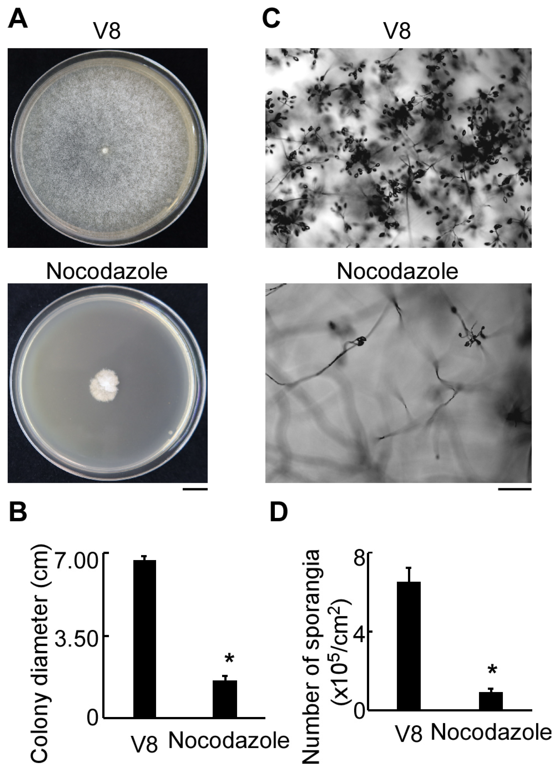
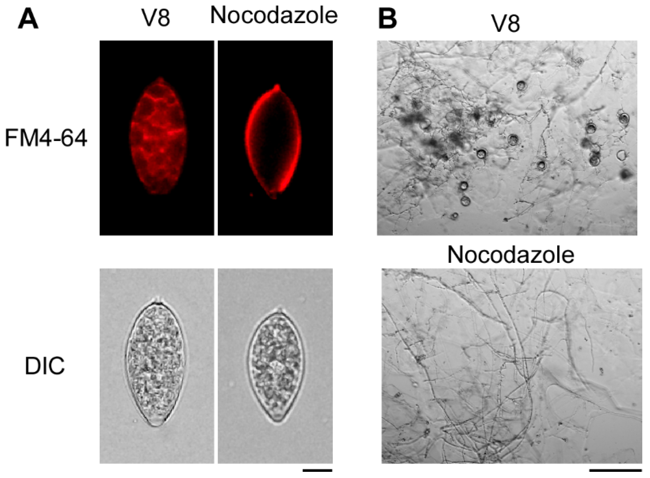
3.1. Cell Cycle Inhibition Impaired Vegetative Growth, Sporangia Production, Zoospore Release, and Oospore Formation in P. litchii

To investigate the effect of cell cycle progression of P. litchii, the pathogen was cultured on V8 media treated with or without nocodazole, a cell cycle inhibitor. As shown in Figure 1A, the growth of P. litchii mycelium on V8 media containing nocodazole was much slower than that of the control. At 5 days, the colony diameter of P. litchii on the nocodazole-treated media was only 1.62 cm, while it reached up to 6.73 cm on the V8 media (Figure 1B). In addition, the numbers of sporangia decreased by 85% in the treated group compared to the control group (Figure 1C,D). To assess the ability of sporangia to release zoospores, the sporangia were stained with FM4-64 following low-temperature incubation and subsequently observed under a fluorescence microscope. The sporangia from the control group exhibited normal cleavage and released a significant number of zoospores, whereas the sporangia produced in the nocodazole-treated media were unable to cleave, resulting in no zoospore formation (Figure 2A). In addition, the P. litchii treated with nocodazole did not produce oospores (Figure 2B). Collectively, these data demonstrate that the cell cycle is required for the growth, sporangia production, zoospore release, and oospore formation of P. litchii.

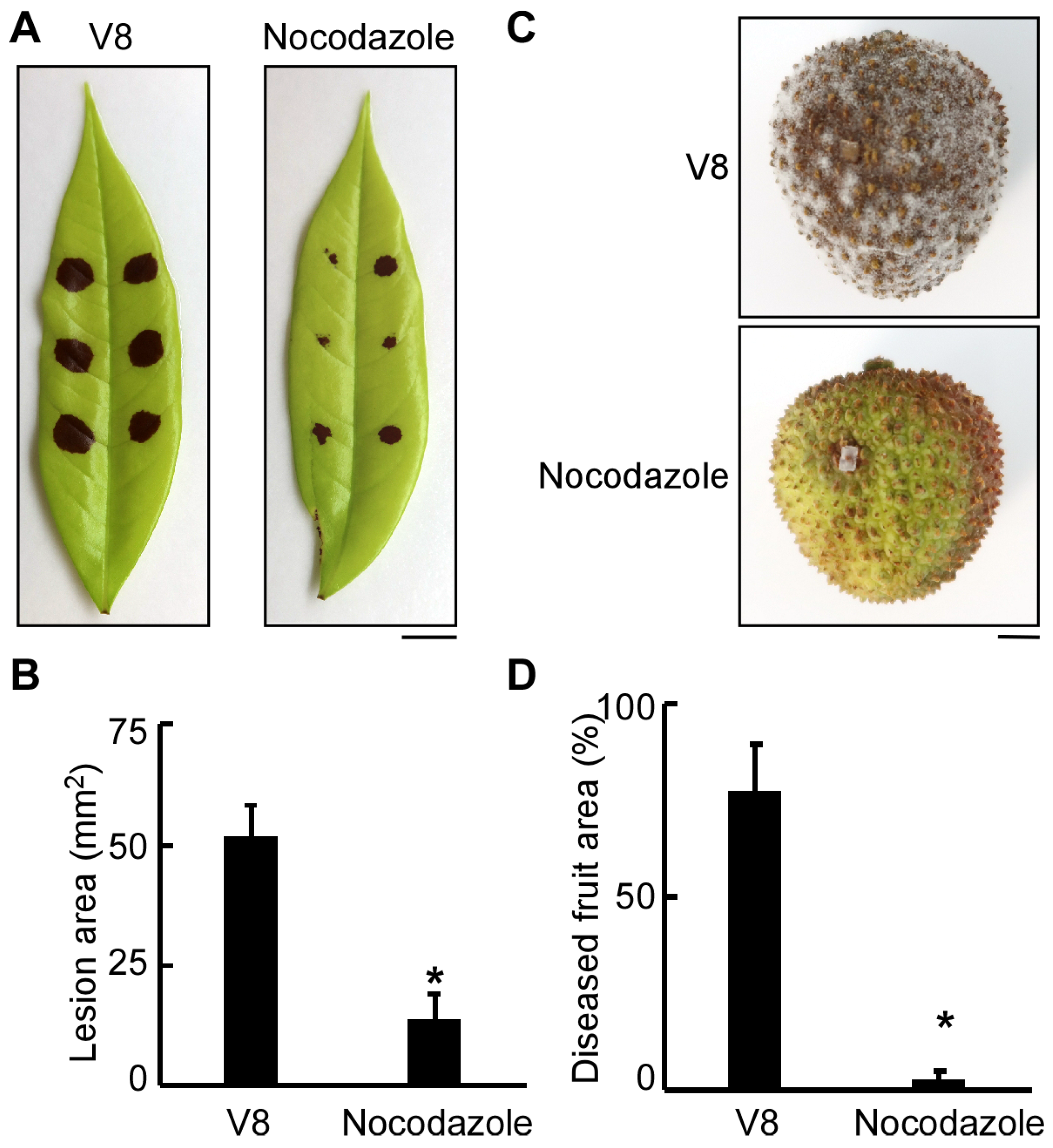
3.2. Normal Progression of Cell Division Is Essential for the Pathogenicity of P. litchii

To determine whether the cell cycle contributes to the virulence of P. litchii, the sporangia and mycelial plugs of Shs3 grown on V8 media with or without nocodazole were used to inoculate the detached litchi leaves and fruits, respectively. On litchi leaves, the average lesion area caused by the nocodazole-treated Shs3 strain was only 12 mm2, while it reached 52 mm2 in the control group, about 4–5 times higher than that in the nocodazole-treated group (Figure 3A,B). On litchi fruits, the average diseased area was less than 5% of the entire fruit surface in the nocodazole-treated group, whereas it reached up to 75% in the control group (Figure 3C,D). Together, these results indicate that the cell cycle is necessary to the virulence of P. litchii.

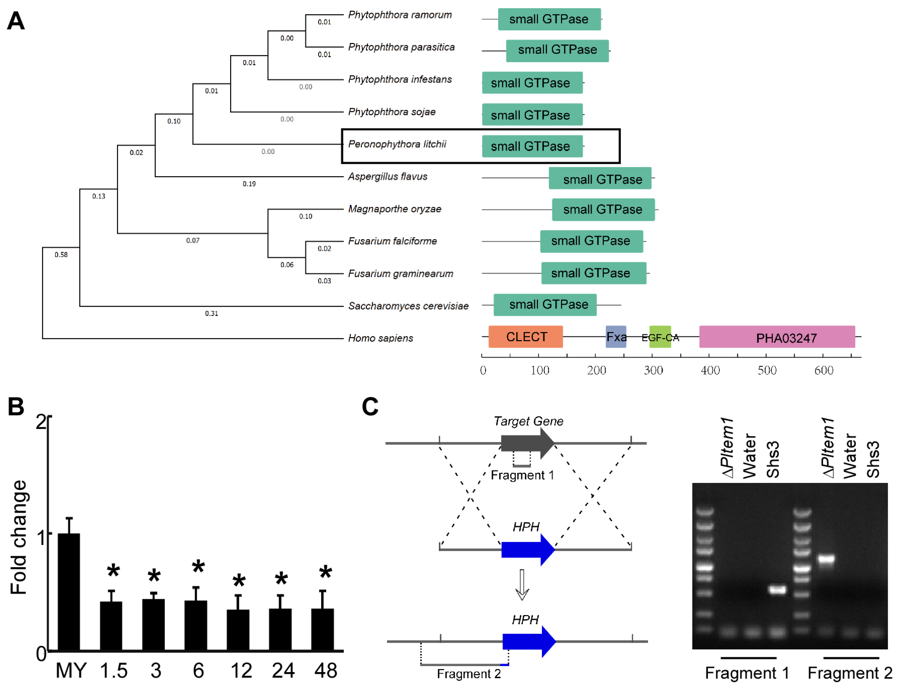
3.3. Characterization of Small GTPase PlTem1 in P. litchii

Tem1 is a key regulator of the mitotic exit network (MEN), and MEN mediates the transition from mitosis to cytokinesis, which is an important step in cell cycle progress [12,15]. Based on the results presented above, it was hypothesized that PlTem1 may play critical roles in P. litchii. Notably, PlTem1 contains a conserved small GTPase domain (Figure 4A). To investigate whether PlTem1 is involved in the infection stage, the transcripts of PlTEM1 were quantified in litchi leaves at 0, 1.5, 3, 6, 12, 24, and 48 hpi. The expression level of PlTEM1 decreased significantly during the infection stage (Figure 4B), which indicates that PlTem1 may play a negative regulatory role during the infection stage. This is also in accordance with the regulatory role of PlTem1 in other species, suggesting that PlTem1 may also play its conserved role as a negative regulatory factor involved in the regulation of the cell division stage in P. litchii.
To explore the functions of PlTem1 in P. litchii, the deletion mutant of this gene was created via a CRISPR/Cas9-mediated genome editing system [7,9,10]. Following PEG-mediated transformation, candidate transformants exhibiting G418 resistance were identified through screening, and subsequently confirmed as mutants via genomic PCR and sequencing. A specific fragment 1 of PlTEM1 was amplified from the Shs3 strain, but not from the candidate ΔPltem1 mutant. Additionally, a 1.5 kb band for fragment 2 was only amplified from the candidate ΔPltem1 mutant using the specific primers, and the sequencing result showed that PlTEM1 was replaced by HPH (Figure 4C). These results demonstrate that PlTem1 was successfully knocked out the ΔPltem1 strains.

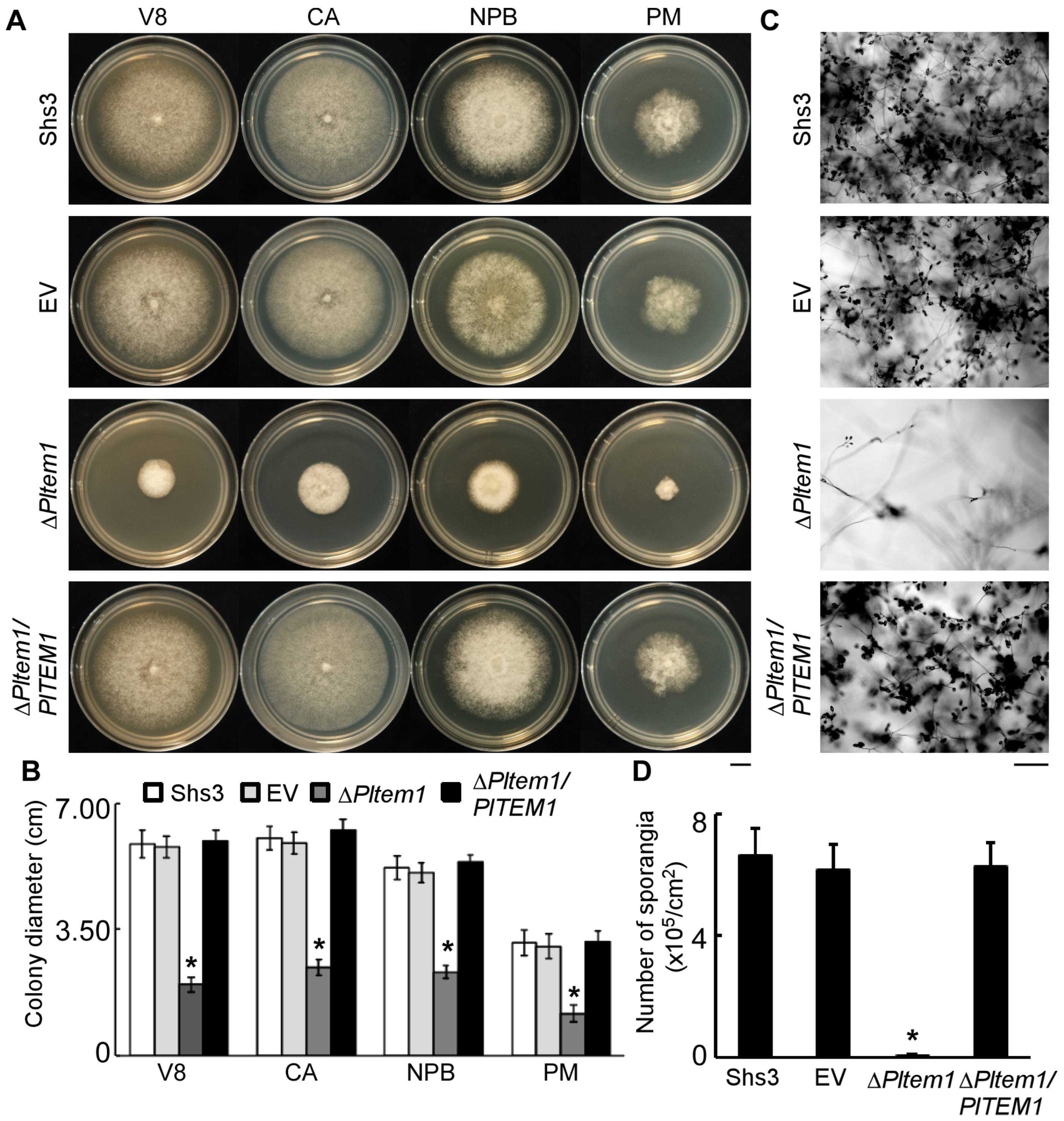
3.4. Tem1 Plays an Important Role in the Vegetative Hyphae and Sporangia of P. litchii

To characterize the biological function of PlTem1 in the mycelial growth of P. litchii, strains (such as Shs3, EV, ΔPltem1, and ΔPltem1/PlTEM1) were cultured on V8, CA, NPB, and PM plates, respectively. After 5 days, the colony diameters of all strains were measured, and the sporangia of these strains were harvested and counted. Compared with the Shs3 and EV strains, the average colony size of the ΔPltem1 strain was significantly reduced, while the aerial mycelium of the ΔPltem1 strain was notably increased (Figure 5A,B). However, there was no significant difference in mycelial growth between the complemented strain ΔPltem1/PlTEM1 and the control strains (Shs3 and EV). These results demonstrate that PlTem1 acts as an essential player in the mycelial growth of P. litchii.
Additionally, the number of sporangia produced by the ΔPltem1 strain was much lower than that of Shs3 and EV, while the ΔPltem1/PlTEM1 strain exhibited no significant change compared to Shs3 and EV in the number of sporangia (Figure 5C,D).

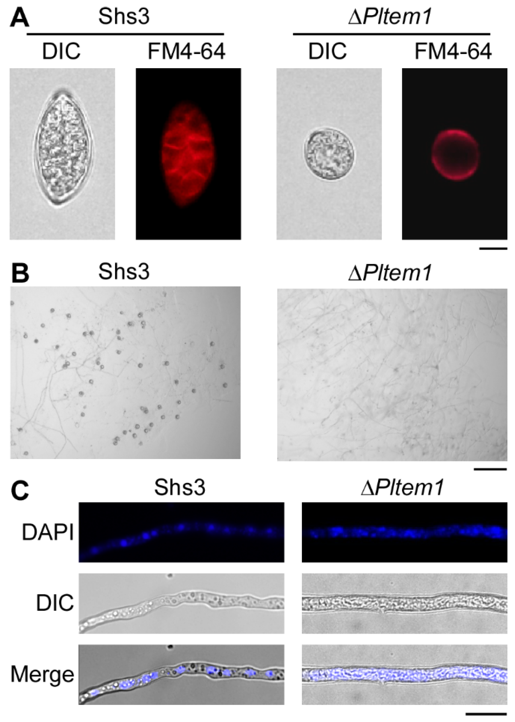
3.5. The Deletion of PlTEM1 Inhibits P. litchii to Produce Zoospores and Oospores, and Its Nuclei Cannot Divide with Abnormal Morphology

The formation of zoospores is crucial for the infection of the host by the diploid pathogenic oomycete P. litchii. Zoospores are formed by the cleavage of sporangia under certain conditions. When PlTEM1 is knocked out, the morphology of sporangia becomes distorted. Moreover, after low-temperature induction of sporangia, the mutant strain ΔPltem1 cannot cleave. In addition, even as the induction time increased, it still failed to cleave to form zoospores (Figure 6A). Meanwhile, observations of its sexual reproduction stage show that ΔPltem1 is unable to form oospores (Figure 6B), indicating that the deletion of PlTEM1 also has a severe impact on sexual reproduction. As a conserved small G-protein hydrolase involved in regulating the cell division process, the nuclear division was observed under a fluorescence microscope. Compared with the intact nuclear morphology of the wild type with uniform aggregation, ΔPltem1 presents a diffused and shapeless state of nuclei (Figure 6C). Taken together, these results indicate that PlTem1 is crucial for both the mitosis and meiosis processes of P. litchii.

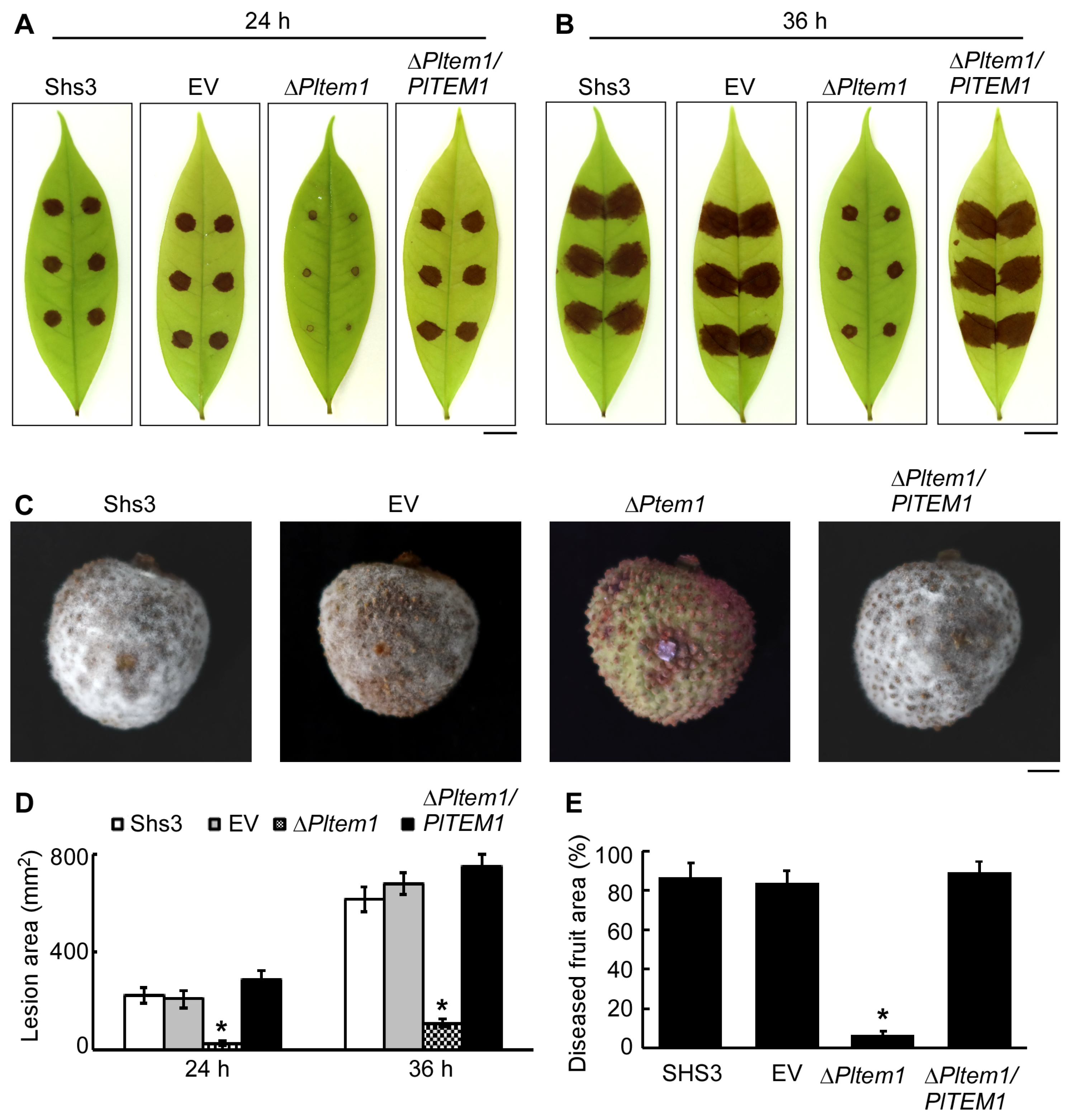
3.6. PlTem1 Is Essential for P. litchii Pathogenicity

To confirm the contribution of PlTem1 to the virulence of P. litchii, the Shs3, EV, ΔPltem1, and ΔPltem1/PlTEM1 strains were utilized to inoculate litchi leaves and fruits, respectively. At 24 hpi, distinct lesions were observed on the litchi leaves inoculated with the Shs3 and EV strains, but the litchi leaf inoculated with ΔPltem1 showed no visible changes (Figure 7A). At 36 hpi, the lesions enlarged significantly on the litchi leaves infected by the control strains (Shs3 and EV), and the lesion area was ~3 times higher than that at 24 hpi, reaching ~650 mm2 (Figure 7B,D). However, there were only small spots on the leaves inoculated with the ΔPltem1 strain (Figure 7B). Intriguingly, the lesion area caused by ΔPltem1/PlTEM1 showed no significant differences compared to that of controls (Figure 7A,B,D), suggesting that PlTEM1 restored the pathogenicity of the ΔPltem1 strain. Similarly, on litchi fruits, the diseased area caused by ΔPltem1 accounted for 8% of the whole fruit surface, which is ~10 times lower than that in the controls (Figure 7C,E). In addition, the virulence of the ΔPltem1/PlTEM1 strain toward litchi fruits was the same as that of the controls. Taken together, these data indicate that PlTem1 is critical for the pathogenicity of P. litchii.

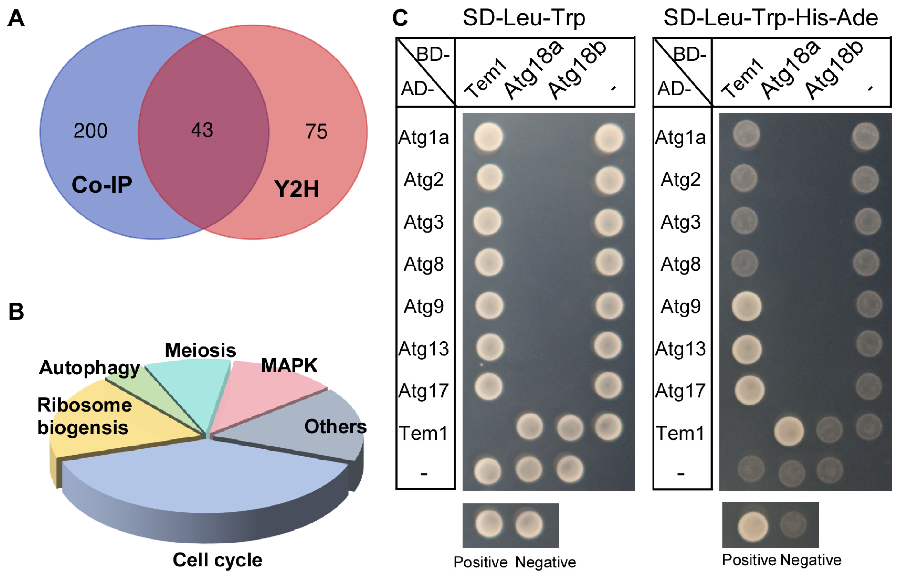
3.7. PlTem1 Interacts with Multiple Autophagy-Related Proteins

To investigate the role of PlTem1 in regulating the pathogenicity of P. litchii, Co-IP and Y2H assays were employed to screen for the PlTem1-interacting proteins, respectively. A total of 43 proteins were identified that were associated with PlTem1 across both screening systems (Figure 8A). These proteins were found to play significant roles as regulators in various biological processes, including the cell cycle, ribosome biogenesis, autophagy, meiosis, and MAPK pathway (Figure 8B). To verify the data acquired from the Co-IP and Y2H screening, several autophagy-related proteins (PlAtg1a, PlAtg2, PlAtg3, PlAtg8, PlAtg9, PlAtg13, PlAtg17, PlAtg18a, and PlAtg18b) were chosen to test their association with PlTem1 via Y2H. As shown in Figure 8C, all tested yeast strains grew normally on SD/-Leu-Trp plates, while only four yeast strains (harboring AD-PlAtg9 and BD-PlTem1, AD-PlAtg13 and BD-PlTem1, AD-PlAtg17 and BD-PlTem1, or AD-PlTem1 and PlAtg18a vectors) grew well on SD/-Leu-Trp-His-Ade plates similar to the positive strain, indicating that PlTem1 interacts with PlAtg9, PlAtg13, PlAtg17, and PlAtg18a, respectively.

4. Discussion

4.1. The Cell Cycle Is Necessary for the Growth and Pathogenicity of Fungi and Oomycetes

Research on secretory proteins has attracted much attention in order to advance our understanding of the pathogenicity of P. litchii. Effectors and cell wall-degrading enzymes, secreted by P. litchii, were reported to contribute to the virulence of the pathogen [10,31,32]. For instance, PlPAE5 associates with and destabilizes LcLTP1, a positive regulator of plant immunity, thereby inhibiting SA-dependent defense responses and resulting in increased host susceptibility [31]. In addition to secretory proteins, investigating the roles of other proteins may provide further insights that enhance our understanding of P. litchii. Here, cell division is firstly reported to be important for the growth, development, and virulence of P. litchii.
Normal cell division was demonstrated to be critical for vegetative growth, the formation of penetration structure, and the pathogenicity of filamentous fungi [20,21]. For instance, treating M. oryzae with nocodazole, an inhibitor of cell division, altered the distributions of chitin in the cell wall, led to abnormal hyphal branching, and decreased the pathogenicity of the pathogen [21]. Similarly, in our study, the suppression of cell division in P. litchii inhibited mycelial growth, reduced sporangia production, and significantly weakened the ability of sporangia to cleave and release zoospores. Since the cleavage of sporangia to form zoospores—which subsequently germinate and infect the host—represents a key infection mechanism of P. litchii, the inhibition of cell division severely compromised its pathogenicity. Collectively, these findings provide evidence that cell division is essential for the growth and pathogenicity of fungi and oomycetes. Furthermore, these studies also imply the essential roles of the cell division in other pathogens (such as bacteria, viruses, and nematodes), indicating that it will be necessary to confirm this speculation in future studies.

4.2. Tem1 Plays a Conserved Role in Regulating the Virulence of Multiple Pathogens

Tem1 serves as a key component in the mitotic exit network (MEN), which regulates the cell cycle process. In our study, the ΔPltem1 strain exhibited phenotypes similar to those of the P. litchii strain treated with nocodazole, supporting the role of PlTem1 in mediating cell division in P. litchii. Consistent with the function of PlTem1 in P. litchii, FgTem1 was also found to be essential for the pathogenicity of F. graminearum [23]. This implies that Tem1 plays a conserved role in regulating the virulence of multiple pathogens. Generally, Tem1 fulfills its function through interaction with other components of the MEN pathway. For instance, the FgBub2-FgBfa1 complex interacts with FgTem1 and serves as its GTPase-activating protein. FgCdc11 interacts with FgTem1 and regulates its localization [23]. In our study, PlTem1 was found to interact with several proteins involved in cell cycle progression. Furthermore, PlTem1 is associated with autophagy-related proteins and MAP kinase proteins. Intriguingly, the autophagy genes of P. litchii play essential roles in growth, development, and pathogenesis [33,34]. In addition, the transcripts of PlMAPK10 were specifically elevated in the infection-related structures of P. litchii, and this is required for the full virulence of the pathogen [35]. However, the regulatory mechanisms between PlTem1 and these proteins also need to be further investigated.
Sporangia production and zoospore release are important steps in the asexual reproduction process of oomycetes, which is required for the transmission of these oomycetes across different hosts. Intriguingly, our study found that the knockout of PlTEM1 led to a dramatical reduction in sporangia production, altered sporangial morphology, with mutant strains exhibiting rounder and smaller sporangia compared to the wild-type, and impaired zoospore release capability. Furthermore, PlTem1 plays a role in regulating the virulence of P. litchii, and the ΔPltem1 strain exhibited a decreased ability to infect the litchi leaves and fruits. These findings indicate that PlTem1 may be considered as a potential target for the development of new pesticides to control the spread of litchi downy blight.

4.3. PlTem1 Has the Potential to Be an Important Bridge Between Autophagy and Cell Division in P. litchii

Cell division is an important, fundamental life process regulated by a large number of important proteins, and its orderly progression is crucial for normal growth and development. Only when it develops in coordination and balance with other life processes can the normal growth and development of the organisms be ensured. As discussed above, the orderly progression of cell division is necessary for the growth, development, and pathogenicity of pathogens. To explore how the cell division and PlTem1 regulate the pathogenic process of P. litchii, the macroscopic and microscopic structures and phenotypes of the ΔPltem1 strain at various growth and development stages were observed. We found that the ΔPltem1 mutant showed an abnormality in the formation of autophagosomes. Intriguingly, multiple proteins involved in autophagy regulation were screened and confirmed to interact with PlTem1. These results provide new evidence for the participation of PlTem1 in coordinating the autophagy process. It is possible that PlTem1 functions as a crucial bridge between autophagy and cell division in P. litchii.

5. Conclusions

This study elucidates the roles of the cell cycle and PlTem1 in the growth, development, and virulence of P. litchii. Suppression of the cell cycle or knockout of PlTEM1 inhibited mycelial growth, impaired sporangia production, zoospore release, and oospore formation, ultimately decreasing the virulence of P. litchii. Additionally, the PlTem1-interacting proteins were characterized as important regulators in the cell cycle and autophagy process. Future studies will focus on exploring the regulatory mechanisms between PlTem1 and autophagy-related proteins. These studies may provide new targets for developing pesticides to control oomycete diseases.

Author Contributions

Data curation, W.F., D.H., G.L. and G.Y.; writing—original draft preparation, W.F. and H.W.; writing—review and editing, L.Y., C.Y. and Q.C.; project administration, Q.C.; funding acquisition, W.F. All authors have read and agreed to the published version of the manuscript.

Funding

This research was funded by the Hainan Provincial Natural Science Foundation of China (grant no. 322QN237 to W.F.) and the Scientific Research Foundation of Hainan University (grant no. KYQD(ZR)-22090 to W.F.).

Data Availability Statement

The original contributions presented in the study are included in the article, further inquiries can be directed to Q.C.

Conflicts of Interest

The authors declare no competing interests.

References

  1. Jiang, Y.; Wang, Y.; Song, L.; Liu, H.; Lichter, A.; Kerdchoechuen, O.; Joyce, D.C.; Shi, J. Postharvest characteristics and handling of litchi fruit. Aust. J. Exp. Agric. 2006, 46, 1541–1556. [Google Scholar] [CrossRef]
  2. Zhao, L.; Wang, K.; Wang, K.; Zhu, J.; Hu, Z. Nutrient components, health benefits, and safety of litchi (Litchi chinensis Sonn.): A review. Compr. Rev. Food Sci. Food Saf. 2020, 19, 2139–2163. [Google Scholar] [CrossRef] [PubMed]
  3. Chi, P.K.; Pang, X.P.; Liu, R. On downy blight of Litchi chinensis (Sonn. I). Pathog. Its Infect. Process. Acta Phytopathol. Sin. 1984, 2, 113–119. [Google Scholar]
  4. Wang, H.; Sun, H.; Ma, J.; Stammler, G.; Zhou, M. Fungicide effectiveness during the various developmental stages of Peronophythora litchii in vitro. J. Phytopathol. 2009, 157, 407–412. [Google Scholar] [CrossRef]
  5. Jing, G.; Huang, H.; Yang, B.; Li, J.; Zheng, X.; Jiang, Y. Effect of pyrogallol on the physiology and biochemistry of litchi fruit during storage. Chem. Cent. J. 2013, 7, 19–29. [Google Scholar] [CrossRef]
  6. Zheng, L.; Situ, J.; Zhu, Q.; Xi, P.; Zheng, Y.; Liu, H.; Zhou, X.; Jiang, Z. Identification of volatile organic compounds for the biocontrol of postharvest litchi fruit pathogen Peronophythora litchii. Postharvest Biol. Technol. 2019, 155, 37–46. [Google Scholar] [CrossRef]
  7. Fang, Y.; Tyler, B.M. Efficient disruption and replacement of an effector gene in the oomycete Phytophthora sojae using CRISPR/Cas9. Mol. Plant Pathol. 2016, 17, 127–139. [Google Scholar] [CrossRef]
  8. Ye, W.; Wang, Y.; Shen, D.; Li, D.; Pu, T.; Jiang, Z.; Zhang, Z.; Zheng, X.; Tyler, B.; Wang, Y. Sequencing of the litchi downy blight pathogen reveals it is a Phytophthora species with downy mildew-like characteristics. Mol. Plant-Microbe Interact. 2016, 29, 573–583. [Google Scholar] [CrossRef]
  9. Fang, Y.; Cui, L.; Gu, B.; Arredondo, F.; Tyler, B.M. Efficient genome editing in the oomycete Phytophthora sojae using CRISPR/cas9. Curr. Protoc. Microbiol. 2017, 44, 21A.1.1–21A.1.26. [Google Scholar] [CrossRef]
  10. Situ, J.; Jiang, L.; Shao, Y.; Kong, G.; Xi, P.; Jiang, Z. Establishment of CRISPR/Cas9 genome editing system in Peronophythora litchii. J. Fungal Res. 2020, 18, 181–188. [Google Scholar]
  11. Guo, H.; Bao, J.; Lin, L.; Wang, Z.; Shi, M.; Huang, Y.; Wang, R.; Li, B.; Liu, P.; Chen, Q. Genome sequence data of Peronophythora litchii, an oomycete pathogen causing litchi downy blight. Mol. Plant-Microbe Interact. 2021, 34, 707–710. [Google Scholar] [CrossRef] [PubMed]
  12. Nigg, E.A. Mitotic kinases as regulators of cell division and its checkpoints. Nat. Rev. Mol. Cell Biol. 2001, 2, 21–32. [Google Scholar] [CrossRef] [PubMed]
  13. Stegmeier, F.; Amon, A. Closing mitosis: The functions of the Cdc14 phosphatase and its regulation. Annu. Rev. Genet. 2004, 38, 203–232. [Google Scholar] [CrossRef] [PubMed]
  14. Hotz, M.; Barral, Y. The mitotic exit network: New turns on old pathways. Trends Cell Biol. 2014, 24, 145–152. [Google Scholar] [CrossRef]
  15. Scarfone, I.; Piatti, S. Coupling spindle position with mitotic exit in budding yeast: The multifaceted role of the small GTPase Tem1. Small GTPases 2015, 6, 196–201. [Google Scholar] [CrossRef]
  16. Rock, J.M.; Amon, A. Cdc15 integrates Tem1 GTPase-mediated spatial signals with Polo kinase-mediated temporal cues to activate mitotic exit. Genes Dev. 2011, 25, 1943–1954. [Google Scholar] [CrossRef]
  17. Musacchio, A. The molecular biology of spindle assembly checkpoint signaling dynamics. Curr. Curr. Biol. 2015, 25, R1002–R1018. [Google Scholar] [CrossRef]
  18. Stukenberg, P.T.; Burke, D.J. Connecting the microtubule attachment status of each kinetochore to cell cycle arrest through the spindle assembly checkpoint. Chromosoma 2015, 124, 463–480. [Google Scholar] [CrossRef]
  19. Fukada, F.; Kodama, S.; Nishiuchi, T.; Kajikawa, N.; Kubo, Y. Plant pathogenic fungi Colletotrichum and Magnaporthe share a common G1 phase monitoring strategy for proper appressorium development. New Phytol. 2019, 222, 1909–1923. [Google Scholar] [CrossRef]
  20. Feng, W.; Yin, Z.; Wu, H.; Liu, P.; Liu, X.; Liu, M.; Yu, R.; Gao, C.; Zhang, H.; Zheng, X.; et al. Balancing of the mitotic exit network and cell wall integrity signaling governs the development and pathogenicity in Magnaporthe oryzae. PLoS Pathogens 2021, 17, e1009080. [Google Scholar] [CrossRef]
  21. Feng, W.; Wang, J.; Liu, X.; Wu, H.; Liu, M.; Zhang, H.; Zheng, X.; Wang, P.; Zhang, Z. Distinctive phosphorylation pattern during mitotic exit network (MEN) regulation is important for the development and pathogenicity of Magnaporthe oryzae. Stress Biol. 2022, 2, 41. [Google Scholar] [CrossRef] [PubMed]
  22. Xu, J.; Liu, X.; Zhang, W.; Feng, W.; Liu, M.; Yang, L.; Yang, Z.; Zhang, H.; Zhang, Z.; Wang, P. Hydrophobic cue-induced appressorium formation depends on MoSep1-mediated MoRgs7 phosphorylation and internalization in Magnaporthe oryzae. PLoS Genet. 2023, 19, e1010748. [Google Scholar] [CrossRef] [PubMed]
  23. Miao, P.; Mao, X.; Chen, S.; Abubakar, Y.S.; Li, Y.; Zheng, W.; Zhou, J.; Wang, Z.; Zheng, H. The mitotic exit mediated by small GTPase Tem1 is essential for the pathogenicity of Fusarium graminearum. PLoS Pathog. 2023, 19, e1011255. [Google Scholar] [CrossRef]
  24. Just, W.W.; Peränen, J. Small GTPases in peroxisome dynamics. Biochim. Biophys. Acta 2016, 1863, 1006–1013. [Google Scholar] [CrossRef]
  25. Dautt-Castro, M.; Rosendo-Vargas, M.; Casas-Flores, S. The small GTPases in fungal signaling conservation and function. Cells 2021, 10, 1039. [Google Scholar] [CrossRef]
  26. Livak, K.J.; Schmittgen, T.D.L. Analysis of relative gene expression data using real-time quantitative PCR and the 2−ΔΔCT method. Methods 2001, 25, 402–408. [Google Scholar] [CrossRef]
  27. Qiu, M.; Li, Y.; Ye, W.W.; Zheng, X.B.; Wang, Y.C. A CRISPR/Cas9-mediated in situ complementation method for Phytophthora sojae mutants. Mol. Plant Pathol. 2021, 22, 373–381. [Google Scholar] [CrossRef]
  28. Lin, L.; Ye, W.; Wu, J.; Xuan, M.; Li, Y.; Gao, J.; Wang, Y.; Wang, Y.; Dong, S.; Wang, Y. The MADS-box transcription factor PsMAD1 is involved in zoosporogenesis and pathogenesis of Phytophthora sojae. Front. Microbiol. 2018, 9, 2259. [Google Scholar] [CrossRef]
  29. Huang, J.; Xi, P.; Deng, Y.; Huang, W.; Wang, J.; Zhao, Q.; Yang, W.; Li, W.; Situ, J.; Jiang, L.; et al. The mitogen-activated protein kinase PlMAPK2 is involved in zoosporogenesis and pathogenicity of Peronophythora litchii. Int. J. Mol. Sci. 2021, 22, 3524. [Google Scholar] [CrossRef]
  30. Yin, Z.; Feng, W.; Chen, C.; Xu, J.; Li, Y.; Yang, L.; Wang, J.; Liu, X.; Wang, W.; Gao, C.; et al. Shedding light on autophagy coordinating with cell wall integrity signaling to govern pathogenicity of Magnaporthe oryzae. Autophagy 2020, 16, 900–916. [Google Scholar] [CrossRef]
  31. Situ, J.; Song, Y.; Feng, D.; Wan, L.; Li, W.; Ning, Y.; Huang, W.; Li, M.; Xi, P.; Deng, Y.; et al. Oomycete pathogen pectin acetylesterase targets host lipid transfer protein to reduce salicylic acid signaling. Plant Physiol. 2024, 194, 1779–1793. [Google Scholar] [CrossRef] [PubMed]
  32. Li, W.; Li, P.; Deng, Y.; Situ, J.; He, Z.; Zhou, W.; Li, M.; Xi, P.; Liang, X.; Kong, G.; et al. A plant cell death-inducing protein from litchi interacts with Peronophythora litchii pectate lyase and enhances plant resistance. Nat. Commun. 2024, 15, 22. [Google Scholar] [CrossRef]
  33. Wang, J.; Zhou, G.; Huang, W.; Li, W.; Feng, D.; Liu, L.; Xi, P.; Jiang, Z.; Kong, G. Autophagy-related gene PlATG6a is involved in mycelial growth, asexual reproduction and tolerance to salt and oxidative stresses in Peronophythora litchii. Int. J. Mol. Sci. 2022, 23, 1839. [Google Scholar] [CrossRef] [PubMed]
  34. Yu, G.; Li, W.; Yang, C.; Zhang, X.; Luo, M.; Chen, T.; Wang, X.; Wang, R.; Chen, Q. PlAtg8-mediated autophagy regulates vegetative growth, sporangial cleavage, and pathogenesis in Peronophythora litchii. Microbiol. Microbiol. Microbiol. Spectr. 2024, 12, e0353123. [Google Scholar] [CrossRef] [PubMed]
  35. Jiang, L.; Situ, J.; Deng, Y.Z.; Wan, L.; Xu, D.; Chen, Y.; Xi, P.; Jiang, Z. PlMAPK10, a mitogen-activated protein kinase (MAPK) in Peronophythora litchii, is required for mycelial growth, sporulation, laccase activity, and plant infection. Front. Microbiol. 2018, 9, 426. [Google Scholar] [CrossRef]
Figure 1. The cell cycle is required for the vegetative growth and sporangia production of P. litchii. (A) The wild-type strain Shs3 was cultured on V8 media with or without the cell cycle inhibitor, nocodazole, in an incubator at 25 °C. After 5 days, the representative pictures were photographed. Bar = 1 cm. (B) The average diameter of the colonies grown on V8 media with or without nocodazole for 5 days. Bar = 50 μm. (C,D) The pictures and numbers of sporangia produced by Shs3 grown on V8 media with or without nocodazole. Values are displayed as means ± standard error, and statistically significant differences are indicated by an asterisk (Student’s t-test, p < 0.05).
Agronomy 15 01619 g001
Figure 2. The cell cycle is required for the zoospore release and oospore production of P. litchii. (A) The sporangia cleavage of Shs3, grown on V8 media with or without nocodazole, were stained via FM4-64 after low-temperature induction and visualized under fluorescence microscopy. Bar = 10 μm. (B) The oospores produced by Shs3 grown on V8 media with or without nocodazole. Bar = 100 μm.
Agronomy 15 01619 g002
Figure 3. The cell cycle contributes to the pathogenicity of P. litchii. (A,B) The symptoms of leaf spots and the lesion area on litchi leaves inoculated with the sporangia produced by Shs3 grown on V8 media with or without nocodazole. The representative pictures were photographed at 24 hpi. Bar = 1 cm. (C,D) The symptoms of downy blight and the diseased area on litchi fruits inoculated with the mycelial plugs of Shs3 grown on V8 media with or without nocodazole. The representative pictures were photographed at 4 d after inoculation. Values are displayed as means ± standard error, and statistically significant differences are indicated by an asterisk (Student’s t-test, p < 0.05). Bar = 1 cm.
Agronomy 15 01619 g003
Figure 4. Characterization and deletion of PlTEM1. (A) The phylogenetic tree of PlTem1 homologs was constructed via MEGA7.0 software using the Maximum Likelihood method with 1000 bootstrap replications. Amino acid sequences for constructing the tree were from Phytophthora ramorum, P. parasitica, P. infestans, P. sojae, P. litchii, Aspergillus flavus, M. oryzae, F. falciforme, F. graminearum, Saccharomyces cerevisiae, and Homo sapiens. (B) The expression analysis of PlTEM1 in litchi leaves after inoculation with P. litchii at 0, 1.5, 3, 6, 12, 24, and 48 h. Values are displayed as means ± standard error, and statistically significant differences are indicated by an asterisk (Student’s t-test, p < 0.05). (C) Schematic diagram and PCR analysis of PlTem1 deletion. PlTem1 was replaced by the HPH gene using a CRISPR/Cas9-mediated genome editing system. Genomic DNA from ΔPltem1 and the Shs3 strain was used for PCR analysis, and water was used as a negative control.
Agronomy 15 01619 g004
Figure 5. PlTem1 positively regulates the vegetative growth and sporangia production of P. litchii. (A) The Shs3, EV (the transformant harboring empty vectors), ΔPltem1, and ΔPltem1/PlTEM1 strains were cultured on V8, CA, NPB, and PM plates in an incubator at 25 °C, respectively. The representative pictures were photographed after 5 days. Bar = 1 cm. (B) The average diameter of the colonies mentioned above. Bar = 100 μm. (C,D) The pictures and numbers of sporangia produced by the Shs3, EV, ΔPltem1, and ΔPltem1/PlTEM1 strains. Values are displayed as means ± standard error, and statistically significant differences are indicated by an asterisk (Student’s t-test, p < 0.05).
Agronomy 15 01619 g005
Figure 6. PlTem1 is important for both asexual and sexual reproduction of P. litchii. (A) The sporangia cleavage of the Shs3 and ΔPltem1 strains were stained via FM4-64 after low-temperature induction and visualized under fluorescence microscopy. Bar = 10 μm. (B) The oospores produced by Shs3 and ΔPltem1 strains. Bar = 100 μm. (C) The nuclei of the Shs3 and ΔPltem1 strains were stained with DAPI and visualized by fluorescence microscopy. Bar = 10 μm.
Agronomy 15 01619 g006
Figure 7. PlTem1 plays a critical role in the pathogenicity of P. litchii. (A,B) The symptoms of leaf spots on litchi leaves inoculated with the Shs3, EV, ΔPltem1, and ΔPltem1/PlTEM1 strains. The representative pictures were photographed at 24 and 36 hpi, respectively. Bar = 1 cm. (C) The symptoms of downy blight on litchi fruits inoculated with the Shs3, EV, ΔPltem1, and ΔPltem1/PlTEM1 strains. The representative pictures were photographed at 4 d after inoculation. Bar = 1 cm. (D,E) The lesion area on litchi leaves and fruits inoculated with the Shs3, EV, ΔPltem1, and ΔPltem1/PlTEM1 strains. Values are displayed as means ± standard error, and statistically significant differences are indicated by an asterisk (Student’s t-test, p < 0.05).
Agronomy 15 01619 g007
Figure 8. The PlTem1-interacting proteins in multi-pathways. (A) The Venn diagram of the PlTem1-interacting proteins screened via Co-IP and Y2H. (B) Functional clustering analysis of PlTem1-interacting proteins. (C) Confirmation of the association between PlTem1 and autophagy-related proteins. AD-Atg1a, AD-Atg2, AD-Atg3, AD-Atg8, AD-Atg9, AD-Atg13, and AD-Atg17 were, respectively, co-transformed with BD-Tem1 or BD-Lam. BD-Atg18a and BD-Atg18b were separately co-transformed with AD-Tem1 or AD-T7. The pairs of AD-T7 and BD-Lam, as well as AD-T7 and BD-53, were used as a negative and positive control, respectively. These yeast transformants were grown on SD-Leu-Trp and SD/-Leu/-Trp/-His/-Ade plates for 8 d.
Agronomy 15 01619 g008
Disclaimer/Publisher’s Note: The statements, opinions and data contained in all publications are solely those of the individual author(s) and contributor(s) and not of MDPI and/or the editor(s). MDPI and/or the editor(s) disclaim responsibility for any injury to people or property resulting from any ideas, methods, instructions or products referred to in the content.

