Article

The Identification, Environmental Factors, and Fungicide Sensitivity of Colletotrichum siamense Causing Leaf Disease of Oil Palm (Elaeis guineensis) in China

1
The Institute of Vegetables, Hainan Academy of Agricultural Sciences, Haikou 571100, China
2
National Pesticide Engineering Research Center (Tianjin), College of Chemistry, Nankai University, Tianjin 300071, China
3
Service Center for Ecological Energy, Linxiang Agricultural and Rural Bureau, Yueyang 414300, China
4
Tianjin Academy of Agricultural Sciences, Tianjin 300381, China
*
Authors to whom correspondence should be addressed.
Agronomy 2025, 15(6), 1331; https://doi.org/10.3390/agronomy15061331
Submission received: 8 April 2025 / Revised: 7 May 2025 / Accepted: 23 May 2025 / Published: 29 May 2025
(This article belongs to the Section Pest and Disease Management)

Abstract

This study aimed to identify the pathogen of oil palm (Elaeis guineensis) leaf spot disease in Hainan Province, China and examine the effects of environmental factors and fungicide sensitivity on the pathogen. The research confirmed that the pathogen responsible for this novel leaf spot disease was Colletotrichum siamense, marking the first report of this pathogen on oil palm in China. Field observations revealed summer-onset disease symptoms with concomitant leaf damage. The pathogen demonstrated optimal growth at a temperature of 30 °C and pH of 7.0, indicating its adaptability to prevailing climatic conditions in the region. Laboratory tests assessed the effects of various environmental factors on mycelial growth, revealing a marked decline in growth at temperatures below 20 °C and above 35 °C, as well as at acidic pH levels. Fungicide sensitivity assays identified pyraclostrobin, tebuconazole, prochloraz, and carbendazim as the most effective compounds, significantly inhibiting the growth of C. siamense with low EC50 values. These findings provide essential information for developing effective disease management strategies to combat leaf spot disease in oil palm plantations.

1. Introduction

Oil palm (Elaeis guineensis Jacp.) is one of the most widely cultivated oil plants in southern Asian countries, where its plantations have developed rapidly [1]. This is driven by the increasing global demand for palm oil, the processed product of oil palm [2]. As non-renewable fossil fuels are depleted, palm oil is gradually becoming one of the raw materials for biofuels. The total export value of palm oil from Indonesia and Malaysia reached USD 34.6 billion in 2023 [3,4]. Although oil palm is typically grown as an ornamental plant in China, it has become the third largest consumer of palm oil [5]. In recent years, small-scale oil palm plantations and potential land for oil palm expansion have gradually emerged in southern China [6,7]. Therefore, concerns regarding the health of oil palms grown in China are increasing.
The leaf spot disease on oil palms caused by microbial pathogens has rarely been reported in China. According to the published research, the disease is mainly caused by Pestalotiopsis sp., Phoma sp., Colletotrichum sp., Curvularia sp., Gloeosporium sp., helminthosporium sp., and Pestalotia sp. [8,9,10]. Notably, the fungi of the genus Colletotrichum is one of the top ten economically important fungal pathogens with a wide range of hosts, which seriously threatens cash crops [11,12]. Previous studies show that pathogenic Colletotrichum species usually cause leaf, twig, and fruit lesions in the plant. The common symptoms of this disease include spots on the leaves, necrosis of the twigs, and sunken fruit [13,14]. It has been reported that the members of the Colletotrichum species, including C. gloeosporioides, C. acutatum, and C. capsica, are capable of inflicting the aforementioned damage on field crops [15,16].
Both morphological and molecular phylogenetic analyses are fundamental to contemporary fungal research. Morphological analysis could reveal the size and shape of the pathogen [17], while molecular phylogenetic analysis, typically using multi-locus sequence typing, including an internal transcription spacer (ITS), the large subunit (LSU), β-tubulin (TUB), actin (ACT), the translation elongation factor (TEF), mating type gene (MAT), chitin synthase (CHS), and glyceraldehyde-3-phosphate dehydrogenase (GAPDH), could be used to distinguish the pathogens with overlapping features [18,19]. In order to further identify and characterize the pathogen responsible for leaf spot disease in oil palm, we performed pathogenicity assessments to fulfill Koch’s postulates, and characterized the pathogen’s growth under varying temperature and pH conditions, as well as evaluating the pathogen’s responses to chemical compounds in vitro to the pathogen from oil palm isolates. This research aimed to identify the pathogen responsible for oil palm leaf spot disease and to propose management strategies for the condition based on insights from previous studies.

2. Materials and Methods

2.1. Sample Collection and Disease Assessment

In May and July of 2017, a new and devastating leaf spot disease was observed on young and mature leaves of oil palm (Tenera variety) in Wenchang, Hainan Province, China. A survey conducted on 100 two-year-old oil palm plants revealed that the disease caused significant damage during the typhoon season in Hainan Province, with an incidence rate of 20% to 25% and indications of further deterioration. The plants were initially subjected to disease severity rating. Disease severity was measured using the following disease grading scale (0–5): 0 = no visible symptom; 1 = 1–10% of leaf area affected; 2 = 11–20% of leaf area affected; 3 = 21–50% of leaf area affected; 4 = 51–80% of leaf area affected; and 5:80% of leaf area affected. To ensure sample uniformity, we collected three oil palm leaves exhibiting concentrated symptoms, with a disease severity rating of 3 from a single oil palm (five sampling sites were established, each with a single symptomatic oil palm specimen selected) [20]. Firstly, the tissues were surface sterilized by dipping in 70% ethanol, followed by 30s in a 0.1% HgCl2 solution. After three rinses with sterile water, the tissues were placed on potato dextrose agar (PDA) and incubated in the dark at 28 °C. The single conidium-derived isolates were selected from the plates according to a previously described method [21], and temporarily named WC-OIL PALM. All cultures were stored at −80 °C.

2.2. PCR Amplification and Sequence Analysis

For molecular characterization, sequences of the internal transcribed spacer (ITS), glyceraldehyde-3-phosphate dehydrogenase (GAPDH), chitin synthase (CHS-1), actin (ACT), and β-tubulin (TUB2) regions were obtained. These elements were amplified from isolates using the following primer pair: ITS1/ITS4 [22], Bt2a/Bt2b [23], GDF/GDR [24], CHS-79F/CHS-345R, ACT-512F/ACT-783R, and CL1C/CL2C, respectively [25].

2.3. Phylogenetic Analyses

TUB2, GAPDH, CHS-1, ITS, CAL, and ACT locus sequence data from the three isolates, along with reference sequences retrieved from the database of National Center for Biotechnology Information (NCBI, Bethesda, MD, USA) (Table A1), were initially analyzed using DNAMAN 9.0 (DNAMAN, Lynnon Biosoft, San Ramon, CA, USA). All sequences derived from different loci in each isolate were concatenated for a single nucleotide alignment. Nucleotide sequences were aligned using CLUSTAL W implemented in MEGA 7.0 followed by manual modification. Gaps were coded as missing data [26,27]. Phylogenetic trees were generated using maximum likelihood analysis with the Kimura two-parameter method on the concatenated alignments. The statistical significance of the tree branches was assessed using 1000 bootstrap replications [28]. Phylograms with the representative bootstrap values (≥50%) above each branch were generated using the tree explorer available in MEGA 7.0 [29].

2.4. Morphological Observation

Microscopic examinations were carried out after 7 days of hyphal tip growth on potato dextrose agar (PDA) plates in the dark at 28 °C. Sterile water was used as the mounting medium for microscopy. The shape, size, color, and opening of ascomata and conidiomata, as well as zone lines, were observed under a microscope (ZEISS Scope A1, Carl Zeiss AG, Jena, Germany) using the standard protocol [30].

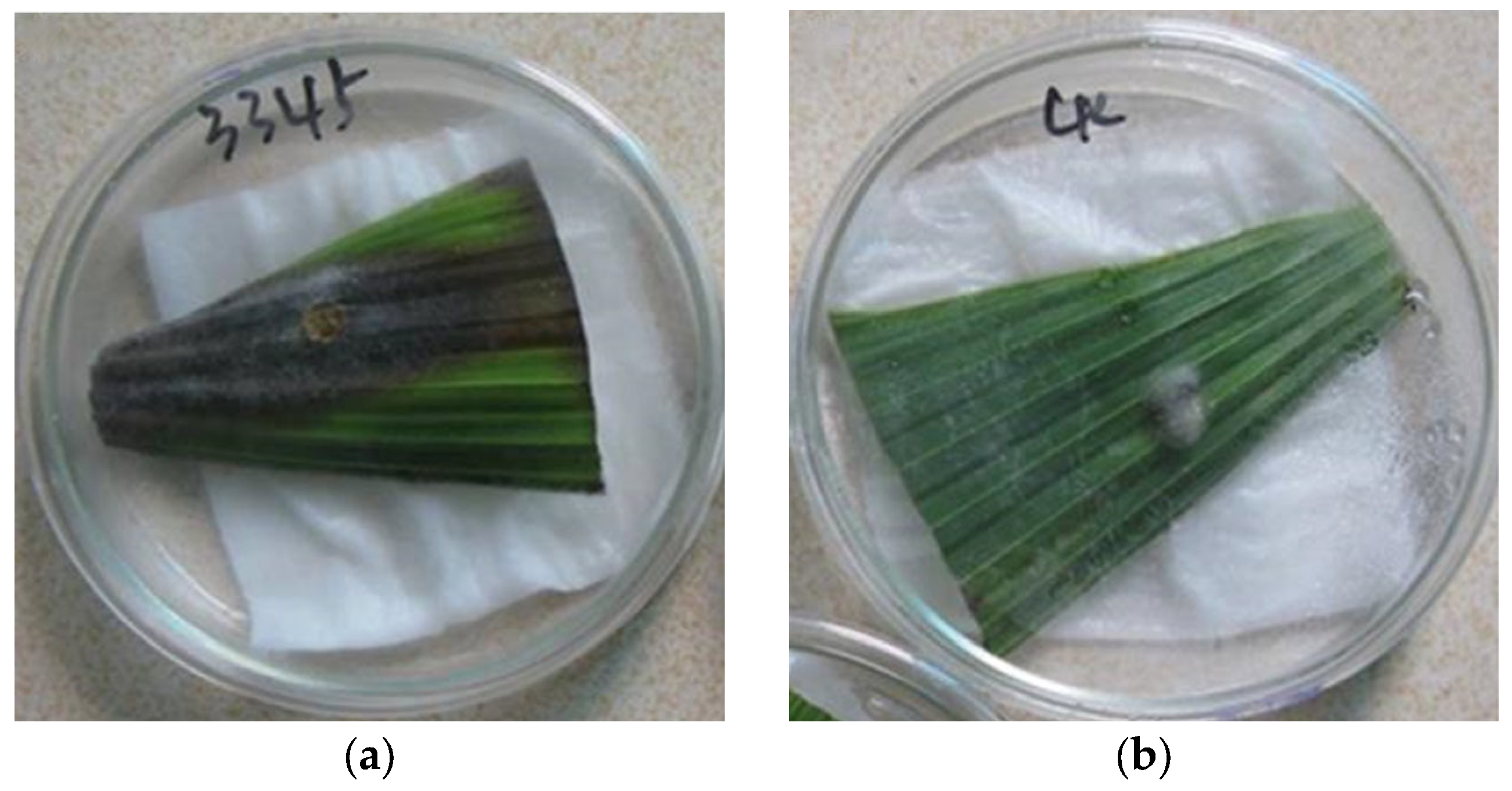
2.5. Pathogenicity Test

For the pathogenicity test, a 2-year-old healthy leaf was wounded with a fine needle and inoculated with mycelial plugs (5 mm diameter plugs). Another set of leaves were treated with pure PDA plugs as a control. All leaves were placed in sterile Petri dishes and subsequently transferred into growth chamber at 28 °C and 90% relative humidity. Three biological replicates were established for both experimental and control groups and the pathogenicity test was repeated four times.

2.6. Effects of pH and Temperature on Mycelial Growth In Vitro

The pathogen isolate was grown on a standard PDA medium (pH = 5.6) and used for assessing the effect of temperature on radial growth. Temperatures ranged from 5 to 35 °C at intervals of 5 °C. The pH effects on the radial growth were investigated on modified PDA medium at different pH values ranging from 4.0 to 8.0, adjusted using McIlvaine buffers at 28 °C in darkness [19,31]. A 5 mm PDA plug of mycelium of isolate was transferred to the center of medium plates for radial growth measurements. Four replicate observations were performed for each temperature and pH condition tested. The colony diameter was measured after 7 days of incubation and the isolates were also scored for pigmentation of the mycelia.
DNA from parallel isolate samples was extracted with a modified Fungal DNA Midi Kit (Omega Bio-Tek, Inc., Norcross, GA, USA). DNA derived from TUB2, GAPDH, CHS-1, ITS, CAL, and ACT loci was amplified using primers specific to conserved fungal sequences (Table A1). Each amplification reaction included 25 µL of Dream Taq Green PCR Master MIX (Thermo Fisher Scientific, Waltham, MA, USA), 5 µL (10 µM) of each primer set, and 14 µL of ddH2O in a final volume of 50 µL. The PCR program for GAPDH, ACT, CAL, and TUB2 included a denaturation step at 94 °C for 2 min, followed by 35 cycles at 94 °C for 45 s, 60 °C for 45 s, and 72 °C for 1 min, and a final cycle at 72 °C for 10 min. The PCR program for the ITS region included a 2 min denaturing step at 94 °C followed by 34 cycles at 94 °C for 1 min, 55 °C for 30 s, and 72 °C for 1 min, and a final cycle of 10 min at 72 °C, respectively [17,32,33]. All PCR reactions were performed on a Gene Amp PCR System 9700 (Applied Biosystems, Foster City, CA, USA). Amplified products were sequenced by Sangon Biotech Co., Ltd. (Shanghai, China).

2.7. Reponses to Chemical Compounds In Vitro

All fungicides, except for Polyoxin formulation (31–34%, technical grade [TG]), which was obtained from Kaken Pharmaceutical Co., Ltd. (Tokyo, Japan), were purchased from China, and included pyraclostrobin formulation (98%, TG) from ADAMA Huifeng Co., Ltd. (Jiangsu, China), tebuconazole formulation (95%, TG) from Bayer Aktiengesellschaf Co., Ltd. (Leverkusen, Germany), thiram formulation (97.5%, TG) from Qingdao Xinrun Biological Technology Co., Ltd. (Qingdao, China), prochloraz formulation (95%, TG) from Jiangsu Yangnong Chemical Co., Ltd. (Yangzhou, China), mancozeb formulation (96%, TG) from Limin Chemical Co., Ltd. (Xuzhou, China), chlorothalonil formulation (98%, TG) from Syngenta Crop Protection Co., Ltd. (Shanghai, China), as well as carbendazim formulation (80%, WP) from Shandong Yijia Agrochemical Co., Ltd. (Shouguang, China). The TG pesticides were first dissolved in dimethyl sulfoxide (DMSO), then diluted with sterile water containing 1‰ Tween-80 to achieve the desired concentrations for subsequent use. The liquid pesticide solutions were mixed thoroughly with a un-solidified, high-pressure sterilized PDA medium at a ratio of 1: 9 (v/v) in culture dishes. The final concentrations of each chemical reagent in the culture dishes were set to 100, 50, 25, 12.5, 6.25, and 3.125 ppm of available ingredient [a.i.], respectively. After solidification, 5 mm mycelial disks of the pathogen isolate WC-OIL PALM were inoculated. Meanwhile, the PDA medium supplemented with the same concentration of DMSO served as the control. Each treatment was repeated in triplicate. The plates were incubated at 25 °C for 7 days in the dark and the diameter of the fungal colony was measured to calculate the pathogen’s inhibition rate to determine the effective concentration for 50% inhibition (EC50), and when the lethal concentration for 90% (LC90) of mycelial viability occurred for each compound, we performed a regression analysis correlating the relative inhibition of mycelial growth with the Log10 various chemical preparations [34]. The experiment was conducted twice with three plates per concentration.

2.8. Statistical Analysis

Fisher’s Least Significant Difference (LSD) test was used to compare the discrepancies in the pathogen’s growth under varying temperature and pH conditions via ANOVA with IBM SPSS 26.0 (Statistical Package for the Social Science, SPSS, Chicago, IL, USA) (p ≤ 0.05) for statistical significance. The EC50 and LC90 values were calculated using the software Prism 9.0 (GraphPad Prism, Dotmatics Co., Boston, MA, USA) and Data Processing System20.05 (DPS, Hangzhou Ruifeng Information Technology Co., Hangzhou, China).

3. Results

3.1. Disease Symptoms

The causal agent of anthracnose in oil palm leaves is C. siamense, which causes polytopic disease attacking oil palm leaves and fruits. The disease occurred widely in oil palm plantations in Hainan Province. In the early stages of the disease, the spots on diseased leaves were transparent and had light brown spots. As the C. siamense continued to exert effects, brown circular and irregularly shaped lesions formed on the leaves, with distinct boundaries, a wheeling pattern, and the edge of the lesion was surrounded by apparent yellow halos (Figure 1). In the later stages of the disease, the lesions transformed to a grayish-white color and eventually became necrotic, the plants exhibiting such symptomatic manifestations were assigned a disease severity rating of grade 3. When the disease progressed to a severe stage, the leaves exhibited significant wilting, withered, and died, either partially or completely. During the disease-prone season in Hainan, light yellow or orange sporangial mounds were frequently observed on the affected plants.

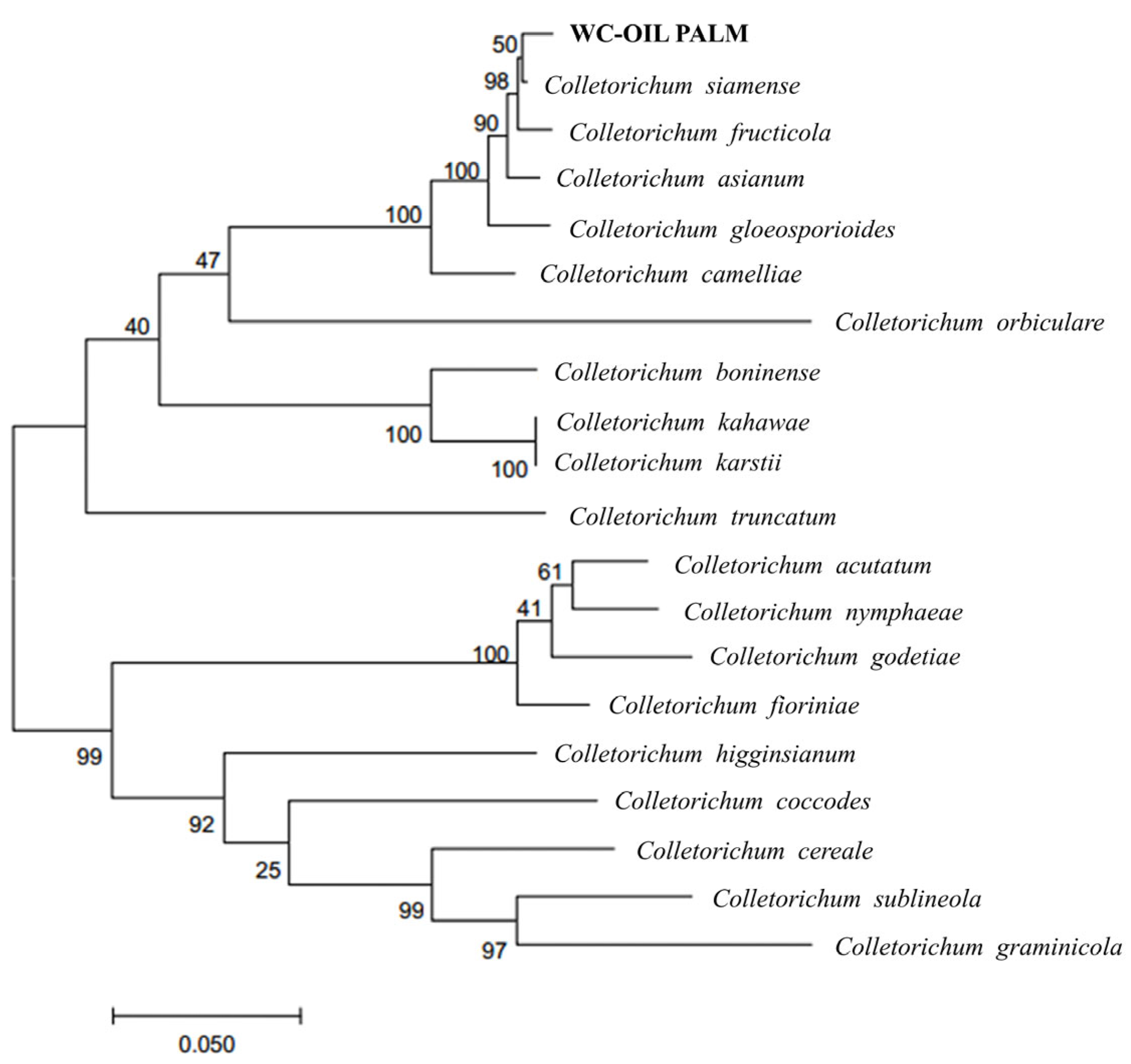
3.2. Species Identification and Phylogenic Analysis Within the Genus Colletotrichum

The PCR products of the genomic DNA of the pathogen WC-OIL PALM were sequenced by the Biotechnology Institute Co., Ltd. (Shanghai, China), and deposited in Genbank, with the following accession numbers: ITS gene (KX242310, 533 bp), CHS gene (KX242306, 299 bp), TUB2 gene (KX242307, 482 bp), ACT gene (KX242309, 282 bp), and GAPDH gene (KX242308, 277 bp). NCBI Blast comparison revealed that the similarity between these genes and Colletotrichum siamense was 99–100%.
The ITS, GAPDH, ACT, and TUB2 sequences from the Colletotrichum genus with specific sources were downloaded from GenBank (Table 1). The relevant sequences were concatenated in the order of ITS-GAPDH-ACT-TUB2, and the phylogenetic tree was constructed using the Tamura–Nei model and maximum likelihood method [35]. The percentage of related taxonomic groups clustered together is shown on the branches. In the pairwise distance matrix estimated by the Maximum Composite Likelihood (MCL) method, the Neighbor-Join and BioNJ algorithms were used to automatically obtain an initial tree for the heuristic search [36]. Then, the topology structure with the highest log-likelihood values was selected. The tree was drawn proportionally, with branch lengths measured by the number of substitutions at each site. The analysis involved 20 nucleotide sequences, and codon positions included 1 + 2 + 3 + non-coding. There was a total of 2105 analyzed sites in the final dataset. Phylogenetic tree analysis results showed that the identified pathogen clustered with C. siamense in the same branch (Figure 2).

3.3. Morphology Characterization

C. siamense was cultivated on PDA medium at 25 °C for 5 days. Initially, the colonies appeared white and turned to a deep gray hue after the incubation period, exhibiting a circular morphology with smooth margins and a velvety surface texture (Figure 3a). The conidia were transparent, asexual, and exhibited a curved or slightly curved shape, with a rounded apex and a truncated base. Microscopic analysis revealed that the size of the conidia ranged from 9.3 to 18.8 μm (length) × 2.9 to 5.7 μm (width) (n = 100). The acervulus were circular and black, densely populated with numerous conidia, and the conidiophore clusters were closely arranged around the conidia without any branching (Figure 3b). The pathogen was successfully reisolated and confirmed as C. siamense via morphological and molecular analysis [37,38].

3.4. The Result of Pathogenicity Test

Pathogenicity testing showed that the selected representative strain, WC-OIL PALM, caused disease in 100% of inoculated oil palm leaves, while the control leaves remained healthy (Figure 4a). The cultivation conditions in the light incubator were set at 25 °C and 90% humidity, and the leaves began showing disease symptoms 5 days after inoculation. The initial symptoms were small circular spots centered on the injury, and the lesion gradually increased over time. After 10 days, the lesion color changed to brown (Figure 4b), and the symptoms were consistent with natural field symptoms. The re-isolation of the pathogen from infected lesions yielded Colletotrichum siamense, consistent with Koch’s postulates.

3.5. Effect of Various Environmental Conditions on the Mycelial Growth of C. siamense

The results indicated that the pathogen C. siamense showed the ability to grow within a certain range of temperatures and pH values. The pathogen demonstrated remarkable vitality between 20 °C and 35 °C. Significant differences were observed in the daily growth diameter of the fungi over a 7-day observation period within this temperature range (p ≤ 0.05). In particular, C. siamense exhibited the highest growth rate at a temperature of 30 °C, and its mycelial diameter on the seventh day was twice that of the mycelium under 20 °C cultivation conditions; however, when C. siamense was cultured at temperatures of 5 °C, 10 °C, 37 °C, and 40 °C, its growth in the Petri dish was significantly inhibited (Figure 5a). At ambient temperatures of 5 °C and 10 °C, while there was a significant difference in the 7-day growth mycelial diameter compared to earlier days (p ≤ 0.05), their diameters still had not exceeded 10 mm and 20 mm, respectively. At 37 °C, the final-day diameter of the mycelium was significantly smaller than the initial diameter. The growth of C. siamense remained relatively stable, with no significant changes at a cultivation condition at 40 °C (p > 0.05). When the pH of the culture environment ranged from 5.0 to 8.0, the growth of C. siamense was not significantly inhibited. Significant differences were observed in the daily growth mycelial diameters within the pH range of 5.0–8.0 (p ≤ 0.05). On the 7th day, the mycelial diameter of the pathogen was less than 80 mm at cultivation at pH 5.0. At pH 6.0, 7.0, and 8.0, the mycelial diameter of the pathogen reached nearly 80 mm (Figure 5b).

3.6. Reponses of C. siamense to Chemical Compounds In Vitro

Eight compounds were tested at different gradient concentrations (100, 50, 25, 12.5, 6.25, and 3.125 ppm) to evaluate their inhibitory effects on thye mycelial growth of C. siamense, and both the EC50 and the LC90 data were derived from assessing the inhibition of the pathogen via various fungicides (Table 2). After 3-day post-exposure, the prochloraz exhibited a more remarkable inhibitory effect on the mycelial growth of C. siamense compared to the other fungicides. Meanwhile, pyraclostrobin, tebuconazole, and carbendazim also exhibited high bioactivity against C. siamense (EC50, 0.10–0.35 µg/mL). By contrast, the EC50 value of polyoxin B, thiram, and chlorothalonil exceeded 10 µg/mL (EC50, 10.10–15.54 µg/mL); only mancozeb demonstrated moderate efficacy on the pathogen (EC50 of 8.75 µg/mL). Furthermore, the rankings of the LC90 and EC50 values for the tested chemical preparations were different. Thiram stood out, with the highest EC50 value of the various chemical compounds, showcasing weaker antifungal activity against the pathogen in comparison to fungicide polyoxin B and chlorothalonil, but the LC90 values for the aforementioned pesticides all exceeded 300 µg/mL (LC90, 309.95–5857.70 µg/mL). Among the eight fungicides, C. siamense exhibited the highest sensitivity to the effects of prochloraz, carbendazim, and tebuconazole (LC90, 0.19–1.24 µg/mL), and displayed a relatively moderate response to the influence of mancozeb (LC90 of 64.58 µg/mL), which was consistent with the EC50 measurement results.

4. Discussion

The genus Colletotrichum includes diverse fungal species that cause diseases in a wide range of plant hosts. In the current study, leaf spot symptoms associated with Colletotrichum species were observed on sweet cherry, luffa sponge gourd, and tobacco [39,40,41]. Nearly a decade ago, C. siamense was isolated from Camellia sp. as a pathogen in China [32]. Even now, C. siamense has been reported to cause tea anthracnose, fruit rot of Capsicum spp., and rubber leaf spot disease [42,43,44]. In this study, the isolate from oil palm, in Hainan province, southern China, was identified as C. siamense through morphological characteristic and phylogenetic analysis. Several findings echo those of prior studies, which may be attributed to the specific crop species, as well as the geographical environment [45]. To the best of our knowledge, this was the first report of C. siamense causing leaf spot disease of oil palm in China.
Detailed morphology studies revealed the main characteristics of C. siamense, including colony colors, shapes, and sizes of the conidia. According to previous research, during the cultivation of C. siamense, which was isolated from Parthenocissus semicordata on PDA medium, the colony color progressively changed from white to gray. This phenomenon assisted us in identifying the strain [46]. C. siamense does not generate ascospores directly, and the mycelial growth is slower than that of C. fructicola, C. sojae, C. plurivorum, C. ovataense, C. cigarro, and C. gloeosporioides s.s. when cultured on PDA. Moreover, C. siamense produces smaller conidia than those of most other Colletotrichum species [38]. For example, the conidia of C. gloeosporioides, isolated from Atractylodes ovata and Areca catechu L., were found to be longer than those of C. siamense [47]. These results further prove that multiple morphological characteristics contribute to distinguishing C. siamense from other Colletotrichum species.
The identification of Colletotrichum species has been a major challenge over the years. Therefore, to accurately identify Colletotrichum species, it is necessary to conduct phylogenetic analysis of concatenated sequences utilizing various loci. Besides the extensive use of ITS regions for fungal molecular identification in the current study, we conducted a concatenated phylogenetic analysis using GAPDH, CHS, ACT, and TUB2 gene sequences to identify C. siamense. To identify other distinct Colletotrichum species, it is essential to amplify specific regions in biomolecular experiments. For instance, to identify C. fructicola, the partial mating type (Mat 1–2) gene region (ApMat) can be utilized. Meanwhile, the Histone H3 (HIS) gene region serves as a useful marker for identifying C. acutatum [40,48]. Notably, a loop-mediated isothermal amplification (LAMP) method has been developed, which enables the direct identification of plant diseases caused by C. siamense at varying severity levels by amplifying the CAL gene, using highly specific primers with a sensitivity of 1 pg/μL. This represents a 10-fold increase in sensitivity compared to conventional PCR methods [43].
Environmental conditions play an important role in fungal growth, development, and adaptability [49]. In this study, we found that the optimal growth temperature for C. siamense was 30 °C, which was almost the same as for C. gloeosporioides (29.5 °C) isolated from papaya and C. aenigma (30 °C) isolated from Camellia sinensis [50,51]. This is because most Colletotrichum species produce fresh conidia quickly from acervuli, and then the level of appressoria formation gradually improves when the ambient temperature ranges from 10 to 30 °C [52]. The optimal growth temperature range for Colletotrichum siamense was determined to be 25–30 °C, which was consistent with the growth characteristics observed in C. siamense strains isolated from Viburnum odoratissimum [53]. When the ambient temperature surpassed 35 °C, the growth of the pathogen became inhibited, consequently leading to a reduction in the incidence of plant anthracnose caused by Colletotrichum spp. [54]. C. siamense exhibited the greatest mycelial growth diameter in neutral pH conditions in the current study. C. asianum and C. fructicola, the causative agents of mango (Mangifera indica L.) anthracnose, together with C. liriopes, which induces Zhi mu (Anemarrhena asphodeloides) anthracnose, flourish best in environments with pH 6.0. For C. spaethianum and C. gloeosporioides isolated from mango fruits infected with anthracnose, pH 4.0 is the most conducive to their growth. The above differences in physiological characteristics among species are likely attributable to the varying environmental conditions across different geographical regions; pH can regulate the secretion of pectate lyase and polygalacturonase, the pathogenic factors of C. gloeosporiides. The former can stimulate the activity of pathogenic enzymes in pathogens [55,56]. Thus, when sterilizing Colletotrichum species, pH regulators are commonly added to optimize the environment for further inhibiting the reproduction of target pathogens in soil, which creates a less favorable environment for the pathogen’s proliferation [57].
At the level of agricultural management strategies, the application of fungicides remains the most direct and effective method for enhancing crop yield and quality while managing the disease caused by Colletotrichum species in economic crops, including kidney bean, peach, and olive [58,59,60]. In this study, the sensitivity of C. siamense isolate to the eight commercial fungicides frequently used to control oil palm leaf spot disease was analyzed, and the pathogen in oil palm was highly sensitive to pyraclostrobin, tebuconazole, prochloraz, and carbendazim compared to the other four fungicides. Additionally, Li et al. [53] demonstrated that both difenoconazole and pyraclostrobin exhibited comparable efficacy to prochloraz in controlling C. siamense-induced plant disease, while previous studies have shown that C. siamense showed high resistance to the dicarboximide (DCF) fungicide iprodione and succinate dehydrogenase inhibitor (SDHI) fungicide fluopyram [61]. For the other Colletotrichum species, C. spaethianum and C. liriopes were effectively inhibited by pyraclostrobin and prochloraz, both of which exhibit an EC50 value in C. spaethianum that was less than 1 mg/L [56]. C. melonis isolated from apple flowers and fruitlets was more sensitive to tebuconazole than C. nymphaeae, with the range of the EC50 value being 0.077–0.116 µg/mL [62]. The varying responses of different Colletotrichum species to fungicides are influenced by multiple factors. Beyond geographical location and environmental conditions, the inherent characteristics of each strain play a crucial role. For instance, the higher resistance of C. siamense to carbendazim compared to prochloraz is associated with F200Y mutations in the β-tubulin 2 (TUB2) gene [61].

5. Conclusions

Based on the findings of the current research study, C. siamense exhibits multiple hosts and a broad distribution range. This study reports the first isolation of the pathogen from oil palm affected by leaf spot disease in China. Investigations into the effects of pH and temperature on the growth environment of C. siamense revealed that the pathogen proliferates rapidly at 30 °C and a pH of 7.0. Moreover, fungicides such as pyraclostrobin, tebuconazole, prochloraz, and carbendazim are found to effectively inhibit the growth and development of this pathogen. This research provides crucial guidance and reference for developing strategies to prevent and control oil palm leaf spot disease in various regions.

Author Contributions

Conceptualization, Z.W. and C.J.; methodology, H.L. and X.S.; software, Z.L.; validation, H.L; formal analysis, M.Z. and Y.C.; investigation, X.S. and Q.B.; resources, X.S.; data curation, Q.P.; writing—original draft preparation, H.L.; writing—review and editing, X.S.; visualization, Q.B.; supervision, X.S. and Q.B.; project administration, X.S.; funding acquisition, X.S. All authors have read and agreed to the published version of the manuscript.

Funding

This study was supported by the Hainan Provincial Department of Science and Technology, who funded this research work through the projects Innovation Platform for Academics of Hainan Province (No. YZPTZX202212) and Science and Technology special fund of Hainan Province (No. ZDYF2024XDNY242).

Data Availability Statement

The authors confirm that the data supporting the findings of this study are available within the article.

Conflicts of Interest

The authors declare no conflicts of interest.

Abbreviations

The following abbreviations are used in this manuscript:
EC50Effective concertation, the minimum concentration of a chemical substance required to induce 50% inhibition of the growth of C. siamense after 3 days of in vitro exposure
LC90Lethal concentration, the minimum concentration of a chemical substance required to induce 90% mortality of C. siamense after 3 days of in vitro exposure
95%CI95% confidence interval for the LC90 (EC50)

Appendix A

Table A1. Primers used in this study for PCR amplification.
Gene NameGene FunctionPrimer NamePrimer Sequence
ITSTaxonomic identification and phylogenetic reconstructionITS1TCCGTAGGTGAACCTGCGG
ITS4TCCTCCGCTTATTGATATGC
GAPDHHousekeeping geneGDFGCCGTCAACGACCCCTTCATTGA
GDRGGGTGGAGTCGTACTTGAGCATGT
CHS-1Mediating chitin biosynthesisCHS-79FTGGGGCAAGGATGCTTGGAAGAAG
CHS-345RTCGAAGAACCATCTGTGAGAGTTG
ACTCommon reference geneACT-512FATGTGCAAGGCCGTTTCGC
ACT-783RTACGAGTCCTTCTGGCCCAT
CALModulating Ca2+-dependent signaling cascadesCL1CGATTCAAGGAGGCCTTCTC
CL2CCTTCTGCATCATGAGCTGGAC
TUB2Builds microtubules for cell division and structureBt2aGGTAACCAAATCGGTGCTGCTTTC
Bt2bACCCTCAGTGTAGTGACCCTTGGC
Table A2. Locus reference genomic sequences downloaded from NCBI.
Gene NameGene FunctionPrimer NamePrimer Sequence
CALModulating Ca2+-dependent signaling cascadesCALDF1AGCAAGTCTCCGAGTTCAAGG
CALDR1CTTCTGCATCATCAYCTGGACG
TEF-1αHousekeeping geneEF-1HaATGGGTAAGGAAGACAAGAC
EF-2TbGGAAGTACCAGTGATCATGTT
EFdFAAGGAYGNCARACYCGNGARCAYGC
EF1-2218RATGACACCRACRGCRACRGTYTG

References

  1. Wei, L.; John Martin, J.J.; Zhang, H.; Zhang, R.; Cao, H. Problems and prospects of improving abiotic stress tolerance and pathogen resistance of oil palm. Plants 2021, 10, 2622. [Google Scholar] [CrossRef] [PubMed]
  2. Nurrochmat, D.R.; Boer, R.; Ardiansyah, M.; Immanuel, G.; Purwawangsa, H. Policy forum: Reconciling palm oil targets and reduced deforestation: Landswap and agrarian reform in Indonesia. Forest Policy Econ. 2020, 119, 102291. [Google Scholar] [CrossRef]
  3. Bentivoglio, D.; Finco, A.; Bucci, G. Factors affecting the indonesian palm oil market in food and fuel industry: Evidence from a time series analysis. Int. J. Energy Econ. Policy 2018, 8, 49–57. [Google Scholar]
  4. Workman, D. Palm Oil Exports by Country. Available online: https://www.worldstopexports.com/palm-oil-exports-by-country/ (accessed on 23 August 2024).
  5. Wang, R.; Lee, K.E.; Mokhtar, M.; Goh, T.L. The challenges of palm oil sustainable consumption and production in China: An institutional theory perspective. Sustainability 2022, 14, 4856. [Google Scholar] [CrossRef]
  6. Zeng, X.H.; Pan, D.L.; Liu, Z.; Chen, J.M.; Lin, W.F. Evaluation of cold tolerant high yielding oil palm germplasm in Guangdong Province of south China, a northern tropical region. J. Oil Palm Res. 2016, 28, 266–280. [Google Scholar] [CrossRef]
  7. Zhao, Q.; Yu, L.; Li, X.; Xu, Y.; Du, Z.; Kanniah, K.; Li, C.; Cai, W.; Lin, H.; Peng, D.; et al. The expansion and remaining suitable areas of global oil palm plantations. Glob. Sustain. 2024, 7, e9. [Google Scholar] [CrossRef]
  8. Shen, H.; Zheng, L.; Zhang, J.; Lin, B.; Xiaoming, P.; Qin, X.; Li, J.; Xie, C. First Report of Pestalotiopsis microspora Causing Leaf Spot of Oil Palm (Elaeis guineensis) in China. Plant Dis. 2014, 98, 140722152922009. [Google Scholar] [CrossRef]
  9. Zheng, L.; Xi, P.; Tu, J.-J.; Chen, X.; Li, J.; Qin, X.; Shen, H.; Xie, C.-P. First report of Phoma herbarum causing leaf spot of oil palm (Elaeis guineensis) in China. Plant Dis. 2016, 101, 629. [Google Scholar] [CrossRef]
  10. Escalante, M.G.; Damas, D.; Márquez, D.; Gelvez, W.; Chacón, H.; Díaz, A.; Moreno, B. Diagnosis and evaluation of pestalotiopsis, and insect vectors, in an oil palm plantation at the south of Maracaibo Lake, Venezuela. Bioagro 2010, 22, 211–216. [Google Scholar]
  11. Penet, L.; Barthe, E.; Alleyne, A.; Blazy, J.M. Disease risk perception and diversity of management strategies by farmers: The case of anthracnose caused by Colletotrichum gloeosporioides on water yams (Dioscorea alata) in Guadeloupe. Crop Prot. 2016, 88, 7–17. [Google Scholar] [CrossRef]
  12. Ji, Y.; Li, X.; Gao, Q.-H.; Geng, C.; Duan, K. Colletotrichum species pathogenic to strawberry: Discovery history, global diversity, prevalence in China, and the host range of top two species. Phytopathol. Res. 2022, 4, 42. [Google Scholar] [CrossRef]
  13. Chang, L.; Li, Y.; Gao, Z.; Bonello, P.; Cleary, M.; Munck, I.A.; Santini, A.; Sun, H. Novel pathogen–plant host interaction: Colletotrichum jiangxiense and Fraxinus americana L. (white ash) in a sentinel garden in China. Plants 2023, 12, 4001. [Google Scholar] [CrossRef] [PubMed]
  14. Guo, Z.; Luo, C.X.; Wu, H.J.; Peng, B.; Kang, B.S.; Liu, L.M.; Zhang, M.; Gu, Q.S. Colletotrichum species associated with anthracnose disease of watermelon (Citrullus lanatus) in China. J. Fungi 2022, 8, 790. [Google Scholar] [CrossRef]
  15. Dowling, M.; Peres, N.; Villani, S.; Schnabel, G. Managing Colletotrichum on fruit crops: A “complex” challenge. Plant Dis. 2020, 104, 2301–2316. [Google Scholar] [CrossRef]
  16. Montri, P.; Taylor, P.W.J.; Mongkolporn, O. Pathotypes of Colletotrichum capsici, the causal agent of Chili anthracnose, in Thailand. Plant Dis. 2009, 93, 17–20. [Google Scholar] [CrossRef]
  17. Lin, Z.; Wei, J.; Zhang, M.; Xu, S.; Powell, C.A. Identification and characterization of a new fungal pathogen causing twisted leaf disease of sugarcane in China. Plant Dis. 2015, 99, 325–332. [Google Scholar] [CrossRef]
  18. Cannon, P.F.; Damm, U.; Johnston, P.R.; Weir, B.S. Colletotrichum–current status and future directions. Stud. in Mycol. 2012, 73, 181–213. [Google Scholar] [CrossRef]
  19. Chen, J.; Cong, L.; Zhou, R.; Li, Z.; Piao, J.; Hao, N. Identification and characterization of Sclerotium delphinii causing southern blight on Aconitum kusnezoffii in northeast China. Plant Dis. 2022, 8, 106. [Google Scholar] [CrossRef]
  20. Ahmed, S.A.; Nagadi, S.A.; Abo-Elyousr, K.A.M.; El-Fawy, M.M. Mitigating helminthosporium leaf spot disease in sesame: Evaluating the efficacy of castor essential oil and sodium bicarbonate on disease management and crop yield enhancement. J. Plant Pathol. 2024, 106, 683–694. [Google Scholar] [CrossRef]
  21. Mishra, R.K.; Mishra, M.; Bohra, A.; Naik SJ, S.; Saabale, P.; Kumar, K.; Patil, P.G.; Srivastava, D.; Singh, N.P. First report of Fusarium equiseti causing wilt of Cajanus scarabaeoides, a wild relative of Pigeonpea, in India. Plant Dis. 2021, 105, 2735. [Google Scholar] [CrossRef]
  22. Innis, M.A.; Gelfand, D.H.; Sninsky, J.J.; White, T.J. PCR protocols: A guide to methods and applications. Trends Biotechnol. 1990, 8, 335. [Google Scholar] [CrossRef]
  23. Glass, N.L.; Donaldson, G.C. Development of primer sets designed for use with the PCR to amplify conserved genes from filamentous ascomycetes. Appl. Environ. Microb. 1995, 61, 1323–1330. [Google Scholar] [CrossRef] [PubMed]
  24. Templeton, A.R.; Crandall, K.A.; Sing, C.F. A cladistic analysis of phenotypic associations with haplotypes inferred from restriction endonuclease mapping and DNA sequence data. III. cladogram estimation. Genetics 1992, 132, 619–633. [Google Scholar] [CrossRef] [PubMed]
  25. Weir, B.S.; Johnston, P.R.; Damm, U. The Colletotrichum gloeosporioides species complex. Stud. Mycol. 2012, 73, 115–180. [Google Scholar] [CrossRef]
  26. Moral, J.; Agustí-Brisach, C.; Raya, M.C.; Jurado-Bello, J.; López-Moral, A.; Roca, L.F.; Chattaoui, M.; Rhouma, A.; Nigro, F.; Sergeeva, V.; et al. Diversity of Colletotrichum species associated with olive anthracnose worldwide. J. Fungi 2021, 7, 741. [Google Scholar] [CrossRef]
  27. Zhang, M.; Forte-Perri, V.; Sun, W.; Tang, L.; Huang, S.; Guo, T.; Chen, X.; Li, Q. Identification and observation of infection processes of Colletotrichum species associated with persimmon anthracnose in Guangxi, China. Plant Dis. 2023, 107, 1670–1679. [Google Scholar] [CrossRef]
  28. Yağmur, A.; Demir, S.; Canpolat, S.; Rezaee Danesh, Y.; Farda, B.; Djebaili, R.; Pace, L.; Pellegrini, M.J.P. Onion Fusarium basal rot disease control by arbuscular mycorrhizal fungi and Trichoderma harzianum. Plants 2024, 13, 386. [Google Scholar] [CrossRef]
  29. Hall, B.G. Building phylogenetic trees from molecular data with MEGA. Mol. Biol. Evolution. 2013, 30, 1229–1235. [Google Scholar] [CrossRef]
  30. Senanayake, I.; Rathnayaka, A.; Sandamali, D.; Calabon, M.; Gentekaki, E.; Lee, H.; Pem, D.; Dissanayake, L.; Wijesinghe, S.; Bundhun, D.; et al. Morphological approaches in studying fungi: Collection, examination, isolation, sporulation and preservation. Mycosphere 2020, 11, 2678–2754. [Google Scholar] [CrossRef]
  31. Dhaver, P.; Pletschke, B.; Sithole, B.; Govinden, R. Isolation, screening, preliminary optimisation and characterisation of thermostable xylanase production under submerged fermentation by fungi in Durban, South Africa. Mycology 2022, 13, 271–292. [Google Scholar] [CrossRef]
  32. Liu, F.; Weir, B.S.; Damm, U.; Crous, P.W.; Wang, Y.; Liu, B.; Meng, Z.; Cai, L. Unravelling Colletotrichum species associated with Camellia: Employing ApMat and GS loci to resolve species in the C. gloeosporioides complex. Persoonia Mol. Phylogeny Evol. Fungi 2015, 35, 63–86. [Google Scholar] [CrossRef] [PubMed]
  33. Mo, J.; Zhao, G.; Li, Q.; Solangi, G.S.; Tang, L.; Guo, T.; Huang, S.; Hsiang, T. Identification and characterization of Colletotrichum species associated with mango anthracnose in Guangxi, China. Plant Dis. 2018, 102, 1283–1289. [Google Scholar] [CrossRef] [PubMed]
  34. Abdel-Kawy, M.A.; Michel, C.G.; Kirollos, F.N.; Hussien, R.A.A.; Al-Mahallawi, A.M.; Sedeek, M.S. Chemical composition and potentiation of insecticidal and fungicidal activities of Citrus trifoliata L. fruits essential oil against Spodoptera littoralis, Fusarium oxysporum and Fusarium solani via nano-cubosomes. Nat. Prod. Res. 2021, 35, 2438–2443. [Google Scholar] [CrossRef]
  35. Riolo, M.; Aloi, F.; Pane, A.; Cara, M.; Cacciola, S.O. Twig and shoot dieback of citrus, a new disease caused by Colletotrichum species. Cells 2021, 10, 449. [Google Scholar] [CrossRef]
  36. Pandey, A.K.; Samota, M.K.; Tanti, A.J.; Babu, A. Trichoderma reesei induces defense-related biochemical markers associated with resistance to Fusarium dieback in tea crop. Biol. Control 2023, 180, 105200. [Google Scholar] [CrossRef]
  37. Lee, D.M.; Hassan, O.; Chang, T. Identification, characterization, and pathogenicity of Colletotrichum species causing anthracnose of peach in Korea. Mycobiology 2020, 48, 210–218. [Google Scholar] [CrossRef]
  38. Hassan, O.; Kim, J.S.; Romain, B.B.N.D.; Chang, T. An account of Colletotrichum species associated with anthracnose of Atractylodes ovata in South Korea based on morphology and molecular data. PLoS ONE 2022, 17, e0263084. [Google Scholar] [CrossRef]
  39. Wang, Y.R.; Hu, Z.; Zhong, J.; Chen, Y.; Zhu, J.Z. First report of Colletotrichum cliviicola causing leaf spot on tobacco (Nicotiana tabacum) in Hunan Province of China. Plant Dis. 2022, 106, 316. [Google Scholar] [CrossRef]
  40. Zhou, Y.; Zhang, W.; Li, Y.; Ji, S.; Li, X.; Hyde, K.D.; Zhang, K.; Phillips, A.J.L.; Manawasinghe, I.S.; Yan, J. Identification and characterization of Colletotrichum species associated with cherry leaf spot disease in China. Plant Dis. 2023, 107, 500–513. [Google Scholar] [CrossRef]
  41. Li, P.; Zhu, J.Z.; Li, X.G.; Zhong, J. Identification and characterization of Colletotrichum fructicola and Colletotrichum siamense causing anthracnose on luffa sponge gourd in China. Plant Dis. 2022, 11, 1537. [Google Scholar] [CrossRef]
  42. Anuradha; Sharma, A.; Sood, S.; Badiyal, A.; Sood, T. Fruit rot of Capsicum spp.: Implications and management strategies. J. Hortic. Sci. Biotech. 2023, 98, 715–731. [Google Scholar] [CrossRef]
  43. Zou, H.Y.; Li, T.W.; Zhang, J.; Shao, H.J.; Kageyama, K.; Feng, W.Z. Rapid detection of Colletotrichum siamense from infected tea plants using filter-disc DNA extraction and loop-mediated isothermal amplification. Plant Dis. 2024, 108, 35–40. [Google Scholar] [CrossRef] [PubMed]
  44. Chen, L.; Xu, L.; Li, X.; Wang, Y.; Feng, Y.; Huang, G.J.A. The diseases and pests of rubber tree and their natural control potential: A bibliometric analysis. Agronomy 2023, 13, 1965. [Google Scholar] [CrossRef]
  45. Wang, Q.; Huang, D.; Tu, W.Y.; Ma, F.W.; Liu, C.H. Overexpression of auxin/indole-3-acetic acid gene MdIAA24 enhances Glomerella leaf spot resistance in apple (Malus domestica). Hortic. Plant J. 2024, 10, 15–24. [Google Scholar] [CrossRef]
  46. Fang, Y.; Zhao, P.; Wang, S.; Cui, X.; Zhang, T.; Ye, R.; Zeng, Y.; Zhang, Y. Colletotrichum siamense causing leaf spot on Parthenocissus semicordata in China. Crop Prot. 2024, 185, 106878. [Google Scholar] [CrossRef]
  47. Cao, X.; Xu, X.; Che, H.; West, J.S.; Luo, D. Eight Colletotrichum species, including a novel species, are associated with areca palm anthracnose in Hainan, China. Plant Dis. 2020, 104, 1369–1377. [Google Scholar] [CrossRef]
  48. Kennedy, A.H.; Schoch, C.L.; Marrero, G.; Brover, V.; Robbertse, B. Publicly available and validated DNA Reference sequences are critical to fungal identification and global plant protection efforts: A use-case in Colletotrichum. Plant Dis. 2022, 106, 1573–1596. [Google Scholar] [CrossRef]
  49. Gadd, G.M.; Fomina, M.; Pinzari, F. Fungal biodeterioration and preservation of cultural heritage, artwork, and historical artifacts: Extremophily and adaptation. Microbiol. Mol. Bio Rev. 2024, 88, e00200-00222. [Google Scholar] [CrossRef]
  50. Sandoval-Contreras, T.; Iñiguez-Moreno, M.; Garrido-Sánchez, L.; Ragazzo-Sánchez, J.A.; Narváez-Zapata, J.A.; Ascencio, F.; Calderón-Santoyo, M. Predictive model for the effect of environmental conditions on the postharvest development of Colletotrichum gloeosporioides strains isolated from papaya (Carica papaya L.). J. Food Protect 2020, 83, 1495–1504. [Google Scholar] [CrossRef]
  51. Lin, S.-R.; Lin, Y.-H.; Ariyawansa, H.A.; Chang, Y.-C.; Yu, S.-Y.; Tsai, I.; Chung, C.-L.; Hung, T.-H. Analysis of the pathogenicity and phylogeny of Colletotrichum species associated with brown blight of tea (Camellia sinensis) in Taiwan. Plant Dis. 2023, 107, 97–106. [Google Scholar] [CrossRef]
  52. Brugneti, F.; Rossini, L.; Drais, M.I.; Turco, S.; Mazzaglia, A. Effect of temperature on in vitro germination and growth of Colletotrichum fioriniae, a new emerging pathogen of olive fruits. Env. Microbiol. Rep. 2024, 16, e13275. [Google Scholar] [CrossRef] [PubMed]
  53. Li, H.; Liao, Y.-C.-Z.; Wan, Y.; Li, D.-W.; Zhu, L.-H. Colletotrichum siamense, a novel causal agent of Viburnum odoratissimum leaf blotch and its sensitivity to fungicides. J. Fungi 2023, 9, 882. [Google Scholar] [CrossRef] [PubMed]
  54. Iqra; ul Haq, I.; Waseem Khan Qadri, R.; Amrao, L.; Ijaz, S. Effect of environmental conditions (temperature and precipitation) on severity of guava die-back caused by Colletotrichum spp. under climatic conditions of Pakistan. J. Plant Pathol. 2022, 104, 179–190. [Google Scholar] [CrossRef]
  55. Tao, R.; Yang, B.; Lin, L.; Munir, S.; Li, Y.; Wang, X.; Huang, M. Biological characterization of emerging fungal pathogen Colletotrichum associated with mango (Mangifera indica L.) post-harvest anthracnose from Vietnam. Mol. Biol. Rep. 2024, 51, 557. [Google Scholar] [CrossRef]
  56. Wei, R.; Wang, R.; Li, Y.; Yue, M.; Ding, W. Identification, biological characteristics and fungicide sensitivity of Colletotrichum species that cause anthracnose on Anemarrhena asphodeloides in China. J. Phytopathol. 2022, 170, 373–381. [Google Scholar] [CrossRef]
  57. Delna, R.; Sharma, G.; Rawat, S. Impact of rice-husk biochar on Colletotrichum falcatum, the pathogen of sugarcane red rot disease. Indian Phytopathol. 2022, 75, 325–329. [Google Scholar] [CrossRef]
  58. Ishii, H.; Watanabe, H.; Yamaoka, Y.; Schnabel, G. Sensitivity to fungicides in isolates of Colletotrichum gloeosporioides and C. acutatum species complexes and efficacy against anthracnose diseases. Pestic. Biochem. Phys. 2022, 182, 105049. [Google Scholar] [CrossRef]
  59. Usman, H.M.; Tan, Q.; Karim, M.M.; Adnan, M.; Yin, W.-X.; Zhu, F.-X.; Luo, C.-X. Sensitivity of Colletotrichum fructicola and Colletotrichum siamense of peach in China to multiple classes of fungicides and characterization of pyraclostrobin-resistant isolates. Plant Dis. 2021, 105, 3459–3465. [Google Scholar] [CrossRef]
  60. Nawaz, H.H.; Manzoor, A.; Iqbal, M.Z.; Ansar, M.R.; Ali, M.; Muhammad Kakar, K.; Ali Awan, A.; Weiguo, M. Colletotrichum acutatum: Causal agent of olive anthracnose isolation, characterization, and fungicide susceptibility screening in Punjab, Pakistan. Plant Dis. 2023, 107, 1329–1342. [Google Scholar] [CrossRef]
  61. Karim, M.M.; Usman, H.M.; Tan, Q.; Hu, J.-J.; Fan, F.; Hussain, R.; Luo, C.-X. Fungicide resistance in Colletotrichum fructicola and Colletotrichum siamense causing peach anthracnose in China. Pestic. Biochem. Phys. 2024, 203, 106006. [Google Scholar] [CrossRef]
  62. Gelain, J.; Lykins, S.; Rosa, P.F.; Soares, A.T.; Dowling, M.; Schnabel, G.; May De Mio, L.L. Identification and fungicide sensitivity of Colletotrichum spp. from apple flowers and fruitlets in Brazil. Plant Dis. 2023, 107, 1183–1191. [Google Scholar] [CrossRef]
Figure 1. Symptoms of leaf spot disease on oil palm. (a) Early-stage symptoms of the disease and (b) late-stage symptoms of the disease.
Agronomy 15 01331 g001
Figure 2. Phylogenetic tree of the genus Colletotrichum constructed based on ITS-GAPDH-ACT-TUB2 sequence. The tree was constructed using maximum likelihood and bootstrap values were shown next to the branches.
Agronomy 15 01331 g002
Figure 3. Morphological characteristics of C. Siamense strain. (a) Front view of colony on PDA culture medium. (b) The micrograph of C. siamense.
Agronomy 15 01331 g003
Figure 4. The symptoms of isolated inoculation of C. Siamense on oil palm leaves. (a) Oil palm leaves inoculated with WC-OIL PALM mycelium (Experimental group). (b) Oil palm leaves inoculated only with PDA block (Control group).
Agronomy 15 01331 g004
Figure 5. Mycelial growth under different environmental conditions. (a) The effect of pH on the growth of C. siamense. (b) The effect of temperature on the growth of C. siamense. Different superscripts letters mean significantly different (p < 0.05).
Agronomy 15 01331 g005
Table 1. Origin of 19 kinds of Colletotrichum species.
SpeciesOriginGenBank Accession Number
ITSGAPDHACTTUB2
C. siamenseChinaMG830351MG830377MG830429MG830326
C. boninenseJapanJQ005153JQ005240JQ005501JQ005588
C. asianumIndiaJQ894679JQ894623JQ894545JQ894601
C. gloeosporioidesItalyJX010152JX010056JX009531JX010445
C. camelliaeChinaKJ955094KJ954795KJ954376KJ955243
C. fructicolaChinaMH370509MH370516MH370530MH370551
C. kahawaeColombiaJQ005215JQ005302JQ005563JQ005649
C. sublineolaChinaMF405438KY038879KY038877KY038881
C. graminicolaBrazilMF803827MF803828MF803829MF803830
C. cerealeChinaJX625161KC843518KC843535JX625188
C. karstiiColombiaJQ005215JQ005302JQ005563JQ005649
C. truncatumBrazilMG543285MG543284MG543286MG543283
C. coccodesThe NetherlandsHM171679HM171673HM171667JX546873
C. higginsianumChinaMF033888MF033889MF033892MF033895
C. orbiculareIndiaKP898982KP898936KP899005KP899052
C. godetiaeItalyKY293406KY293404KY293402KY293407
C. fioriniaeUKJQ948344JQ948674JQ949665JQ949995
C. acutatumAustraliaJQ005776JQ948677JQ005839JQ005860
C. nymphaeaeRepublic of KoreaLC428839LC428842LC428840LC428841
Table 2. The in vitro inhibitory effects of various fungicides on C. siamense.
CompoundEC50 (95%CI)r2 *LC90 (95%CI)r #
Polyoxin B12.74 (4.91, 33.98)0.97775857.70
(1629.00, 45075.08)
0.9955
Pyraclostrobin0.35 (0.20, 0.63)0.9751109.56
(57.12, 246.85)
0.9962
Tebuconazole0.10 (0.06, 0.18)0.97851.24
(1.02, 1.53)
0.9532
Thiram15.54 (9.80, 24.88)0.9938309.95
(171.19, 708.54)
0.9911
Prochloraz0.02 (0.01, 0.04)0.96830.19
(0.15, 0.23)
0.9554
Mancozeb8.75 (5.03, 15.03)0.990964.58
(44.06, 107.28)
0.9902
Chlorothalonil 10.10 (2.97, 33.61)0.9557402.70
(193.63, 1200.55)
0.9906
Carbendazim0.1381 (0.11, 0.18)0.99380.91
(0.72, 1.14)
0.9496
* Correlation coefficient, utilized to assess the goodness of non-linear fitting for EC50. # Correlation coefficient, utilized to assess the goodness of non-linear fitting for LC90.
Disclaimer/Publisher’s Note: The statements, opinions and data contained in all publications are solely those of the individual author(s) and contributor(s) and not of MDPI and/or the editor(s). MDPI and/or the editor(s) disclaim responsibility for any injury to people or property resulting from any ideas, methods, instructions or products referred to in the content.

