Article

CRISPR/Cas9-Mediated Knockout of PxPGRP4 Influences Midgut Microbial Homeostasis and Immune Responses in Plutella xylostella

1
State Key Laboratory of Green Pesticide, “Belt and Road” Technology Industry and Innovation Institute for Green and Biological Control of Agricultural Pests, College of Plant Protection, South China Agricultural University, Guangzhou 510642, China
2
Department of Entomology, College of Plant Protection, Yangzhou University, Yangzhou 225009, China
*
Author to whom correspondence should be addressed.
These authors contributed equally to this work.
Agronomy 2025, 15(6), 1294; https://doi.org/10.3390/agronomy15061294 (registering DOI)
Submission received: 20 March 2025 / Revised: 24 April 2025 / Accepted: 23 May 2025 / Published: 25 May 2025

Abstract

:
Peptidoglycan recognition proteins (PGRPs) are essential for innate immune recognition and regulation from insects to mammals. However, the specific role of PGRPs in responding to Bacillus thuringiensis (Bt) infection and maintaining midgut microbial homeostasis in Plutella xylostella remains poorly understood. In this study, we identified and characterized a PGRP gene from P. xylostella, designated PxPGRP4. The spatiotemporal expression analysis revealed that PxPGRP4 is predominantly expressed in the midgut of naïve larvae and at adult stages. A homozygous mutant strain featuring a four-base pair nucleotide deletion was successfully generated through CRISPR/Cas9-mediated knockout of PxPGRP4. The bioassay results indicated that the susceptibility of P. xylostella larvae to Cry1Ac protoxin was significantly increased by the loss of PxPGRP4 expression. Furthermore, 16S rRNA sequencing and qPCR analysis revealed that the PxPGRP4 mutants exhibited a significantly reduced total bacterial load and altered microbiota composition in the midgut compared to the wild-type strain, with a shift in the dominant bacterial family from Enterobacteriaceae to Enterococcaceae. Additionally, the knockout of PxPGRP4 resulted in significant alterations in the expression of midgut immune-related genes. These findings highlight the crucial role of PxPGRP4 as a modulator of midgut microbiota and immune responses and provide valuable insights into Bt resistance management.

1. Introduction

Bacillus thuringiensis (Bt), a gram-positive bacterium, produces crystalline (Cry) protein toxins such as Cry1Ac protoxin, which bind to midgut epithelial receptors in susceptible insects, forming pores that disrupt gut integrity [1]. Bt exhibits targeted pathogenicity toward various orders of insects, including Lepidoptera, Diptera, and Coleoptera, while remaining nontoxic to non-target organisms such as humans and animals [1]. Consequently, it has been employed as a biological pesticide and widely integrated into transgenic crops expressing various Bt toxin genes [2,3]. The extensive cultivation of transgenic Bt crops has resulted in a significant reduction in the application of chemical insecticides, enhanced natural pest control, and increased agricultural profitability [4]. However, the effectiveness of pest control using Bt-based products is diminished due to the evolution of insect resistance in field conditions [5]. Bt endotoxins interact with epithelial protein receptors, leading to pore formation and subsequent gut cell lysis [3,6]. During this process, mutations in receptor genes and alterations in the gut microenvironment—encompassing gut immunity and microbiota—significantly influence Bt toxicity [7,8,9].
The gut microbiota of insects significantly influences various host life processes, including immunity, reproduction, metabolism, development, and resistance to insecticides [10,11,12,13,14,15]. The composition of the microbial community within the gastrointestinal tract of insects is shaped by multiple factors, such as the host’s diet, environmental conditions, and the developmental stage (egg, larva, pupa, or adult) of the insect [16,17]. Evidence suggests that gut commensal bacteria play a regulatory role in Bt pathogenicity. For instance, an increase in midgut microbiota load enhances immune priming and tolerance to Bt in Spodoptera exigua [18], while the presence of gut microbiota has been shown to increase host mortality following Bt GS57 infection [19]. Previous studies indicate that Cry1Ac treatment alters the composition of midgut microbiota in P. xylostella larvae, and the absence of gut microbiota leads to a significant reduction in susceptibility to the Bt Cry1Ac protoxin [20]. Thus, maintaining homeostasis of insect gut microbiota is essential for normal physiological functions and defense against invading pathogens. A growing body of evidence has demonstrated that the gut immune response regulates the structure and diversity of gut microbiota [21,22,23,24]. In Drosophila melanogaster, the composition and abundance of gut microbiota are actively modulated by immune effectors, including antimicrobial peptides (AMPs) and lysozymes [22]. The immune deficiency (IMD) signaling pathways serve as the primary innate immune pathway in the insect gut, playing crucial roles in defense against microbial pathogens, which mainly regulate the synthesis of AMPs in the insect gut [25]. Silencing Relish, an NF-kappaB transcription factor, enhances the endogenous gut microbiota by modulating AMP synthesis in the palm weevil pest, Rhynchophorus ferrugineus [26]. Peptidoglycan recognition proteins (PGRPs) detect peptidoglycans (PGNs) released by bacteria and play a crucial regulatory role in coordinating the immune response to infectious signals and intestinal symbiotic bacteria [25,27].
We hypothesized that PxPGRP4 maintains gut microbial homeostasis and modulates Cry1Ac susceptibility by regulating the IMD pathway. To test this, we employ CRISPR/Cas9-mediated knockout to generate homozygous PxPGRP4 mutants in P. xylostella larvae. The results demonstrate that PxPGRP4 regulates the synthesis of AMPs in the midgut and influences the abundance and community structure of the host’s midgut microbiota. Furthermore, the loss of PxPGRP4 expression led to a 31-fold increase in sensitivity to Bt Cry1Ac protoxin in P. xylostella larvae. These findings highlight PxPGRP4 as a potential target for disrupting gut immunity–microbiota crosstalk, offering a strategy to delay Bt resistance evolution in agroecosystems.

2. Materials and Methods

2.1. Insect Rearing

The diamondback moth P. xylostella was reared for multiple generations to develop the susceptible strain. Rearing was performed according to the protocols mentioned in previous studies [28]. An artificial diet (40 g of yeast, 75 g of wheat germ powder, 2 g of multivitamins, 2 g of sorbic acid, 2 g of nipagin, 2 g of ascorbic acid, 20 g of sucrose, 6 g of radish seed powder, 12 g of agar powder, 2 mL of canola oil, and 3 to 4 drops linoleic acid in 500 mL of distilled water) was utilized instead of a natural diet, i.e., Chinese broccoli.

2.2. Preparation of Cry1Ac Protoxin and Bioassays

Cry1Ac protoxin was made using the Bt HD-73 strain (courtesy: Prof. Zhang Jie, IPP, CAAS, Beijing, China) as mentioned previously [29]. The bioassay was carried out to evaluate P. xylostella sensitivity to Cry1Ac protoxin. Four replicates for five Cry1Ac protoxin concentrations, along with a toxin-free control were bioassayed. For each replicate, 250 μL of Cry1Ac diluent was added to 1 g artificial diet in a petri dish (90 mm), mixed well, and then allowed to air-dry. After being starved for 4 h, a total of 40 freshly molted 3rd instar larvae were placed in the petri dish for each treatment in four repetitions. After 48 h, Abbott’s formula was utilized to correct the mortality rate [30], and probit analysis was used to determine the LC50 values [31].

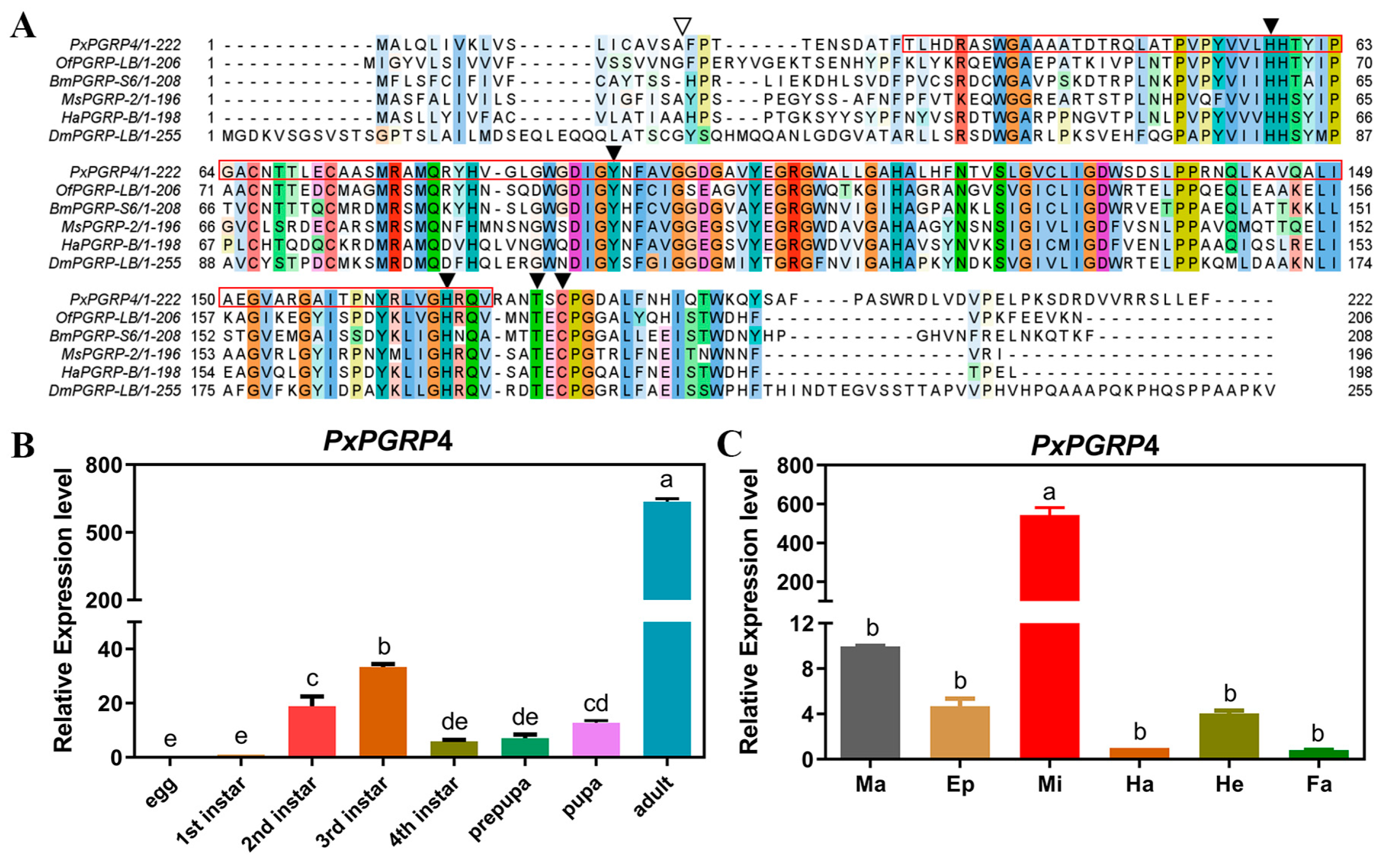
2.3. Cloning and Sequence Analysis of PxPGRP4

Using TRIzol reagent (Invitrogen, Carlsbad, CA, USA), total RNA was extracted from the midgut of the P. xylostella. RNA concentration and integrity were evaluated using a NanoDrop 2000 and 1% agarose gel electrophoresis. Subsequently, cDNA was synthesized with the PrimeScript 1st Strand cDNA Synthesis Kit (Takara, Dalian, China). According to P. xylostella transcriptome sequencing with particular primers, the putative PxPGRP4 (LOC105386207) was chosen for cloning (Table S1). PCR amplification was conducted using 2 × SuperTaq PCR StarMix (GenStar, Shenzhen, China) under the following conditions: initial denaturation at 94 °C for 3 min; 35 cycles of 94 °C for 30 s, 55 °C for 30 s, and 72 °C for 1 min, followed by a final extension at 72 °C for 10 min. The PCR products were purified using a universal DNA purification kit (Tiangen, Beijing, China) and subsequently ligated into the pClone007 vector (Tsingke, Beijing, China) for Sanger sequencing.
To determine the complete open reading frame, the confirmed cDNA sequence of PxPGRP4 was investigated with ORF Finder tools (https://www.ncbi.nlm.nih.gov/orffinder/, accessed on 10 April 2020). The molecular weight and theoretical isoelectric point were calculated using the pI/Mw tool (http://web.expasy.org/protparam/, accessed on 10 April 2020). Multiple sequence alignments were performed using ClustalW (http://www.ebi.ac.uk/Tools/msa/clustalw2/, accessed on 10 April 2020). The signal peptide cleavage site was predicted by the Signal P4.1 server (http://www.cbs.dtu.dk/services/SignalP/, accessed on 10 April 2020). The SMART program (https://smart.embl.de/, accessed on 12 April 2020) predicted its functional domains [32]. Using the neighbor-joining method, MEGA 7.0 was used to create a phylogenetic tree [33].

2.4. Spatiotemporal Expression Profile Analysis of PxPGRP4

The samples were collected from different stages of P. xylostella, i.e., eggs and larvae (first, second, third, and fourth), prepupae, pupae, and adults, to evaluate the expression profiles of PxPGRP4 at various developmental phases and in different body tissues of P. xylostella. In sterilized phosphate-buffered saline (PBS), P. xylostella larvae of the fourth instar were dissected, and tissues including hemocytes, midgut, fat body, Malpighian tubules, epidermis, and head were collected. The above samples were frozen in liquid nitrogen. After liquid nitrogen-mediated freezing, the samples were transferred to a −80 °C freezer. Total RNA was isolated using TRIzol reagent (Invitrogen, Carlsbad, CA, USA), and cDNA was synthesized with the PrimeScript 1st Strand cDNA Synthesis Kit (Takara, Dalian, China) following the previously described protocol. Quantitative real-time PCR (qPCR) was carried out using SYBR Green (Invitrogen, Carlsbad, CA, USA) dye on the CFX96 system (Bio-Rad, Hercules, CA, USA) with gene-specific primers (Table S1). The PCR protocol was configured as follows: initial denaturation at 95 °C for 30 s, followed by 40 cycles of 95 °C for 5 s and 60 °C for 30 s. A dissociation curve ranging from 65 °C to 95 °C was generated to verify the specificity of the PCR products. Ribosomal protein S13 (RPS13) was used as an internal reference gene for normalization. Each experiment was performed in triplicate, and relative expression levels were calculated using the 2(−ΔΔCT) method [34].

2.5. PxPGRP4 Fusion Expression and Polyclonal Antibody Preparation

Reverse transcription PCR was used to amplify the open reading frame (ORF) of PxPGRP4 cDNA. The purified PCR product was digested with BamH I and Sac I restriction enzymes and ligated into the BamH I/Sac I digested expression vector pET-32a (+). For the expression of the protein fusion PxPGRP4, the generated plasmid was added to competent BL21 (DE3) cells. BL21 (DE3) cells produced the fusion protein after 21 h at 16 °C and 100 rpm stimulation with 0.2 mM isopropyl-D-thiogalactoside (IPTG).
The bacterial pellets were centrifuged and subsequently resuspended in a lysis buffer (50 mM Tris-HCl, 500 mM NaCl, pH 8.0). Following cell disruption using a high-pressure frozen cell crusher, the lysate was collected and centrifuged at 12,000 rpm for 20 min at 4 °C. The supernatant was collected and subjected to Ni-NTA column chromatography as instructed by Sangon Biotech (Shanghai, China) to purify the recombinant fusion protein. Then, New Zealand white rabbits were injected with 1 mg of purified recombinant protein to create polyclonal antibodies, and antiserum was collected after three booster doses of 100 μg each (ABclonal Biotech, Wuhan, China). The serum titer was determined by an indirect enzyme-linked immunosorbent assay (ELISA) and reached an endpoint of 1:12,800 [35]. Preimmune and anti-PxPGRP4 sera were subjected to purification via ammonium sulfate precipitation, and their protein concentrations were quantified using a BCA assay kit (Sangon Biotech, Shanghai, China).

2.6. The Preparation of sgRNA and Cas9 Protein

The CRISPR gRNA design was performed using the ATUM online tool (https://www.atum.bio/eCommerce/cas9/input, accessed on 5 September 2020), and two 20 bp targeting sites (5′-AGCCACGCCAGTCCCGTACG-3′ and 5′-CCAGCATGCAAGCCATGCAG-3′) situated in exon 2 of PxPGRP4 were chosen to design the sgRNA. Using the Genomic DNA Isolation Kit (Tiangen, Beijing, China), genomic DNA was recovered from wild-type P. xylostella adults. The two sgRNAs were aligned against the P. xylostella genome using the Cas-OFFinder web tool (http://www.rgenome.net/cas-offinder/, accessed on 5 September 2020), with a mismatched number of 3. This was completed to predict any potential off-target effects. A 530 bp genomic DNA fragment encompassing the target sites in exon 2 was amplified using specific primers (PGRP4-cF/cR, Table S1) and I-5™ 2× High-Fidelity Master Mix (Tsingke, Beijing, China) for single nucleotide polymorphism (SNP) detection. The PCR protocol included an initial denaturation step at 98 °C for 2 min, followed by 35 cycles of denaturation at 98 °C for 10 s, annealing at 55 °C for 10 s, and extension at 72 °C for 10 s, with a final extension at 72 °C for 2 min. The relevant PCR products for sgRNA in vitro transcription were created. The forward primer contained a T7 polymerase binding site (italicized), the sgRNA target sequence (underlined), and a partial sequence from the sgRNA scaffold plasmid (CRISPR-F, Table S1). The 80 bp sgRNA scaffold sequence was chemically synthesized and subsequently subcloned into the pUC57 vector to generate the scaffold plasmid. The reverse primer (CRISPR-R, Table S1) was designed to include a partial sequence complementary to the sgRNA scaffold plasmid. The recommendations from the I-5™ 2×High-Fidelity Master Mix Manual (Tsingke, Beijing, China) were used for the PCR program’s parameters. The scaffold plasmid was subsequently used as the template. Following the manufacturer’s instructions, PCR products were purified using the OMEGA Gel Extraction Kit (OMEGA, Norcross, GA, USA) and then utilized for in vitro transcription with the Hiscribe T7 Quick High-Yield RNA Synthesis Kit (NEB, Ipswich, MA, USA). The GenScript Biotech company was the source of the Cas9 protein (GenScript, Nanjing, China).

2.7. sgRNA/Cas9 Protein Microinjection

Fresh P. xylostella eggs were collected at the pre-blastoderm stage and placed on a 9 cm2 parafilm sheet. The sheet was precoated with cabbage leaf extract to provide near-natural conditions and attract adults. The sheets were renewed at 15 min intervals. A ribonucleoprotein complex was prepared by incubating 150 ng/μL sgRNA with 300 ng/μL Cas9 protein (GenScript, Nanjing, China) at 37 °C for 10 min. This complex was then microinjected into the eggs using a FemtoJet 4i and InjectMan 4 microinjection system (Eppendorf, Hamburg, Germany). The uninjected embryos were set as the negative control. Those embryos were transferred to the hatching chamber for routine rearing. After hatching, larvae were reared according to the standard protocols mentioned in Section 2.1.

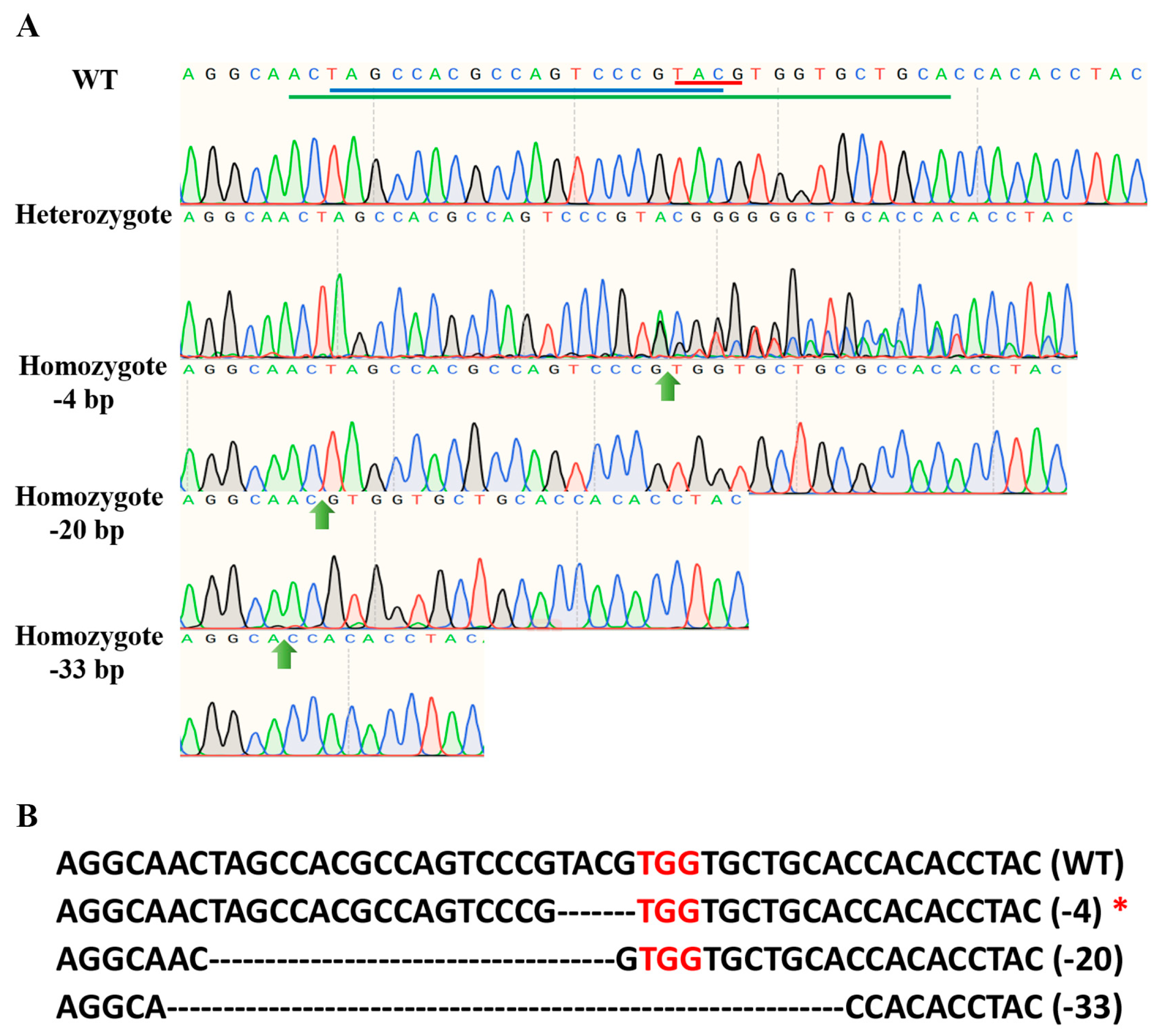
2.8. Genetic Crosses and PCR-Based Genotyping

To create stable homozygous lines of the PxPGRP4 gene, a number of successive crosses were performed. Generation 0 (G0) refers to eggs that were injected, and after hatching, larvae reached the adult stage. To create the initial transgenic lines (G1), the mating of virgin G0 adults with virgin wild-type adults was individually conducted. To detect mutations in the adults, genomic DNA was extracted and subjected to PCR amplification of the PxPGRP4 gene using specific primers PGRP4-cF and PGRP4-cR (Table S1), followed by direct Sanger sequencing (Tsingke, Guangzhou, China). According to the sequencing chromatograms, which showed multiple peaks around the sgRNA target sites in the G0, the corresponding offspring (G1) were kept alive. Homozygous mutants (G2) were created by randomly self-crossing members of G1. PCR-based genotyping was used to identify homozygous mutants (G2), and self-crossing was used to create stable homozygous PxPGRP4 lines in G3.

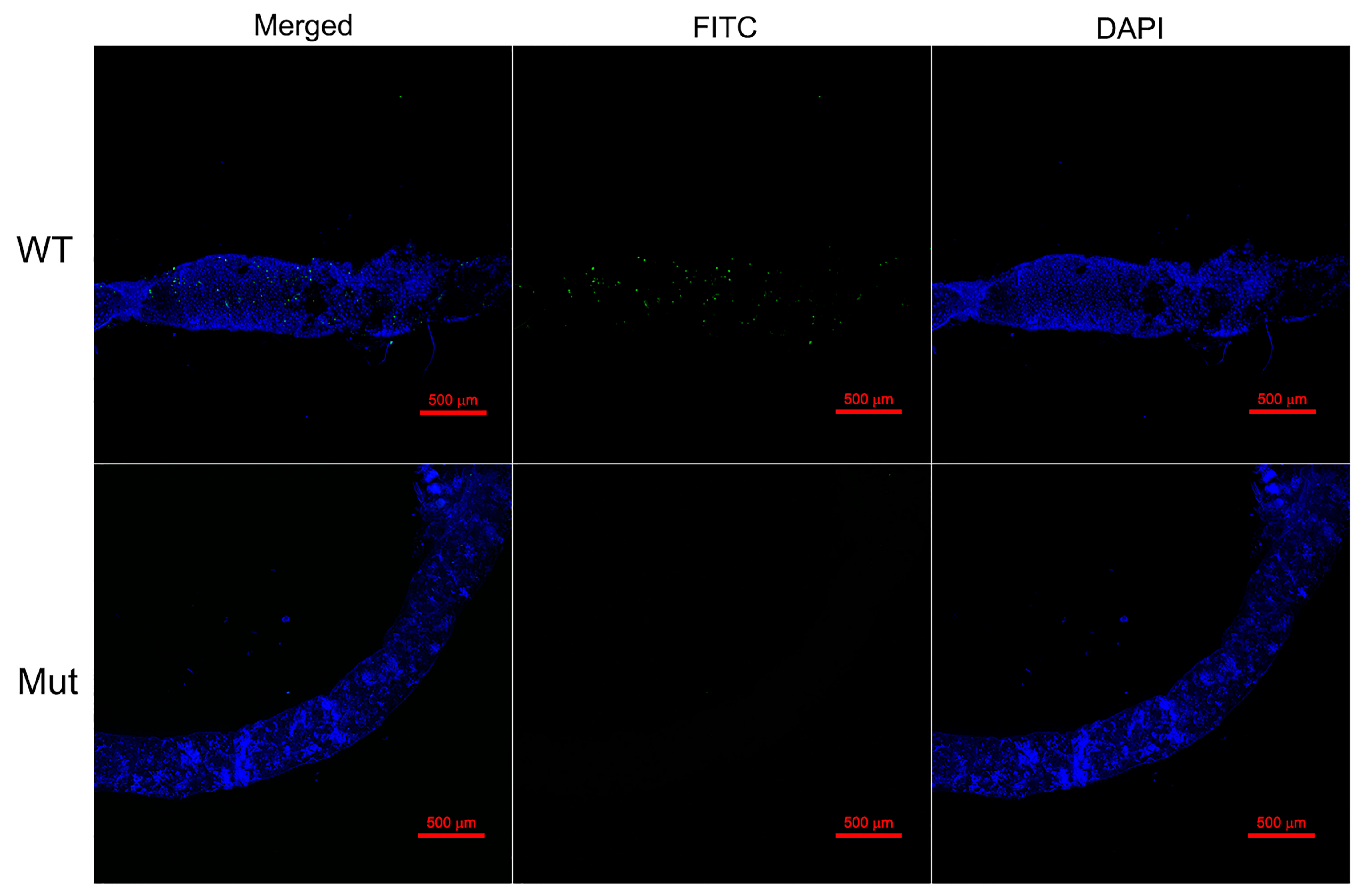
2.9. Immunofluorescence Assay

The gut samples from mutant and wild-type strains were treated with paraformaldehyde (4%) for 30 min, blocked for an interval of 2 h with 1% BSA (dissolved in 1% PBST) under laboratory conditions, and then incubated with the primary antibody (anti-PxPGRP4) at a 1:1000 dilution at 4 °C overnight for the immunofluorescence experiment. Following three washes with 1× PBST, the samples were incubated with a FITC-conjugated secondary antibody (mouse anti-rabbit, Sangon Biotech) at a 1:5000 dilution for 2 h at room temperature. Fluorescence was visualized using a Nikon A1 confocal laser-scanning microscope (Nikon, Tokyo, Japan).

2.10. Midgut Microbiota Analysis

The Omega Stool DNA Kit (Omega Bio-Tek, Norcross, GA, USA) was used to isolate midgut bacterial genomic DNA from the collected samples (per the standard instructions). The hypervariable regions (V3 and V4) of the 16S rRNA gene were amplified from genomic DNA using primers 338F (ACTCCTACGGGAGGCAGCAG) and 806R (GGACTACNNGGGTATCTAAT). PCR amplification was conducted under the following conditions: initial denaturation at 94 °C for 5 min; 30 cycles of 94 °C for 30 s, 50 °C for 30 s, and 72 °C for 1 min, followed by a final extension at 72 °C for 7 min. The resulting PCR products were purified with AMPure XP beads (Beckman Coulter, Inc., Indianapolis, IN, USA). After purification, the Illumina MiSeq PE300 platform was used for pooling and sequencing (Allwegene Tech, Beijing, China). Following sequencing, Pear version 0.9.6 was used to merge paired-end reads. All sequence analysis steps were carried out using the QIIME version 1.8.0 algorithm. The raw paired-end reads underwent quality filtering, chimera removal, and operational taxonomic unit (OTU) clustering using the Vsearch v2.7.1 algorithm. Chimeras were identified through both de novo and reference-based detection methods implemented by UCHIME within the pipeline [36]. Finally, OTUs were clustered at ≥97% similarity, according to USEARCH. A representative sequence of each OTU was selected, and taxonomic information was annotated using the Silva 128 reference database.

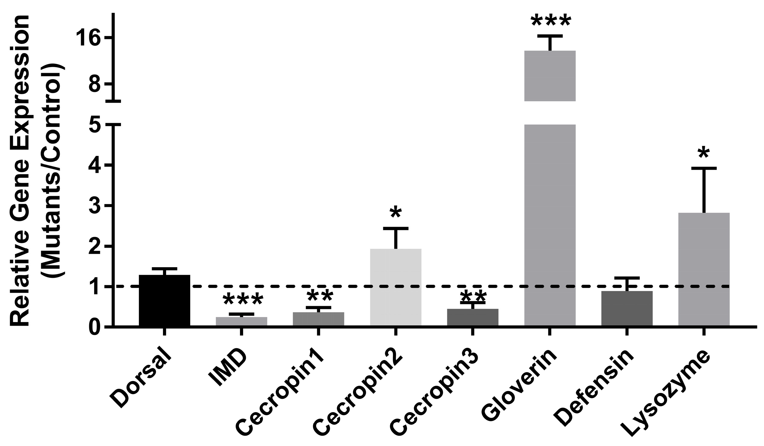
2.11. Bacterial Quantitation and Gene Expression Analysis

qPCR was conducted on genomic DNA using either a general bacterial (Eub) primer or a specific primer targeting the 16S rRNA gene [37]. The ΔΔCt method was employed to analyze the relative quantification changes in midgut bacteria, and a load of midgut bacteria was normalized to the P. xylostella housekeeping gene RPS13. Gene expression analysis was performed as elaborated in Section 2.4. Each experiment was performed in triplicate, and the primers used for qPCR are detailed in Table S1.

2.12. Statistical Analysis

Statistical analyses were performed using SPSS version 22.0 software. Pairwise comparisons were conducted using Student’s t-test, while multiple comparisons were analyzed using one-way analysis of variance (ANOVA) followed by Fisher’s least significant difference (LSD) test. The results were visualized using GraphPad Prism version 7.0 and are performed as mean ± standard deviation (SD) based on three replicates.

3. Results

3.1. PxPGRP4 Characterization

The PxPGRP4 gene contains an open reading frame (ORF) of 615 base pairs, which encodes a protein of 204 amino acids (aa). The theoretical molecular weight of the encoded protein is estimated to be 22.24 kDa, with an isoelectric point of 7.43. The precursor peptide corresponding to PxPGRP4 is predicted to possess a signal peptide, with the cleavage site located between the 18th and 19th amino acid residues (VSA-FP). Additionally, the protein features five amidase catalytic sites and three Zn2+ binding residues. Conserved domain analysis indicates that the PGRP domain spans amino acids 30 to 170 (Figure 1A). Furthermore, the deduced amino acid sequence of PxPGRP4 was compared with PGRP sequences from other insects obtained from the NCBI database. Phylogenetic analysis revealed that PxPGRP4 is closely related to the well-characterized PGRPs of Bombyx mori (NP_001036858.1) and Helicoverpa armigera (AFP23117), suggesting a conserved evolutionary role in insect immunity (Figure S1).

3.2. Expression Profile of PxPGRP4

The sub-lethal concentration (LC25) of Cry1Ac protoxin significantly stimulated the expression of PxPGRP4 in the third instar larvae of P. xylostella, demonstrating fold changes of 3.15 at 12 h, 3.86 at 24 h, and 2.53 at 36 h post-Cry1Ac treatment [20]. The developmental expression profile of PxPGRP4 indicated that transcription was detectable from the first instar larvae, peaked during the adult stage, and exhibited moderate expression levels during the second and third instar larval stages (Figure 1B). In third instar larvae, the tissue profile of PxPGRP4 demonstrated the highest expression specifically in the midgut, while remaining at low levels in several other tissues, including the Malpighian tubules, epidermis, head, hemocytes, and fat body (Figure 1C).

3.3. Recombinant Expression and Purification of PxPGRP4

The recombinant protein PxPGRP4 was expressed as a fusion protein using the pET-32a (+) expression vector, which incorporates a 6×His tag. Subsequently, the fusion protein was purified using a Ni2+-NTA column. The SDS-PAGE analysis revealed the presence of a specific band in the vicinity of 40 kDa (Figure S2A). Western blotting analysis demonstrated that the protein exhibited strong cross-reactivity with the anti-His-tag monoclonal antibody (Figure S2B), thereby further confirming its identity as the fusion protein PxPGRP4–6×His.

3.4. Establishment of the PxPGRP4 Homozygous Mutant

PxPGRP4 mutations were generated by microinjecting Cas9 protein along with specific sgRNA (without predicted off-target activity) into 520 eggs at the pre-blastoderm stage of P. xylostella. Among the 520 injected eggs, 52.9% (275 eggs) successfully hatched and developed into adults, designated as the G0 generation. Sequencing analysis of 96 G0-crossed adult pairs revealed a mutation efficiency of 34.57% at the target site located in exon 2 of the PxPGRP4 gene. These findings were corroborated by sequencing chromatograms, which exhibited multiple peaks at the target sites, indicating successful gene editing (Figure 2A). To establish stable homozygous mutant lines, the mutated adults were self-crossed over three generations (G1–G3), resulting in three distinct lines: homozygote-1 (carrying a 4 bp deletion), homozygote-2 (with a 20 bp deletion), and homozygote-3 (with a 33 bp deletion). Frameshift mutations resulted in truncated proteins in the homozygote-1 and homozygote-2 mutant lines (Figure 2B). The presence of PxPGRP4 was confirmed through intestinal immunofluorescence detection, which validated the knockout effect and protein distribution. The results indicated that PxPGRP4 protein was absent in the gut of the homozygous mutant strain, whereas it was widely distributed in the wild-type P. xylostella strain (Figure 3).

3.5. PxPGRP4 Knockout Affect Cry1Ac Insecticidal Activity

To investigate the relationship between PxPGRP4 and the insecticidal potential of the Cry1Ac protoxin, a stable knockout strain of PxPGRP4 was generated through the CRISPR/Cas9 genome editing system, as previously outlined. The introduced deletion mutations were anticipated to result in the truncation of the PxPGRP4 protein. Susceptibility to the Cry1Ac protoxin was assessed in both the mutants and the original wild-type strain, which served as a control. Notably, the bioassay results showed that the semi-lethal concentration (LC50) of the wild-type strain [12.153 (10.024–14.367) μg/g] for the Cry1Ac protoxin was approximately 31-fold higher than that of the PxPGRP4 mutant strain [0.389 (0.342–0.458) μg/g] (Table 1). These findings demonstrate that the deletion of PxPGRP4 significantly enhances the susceptibility of P. xylostella larvae to Bt Cry1Ac protoxin.

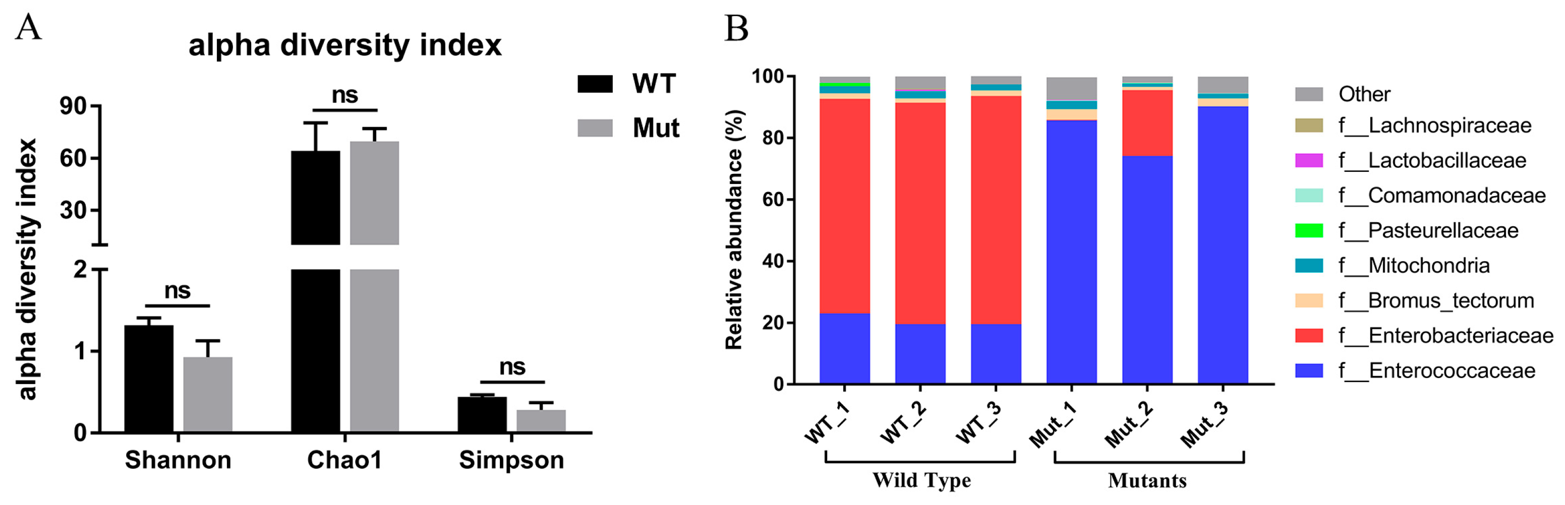
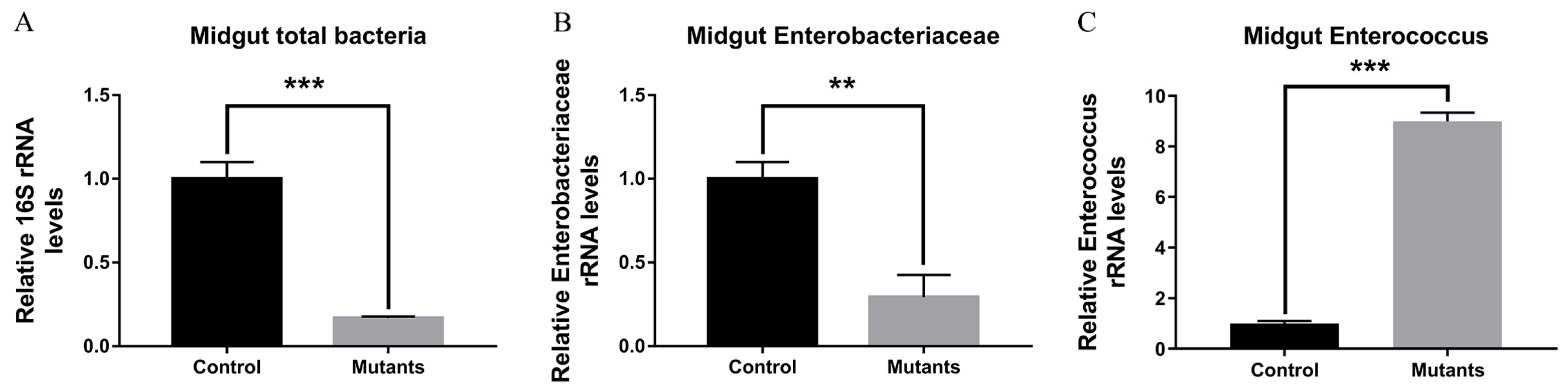
3.6. The Effects of Defective PxPGRP4 on Midgut Homeostasis

To investigate the role that PxPGRP4 played in initiating and regulating innate immune signaling pathways in P. xylostella larvae, we measured the expression of midgut immune-related genes in both PxPGRP4 mutants and wild-type strains. Compared to the wild-type strain, qPCR results indicated that the expression levels of IMD, Cecropin1, and Cecropin3 were significantly decreased in the PxPGRP4 mutants. Conversely, the transcript levels of Cecropin2, Gloverin, and Lysozyme were significantly increased, while the transcript levels of Dorsal and Defensin exhibited no significant changes (Figure 4).
Given that the knockout of PxPGRP4 altered the expression of AMPs in the midgut, it is reasonable to speculate that the midgut microbiota may also be affected. To investigate this hypothesis, 16S rRNA sequencing was employed to analyze the diversity and relative abundance of midgut microbiota in both PxPGRP4 mutant and wild-type strains. As anticipated, the results of 16S rRNA sequencing revealed that the alpha diversity indices (Shannon, Chao1, and Simpson) of midgut bacteria did not exhibit significant differences between the PxPGRP4 mutant and wild-type strains (Figure 5A). However, the relative abundance of midgut bacteria showed significant alterations following the knockout of PxPGRP4. In the wild-type strain, bacteria from the Enterobacteriaceae family predominated the midgut microbiota, with a mean relative abundance of 71.87 ± 1.80%. Conversely, in the PxPGRP4 mutant strain, the dominant bacteria shifted to the Enterococcaceae family, which exhibited a mean relative abundance of 83.4 ± 6.75%. Additionally, the relative abundance of Enterobacteriaceae bacteria decreased to 7.18 ± 10.00% (Figure 5B).
We further investigated whether the knockout of PxPGRP4 influenced the total bacterial load in the midgut of P. xylostella larvae. The relative abundances of Enterobacteriaceae and Enterococcus were assessed using qPCR with primers targeting specific regions of the 16S rRNA gene (Table S1). The results revealed a significant reduction in the total midgut bacterial load in the PxPGRP4 mutant strain (p < 0.001) (Figure 6A). Moreover, consistent with the sequencing results, the abundance of Enterobacteriaceae significantly decreased (p < 0.01) (Figure 6B). Conversely, Enterococcus significantly increased the midgut abundance following the knockout of PxPGRP4 (p < 0.001) (Figure 6C). These findings suggest that midgut homeostasis, which involves both the immune response and microbiota, was disrupted due to the knockout of PxPGRP4.

4. Discussion

PGRPs exhibit a remarkable level of conservation among various species, spanning from invertebrates to vertebrates, and share structural similarities with the bacteriophage T7 lysozyme [25]. These PGRPs were classified into two groups based on their amidase activity: (1) those that have lost their ancestral peptidoglycan hydrolytic function and (2) those that retain this ancestral feature [38]. The latter group functions as a zinc-dependent N-acetylmuramoyl-L-alanine amidase, which is essential for innate immunity [25,39]. We identified and characterized the PxPGRP4 gene from P. xylostella, which encodes a secreted protein. This protein possesses the typical PGRP domain and five active sites for amidase enzymatic activity, making it comparable to Drosophila PGRP-LB. In Drosophila, the recognition of lysine-containing peptidoglycan (PGN) is mediated by PGRP-SA and PGRP-SD, resulting in the activation of the Toll pathway. In contrast, PGRP-LC and PGRP-LE are involved in recognizing DAP-type PGN and activating the IMD pathway [40]. Research has shown that the knockdown of PGRP-S1 in P. xylostella significantly decreases the transcription levels of downstream AMP genes, such as Cecropin, Defensin, Lysozyme1, and Lysozyme2 [41]. Our previous studies indicated that treatment with Bt Cry1Ac protoxin leads to dysbiosis of the midgut microbiota and a significant immunological response in the midgut of P. xylostella larvae [20]. However, the key regulator genes responsible for maintaining midgut homeostasis in P. xylostella remain inadequately characterized. In the current study, we observed that the transcript levels of midgut immune-related genes (IMD, Cecropin1, and Cecropin3) were significantly reduced following the knockout of PxPGRP4 in the mutant strain compared to the control. In contrast, the expression levels of Gloverin, Cecropin2, and Lysozyme were markedly up-regulated, which suggests that PxPGRP4 positively regulates the IMD pathway gene expression, and Gloverin and Cecropin2 may be not the IMD pathway-specific effector genes. These findings indicate that PxPGRP4 plays a crucial role in regulating the activation of the IMD pathway in the midgut of P. xylostella larvae as well as the synthesis of immune effector genes. Further research is warranted to elucidate the mechanisms by which PxPGRP4 regulates the midgut immune signaling pathway in P. xylostella.
The maintenance of midgut microbiota homeostasis is pivotal for host physiology and metabolism. Multiple studies have indicated that the immune system of insects, including components such as C-type lectin, PGRP-SC, and Relish, plays a critical role in regulating the diversity and abundance of gut bacteria across various insect orders [21,22,26,42]. Our data clearly demonstrate that the PxPGRP4 mutant strain exhibited a significantly reduced total bacterial load and an altered relative abundance of midgut bacteria compared to the wild-type strain. However, midgut bacterial diversity did not change significantly in our study. Similarly, silencing PGRP-LB in R. ferrugineus significantly altered the gut microbiota composition, resulting in elevated expression of AMPs and a reduced relative abundance of Enterobacteriaceae compared to the control group [43]. In Musca domestica (house fly), PGRP-SC has been shown to play a pivotal role in modulating the structure and composition of the gut microbiota [21]. This is accomplished by regulating the severity of the gut immune response, which includes the degradation of various pathogen-associated molecular patterns (PAMPs) and the suppression of AMP expression.
Current evidence indicates that the dominant bacterial family shifts from Enterobacteriaceae to Enterococcus following the loss of PxPGRP4 expression. It has been reported that inoculation with Enterococcus pernyi can induce the expression of AMPs such as Attacin, Ceropin, Gloverin, Lysozyme, and Lebocin in Antheraea pernyi [44]. Enterobacteriaceae and Enterococcus exhibit distinct functions within the insect gut, particularly in response to pathogenic invasions. In Manduca sexta, the presence of the midgut commensal bacterium Enterococcus faecalis has been associated with increased mortality due to Bt toxin, primarily resulting from the induction of sepsis in the hemocoel [45]. We hypothesize that the observed increase in Enterococcus abundance in the midgut of P. xylostella could potentially facilitate its translocation into the hemocoel, which is aided by Bt Cry1Ac protoxin, finally contributing to the host insect’s mortality induced by sepsis. However, further experimental validation is needed to test this possibility.
The functional mechanism of Bt toxicity, particularly the manipulation of Cry toxin protein receptors located on the brush border membrane vesicles (BBMVs) of the host midgut, has been extensively documented in various studies [46,47,48,49,50]. Evidence indicated that the MAPK signaling pathway and its associated transcription factors play a regulatory role in the expression of Cry1Ac toxin receptors, thereby influencing the emergence of Cry1Ac resistance in P. xylostella [50,51,52]. However, the complex interactions between host immunity, Bt infection, and gut microbiota remain inadequately understood and necessitate further investigation. Specifically, the regulatory function of crucial immune genes during this interaction requires additional study. In Anopheles stephensi, the knockdown of PGRP-LD changed the structural integrity of the peritrophic matrix and disrupted spatial homeostasis of gut microbiota, finally increasing susceptibility to Plasmodium berghei infection [24]. Whether PxPGRP4 regulates midgut barrier integrity through maintaining gut microbiota homeostasis is also worth further research and confirmation. Relish acts as a key regulatory gene that contributes to maintaining gut homeostasis alongside PGRP. In the model insect Galleria mellonella, suppression of the Relish gene resulted in a significant reduction in the expression of AMPs and a concomitant increase in gut bacterial load [53]. Similarly, silencing the 102 Sl gene in S. littoralis diminished immunocompetence and significantly heightened larval susceptibility to Bt or its toxins [8]. The present study suggests that the loss of PxPGRP4 resulted in a significant proliferation of Enterococcaceae bacteria in the midgut of P. xylostella and then promoted the progression of sepsis in the hemocoel after translocation aided by Cry1Ac, which finally enhanced the insecticidal activity of Bt Cry1Ac protoxin. From an application perspective, PxPGRP4 could be exploited through the following: (1) CRISPR-based gene drives to spread susceptibility alleles in field populations, (2) small-molecule inhibitors of PGRP-amidase activity to mimic knockout effects, or (3) stacked transgenic crops combining Cry1Ac with PxPGRP4-targeting RNAi.

5. Conclusions

Our study demonstrates that PxPGRP4 serves as a critical regulator of midgut immune responses and microbial homeostasis, significantly influencing the composition and abundance of midgut microbiota as well as modulating the expression of AMPs in P. xylostella larvae. Furthermore, it plays a crucial role in determining the susceptibility of P. xylostella larvae to Cry1Ac protoxin. Ongoing research into the regulatory role of PxPGRP4 in immune signaling pathways, along with the influence of gut commensal bacteria on Cry1Ac toxicity, has the potential to yield new insights for enhancing the insecticidal efficacy of Bt Cry toxins while addressing issues related to Bt resistance.

Supplementary Materials

The following supporting information can be downloaded at: https://www.mdpi.com/article/10.3390/agronomy15061294/s1, Figure S1: Phylogenetic analysis of PxPGRP4 with other known insect PGRPs; Figure S2: SDS-PAGE and Western blotting analysis of PxPGRP4 purified protein; Table S1: Primers used in this study.

Author Contributions

Conceptualization, F.J., X.X. and S.L.; methodology, S.L., X.X. and C.F.; software, D.F., M.L; validation, S.L., M.L., C.F. and D.F.; formal analysis, S.L., X.X. and C.F.; investigation, S.L., M.L. and D.F.; resources, F.J., X.X.; data curation, S.L., M.L. and D.F.; writing—original draft preparation, S.L., M.L.; writing—review and editing, F.J., X.X., C.F. and S.L.; visualization, D.F., X.X. and M.L.; supervision, F.J., X.X.; project administration, F.J., X.X. and C.F.; funding acquisition, F.J., X.X. and S.L. All authors have read and agreed to the published version of the manuscript.

Funding

This research was funded by the National Natural Science Foundation of China (31972345, 32172498, 32202281) and the Natural Science Foundation of Guangdong, China (2023A1515010305).

Data Availability Statement

The 16S rRNA sequence data used in this study have been deposited in the NCBI Sequence Read Archive (SRA) repository, and the BioProject accession number is PRJNA1144179.

Conflicts of Interest

The authors declare no conflicts of interest.

References

  1. Bravo, A.; Gill, S.S.; Soberon, M. Mode of action of Bacillus thuringiensis Cry and Cyt toxins and their potential for insect control. Toxicon 2007, 49, 423–435. [Google Scholar] [CrossRef] [PubMed]
  2. Romeis, J.; Meissle, M.; Bigler, F. Transgenic crops expressing Bacillus thuringiensis toxins and biological control. Nat. Biotechnol. 2006, 24, 63–71. [Google Scholar] [CrossRef]
  3. Bel, Y.; Ferre, J.; Hernandez-Martinez, P. Bacillus thuringiensis toxins: Functional characterization and mechanism of action. Toxins 2020, 12, 785. [Google Scholar] [CrossRef] [PubMed]
  4. Kumar, S.; Chandra, A.; Pandey, K.C. Bacillus thuringiensis (Bt) transgenic crop: An environment friendly insect-pest management strategy. J. Environ. Biol. 2008, 29, 641–653. [Google Scholar]
  5. Tabashnik, B.E.; Van Rensburg, J.B.; Carriere, Y. Field-evolved insect resistance to Bt crops: Definition, theory, and data. J. Econ. Entomol. 2009, 102, 2011–2025. [Google Scholar] [CrossRef]
  6. Melo, A.L.; Soccol, V.T.; Soccol, C.R. Bacillus thuringiensis: Mechanism of action, resistance, and new applications: A review. Crit. Rev. Biotechnol. 2016, 36, 317–326. [Google Scholar] [CrossRef] [PubMed]
  7. Li, S.; De Mandal, S.; Xu, X.; Jin, F. The tripartite interaction of host immunity-Bacillus thuringiensis infection-gut microbiota. Toxins 2020, 12, 514. [Google Scholar] [CrossRef]
  8. Caccia, S.; Di Lelio, I.; La Storia, A.; Marinelli, A.; Varricchio, P.; Franzetti, E.; Banyuls, N.; Tettamanti, G.; Casartelli, M.; Giordana, B.; et al. Midgut microbiota and host immunocompetence underlie Bacillus thuringiensis killing mechanism. Proc. Natl. Acad. Sci. USA 2016, 113, 9486–9491. [Google Scholar] [CrossRef]
  9. Pardo-Lopez, L.; Soberon, M.; Bravo, A. Bacillus thuringiensis insecticidal three-domain Cry toxins: Mode of action, insect resistance and consequences for crop protection. FEMS Microbiol. Rev. 2013, 37, 3–22. [Google Scholar] [CrossRef]
  10. Ceja-Navarro, J.A.; Vega, F.E.; Karaoz, U.; Hao, Z.; Jenkins, S.; Lim, H.C.; Kosina, P.; Infante, F.; Northen, T.R.; Brodie, E.L. Gut microbiota mediate caffeine detoxification in the primary insect pest of coffee. Nat. Commun. 2015, 6, 7618. [Google Scholar] [CrossRef]
  11. Storelli, G.; Defaye, A.; Erkosar, B.; Hols, P.; Royet, J.; Leulier, F. Lactobacillus plantarum Promotes Drosophila Systemic Growth by Modulating Hormonal Signals through TOR-Dependent Nutrient Sensing. Cell Metab. 2011, 14, 403–414. [Google Scholar] [CrossRef] [PubMed]
  12. Cai, Z.H.; Yao, Z.C.; Li, Y.S.; Xi, Z.Y.; Bourtzis, K.; Zhao, Z.; Bai, S.; Zhang, H.Y. Intestinal probiotics restore the ecological fitness decline of Bactrocera dorsalis by irradiation. Evol. Appl. 2018, 11, 1946–1963. [Google Scholar] [CrossRef]
  13. Shin, S.C.; Kim, S.H.; You, H.; Kim, B.; Kim, A.C.; Lee, K.A.; Yoon, J.H.; Ryu, J.H.; Lee, W.J. Drosophila microbiome modulates host developmental and metabolic homeostasis via insulin signaling. Science 2011, 334, 670–674. [Google Scholar] [CrossRef] [PubMed]
  14. Yuan, C.F.; Xing, L.S.; Wang, M.L.; Hu, Z.H.; Zou, Z. Microbiota modulates gut immunity and promotes baculovirus infection in Helicoverpa armigera. Insect Sci. 2021, 28, 1766–1779. [Google Scholar] [CrossRef]
  15. Xia, X.; Sun, B.; Gurr, G.M.; Vasseur, L.; Xue, M.; You, M. Gut microbiota mediate insecticide resistance in the diamondback moth, Plutella xylostella (L.). Front. Microbiol. 2018, 9, 25. [Google Scholar] [CrossRef] [PubMed]
  16. Wang, S.C.; Wang, L.Y.; Fan, X.; Yu, C.; Feng, L.; Yi, L. An insight into diversity and functionalities of gut microbiota in insects. Curr. Microbiol. 2020, 77, 1976–1986. [Google Scholar] [CrossRef]
  17. Yun, J.H.; Roh, S.W.; Whon, T.W.; Jung, M.J.; Kim, M.S.; Park, D.S.; Yoon, C.; Nam, Y.D.; Kim, Y.J.; Choi, J.H.; et al. Insect gut bacterial diversity determined by environmental habitat, diet, developmental stage, and phylogeny of host. Appl. Environ. Microbiol. 2014, 80, 5254–5264. [Google Scholar] [CrossRef]
  18. Hernandez-Martinez, P.; Naseri, B.; Navarro-Cerrillo, G.; Escriche, B.; Ferre, J.; Herrero, S. Increase in midgut microbiota load induces an apparent immune priming and increases tolerance to Bacillus thuringiensis. Environ. Microbiol. 2010, 12, 2730–2737. [Google Scholar] [CrossRef]
  19. Li, Y.Z.; Zhao, D.; Wu, H.; Ji, Y.J.; Liu, Z.R.; Guo, X.C.; Guo, W.; Bi, Y. Bt GS57 interaction with gut microbiota accelerates Spodoptera exigua mortality. Front. Microbiol. 2022, 13, 835227. [Google Scholar] [CrossRef]
  20. Li, S.Z.; Xu, X.X.; De Mandal, S.; Shakeel, M.; Hua, Y.Y.; Shoukat, R.F.; Fu, D.R.; Jin, F.L. Gut microbiota mediate Plutella xylostella susceptibility to Bt Cry1Ac protoxin is associated with host immune response. Environ. Pollut. 2021, 271, 116271. [Google Scholar] [CrossRef]
  21. Liang, Y.; Yang, L.; Wang, Y.; Tang, T.; Liu, F.; Zhang, F. Peptidoglycan recognition protein SC (PGRP-SC) shapes gut microbiota richness, diversity and composition by modulating immunity in the house fly Musca domestica. Insect Mol. Biol. 2023, 32, 200–212. [Google Scholar] [CrossRef] [PubMed]
  22. Marra, A.; Hanson, M.A.; Kondo, S.; Erkosar, B.; Lemaitre, B. Drosophila antimicrobial peptides and lysozymes regulate gut microbiota composition and abundance. mBio 2021, 12, e0082421. [Google Scholar] [CrossRef] [PubMed]
  23. Pang, X.; Xiao, X.; Liu, Y.; Zhang, R.; Liu, J.; Liu, Q.; Wang, P.; Cheng, G. Mosquito C-type lectins maintain gut microbiome homeostasis. Nat. Microbiol. 2016, 1, 16023. [Google Scholar] [CrossRef]
  24. Song, X.; Wang, M.; Dong, L.; Zhu, H.; Wang, J. PGRP-LD mediates A. stephensi vector competency by regulating homeostasis of microbiota-induced peritrophic matrix synthesis. PLoS Pathog. 2018, 14, e1006899. [Google Scholar] [CrossRef]
  25. Lemaitre, B.; Hoffmann, J. The host defense of Drosophila melanogaster. Annu. Rev. Immunol. 2007, 25, 697–743. [Google Scholar] [CrossRef]
  26. Xiao, R.; Wang, X.; Xie, E.; Ji, T.; Li, X.; Muhammad, A.; Yin, X.; Hou, Y.; Shi, Z. An IMD-like pathway mediates the intestinal immunity to modulate the homeostasis of gut microbiota in Rhynchophorus ferrugineus Olivier (Coleoptera: Dryophthoridae). Dev. Comp. Immunol. 2019, 97, 20–27. [Google Scholar] [CrossRef] [PubMed]
  27. Bosco-Drayon, V.; Poidevin, M.; Boneca, I.G.; Narbonne-Reveau, K.; Royet, J.; Charroux, B. Peptidoglycan sensing by the receptor PGRP-LE in the Drosophila gut induces immune responses to infectious bacteria and tolerance to microbiota. Cell Host Microbe 2012, 12, 153–165. [Google Scholar] [CrossRef]
  28. Li, S.; Xu, X.; Shakeel, M.; Xu, J.; Zheng, Z.; Zheng, J.; Yu, X.; Zhao, Q.; Jin, F. Bacillus thuringiensis suppresses the humoral immune system to overcome defense mechanism of Plutella xylostella. Front. Physiol. 2018, 9, 1478. [Google Scholar] [CrossRef]
  29. Zhou, Z.S.; Yang, S.J.; Shu, C.L.; Song, F.P.; Zhou, X.P.; Zhang, J. Comparison and optimization of the method for Cry1Ac protoxin preparation in HD73 strain. J. Integr. Agric. 2015, 14, 1598–1603. [Google Scholar] [CrossRef]
  30. Abbott, W.S. A method of computing the effectiveness of an insecticide. 1925. J. Am. Mosq. Control. Assoc. 1987, 3, 302–303. [Google Scholar]
  31. Finney, D. Probit Analysis, 3rd ed.; Cambridge University Press: London, UK, 1971. [Google Scholar]
  32. Letunic, I.; Bork, P. 20 years of the SMART protein domain annotation resource. Nucleic Acids Res. 2018, 46, D493–D496. [Google Scholar] [CrossRef] [PubMed]
  33. Saitou, N.; Nei, M. The neighbor-joining method: A new method for reconstructing phylogenetic trees. Mol. Biol. Evol. 1987, 4, 406–425. [Google Scholar] [CrossRef] [PubMed]
  34. Livak, K.J.; Schmittgen, T.D. Analysis of relative gene expression data using real-time quantitative PCR and the 2−ΔΔCT Method. Methods 2001, 25, 402–408. [Google Scholar] [CrossRef]
  35. Kastrup, J.; Feldt-Rasmussen, U.; Bartram, H.R.; Witten, J.; Sand Hansen, H. An enzyme linked immunosorbent assay (ELISA) for measurement of human serum thyroglobulin. Evaluation of the influence of thyroglobulin auto-antibodies. Scand. J. Clin. Lab. Investig. 1985, 45, 471–476. [Google Scholar] [CrossRef] [PubMed]
  36. Edgar, R.C.; Haas, B.J.; Clemente, J.C.; Quince, C.; Knight, R. UCHIME improves sensitivity and speed of chimera detection. Bioinformatics 2011, 27, 2194–2200. [Google Scholar] [CrossRef]
  37. Denman, S.E.; McSweeney, C.S. Development of a real-time PCR assay for monitoring anaerobic fungal and cellulolytic bacterial populations within the rumen. Fems Microbiol. Ecol. 2006, 58, 572–582. [Google Scholar] [CrossRef]
  38. Royet, J.; Dziarski, R. Peptidoglycan recognition proteins: Pleiotropic sensors and effectors of antimicrobial defences. Nat. Rev. Microbiol. 2007, 5, 264–277. [Google Scholar] [CrossRef]
  39. Kurata, S. Peptidoglycan recognition proteins in Drosophila immunity. Dev. Comp. Immunol. 2014, 42, 36–41. [Google Scholar] [CrossRef]
  40. Werner, T.; Liu, G.; Kang, D.; Ekengren, S.; Steiner, H.; Hultmark, D. A family of peptidoglycan recognition proteins in the fruit fly Drosophila melanogaster. Proc. Natl. Acad. Sci. USA 2000, 97, 13772–13777. [Google Scholar] [CrossRef]
  41. Zhang, Z.; Kong, J.; De Mandal, S.; Li, S.; Zheng, Z.; Jin, F.; Xu, X. An immune-responsive PGRP-S1 regulates the expression of antibacterial peptide genes in diamondback moth, Plutella xylostella (L.). Int. J. Biol. Macromol. 2020, 142, 114–124. [Google Scholar] [CrossRef]
  42. Wang, W.W.; Wang, G.J.; Zhuo, X.R.; Liu, Y.; Tang, L.; Liu, X.S.; Wang, J.L. C-type lectin-mediated microbial homeostasis is critical for Helicoverpa armigera larval growth and development. PLoS Pathog. 2020, 16, e1008901. [Google Scholar] [CrossRef] [PubMed]
  43. Dawadi, B.; Wang, X.H.; Xiao, R.; Muhammad, A.; Hou, Y.M.; Shi, Z.H. PGRP-LB homolog acts as a negative modulator of immunity in maintaining the gut-microbe symbiosis of red palm weevil, Rhynchophorus ferrugineus Olivier. Dev. Comp. Immunol. 2018, 86, 65–77. [Google Scholar] [CrossRef] [PubMed]
  44. Liu, W.; Wang, Y.; Zhou, J.; Zhang, Y.; Ma, Y.; Wang, D.; Jiang, Y.; Shi, S.; Qin, L. Peptidoglycan recognition proteins regulate immune response of Antheraea pernyi in different ways. J. Invertebr. Pathol. 2019, 166, 107204. [Google Scholar] [CrossRef]
  45. Mason, K.L.; Stepien, T.A.; Blum, J.E.; Holt, J.F.; Labbe, N.H.; Rush, J.S.; Raffa, K.F.; Handelsman, J. From commensal to pathogen: Translocation of Enterococcus faecalis from the midgut to the hemocoel of Manduca sexta. mBio 2011, 2, e00065-11. [Google Scholar] [CrossRef]
  46. Batool, K.; Alam, I.; Jin, L.; Xu, J.; Wu, C.; Wang, J.; Huang, E.; Guan, X.; Yu, X.Q.; Zhang, L. CTLGA9 interacts with ALP1 and APN receptors to modulate Cry11Aa toxicity in Aedes aegypti. J. Agric. Food Chem. 2019, 67, 8896–8904. [Google Scholar] [CrossRef]
  47. Gao, M.; Dong, S.; Hu, X.; Zhang, X.; Liu, Y.; Zhong, J.; Lu, L.; Wang, Y.; Chen, L.; Liu, X. Roles of midgut cadherin from two moths in different Bacillus thuringiensis action mechanisms: Correlation among toxin binding, cellular toxicity, and synergism. J. Agric. Food Chem. 2019, 67, 13237–13246. [Google Scholar] [CrossRef] [PubMed]
  48. Guo, Z.; Kang, S.; Chen, D.; Wu, Q.; Wang, S.; Xie, W.; Zhu, X.; Baxter, S.W.; Zhou, X.; Jurat-Fuentes, J.L.; et al. MAPK signaling pathway alters expression of midgut ALP and ABCC genes and causes resistance to Bacillus thuringiensis Cry1Ac toxin in diamondback moth. PLoS Genet. 2015, 11, e1005124. [Google Scholar] [CrossRef]
  49. Liao, C.; Jin, M.; Cheng, Y.; Yang, Y.; Soberón, M.; Bravo, A.; Liu, K.; Xiao, Y. Bacillus thuringiensis Cry1Ac protoxin and activated toxin exert differential toxicity due to a synergistic interplay of Cadherin with ABCC transporters in the cotton bollworm. Appl. Environ. Microbiol. 2022, 88, e0250521. [Google Scholar] [CrossRef]
  50. Qin, J.; Guo, L.; Ye, F.; Kang, S.; Sun, D.; Zhu, L.; Bai, Y.; Cheng, Z.; Xu, L.; Ouyang, C.; et al. MAPK-activated transcription factor PxJun suppresses PxABCB1 expression and confers resistance to Bacillus thuringiensis Cry1Ac toxin in Plutella xylostella (L.). Appl. Environ. Microbiol. 2021, 87, e0046621. [Google Scholar] [CrossRef]
  51. Guo, L.; Cheng, Z.; Qin, J.; Sun, D.; Wang, S.; Wu, Q.; Crickmore, N.; Zhou, X.; Bravo, A.; Soberon, M.; et al. MAPK-mediated transcription factor GATAd contributes to Cry1Ac resistance in diamondback moth by reducing PxmALP expression. PLoS Genet. 2022, 18, e1010037. [Google Scholar] [CrossRef]
  52. Xu, L.; Qin, J.; Fu, W.; Wang, S.; Wu, Q.; Zhou, X.; Crickmore, N.; Guo, Z.; Zhang, Y. MAP4K4 controlled transcription factor POUM1 regulates PxABCG1 expression influencing Cry1Ac resistance in Plutella xylostella (L.). Pestic. Biochem. Physiol. 2022, 182, 105053. [Google Scholar] [CrossRef] [PubMed]
  53. Sarvari, M.; Mikani, A.; Mehrabadi, M. The innate immune gene Relish and Caudal jointly contribute to the gut immune homeostasis by regulating antimicrobial peptides in Galleria mellonella. Dev. Comp. Immunol. 2020, 110, 103732. [Google Scholar] [CrossRef] [PubMed]
Figure 1. (A) Alignments of amino acid sequences of PxPGRP4 with other insect PGRPs. Amino acid sequences of insect PGRPs were retrieved from the GenBank: Of, O. furnacalis (PGRP-LB: XP_028159392.1); Bm, B. mori (PGRP-S6: NP_001036858.1); Ms, M. sexta (PGRP-2: ACX49764); Ha, H. armigera (PGRP-B: AFP23116); Dm, D. melanogaster (PGRP-LB: NP_001247052.1). ▽ Indicates signal peptide cleavage site (VSA-FP), ▼ indicates key amino acid sites that determine zinc ion/amidase activity (H-Y-H-T-C), and red line box indicates conserved PGRP domain (30–170 aa). Developmental stages (B) and tissue distribution (C) expression patterns of PxPGRP4. Ma, Malpighian tubule; Ep, epidermis; Mi, midgut; Ha, head; He, hemocyte; Fa, fat body. Data are presented as mean ± standard deviation (SD) from three independent experiments. Statistical significance was assessed using LSD and Duncan tests (α = 0.05), with differences indicated by lowercase letters (a–e) above the columns (p ≤ 0.05).
Agronomy 15 01294 g001
Figure 2. Sequencing and identification of the mutant genotypes in PxPGRP4. (A) Sequencing chromatograms of PCR products from G0 to G2 individuals showing mutations at the target site. Homozygote −4 bp (4-base deletion) is underlined in red, homozygote −20 bp (20-base deletion) is underlined in blue, homozygote −33 bp (33-base deletion) is underlined in green, and the cleavage site is indicated with a green arrow. (B) Various indel mutations flanking the CRISPR target sites of PxPGRP4 were identified in G2 larvae through sequencing of individual PCR products. The deleted bases are represented by dashes, with the number of deleted bases indicated to the right of each allele (−, deletion). The asterisk represents the mutant genotype used for subsequent experiments.
Agronomy 15 01294 g002
Figure 3. Intestinal immunofluorescence (IF) detection of PxPGRP4. Gut samples from WT (wild-type) and Mut (PxPGRP4 mutants) were treated with PxPGRP4 polyclonal antibody and FITC-conjugated secondary antibody (mouse anti-rabbit) (green). Nuclei were stained with DAPI (blue). Images are representative of three independent experiments. Scale bars, 500 μm.
Agronomy 15 01294 g003
Figure 4. The relative expression levels of immunity-related genes in the midgut of the wild-type strain (Control) and PxPGRP4 mutants (Mutants) were analyzed. Error bars represent standard deviation (SD) with a sample size of n = 3. Statistical significance was assessed using Student’s t-test, with p-values indicated as follows: * p < 0.05; ** p < 0.01; *** p < 0.001. The data presented are from one of three independent experiments.
Agronomy 15 01294 g004
Figure 5. The effect of PxPGRP4 knockout on midgut microbial diversity (A) and relative abundance (B) in P. xylostella is presented. WT, wild-type; Mut, PxPGRP4 mutants. The notation “ns” indicates no significant difference in pairwise comparisons, and the significance of the differences was analyzed using Student’s t-test.
Agronomy 15 01294 g005
Figure 6. The relative abundance of total bacteria (A), Enterobacteriaceae (B), and Enterococcus (C) in the midgut of the wild-type strain (Control) and PxPGRP4 mutants (Mutants) was assessed. Quantification was performed using 16S rRNA gene-based quantitative PCR (qPCR) analysis, with three biological replicates conducted for accuracy. Error bars indicate standard deviation (SD, n = 3). Statistical significance was evaluated using Student’s t-test, with significance levels indicated as ** p < 0.01 and *** p < 0.001.
Agronomy 15 01294 g006
Table 1. Toxicity of Cry1Ac protoxin to 3rd instar larvae of P. xylostella PxPGRP4 mutant and wild-type strains after 48 h of exposure.
StrainsnSlope ± SELC50 (μg/g)95% Fiducial Limits
Wild Type2401.475 ± 0.17812.15310.024–14.367
Mutant2401.767 ± 0.2230.3890.342–0.458
n: number of larvae in the probit analysis. SE: standard error.
Disclaimer/Publisher’s Note: The statements, opinions and data contained in all publications are solely those of the individual author(s) and contributor(s) and not of MDPI and/or the editor(s). MDPI and/or the editor(s) disclaim responsibility for any injury to people or property resulting from any ideas, methods, instructions or products referred to in the content.

