Article

Fungal Biota and Mycotoxins Contamination in Soybean Expeller

by
Gisele Maciel
1,*,
Claudia Carla Castellari
2,
Leandro Marcelo Cardoso
1,
María Bernadette Abadía
1,
Patrizia Busato
3 and
Ricardo Enrique Bartosik
1
1
IPADS Balcarce (INTA-CONICET), RN 226 km 73.5, Balcarce 7620, Argentina
2
Laboratorio de Microbiología de Suelos y Alimentos, Facultad de Ciencias Agrarias, Universidad Nacional de Mar del Plata, RN 226 km 73.5, Balcarce 7620, Argentina
3
Interuniversity Department of Regional and Urban Studies and Planning (DIST), Polytechnic of Turin, Viale Pier Andrea Mattioli 39, 10125 Torino, Italy
*
Author to whom correspondence should be addressed.
Agronomy 2025, 15(4), 807; https://doi.org/10.3390/agronomy15040807
Submission received: 12 February 2025 / Revised: 21 March 2025 / Accepted: 22 March 2025 / Published: 25 March 2025
(This article belongs to the Section Agricultural Biosystem and Biological Engineering)

Abstract

:
Soybean expeller (SBE), a by-product of soybean oil extraction through the extruding-expelling process, is widely used as a protein source in animal feed and soy-based foods. This study evaluated fungal contamination and mycotoxin levels in SBE samples from 11 extruding-expelling facilities in Argentina, assessing fungal load, moisture content (MC), and mycotoxin profiles. Fungal biota was quantified through colony forming unit (CFU) counts and identified via morphological analysis, while mycotoxins were quantified using liquid chromatography and tandem mass spectrometry. CFU counts were low (0 to 4 CFU g−1 DM), with Penicillium spp. (28.0%) and Mucoraceae (family) (25.6%) being the most frequently isolated genera. Deoxynivalenol (DON) and aflatoxins (AFB1, AFB2, AFG1, and AFG2) were detected in 20% to 40% of the samples. The average concentration was 215.19 µg kg−1 for DON and 41.68, 0.39, and 0.34 µg kg−1 for AFB1, AFG1, and AFG2, respectively. Although most mycotoxin concentrations were below regulatory limits, a few samples exceeded the threshold for DON (8.6%) and AFB1 (2.9%). Co-occurrence of two mycotoxins was observed in 60% of the samples. These results highlight the importance of monitoring fungal contamination and mycotoxin levels to ensure the safety and quality of SBE for feed and food applications.

1. Introduction

Soybean expeller (SBE), a by-product obtained during the extruding-expelling process of soybean oil extraction [1], is highly valued as a protein-rich component in animal feed and soy-based food products [2,3,4,5]. In Argentina, nearly one million tons of SBE are produced annually [6] in 373 extruding-expelling facilities, primarily located in the Pampa’s region [7]. The average composition of SBE in Argentina is 7.2% moisture, 44.2% protein, and 8.1% residual oil [8,9], aligning with the values set by Standard XIX [10] for commercialization. Despite its economics and nutritional importance, SBE is susceptible to fungal contamination and mycotoxin accumulation, which can pose significant risk to both animal and human health [11,12].
Fungal contamination in soybean can occur during cultivation in the field, transportation, storage, and processing [13]. Under improper storage conditions—such as high relative humidity (>70%) and temperature [14,15,16,17]—fungus can proliferate in soybean seed and expeller, leading to the production of mycotoxins and overall quality degradation [18,19,20,21,22]. Common mycotoxins associated with soybean products include deoxynivalenol (DON) and aflatoxins (AFs), produced by fungal genera such as Fusarium and Aspergillus [23,24,25,26]. These toxins are a major concern due to their adverse health effects and the lack of established regulation limits in Argentina for food and feed applications.
DON is a mycotoxin produced by Fusarium culmorum and Fusarium graminearum, commonly found in various cereal crops such as wheat, maize, barley, oats, and rye [27]. These fungi synthesize mycotoxins in field crops, leading to contamination at pre-harvest stages. DON is a widespread contaminant in wheat, with a reported prevalence of 57% in 11,444 tested samples globally [28]. At the cellular level, DON inhibits DNA, RNA, and protein synthesis by targeting ribosomes, resulting in erythrocyte hemolysis [29]. Its systemic toxic effects include feed refusal, reduced weight gain, gastroenteritis, diarrhea, emesis, nutrient malabsorption, cardiotoxicity, teratogenicity, and immunotoxicity [30,31].
Aflatoxins are a class of mycotoxins produced by Aspergillus species, primarily Aspergillus flavus and Aspergillus parasiticus [32]. These fungi are commonly found in soil and various organic materials and are known to synthesize toxins during grain storage. While A. flavus strains produce only aflatoxins B1 (AFB1) and B2 (AFB2), A. parasiticus strains can synthesize AFB1, AFB2, G1 (AFG1), and G2 (AFG2) [33]. Notably, AFB1 is the most potent hepatocarcinogen identified in mammals and is classified as a Group I carcinogen by the IARC [30,34].
Aflatoxins are among the most extensively studied mycotoxins worldwide due to their significant impact on food safety and health. They have been linked to various diseases, including aflatoxicosis in livestock, domestic animals, and humans. Their potent carcinogenicity in susceptible laboratory animals and acute toxicological effects in humans have led to heightened global concern and regulatory attention [35,36].
Previous studies have documented mycotoxin contamination in soybean and related by-products, such as soybean meal [19,37,38,39]; however, limited information is available regarding mycotoxin levels specifically in SBE. Additionally, mycotoxigenic species belonging to the genera Alternaria, Fusarium, Penicillium, and Aspergillus have been reported in both recently harvested and stored soybeans [19,22,40,41,42,43]. Despite the widespread use of SBE as a feed ingredient, the relationship between fungal load, moisture content (MC), and mycotoxin contamination in SBE remains poorly understood, making its microbiological safety a critical concern. However, no national or international standards specifically regulate the microbiological quality or mycotoxin presence in SBE. In response, major animal feed manufacturers have begun assessing microbiological quality based on European regulations. Mycological criteria—such as the threshold of ≤104 CFU/g—are currently established only for other soybean-derived products used in feed [44].
This study aims to address these gaps by evaluating the microbiological quality of SBE obtained from various extruding-expelling facilities located in the Argentine Pampa’s region by (1) quantifying and identifying the fungal biota, including filamentous fungi and yeasts, present in SBE; (2) assessing the correlation between SBE MC and fungal biota load; and (3) determining the presence of mycotoxins in SBE. The findings will contribute to improving quality control practices and enhancing the safety of SBE for feed and food applications.

2. Materials and Methods

2.1. Sampling of Soybean Expeller

SBE samples were obtained during the 2015 and 2016 harvest seasons from 11 extruding-expelling facilities located in the Pampa’s region of Argentina, covering the provinces of Buenos Aires (6), Santa Fe (2), Córdoba (2) and Entre Ríos (1). The 11 facilities represent 3% of the extruding-expelling facilities of Argentina. In each facility, three to eight separate SBE samples (obtained in three consecutive days, 5 kg each) were collected directly from the screw press outlet (Figure 1) and placed in double hermetic plastic bags to avoid moisture variations until use (n = 33). The initial sampling plan considered three samples per facility; however, logistical factors allowed for additional sampling in some locations, resulting in an unbalanced dataset, which was only used for mycotoxin analysis (n = 41). Of each sample, approximately 4.5 kg were stored at 4 °C for fungal biota analysis, following the same procedure as described by Castellari et al. [18]. This storage was for preservation purposes, preventing fungal growth until analysis, which was conducted within seven days. Additionally, 0.5 kg was stored at −18 °C for mycotoxin determination and was analyzed within one month.

2.2. Moisture Content Measurement

The MC of each SBE sample was determined in triplicate with the oven-drying method [45] at 103 °C for 72 h. Results were expressed on a dry basis (% d.b.).

2.3. Quantification and Identification Methods of Fungal Biota

Fungal biota (filamentous fungi and yeasts) was evaluated at the Soil and Food Microbiology Lab (FCA-INTA Balcarce, Balcarce, Argentina) by the method of serial dilutions. Ten grams of each SBE sample was homogenized in 90 mL of peptone water solution (one g casein peptone/L, Britania®, Ciudad Autónoma de Buenos Aires, Argentina) for 15 min at 120 rpm in an orbital plane shaker (BM021, BIOMINT, Florida Oeste, Argentina), with four independent replicates. The 10th-fold serial dilution (10−1) method in a tube was used, and 0.1 mL aliquots from each dilution were inoculated in duplicate in Petri dishes with potato dextrose agar (PDA, Britania®, Ciudad Autónoma de Buenos Aires, Argentina) at 2% and 0.1 g/L of chloramphenicol (Anedra®, Troncos del Talar, Argentina) to inhibit bacterial growth. PDA with antibiotics medium was used for the cultivation and enumeration of yeast and filamentous fungi (molds) from SBE [46]. After five to seven days of incubation at 28 °C, filamentous fungi and yeast colonies were counted, and the results expressed as colony-forming units per gram of dry matter (CFU g−1 DM) [47].
The identification of fungal biota in SBE was performed in the Petri dishes based on vegetative hyphae (in vitro culture medium: malt extract medium-MEA and sucrose medium-CYA) and microscopic (Olympus BH2, PR, USA) (asexual spores and sexual spores) characteristics [47,48]. The results were expressed as isolation frequency (Equation (1)), defined as the percentage of samples in which each genus was detected, and as relative density (Equation (2)), defined as the proportion of each genus isolated relative to the total genus isolated at each specific extruding-expelling facility [49,50].
I s o l a t i o n   f r e q u e n c y % = n s N × 100
where ns is the number of samples contaminated with a specific genus, and N is the total number of samples.
R e l a t i v e   d e n s i t y % = n i N i × 100
where ni is the number of isolates belonging to a specific genus, Ni is the total number of genus isolated from a specific extruding-expelling facility.

2.4. Mycotoxins Analysis

The mycotoxins analysis was carried out at the Institute of Food Technology (INTA Castelar, Castelar, Argentina). The evaluated mycotoxins were AFB1, AFB2, AFG1, AFG2, nivalenol (NIV), DON, 13 acetyl DON, 15 acetyl DON, ochratoxin A, and zearalenone. The mycotoxin analysis was performed in SBE collected from all facilities except facilities 5 and 6, due to operational restrictions (n = 35). For mycotoxin analysis, 500 g from each SBE sample was ground in a laboratory grinder (Fw-100, Arcano, Beijing, China) and homogenized. The determination was carried out as described by Castañares et al. [51] with some experimental modifications. Firstly, 10 g of each sample was suspended in 20 mL of extraction solvent (acetonitrile: water: acetic acid; 79:20:1) with the addition of 2 g of NaCl. The samples were homogenized with Ultra-Turrax® (IKA Labortecknik, Staufen, Germany) for 3 min, sonicated for 60 min, and centrifuged for 5 min at 3000 rpm (LC-75R, Luguimac®, Buenos Aires, Argentina). A volume of 10 mL extract was transferred into glass vials and evaporated to dryness at 45 °C under a stream of N2. The samples were resuspended in 2 mL of acetonitrile:water (70:30). They were then diluted with 28 mL of ultrapure water and purified with an SPE C18 cartridge (Strata C18-E, 55 µm, 70A, 500 mg/6 mL, Phenomenex, Chesire, UK). The cartridge was washed with 2 mL of methanol:water (1:99) and eluted with 1 mL of methanol:water (70:30). Finally, they were filtered through a 0.2 µm polyvinylidene fluoride (PVDF) membrane and injected into a liquid chromatogram/tandem mass spectrometer (LTQ-XL™ Linear Ion Trap Thermo, San Jose, CA, USA). Chromatographic separations were performed with a C18 100 2.1 mm Hypersil™ ODS (Thermo Fisher, Waltham, MA, USA) (5 mm particle size) column. A solution of ammonium formate (10 mM) and acetonitrile was used as a mobile phase. Samples (10 µL) were analyzed at a flow rate of 0.2 mL/min at 45 °C. The mass-spectrometer acquisition settings were electrospray ionization (ESI) negative or positive (Table A1), retention time and abundance of the confirmation ion (Ion C) relative to that of quantification ion (Ion Q) were used as identification criteria.
The quantitative determination was performed using the following Sigma-Aldrich standards: B-trichothecene mix (part number: 34134), aflatoxin mix (part number: CRM46303), ochratoxin A solution (part number: 34037), and zearalenone solution (part number: 34126). Non-detected values were assumed as half the limit of detection (LOD), and values below the limit of quantification (LOQ) were assumed as half the LOQ [52,53].

2.5. Statistical Analysis

CFU counts and SBE MC data were analyzed as a completely randomized design using the R software (version 4.3.0, 21 April 2023) [54]. Each facility was considered a different treatment, and each sample (collected over three consecutive days) represents an independent experimental unit. A non-parametric Kruskal–Wallis test was performed for CFU data, followed by a Dunn’s test with Bonferroni correction (α = 0.05) [55,56]. In addition, a Pearson correlation analysis was carried out between counts of CFU g−1 DM and SBE MC.
Mycotoxin concentration in SBE samples from different extruded-expelling facilities was characterized through descriptive statistics (means and standard deviation (SD) was performed only with quantified samples). A Pearson correlation was performed to analyze linear relationships between pairs of mycotoxins. All statistical analyses were performed using the R software (version 4.3.0, 21 April 2023) [54].

3. Results

3.1. Quantification and Identification of Fungal Biota

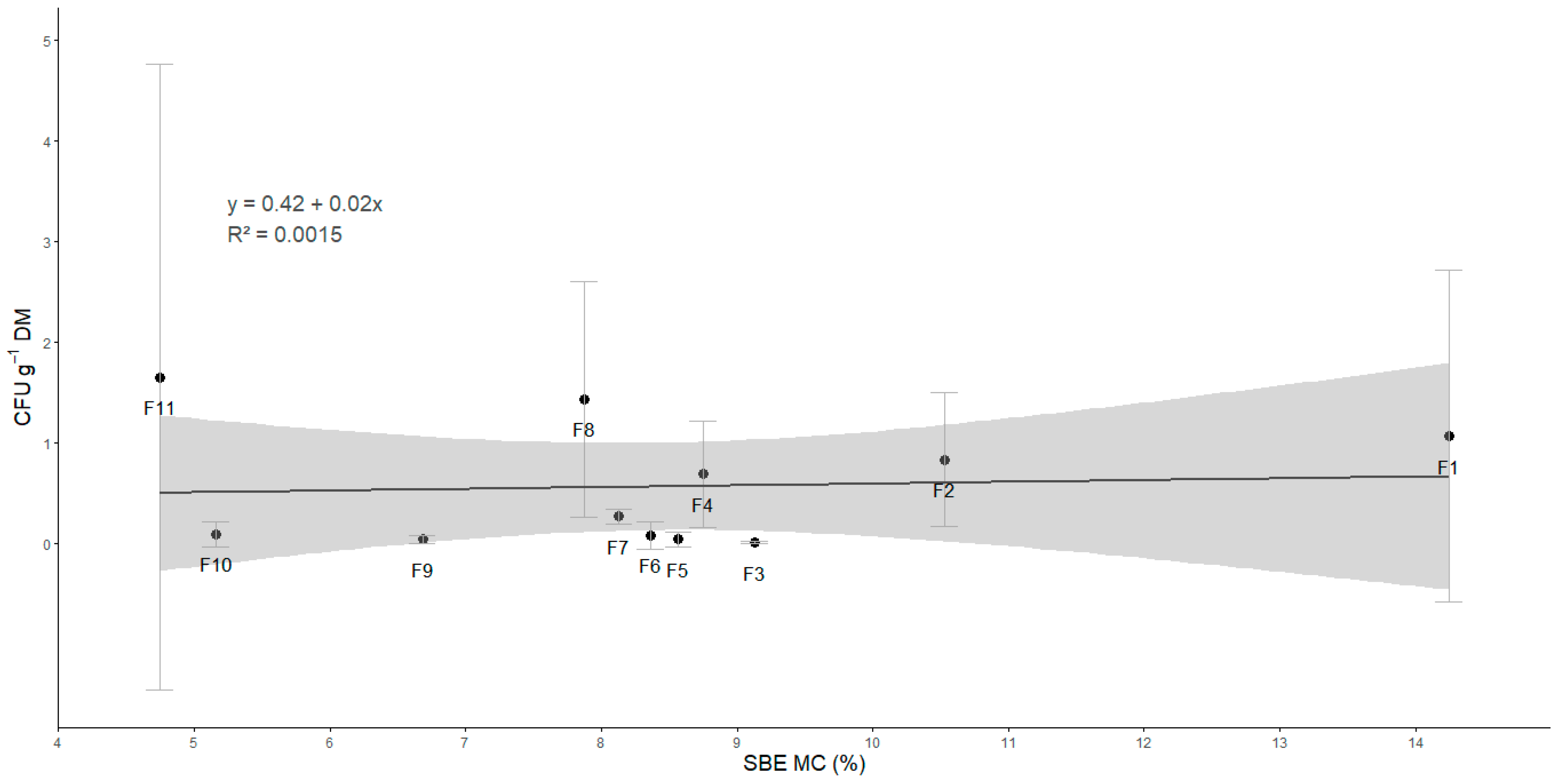
The Figure 2 presents the mean fungal counts (CFU g−1 DM) alongside the mean MC of SBE samples from different extruding-expelling facilities. Fungal counts in SBE samples were low, with mean values ranging from 0 to 4 CFU g−1 DM. The highest average was 1.66 ± 3.99 CFU g−1 DM (recorded in Facility 11), while the lowest average was 0.01 ± 0.03 CFU g−1 DM (Facility 3). The highest CFU g−1 DM values were 16.17, 15.36, and 14.64 in one of the four replicates of samples corresponding to Facilities 8, 11, and 1, respectively. No significant differences in CFU averages were found among the facilities (p > 0.05 in the Dunn test).
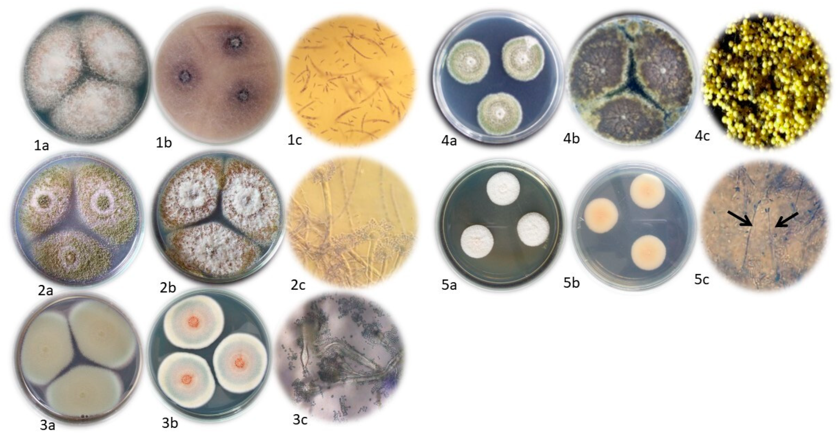
Seven genera of filamentous fungi belonging to upper taxonomic classes were identified in the SBE samples, including Acremonium sp., Alternaria sp., Aspergillus spp., Cladosporium sp., Eurotium sp., Fusarium sp. and Penicillium spp., as well as one genus from the lower taxonomic class Mucoraceae (family). Figure 3 shows the macroscopic morphology of some of these genera, which served as the basis for their identification. Due to unforeseen data loss, images for every detected genus could not be provided. Penicillium and Mucoraceae were the most frequently isolated (28.0% and 25.6%, respectively), followed by Aspergillus (15.5%) and Cladosporium (14.3%). The isolation frequency of the remaining fungi were below 10%, while the yeasts exhibited an isolation frequency of 11.9%. These results indicate that no single genus predominated in the SBE samples.
An analysis of the relative density of extruding-expelling facilities revealed that total yeasts, Penicillium, and Mucoraceae were detected in the most of the facilities evaluated, although the percentage in the composition of the total mycobiota was variable (Table 1). Total yeasts were found in all facilities, with relative density values ranging from 0.5 to 100%. Penicillium was present in nine facilities, with values ranging from 1.9 to 93.8%, and Mucoraceae was identified in eight facilities, with relative density values between 0.9 and 79%. Among potentially mycotoxigenic genera (Alternaria, Aspergillus and Fusarium), Aspergillus was isolated in six facilities with relative density values ranging from 0.3% to 68.8%. Alternaria was identified in three facilities with values between 0.3 and 0.9%, while Fusarium was isolated in only one facility with a relative density value of 46%.

3.2. Relationship Between Soybean Expeller Moisture Content and Fungal Biota Load

The average MC of SBE ranged from 4.74% to 14.25% across the 11 extruding-expelling facilities (Figure 2). Despite this variability, the fungal biota load, expressed as CFUg−1 DM, remained relatively constant, ranging from 0 to 4. No significant linear relationship (p > 0.05) was found between CFU counts and SBE MC, as evidenced by the low Pearson correlation coefficient (r = 0.039).

3.3. Detection and Quantification of Mycotoxins

Only DON and aflatoxins (AFs) were detected in the SBE samples (Table 2). DON and AFB1 were quantified in 20.0% and 25.7% of samples, respectively, with average concentrations of 215.19 μg kg−1 for DON and 41.68 μg kg−1 for AFB1 (Table 2). The detected DON levels ranged from 1.50 μg kg−1 (quantification limit) to 2385.20 μg kg−1. Among AFs, AFB1 exhibited the highest concentration, reaching 378 μg kg−1. AFG1 and AFG2 were quantified in 40% of the samples but at low concentrations, with average values of 0.39 μg kg−1 and 0.34 μg kg−1, respectively. AFB2 levels were below the quantification limit. The other analyzed mycotoxins, including NIV, 13 acetyl DON, 15 acetyl DON, ochratoxin A and zearalenone, were not detected.
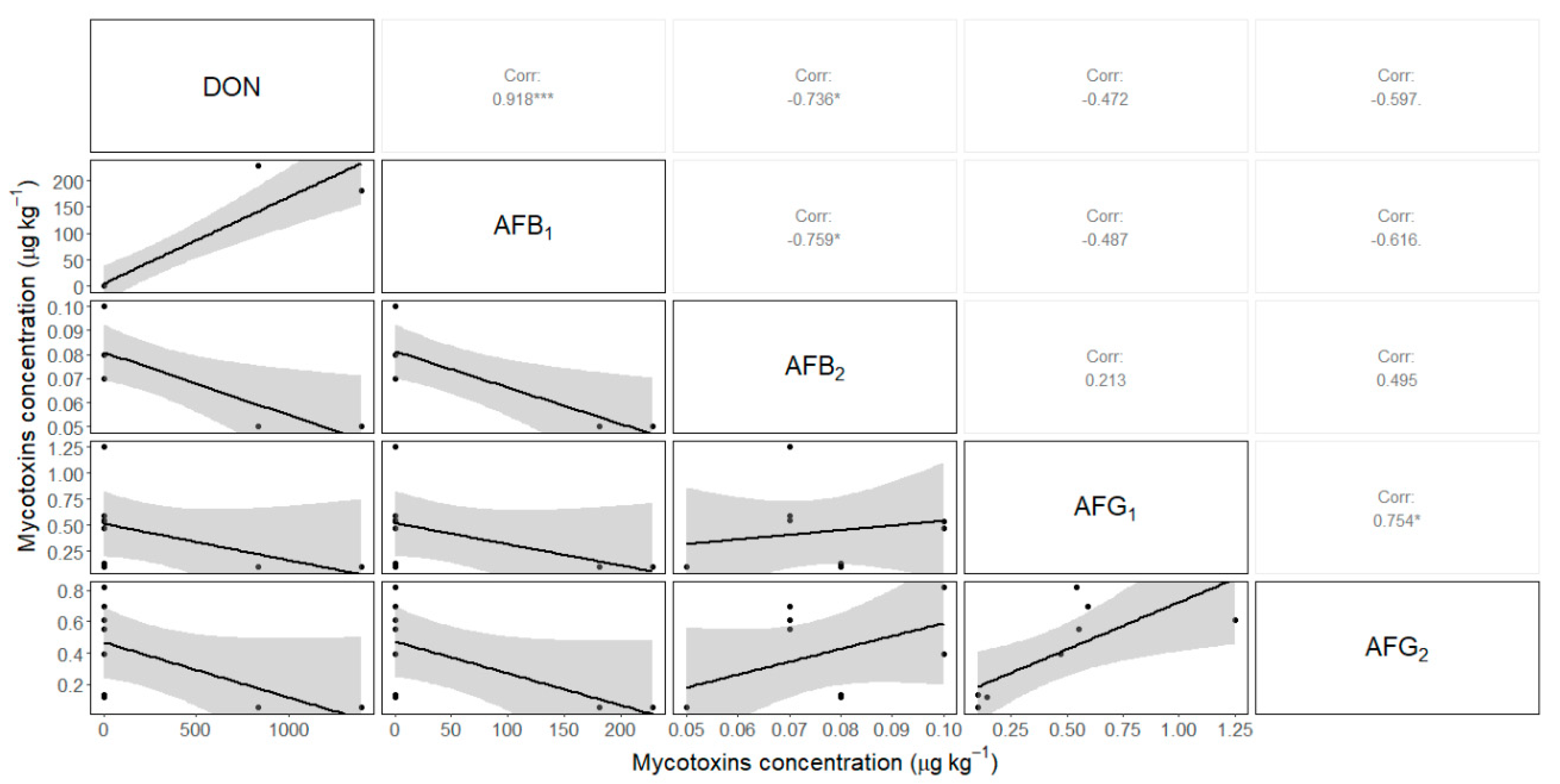
Positive linear correlations were found between DON and AFB1 (0.918), AFG1 and AFG2 (0.754), and between DON and AFB2 (0.736). In contrast, a negative correlation was identified between AFB1 and AFB2 (−0.759) (Figure 4). Co-occurrence of two mycotoxins was observed in 60% of the analyzed samples. The combination of AFG1 and AFG2 was found in 40% of the samples, while DON and AFB1 co-occurred in 20% of the samples.

4. Discussion

4.1. Implications of Fungal Biota Quantification and Identification

The fungal counts reported in this study for SBE were low (ranging from 0 to 4 CFU g−1 DM). Currently, no specific regulations define maximum fungal load limits for SBE, and the literature on CFU counts for soybean by-products, such as SBE, is scarce, limiting comparative analyses. Nevertheless, some studies have reported CFU counts for soybeans and soybean meal. For example, Good Manufacturing Practices (GMP 14) recommend a maximum fungal load of 104 CFU g−1 DM for soybean meal [13]. The CFU counts for SBE in this study were lower than those reported by Ghaemmaghami et al. [38] (6 × 10² CFU g−1 DM) for soybean meal and pellets and by Egbuta et al. [57] (106 CFU g−1 DM) for processed soybean, using similar CFU count analysis techniques. Therefore, these results indicate that the SBE exhibits good mycological quality, making it suitable for animal consumption.
The low CFU counts found in all SBE samples may be attributed to several factors. Generally, soybeans have lower microbial loads compared to other grains [19,58], which may be related to their chemical composition, particularly their low carbohydrate content [59,60]. Additionally, the processes involved in converting soybeans into SBE likely reduce fungal load [61]. Specially, the extruding-expelling process, which involves simultaneous pressure and temperature treatments, is highly effective in reducing fungal contamination. During extrusion, soybean seeds are processed through a continuous screw system that generates frictional heat (110–150 °C) and high pressure (40 MPa) for a short time (20–30 s) [1,2].
The combined effect of heat and pressure significantly reduce fungal viability [38]. For example, Fusarium species, such as F. graminearum, do not resist pressures greater than 1.01 MPa, while A. flavus and A. alternata are inhibited at pressures exceeding 0.505 MPa [62].
Temperature, by itself, also has an effect of microflora. In this study, the temperature recorded in the extruding-expelling processes in the different facilities ranged from 125 °C to 168 °C, significantly exceeding the temperature threshold (83° C for 7 s) required to inactivate most mesophilic flora [63]. In consequence, the lack of correlation between SBE MC and normalized CFU in our samples (Figure 2) may be attributed to the effect of high temperature/pressure treatment during the extruding process, which likely reduced the CFU levels.
The most frequently identified fungal genera in SBE samples were Penicillium, Aspergillus, Acremonium, Cladosporium, Eurotium, Alternaria, and Mucoraceae (family). These genera are consistent with those previously reported in soybean, although their isolation frequencies varied [13,22,43]. Differences in isolation frequencies may be attributed to variations in the fungal composition of soybean seed [64] and in the conditions of the extruding-expelling process [38], as mentioned above (high temperature and high pressure) [1,2].
The low occurrence of field fungi, such as Fusarium and Alternaria, could be explained by the low MC of the soybean before processing. Chiotta et al. [65] found that Fusarium and Alternaria were the most common fungi in recently harvested soybeans, whereas Aspergillus and Penicillium, which thrive under lower relative humidity, were more prevalent during storage. Barros et al. [40] reported that, during storage, the diversity of fungal genera decreased, with xerophytic genera (Aspergillus and Eurotium) becoming dominant, while field fungi such as Fusarium disappeared.
The hygienic quality of feed and food products must be ensured through proper sanitary conditions. Fungal presence is a recognized indicator of feed hygiene [66]. However, in Argentina, no specific regulations set fungal load limits for feed.
Field observations showed that surveyed extrusion-expelling facilities lack strict sanitation protocols, likely because most produce expeller for feed rather than food. However, facilities processing SBE for food adhere to strict hygiene regulations [67]. Despite this, extrusion effectively reduces fungal contamination, ensuring a microbiologically safe product. This treatment significantly impacts the viability of both somatic and reproductive fungal structures [68,69]. Previous studies [38,66,70] also found that extrusion significantly reduces microbial loads in pellets, aligning with our findings.

4.2. Implications of Soybean Expeller Moisture Content on Fungal Biota Load

The SBE MC showed significant variability across facilities (Figure 2). Some facilities produced by-products with MC below the safe storage MC, while others exceeded it [71], increasing microbiological risks during storage and posing potential safety concerns for feed and food applications. This variability can be attributed to differences in the raw material MC [72] and the conditions of the extruding-expelling process [2]. The SBE MC observed in this study, ranging from 4.74% to 14.25%, aligns with previous reports from Argentine facilities [8,9] and complies with the maximum permitted MC of 14.3% (12.5% w.b.) established by the XIX standard for commercialization in Argentina [10].
These results suggest that, within the tested MC range, fungal proliferation is not only influenced by MC, indicating that additional factors may contribute to the observed variability in fungal biota load among facilities. One possible explanation for the absence of correlation between SBE MC and CFU in our samples might be the high-temperature heat treatment during extruding process, which likely reduces CFU levels by adversely affecting most mesophilic flora, as reported in previous studies [62,63].

4.3. Assessment of Mycotoxins Contamination and Its Implications in Soybean Expeller

No studies reporting mycotoxin levels in SBE were identified. However, the presence of mycotoxins produced by Fusarium, Alternaria, and Aspergillus in soybean seeds and by-products, such as soybean meal, has been documented [25,65,73].
In this study, DON, AFB1, AFG1, and AFG2 were the only mycotoxins quantified in SBE, with detection frequency ranging from 20 to 40% of analyzed samples (Table 2). Similar mycotoxins were reported by Abia et al. [74] in soybean meal, though with differing occurrence rates. While DON, AFB1, and AFG1 were detected, AFB2, AFG2, and ochratoxins were absent. Soybeans do not provide a favorable matrix for fungal growth due to their low MC and composition, particularly their high protein/carbohydrate ratio [75]. For this reason, reports of mycotoxins in soybeans are scarce in Argentina, and the detected concentrations are variable.
Low levels of AFs and high levels of DON were detected in soybean samples by Barros et al. [13], while Oviedo et al. [76] identified alternariol and alternariol monomethyl ether. D’Espósito et al. [77] found a higher incidence of AFB1 (26.66%, 1.44–7.43 µg kg−1) than DON (6.66%, 297–490 µg kg−1) but in soybean samples obtained from silo bags. Compared to SBE samples in this study, AFB1 incidences were similar, while DON incidences were lower. However, the DON and AFB1 concentration ranges in SBE were significantly higher (Table 2).
DON is commonly associated with Fusarium species, which produce the toxin in the field [78,79]. Reports from Argentina (Córdoba province) confirm the presence of F. graminearum and DON during the grain filling and ripening of soybean under high humidity conditions [65,80]. AFs are significant food and feed contaminants produced mainly by A. flavus (producing AFB) and A. parasiticus (producing both AFB and AFG) [73,81,82,83]. Only 25–40% of A. flavus strains produce AFs [84]. AFB1, AFG1, and AFG2 were quantified in SBE samples at varying concentrations and frequencies (Table 2). AFB1 was quantified in 25.7% of the SBE samples, with no direct correlation to the low relative density of Aspergillus spp. (Table 1). Greco et al. [79] reported AFs in 50% of feed samples containing SBE, with a mean concentration of 2.82 µg kg−1, significantly lower than the mean concentration in this study (10.62 µg kg−1).
The presence of AFG1 and AFG2 in SBE samples, not previously reported, suggest contamination of A. parasiticus, despite its absence in these samples. AFG concentrations were lower than AFB, indicating A. flavus as a likely contributor, given its adaptation to temperate conditions [85].
Mycotoxin contamination does not always correlate with fungal isolation, as toxins can persist after fungal growth ceases due to unfavorable conditions [79]. Industrial processes, such as extrusion, can reduce mycotoxin concentrations [23,86]. However, the effect on each mycotoxin can be different. While DON degrades above 150 °C [44] to 180 °C [87,88,89], AFs are thermally stable, decomposing only at temperatures from 237 °C (AFG) to 306 °C (AFB) [90]. Consequently, the temperatures reached during the extrusion process likely influenced the observed mycotoxin level in the SBE [91].
Notably, a significant positive correlation was observed between DON and AFB1, as well as between AFG1 and AFG2. Conversely, a significant negative correlation was found between DON and AFB2 and between AFB1 and AFB2 (Figure 4). Co-occurrence of mycotoxins was noted in 60% of SBE samples, with 40% showing AFG1 and AFG2, and 20% showing DON and AFB1 (Table 2). Such co-occurrence aligns with studies indicating frequent multi-mycotoxin contamination in raw materials and feed ingredients [39,52,57,92].
DON and AFs concentration in SBE highlights a potential risk to animal and human health. In 8.6% of the samples, DON level exceeded the European Union threshold of 750 ppb (or 750 µg kg−1) for feed ingredients [93]. For AFs in food and feed ingredients, the accepted levels are 20 and 300 µg kg−1, respectively [94]. In the present study, the mean levels of AFB1, AFG1, and AFG2 were 41.70, 0.39, and 0.38 µg kg−1, respectively. These levels exceeded the specification for food but fell below the specification for feed. However, AFB1 concentrations also surpassed food safety limits in isolated cases, reaching 378 µg kg−1 (2.9% of samples). Since there are no local regulations in Argentina regarding mycotoxin levels in SBE, the results were compared with international standards and regulations to assess their safety and quality. This comparison ensures that the findings align with widely recognized guidelines, helping to establish a clearer context for SBE usage in the industry.
Given widespread use of SBE in feed and food formulations, these findings underscore the need for systematic monitoring and adherence to Good Manufacturing Practices (GMP). This study provides clear evidence of raw material (soybean) contamination with mycotoxigenic fungi and their associated mycotoxins, either in the field or during storage. While fungal presence is controlled during the extruding process, as previously explained, mycotoxins are not inactivated during processing. Consequently, the final product may remain contaminated, posing potential health risks to animals consuming SBE directly or as a component of feed formulations. Implementing GMP measures, including drying soybeans, optimizing storage conditions, and regular facility cleaning [95], is critical for minimizing fungal growth and mycotoxin contamination [96,97,98]. However, the facilities analyzed in this study currently lack mycotoxin control measures, further emphasizing the need for preventive strategies to ensure the safety and quality of soybean by-products for industrial use.

5. Conclusions

The low CFU counts observed in SBE samples suggest that the product is generally of good quality for animal consumption. No correlation was found between the SBE MC and the CFU of fungal biota, probably due to the heat treatment applied during the extruding process. Penicillium and Mucoraceae (family) were the most frequently isolated fungal genera, among five others. While the concentrations of DON and AFs (B1, B2, G1, and G2) in the SBE were generally low, some samples exceeded established regulatory limits for food and feed. This suggests that, although thermal treatment during extrusion reduces fungal load, it might not be sufficient to substantially lower mycotoxin concentration. Notably, 60% of SBE samples presented a co-occurrence of at least two mycotoxins.
These findings emphasize the critical importance of monitoring and controlling the SBE MC during storage and conducting regular mycotoxin testing in both whole soybeans and the resulting SBE to ensure safety and quality. Furthermore, the detection of mycotoxins exceeding regulatory thresholds in some SBE samples underscores a potential risk to both animal and human health, emphasizing the need for stringent quality control measures.

Author Contributions

Conceptualization, G.M. and R.E.B.; Data curation, G.M.; Formal analysis, G.M., C.C.C., L.M.C. and M.B.A.; Funding acquisition, L.M.C., P.B. and R.E.B.; Investigation, C.C.C. and M.B.A.; Methodology, G.M. and C.C.C.; Project administration, R.E.B.; Resources, L.M.C. and R.E.B.; Supervision, R.E.B.; Writing—original draft, G.M.; Writing—review and editing, C.C.C., L.M.C., M.B.A., P.B. and R.E.B. All authors have read and agreed to the published version of the manuscript.

Funding

This research was funded by the Instituto Nacional de Tecnología Agropecuaria (INTA) Projects P.E. I119, P.E. I120 and REC I165.

Data Availability Statement

Dataset available on request from the authors.

Acknowledgments

The authors are grateful to Leandro S. Cambareri, Martín Mansilla, and Diego Cristos for their technical support.

Conflicts of Interest

The authors declare no conflicts of interest.

Abbreviations

The following abbreviations are used in this manuscript:
SBESoybean expeller
MCMoisture content
CFUColony forming unit
DONDeoxynivalenol
AFAflatoxin
d.b.Dry basis
w.b.Wet basis
DMDry matter
PDAPotato dextrose agar
LODLimit of detection
LOQLimit of quantification
ANOVAAnalysis of variance
SDStandard deviation
GMPGood Manufacturing Practices

Appendix A

Table A1. List of analytes determined in the negative or positive ionization mode and parameters for mass-spectrometry.
Analyte NameIonizationPrecursor Ion (m/z)Ion SpeciesProduct Ion (m/z)Collision Cell
Ion QIon C
DONESI+297[M+H]+24923139
NivalenolESI-311[M−H]18328139
13 acetil DONESI+339[M+H]+23121339
15 acetil DONESI+339[M+H]+32113739
NivalenolESI-371[M+OAc]31128145
AFB1ESI+313[M+H]+28424125
AFB2ESI+315[M+H]+28625928
AFG1ESI+329[M+H]+24320025
AFG2ESI+331[M+H]+31324528
Note: Ion C is cualification product ions, Ion Q is quantification product ions, ESI is electrospray ionization, [M+H]+ is protonated adduct, [M−H] is deprotonated adduct, and [M+OAc] is acetate adduct.

References

  1. Nelson, A.I.; Wijeratne, W.B.; Yeh, S.W.; Wei, T.M.; Wei, L.S. Dry Extrusion as an Aid to Mechanical Expelling of Oil from Soybeans. J. Am. Oil Chem. Soc. 1987, 64, 1341–1347. [Google Scholar] [CrossRef]
  2. Bargale, P.C.; Ford, R.J.; Sosulski, F.W.; Wulfsohn, D.; Irudayaraj, J. Mechanical Oil Expression from Extruded Soybean Samples. J. Am. Oil Chem. Soc. 1999, 76, 223–229. [Google Scholar]
  3. Bragachini, M.; Ustarroz, F.; Saavedra, A.E.; Méndez, J.M.; Mathier, D.; Bragachini, M.; Sosa, N.; Alladio, R.M.; Accoroni, C.; Hennung, H. Evolución Del Sistema Productivo Agropecuario Argentino; Manfredi: Córdoba, Argentina, 2017. [Google Scholar]
  4. Medic, J.; Atkinson, C.; Hurburgh, C.R. Current Knowledge in Soybean Composition. J. Am. Oil Chem. Soc. 2014, 91, 363–384. [Google Scholar] [CrossRef]
  5. Papier, K.; Tong, T.Y.; Appleby, P.N.; Bradbury, K.E.; Fensom, G.K.; Knuppel, A.; Perez-Cornago, A.; Schmidt, J.A.; Travis, R.C.; Key, T.J. Comparison of Major Protein-Source Foods and Other Food Groups in Meat-Eaters and Non-Meat-Eaters in the Epic-Oxford Cohort. Nutrients 2019, 11, 824. [Google Scholar] [CrossRef]
  6. SAGyP. Molienda de Granos Oleaginosos, Producción de Aceites, Pellets y Expellers. Available online: https://www.magyp.gob.ar/sitio/areas/ss_mercados_agropecuarios/areas/granos/_archivos/000058_Estad%C3%ADsticas/000032_Evolucion%20de%20la%20Molienda%20(Cereales%20y%20Oleaginosas)/000002_Evoluci%C3%B3n%20de%20la%20Molienda%20Mensual%20-%20Oleaginosas/000002_Evoluci%C3%B3n%20de%20la%20Molienda%20Mensual%20-%20Oleaginosas.php (accessed on 25 September 2024).
  7. RUCA-MAGyP. Registro Único de La Cadena Agroalimentaria. Available online: https://ruca.magyp.gob.ar/padron/ (accessed on 24 November 2023).
  8. Maciel, G.; Wagner, J.R.; Juan, N.A.; San Martino, S.; Bartosik, R.E. Assessment of the Main Sources of Variability of Soybean (Glycine Max) Expeller Composition and Quality: A Field Study. Agric. Eng. Int. CIGR J. 2020, 22, 211–220. [Google Scholar]
  9. Juan, N.A.; Massigogue, J.I.; Errasquin, L.; Méndez, J.M.; Ochandio, D.C.; Saavedra, A.E.; Paolilli, M.C.; Alladio, R.M.; Accoroni, C.; Behr, E.F. Calidad de la Soja Procesada y del Expeller Producido por la Industria de Extrusado-Prensado en Argentina; Ediciones INTA: Buenos Aires, Argentina, 2015. [Google Scholar]
  10. SAGPyA. Resolución SAGPyA N°317/99. Norma XIX: Subproductos de Oleaginosos; SAGPyA: Buenos Aires, Argentina, 1999; p. 10. [Google Scholar]
  11. Kumar, C.; Karim, M.A.; Joardder, M.U.H. Intermittent Drying of Food Products: A Critical Review. J. Food Eng. 2014, 121, 48–57. [Google Scholar] [CrossRef]
  12. Smith, J.E.; Solomons, G.; Lewis, C.; Anderson, J.G. Role of Mycotoxins in Human and Animal Nutrition and Health. Nat. Toxins 1995, 3, 187–192. [Google Scholar] [CrossRef]
  13. Barros, G.G.; Oviedo, M.S.; Ramirez, M.L.; Chulze, S.N. Safety Aspect in Soybean Food and Feed Chains: Fungal and Mycotoxins Contamination. In Soybean—Biochemistry, Chemistry and Psysiology; IntechOpen: London, UK, 2011; pp. 7–20. [Google Scholar]
  14. Fleurat-Lessard, F. Integrated Management of the Risks of Stored Grain Spoilage by Seedborne Fungi and Contamination by Storage Mould Mycotoxins—An Update. J. Stored Prod. Res. 2017, 71, 22–40. [Google Scholar] [CrossRef]
  15. Magan, N.; Sanchis, V.; Aldred, D. The Role of Spoilage Fungi in Seed Deterioration. In Fungal Biotechnology in Agricultural, Food and Environmental Applications; CRC Press: Boca Raton, FL, USA, 2004; Chapter 28; pp. 311–323. [Google Scholar]
  16. Sirohi, R.; Tarafdar, A.; Kumar Gaur, V.; Singh, S.; Sindhu, R.; Rajasekharan, R.; Madhavan, A.; Binod, P.; Kumar, S.; Pandey, A. Technologies for Disinfection of Food Grains: Advances and Way Forward. Food Res. Int. 2021, 145, 110396. [Google Scholar] [CrossRef]
  17. Suleiman, R.A.; Rosentrater, K.A.; Bern, C.J. Effects of Deterioration Parameters on Storage of Maize. In Proceedings of the ASABE Annual International Meeting, Kansas City, MO, USA, 21–24 July 2013; p. 51. [Google Scholar]
  18. Castellari, C.C.; Cendoya, M.G.; Marcos Valle, F.J.; Barrera, V.; Pacin, A.M. Factores Extrínsecos e Intrínsecos Asociados a Poblaciones Fúngicas Micotoxigénicas de Granos de Maíz (Zea mays L.) Almacenados En Silos Bolsa En Argentina. Rev. Argent. Microbiol. 2015, 47, 350–359. [Google Scholar] [CrossRef]
  19. Garcia, L.P.; Savi, G.D.; Santos, K.; Scussel, V.M. Fumonisins and Fungi in Dry Soybeans (Glycine max L.) for Human Consumption. Food Addit. Contam. Part B Surveill. 2016, 9, 79–84. [Google Scholar] [CrossRef] [PubMed]
  20. Gomes, D.P.; Kronka, A.Z.; Barrozo, L.M.; Silva, R.P.D.; Souza, A.L.; Silva, B.M.S.e.; Panizzi, R.D.C. Efeito Da Colhedora, Velocidade e Ponto de Coleta Na Contaminação de Sementes de Soja Por Fungos. Rev. Bras. Sementes 2009, 31, 160–166. [Google Scholar] [CrossRef]
  21. Njobeh, P.B.; Dutton, M.F.; Koch, S.H.; Chuturgoon, A.; Stoev, S.; Seifert, K. Contamination with Storage Fungi of Human Food from Cameroon. Int. J. Food Microbiol. 2009, 135, 193–198. [Google Scholar] [CrossRef] [PubMed]
  22. Zelaya, M.J.; González, H.H.L.; Resnik, S.L.; Pacin, A.M. Mycobiota and Potential Mycotoxin Contamination of Soybean RR in Different Production Areas in Argentina. Int. Res. J. Plant Sci. 2013, 4, 133–143. [Google Scholar]
  23. Bullerman, L.B.; Bianchini, A. Good Food-Processing Techniques: Stability of Mycotoxins in Processed Maize-Based Foods. In Mycotoxin Reduction in Grain Chains; John Wiley & Sons: Hoboken, NJ, USA, 2014; pp. 89–100. [Google Scholar]
  24. Kumera, N.; Mohammed, A. Mycotoxin Occurrence in Grains and the Role of Postharvest Management as a Mitigation Strategies. A Review. Food Cont. 2017, 78, 412–425. [Google Scholar]
  25. Schollenberger, M.; Müller, H.M.; Rüfle, M.; Suchy, S.; Plank, S.; Drochner, W. Natural Occurrence of 16 Fusarium Toxins in Grains and Feedstuffs of Plant Origin from Germany. Mycopathologia 2006, 161, 43–52. [Google Scholar] [CrossRef]
  26. Waskiewicz, A. Natural Occurrence of Mycotoxins in Food. Encycl. Food Microbiol. 2014, 2, 880–886. [Google Scholar]
  27. Mishra, S.; Ansari, K.M.; Dwivedi, P.D.; Pandey, H.P.; Das, M. Occurrence of Deoxynivalenol in Cereals and Exposure Risk Assessment in Indian Population. Food Control 2013, 30, 549–555. [Google Scholar] [CrossRef]
  28. WHO. Safety Evaluation of Certain Mycotoxins in Food. In FAO Food and Nutrition Paper 74; WHO Food Additives Series 47; WHO: Genève, Switzerland, 2001; pp. 419–455. [Google Scholar]
  29. Rotter, B.A.; Prelusky, D.B.; Pestka, J.J. Invited Review: Toxicology of deoxynivalenol (vomitoxin). J. Toxicol. Environ. Health 1996, 48, 1–34. [Google Scholar] [CrossRef]
  30. International Agency for Research on Cancer. Monographs on the Evaluation of Carcinogenic Risk of Chemicals to Humanas: Some Naturally Occurring Substances: Food Ítems and Constituents, Heterocyclic Aromatic Amines and Mycotoxins. Hum. Papillomaviruses 1993, 56, 609. [Google Scholar]
  31. Amuzie, C.J.; Pestka, J.J. Suppression of Insulin-Like Growth Factor Acid-Labile Subunit Expression—A Novel Mechanism for Deoxynivalenol-Induced Growth Retardation. Toxicol. Sci. 2010, 3, 412–421. [Google Scholar]
  32. Ellis, W.O.; Smith, J.P.; Simpson, B.K.; Oldham, J.H.; Scott, P.M. Aflatoxins in Food: Occurrence, Biosynthesis, Effects on Organisms, Detection, and Methods of Control. Crit. Rev. Food Sci. Nutr. 1991, 30, 403–439. [Google Scholar] [CrossRef] [PubMed]
  33. Bennett, J.; Klich, M. Mycotoxins: Clinical Microbiology Reviews. ASM 2003, 16, 497–516. [Google Scholar]
  34. International Agency for Research on Cancer. Monographs on the Evaluation of Carcinogenic Risk of Chemicals to Humanas: Some Traditionally Herbal Medicines, Some Mycotoxins, Naphthalene and Styrene; WHO: Genève, Switzerland, 2002; Volume 82, p. 601. [Google Scholar]
  35. Arapcheska, M.; Jovanovska, V.; Jankuloski, Z.; Hajruali –Musliu, Z.; Uzunov, R. Imapct of Aflatoxins on Animal and Human Health. Int. J. Innov. Sci. Eng. Technol. 2015, 2, 156–161. [Google Scholar]
  36. Joint FAO/WHO Expert Committee on Food Additives. Safety Evaluation of Certain Food Additives and Contaminants; Joint FAO/WHO Expert Committee on Food Additives AFLA: Geneva, Switzerland, 1998. [Google Scholar]
  37. Bhat, R.; Reddy, K.R.N. Challenges and Issues Concerning Mycotoxins Contamination in Oil Seeds and Their Edible Oils: Updates from Last Decade. Food Chem. 2017, 215, 425–437. [Google Scholar] [CrossRef]
  38. Ghaemmaghami, S.S.; Modirsaneii, M.; Khosravi, A.R.; Razzaghi-Abyaneh, M. Study on Mycoflora of Poultry Feed Ingredients and Finished Feed in Iran. Iran J. Microbiol. 2016, 8, 47–54. [Google Scholar]
  39. Gutleb, A.C.; Caloni, F.; Giraud, F.; Cortinovis, C.; Pizzo, F.; Hoffmann, L.; Bohn, T.; Pasquali, M. Detection of Multiple Mycotoxin Occurrences in Soy Animal Feed by Traditional Mycological Identification Combined with Molecular Species Identification. Toxicol. Rep. 2015, 2, 275–279. [Google Scholar] [CrossRef]
  40. Barros, G.; Gacía, D.; Oviedo, M.S.; Ramirez, M.L.; Torres, A.; Chulze, S. Influencia Del Almacenamiento Sobre La Micoflora de Soja Utilizada Para La Producción de Harina. In VI Congreso Latinoamericano de Micología; Asociacion Latinoamericana de Micologia: Mar del Plata, Argentina; Buenos Aires, Argentina, 2008. [Google Scholar]
  41. Boca, R.T.; Pacin, A.M.; González, H.H.L.; Resnik, S.L.; Souza, J.C. Soja y Mocotoxinas: Flora Fùngica—Variedades—Prácticas Agronómicas. Aceites Grasas 2003, Tomo XIII, 510–515. [Google Scholar]
  42. Broggi, L.E.; Pacin, A.M.; Gasparovie, A.; Sacchi, C.; Rothermel, A.; Gallay, A.; Resnik, S. Natural Occurrence of Aflatoxins, Deoxynivalenol, Fumonisins and Zearalenone in Maize from Entre Ríos Province, Argentina. Mycotoxin Res. 2007, 23, 59–64. [Google Scholar] [CrossRef]
  43. Garrido, C.E.; González, H.L.; Salas, M.P.; Resnik, S.L.; Pacin, A.M. Mycoflora and Mycotoxin Contamination of Roundup Ready Soybean Harvested in the Pampean Region, Argentina. Mycotoxin Res. 2013, 29, 147–157. [Google Scholar]
  44. Gimeno, A.; Martins, M.L. Micotoxinas y Micotoxicosis en Animales y Humanos, 3rd ed.; Special Nutrients, Inc.: Miami, FL, USA, 2011. [Google Scholar]
  45. ASAE Standard—S352.2; Moisture Measurement—Unground Grain and Seeds. ASAE: St. Joseph, MI, USA, 2003; p. 593.
  46. Atlas, R.M. Handbook of Microbiological Media, 3rd ed.; CRC Press: Boca Raton, FL, USA, 2004; ISBN 978-0-429-12903-2. [Google Scholar]
  47. Pitt, J.I.; Hocking, A.D. Fungi and Food Spoilage; Springer International Publishing: Berlin/Heidelberg, Germany, 2009; ISBN 978-0-387-92206-5. [Google Scholar]
  48. Nelson, P.E.; Toussoun, T.A.; Marasas, W.F.O. Fusarium Species. An Illustrated Manual of Identification; The Pennsylvania State University Press: University Park, PA, USA, 1983. [Google Scholar]
  49. Barbosa, T.S.; Pereyra, C.M.; Soleiro, C.A.; Dias, E.O.; Oliveira, A.A.; Keller, K.M.; Silva, P.P.O.; Cavaglieri, L.R.; Rosa, C.A.R. Mycobiota and Mycotoxins Present in Finished Fish Feeds from Farms in the Rio de Janeiro State, Brazil. Int. Aquat. Res. 2013, 5, 3. [Google Scholar] [CrossRef]
  50. Zelaya, M.J. Estudio de La Micoflora Contaminante Del Germoplasma de Soja En Argentina; Universidad de Buenos Aires: Buenos Aires, Argentina, 2006. [Google Scholar]
  51. Castañares, E.; Martínez, M.; Cristos, D.; Rojas, D.; Lara, B.; Stenglein, S.; Dinolfo, M.I. Fusarium Species and Mycotoxin Contamination in Maize in Buenos Aires Province, Argentina. Eur. J. Plant Pathol. 2019, 155, 1265–1275. [Google Scholar] [CrossRef]
  52. Calori-Domingues, M.A.; Iwahashi, P.M.R.; Ponce, G.H.; Gloria, E.M.d.; Dias, C.T.d.S.; Button, D.C.; De Camargo, A.C. Aflatoxin B1 and Zearalenone in Soybeans: Occurrence and Distribution in Whole and Defective Kernels. Food Addit. Contam. Part B Surveill. 2018, 11, 273–280. [Google Scholar] [CrossRef] [PubMed]
  53. Schothorst, R.; van Egmond, H.P. Report from SCOOP Task 3.2.10 “Collection of Occurrence Data of Fusarium Toxins in Food and Assessment of Dietary Intake by the Population of EU Member States” Subtask: Trichothecenes. Tox. Let. 2004, 153, 133–143. [Google Scholar] [CrossRef] [PubMed]
  54. R Core Team. R: A Language and Environment for Statistical Computing; R Core Team: Vienna, Austria, 2023; Available online: https://www.r-project.org/ (accessed on 21 April 2023).
  55. Kruskal, W.H.; Wallis, W.A. Use of Ranks in OneCriterion Variance Analysis. J. Am. Stat. Assoc. 1952, 47, 583–621. [Google Scholar] [CrossRef]
  56. Dunn, J.T. Multiple Comparisons Using Rank Sums. Technometrics 1964, 6, 241–252. [Google Scholar] [CrossRef]
  57. Egbuta, M.A.; Mwanza, M.; Phoku, J.Z.; Chilaka, C.A.; Dutton, M.F. Comparative Analysis of Mycotoxigenic Fungi and Mycotoxins Contaminating Soya Bean Seeds and Processed Soya Bean from Nigerian Markets. Adv. Microbiol. 2016, 06, 1130–1139. [Google Scholar] [CrossRef]
  58. Roigé, M.B.; Aranguren, S.M.; Riccio, M.B.; Pereyra, S.; Soraci, A.L.; Tapia, M.O. Mycobiota and Mycotoxins in Fermented Feed, Wheat Grains and Corn Grains in Southeastern Buenos Aires Province, Argentina. Rev. Iberoam. Micol. 2009, 26, 233–237. [Google Scholar] [CrossRef]
  59. Ochandio, D.; Bartosik, R.; Gastón, A.; Abalone, R.; Arias Barreto, A.; Yommi, A. Modelling Respiration Rate of Soybean Seeds (Glycine max (L.)) in Hermetic Storage. J. Stored Prod. Res. 2017, 74, 36–45. [Google Scholar] [CrossRef]
  60. Piotrowska, M.; Śliżewska, K.; Biernasiak, J. Chapter 8: Mycotoxins in Cereal and Soybean-Based Food and Feed. In Soybean—Pest Resistance; El-Shemy, H.A., Ed.; InTech: Rijeka, Croatia, 2013; pp. 185–230. ISBN 978-953-51-0978-5. [Google Scholar]
  61. Coradi, P.C.; De Lacerda Filho, A.F.; Paes Chaves, J.B.; De Castro Melo, E. Effects of the Feed Processing in the Reduction of the Microbiological Contamination on the Final Product. Rev. Bras. Prod. Agroindustriais 2013, 15, 81–92. [Google Scholar]
  62. Buerman, E.C.; Worobo, R.W.; Padilla-Zakour, O.I. High Pressure Processing of Spoilage Fungi as Affected by Water Activity in a Diluted Apple Juice Concentrate. Food Cont. 2019, 8, 106779. [Google Scholar] [CrossRef]
  63. Okelo, P.O.; Wagner, D.D.; Carr, L.E.; Wheaton, F.W.; Douglass, L.W.; Joseph, S.W. Optimization of Extrusion Conditions for Elimination of Mesophilic Bacteria during Thermal Processing of Animal Feed Mash. Anim. Feed Sci. Technol. 2006, 129, 116–137. [Google Scholar] [CrossRef]
  64. Zelaya, M.J.; González, H.L.; Resnik, S.L.; Martinez, M.J. Microflora Contaminante En Soja Cosechada En La Principal Zona de Producción de La República Argentina. In Proceedings of the X Congreso Argentino de Ciencia y Tecnología de Alimentos. Primer Simposio Internacional de Nuevas Tecnologías, Mar del Plata, Argentina, 18–20 May 2005; p. 318. [Google Scholar]
  65. Chiotta, M.L.; Fumero, M.V.; Cendoya, E.; Palazzini, J.M.; Alaniz-Zanon, M.S.; Ramirez, M.L.; Chulze, S.N. Toxigenic Fungal Species and Natural Occurrence of Mycotoxins in Crops Harvested in Argentina. Rev. Argent. Microbiol. 2020, 52, 339–347. [Google Scholar] [CrossRef] [PubMed]
  66. Del Pilar Monge, M.; Magnoli, C.E.; Chiacchiera, S.M. Survey of Aspergillus and Fusarium Species and Their Mycotoxins in Raw Materials and Poultry Feeds from Córdoba, Argentina. Mycotoxin Res. 2012, 28, 111–122. [Google Scholar] [CrossRef]
  67. CAA. Código Alimentario Argentino Capítulo II: Condiciones Generales de Las Fábricas y Comercios de Alimentos; CAA: Buenos Aires, Argentina, 2023; p. 78. [Google Scholar]
  68. Jay, J.M.; Loessner, M.J.; Golden, D.A.; García de Fernando Minguillón, G.D. Microbiología Moderna de Los Alimentos; Acribia: Zaragoza, España, 2009; ISBN 84-200-1125-8. [Google Scholar]
  69. Hong, T.D.; Ellis, R.H.; Moore, D. Development of a Model to Predict the Effect of Temperature and Moisture on Fungal Spore Longevity. Ann. Bot. 1997, 79, 121–128. [Google Scholar] [CrossRef]
  70. Dalcero, A.; Magnoli, C.; Luna, M.; Ancasi, G.; Reynoso, M.M.; Chiacchiera, S.; Miazzo, R.; Palacio, G. Mycoflora and Naturally Occurring Mycotoxins in Poultry Feeds in Argentina. Mycopathologia 1998, 141, 37–43. [Google Scholar] [CrossRef]
  71. Maciel, G.; de la Torre, D.A.; Cardoso, L.M.; Cendoya, M.G.; Wagner, J.R.; Bartosik, R.E. Determination of Safe Storage Moisture Content of Soybean Expeller by Means of Sorption Isotherms and Product Respiration. J. Stored Prod. Res. 2020, 86, 101567. [Google Scholar] [CrossRef]
  72. Zhu, S.; Riaz, M.N.; Lusas, E.W. Effect of Different Extrusion Temperatures and Moisture Content on Lipoxygenase Inactivation and Protein Solubility in Soybeans. J. Agric. Food Chem. 1996, 44, 3315–3318. [Google Scholar] [CrossRef]
  73. Pacin, A. ¿Existe UN Diagnóstico Sobre Micotoxinas en Soja en Argentina? In Proceedings of the Workshop Calidad de la Producción y Granos con Valor Agregado, Mercosoja, Rosario-Santa Fe, Argentina, 2006; pp. 285–287. [Google Scholar]
  74. Abia, W.A.; Warth, B.; Sulyok, M.; Krska, R.; Tchana, A.N.; Njobeh, P.B.; Dutton, M.F.; Moundipa, P.F. Determination of Multi-Mycotoxin Occurrence in Cereals, Nuts and Their Products in Cameroon by Liquid Chromatography Tandem Mass Spectrometry (LC-MS/MS). Food Control 2013, 31, 438–453. [Google Scholar] [CrossRef]
  75. Pereira, C.S.; Cunha, S.C.; Fernandes, J.O. Prevalent Mycotoxins in Animal Feed: Occurrence and Analytical Methods. Toxins 2019, 11, 290. [Google Scholar] [CrossRef]
  76. Oviedo, M.S.; Ramirez, M.L.; Barros, G.G.; Chulze, S.N. Influence of Water Activity and Temperature on Growth and Mycotoxin Production by Alternaria Alternata on Irradiated Soya Beans. Int. J. Food Microbiol. 2011, 149, 127–132. [Google Scholar] [CrossRef] [PubMed]
  77. D’Espósito, R.E.; Jimeno, M.A.; Metz, M.P.; Cardozo, G. Aislamiento Micotoxinas En Silos Bolsa Con Granos de Maíz y Soja. In Proceedings of the I Jornada Nacional de Agroalimentos y Sustentabilidad, Córdoba, Argentina, 7 June 2019; pp. 1–3. [Google Scholar]
  78. Joint FAO/WHO Expert Committee on Food Additives. Summary and Conclusions. Seventy-Second Meeting of the Joint FAO/WHO Expert Committee on Food Additives (JECFA/72/SC); WHO: Geneva, Switzerland, 2010; p. 16. [Google Scholar]
  79. Greco, M.; Pardo, A.; Pose, G. Mycotoxigenic Fungi and Natural Co-Occurrence of Mycotoxins in Rainbow Trout (Oncorhynchus Mykiss) Feeds. Toxins 2015, 7, 4595–4609. [Google Scholar] [CrossRef] [PubMed]
  80. Barros, G.; Alaniz Zanon, M.S.; Abod, A.; Oviedo, M.S.; Ramirez, M.L.; Reynoso, M.M.; Torres, A.; Chulze, S. Natural Deoxynivalenol Occurrence and Genotype and Chemotype Determination of a Field Population of the Fusarium Graminearum Complex Associated with Soybean in Argentina. Food Addit. Contam. Part A Chem. Anal. Control. Expo. Risk Assess. 2012, 29, 293–303. [Google Scholar] [CrossRef] [PubMed]
  81. Ahammed, S.K.; Anandam, R.J.; Prasad Babu, G.; Munikrishnaiah, M.; Gopal, K. Studies on Seed Mycroflora of Soybean and Its Effect on Seed and Seedling Quality Characters. Legume Res. 2006, 29, 186–190. [Google Scholar]
  82. EFSA. Aflatoxins (Sum of B1, B2, G1, G2) in Cereals and Cereal-derived Food Products; EFSA: Parma, Italy, 2013; p. 11. [Google Scholar]
  83. Vaamonde, G.; Degrossi, C.; Comerio, R.; Fernandez Pinto, Y.V. Provincia de cordoba (argentina): Caracteristicas diferenciales y capacidad aflatoxicogenicA. Bol. Soc. Argent. Bot. 1995, 30, 191–198. [Google Scholar]
  84. Abarca, M.L.; Bragulat, M.R.; Castella, G.; Cabanes, F.J. Mycoflora and Aflatoxin-Producing Strains in Animal Mixed Feeds; Elsevier: Amsterdam, The Netherlands, 1994; Volume 57, pp. 256–258. [Google Scholar]
  85. Gourama, H.; Bullerman, L.B. Aspergillus Flavus and Aspergillus Parasiticus: Aflatoxigenic Fungi of Concern in Foods and Feeds T: A Review. J. Food Prot. 1995, 58, 1395–1404. [Google Scholar]
  86. Ricca, A.P. Gestion de La Inocuidad En Los Granos Para Potenciar Su Valor Agregado. In Integración Asociativa del Campo a la Góndola; Ediciones, I., Ed.; 1° Congreso de Valor Agregado en Origen; Manfredi: Códoba, Argentina, 2012; pp. 225–229. [Google Scholar]
  87. Ehling, G.; Cockburn, A.; Snowdon, P.; Buschhaus, H. The Significance of the Fusarium Toxin Deoxynivalenol (DON) for Human and Animal Health. Cereal Res. Commun. 1997, 25, 443–447. [Google Scholar]
  88. Hazel, C.M.; Patel, S. Influence of Processing on Trichothecene Levels. Toxicol. Lett. 2004, 153, 51–59. [Google Scholar] [CrossRef]
  89. Lori, G.A.; Rizzo, I. Deoxinivalenol. In Micotoxinas en Alimentos; Editorial Díaz de Santos, S.A.: Madrid, Spain, 2007; pp. 269–292. ISBN 978-84-9969-962-2. [Google Scholar]
  90. Jalili, M. A Review on Aflatoxins Reduction in Food. Iran. J. Health Saf. Environ. 2015, 3, 445–459. [Google Scholar]
  91. Cazzaniga, D.; Basílico, J.C.; González, R.J.; Torres, R.L.; De Greef, D.M. Mycotoxins Inactivation by Extrusion Cooking of Corn Flour. Lett. Appl. Microbiol. 2001, 33, 144–147. [Google Scholar] [CrossRef]
  92. Binder, E.M.; Tan, L.M.; Chin, L.J.; Handl, J.; Richard, J. Worldwide Occurrence of Mycotoxins in Commodities, Feeds and Feed Ingredients. Anim. Feed Sci. Technol. 2007, 137, 265–282. [Google Scholar] [CrossRef]
  93. Regulation EC. Regulation (EC) N° 1881/2006. Setting Maximum Levels for Certain Contaminants in Foodstuffs; EU: Brussels, Belgium, 2006; Volume L 364. [Google Scholar]
  94. FDA. Guidance for Industry: Action Levels for Poisonous or Deleterious Substances in Human Food and Animal; FDA: Silver Spring, MD, USA, 2000. [Google Scholar]
  95. Abadia, B.; Bartosik, R. Manual de Buenas Prácticas En Poscosecha de Granos: Hacia El Agregado de Valor En Origen de La Producción Primaria; Bernadette, A., Bartosik, R., Eds.; Ediciones INTA: Buenos Aires, Argentina, 2013; ISBN 978-987-679-264-6. [Google Scholar]
  96. Lusas, E.W. Soybean Processing and Utilization. In Soybeans: Improvement, Production, and Uses; American Society of Agronomy, Crop Science Society of America, Soil Science Society of America: Madison, WI, USA, 2004; Volume 16, pp. 949–1045. [Google Scholar]
  97. Magan, N.; Aldred, D.; Baxter, E.S. Good Postharvest Storage Practices for Wheat Grain. In Mycotoxin Reduction in Grain Chains; John Wiley & Sons: Hoboken, NJ, USA, 2014. [Google Scholar]
  98. Toomer, O.T.; Oviedo, E.O.; Ali, M.; Patino, D.; Joseph, M.; Frinsko, M.; Vu, T.; Maharjan, P.; Fallen, B.; Mian, R. Current Agronomic Practices, Harvest & Post-Harvest Processing of Soybeans (Glycine max)—A Review. Agronomy 2023, 13, 427. [Google Scholar] [CrossRef]
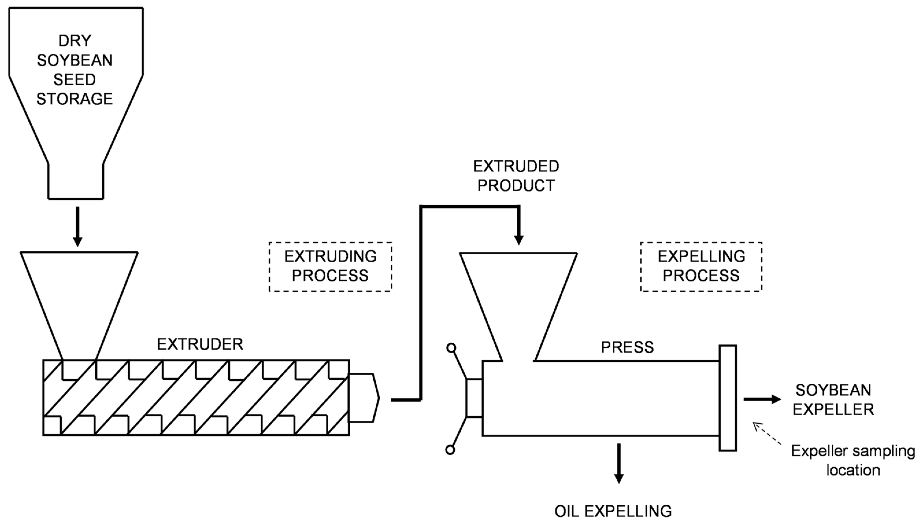
Figure 1. Conceptual diagram of extruding-expelling process.
Agronomy 15 00807 g001
Figure 2. Relationship between average values of soybean expeller moisture content (SBE MC) and counts of colony forming units (CFU g−1 DM) for different extruding-expelling facilities. The dots represent the averages, and the bars, the standard deviation. R2 is the determination coefficient of the fitted linear model. F1 to F11 corresponds to Facility 1 to 11, respectively.
Agronomy 15 00807 g002
Figure 3. Macroscopic morphology of fungal colonies grown on 90 mm Petri dishes with MEA (a) and CYA (b) media, along with reproductive structures and spores of the identified fungi (c): (1a,1b) Fusarium sp.; (1c) asexual spores (optical microscope, 400×); (2a,2b) Aspergillus flavus; (2c) asexual reproductive structures (optical microscope, 400×); (3a,3b) Penicillium sp.; (3c) asexual reproductive structures (optical microscope, 400×); (4a,4b) Eurotium sp.; (4c) cleistothecia (optical microscope, 10×); (5a,5b) Acremonium sp.; (5c) asexual reproductive structures (optical microscope, 400×), black arrows indicate conidiophores. Photographs were captured using an Olympus camera (850,000 pixels, 3× zoom) (CAMEDIA, Tokyo, Japan).
Agronomy 15 00807 g003
Figure 4. Correlation of mycotoxins concentrations in soybean expeller samples. Panels on the lower left side show the distribution of concentrations (in μg kg−1) for each combination of two mycotoxins. Panels in the diagonal show each mycotoxin. Panels on the upper right side show the correlation coefficient for each combination of two mycotoxins. *** p < 0.001; * p < 0.05.
Agronomy 15 00807 g004
Table 1. Relative density of mycotoxigenic fungi and total yeast in soybean expeller samples discriminated by extruded-expelling facilities.
Facility IDAlternaria spp.Aspergillus spp.Fusarium spp.Mucoraceae (family)Penicillium spp.Total
Yeasts
Other
Facility 10.50.5077.07.83.610.6
Facility 206.246.00.94.537.45.0
Facility 3000001000
Facility 40002.67.352.337.7
Facility 5068.801.329.00.50.3
Facility 60000052.647.4
Facility 70.92.201.91.90.592.5
Facility 80.30.3079.02.116.22.1
Facility 9018.2027.336.49.19.1
Facility 10000030.055.015.0
Facility 110001.693.82.72.0
Note: relative density was expressed as percentage (%). Facility ID is extruding-expelling facility identification.
Table 2. Mycotoxins concentration in soybean expeller samples from different extruded-expelling facilities.
Facility IDRepetitionDONAFs
B1B2G1G2
LOD (μg kg−1)30.20.10.20.1
LOQ (μg kg−1)100.30.20.30.2
Facility 11nd<LOQ<LOQ0.91.3
2ndndnd0.50.6
3ndndndndnd
4ndndnd0.3<LOQ
5nd<LOQ<LOQ1.00.6
Facility 21ndndnd0.70.9
2nd<LOQ<LOQ0.60.6
3ndndnd0.50.6
Facility 31nd<LOQ<LOQndnd
2ndndndndnd
3nd<LOQ<LOQnd0.3
Facility 51nd0.3ndndnd
2nd0.3ndndnd
3nd<LOQ<LOQndnd
4nd<LOQ<LOQ0.40.6
5ndndndndnd
6nd<LOQ<LOQndnd
7nd<LOQ<LOQndnd
8ndndndndnd
Facility 71nd<LOQ<LOQ0.30.5
2nd<LOQ<LOQ0.70.9
3nd<LOQ<LOQ0.71.1
Facility 811038.4159.0ndndnd
22385.2256.5ndndnd
3741.6127.0ndndnd
Facility 91nd<LOQ<LOQ0.60.5
2nd<LOQ<LOQ0.70.6
3nd<LOQ<LOQ<LOQ<LOQ
Facility 101ndndndndnd
2nd<LOQ<LOQ3.61.7
3ndndndndnd
Facility 111529.2378.0ndndnd
21573.8231.8ndndnd
3706.7180.4ndndnd
4514.6122.0ndndnd
Minimum (μg kg−1)1.500.100.050.100.05
1st Quartil (μg kg−1)1.500.100.050.100.05
Median (μg kg−1)1.500.150.050.100.05
Mean (μg kg−1)215.1941.680.070.390.34
3rd Quartil (μg kg−1)1.500.210.100.570.60
Maximum (μg kg−1)2385.20378.000.103.551.74
Positives *7901414
Percentage of positives (%)20.025.7040.040.0
Note: DON is deoxynivalenol, AFs is aflatoxins, LOD is limit of detection, LOQ is limit of quantification, nd is not detected, <LOQ is less than limit of quantification, and * is samples with a mycotoxin concentration above the limit of quantification.
Disclaimer/Publisher’s Note: The statements, opinions and data contained in all publications are solely those of the individual author(s) and contributor(s) and not of MDPI and/or the editor(s). MDPI and/or the editor(s) disclaim responsibility for any injury to people or property resulting from any ideas, methods, instructions or products referred to in the content.