Article

Research on the Pathogenic Mechanism of Effector FvCfem7 in Fusarium verticillioides

1
Fujian Universities Key Laboratory for Plant-Microbe Interaction, College of Life Science, Fujian Agriculture and Forestry University, Fuzhou 350002, China
2
National Key Laboratory of Agricultural and Forestry Biosafety, Fuzhou 350002, China
3
Key Laboratory of Biopesticide and Chemical Biology, Ministry of Education, Fujian Agriculture and Forestry University, Fuzhou 350002, China
*
Authors to whom correspondence should be addressed.
Agronomy 2025, 15(12), 2706; https://doi.org/10.3390/agronomy15122706
Submission received: 24 October 2025 / Revised: 18 November 2025 / Accepted: 19 November 2025 / Published: 25 November 2025
(This article belongs to the Section Pest and Disease Management)

Abstract

Fusarium verticillioides, a hemibiotrophic pathogen, infects a range of important crops and contaminates grains with fumonisin B1 (FB1) toxins, posing serious threats to yield, quality, and food safety. Secreted proteins containing Common Fungal Extracellular Membrane (CFEM) domains are known to contribute to the pathogenicity of several fungi, yet their functions in F. verticillioides remain poorly understood. In this study, we first identified the truncated protein FvCfem7ΔSP without signal-peptide-triggered host immune responses in tobacco. The knockout mutant ΔFvcfem7 exhibited significantly enhanced virulence, while the constitutive overexpression of the FvCFEM7-OE strain showed reduced pathogenicity. Notably, foliar spraying of recombinant FvCfemΔSP protein suppressed fungal infection. FvCfem7 accumulated specifically in haustorium-like structures during early infection of maize leaves and onion. However, heterologous expression of FvCfemΔSP in Nicotiana benthamiana leaves and maize protoplasts can be localized in their cytoplasm and nucleus, although its potential transport mechanism remains to be elucidated. Further analysis revealed that FvCfem7 interacts with specific members of ZmPR5, as well as ZmPR1 and ZmPR4. The ΔFvcfem7 mutant suppressed ZmPR1 induction while enhancing ZmPR5 expression at 24 hpi, which suggests that FvCfem7 modulates the expression of PR proteins at the early invasion stage. In summary, FvCfem7 was identified as a CFEM effector that is recognized and hijacked by PR proteins, thereby triggering immune defenses, while its host-targeting function was also characterized.

1. Introduction

1.1. Hemibiotrophic Pathogen Effectors

Fungal effectors are crucial modulators of fungus–plant interactions. Most classical effector proteins identified to date are characterized by their relatively small size (≤300 amino acids), cysteine-rich sequences, and tertiary structures often stabilized by disulfide bridges, such as Cladosporium fulvum Avr4 and Avr9 [1,2,3]. While small proteins are common, larger-molecular-weight proteins can also function as effectors [4]. A well-known conserved motif of effector is RXLR (Arg-X-Leu-Arg). Other characteristic motifs located in N- or C-terminal regions include Crinkler (CRN), lysin motif (LysM), RGD, DELD, EAR, RYWT, Y/F/WXC, and the Common Fungal Extracellular Membrane domain, CFEM [5]. Fungal pathogen effectors typically contain a signal peptide and lack transmembrane regions, playing crucial roles in pathogenicity [6]. Effectors can trigger plant resistance or susceptibility by either inducing or suppressing host cell death [7], and they may also exhibit host-stage-specific expression [8,9].
The outcome of a fungus–plant interaction is largely determined by the specific repertoire of effectors deployed. Hemibiotrophic pathogens typically possess a higher total number of secreted proteins [10], reflecting the combined features of both necrotrophic and biotrophic fungi in their secretomes [11]. Global transcriptomic analysis of the hemibiotroph Colletotrichum higginsianum revealed that genes encoding unannotated secreted proteins are predominantly expressed during the initial biotrophic phase, while secreted lytic enzymes, including plant cell wall-degrading enzymes (PCWDEs), are upregulated in the subsequent necrotrophic phase [8]. A similar expression shift was observed in the endophyte Piriformospora indica, which transitions into a cell death-associated phase during later infection stages [12].

1.2. Cfem Effector

The CFEM domain, unique to fungi, typically spans approximately 60 amino acids and contains eight conserved cysteine residues [13] and is more abundant in pathogenic species compared to non-pathogenic ones [14]. CFEM-containing proteins are enriched in various pathogenic fungi, including Aspergillus fumigatus [15], Botryotinia fuckeliana [16], Sclerotinia sclerotiorum [16], Fusarium oxysporum [17], Magnaporthe oryzae [18], Fusarium graminearum [19], and F. verticillioides [20]. In the pathogen F. verticillioides, 19 FvCFEM proteins have been identified and classified into Pth11-like proteins (with multiple transmembrane domains) and non-Pth11-like proteins (putative secreted effectors). Notably, four proteins (Cfem14, 5, 8, and 19) lack a signal peptide (SP). However, seven non-Pth11-like proteins with an SP (CFEMfem18, 7, 10, 9, 13, 12, and 17) induce significant cell death [20].
CFEM proteins exhibit significant functional diversity across fungal species. Beyond their primary role in maintaining cell wall integrity (e.g., Saccharomyces Ccw14 [21] and Aspergillus fumigatus CfmA-C [15]), these proteins are also involved in iron acquisition from sources such as heme and hemoglobin [22]. In Candida albicans, four well-characterized CFEM proteins (Csa1, Csa2, Rbt5, and Pga7) play crucial roles in biofilm formation and virulence by facilitating heme-iron acquisition via a conserved aspartic acid residue within the CFEM domain [22,23,24,25]. The CFEM-containing antigen Ag2, a major immunogen of Coccidioides immitis, conferred protective immunity in mice against a lethal challenge in [26].
CFEM proteins mediate virulence by modulating programmed cell death, either inducing or suppressing it. For example, Puccinia striiformis PstCfem1 and Botrytis cinerea BcCfem1 induce cell death [27], and Colletotrichum graminicola CFEM effectors suppress BAX-induced PCD [28]. CFEM proteins like ACI1, Pth11, and WISH in M. oryzae are required for appressorium formation and play important roles in virulence [18,29,30,31]. Furthermore, multiple CFEM effectors interact with host proteins to inhibit immunity. F. graminearum Cfem1/n1/C5 interacts with ZmWAK17-binding proteins and suppresses ZmWAK17-triggered cell death [32]. FvCFEM12 from F. verticillioides interacts with the maize-wall-associated kinase ZmWAK17ET to suppress plant immunity [20]. Verticillium dahliae VdSCP76 and VdSCP77 suppress host immunity through a potential iron-binding site conserved among CFEM family members [33].

1.3. Host Pathogenesis-Related (PR) Protein Interactions with Effectors Affect Immunity

Fungal effectors often target plant defense components PRs, inducing host signaling and metabolic pathways, to its facilitate colonization. PR proteins with enzymatic activities include constitutive enzymes such as β-1,3-glucanases, chitinases, peroxidases, and ribonucleases, which are important in the early events of higher plant defense against phytopathogens. Plant chitinases, classified into PR families PR-3, PR-4, PR-8, and PR-11, play a crucial role in plant immunity by inhibiting fungal growth and reproduction, often through the induction of hypersensitive reactions and other defense responses [34,35]. PR1 belongs to a widespread superfamily of proteins sharing a CAP domain. PR5/thaumatin-like proteins (TLPs) contain a conserved G-x-[GF]-x-C-x-T-[GA]-D-C-x(1,2)-[GQ]-x(2,3)-C motif.
Several PRS were found to interact with effectors to affect host immunity. F. Oxysporum FolSCP1, interacting with tomato SlPR5, effectively attenuated the antifungal activity of the tomato PR-5 protein [36]. PR1 is targeted by several necrotrophic effectors, such as ToxA, Tox3, and the cerato-platanin protein CP1. The necrotic function of SnToxA from Parastagonospora nodorum is mediated by specifically targeting TaPR1-5, a homodimeric PR1 isoform in wheat. This site-specific interaction disrupts TaPR1-5′s immune function, likely by interfering with CAPE1 signaling [37,38]. Another P. nodorum effector, SnTox3, interacts with multiple wheat PR1 isoforms and inhibits CAPE1 release, thereby suppressing immune defense [38,39]. Similarly, the cerato-platanin protein SsCP1 from Sclerotiorum sclerotiorum interacts with Arabidopsis PR1 in the apoplast and promotes infection, whereas PR1 overexpression enhances resistance [40]. In F. oxysporum f. sp. lycopersici, FolSvp1 directly binds PR1 and translocates it from the apoplast to the nucleus, preventing CAPE1 generation and suppressing immunity [20]. Conversely, potato PR1 can translocate into oomycete cells and inhibit growth by targeting the AMP-activated protein kinase complex, suggesting possible cross-kingdom movement [41].

1.4. F. verticillioides–Host Interactions and Control Strategies

F. verticillioides is a hemibiotrophic fungal pathogen that causes severe diseases such as sugarcane shoot rot, corn ear rot, stem rot, and leaf rot worldwide. It contaminates grains with the mycotoxin fumonisin B1 (FB1) and can directly penetrate plant cell walls through attachment-like structures to establish infection [42]. Current control strategies, including breeding resistant maize varieties, are limited by the lack of fully resistant genes and the pathogen’s high adaptability [43,44]. Necrotrophic effectors from P. nodorum and virulence-related proteins from Zymoseptoria tritici have been used to identify resistance genes and quantitative trait loci (QTLs) in wheat [45]. Molecular findings—such as CFEM proteins (e.g., FvCFEM12) suppressing host immunity via interactions with ZmWAK17 [20,32] or a LysM-containing FvLcp1 inhibiting BAX-triggered cell death in Nicotiana benthamiana [46]—offer untapped potential for addressing these challenges.
Understanding effector–host interactions (e.g., how F. verticillioides targets PR proteins or degrades maize chitinases [47]) can drive the development of novel disease control tools: for instance, targeting CFEM-mediated virulence pathways to reduce infection or leveraging PR protein functions to enhance crop resistance. In this study, we investigated the function of FvCfem7 in F. verticillioides-plant interactions.

2. Materials and Methods

2.1. Bioinformatics and Phylogenetic Analyses

The coding sequence (CDS) of FVEG_02239 was retrieved from the FungiDB database (https://fungidb.org/fungidb/app/record/gene/FVEG_02239) (accessed on 12 October 2022). Protein domains were predicted via the SMART online tool (https://smart.embl.de/) (accessed on 12 October 2022) (Figure S1a), and the presence of a signal peptide was further predicted using SignalP 6.0 (https://services.healthtech.dtu.dk/services/SignalP-6.0/) (accessed on 12 October 2022) (Figure S1b). Additional CFEM protein sequences from Fusarium graminearum, referenced from a previous study [32], were also acquired from FungiDB. A phylogenetic tree was constructed using MEGA11 software with the Maximum Likelihood method and 1000 bootstrap replicates.

2.2. A Yeast Invertase Secretion Assay

To validate the function of the predicted signal peptide, a yeast invertase secretion assay was performed. The putative signal peptide sequence of FvCfem7 was cloned into the pSUC2 vector and transformed into S. cerevisiae YTK12, a strain deficient in invertase that cannot utilize raffinose as a sole carbon source. Positive transformants were selected and streaked onto both CMD-W medium (containing 0.67% yeast nitrogen base without amino acids, 2% sucrose, 0.1% glucose, and 0.075% DO Supplement (-Trp)) and YPRAA medium (containing 1% yeast extract, 2% peptone, 2% raffinose, and 2 μg/mL antimycin A). Untransformed YTK12 and the YTK12 carrying the empty pSUC2 vector served as negative controls, while the signal peptide of the oomycete effector Avr1b was used as a positive control.

2.3. Transient Expression in Tobacco

The PCR product (primers listed in Table S1) of the coding sequences of FvCFEM7 and FvCFEM7ΔSP (lacking the signal peptide), each driven by the CaMV 35S promoter, was cloned into the pCXSN vector using a one-step cloning method. The resulting recombinant plasmids were transformed into Agrobacterium tumefaciens strain GV3101. Positive clones were selected on LB medium supplemented with rifampicin and kanamycin, followed by verification via PCR and Sanger sequencing. For the transient expression assays, a verified positive monoclonal colony was cultured at 28 °C and 200 rpm for approximately 16 h. Bacterial cells were then harvested and resuspended to a final OD600 of 0.4–0.6. To assess the ability of the proteins to induce cell death, the abaxial surfaces of leaves from 3–4-week-old tobacco seedlings were infiltrated with bacterial suspensions carrying the following constructs using a 1 mL needleless syringe: empty pCXSN vector (negative control), INF1 (positive control), FvCFEM7, or FvCFEM7ΔSP. The appearance of necrotic lesions was observed and recorded 2–3 days post-infiltration. To evaluate for cell death suppression activity, tobacco leaves were first infiltrated with suspensions of the following constructs: empty pCXSN vector, AVR protein (a positive control), FvCFEM7, or FvCFEM7ΔSP. After 24 h, the same leaf regions were challenged with a second infiltration of A. tumefaciens GV3101 suspension carrying the INF1 gene (OD600 = 0.2). Necrosis was observed and recorded 2–3 days after the second infiltration. All infiltration assays were independently repeated 3–4 times.

2.4. Prokaryotic Heterologous Protein Expression and Disease Protection Assay

A recombinant vector encoding an N-terminal tagged, tac promoter-driven FvCFEM7ΔSP-GST fusion protein was constructed and transformed into E. coli BL21 (DE3) cells (Tsingke Biotech, Beijing, China, DLC201). Positive transformants were confirmed, and the fusion protein expression was induced with 0.4 mM isopropyl β-D-1-thiogalactopyranoside (IPTG) at 16 °C and 160 rpm for 24 h. Cells collected from 100 mL cultures were collected by centrifugation and lysed in PBS buffer supplemented with 1 mM PMSF, 1 mM DTT, and 1% Triton X-100. The cell suspension was subsequently subjected to ultrasonic disruption for 12 min, and the lysate was purified using GST affinity beads. The FvCFEM7ΔSP-GST was eluted and verified by Western blot analysis using an anti-GST antibody. Subsequently, the purified protein was diluted to a working concentration of 10 µM. For the disease protection assay, the 3rd and 4th functional leaves of 4-week-old maize seedlings (cultivar B73) were evenly sprayed with either purified GST protein (control) or the FvCFEM7ΔSP-GST fusion protein. Treated plants were incubated in a controlled environment at 50% humidity, in the dark, overnight. The following day, a 0.6 mm mycelial plug from a 3-day-old F. verticillioides 7600 culture was inoculated onto the center of each treated leaf and secured with parafilm. Disease progression was evaluated by measuring the lesion area on the leaves five days post-inoculation.

2.5. Strain and Growth Conditions

F. verticillioides 7600 (Fv7600) and its derivative strains were cultured on Potato Dextrose Agar (PDA), Minimal Medium (MM), or Complete Medium (CM) at 25 °C for three days to observe colony morphology and spores. To quantify spores, the colony from three-day PDA cultures was washed with 2 mL of ddH2O, and the suspension was filtered through miracloth and then counted with a hemocytometer under a microscope after performing the necessary dilutions. Stress response assays were conducted in MM supplemented with the following stressors: membrane stress (0.01% sodium dodecyl sulfate, SDS), oxidative stress (10 mM H2O2), and cell wall stress (100 µg/mL Congo Red, CR).

2.6. RNA Extraction and Quantitative PCR Analysis

Total RNA was extracted by an Eastep® Super Total RNA Extraction Kit (LS1040) (Promega, Shanghai, China) from Fv7600 mycelia cultured at 25 °C for two days, as well as from maize leaves inoculated with Fv7600 for three days. Quantitative reverse transcription PCR (qRT-PCR) was conducted as described by Wen (2025) [48]. Briefly, cDNA was synthesized using a reverse transcription kit (Vazyme, Nanjing, China). The actin gene (FVEG_02048) served as the internal reference gene, and qRT-PCR was conducted using the SuperReal PreMix (SYBR Green) kit (Takara, Japan). Relative transcript levels were calculated using the 2−ΔΔCT method. Three biological replicates were analyzed for each gene.

2.7. Generation of ∆Fvcfem7, ∆Fvcfem7-C, and FvCFEM7-OE Strains

The FvCFEM7 gene was knocked out via homologous recombination. Specifically, the upstream and downstream sequences of FvCFEM7 were fused to a hygromycin resistance (HPH) gene cassette (Figure S2). Putative mutants were screened by PCR and further validated by Southern blot analysis (Figure S2). For Southern blot verification, genomic DNA from wild-type and mutant strains was digested with Xho I and hybridized with Probe B (derived from the downstream sequence). The sizes of the hybridized fragments were consistent with the expected results (Figure S2). For genetic complementation, a pKNT vector harboring FvCFEM7 (driven by its native promoter and C-terminally fused to red fluorescent protein (RFP)) was constructed and transformed into ∆Fvcfem7 protoplasts. Resistant transformants were obtained, and the complemented strain (∆Fvcfem7-C) was validated by PCR and qRT-PCR (Figure S2). Additionally, an overexpression construct (RP27:FvCFEM7) was introduced into ∆Fvcfem7 protoplasts. The resulting overexpression strain (FvCFEM7-OE) was confirmed by qRT-PCR (Figure S2).

2.8. Phylogenetic Analysis of Maize and Sugarcane

Maize leaves were inoculated following the same method described in the “Prokaryotic heterologous protein expression and disease protection assay” section. For sugarcane stem inoculation, conidial suspensions of tested strains were collected and adjusted to 1 × 10⁶/mL with 2 μL/mL 2% sterile Tween 20. Stems of the susceptible sugarcane cultivar R570 were surface-sterilized, and a 1 cm deep pinhole was made with a sterilized needle and filled with a sterilized toothpick that was sterilized first and then soaked in the spore suspension. The wound was sealed with parafilm; controls were treated with ddH2O. At 5–7 days post-inoculation, stems were longitudinally sectioned through the inoculation site for observation and photography. The infected area was quantified using the ImageJ 1.8.0 software. All experiments were repeated at least three times.

2.9. Subcellular Localization Observation

To determine the subcellular localization of FvCfem7, FvCFEM7 with its native promoter was cloned into the pMcherry vector and subsequently introduced into the ΔFvcfem7 mutant protoplasts. Transformants were selected based on geneticin resistance and verified by qRT-PCR (Figure S2). Confocal microscopy confirmed the presence of specific RFP fluorescence in the vegetative hyphae, successfully generating the FvCfem7ΔSP-RFP strain. We performed imaging of F. verticillioides mycelia and conidia on a Nikon A1R confocal microscope (Nikon, Tokyo, Japan), with GFP excitation at 488 nm and mCherry (RFP) excitation at 561 nm.
The FvCfem7ΔSP-RFP strain was then inoculated onto three-week-old maize (B73) leaves and onion epidermis using the inoculation method described in the disease protection assay. At various infection time points (36 and 48 h post-inoculation), the epidermal layers at the infection sites were carefully peeled and examined for fluorescence using confocal microscopy (Nikon, Tokyo, Japan). To determine whether FvCFEM7ΔSP localizes to the extracellular space or the cytoplasm, a plasmolysis assay was performed on onion epidermal cells. The tiered single-layer onion inner epidermis was placed on a glass slide, treated with a 30% sterile sucrose solution for 3–5 min to induce osmotic stress, and observed under the confocal microscope.
To examine the localization of FvCfem7 in plant cells, a fusion construct, pCXSN:FvCFEM7ΔSP:GFP, was generated for transient expression in tobacco leaves. To test for a functional nuclear localization signal in FvCFEM7ΔSP, the pCXSN:FvCFEM7ΔSP construct was co-expressed with the nuclear marker pCXSN:RFP-OsHis1 in A. tumefaciens GV3101. Epidermal layers of the infiltrated tobacco leaves were peeled 24 h post-infiltration and observed immediately under the confocal microscope. Additionally, a maize protoplast localization vector, PHF223:GFP:FvCFEM7ΔSP, was constructed. This vector was co-transformed with the nuclear marker PHF223:RFP-OsHis1 into maize protoplasts via transient transfection to confirm subcellular localization.

2.10. Yeast Two-Hybrid Library Screening and Assay

To further elucidate the molecular mechanism of FvCFEM7, a yeast two-hybrid (Y2H) library was constructed using cDNA from F. verticillioides (FV7600)-infected maize B73 to screen for proteins interacting with FvCFEM7.
Total RNA was extracted from B73 maize leaves infected with FV7600 for 48 h using the NucleoSpin® RNA Kit (740955, Takara, Kyoto, Japan). cDNA was then synthesized from the infected leaf RNA using the SMARTerTM cDNA Library Construction Kit (634889, Takara). The synthesized cDNA was normalized via the duplex-specific nuclease (DSN) method using the TRIMMER DIRECT cDNA Normalization Kit (NK002, Evrogen), followed by amplification with the Advantage® 2 PCR Kit (639207, Clontech). The normalized cDNA was digested with Sfi I enzyme, purified, and ligated into the pGADT7-Sfi I vector overnight at 12 °C. The resulting ligation product was gently mixed with yeast competent cells HST08 to generate the primary plasmid library.
The cDNA library plasmid and the bait plasmid pGBKT7-FvCFEM7ΔSP were co-transformed into the yeast strain AH109. Positive clones were selected on solid SD/-Leu-Trp-His-Ade medium supplemented with X-α-gal. Plasmids were extracted from the positive yeast clones and sequenced. Obtained sequences were aligned against the NCBI and MaizeGDB databases (https://maizegdb.org/) to predict the functions of the candidate interacting proteins. Coding sequences of the candidate interactors were cloned into the pGADT7 vector for subsequent one-to-one verification with the bait protein.
For the Y2H assay, the bait vector pGBKT7-FvCFEM7ΔSP was constructed and validated to ensure no self-activation or cytotoxicity. The following plasmid pairs were co-transformed into S. cerevisiae AH109: (1) pGBKT7-FvCFEM7ΔSP + pGADT7 (empty vector control), (2) pGBKT7-FvCFEM7ΔSP + pGADT7-candidate interacting protein (test group), (3) pGBKT7-lam + pGADT7-T (negative control), and (4) pGBKT7-53 + pGADT7-T (positive control). Protein–protein interactions were assessed based on transformant growth on SD/-Leu-Trp-His-Ade medium.

2.11. Statistical Analysis

Statistical analyses were performed using the GraphPad Prism 8 software. Significant differences in the charts were determined via two-way analysis of variance (ANOVA) or Student’s t-test. Significance levels were defined as follows: * p < 0.05, ** p < 0.01, *** p < 0.001; ns indicates no significant difference.

3. Results

3.1. Fvcfem7 Serves as an Important Candidate Effector in F. verticillioides

Canonical effectors typically exhibit altered expression patterns during the early stages of pathogen invasion. Transcriptome analysis of maize leaves at early infection stages (48 h and 96 h post-inoculation with F. verticillioides) revealed that FVEG_02239 was downregulated by 43% and 85%, respectively. RT-qPCR was subsequently performed to quantify the expression dynamics of this gene at various infection time points (12–96 h) on maize (cultivar B73) leaves, with β-tubulin (FVEG_04081) as the internal reference. The results demonstrated a progressive downregulation of this gene throughout the infection period (Figure 1a), suggesting its potential involvement in mediating the response of F. verticillioides to plant immune defenses.
Bioinformatic analysis indicated that the protein encoded by FVEG_02239 contains an N-terminal signal peptide, a CFEM domain, and multiple low-complexity repetitive amino acid regions (Figure S1a). SignalP 6.0 prediction identified a signal peptide spanning amino acid residues 1–15 at the N-terminus (Figure S1b). A phylogenetic tree was constructed in MEGA11 using FVEG_02239 and other CFEM family proteins from Fusarium graminearum retrieved from the NCBI database [19,32]. Phylogenetic analysis showed that FVEG_02239 shares the closest evolutionary relationship with FgCFEM7. Sequence alignment revealed that the FVEG_02239-encoded protein shares 70.95% sequence identity with FgCfem7 (FGRAMPH1_01G10249) but only 26.00% identity with FgCfem10 (FGRAMPH1_01G13195). The CFEM7 domain exhibits significant sequence conservation across different fungal species (Figure S2). FVEG_02239 also shows sequence similarity with Cfem7 homologs from other Fusarium species and M. oryzae (Figure S2) and exhibits varying degrees of identity with other FgCFEM family members (Table S2). Based on these results, FVEG_02239 was designated as FvCfem7 (Figure S2). Notably, Li (2025) [20] previously referred to the gene as FvCfem10.
To validate the functional activity of the predicted signal peptide in FvCfem7, a yeast invertase secretion assay was performed. The S. cerevisiae YTK12 strain transformed with the FvCfem7 signal peptide (FvCfem7SP) construct was cultured on CMD-W and YPRAA media, and its invertase secretion activity was evaluated using the TTC reagent. Similar to the positive control (Avr1bSP), yeast transformants expressing FvCfem7SP exhibited normal growth on YRPAA medium (Figure 1b). Furthermore, invertase secretion was confirmed by the red color development in 2,3,5-triphenyltetrazolium chloride (TTC) assays using culture supernatants from YRPAA-grown cultures of both Avr1bSP and FvCfem7SP transformants (Figure 1b). These results collectively demonstrate that the predicted signal peptide of FvCfem7 is functionally competent in mediating protein secretion.
To test whether FvCfem7 (with or without the signal peptide) induces programmed cell death (PCD) in plants, we performed transient expression assays on N. benthamiana via Agrobacterium-mediated transformation, with the empty vector as a negative control and INF 1(INF-associated X) as a positive control. The results showed that FvCfem7ΔSP (FvCfem7 lacking the signal peptide) induced significant PCD, although it failed to suppress INF 1-induced PCD (Figure 2a), suggesting that FvCFEM7ΔSP may trigger plant immunity.
To further verify whether FvCfem7ΔSP elicits host immunity, we heterologously expressed FvCfem7△sp in prokaryotic cells and purified the FvCfem7ΔSP-GST fusion protein. Western blot analysis with an anti-GST antibody confirmed the successful purification of FvCfem7ΔSP-GST, with an expected molecular weight of 45.4 kDa (Figure 2b). The purified FvCfem7ΔSP-GST protein was then sprayed onto maize leaves prior to inoculation with F. verticillioides strain Fv7600, with GST protein, with GST alone serving as a negative control. At five days post-inoculation, leaves treated with GST protein showed extensive necrosis at the inoculation sites, whereas leaves treated with FvCfem7ΔSP protein exhibited significantly reduced lesion areas and displayed immune-associated necrotic spots (Figure 2c). These results indicate that exogenous application of purified FvCfem7ΔSP protein significantly impaired the pathogenicity of F. verticillioides. These findings suggest that FvCfem7ΔSP can induce plant cell death and trigger immunity during the early biotrophic stage of infection. Furthermore, it would be valuable to investigate whether the application of this FvCfem7 to uninfected leaves elicits a similar response.

3.2. FvCfem7 Negatively Regulates the Pathogenicity of F. verticillioides

To further investigate the role of FvCfem7 in pathogenicity, we constructed FvCfem7 deletion mutants ∆Fvcfem7, complemented strains ∆Fvcfem7-C, and overexpressed FvCFEM7-OE strains and confirmed their genotypes (Figure S3a–e). Southern blot analysis confirmed the successful generation of four independent knockout mutants: as expected, the wild-type (WT) hybridization fragment for DNA hybridization was approximately 4.3 kb, while the target fragment in the ∆Fvcfem7 mutant was about 5.7 kb (Figure S2). Similar to other characterized CFEM effectors, such as CfEC12 and FgCfem1, FvCfem7 does not noticeably affect fungal morphology. Both ∆Fvcfem7 and FvCFEM7-OE strains showed no effects on vegetative growth or conidiation (Figure S4a,b). Under stress conditions induced by 10 mM of H2O2 and 0.01% SDS, the mutant strain exhibited enhanced growth compared to the WT strain. However, no significant growth differences were observed in response to other stresses, such as CR, CFW, or high concentrations of NaCl (Figure S4c,d). These results suggest that the FvCFEM7 gene may be involved in maintaining cell membrane integrity and antioxidant defense mechanisms of F. verticillioides.
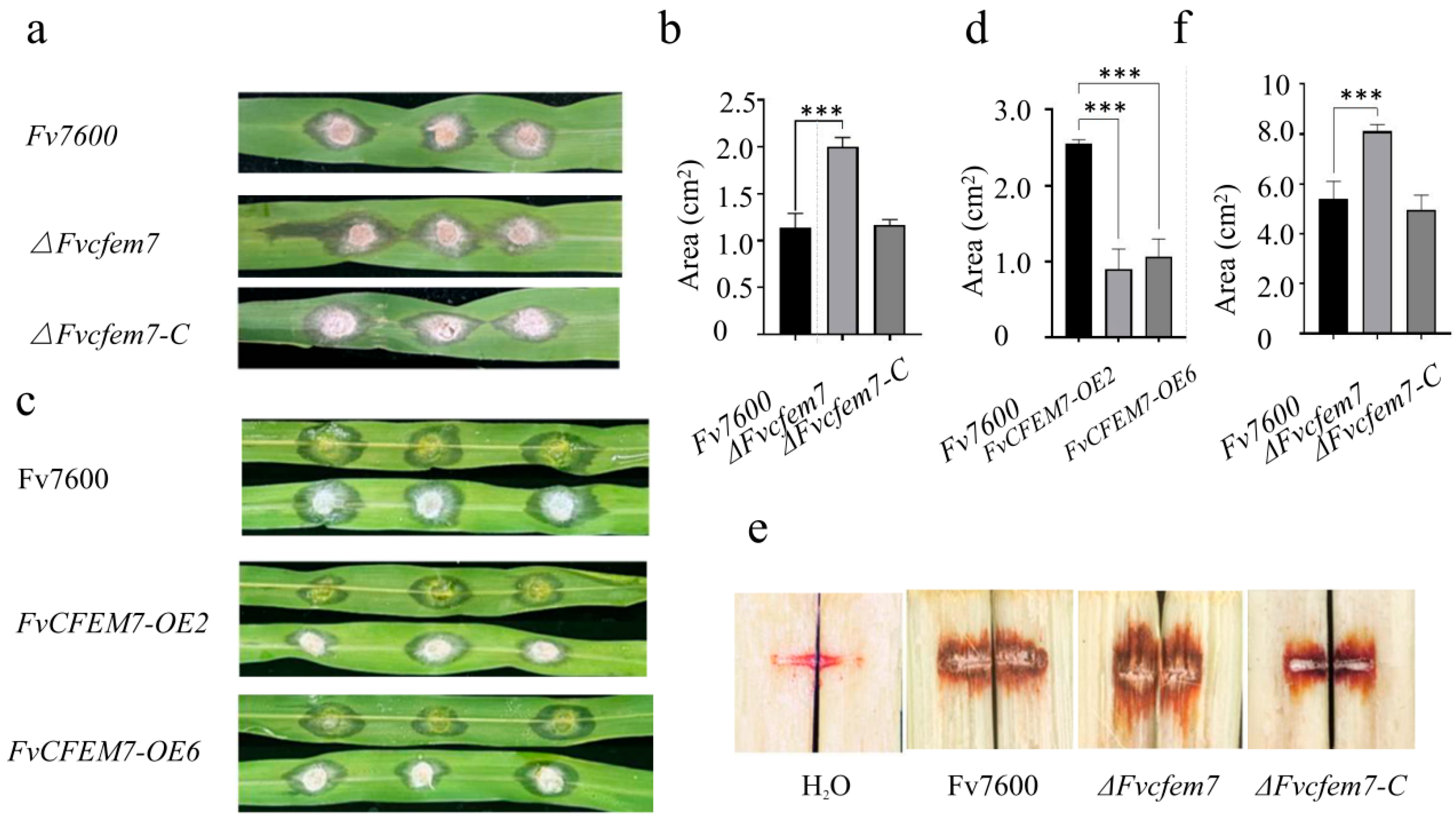
Pathogenicity assays were conducted on leaves of the susceptible maize cultivar B73 by inoculation with the WT strain, ∆Fvcfem7 deletion mutants, complemented strains (∆Fvcfem7-C), and overexpression strain (FvCFEM7-OE). The ∆Fvcfem7 mutants developed significantly larger lesions than the WT, whereas the ∆Fvcfem7-C exhibited lesions similar in size to those of the WT (Figure 3a,b). In contrast, the FvCFEM7-OE strain caused significantly smaller lesions compared to the WT (Figure 3c,d). Similar pathogenicity tests were performed on the susceptible sugarcane cultivar R570. Multiple strains (WT, ∆Fvcfem7, and ∆Fvcfem7-C) were inoculated into sugarcane stalks, and lesion areas were measured 7 days post-inoculation. Consistent with the maize assay results, ∆Fvcfem7 mutants induced significantly larger lesions than the WT and complemented strains (Figure 3e,f). These results demonstrate that FvCfem7 negatively regulates the full virulence of F. verticillioides during host infection.

3.3. Differential Localization of Fvcfem7ΔSP In Vitro, During Hyphal Infection, and in Host Tissues

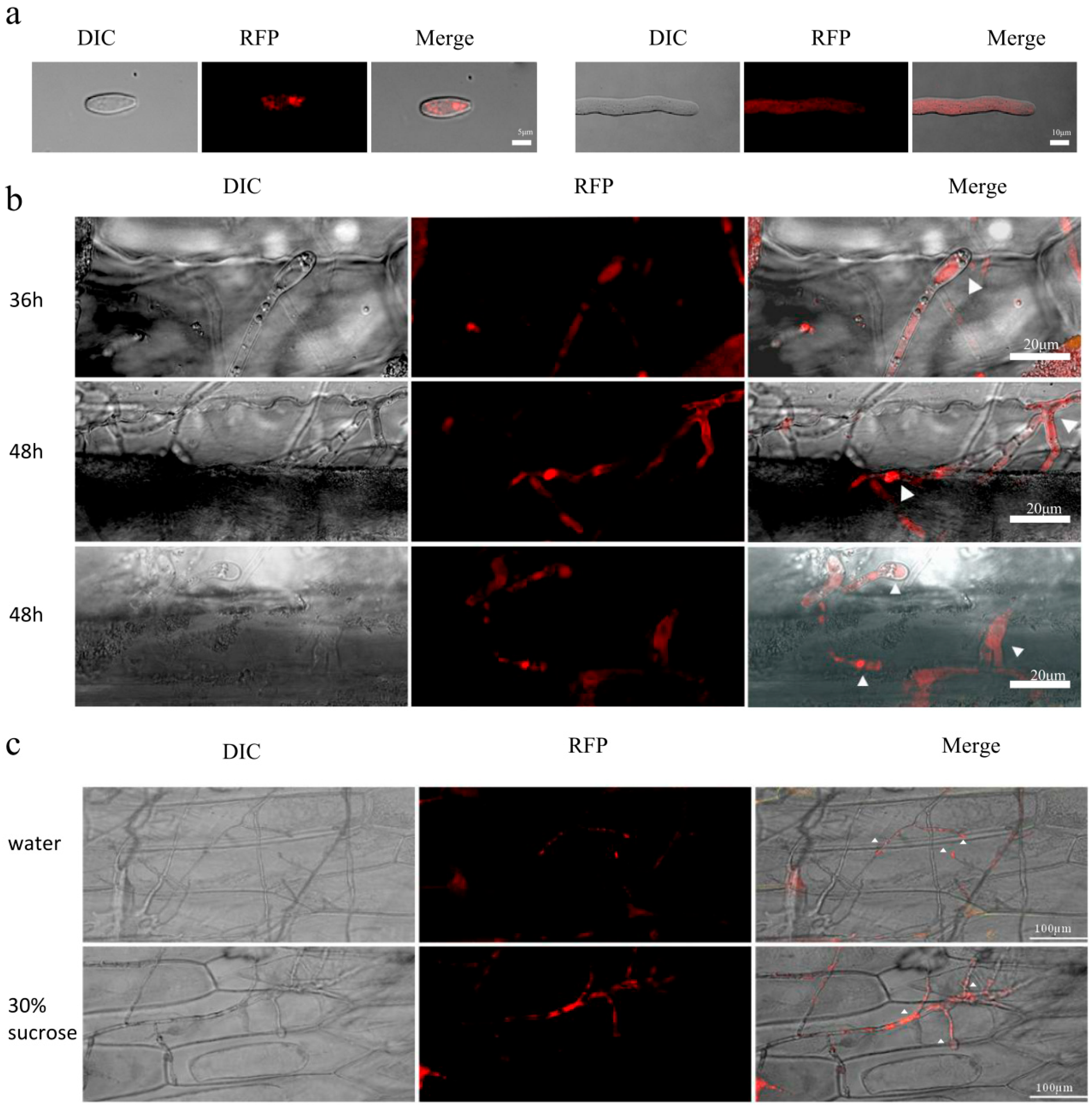
To further explore the function of FvCfem7, we analyzed its subcellular localization using the complemented strain ∆Fvcfem7-C expressing a red fluorescent protein (RFP)-tagged fusion protein. The FvCFEM7:RFP fluorescence exhibited stage-specific subcellular localization in F. verticillioides. In spores cultured in synthetic media, the fluorescent signal appeared as puncta; in hyphae, however, it became widely distributed in multiple subcellular compartments, including the cytoplasm and nucleus (Figure 4a).
During the early infection stages (12–48 h) in maize leaves, FvCfem7 accumulated in enlarged, vesicle-like infection structures resembling haustoria (Figure 4b). This aggregation became more prominent as hyphae penetrated the plant intercellular spaces. At approximately 48 hpi, the signal appeared dispersed around these aggregated haustorium-like structures and within hyphae, with localization extending to the peripheral membrane regions of invading hyphae (Figure 4b). In the late infection stages (after 72 h), the fluorescence signal was substantially diminished. F. verticillioides has been reported to form haustorium-like structures during infection, similar to those observed in Verticillium dahliae [49]. These infection structures may play a critical role in successful host cell invasion, analogous to the appressoria of Magnaporthe oryzae.
To determine whether FvCfem7 functions as an extracellular or cytoplasmic effector, we examined its localization during infection of onion epidermal cells. F. verticillioides formed haustorium-like structures at the tips of infected hyphae, which were primarily localized at the host cell wall interface two days after inoculation (dpi). When infecting onion epidermal cells, FvCfem7 aggregated in the infected haustorium-like infection structures at the cell wall of the infection interface. Cytoplasmic wall separation experiments, performed by treating infected onion epidermis with 30% sucrose solution, revealed that FvCfem7 was present in both cytoplasmic and extracellular regions. The signal was intense in the extracellular space surrounding the haustorium-like structures, while also showing clear aggregation in the cytoplasm (Figure 4b,c). These results indicate that FvCfem7 is constitutively expressed during vegetative hyphal growth but becomes enriched in haustorium-like structures during infection—an enrichment is especially evident in hyphae penetrating plant cells and intercellular spaces. This pattern of enrichment is consistent with observations in Pyricularia oryzae. Fluorescently labeled Avr-Pita1 and Pwl effector proteins were shown to be secreted and aggregated in a specific structure, the biotrophic interfacial complex (BIC) [6], while Bas4 was secreted and aggregated in another specific structure, the extra-invasive hyphal membrane (EIHM) [50]. The dual localization of FvCfem7 suggests that it may function as both an extracellular and a cytoplasmic effector in F. verticillioides, potentially playing an important role in maize infection. We speculate that the dynamic localization of FvCfem7 reflects multifunctional roles, participating in intracellular metabolic regulation while also contributing to extracellular signaling and defense modulation.
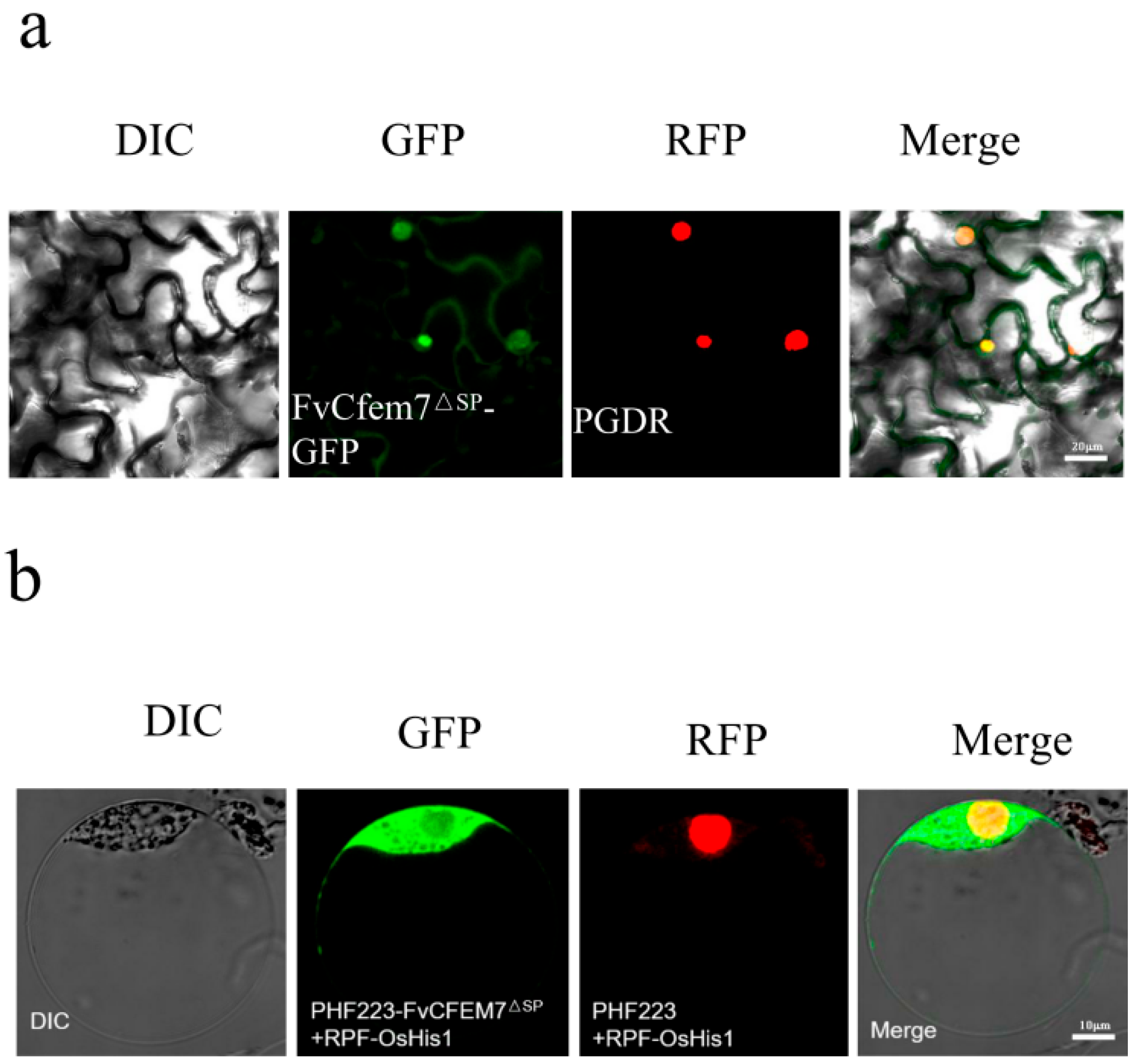
To investigate the molecular mechanisms of FvCFEM7ΔSP–host interactions and to identify potential interacting proteins, we first analyzed its subcellular localization in host cells through transient expression mediated via Agrobacterium or direct transformation into protoplasts. The fusion construct pCXSN-FvCFEM7△sp-GFP was transiently expressed in tobacco leaves through Agrobacterium-mediated transformation. FvCFEM7ΔSP showed strong localization in the nucleus, with a faint signal detected at the cell membrane (Figure 5a). We also expressed the PHF223-GFP-FvCfem7ΔSP construct in maize protoplasts, which showed weak nuclear and cell membrane signals but strong cytoplasmic localization (Figure 5b). Combined with the results from tobacco leaves and maize protoplasts, FvCFEM7ΔSP exhibited a nuclear localization tendency, although no exclusive nuclear localization signal was identified. This suggests that the protein may be translocated into the nucleus through interaction with other host factors.
In summary, FvCfem7 initially localizes to haustorium-like structures in invading hyphae and later accumulates at the host plasma membrane interface prior to the cell death suppression. Collectively, FvCfem7ΔSP exhibits differential localization under synthetic culture conditions, during hyphal infection, and within host tissues.

3.4. Fvcfem7ΔSP Interacts with PR Proteins

To further elucidate the mechanism of action of FvCFEM7, we screened for its interacting proteins using a yeast two-hybrid (Y2H) system with a cDNA library derived from maize B73 infected with F. verticillioides strain FV7600. The bait vector pGBKT7-FvCFEM7ΔSP was constructed and confirmed to be free of self-activation and toxicity in yeast. Prey AD plasmids from the infected maize cDNA library were co-transformed with the bait plasmid into yeast AH109 cells. In the comparative analyses of the NCBI, maize, and F. verticillioides genome databases, we identified PR5-2—a member of the pathogenesis-related (PR) protein family 5—as a candidate interactor (Figure S5).
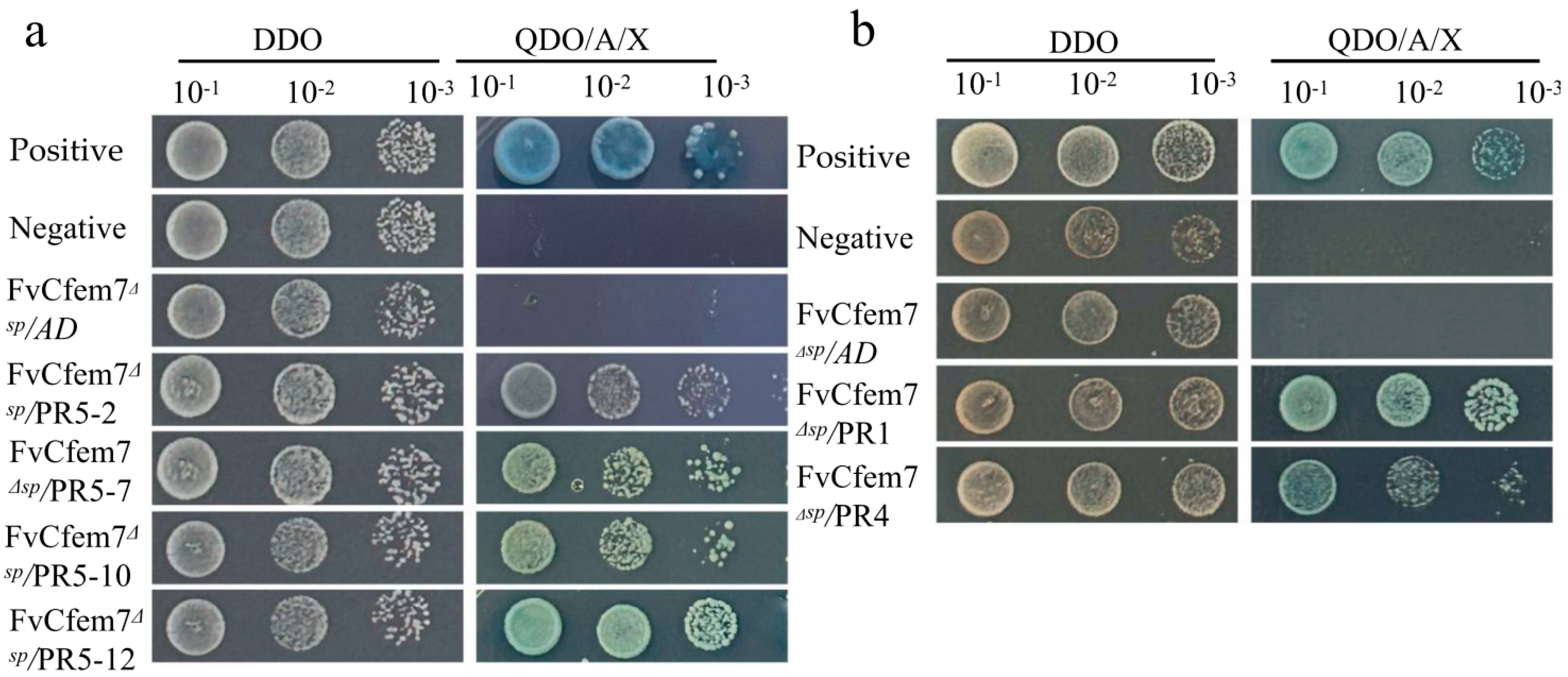
Subsequently, to validate the interaction between FvCfem7 and PR5-2, as well as other PR5 family proteins, we constructed expression vectors for multiple PR5 family members. One-to-one Y2H assays confirmed that FvCfem7 interacts with several PR5 family proteins (Figure 6a), suggesting that FvCfem7 may participate in host immune responses via these interactions. Given that PR family proteins often function cooperatively and exhibit functional crosstalk [51], we further tested whether FvCfem7 interacts with other PR family members. AD vectors containing maize ZmPR1 and ZmPR4 genes were constructed, and one-to-one Y2H validation demonstrated that FvCfem7 also interacts with both ZmPR1 and ZmPR4 in yeast (Figure 6b).

3.5. Effect of Fvcfem7 on the Expression of Interacting Maize PR Proteins

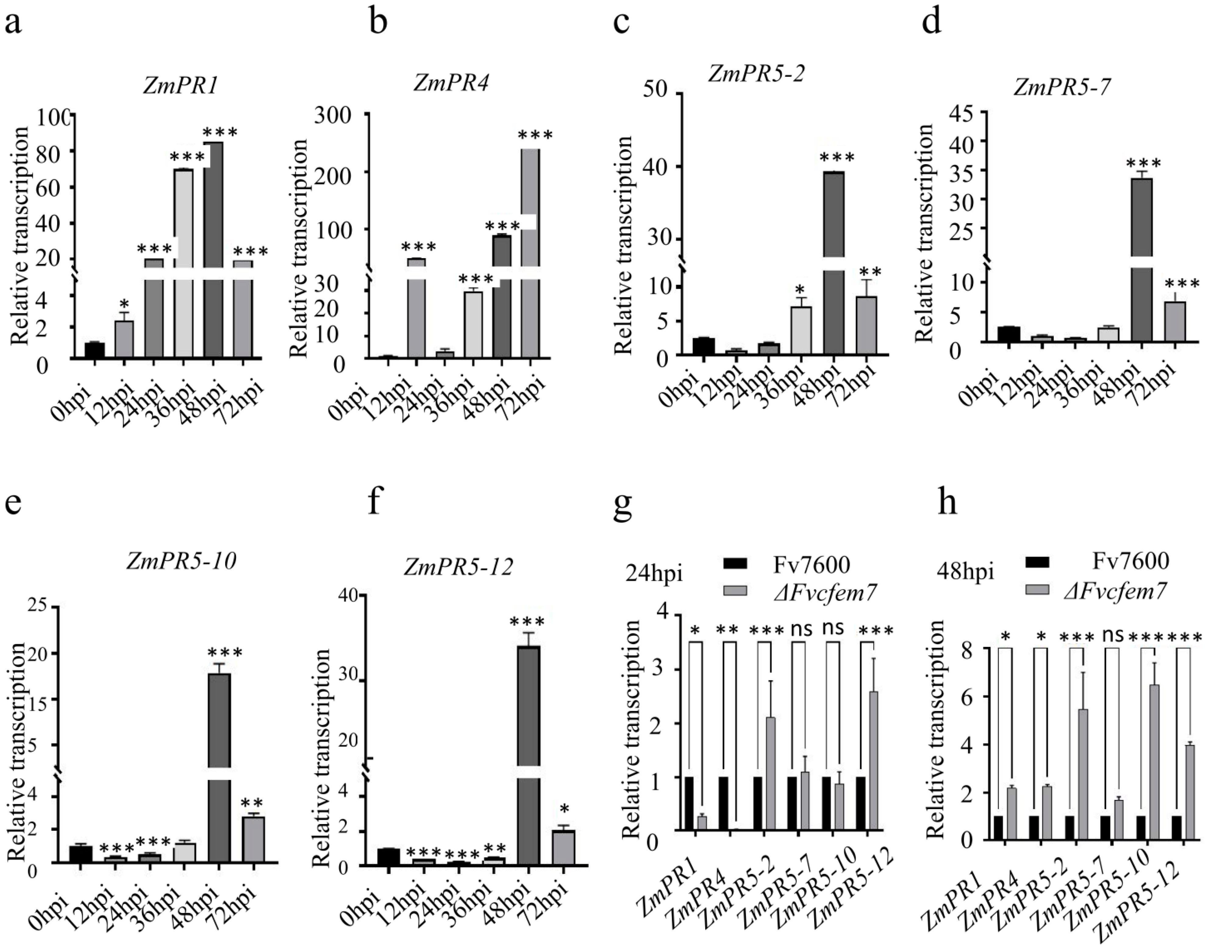
To investigate whether F. verticillioides FvCfem7 modulates the plant immune system through interactions with maize PR proteins, we analyzed the expression profiles of maize PR genes at various time points post-infection (0–72 h post-inoculation, hpi). Using RT-qPCR, we compared the expression patterns of these genes in maize B73 leaves infected at 24 hpi and 48 hpi with the wild-type (WT) strain versus the ∆Fvcfem7 mutant at 24 hpi and 48 hpi.
RNA was extracted from infected leaves at different time points (0–72 hpi). The results showed that PR1 and PR4 were highly expressed both in the early (0–48 hpi) and late (up to 72 hpi) stages of infection, with PR4 significantly upregulated in the early stages (0–48 hpi), indicating its critical role in early immune defense (Figure 7a,b). In contrast, the expression of PR5 family members peaked sharply at 48 hpi, with consistent expression patterns observed across family members (Figure 7c–f). Collectively, these results suggest that ZmPR1 and ZmPR4 primarily contribute to maize immune defense during the early stage of F. verticillioides infection (0–48 hpi), whereas PR5 family members play a dominant role in the later stage (after 48 hpi).
Based on the importance of different PR proteins at two key infection time points—24 hpi and 48 hpi—we compared the expression of PR genes in response to infection by the WT and ∆Fvcfem7 using RT-qPCR. At 24 hpi, the expression levels of ZmPR1 and ZmPR4 in maize leaves were significantly downregulated, while some PR5 family members were upregulated (Figure 7g). At 48 hpi, PR5 family members that interact with FvCfem7 showed a significant upward expression trend (Figure 7h).

4. Discussion

Plant–pathogen interactions are driven by antagonistic coevolution, where plants favor incompatible (resistant) responses and pathogens favor compatible (susceptible) interactions. This dynamic is classically described by the gene-for-gene model [52], where specific host resistance (R) gene products recognize corresponding pathogen avirulence (Avr) effectors, thereby triggering an incompatible interaction. The failure of this recognition—due to allelic variation or the absence of either component—results in compatibility [53]. The effector evolution trajectory reflects a trade-off between evading host detection and retaining virulence functions. For example, the Avr-Pita effector in Magnaporthe oryzae has undergone multiple translocations via mobile genetic elements, with parasexual transfer facilitating its persistence and recovery in populations [54].
Against this evolutionary backdrop, our comparative transcriptome analysis of F. verticillioides grown in CM culture versus maize-infected media identified differentially expressed genes (DEGs), including FvCfem7, which was significantly downregulated during maize infection. We hypothesize that this downregulation occurs because FvCfem7 is recognized by host targets and counteracted by host PR proteins, prompting the pathogen to reduce its expression as a strategy to disengage from this specific “arms race”. This scenario aligns with the “boom-and-bust” cycle, where Avr genes can be reactivated once corresponding R genes are lost from the host populations [10]. Thus, FvCFEM7 expression might be restored if its pathogenicity-influencing host interaction partner is eliminated.
CFEM effectors exhibit diverse fungal biological functions. While some (e.g., CfEC12, FgCfem1) have no obvious morphology effects [32,55], others are essential for spore formation and infection structure development—including PeCfem5, PeCfem8 [56], and Colletotrichum gloeosporioides CgCfem1 [43]. Although FvCFEM7 is expressed during vegetative growth of F. verticillioides, the ∆Fvcfem7 mutant displayed no growth defects (Figure S3a), indicating functional redundancy at this stage.
To clarify FvCfem7′s role in infection, we analyzed its subcellular localization using an mRFP-FvCFEM7 fusion protein. The protein formed a punctum in the spore cytoplasm, dispersed throughout germinating spore and vegetative hyphae cytoplasm, and exhibited stage-specific localization and expression on the surface of the infected hyphae. This pattern of FvCfem7 differs from FgCfem1/FgCfemn1, which localizes to spore surfaces and septa and persists during germination, though they all concentrate on the invading hyphal surface [32]. In contrast, Cfem18-RFP was undetectable, and SP-mRFP-Cfem5 showed weak, transient spore localization but no hyphal signal [32]. Notably, the mRFP-FvCFEM7 signal diminished in late-stage invading hyphae, resembling SP-mRFP-FgCfem5′s transient localization. While Zuo (2022) [32] attributed Cfem5′s signal loss to host tissue diffusion, our qRT-PCR data revealed FvCFEM7 suppression as early as 12 h post-inoculation (hpi), with continued downregulation until 96 hpi (Figure 1a), indicating active transcriptional repression.
Effector expression is tightly regulated by infection stages and host cell type [57], shaped by ongoing plant–pathogen coevolution. Combining localization and functional data, we propose that FvCfem7—unlike FgCfem1/FgCfemn1, which reduces host resistance [32]—may have been hijacked by the host to enhance resistance.
During initial infection, plant chitinases and β-1,3-glucanases degrade fungal cell walls, releasing elicitors that activate pattern-triggered immunity (PTI), inducing PR protein production and pathogen cell degradation [58,59,60,61]. Fungal effectors can either induce or suppress plant cell death in a coevolutionary arms race [62,63]. A second wave of coevolution follows, where pathogens mutate, lose, or acquire novel effectors to evade effector-triggered immunity (ETI), while plants evolve new R proteins, creating a cyclic ETI-ETS-ETI arms race [64].
In our study, FvCfem7ΔSP was detected at the host–pathogen interface during early F. verticillioides infection. Using FvCfem7ΔSP as bait in a yeast two-hybrid (Y2H) screen of infected maize leaf cDNA libraries, we identified ZmPR5 as an interacting partner, confirmed by follow-up Y2H assays. We propose that during long-term host–pathogen coevolution, FvCfem7ΔSP is recognized by PR proteins (e.g., ZmPR1, ZmPR4, and ZmPR5), triggering immune responses. Consequently, the ∆Fvcfem7 mutant exhibits dampened immunity and increased pathogenicity, while exogenous FvCfem7ΔSP application enhances immunity and inhibits fungal growth. Thus, FvCfem7 functions as a host-hijacked effector that boosts resistance via immune induction. Notably, ∆Fvcfem7 did not impair fungal growth but negatively regulated pathogenicity, raising questions about its evolutionary retention. Pathogen fitness may depend on novel effectors emerging to replace arms race losses or target new host components.
Temporal PR gene expression patterns further clarify FvCfem7′s role: at 48 hpi (biotrophy-to-necrotrophy transition), ZmPR1, ZmPR4, and ZmPR5 expression were significantly higher in ∆Fvcfem7 mutants than in the wild type. However, at 24 hpi (invasion stage), ZmPR1 and ZmPR4 expression were markedly downregulated in ∆Fvcfem7-infected leaves, while some ZmPR5 family members were induced. Given PR1 and PR4‘s critical roles in early anti-pathogen immunity [51], we propose that FvCfem7-induced ZmPR1/ZmPR4 expression at 24 hpi promotes programmed cell death (PCD) to initially suppress pathogenicity. By 48 hpi, this PCD may facilitate necrotrophy, enhancing ∆Fvcfem7 pathogenicity. Collectively, these findings demonstrate FvCfem7 induces maize immune responses during the early biotrophic stage, likely by modulating PR protein expression. FvCfem7-induced ZmPR1/ZmPR4 induction within 24 hpi enhances resistance, explaining ∆Fvcfem7′s increased pathogenicity and potential FvCFEM7-OE strain hypovirulence. In addition, detecting the expression level of pathogenesis-related proteins (PRs) via quantitative polymerase chain reaction (qPCR) in leaves sprayed with purified FvCfem7 will more comprehensively demonstrate the ability of this effector to induce and stimulate PRs. This work provides key insights into FvCfem7′s molecular role in plant–pathogen interactions. This interplay informs the development of strategies for breeding resistant cultivars and monitoring pathogen dynamics. Specifically, the expression level of the effector FvCfem7 can be used as a reliable criterion to screen for strains with elevated virulence. Furthermore, it paves the way for engineering crop varieties capable of activating PR defenses in a spatially and temporally controlled manner, thereby minimizing potential yield penalties.

5. Conclusions

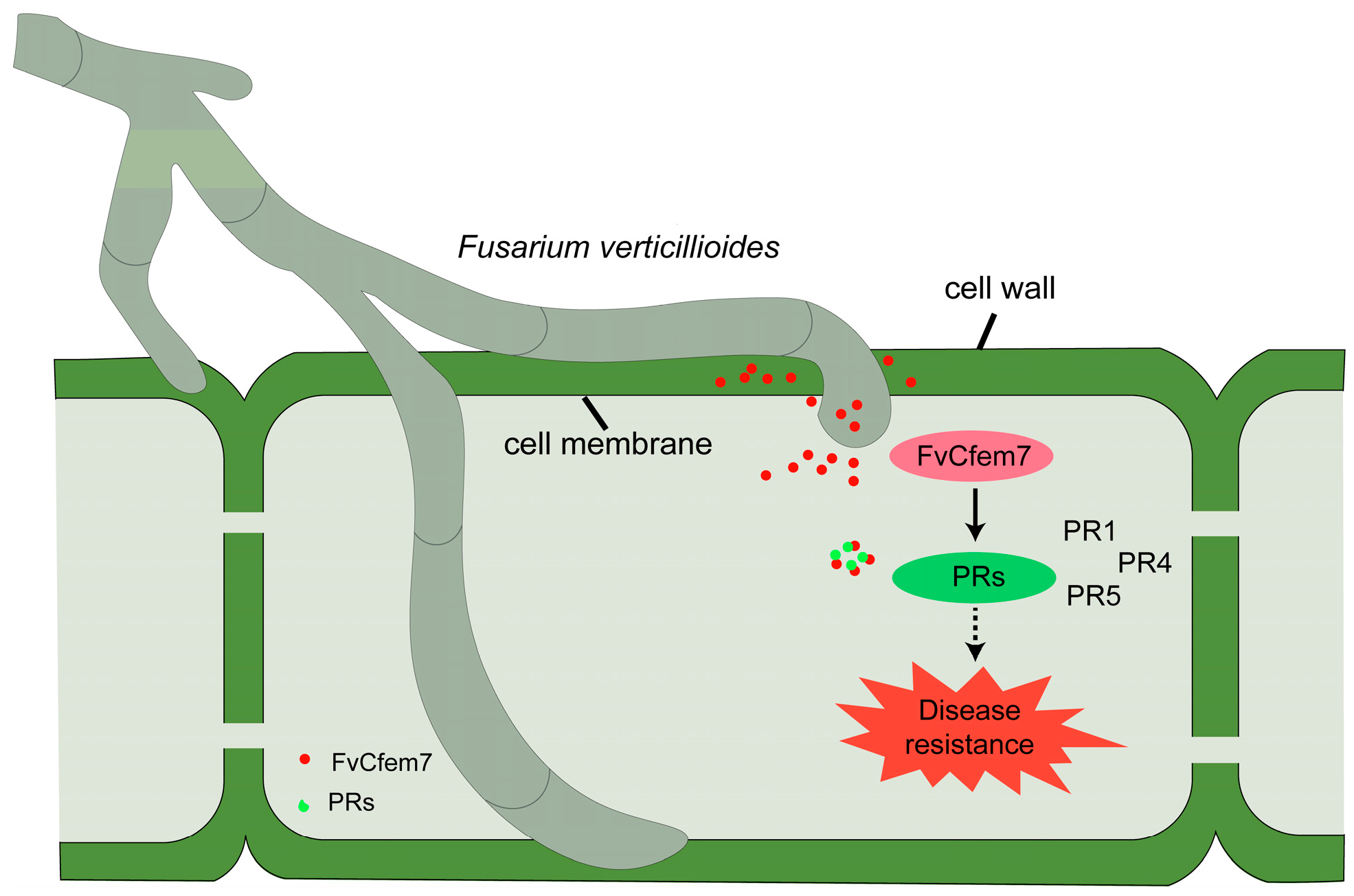
In summary, this study demonstrates that the F. verticillioides effector FvCfem7—whose expression is downregulated during infection—modulates plant immunity by interacting with host PR proteins. Exogenous application of the FvCfem7 recombinant protein can induce plant resistance. We propose a preliminary model for the negative feedback regulation of pathogenicity caused by FvCfem7 in F. verticillioides (Figure 8), providing a theoretical foundation for developing novel disease control strategies.

Supplementary Materials

The following supporting information can be downloaded at: https://www.mdpi.com/article/10.3390/agronomy15122706/s1. Figure S1: Bioinformatics analysis of the structural domain of protein FVEG_02239. (a) Schematic diagram of the domain of the FvCfem7 protein generated by IBS1.0. The light gray line represents the full-length protein, the orange rectangle indicates the signal peptide, the gray rectangle represents the CFEM domain, and the green region denotes low complexity. (b) A signal peptide of the FvCFEM7 was predicted using SignalP 6.0. In total, 1–15 amino acids are SEC/SPI-type signal peptides. Figure S2: Identification of FvCfem7 protein. (a) Multiple sequence alignment of FvCFEM7 with orthologs from Fusarium graminearum (FGCFEM7, FGRAMPH1_01G13195), Fusarium fujikuroi (FfCFEM7, KLO84274.1), Fusarium oxysporum (FoCFEM7, KAJ9420558.1), Pyricularia oryzae (MoCFEM7,XP_003720604.1). (b) Phylogenetic tree of FvCFEM7 and several selected fungi CFEM7. FgCFEM10 (FGRAMPH1_01G13195, Fusarium graminearum), CpCFEM7 (XP_036666947.1, Candida parapsilosis). Figure S3: Construction and validation of the FvCFEM7 gene deletion mutant (∆Fvcfem7) and complemented transformant strain (FvCFEM7-OE). (a) Schematic diagram of the gene knockout strategy via homologous recombination. (b) PCR validation of deletion mutant transformants. (c) Schematic diagram of the Southern blot hybridization strategy for identifying deletion mutants. (d) Southern blot hybridization analysis. (e) RT-qPCR analysis of FvCFEM7 gene expression levels in overexpression strains (FvCFEM7-OE), FvCfem7ΔSP-RFP strain, and ΔFvcfem7-C, using β-tubulin (FVEG_04081) as the reference gene. *** indicates significant differences (p < 0.001, t-test). Figure S4: FvCfem7 does not affect vegetative growth or sporulation in F. verticillioides, but it affects various stress tolerance factors. (a) Vegetative growth of wild-type (WT), ∆Fvcfem7, and complemented strains (∆Fvcfem7-C) on CM, MM, and PDA media. (b) Sporulation analysis of tested strains on PDA sporulation medium. (c,d) Stress tolerance assays of WT, ∆Fvcfem7, and ∆Fvcfem7-C strains. (c) The corresponding inhibition rates of the colony diameter (d). Figure S5: Screening for CFfem7-interacting proteins using a yeast hybrid (Y2H) library. The Y2H library was constructed from Fusarium verticillioides-infected maize leaves. (a) FvCFEM7 shows no self-activation in the Y2H system. (b) Positive colonies from the initial Y2H screening grown on QDO/A agar plates (SD/- Leu -Trp -His- Ade 3-AT) and stained blue with X-gal. Table S1: The primers used in this study. Table S2: FvCFEM identity with FgCFEM.

Author Contributions

Conceptualization, W.Y. and M.W.; methodology, M.W., G.W. and W.Y.; analysis, M.W., G.W., Y.L., S.L., L.K. and X.L.; writing—original draft preparation, W.Y. and G.L.; writing—review and editing, W.Y., Z.W. and G.L.; supervision, Z.W. and W.Y.; funding acquisition, W.Y. All authors have read and agreed to the published version of the manuscript.

Funding

This research was funded by the Natural Science Foundation of Fujian Province (2024J01399), the Major Science and Technology Projects in Fujian Province (2022NZ0106), and the open funds of the State Key Laboratory of Agricultural Microbiology (AMLKF202502).

Institutional Review Board Statement

Not applicable.

Data Availability Statement

The original contributions presented in the study are included in the article/Supplementary Materials. Further inquiries can be directed to the corresponding authors.

Conflicts of Interest

The authors declare no conflicts of interest.

References

  1. Hooven, H.W.v.D.; Burg, H.A.v.D.; Vossen, P.; Boeren, S.; de Wit, P.J.G.M.; Vervoort, J. Disulfide bond structure of the AVR9 elicitor of the fungal tomato pathogen Cladosporium fulvum: Evidence for a cystine knot. Biochemistry 2001, 40, 3458–3466. [Google Scholar] [CrossRef] [PubMed]
  2. Burg, H.A.v.D.; Westerink, N.; Francoijs, K.-J.; Roth, R.; Woestenenk, E.; Boeren, S.; de Wit, P.J.G.M.; Joosten, M.H.A.J.; Vervoort, J. Natural disulfide bond-disrupted mutants of AVR4 of the tomato pathogen Cladosporium fulvum are sensitive to proteolysis, circumvent Cf-4-mediated resistance, but retain their chitin binding ability. J. Biol. Chem. 2003, 278, 27340–27346. [Google Scholar] [CrossRef]
  3. Gan, P.; Ikeda, K.; Irieda, H.; Narusaka, M.; O’Connell, R.J.; Narusaka, Y.; Takano, Y.; Kubo, Y.; Shirasu, K. Comparative genomic and transcriptomic analyses reveal the hemibiotrophic stage shift of Colletotrichum fungi. New Phytol. 2013, 197, 1236–1249. [Google Scholar] [CrossRef]
  4. Djamei, A.; Schipper, K.; Rabe, F.; Ghosh, A.; Vincon, V.; Kahnt, J.; Osorio, S.; Tohge, T.; Fernie, A.R.; Feussner, I.; et al. Metabolic priming by a secreted fungal effector. Nature 2011, 478, 395–398. [Google Scholar] [CrossRef]
  5. Liu, L.; Xu, L.; Jia, Q.; Pan, R.; Oelmüller, R.; Zhang, W.; Wu, C. Arms race: Diverse effector proteins with conserved motifs. Plant Signal Behav. 2019, 14, 1557008. [Google Scholar] [CrossRef]
  6. Giraldo, M.C.; Valent, B. Filamentous plant pathogen effectors in action. Nat. Rev. Microbiol. 2013, 11, 800–814. [Google Scholar] [CrossRef]
  7. Lu, S.; Edwards, M.C. Genome-wide analysis of small secreted cysteine-rich proteins identifies candidate effector proteins potentially involved in Fusarium graminearum-wheat interactions. Phytopathology 2016, 106, 166–176. [Google Scholar] [CrossRef]
  8. O’Connell, R.J.; Thon, M.R.; Hacquard, S.; Amyotte, S.G.; Kleemann, J.; Torres, M.F.; Damm, U.; Buiate, E.A.; Epstein, L.; Alkan, N.; et al. Lifestyle transitions in plant pathogenic Colletotrichum fungi deciphered by genome and transcriptome analyses. Nat. Genet. 2012, 44, 1060–1065. [Google Scholar] [CrossRef] [PubMed]
  9. Stergiopoulos, I.; de Wit, P.J. Fungal effector proteins. Annu. Rev. Phytopathol. 2009, 47, 233–263. [Google Scholar] [CrossRef] [PubMed]
  10. Lo Presti, L.; Lanver, D.; Schweizer, G.; Tanaka, S.; Liang, L.; Tollot, M.; Zuccaro, A.; Reissmann, S.; Kahmann, R. Fungal effectors and plant susceptibility. Annu. Rev. Plant Biol. 2015, 66, 513–545. [Google Scholar] [CrossRef]
  11. Lowe, R.G.T.; Howlett, B.J. Indifferent, affectionate, or deceitful: Lifestyles and secretomes of fungi. PLoS Pathog. 2012, 8, e1002515. [Google Scholar] [CrossRef]
  12. Lahrmann, U.; Ding, Y.; Banhara, A.; Rath, M.; Hajirezaei, M.R.; Döhlemann, S.; von Wirén, N.; Parniske, M.; Zuccaro, A. Host-related metabolic cues affect colonization strategies of a root endophyte. Proc. Natl. Acad. Sci. USA 2013, 110, 13965–13970. [Google Scholar] [CrossRef]
  13. Kulkarni, R.D.; Kelkar, H.S.; Dean, R.A. An eight-cysteine-containing CFEM domain unique to a group of fungal membrane proteins. Trends Biochem. Sci. 2003, 28, 118–121. [Google Scholar] [CrossRef]
  14. Zhang, Z.-N.; Wu, Q.-Y.; Zhang, G.-Z.; Zhu, Y.-Y.; Murphy, R.W.; Liu, Z.; Zou, C.-G. Systematic analyses reveal uniqueness and origin of the CFEM domain in fungi. Sci. Rep. 2015, 5, 13032. [Google Scholar] [CrossRef]
  15. Vaknin, Y.; Shadkchan, Y.; Levdansky, E.; Morozov, M.; Romano, J.; Osherov, N. The three Aspergillus fumigatus CFEM-domain GPI-anchored proteins (CfmA-C) affect cell-well stability but do not play a role in fungal virulence. Fungal Genet. Biol. 2014, 63, 55–64. [Google Scholar] [CrossRef] [PubMed]
  16. Heard, S.; Brown, N.A.; Hammond-Kosack, K. An interspecies comparative analysis of the predicted secretomes of the necrotrophic plant pathogens Sclerotinia sclerotiorum and Botrytis cinerea. PLoS ONE 2015, 10, e0130534. [Google Scholar] [CrossRef]
  17. Ling, J.; Zeng, F.; Cao, Y.; Zhang, J.; Chen, G.; Mao, Z.; Yang, Y.; Xie, B. Identification of a class of CFEM proteins containing a new conserved motif in Fusarium oxyporum. Physiol. Mol. Plant Pathol. 2015, 89, 41–48. [Google Scholar] [CrossRef]
  18. Kou, Y.; Tan, Y.H.; Ramanujam, R.; Naqvi, N.I. Structure-function analyses of the Pth11 receptor reveal an important role for CFEM motif and redox regulation in rice blast. New Phytol. 2017, 214, 330–342. [Google Scholar] [CrossRef] [PubMed]
  19. Chen, L.; Wang, H.; Yang, J.; Yang, X.; Zhang, M.; Zhao, Z.; Fan, Y.; Wang, C.; Wang, J. Bioinformatics and transcriptome. Analysis of CFEM proteins in Fusarium graminearum. J. Fungi 2021, 7, 871. [Google Scholar] [CrossRef]
  20. Li, H.; Ishfaq, S.; Liang, X.; Wang, R.; Wei, H.; Guo, W. A novel CFEM effector in Fusarium verticillioides required for virulence involved in plant immunity suppression and fungal cell wall integrity. Int. J. Mol. Sci. 2025, 26, 4369. [Google Scholar] [CrossRef]
  21. Moukadiri, I.; Armero, J.; Abad, A.; Sentandreu, R.; Zueco, J. Identification of a mannoprotein present in the inner layer of the cell wall of Saccharomyces cerevisiae. J. Bacteriol. 1997, 179, 2154–2162. [Google Scholar] [CrossRef]
  22. Kuznets, G.; Vigonsky, E.; Weissman, Z.; Lalli, D.; Gildor, T.; Kauffman, S.J.; Turano, P.; Becker, J.; Lewinson, O.; Kornitzer, D. A relay network of extracellular heme-binding proteins drives C. albicans iron acquisition from hemoglobin. PLoS Pathog. 2014, 10, e1004407. [Google Scholar] [CrossRef]
  23. Weissman, Z.; Kornitzer, D. A family of Candida cell surface haem-binding proteins involved in haemin and haemoglobin-iron utilization. Mol. Microbiol. 2004, 53, 1209–1220. [Google Scholar] [CrossRef]
  24. Nasser, L.; Weissman, Z.; Pinsky, M.; Amartely, H.; Dvir, H.; Kornitzer, D. Structural basis of haem-iron acquisition by fungal pathogens. Nat. Microbiol. 2016, 1, 16156. [Google Scholar] [CrossRef]
  25. Chen, C.; Pande, K.; French, S.D.; Tuch, B.B.; Noble, S.M. An iron homeostasis regulatory circuit with reciprocal roles in Candida albicans commensalism and pathogenesis. Cell Host Microbe 2011, 10, 118–135. [Google Scholar] [CrossRef]
  26. Zhu, Y.; Yang, C.; Magee, D.; Cox, R.A. Coccidioides immitis antigen 2: Analysis of gene and protein. Gene 1996, 181, 121–125. [Google Scholar] [CrossRef] [PubMed]
  27. Zhu, W.; Wei, W.; Wu, Y.; Zhou, Y.; Peng, F.; Zhang, S.; Chen, P.; Xu, X. BcCFEM1, a CFEM domain-containing protein with putative gpi-anchored site, is involved in pathogenicity, conidial production, and stress tolerance in Botrytis cinerea. Front. Microbiol. 2017, 8, 1807. [Google Scholar] [CrossRef] [PubMed]
  28. Gong, A.-D.; Jing, Z.-Y.; Zhang, K.; Tan, Q.-Q.; Wang, G.-L.; Liu, W.-D. Bioinformatic analysis and functional characterization of the CFEM proteins in maize anthracnose fungus Colletotrichum graminicola. J. Integr. Agric. 2020, 19, 541–550. [Google Scholar] [CrossRef]
  29. Choi, W.; Dean, R.A. The adenylate cyclase gene MAC1 of Magnaporthe grisea controls appressorium formation and other aspects of growth and development. Plant Cell 1997, 9, 1973–1983. [Google Scholar] [CrossRef]
  30. DeZwaan, T.M.; Carroll, A.M.; Valent, B.; Sweigard, J.A. Magnaporthe grisea Pth11p is a novel plasma membrane protein that mediates appressorium differentiation in response to inductive substrate cues. Plant Cell 1999, 11, 2013–2030. [Google Scholar] [CrossRef]
  31. Sabnam, N.; Barman, S.R. WISH, a novel CFEM GPCR is indispensable for surface sensing, asexual and pathogenic differentiation in rice blast fungus. Fungal Genet. Biol. 2017, 105, 37–51. [Google Scholar] [CrossRef]
  32. Zuo, N.; Bai, W.-Z.; Wei, W.-Q.; Yuan, T.-L.; Zhang, D.; Wang, Y.-Z.; Tang, W.-H. Fungal CFEM effectors negatively regulate a maize wall-associated kinase by interacting with its alternatively spliced variant to dampen resistance. Cell Rep. 2022, 41, 111877. [Google Scholar] [CrossRef] [PubMed]
  33. Wang, D.; Zhang, D.-D.; Song, J.; Li, J.-J.; Wang, J.; Li, R.; Klosterman, S.J.; Kong, Z.-Q.; Lin, F.-Z.; Dai, X.-F.; et al. Verticillium dahliae CFEM proteins manipulate host immunity and differentially contribute to virulence. BMC Biol. 2022, 20, 55. [Google Scholar] [CrossRef]
  34. Punja, Z.K.; Zhang, Y.Y. Plant chitinases and their roles in resistance to fungal diseases. J. Nematol. 1993, 25, 526–540. [Google Scholar] [PubMed]
  35. Collinge, D.B.; Kragh, K.M.; Mikkelsen, J.D.; Nielsen, K.K.; Rasmussen, U.; Vad, K. Plant chitinases. Plant J. 1993, 3, 31–40. [Google Scholar] [CrossRef]
  36. Qian, H.; Xiao, Z.; Cheng, L.; Geng, R.; Ma, Y.; Bi, Y.; Liang, W.; Yang, A. A novel secreted protein of Fusarium oxysporum promotes infection by inhibiting PR-5 protein in plant. Plant Cell Environ. 2025, 48, 1021–1036. [Google Scholar] [CrossRef] [PubMed]
  37. Lu, S.; Faris, J.D.; Sherwood, R.; Friesen, T.L.; Edwards, M.C. A dimeric PR-1-type pathogenesis-related protein interacts with ToxA and potentially mediates ToxA-induced necrosis in sensitive wheat. Mol. Plant Pathol. 2014, 15, 650–663. [Google Scholar] [CrossRef]
  38. Sung, Y.; Outram, M.A.; Breen, S.; Wang, C.; Dagvadorj, B.; Winterberg, B.; Kobe, B.; Williams, S.J.; Solomon, P.S. PR1-mediated defence via C-terminal peptide release is targeted by a fungal pathogen effector. New Phytol. 2021, 229, 3467–3480. [Google Scholar] [CrossRef]
  39. Breen, S.; Williams, S.J.; Winterberg, B.; Kobe, B.; Solomon, P.S. Wheat PR-1 proteins are targeted by necrotrophic pathogen effector proteins. Plant J. 2016, 88, 13–25. [Google Scholar] [CrossRef]
  40. Yang, G.; Tang, L.; Gong, Y.; Xie, J.; Fu, Y.; Jiang, D.; Li, G.; Collinge, D.B.; Chen, W.; Cheng, J. A cerato-platanin protein SsCP1 targets plant PR1 and contributes to virulence of Sclerotinia sclerotiorum. New Phytol. 2018, 217, 739–755. [Google Scholar] [CrossRef]
  41. Luo, X.; Tian, T.; Feng, L.; Yang, X.; Li, L.; Tan, X.; Wu, W.; Li, Z.; Treves, H.; Serneels, F.; et al. Pathogenesis-related protein 1 suppresses oomycete pathogen by targeting against AMPK kinase complex. J. Adv. Res. 2023, 43, 13–26. [Google Scholar] [CrossRef] [PubMed]
  42. Nguyen, T.T.; Dehne, H.W.; Steiner, U. Histopathological assessment of the infection of maize leaves by Fusarium graminearum, F. proliferatum, and F. verticillioides. Fungal Biol. 2016, 120, 1094–1104. [Google Scholar] [CrossRef]
  43. Feng, X.; Xiong, H.; Zheng, D.; Xin, X.; Zhang, X.; Wang, Q.; Wu, F.; Xu, J.; Lu, Y. Identification of Fusarium verticillioides resistance alleles in three maize populations with teosinte gene introgression. Front. Plant Sci. 2022, 13, 942397. [Google Scholar] [CrossRef]
  44. Bai, H.; Si, H.; Zang, J.; Pang, X.; Yu, L.; Cao, H.; Xing, J.; Zhang, K.; Dong, J. Comparative proteomic analysis of the defense response to Gibberella stalk rot in maize and reveals that ZmWRKY83 is involved in plant disease resistance. Front. Plant Sci. 2021, 12, 694973. [Google Scholar] [CrossRef] [PubMed]
  45. Lebrun, M.H.; Langin, T.T.; Kroj, T.T.; Cockram, J.; Oliver, R.; Kema, G.; Valade, R.; Praud, S.; Laurent, V.; Duchalais, L. Wheat effector assisted breeding for resistance to fungal pathogens (WEAB). In Proceedings of the (JJC)-11emes Rencontres de Phytopathologie-Mycologie, Societe Française de Phytopathologie (SFP), Aussois, France, 1 January 2016; Volume 49. [Google Scholar]
  46. Zhang, H.; Kim, M.S.; Huang, J.; Yan, H.; Yang, T.; Song, L.; Yu, W.; Shim, W.B. Transcriptome analysis of maize pathogen Fusarium verticillioides revealed FvLcp1, a secreted protein with type-D fungal LysM and chitin-binding domains, that plays important roles in pathogenesis and mycotoxin production. Microbiol. Res. 2022, 265, 127195. [Google Scholar] [CrossRef] [PubMed]
  47. Naumann, T.A.; Wicklow, D.T.; Price, N.P.J. Identification of a chitinase-modifying protein from Fusarium verticillioides: Truncation of a host resistance protein by a fungalysin metalloprotease. J. Biol. Chem. 2011, 286, 35358–35366. [Google Scholar] [CrossRef]
  48. Wen, G.; Lu, X.; Liang, J.; Liu, Y.; Zhang, X.; Lu, G.; Wang, Z.; Yu, W. The Global Transcription Factor FvCon7 Plays a Role in the Morphology, FB1 Toxin Production, and Pathogenesis of Fusarium verticillioides. Plants 2025, 14, 2725. [Google Scholar] [CrossRef]
  49. Liu, Q.; Li, Y.; Wu, H.; Zhang, B.; Liu, C.; Gao, Y.; Guo, H.; Zhao, J. Hyphopodium-specific signaling is required for plant Iinfection by Verticillium dahliae. J. Fungi. 2023, 9, 484. [Google Scholar] [CrossRef]
  50. Hogenhout, S.A.; Van der Hoorn, R.A.; Terauchi, R.; Kamoun, S. Emerging concepts in effector biology of plant-associated organisms. Mol. Plant Microbe Interact. 2009, 22, 115–122. [Google Scholar] [CrossRef]
  51. Dos Santos, C.; Franco, O.L. Pathogenesis-related proteins (PRs) with enzyme activity activating plant defense responses. Plants 2023, 12, 2226. [Google Scholar] [CrossRef]
  52. Flor, H.H. The complementary systems in flax and flax rust. Adv. Genet. 1956, 8, 29–54. [Google Scholar]
  53. Dodds, P.N.; Rathjen, J.P. Plant immunity: Towards an integrated view of plant-pathogen interactions. Nat. Rev. Genet. 2010, 11, 539–548. [Google Scholar] [CrossRef] [PubMed]
  54. Chen, X.-L.; Shi, T.; Yang, J.; Shi, W.; Gao, X.; Chen, D.; Xu, X.; Xu, J.-R.; Talbot, N.J.; Peng, Y.-L. N-glycosylation of effector proteins by an α-1,3-mannosyltransferase is required for the rice blast fungus to evade host innate immunity. Plant Cell 2014, 26, 1360–1376. [Google Scholar] [CrossRef]
  55. Shang, S.; Liu, G.; Zhang, S.; Liang, X.; Zhang, R.; Sun, G. A fungal CFEM-containing effector targets NPR1 regulator NIMIN2 to suppress plant immunity. Plant Biotechnol. J. 2024, 22, 82–97. [Google Scholar] [CrossRef]
  56. Wang, C.; Yang, Q.; Wang, W.; Li, Y.; Guo, Y.; Zhang, D.; Ma, X.; Song, W.; Zhao, J.; Xu, M. A transposon-directed epigenetic change in ZmCCT underlies quantitative resistance to Gibberella stalk rot in maize. New Phytol. 2017, 215, 1503–1515. [Google Scholar] [CrossRef] [PubMed]
  57. Okmen, B.; Doehlemann, G. Inside plant: Biotrophic strategies to modulate host immunity and metabolism. Curr. Opin. Plant Biol. 2014, 20, 19–25. [Google Scholar] [CrossRef]
  58. Jashni, M.K.; Mehrabi, R.; Collemare, J.; Mesarich, C.H.; de Wit, P.J.G.M. The battle in the apoplast: Further insights into the roles of proteases and their inhibitors in plant–pathogen interactions. Front. Plant Sci. 2015, 6, 584. [Google Scholar] [CrossRef]
  59. Fesel, P.H.; Zuccaro, A. β-Glucan: Crucial component of the fungal cell wall and elusive MAMP in plants. Fungal Genet. Biol. 2016, 90, 53–60. [Google Scholar] [CrossRef]
  60. Héloir, M.-C.; Adrian, M.; Brulé, D.; Claverie, J.; Cordelier, S.; Daire, X.; Dorey, S.; Gauthier, A.; Lemaître-Guillier, C.; Negrel, J.; et al. Recognition of elicitors in grapevine: From MAMP and DAMP perception to induced resistance. Front. Plant Sci. 2019, 10, 1117. [Google Scholar] [CrossRef]
  61. Jamiołkowska, A. Natural compounds as elicitors of plant resistance against diseases and new biocontrol strategies. Agronomy 2020, 10, 173. [Google Scholar] [CrossRef]
  62. Abramovitch, R.B.; Martin, G.B. Strategies used by bacterial pathogens to suppress plant defenses. Curr. Opin. Plant Biol. 2004, 7, 356–364. [Google Scholar] [CrossRef] [PubMed]
  63. Mukhtar, M.S.; McCormack, M.E.; Argueso, C.T.; Pajerowska-Mukhtar, K.M. Pathogen tactics to manipulate plant cell death. Curr. Biol. 2016, 26, R608–R619. [Google Scholar] [CrossRef] [PubMed]
  64. Jones, J.D.G.; Dangl, J.L. The plant immune system. Nature 2006, 444, 323–329. [Google Scholar] [CrossRef] [PubMed]
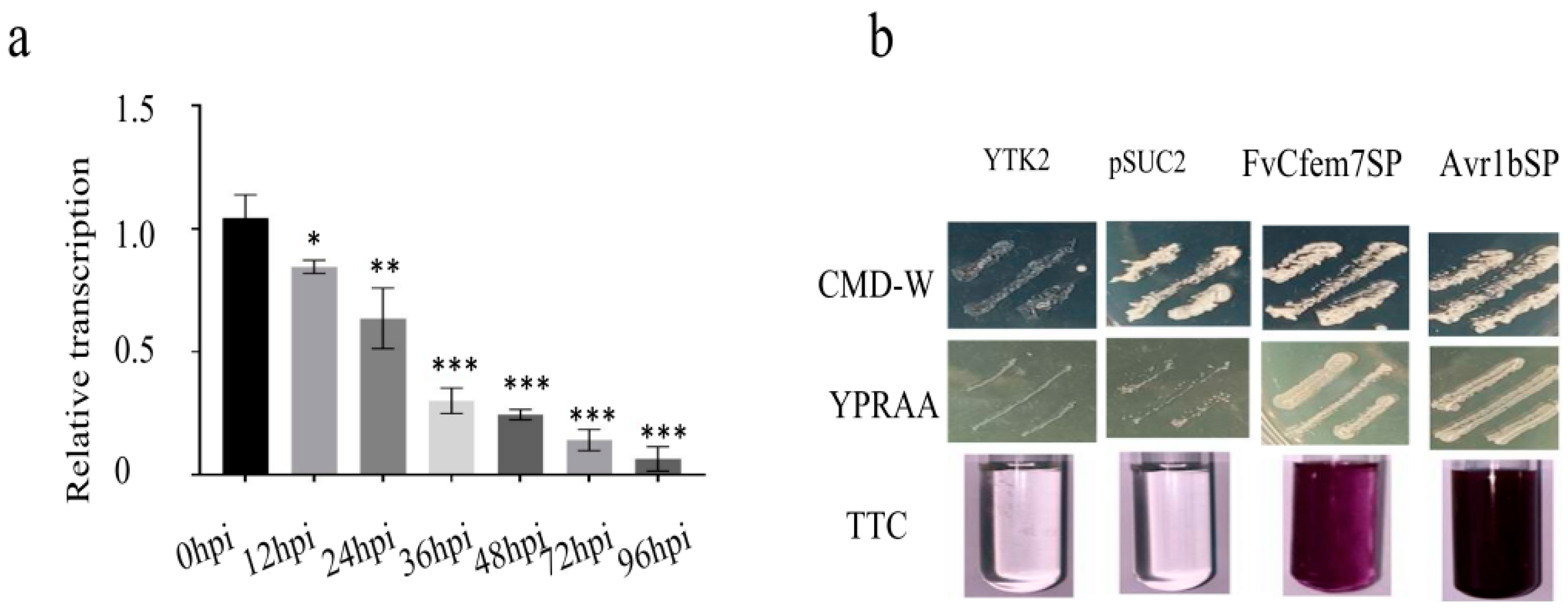
Figure 1. Transcriptional profile of the FvCFEM7 gene in infected maize and functional characterization of its signal peptide. (a) Reverse transcription–quantitative PCR analysis of FvCFEM7 expression levels of FvCFEM7 relative to the F. verticillioides β-tubulin gene (FVEG_04081) in maize leaves at 0–96 h post-inoculation (hpi) with F. verticillioides. Error bars represent standard errors. *, **, and *** indicate statistical significance at p < 0.05, p < 0.01, and p < 0.001, respectively. (b) Functional validation of the putative N-terminal signal peptide of FvCfem7 using a yeast signal trap assay. The transformed Saccharomyces cerevisiae YTK12 strain harboring the FvCfem7 signal peptide (FvCfem7SP) grew on both CMD-W and YPRAA media and exhibited invertase activity, as indicated by a red color change in the presence of 2,3,5-triphenyltetrazolium chloride (TTC) reagent. Untransformed YTK12 and YTK12 carrying the empty pSUC2 vector served as negative controls, while the signal peptide of the oomycete effector Avr1b was used as a positive control.
Agronomy 15 02706 g001
Figure 2. Signal peptide deletion (FvCfem7ΔSP) induced hypersensitive cell death in N. benthamiana, and spraying FvCfem7ΔSP impaired F. verticillioides pathogenicity. (a) Agrobacterium tumefaciens strains expressing the indicated FvCfem7ΔSP or full-length FvCfem7 were infiltrated into N. benthamiana leaves. Leaves were photographed 2–3 days post-infiltration (dpi). For the suppression of the INF1-induced PCD suppression assay, A. tumefaciens expressing FvCfem7ΔSP or full-length FvCfem7 was re-infiltrated into N. benthamiana leaves 24 h after initial infiltration with INF1-expressing A. tumefaciens. Leaves were photographed 2–3 days after the second infiltration. (b) Western blot (WB) analysis with an anti-GST antibody confirming the purification of heterologously expressed FvCfem7ΔSP. (c) In vitro spraying of purified FvCfem7ΔSP significantly enhanced plant disease resistance. Leaves sprayed with purified GST protein served as the control.
Agronomy 15 02706 g002
Figure 3. Virulence assays of FvCFEM7 deletion mutant ∆Fvcfem7, and complemented overexpression strain (FvCFEM7-OE) of F. verticillioides on maize and sugarcane. (a,b) Pathogenicity of ∆Fvcfem7 on maize leaves (a) and statistical analysis of lesion areas (b). (c,d) Pathogenicity of FvCFEM7-OE on maize leaves (c) and statistical analysis of lesion areas (d). (e,f) Pathogenicity of the ∆Fvcfem7 strain on sugarcane stalks (e) and statistical analysis of lesion areas (f). *** indicates significant differences at p < 0.001 (t-test).
Agronomy 15 02706 g003
Figure 4. Subcellular localization of FvCfem7ΔSP in F. verticillioides during in vitro growth and host infection. (a) Localization of FvCfem7ΔSP-mRFP in F. verticillioides conidia and hyphae grown in vitro. Scale bars = 5 μm (incondia) and 10 μm (hypha). (b) Localization of the FvCfem7ΔSP-mRFP fusion protein at the hyphal surface of the host–pathogen interface in maize leaves at 36 hpi and 48 hpi. Scale bars = 20 μm. (c) Subcellular localization of FvCFEM7 during F. verticillioides infection of onion epidermal cells at 2 dpi. Scale bars = 100 μm. The haustorium-like structures or extra-invasive hyphal membrane (EIHM) where CFEM7 gathers is maiked by the white triangle.
Agronomy 15 02706 g004
Figure 5. Subcellular localization of heterologously expressed FvCfem7ΔSP in plant cells. (a) Localization of C-terminal mCherry-tagged FvCfem7ΔSP transiently expressed in N. benthamiana leaves. Scale bars = 20 μm. (b) Localization of FvCfem7ΔSP in maize protoplasts. Scale bars = 10 μm.
Agronomy 15 02706 g005
Figure 6. Yeast two-hybrid (Y2H) assay revealing interactions between F. verticillioides CFEM proteins and maize ZmPRs. (a) Y2H assay examining interactions between FvCFEM7 and ZmPR5-2, -5, -10, and -12. Interactions were assessed based on growth on DDO agar (SD/-Leu-Trp deficiency medium), QDO/3AT agar plates (SD/-Leu-Trp-His-Ade + 3-AT) plates, and α-galactosidase activity. 3AT:3-amino-1,2,4-triazole. (b) Y2H assay examining interactions between FvCFEM7 and ZmPR1 or ZmPR4.
Agronomy 15 02706 g006
Figure 7. Expression levels of CFEM7-interacting maize PR protein genes at different infection stages. (af) Relative expression levels of the defense-related genes (ZmPR5-2, -5, -10, -12, ZmPR1, and ZmPR4) in maize leaves at different time points after inoculation with F. verticillioides, normalized to ZmGAPDH (LOC100192489) and determined by reverse transcription–quantitative PCR. (g,h) Effects of FvCfem7 on the expression of its interacting maize PR genes at 24 hpi (g) and 48 hpi (h). The data represent three biological replicates. Error bars represent standard errors. *, **, and *** indicate significant differences at ns-(not significant), p < 0.05, p < 0.01, and p < 0.001, respectively (t-test).
Agronomy 15 02706 g007
Figure 8. Schematic model of the proposed immunity-triggering FvCfem7–PR interaction. PR: host disease related protein that regulates the host’s immune system. FvCfem7, negatively regulateing the pathogenicity of pathogens, is an effector recognized by PR proteins (including PR1, PR4, and PR5).
Agronomy 15 02706 g008
Disclaimer/Publisher’s Note: The statements, opinions and data contained in all publications are solely those of the individual author(s) and contributor(s) and not of MDPI and/or the editor(s). MDPI and/or the editor(s) disclaim responsibility for any injury to people or property resulting from any ideas, methods, instructions or products referred to in the content.