Article

Application of Nanostructured Semiconductor Oxides TiO2-Based as Additives in the Germination Process of Alfalfa

by
Alexis Martínez-Barras
1,2,
Susana Rodríguez-Jurado
3,
Humberto Aguirre-Becerra
3,
Claudia E. Pérez-García
4,
Karen Esquivel Escalante
1,* and
Ana A. Feregrino-Pérez
1,3,*
1
Graduate and Research Division, Engineering Faculty, Universidad Autónoma de Querétaro, Cerro de las Campanas, Santiago de Querétaro 76010, Querétaro, Mexico
2
Unidad Académica de Posgrado/Laboratorio de Bioquímica, Tecnológico Nacional de México/Instituto Tecnológico de Oaxaca, Francisco I. Madero, Oaxaca de Juárez 68033, Oaxaca, Mexico
3
División de Investigación y Posgrado, Facultad de Ingeniería, Universidad Autónoma de Querétaro, Carretera a Chichimequillas Km 1 s/n, Amazcala, Santiago de Querétaro 76265, Querétaro, Mexico
4
Facultad de Química, Universidad Autónoma de Querétaro, Cerro de las Campanas, Santiago de Querétaro 76010, Querétaro, Mexico
*
Authors to whom correspondence should be addressed.
Agronomy 2025, 15(11), 2580; https://doi.org/10.3390/agronomy15112580
Submission received: 7 October 2025 / Revised: 28 October 2025 / Accepted: 31 October 2025 / Published: 10 November 2025

Abstract

Nanotechnology has emerged as a promising approach to enhance agricultural productivity; in this context, the effects of nanoparticles (NPs) on plants depend strongly on their size, composition, and concentration. We evaluated the influence of titanium dioxide (TiO2) and silver-doped titanium dioxide (Ag-TiO2) nanoparticles on seed germination, early growth, metabolite production, and antioxidant responses in alfalfa (Medicago sativa L.). Nanoparticles were synthetized via sol–gel; titanium isopropoxide was used as precursor and isopropanol as organic solvent, silver nitrate was used as dopant. Seeds were treated with nanoparticle suspensions at 0, 1, 5, 10, and 15 ppm. Morphological parameters (germination rate, radicle length, fresh weight, leaf morphology, and chlorophyll index), total phenols, flavonoids, and antioxidant capacity (DPPH and ABTS assays) were evaluated. Results showed a concentration-dependent response in morphological characteristics. TiO2 promoted radicle elongation at 10 ppm (16%) and increased chlorophyll index along all concentrations (from 7% to 17%) but inhibited leaf growth at both 1 and 15 ppm (from 49% to 59%). In contrast, Ag-TiO2 enhanced germination percentage by up to 95% and phenolic accumulation at 5 and 15 ppm (p < 0.05), although leaf length was consistently reduced across all concentrations (from 11% to 17%). Flavonoid levels increased by up to 116% at concentration of 15 ppm (p < 0.05). Antioxidant activity exhibited a contrasting pattern: TiO2 reduced radical scavenging capacity when applied at 10 and 15 ppm, against the control group, from 48.62% to 17.72% and 13.96%, respectively, while Ag-TiO2 maintained the antioxidant capacity when applied at 1 ppm. These findings suggest that nanoparticles in fact influence the germination process and have a noticeable effect on the morphological characteristics of alfalfa’ sprouts.

1. Introduction

Nanotechnology has emerged as a transformative tool in agriculture, owing to the unique physicochemical properties of nanoparticles—such as high surface area, reactivity, and tunable delivery capabilities—which enable enhanced nutrient uptake and stress tolerance in crops [1,2]. Diverse nanoparticle types (including carbon-based, metal oxides, and metal-based formulations) have been used as seed priming agents, foliar sprays, and soil amendments, providing targeted delivery of nutrients and stimulating plant defense mechanisms [2,3]. These nanomaterials are also explored as nano-fertilizers, offering benefits such as improved precision, higher stability, lower environmental leaching, and controlled nutrient release [3,4]. Recent reviews highlight nano-enabled delivery systems (e.g., polymeric carriers, nanoemulsions) and foliar uptake routes (stomal/apoplasticos/simplastico) that can improve efficiency and reduce inputs [5].
Despite the promise of nanotechnology in agriculture, the biological effects of nanoparticles on plants can be highly variable. Plant responses—stimulatory or inhibitory—vary with NP composition, size, surface coating, and concentration, as well as with plant species, genotype, and developmental stage. For instance, titanium dioxide (TiO2) nanoparticles have shown hormetic behavior (stimulation at low doses and inhibition at higher doses) across multiple systems [6], with reports of enhanced germination/growth at low exposure levels and phytotoxicity when overdosed [7,8]. Furthermore, silver nanoparticles (AgNPs) can induce oxidative stress and membrane damage but also trigger defense-related pathways and secondary metabolism, underscoring their dual roles [9,10].
As mentioned previously, and despite growing research interest, many studies have shown that the balance between beneficial and adverse effects is species-specific and strongly dose-dependent; nanoparticle size and surface chemistry further modulate internalization and ion release [11]. The underlying mechanisms are still being clarified, although recent syntheses emphasize redox signaling, phytohormone crosstalk, and nutrient homeostasis as central nodes [2,12]. Additionally, effects might vary with the development stage of application (e.g., nano-priming vs. vegetative foliar sprays) [3,5,9]. Several studies have documented toxicity at higher concentrations in crops such as tomato (Solanum lycopersicum L.), wheat (Triticum aestivum L.), cucumber (Cucumis sativus L.), maize (Zea mays L.), alfalfa (Medicago sativa L.), and lettuce (Lactuca sativa L.), manifested as reduced germination, stunted growth, pigment loss, or perturbed nutrient balance [11,13,14].
Previous research has demonstrated variable outcomes depending on the nanoparticle type and plant species involved. For instance, treatment with TiO2 nanoparticles has been reported to adversely affect maize (Zea mays L.) development, resulting in reduced germination rates, shorter root and shoot lengths, and increased bioaccumulation [11]. Conversely, research on common bean (Phaseolus vulgaris L.) exposed to iron oxide nanoparticles (Fe3O4) frequently shows positive responses at modest doses (↑leaf number, root/shoot length, biomass), and even improved nodulation when combined with rhizobia [15,16]. In contrast, foliar exposure of amaranth (Amaranthus retroflexus L.) to metallic nanoparticles (Ag, Cu) and ZnO has produced marked leaf necrosis and chlorophyll loss, with ZnO often the most damaging in leaf assays [17], and nano-SiO2 (200–800 mg L−1) drastically decreases germination, growth, biomass, and photosynthetic pigments in Triticum aestivum at higher doses [18]. By comparison, in wheat (Triticum aestivum L.), low-dose TiO2 has repeatedly improved germination, root elongation, and chlorophyll, whereas higher doses can suppress these traits—again consistent with hormesis [9,19,20]. Collectively, these findings support that NPs’ impacts can be positive or negative depending on crop, nanomaterial, and dose [11,20,21,22].
Given these contrasting outcomes, it is of significant interest to evaluate the potential effects of TiO2 and silver (Ag)-doped TiO2 NPs as nano-additives in models like alfalfa (Medicago sativa L.) sprouting an agronomically important legume for forage and a functional food rich in bioactives. Enhancing germination and early development in alfalfa via nanotechnologies could improve productivity and quality, with downstream benefits to the feed/food chain [1,3,4,23]. Moreover, alfalfa sprouts’ nutraceutical profile and vitamin content align with current efforts to leverage nano-enabled strategies to bolster plant robustness and food quality in sustainable systems [2,19,23]. Motivated by this complexity, this study aims to report how early seedling development, chlorophyll accumulation, secondary metabolites (phenols and flavonoids), and antioxidant capacity are affected using TiO2 and Ag-doped TiO2 nanoparticles.

2. Materials and Methods

Alfalfa seeds were purchased from “La Semillería,” located in the municipality of Corregidora, Colonia Los Ángeles, kilometer 11.4 on the Celaya-Querétaro highway (20°32′14.15″ N, 100°29′14.37″ W).
For nanoparticle synthesis, titanium isopropoxide 97% (Ti[OCH(CH3)2]4) from Sigma Aldrich (St. Louis, MO, USA) was used as precursor, and silver nitrate (AgNO3) from J.T. Baker (Phillipsburg, NJ, USA) was employed as the dopant. Isopropanol 99% from Sigma Aldrich (St. Louis, MO, USA) was used as solvent.
Reagents for metabolite extraction and biochemical assays included methanol (HPLC grade) and ethanol (analytical grade), both from Sigma Aldrich (St. Louis, MO, USA). Reagents comprised gallic acid (C6H2(OH)3COOH), Rutin, and 2-aminoethyl diphenylborate, along with Folin–Ciocalteu reagent and sodium carbonate (Na2CO3, analytical grade), all supplied by Sigma Aldrich (St. Louis, MO, USA). Antioxidant activity was evaluated using DPPH (2,2-diphenyl-1-picrylhydrazyl), ABTS (2,2′-azino-bis(3-ethylbenzothiazoline-6-sulfonic acid)), potassium persulfate (K2S2O8), and Trolox (6-hydroxy-2,5,7,8-tetramethylchroman-2-carboxylic acid), also provided by Sigma Aldrich (St. Louis, MO, USA).
Finally, for seed disinfection, neutral liquid soap (commercial grade) and a 3% sodium hypochlorite solution were used.

2.1. Nanoparticle Synthesis of TiO2 and Ag-TiO2 by Sol–Gel Method and Physicochemical Material’s Characterization

For the nanoparticle’s formation, 10.5 mL of titanium isopropoxide (97%, Sigma Aldrich) was used as the precursor, and 102 mL of isopropanol (97%, Sigma Aldrich) served as the organic solvent. The precursor was slowly dissolved in the organic solvent and continuously stirred for 20 min under a nitrogen atmosphere [24]. Subsequently, the hydrolysis process was carried out by slowly adding 101.5 mL of water to the precursor–solvent solution, followed by continuous stirring of the mixture in complete darkness for an additional hour. For the synthesis of silver ion (Ag+) doped nanoparticles at 0.1 wt%, silver nitrate (AgNO3) (JT Baker) was used as the doping agent. This salt was previously dissolved in the water volume for the hydrolysis process. Once incorporated into the precursor-solvent mixture and after the hydrolysis reaction was completed, the resulting gel was filtered, then subjected to drying at room temperature, grinding, and finally calcinated at 450 °C for 30 min [24].
The morphology of the samples was analyzed using scanning electron microscopy (SEM) with a JEOL JSM 6060 LV instrument (JEOL Co. Ltd., Peabody, MA, USA) operating at an accelerating voltage of 15 kV. Transmission electron microscopy (TEM) imaging was performed using a JEM 2000FX transmission electron microscope, operating at 15 keV (JEOL Co. Ltd., Peabody, MA, USA). The crystalline structure of the samples was examined by X-ray diffraction (XRD) using a Bruker D8 diffractometer (Bruker Nano Inc., Kennewick, WA, USA), equipped with a sealed copper tube generating Cu Kα radiation (λ = 1.5406 Å), scanning over a 2θ range of 10° to 80° with a step size of 0.01°. The crystallite sizes of TiO2 and Ag-TiO2 nanoparticles were determined using two complementary methods: the Scherrer equation and the Williamson–Hall analysis (W-H). Vibrational analysis of the lattice was performed by Raman spectroscopy using a Horiba Scientific LabRam HR system (Horiba, Kyoto, Japan) with a Nd:YAG laser operating at a wavelength of 532 nm.

2.2. Germination Experiments

Seeds were monitored under controlled conditions for both treatments, following a standardized growth period of nine days in accordance with the guidelines set by the International Seed Testing Association (ISTA). Temperature and relative humidity were experimentally verified daily, averaging 20.8 °C and 51.5%, respectively. These parameters were recorded using an Inkbird IHC-200 hygrostat (St. Louis, MO, USA and a Steren TER-150 temperature sensor (Mexico city, CDMx, Mexico). Subsequently, treatments with TiO2 and Ag-TiO2 nanoparticles were applied to groups of 50 seeds, with three replicates per treatment.
Before initiating the germination process, seeds were disinfected by their immersion for 5 min in a neutral liquid soap solution (commercial grade), and subsequently rinsed with running water. In a second step, seeds were submerged for 5 min in a 3% sodium hypochlorite solution, followed by multiple rinses with distilled water. Excess moisture was carefully removed using absorbent paper. After being disinfected, seeds were subjected to an imbibition process in treatment solutions containing TiO2 or Ag-TiO2 nanoparticles at different concentrations (Control = 0, 1, 5, 10, and 15 ppm) for 2 h. Each concentration was evaluated in triplicate. To ensure uniform nanoparticle dispersion, all solutions were sonicated in an ultrasonic bath (Bransonic M2800-CPX-HE, Emerson Ferguson, MO, USA) for 40 min.
Following imbibition, fifty seeds were randomly placed between two layers of pre-moistened Whatman No. 1 filter paper in Petri dishes (100 × 25 mm). A parallel control (untreated) experiment was conducted under the same conditions. Irrigation was carried out daily by fine spraying with distilled water as required to maintain humidity. The total growth period was standardized to nine days, with germination defined as the visible emergence of cotyledons [25,26].

Germination Percentage

Once the germination protocol was established and standardized, a quantitative assessment of seeds that completed the germination process was conducted until the final day of the experiment. This value was divided by the total number of seeds initially sown and multiplied by one hundred (Equation (1)), to determine the total emergence percentage, a fundamental parameter for evaluating seed viability and vigor under the different applied treatments [27].
%   Germination = Number   of   seedlings   emerged   at   last   count Number   of   seeds   sown × 100

2.3. Metabolites Quantification

A methanolic extraction was performed following the procedure described below. Previously harvested sprouts were separated according to treatment concentrations (control = 0, 1, 5, 10, and 15 ppm) and subsequently grinded in liquid nitrogen. The entire sprout was used for methanolic extraction. The resulting material was then macerated in methanol at a ratio of 1:10 (w/v). The suspension was protected from light and subjected to ultrasonic treatment (Bransonic M2800-CPX-HE, Emerson Ferguson, MO, USA) for 20 min. Afterwards, the mixture was centrifuged (Sorvall Biofuge Primo R, model 75005448; Thermo Scientific, Osterode, Germany) at 8000 rpm for 15 min at 4 °C. The supernatant, corresponding to the soluble methanolic extract, was designated as the total methanolic extract of the sprouts and stored at 4 °C in darkness until further analysis [28].

2.3.1. Quantification of Total Flavonoids

A standard calibration curve was prepared using Rutin solutions at concentrations ranging from 2 to 200 μg/mL (2, 5, 10, 25, 50, 100, 150, and 200 μg/mL). The stock solution was prepared by dissolving 0.025 g of Rutin in 10 mL of methanol. For the UV-Vis spectroscopic analysis, a 96-well microplate was used, and each concentration of the standard was measured in triplicate. In each well, 50 μL of the Rutin solution was mixed with 180 μL of distilled water and 20 μL of 1% 2-aminoethyl diphenylborate. The blank consisted of 230 μL of distilled water and 20 μL of 1% 2-aminoethyl diphenylborate. Absorbance readings were taken at 404 nm.
For the quantification of total flavonoids in the organic samples, 50 μL of the methanolic extract was mixed with 180 μL of distilled water and 20 μL of 1% 2-aminoethyl diphenylborate. Absorbance was measured at 404 nm using a UV-Vis spectrophotometer (Multiskan GO, model 51119300, Thermo Scientific, Vantaa, Finland). Results are expressed as milligrams of Rutin equivalents per gram of organic sample (mg Rutin equivalents/g) [29].

2.3.2. Quantification of Total Phenol

The total phenolic content was determined using the Folin–Ciocalteu method [30]. Initially, a calibration curve was established using gallic acid as the standard (0.1 mg/mL), in combination with the Folin–Ciocalteu reagent, a 20% sodium carbonate solution (Na2CO3), and distilled water. Absorbance measurements were taken at 760 nm using a UV-Vis spectrophotometer (Multiskan GO, model 51119300, Thermo Scientific, Vantaa, Finland).
For sample analysis, 40 μL of methanolic extract were mixed with 460 μL of distilled water, 250 μL of the Folin–Ciocalteu reagent, and 1250 μL of the 20% sodium carbonate solution. Both calibration standards and samples were incubated in darkness for 2 h prior to absorbance measurement at 760 nm. A blank was prepared using only the Folin–Ciocalteu reagent, sodium carbonate, and distilled water. Results were expressed as milligrams of gallic acid equivalents per gram of extract (mg GAE/g).

2.4. Antioxidant Capacity

2.4.1. Antioxidant Capacity Determination by DPPH (2,2-Diphenyl-1-picrylhydrazyl)

Antioxidant activity was conducted under controlled low-light laboratory conditions (40–50 μmol m−2 s−1) and determined using the DPPH (2,2-diphenyl-1-picrylhydrazyl) radical method, following the protocol described in [31]. Initially, a kinetic assay was performed to establish the optimal reaction time by evaluating the neutralization of the DPPH radical by an antioxidant standard. This radical exhibits a characteristic violet coloration that fades to pale yellow upon accepting electrons from reducing agents (e.g., Trolox), indicating free radical scavenging. Kinetics were analyzed over a 0 to 120 min interval, with 60 min identified as the optimal time to reach reaction equilibrium.
The calibration curve was prepared from a Trolox stock solution (1 mM), diluted to concentrations ranging from 50 to 800 μM, mixed with 80% methanol and DPPH reagent. Trolox concentration was then plotted against inhibition percentage to quantify antioxidant activity. Controls included the following: (i) Blank: 20 μL methanol + 200 μL distilled water; (ii) Control: 20 μL methanol + 200 μL DPPH solution.
The analysis of the sample was performed using 20 μL of methanolic extract and 200 μL of DPPH solution. Absorbance readings were taken at 520 nm using a Multiskan GO spectrophotometer (model 51119300, Thermo Scientific, Vantaa, Finland). Results were expressed as percentage of inhibition. Based on the observed readings, Equation (2) was used to calculate the DPPH radical scavenging activity.
% I H B = A b s s a m p l e c o n t r o l c o n t r o l × 100

2.4.2. Determination of Antioxidant Capacity by ABTS (2,2-Azino-bis-3-ethylthiazoline benzenesulfonic acid-6)

For the ABTS radical inhibition assay, 7 mM ABTS solution and a 140 mM potassium persulfate solution were prepared and mixed, then allowed to react in the dark at room temperature for 12 to 16 h to generate the (ABTS•+) radical cation. Subsequently, the absorbance of this solution was adjusted to 0.700 ± 0.05 at 734 nm by dilution with methanol. The spectrophotometric assay was performed by mixing 230 μL of the (ABTS•+) solution with 20 μL of the sample extract, shaking the mixture for 45 s, and measuring absorbance at 734 nm after 5 min of incubation. The control consisted of 230 μL of ABTS•+ and 20 μL of methanol, while the blank contained 230 μL of ethanol and 20 μL of methanol. Antioxidant activity was calculated based on the decrease in absorbance according to Equation (3), using a 1 mM Trolox solution as the standard. This method relies on the ability of various substances to scavenge free radicals. Absorbance readings were taken using a UV-Vis spectrophotometer Multiskan GO (model 51119300, Thermo Scientific, Vantaa, Finland). Results were expressed as percentage inhibition [31].
% I H B = ( 1 ( A b s S a m p l e b l a n k c o n t r o l ) ) × 100

2.5. Chlorophyll Index Measurements

The chlorophyll index was quantified using a Soil–Plant Analysis Development SPAD 502 Plus Chlorophyll Meter from Minolta Co. Ltd (Mexico city, CDMx, Mexico). SPAD values were determined from sprouts in each treatment group taking three readings per sprout for an average amount [32,33].

2.6. Data Analysis

Statistical analysis was performed using the software GraphPad Prism5. A significant statistical difference was determined using a Dunnett and a Tukey pairwise comparison. The data significance value was p ≤ 0.05 in all the analyses.

3. Results

3.1. Physicochemical Characterization of TiO2 and Ag-TiO2 NPs

Figure 1a,b show the SEM image of the samples. SEM revealed agglomerated nanoparticles for both TiO2 and Ag-TiO2. The overall structure is agglomerated, and with some plate-like fragments interspersed among the fine particle clusters. Brightness/contrast is broadly similar to the TiO2 image at this magnification, suggesting no large, Ag-rich islands (which would appear conspicuously brighter in backscattered mode) [34,35].
Figure 2 shows TEM analysis for (a) TiO2 and (b) Ag-TiO2 samples at a scale of 50 nm. TEM showed predominantly spherical particles with mean sizes of 16 nm (TiO2) and 14.5 nm (Ag-TiO2). The presence of the silver was confirmed by the change in the band gap measurements, where the pristine TiO2 NPs were 3.15 eV and the Ag-TiO2 NPs were 3.05 eV, aligning with results previously reported by our research group [24,36].
Figure 3a display the X-ray diffraction (XRD) patterns of TiO2 and Ag-TiO2 nanoparticles synthesized via the sol–gel method. The diffractograms exhibit TiO2 characteristic peaks at 2θ angles of 25.1°, 37.7°, 47.8°, 53.6°, 54.8°, 62.5°, 68.7°, 70.1°, and 75.1°. These experimental 2θ angles from XRD pattern, agree with JCPS card no. 21-1272 (anatase TiO2). The most intense peak corresponds to the (101) plane in both samples, indicating a preferential growth of the anatase phase. Ag-TiO2, no distinct metallic Ag or Ag oxide reflections were observed—commonly reported at low Ag loadings where Ag is highly dispersed or below XRD detection—while the TiO2 matrix remained anatase [37,38].
Figure 3b display the Raman spectra, which exhibit the characteristic vibrational modes of the anatase phase of TiO2, with well-defined signals at 142.7 (Eg), 396.8 (B1g), 517 (B1g/A1g), and 637.7 (Eg) cm−1. Both TiO2 and Ag-TiO2 samples show these features, confirming that anatase is the predominant crystalline phase. No bands attributable to brookite or rutile (~446 and ~609 cm−1), nor peaks assignable to silver species, were observed, consistent with a low dopant concentration and/or homogeneous dispersion of Ag. These results indicate the high phase purity and good structural quality of the synthesized nanoparticles [39,40]. Additional information on the characterization of the nanoparticles can be found in Supplementary Materials (Figure S1).
Regarding the crystal sizes determined by the Scherrer equation and Williamson–Hall (W-H) methods, the W-H method was employed due to its ability to account for strain effects (compression and relaxation) influencing the experimental crystal structure. Table 1 summarizes the results obtained from both methods. Noticeable differences were observed in the calculated crystallite sizes: the Scherrer method yielded an average size of approximately 9 nm, whereas the Williamson–Hall method indicated a larger average size of around 17 nm [41,42].

3.2. Physical Assessment on Sprouts

Germination percentage did not differ significantly among treatments unless otherwise indicated (86%, 88.6%, 92%, 89.3% and 90.6% as well as 83.3%, 94.6%, 92.6%, 90% and 95.3% for 0 ppm, 1 ppm, 5 ppm, 10 ppm, and 15 ppm, for TiO2 and Ag-TiO2, respectively). All assays were conducted after a standardized nine-day growth period.

3.2.1. Fresh Weight

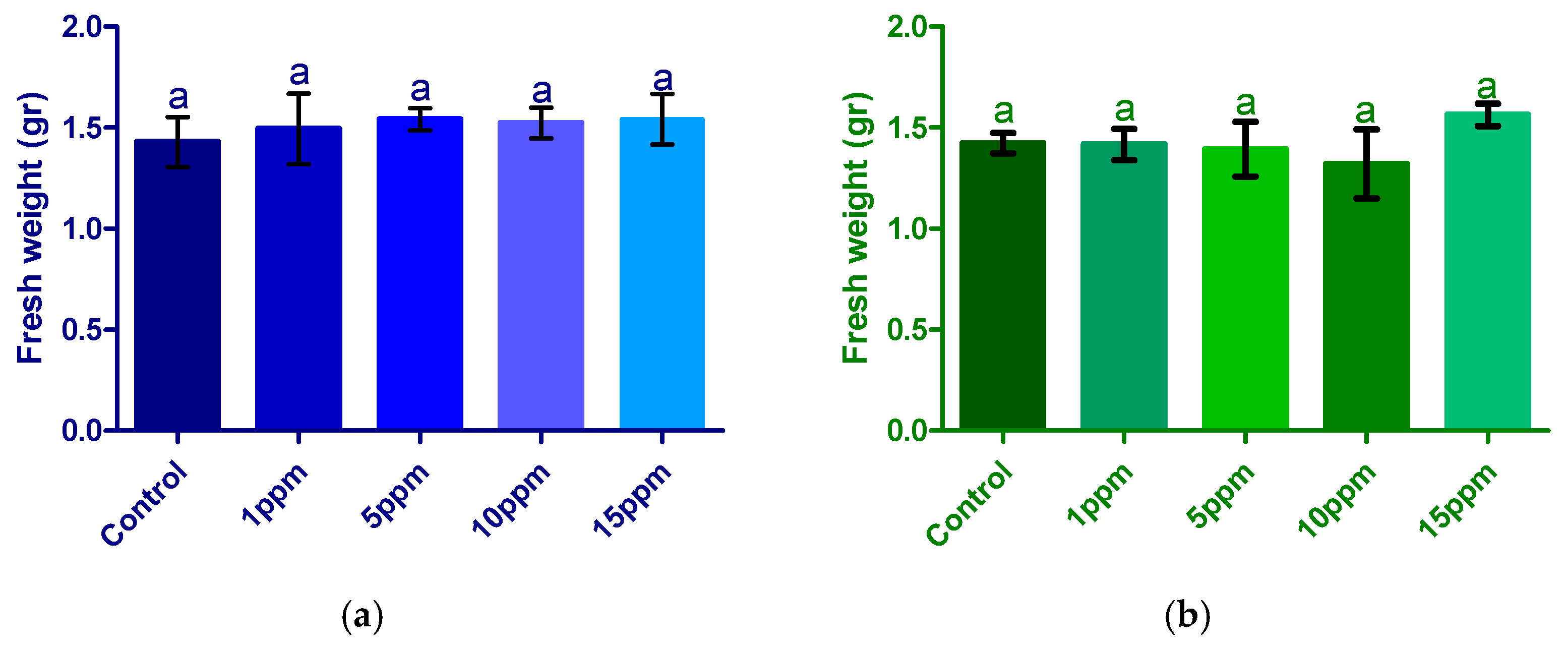
The fresh weight of plants treated with TiO2 and Ag-TiO2 was recorded after nine days of growth. As shown in Figure 4a, gram-values for TiO2 exhibited percentage differences relative to the control (Table 2), while Ag-TiO2 (Figure 4b) treatment displayed a distinct pattern. In TiO2-treated samples, biomass increases of 4% to 8% were observed across all tested concentrations, though without statistically significant differences between them. Conversely, Ag-TiO2 treatment demonstrated an inhibitory effect at all concentrations, with 1% to 7% reductions in fresh weight. These decreases, like the increases observed with TiO2, were not statistically significant relative to the control, and no significant variations were detected among the tested concentrations.
Changes in fresh weight generated from the treatments (with TiO2 and Ag-TiO2) are summarized in Table S1. These modifications are reported as a percentage variation with respect to the control group.
An increase in TiO2 concentration was associated with an increase in biomass production; however, this increase was not statistically significant (p > 0.05), suggesting a potential positive effect of TiO2 on plant growth. Specifically, concentrations of 5 and 15 ppm exhibited an approximate 8% increase compared to the control, which may be related to TiO2’s ability to stimulate photosynthesis and thus enhance biomass production.
Conversely, the Ag-TiO2 treatment showed a decrease in biomass production across most concentrations tested, with a progressive reduction in fresh weight as concentration increased. The 1 and 5 ppm concentrations exhibited slight decreases of 1% and 2%, respectively, without significant differences relative to the control. The 10 ppm concentration showed a 7% reduction, whereas the 15 ppm concentration, unlike the others, demonstrated a 7% increase in biomass.
Nonetheless, it is important to emphasize that none of these percentage changes relative to the control were statistically significant, as all p-values exceeded 0.05 according to Tukey’s and Dunnett’s tests.

3.2.2. Radicle Length

Radicle length was measured from the bifurcation of the cotyledons, i.e., from the first leaves to the shoot tip. These measurements were recorded after 9 days of growth under standardized humidity and temperature conditions.
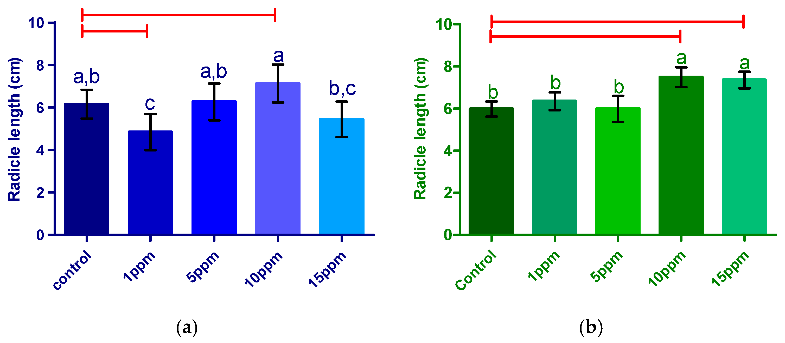
Figure 5 illustrates the differential effects of TiO2 and Ag-TiO2 nanoparticles on radicle length in alfalfa seedlings (Figure S2). In the TiO2 treatment (Figure 5a), a concentration-dependent response is observed: the application of 1 ppm resulted in a significant inhibitory effect compared to the control, while concentrations of 5 and 10 ppm promoted radicle growth, with the maximum length achieved at 10 ppm. Although the 15 ppm concentration also showed a promotive effect relative to the control, it did not significantly exceed the effect observed at 10 ppm and exhibited greater variability, as indicated by the error bars. It is noteworthy that statistical analyses (Tukey’s test) reveal significant differences between certain groups, but also some overlap among treatments, suggesting a non-linear response and the possible existence of an optimal concentration threshold.
For the Ag-TiO2 treatment (Figure 5b), concentrations of 1 and 5 ppm did not produce significant differences compared to the control, whereas 10 and 15 ppm induced a significant increase in radicle length. However, no significant difference was observed between these two higher concentrations, indicating a plateau in the promotive effect from 10 ppm onwards. Importantly, in both treatments, the 10 ppm concentration stands out as the most effective in stimulating radicle growth.
Table S1 summarizes the modifications in root length of alfalfa sprouts after nine days of exposure to different concentrations of TiO2 and Ag-TiO2 nanoparticles. For the TiO2 treatment, a concentration-dependent response was observed: application of 1 ppm resulted in significant inhibition of root growth (−21%), while concentrations of 5 and 10 ppm promoted growth, with increases of 2% and 16% compared to the control, respectively. In contrast, the 15 ppm concentration again showed an inhibitory effect (−12%). These results indicate that the greatest promotion of root growth was achieved at 10 ppm TiO2, suggesting the existence of an optimal concentration for this effect.
For the Ag-TiO2 treatment, concentrations of 1 and 5 ppm did not produce relevant changes compared to the control (6% and 0%, respectively). However, concentrations of 10 and 15 ppm induced notable increases in root length, with increments of 25% and 23%, respectively. This demonstrates that, in the case of Ag-TiO2, the promotive effect on root growth is primarily observed at higher concentrations.
Taken together, the data in Figure S1 highlight that a concentration of 10 ppm, for both TiO2 and Ag-TiO2, represents the most effective threshold for promoting root growth in alfalfa sprouts under the evaluated conditions.

3.2.3. Chlorophyll Index

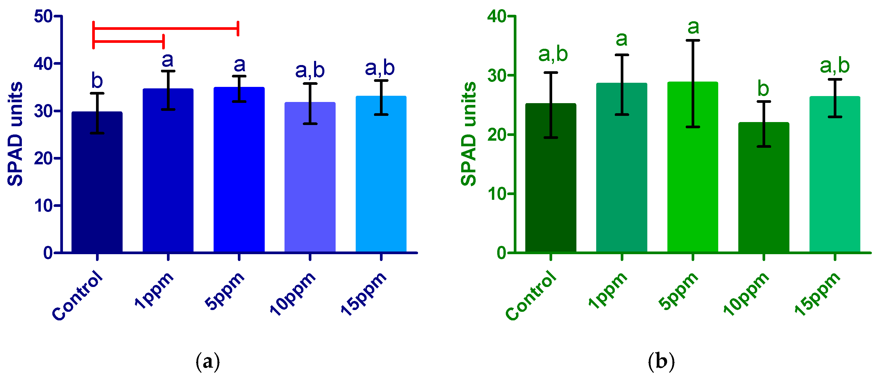
Data were recorded from the measurements made on the true leaves of the sprouts after the established growth period of 9 days. SPAD values were used as an indirect indicator of chlorophyll content [43]. Chlorophyll content was estimated non-destructively using an SPAD 502 Plus Chlorophyll Meter (Konica Minolta Co., Ltd., Tokyo, Japan). The device measures leaf transmittance at two wavelengths (≈650 nm and 940 nm) and calculates an SPAD index that correlates positively with total chlorophyll (a + b) and leaf nitrogen content. Compared with solvent-based extraction methods, SPAD readings are rapid, reliable, and allow repeated measurements on the same tissue without damage, providing an effective indicator of photosynthetic activity and plant physiological status under stress or treatment conditions. TiO2 increased SPAD across concentrations (≈7–17%; significant between some groups, p < 0.05). Ag-TiO2 did not differ significantly from control at 1, 5, and 15 ppm, but 10 ppm decreased SPAD (p < 0.05) (Figure 6).

3.2.4. Morphological Changes on Leaves

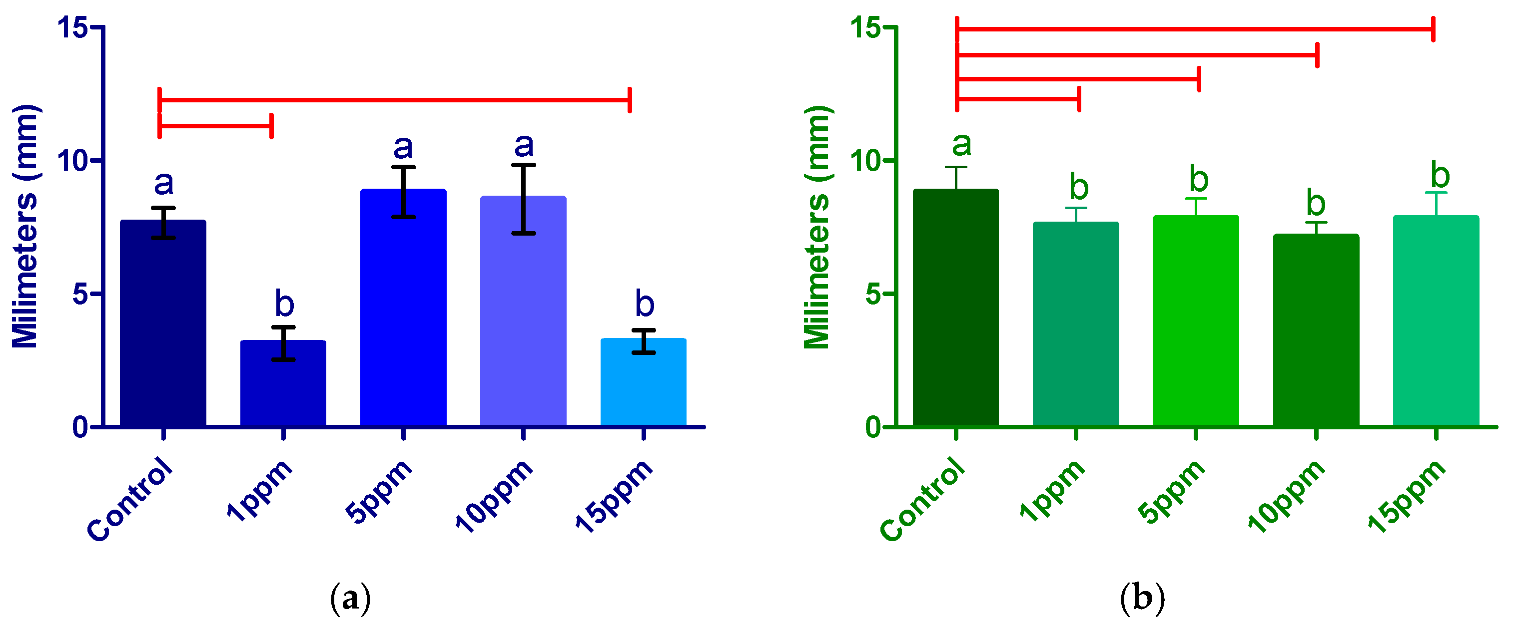
Figure 7a illustrates that treatment with TiO2 nanoparticles produced variable effects on the leaf width of alfalfa, in accordance with the concentration applied Figure S2. Increases of 13% and 4% were observed at 5 and 10 ppm, respectively, while concentrations of 1 and 15 ppm caused a significant 49% reduction in leaf width. This trend aligns with that observed in radicle length, where intermediate concentrations favored growth and extreme concentrations exhibited inhibitory effects. However, according to Tukey’s and Dunnett’s statistical analyses, the increase in leaf width at 5 ppm was not statistically significant (p > 0.05), whereas the reductions at 1 and 15 ppm were significant (p < 0.05), as detailed in Table S1.
In contrast, the Ag-TiO2 treatment (Figure 7b) displayed a different pattern, with increases in leaf width ranging from 3% to 11% across all tested concentrations. The greatest promotive effect was recorded at 15 ppm, while 10 ppm yielded the smallest increase. Nonetheless, none of these differences were statistically significant (p > 0.05). This behavior suggests that, unlike pure TiO2, silver-doped nanoparticles exert a more uniform and moderate effect on foliar growth without marked inhibitory effects.
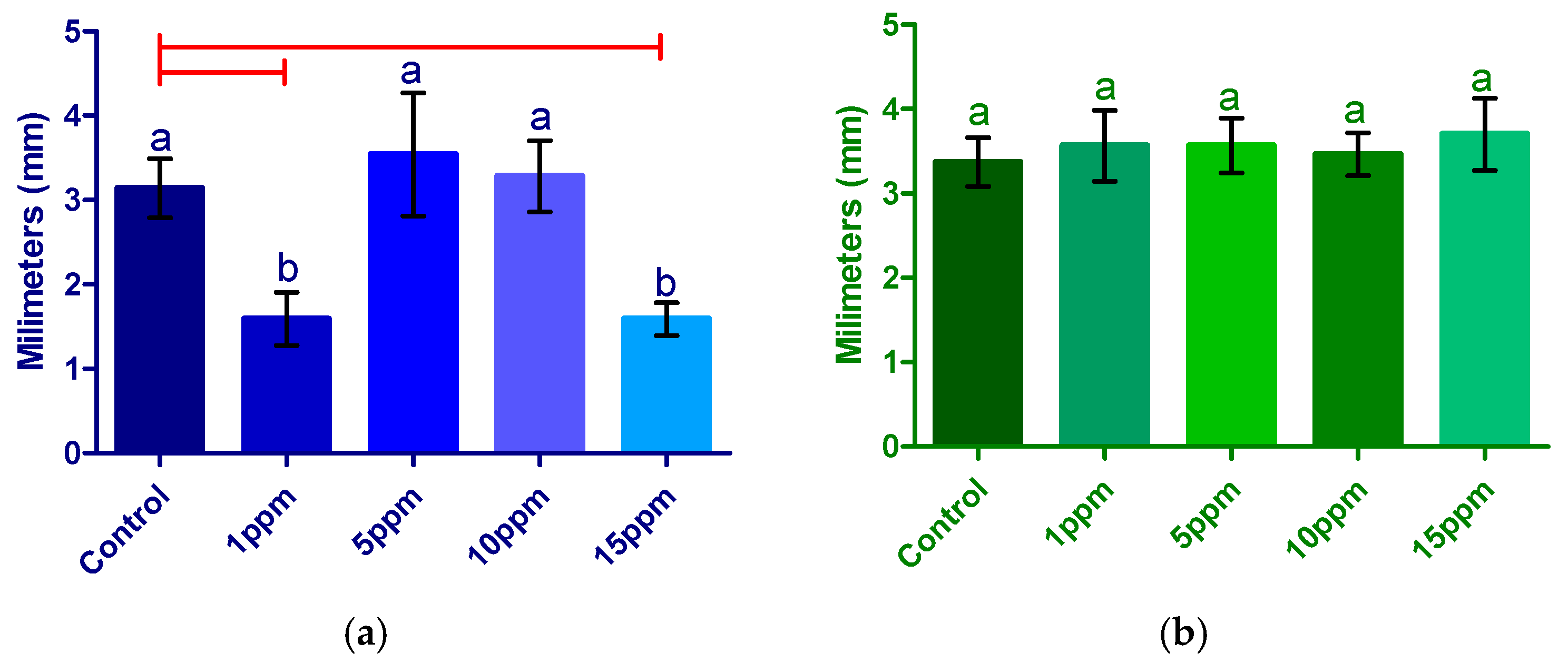
Figure 8a demonstrates that treatment with TiO2 nanoparticles produced contrasting effects on the true leaf length of alfalfa seedlings, depending on the applied concentration (Figure S3a,b). Extreme concentrations of 1 and 15 ppm resulted in significant inhibition of leaf elongation, with reductions of 59% and 58% compared to the control, respectively (Table S1). In contrast, intermediate concentrations of 5 and 10 ppm produced a slight promotive effect, with increases of 13% and 11% in leaf length. However, these increases were not statistically significant compared to the control, according to Tukey’s and Dunnett’s tests (p > 0.05), whereas the reductions observed at the extreme concentrations were significant (p < 0.05). These results suggest that the effect of TiO2 on leaf elongation is dose-dependent, with marked inhibition at low and high concentrations, and a neutral or mildly promotive effect at intermediate concentrations.
In the case of Ag-TiO2 treatment (Figure 8b), a generalized trend of growth inhibition was observed across all concentrations tested. Reductions in leaf length ranged from 11% to 17% relative to the control, with the maximum inhibitory effect at 10 ppm (S1). Unlike the TiO2 treatment, no significant differences were observed among the various Ag-TiO2 concentrations; however, all concentrations differed significantly from the control (p < 0.05), indicating a consistent negative impact of silver doping on leaf development.
Additionally, although Ag-TiO2 treatment promoted an increase in leaf length (Figure 8b), this morphological change did not translate into greater longitudinal growth, but rather resulted in leaves with a more elongated, drop-like shape, in contrast to the oval shape observed in the control group. The 10 ppm concentration of Ag-TiO2 was particularly notable, as it produced both significant inhibition of leaf length and changes in SPAD values, suggesting a relationship between pigmentation and leaf morphology. However, this relationship was not entirely consistent across all concentrations, except for 10 ppm. Overall, the morphological effect of silver-doped nanoparticles on leaves was variable and did not show a clear trend of growth promotion or inhibition. Representative photographs of all treatments group (Control, 1, 5, 10, and 15 ppm for both TiO2 and Ag-TiO2) are shown in Supplementary Materials Section (Figures S2 and S3), where the morphological effects described above will be visualized.

3.3. Metabolic Assessment (Antioxidant Capacity and Total Phenols and Flavonoids)

3.3.1. Determination of Total Phenols

For the TiO2 treatment (Figure 9a), a progressive increase in total phenolic content was observed as the treatment concentration increased. Concentrations of 1 and 5 ppm produced modest increases of 12% and 8% compared to the control, respectively, with no significant difference between them (p > 0.05). However, concentrations of 10 and 15 ppm resulted in substantial increases of 46% and 68%, respectively, which were both statistically significant compared to the control (p < 0.05), highlighting the promotive efficacy of higher concentrations.
In contrast, the Ag-TiO2 treatment (Figure 9b) exhibited a different pattern. The 1 ppm concentration caused a slight inhibition (−2%) relative to the control, while 5 ppm induced the highest increase in total phenolics (62%), followed by 15 ppm (29%) and 10 ppm (15%). The increase observed at 5 ppm was statistically significant compared to the control (p < 0.05), whereas the differences between 10 and 15 ppm were not significant (p > 0.05). These results suggest that the response in total phenolic production depends on both the type of nanoparticle and the applied concentration, with 15 ppm TiO2 being the most effective in promoting phenolic compound accumulation.

3.3.2. Determination of Total Flavonoids

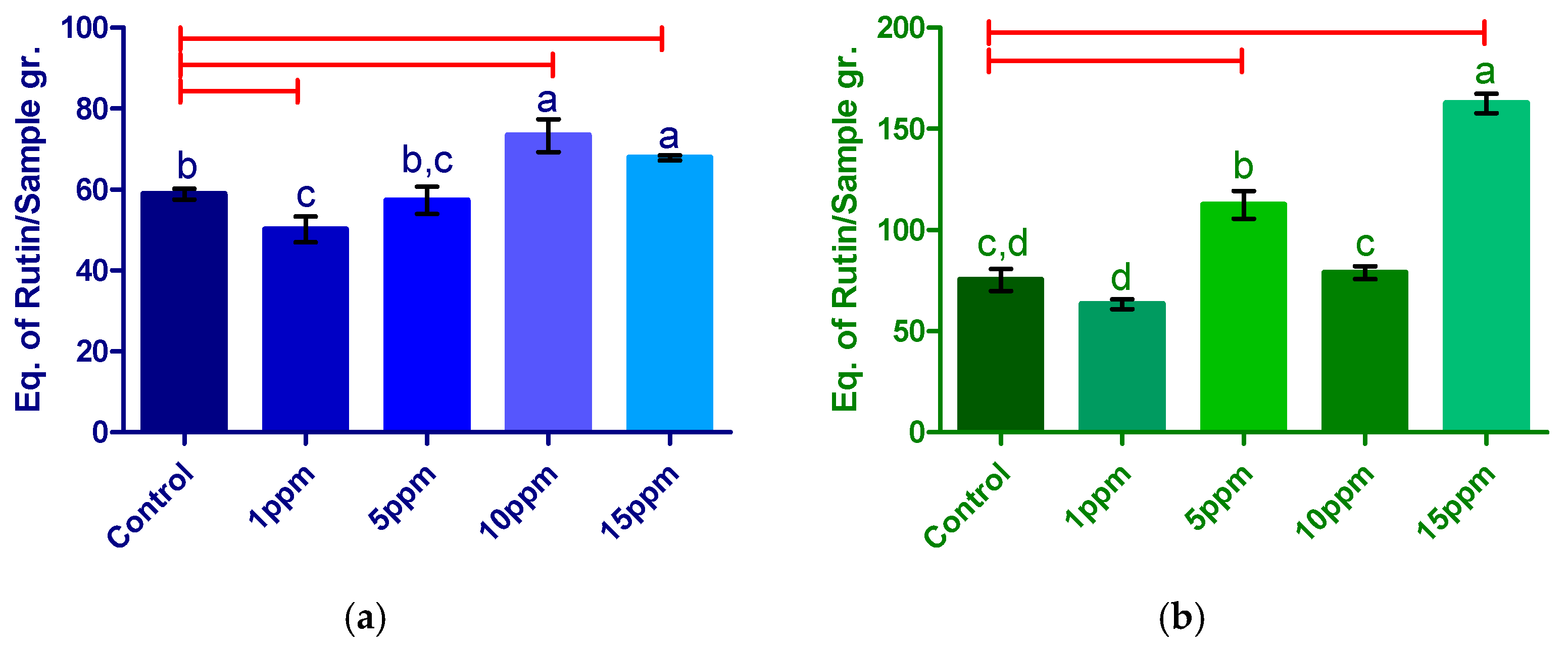
The results for total flavonoid quantification in alfalfa seedlings treated with TiO2 and Ag-TiO2 nanoparticles, synthesized via the sol–gel method, are presented in Figure 10. Values are expressed as mg Rutin equivalents per gram of sample.
For the TiO2 treatment (Figure 10a), a concentration-dependent response was observed. Concentrations of 1 and 5 ppm induced slight inhibitions in total flavonoid levels, with reductions of 15% and 3% compared to the control, respectively, although these differences were not statistically significant (p > 0.05). In contrast, concentrations of 10 and 15 ppm significantly increased flavonoid content by 25% and 15%, respectively (p < 0.05 vs. control). Although the promotive effect was higher at 10 than at 15 ppm, the difference between these concentrations was not statistically significant, despite the clear trend suggesting a maximum at 10 ppm.
For the Ag-TiO2 treatment (Figure 10b), a significant promotion of total flavonoid production was observed at 5 and 15 ppm, with increases of 49% and 116% compared to the control, respectively (p < 0.05). At 10 ppm, an 18% increase was observed, although this was not statistically significant (p > 0.05). Conversely, the 1 ppm concentration resulted in a 16% reduction in flavonoid levels, consistent with the results for total phenols at the same concentration and treatment. Overall, the Ag-TiO2 treatment exhibited a greater promotive effect on total flavonoid production compared to undoped TiO2. It is noteworthy that the 15 ppm concentration in both treatments induced the highest accumulation of secondary metabolites (total phenols and flavonoids), with the most pronounced effect observed for Ag-TiO2.

3.3.3. Determination of Antioxidant Capacity in Sprouts with TiO2 and Ag-TiO2 Treatment

The antioxidant capacity was measured to calculate mean the DPPH radical inhibition in alfalfa sprout extracts after nine days of growth. The statistical analysis of the antioxidant capacity showed the antioxidant activity in alfalfa sprouts treated with TiO2 and Ag-TiO2, as shown in Table 2. TiO2 decreased inhibition at all doses (p < 0.05). Ag-TiO2 treatment did not differ significantly from the control across doses. Consequently, no clear trend was identified linking Ag-TiO2 concentration to increased oxidative damage, as the observed fluctuations were not sufficiently robust to establish a defined pattern.
The results for ABTS radical scavenging activity in alfalfa sprouts treated with TiO2 and Ag-TiO2 are shown in Table 2. TiO2 decreased inhibition at 10 and 15 ppm (p < 0.05); 1–5 ppm showed no significant change. Ag-TiO2 increased inhibition at 1–5 ppm and decreased it at 10–15 ppm (p < 0.05).
Overall, these results demonstrate that both the type of nanoparticle and the applied concentration significantly influence the antioxidant capacity of alfalfa sprouts. While TiO2 tends to decrease antioxidant activity at higher concentrations, Ag-TiO2 can enhance it at low concentrations but exerts an inhibitory effect at higher doses.

4. Discussion

The nanometric characteristic and crystalline properties of TiO2 and Ag-TiO2 were confirmed by SEM, TEM, and XRD analyses, and additional information on the characterization of the nanoparticles can be found in Supplementary Materials (Figure S1). These results support the idea that the suspensions used retained a structure consistent with the anatase phase, with no evidence of significant agglomeration. Therefore, we infer that the observed effects of the treatments are derived primarily from exposure to nanoparticles rather than from possible ionic dissolution, in agreement with previous reports indicating that Ag+ release from low-doped TiO2 (≤0.1 wt%) remains minimal under neutral aqueous conditions [44,45].
Based on this physicochemical stability, the observed biological responses can be attributed to direct interactions between the nanoparticles and germinative tissues. Indeed, seed exposure to TiO2 and Ag-TiO2 during imbibition produced dose-dependent and trait-specific responses. Low doses of TiO2 (1 ppm) promoted several early growth traits (radicle length, leaf size, and pigmentation), whereas medium doses (5–10 ppm) tended to reduce germination percentage. In contrast, treatment with Ag-TiO2 at 15 ppm caused significant alterations in germination, fresh weight, and radicle length, with little effect on leaf morphology.
This response pattern aligns with the biphasic or hormetic behavior widely described for metallic and metal oxide nanomaterials in plants, where stimulation at low doses transforms into inhibition at higher doses and depends on the composition, size, and exposure window [46,47]. In accordance, nano-TiO2 has shown dual responses in multiple plant systems [20]. Under this mechanistic perspective, the higher availability and relative accumulation of Ag from the Ag-TiO2 composite could explain the inhibitory effect observed at 15 ppm, as suggested by Wang et al. [48], who highlight that the reduced size and rapid dissolution of silver-doped nanoparticles intensify their interaction with tissues at early stages of development.
On the other hand, at the tissue level, nano-TiO2 particles are absorbed through the root epidermis and even through foliar pathways, moving through the apoplastic, and symplastic routes, and accumulate in vacuoles, cell walls or chloroplasts, depending on the size, coating, and concentration [49,50]. In contrast, silver nanoparticles and the released Ag+ ions present greater mobility and reactivity. Smaller AgNPs (characterized by faster ionic dissolution) more easily penetrate cell walls, move towards vascular tissues, and are transported to shoots, thus altering root elongation, stem growth, and germination [21]. The Ag-TiO2 nanocomposite likely acts through a dual mechanism: TiO2 acts as a support and redox modulator, while the silver dopant contributes with increased mobility and release of Ag+ ions, which would explain the more intense effects observed at intermediate concentrations (10–15 ppm).
The dose-dependent transition from beneficial to detrimental effects observed in this study is consistent with the hormesis phenomenon described for various metallic and metal-oxide nanomaterials. At low doses, nanoparticles can induce eustress that stimulates antioxidant, enzymatic, and hormonal signaling pathways, while at higher concentrations they exceed homeostatic capacity and cause oxidative stress, photosynthetic disturbances, and nutritional imbalances [14,51]. This type of biphasic response has been widely documented in species such as Oryza sativa L., Triticum aestivum L., and Phaseolus vulgaris L. exposed to TiO2 or AgNPs [20,52].
The observed differences in fresh weight between control and nanoparticle-treated seedlings can be attributed to the imbibition phase, during which seeds likely absorbed enough water to influence hydrolytic activity that mobilizes energy reserves. This behavior is consistent with the findings of Hao et al. [53], who reported that exposure to TiO2 between 5 and 150 ppm did not produce significant changes in rice seedling biomass. Our results reinforce that fresh weight does not necessarily correlate with other early morphological traits: root elongation, germination, and fresh mass responded in a non-parallel and dose-dependent manner [53,54]. With TiO2, root elongation was inhibited at 1 ppm and stimulated at 10 ppm, whereas with Ag-TiO2, promotion was evident between 10 and 15 ppm. This decoupling suggests that fresh stand and early growth traits are partially independent variables, influenced by distinct physiological processes.
Consequently, germination and root elongation were analyzed separately from fresh weight. Intermediate concentrations (5–10 ppm) tended to reduce germination, although without significant differences (p > 0.05), which is consistent with previous reports of uncoupled responses to TiO2 exposure in legumes and cereals, such as Pisum sativum and Triticum aestivum L., where moderate germination reductions were observed without notable changes in biomass [25]. Studies in Vicia faba L. and Phaseolus vulgaris L. also reported inhibition of stem elongation at 15 mg L−1 of TiO2, consistent with the trends observed here, although with differences attributable to particle size and treatment duration [55].
Treatment with Ag-TiO2 induced root morphological alterations comparable to those observed after exposure to pure AgNPs at 10–20 mg L−1, where root growth stimulation is reported, while AgNO3 produces inhibition at similar doses [21]. These effects are attributed to the release of Ag+, which interferes with sulfur metabolism proteins and promotes the release of nitrile compounds with auxin activity [56,57] which could explain the observed root pattern.
Regarding leaf pigmentation, SPAD units increased significantly with TiO2 at 1 and 5 ppm (≈17% compared to the control), suggesting a stimulating effect on chlorophyll biosynthesis. This result contrasts with that reported by Z. Doğaroğlu et al. [26], who observed pigment inhibition at similar concentrations, but agrees with recent studies where TiO2 improves photosynthetic efficiency and chlorophyll content under low radiation or abiotic stress conditions [58]. In contrast, Ag-TiO2 did not significantly modify SPAD values, in disagreement with Gordillo-Delgado et al. [59], who documented increases in spinach treated with smaller nanoparticles (7–8 nm). This contrast confirms that size and dose are critical factors in determining the physiological response [21].
Given the nanometric size of the particles used, intracellular internalization is likely. Proposed mechanisms include passive diffusion across the membrane, endocytosis, and transport via protein channels or aquaporins [60]. Through these routes, nanoparticles can interfere with metabolic processes associated with phase II of germination, including the activation of antioxidant enzymes and the synthesis of secondary metabolites. In this study, the metabolic profile showed a general trend toward increased secondary metabolites, especially with TiO2, whose dose–response effect was more consistent, peaking at 15 ppm. Similar results have been reported by Tighe-Neira et al. [61] and recently by T. Missaoui et al. [62], who observed increases of 20–60% in phenolic and flavonoid compounds in seedlings treated with TiO2 between 10 and 100 mg L−1.
In contrast, Ag-TiO2 exhibited its maximum phenolic induction at 5 ppm, doubling the total phenol content relative to the control, but without a clear linear trend, suggesting metabolic saturation at intermediate concentrations. Antioxidant activity, assessed by DPPH, showed predominantly inhibitory effects, contrary to what has been reported in systems treated with ZnO or Ag [63,64]. Recent studies confirm that both AgNPs and TiO2 can generate reactive oxygen species (ROS) that, in excess, reduce overall antioxidant capacity by oxidizing compounds such as ascorbic acid and soluble phenols [65,66].
In this context, the lower antioxidant capacity detected in plants treated with TiO2 could be associated with the limited generation of photocatalytic electrons under germination conditions, while Ag+ doping, although it increases the photocatalytic of the system, does not necessarily translate into a net antioxidant enhancement. This suggests a complex interaction between doping, concentration, and tissue redox status [51,67]. Additional assays with a broader range of concentrations (>15 ppm) and specific analyses of ROS and antioxidant enzymes (SOD, CAT, APX) are required to confirm this hypothesis.
Finally, the low proportion of Ag+ dopant in the TiO2 used, corroborated by its non-detection in morphological and crystallographic analyses, explains the absence of marked cytotoxic effects and the moderate activation of antioxidant defense mechanisms, reflected in the increase in total phenols and flavonoids. Mahakham et al. [68] and Bedlovičová et al. [69] proposed that silver nanopriming acts through three complementary pathways: nanopore formation in the seed coat, controlled ROS induction, and enzymatic activation of α-amylase. In our case, the relatively low Ag+ concentration likely induced a eustress level that favored germination without significantly altering the levels of antioxidant metabolites, which agrees with recent studies on the regulatory role of ROS in early germination [70,71].

5. Conclusions

The physiological and morphological responses observed in seedlings treated with TiO2 and Ag-TiO2 nanoparticles are strongly dependent on particle size, exposure duration, and seed coat composition, which govern nanoparticle penetration and translocation. Our results suggest and support the theory that smaller nanoparticles (≈14–16 nm) exhibit greater internal mobility, enhancing nutrient uptake and stress modulation, whereas larger particles tend to accumulate in root tissues, limiting benefits and occasionally causing phytotoxic effects. The concentration-dependent changes in leaf size and pigmentation observed in this study confirm that nanoscale properties directly influence photosynthetic capacity and early plant development.
These results contribute to the understanding of nanoparticle–plant interactions by demonstrating how physicochemical characteristics (size, composition, and dose) determine functional outcomes. The study’s relevance lies in providing mechanistic insights useful for designing safer and more efficient nano formulations for agricultural applications, optimizing their benefits while reducing ecological risks.

Supplementary Materials

The following supporting information can be downloaded at: https://www.mdpi.com/article/10.3390/agronomy15112580/s1, Figure S1: Additional information on the characterization of the nanoparticles, as well as the repositories of the work groups where these nanoparticles were used, are available; Table S1: Percentual changes on morphological characteristics of sprouts treated with nanoparticles of TiO2 and Ag-TiO2; Figure S2: Representative images of radicle length, leaf width, and length measurements in germinant treated with TiO2 and Ag-TiO2; Figure S3: Representative photographs of the general morphology of the sprouts treated with Ag-TiO2.

Author Contributions

Writing—original draft preparation, A.M.-B.; writing—review and editing, A.A.F.-P., K.E.E., H.A.-B., C.E.P.-G. and S.R.-J.; resources, supervision and visualization, A.A.F.-P. and K.E.E.; methodology, C.E.P.-G. and S.R.-J.; investigation, H.A.-B.; visualization, analysis, and interpretation for data, A.M.-B. All authors have read and agreed to the published version of the manuscript.

Funding

This research received no external funding.

Data Availability Statement

Data are contained within the article.

Acknowledgments

GenAI tools was used for writing assistance (grammar, punctuation and reduce the similarity percentage in the Section 2). The authors are pleased to acknowledge and thank Secretaría de Ciencia, Humanidades, Tecnología e Innovación (SECIHTI) for the postgraduate scholarship of Susana Rodríguez Jurado. Moreover, the authors would like to thank FONFIVE FIN202420 and FONFIVE FIN202413.

Conflicts of Interest

The authors declare that they have no known competing financial interests or personal relationships that could have appeared to influence the work reported in this paper.

References

  1. Jia, Y.; Klumpp, E.; Bol, R.; Amelung, W. Uptake of metallic nanoparticles containing essential (Cu, Zn and Fe) and non-essential (Ag, Ce and Ti) elements by crops: A meta-analysis. Crit. Rev. Environ. Sci. Technol. 2023, 53, 1512–1533. [Google Scholar] [CrossRef]
  2. Balusamy, S.R.; Joshi, A.S.; Perumalsamy, H.; Mijakovic, I.; Singh, P. Advancing sustainable agriculture: A critical review of smart and eco-friendly nanomaterial applications. J. Nanobiotechnol. 2023, 21, 372. [Google Scholar] [CrossRef] [PubMed]
  3. Faizan, M.; Sharma, P.; Sultan, H.; Alam, P.; Sehar, S.; Rajput, V.D.; Hayat, S. Nano-priming: Improving plant nutrition to support the establishment of sustainable agriculture under heavy metal stress. Plant Nano Biol. 2024, 10, 100096. [Google Scholar] [CrossRef]
  4. Mgadi, K.; Ndaba, B.; Roopnarain, A.; Rama, H.; Adeleke, R. Nanoparticle applications in agriculture: Overview and response of plant-associated microorganisms. Front. Microbiol. 2024, 15, 1354440. [Google Scholar] [CrossRef]
  5. Ansari, M.M.; Shin, M.; Kim, M.; Ghosh, M.; Kim, S.-H.; Son, Y.-O. Nano-enabled strategies in sustainable agriculture for enhanced crop productivity: A comprehensive review. J. Environ. Manag. 2024, 372, 123420. [Google Scholar] [CrossRef]
  6. Chandoliya, R.; Sharma, S.; Sharma, V.; Joshi, R.; Sivanesan, I. Titanium Dioxide Nanoparticle: A Comprehensive Review on Synthesis, Applications and Toxicity. Plants 2024, 13, 2964. [Google Scholar] [CrossRef]
  7. Bakshi, M.; Kumar, S. Investigating Impacts of Nanosized Titanium Dioxide (Nano-TiO2) on Maize (Zea mays L.) and Their Ensuing Implications on Soil Physiochemistry: A Soil Exposure Study. Water Air Soil Pollut. 2023, 234, 559. [Google Scholar] [CrossRef]
  8. Erofeeva, E.A. Hormesis and Nanomaterials. From Biostimulation to Toxicity. In Plant Biostimulation with Nanomaterials; Juárez-Maldonado, A., Benavides-Mendoza, A., Ojeda-Barrios, D.L., Tortella Fuentes, G., Seabra, A.B., Eds.; Springer Nature: Singapore, 2025; pp. 1–19. [Google Scholar] [CrossRef]
  9. Sharma, M.M.M.; Kapoor, D.; Loyal, A.; Kumar, R.; Sharma, P.; Husen, A. Plant Response to Silver Nanoparticles in Terms of Growth, Development, Production, and Protection: An Overview. In Plant Response to Silver Nanoparticles: Plant Growth, Development, Production, and Protection; Husen, A., Ed.; Springer Nature: Singapore, 2025; pp. 1–22. [Google Scholar] [CrossRef]
  10. Alfosea-Simón, F.J.; Burgos, L.; Alburquerque, N. Silver Nanoparticles Help Plants Grow, Alleviate Stresses, and Fight Against Pathogens. Plants 2025, 14, 428. [Google Scholar] [CrossRef]
  11. Muzammil, S.; Ashraf, A.; Siddique, M.H.; Aslam, B.; Rasul, I.; Abbas, R.; Afzal, M.; Faisal, M.; Hayat, S. A review on toxicity of nanomaterials in agriculture: Current scenario and future prospects. Sci. Prog. 2023, 106, 00368504231221672. [Google Scholar] [CrossRef]
  12. Narang, J.; Kapoor, B.; Sharma, S.; Gill, N.S. Mechanistic insights: How nanoparticles modulate plant hormones and defense response signaling under stress. Plant Nano Biol. 2025, 13, 100174. [Google Scholar] [CrossRef]
  13. Mathur, P.; Chakraborty, R.; Aftab, T.; Roy, S. Engineered nanoparticles in plant growth: Phytotoxicity concerns and the strategies for their attenuation. Plant Physiol. Biochem. 2023, 199, 107721. [Google Scholar] [CrossRef]
  14. Francis, D.V.; Abdalla, A.K.; Mahakham, W.; Sarmah, A.K.; Ahmed, Z.F.R. Interaction of plants and metal nanoparticles: Exploring its molecular mechanisms for sustainable agriculture and crop improvement. Environ. Int. 2024, 190, 108859. [Google Scholar] [CrossRef]
  15. Kokina, I.; Plaksenkova, I.; Jermaļonoka, M.; Petrova, A. Impact of iron oxide nanoparticles on yellow medick (Medicago falcata L.) plants. J. Plant Interact. 2020, 15, 1–7. [Google Scholar] [CrossRef]
  16. De Souza-Torres, A.; Govea-Alcaide, E.; Gomez-Padilla, E.; Masunaga, S.H.; Effenberger, F.B.; Rossi, L.M.; Lopez-Sanchez, R.; Jardim, R.F. Fe3O4 nanoparticles and Rhizobium inoculation enhance nodulation, nitrogen fixation and growth of common bean plants grown in soil. Rhizosphere 2021, 17, 100275. [Google Scholar] [CrossRef]
  17. Aruna, A.; Namasivayam, S.K.R.; Chandu, P.S. Evaluation of phytotoxicity of metallic nanoparticles against Amaranthus retroflexus. Asian J. Microbiol. Biotechnol. Environ. Sci. 2014, 16, 41–46. [Google Scholar]
  18. Karimi, J.; Mohsenzadeh, S. Effects of silicon oxide nanoparticles on growth and physiology of wheat seedlings. Russ. J. Plant Physiol. 2016, 63, 119–123. [Google Scholar] [CrossRef]
  19. Imtiaz, M.; Kamran, M.; Arshad, A.; Sardar, S.; Shah, T. Effect of Exogenous Application of Titanium Dioxide Nanoparticles on Growth, Yield, and Yield Components of Wheat (Triticum aestivum L.) under Drought Stress Conditions. Planta Anim. 2025, 4, 217–234. [Google Scholar] [CrossRef]
  20. Trela-Makowej, A.; Orzechowska, A.; Szymańska, R. Less is more: The hormetic effect of titanium dioxide nanoparticles on plants. Sci. Total Environ. 2024, 910, 168669. [Google Scholar] [CrossRef]
  21. Lahuta, L.B.; Szablińska-Piernik, J.; Stałanowska, K.; Głowacka, K.; Horbowicz, M. The Size-Dependent effects of silver nanoparticles on germination, early seedling development and polar metabolite profile of wheat (Triticum aestivum L.). Int. J. Mol. Sci. 2022, 23, 13255. [Google Scholar] [CrossRef]
  22. Sanzari, I.; Leone, A.; Ambrosone, A. Nanotechnology in plant science: To make a long story short. Front. Bioeng. Biotechnol. 2019, 7, 120. [Google Scholar] [CrossRef]
  23. Altabbaa, S.; Mann, N.A.; Chauhan, N.; Utkarsh, K.; Thakur, N.; Mahmoud, G.A.-E. Era connecting nanotechnology with agricultural sustainability: Issues and challenges. Nanotechnol. Environ. Eng. 2023, 8, 481–498. [Google Scholar] [CrossRef]
  24. Esquivel, K.; Nava, R.; Zamudio-Méndez, A.R.; Vega González, M.; Jaime-Acuña, Ó.; Escobar-Alarcón, L.; Peralta-Hernández, J.M.; Pawelec, B.; García Fierro, J.L. Microwave-assisted synthesis of (S)Fe/TiO2 systems: Effects of synthesis conditions and dopant concentration on photoactivity. Appl. Catal. B Environ. 2013, 140, 213–224. [Google Scholar] [CrossRef]
  25. Basahi, M. Seed germination with titanium dioxide nanoparticles enhances water supply, reserve mobilization, oxidative stress and antioxidant enzyme activities in pea. Saudi J. Biol. Sci. 2021, 28, 6500–6507. [Google Scholar] [CrossRef] [PubMed]
  26. Doğaroğlu, Z. Effects of TiO2 and ZnO nanoparticles on germination and antioxidant system of wheat (Triticum aestivum L.). Appl. Ecol. Environ. Res. 2017, 15, 1499–1510. [Google Scholar] [CrossRef]
  27. Parveen, A.; Rao, S. Effect of Nanosilver on Seed Germination and Seedling Growth in Pennisetum glaucum. J. Clust. Sci. 2015, 3, 693–701. [Google Scholar] [CrossRef]
  28. Parola-Contreras, I.; Guevara-González, R.G.; Feregrino-Pérez, A.A.; Reynoso-Camacho, R.; Pérez-Ramírez, I.F.; Ocampo-Velázquez, R.V.; Rojas-Molina, A.; Luna-Vazquez, F.; Tovar-Pérez, E.G.; Parola-Contreras, I.; et al. Phenolic compounds and antioxidant activity of methanolic extracts from leaves and flowers of chilcuague (Heliopsis longipes, Asteraceae). Bot. Sci. 2021, 99, 149–160. [Google Scholar] [CrossRef]
  29. Garcia-Mier, L.; Jimenez-Garcia, S.N.; Guevara-González, R.G.; Feregrino-Perez, A.A.; Contreras-Medina, L.M.; Torres-Pacheco, I. Elicitor Mixtures Significantly Increase Bioactive Compounds, Antioxidant Activity, and Quality Parameters in Sweet Bell Pepper. J. Chem. 2015, 2015, e269296. [Google Scholar] [CrossRef]
  30. Dewanto, V.; Wu, X.; Adom, K.K.; Liu, R.H. Thermal Processing Enhances the Nutritional Value of Tomatoes by Increasing Total Antioxidant Activity. J. Agric. Food Chem. 2002, 50, 3010–3014. [Google Scholar] [CrossRef]
  31. Garcia-Mier, L.; Meneses-Reyes, A.E.; Jimenez-Garcia, S.N.; Mercado Luna, A.; García Trejo, J.F.; Contreras-Medina, L.M.; Feregrino-Perez, A.A. Polyphenol Content and Antioxidant Activity of Stevia and Peppermint as a Result of Organic and Conventional Fertilization. J. Food Qual. 2021, 2021, e6620446. [Google Scholar] [CrossRef]
  32. Doğaroğlu, Z.G.; Ece, F.; Çiftci, B.N.; Yıldırımcan, S.; Erat, S. Evaluation of stress factor on wheat (Triticum aestivum): The effect of ZnO and Ni-doped ZnO nanoparticles. Toxicol. Environ. Chem. 2021, 103, 382–398. [Google Scholar] [CrossRef]
  33. Vishwakarma, M.; Kulhare, P.S.; Tagore, G.S. Estimation of Chlorophyll Using SPAD meter. Int. J. Environ. Clim. Change 2023, 13, 1901–1912. [Google Scholar] [CrossRef]
  34. Datye, A.; DeLaRiva, A. Scanning Electron Microscopy (SEM). In Springer Handbook of Advanced Catalyst Characterization; Wachs, I.E., Bañares, M.A., Eds.; Springer International Publishing: Cham, Switzerland, 2023; pp. 359–380. [Google Scholar] [CrossRef]
  35. Goldstein, J.I.; Newbury, D.E.; Michael, J.R.; Ritchie, N.W.M.; Scott, J.H.J.; Joy, D.C. Backscattered Electrons. In Scanning Electron Microscopy and X-Ray Microanalysis; Springer: New York, NY, USA, 2018; pp. 15–28. [Google Scholar] [CrossRef]
  36. Chauhan, R.; Kumar, A.; Chaudhary, R.P. Structural and optical characterization of Ag-doped TiO2 nanoparticles prepared by a sol–gel method. Res. Chem. Intermed. 2012, 38, 1443–1453. [Google Scholar] [CrossRef]
  37. Narkbuakaew, T.; Sattayaporn, S.; Saito, N.; Sujaridworakun, P. Investigation of the Ag species and synergy of Ag-TiO2 and g-C3N4 for the enhancement of photocatalytic activity under UV–Visible light irradiation. Appl. Surf. Sci. 2022, 573, 151617. [Google Scholar] [CrossRef]
  38. Damkale, S.R.; Arbuj, S.S.; Umarji, G.G.; Rane, S.B.; Kale, B.B. Highly crystalline anatase TiO2 nanocuboids as an efficient photocatalyst for hydrogen generation. RSC Adv. 2021, 11, 7587–7599. [Google Scholar] [CrossRef]
  39. Porkodi, K.; Arokiamary, S.D. Synthesis and spectroscopic characterization of nanostructured anatase titania: A photocatalyst. Mater. Charact. 2007, 58, 495–503. [Google Scholar] [CrossRef]
  40. Ohsaka, T.; Izumi, F.; Fujiki, Y. Raman spectrum of anatase, TiO2. J. Raman Spectrosc. 1978, 7, 321–324. [Google Scholar] [CrossRef]
  41. Sukarman; Kristiawan, B.; Budiana, E.P.; Khoirudin; Abdulah, A. Multi-technique characterization of TiO2 nanoparticles: Crystallite size, microstrain, and phase analysis for nanomaterial applications—A review. Hybrid Adv. 2025, 11, 100523. [Google Scholar] [CrossRef]
  42. Alam, M.A.; Ahmed, S.; Bishwas, R.K.; Mostofa, S.; Jahan, S.A. X-ray crystallographic diffraction study by whole powder pattern fitting (WPPF) method: Refinement of crystalline nanostructure polymorphs TiO2. S. Afr. J. Chem. Eng. 2025, 51, 68–77. [Google Scholar] [CrossRef]
  43. Padilla, F.M.; Teresa Peña-Fleitas, M.; Gallardo, M.; Thompson, R.B. Evaluation of optical sensor measurements of canopy reflectance and of leaf flavonols and chlorophyll contents to assess crop nitrogen status of muskmelon. Eur. J. Agron. 2014, 58, 39–52. [Google Scholar] [CrossRef]
  44. Tripathi, S.; Tiwari, K.; Mahra, S.; Victoria, J.; Rana, S.; Tripathi, D.K.; Sharma, S. Nanoparticles and root traits: Mineral nutrition, stress tolerance and interaction with rhizosphere microbiota. Planta 2024, 260, 34. [Google Scholar] [CrossRef] [PubMed]
  45. Djanaguiraman, M.; Anbazhagan, V.; Dhankher, O.P.; Prasad, P.V.V. Uptake, Translocation, Toxicity, and Impact of Nanoparticles on Plant Physiological Processes. Plants 2024, 13, 3137. [Google Scholar] [CrossRef]
  46. Rafique, R.; Zahra, Z.; Virk, N.; Shahid, M.; Pinelli, E.; Park, T.J.; Kallerhoff, J.; Arshad, M. Dose-dependent physiological responses of Triticum aestivum L. to soil applied TiO2 nanoparticles: Alterations in chlorophyll content, H2O2 production, and genotoxicity. Agric. Ecosyst. Environ. 2018, 255, 95–101. [Google Scholar] [CrossRef]
  47. Iavicoli, I.; Leso, V.; Fontana, L.; Calabrese, E.J. Nanoparticle Exposure and Hormetic Dose–Responses: An Update. Int. J. Mol. Sci. 2018, 19, 805. [Google Scholar] [CrossRef]
  48. Wang, J.; Koo, Y.; Alexander, A.; Yang, Y.; Westerhof, S.; Zhang, Q.; Schnoor, J.L.; Colvin, V.L.; Braam, J.; Alvarez, P.J.J. Phytostimulation of Poplars and Arabidopsis Exposed to Silver Nanoparticles and Ag+ at Sublethal Concentrations. Environ. Sci. Technol. 2013, 47, 5442–5449. [Google Scholar] [CrossRef]
  49. Zaman, W.; Ayaz, A.; Park, S. Nanomaterials in agriculture: A pathway to enhanced plant growth and abiotic stress resistance. Plants 2025, 14, 716. [Google Scholar] [CrossRef] [PubMed]
  50. Deng, Y.; Petersen, E.J.; Challis, K.E.; Rabb, S.A.; Holbrook, R.D.; Ranville, J.F.; Nelson, B.C.; Xing, B. Multiple Method Analysis of TiO2 Nanoparticle Uptake in Rice (Oryza sativa L.) Plants. Environ. Sci. Technol. 2017, 51, 10615–10623. [Google Scholar] [CrossRef]
  51. Cox, A.; Venkatachalam, P.; Sahi, S.; Sharma, N. Silver and titanium dioxide nanoparticle toxicity in plants: A review of current research. Plant Physiol. Biochem. 2016, 107, 147–163. [Google Scholar] [CrossRef]
  52. Khan, I.; Hasan, M.; Kausar, R.; Shehzad, J.; Mustafa, G. Plant Molecular Responses to Nanoparticle Stress. In Plant and Nanoparticles; Chen, J.-T., Ed.; Springer Nature: Singapore, 2022; pp. 239–264. [Google Scholar] [CrossRef]
  53. Hao, Y.; Zhang, Z.-T.; Rui, Y.-K.; Ren, J.-Y.; Hou, T.-Q.; Wu, S.-J.; Rui, M.-M.; Jiang, F.-P.; Liu, L.-M. Effect of Different Nanoparticles on Seed Germination and Seedling Growth in Rice. In Proceedings of the 2nd Annual International Conference on Advanced Material Engineering (AME 2016), Dubai, United Arab Emirates, 23–24 January 2016; Atlantis Press: Dordrecht, The Netherlands, 2016; pp. 166–173. [Google Scholar] [CrossRef]
  54. Haghighi, M.; Teixeira Da Silva, J.A. The effect of N-TiO2 on tomato, onion, and radish seed germination. J. Crop Sci. Biotechnol. 2014, 17, 221–227. [Google Scholar] [CrossRef]
  55. Kushwah, K.S.; Patel, S. Effect of Titanium Dioxide Nanoparticles (TiO2 NPs) on Faba bean (Vicia faba L.) and Induced Asynaptic Mutation: A Meiotic Study. J. Plant Growth Regul. 2020, 39, 1107–1118. [Google Scholar] [CrossRef]
  56. Khodakovskaya, M.V.; De Silva, K.; Nedosekin, D.A.; Dervishi, E.; Biris, A.S.; Shashkov, E.V.; Galanzha, E.I.; Zharov, V.P. Complex genetic, photothermal, and photoacoustic analysis of nanoparticle-plant interactions. Proc. Natl. Acad. Sci. USA 2011, 108, 1028–1033. [Google Scholar] [CrossRef]
  57. Sun, J.; Wang, L.; Li, S.; Yin, L.; Huang, J.; Chen, C. Toxicity of silver nanoparticles to Arabidopsis: Inhibition of root gravitropism by interfering with auxin pathway. Environ. Toxicol. Chem. 2017, 36, 2773–2780. [Google Scholar] [CrossRef]
  58. Vatankhah, A.; Aliniaeifard, S.; Moosavi-Nezhad, M.; Abdi, S.; Mokhtarpour, Z.; Reezi, S.; Tsaniklidis, G.; Fanourakis, D. Plants exposed to titanium dioxide nanoparticles acquired contrasting photosynthetic and morphological strategies depending on the growing light intensity: A case study in radish. Sci. Rep. 2023, 13, 5873. [Google Scholar] [CrossRef]
  59. Gordillo-Delgado, F.; Zuluaga-Acosta, J.; Restrepo-Guerrero, G. Effect of the suspension of Ag-incorporated TiO2 nanoparticles (Ag-TiO2 NPs) on certain growth, physiology and phytotoxicity parameters in spinach seedlings. PLoS ONE 2020, 15, e0244511. [Google Scholar] [CrossRef]
  60. Liu, W.; Zeb, A.; Lian, J.; Wu, J.; Xiong, H.; Tang, J.; Zheng, S. Interactions of metal-based nanoparticles (MBNPs) and metal-oxide nanoparticles (MONPs) with crop plants: A critical review of research progress and prospects. Environ. Rev. 2020, 28, 294–310. [Google Scholar] [CrossRef]
  61. Tighe-Neira, R.; Reyes-Díaz, M.; Nunes-Nesi, A.; Recio, G.; Carmona, E.; Corgne, A.; Rengel, Z.; Inostroza-Blancheteau, C. Titanium dioxide nanoparticles provoke transient increase in photosynthetic performance and differential response in antioxidant system in Raphanus sativus L. Sci. Hortic. 2020, 269, 109418. [Google Scholar] [CrossRef]
  62. Missaoui, T.; Smiri, M.; Chemingui, H.; Hafiane, A. Effect of Nanosized TiO2 on Redox Properties in Fenugreek (Trigonella foenum graecum L.) during Germination. Environ. Process. 2021, 8, 843–867. [Google Scholar] [CrossRef]
  63. García-López, J.I.; Zavala-García, F.; Olivares-Sáenz, E.; Lira-Saldívar, R.H.; Díaz Barriga-Castro, E.; Ruiz-Torres, N.A.; Ramos-Cortez, E.; Vázquez-Alvarado, R.; Niño-Medina, G. Zinc Oxide Nanoparticles Boosts Phenolic Compounds and Antioxidant Activity of Capsicum annuum L. during Germination. Agronomy 2018, 8, 215. [Google Scholar] [CrossRef]
  64. Jiang, H.S.; Yin, L.Y.; Ren, N.N.; Zhao, S.T.; Li, Z.; Zhi, Y.; Shao, H.; Li, W.; Gontero, B. Silver nanoparticles induced reactive oxygen species via photosynthetic energy transport imbalance in an aquatic plant. Nanotoxicology 2017, 11, 157–167. [Google Scholar] [CrossRef]
  65. Venzhik, Y.V.; Deryabin, A.N. Regulation of Pro-/Antioxidant Balance in Higher Plants by Nanoparticles of Metals and Metal Oxides. Russ. J. Plant Physiol. 2023, 70, 14. [Google Scholar] [CrossRef]
  66. Singh, R.; Choudhary, P.; Kumar, S.; Daima, H.K. Mechanistic approaches for crosstalk between nanomaterials and plants: Plant immunomodulation, defense mechanisms, stress resilience, toxicity, and perspectives. Environ. Sci. Nano 2024, 11, 2324–2351. [Google Scholar] [CrossRef]
  67. Kumar, D.; Dhankher, O.P.; Tripathi, R.D.; Seth, C.S. Titanium dioxide nanoparticles potentially regulate the mechanism (s) for photosynthetic attributes, genotoxicity, antioxidants defense machinery, and phytochelatins synthesis in relation to hexavalent chromium toxicity in Helianthus annuus L. J. Hazard. Mater. 2023, 454, 131418. [Google Scholar] [CrossRef] [PubMed]
  68. Mahakham, W.; Sarmah, A.K.; Maensiri, S.; Theerakulpisut, P. Nanopriming technology for enhancing germination and starch metabolism of aged rice seeds using phytosynthesized silver nanoparticles. Sci. Rep. 2017, 7, 8263. [Google Scholar] [CrossRef] [PubMed]
  69. Bedlovičová, Z.; Strapáč, I.; Baláž, M.; Salayová, A. A Brief Overview on Antioxidant Activity Determination of Silver Nanoparticles. Molecules 2020, 25, 3191. [Google Scholar] [CrossRef] [PubMed]
  70. Prażak, R.; Święciło, A.; Krzepiłko, A.; Michałek, S.; Arczewska, M. Impact of Ag Nanoparticles on Seed Germination and Seedling Growth of Green Beans in Normal and Chill Temperatures. Agriculture 2020, 10, 312. [Google Scholar] [CrossRef]
  71. Wang, Y.; Sun, X.; Peng, J.; Li, F.; Ali, F.; Wang, Z. Regulation of seed germination: ROS, epigenetic, and hormonal aspects. J. Adv. Res. 2025, 71, 107–125. [Google Scholar] [CrossRef]
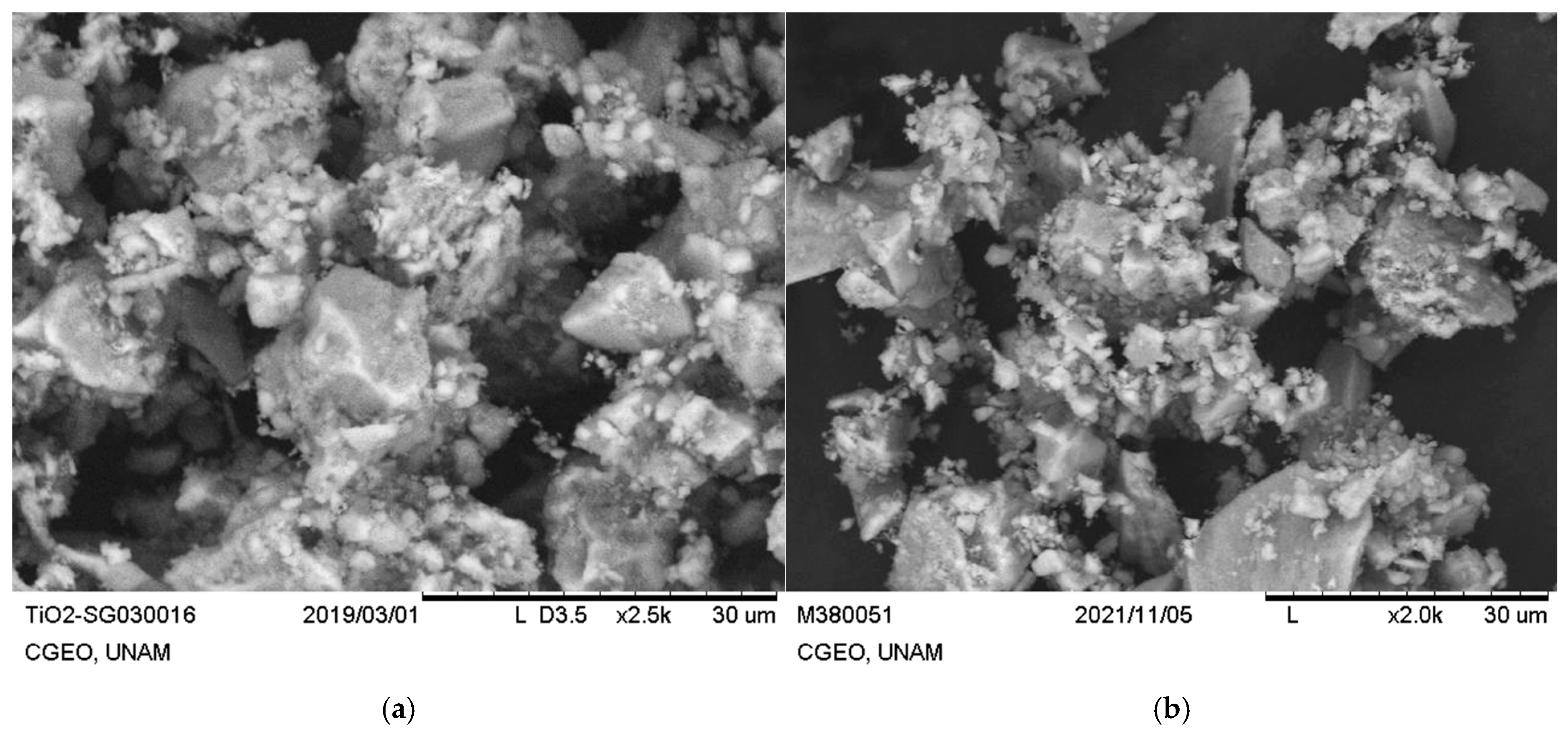
Figure 1. Scanning electron microscopy of (a) TiO2 and (b) Ag-TiO2 nanoparticles synthesized by sol–gel method.
Agronomy 15 02580 g001
Figure 2. Transmission electron microscopy of (a) TiO2 and (b) Ag-TiO2 nanoparticles synthesized by sol–gel technique and size distribution of (c) TiO2 and (d) Ag-TiO2 nanoparticles.
Agronomy 15 02580 g002
Figure 3. (a) X-ray diffraction pattern and (b) Raman spectra of TiO2 and Ag-TiO2 nanoparticles synthesized by sol–gel technique.
Agronomy 15 02580 g003
Figure 4. (a) Fresh weight of alfalfa sprouts treated with TiO2, (b) Fresh weight of alfalfa sprouts treated with Ag-TiO2. Comparison between averages using Tuckey’s test is indicated by letters, averages with different letters indicate significant differences (p < 0.05). Comparison of averages with the control, using Dunnett’s test, shows averages with significant difference (p < 0.05).
Agronomy 15 02580 g004
Figure 5. (a) Radicle length of alfalfa seedlings treated with TiO2; (b) radicle length of alfalfa seedlings treated with Ag-TiO2. Statistical comparisons between group means using Tukey’s test are indicated by different letters; means sharing the same letter are not significantly different (p < 0.05). Comparisons of each treatment mean with the control using Dunnett’s test are highlighted by red bars; significant differences (p < 0.05) are indicated accordingly.
Agronomy 15 02580 g005
Figure 6. (a) Effect of TiO2 and (b) Ag-TiO2 NPs on SPAD units corresponding to true leaf pigmentation in alfalfa sprouts. Comparison between averages using Tuckey’s test is denoted by letters, averages with different letters indicate significant differences (p < 0.05). Comparison of averages with the control, using Dunnett’s test, is indicated by red bars, averages with significant difference (p < 0.05).
Agronomy 15 02580 g006
Figure 7. (a) Effect of TiO2 and (b) Ag-TiO2 NPs on true leaf width in alfalfa sprouts. Letters indicate comparisons among means using Tukey’s test; means with different letters differ significantly (p < 0.05). Red bars represent comparisons with the control using Dunnett’s test.
Agronomy 15 02580 g007
Figure 8. Effect of (a) TiO2 nanoparticles and (b) Ag-TiO2 NPs on true leaf length in alfalfa seedlings. Mean comparisons were analyzed using Tukey’s and Dunnett’s tests (p < 0.05). Means with different letters indicate significant differences among concentrations. Red bars denote significant differences in treatments relative to the control group.
Agronomy 15 02580 g008
Figure 9. mg GAE/sample gr (mg gallic acid equivalents/sample gr). Effect of TiO2 (a) and Ag-TiO2 (b) NPs on total phenols in alfalfa sprouts. Comparison between averages using Tukey’s test is denoted by letters, averages with different letters indicate significant differences (p < 0.05). Comparison of averages with the control, using Dunnett’s test, is indicated by red bars, averages with significant difference (p < 0.05).
Agronomy 15 02580 g009
Figure 10. Effect of TiO2 (a) and Ag-TiO2 (b) NPs on total flavonoids in alfalfa sprouts. Comparison between averages using Tukey’s test is indicated by letters, averages with different letters indicate significant differences (p < 0.05). Comparison of averages with control using Dunnett’s test is indicated by red bars, averages with significant difference (p < 0.05).
Agronomy 15 02580 g010
Table 1. Nanoparticle sizes calculated from Williamson–Hall and Scherrer methods.
MaterialScherrer (nm)W-H (nm)
TiO28.6519.80
Ag-TiO29.6214.00
Table 2. Antioxidant capacity through DPPH and ABTS radical stabilizing activity in alfalfa sprouts treated with TiO2 and Ag-TiO2.
DPPH
TreatmentConcentration (ppm)% of IHB 1
Control048.62 ± 2.82 A
TiO2128.30 ± 3.52 C*
540.85 ± 2.82 B*
1017.72 ± 1.43 D*
1513.96 ± 1.20 D*
Control057.40 ± 1.93 AB
Ag-TiO2154.00 ± 1.72 AB
549.97 ± 3.53 B
1049.17 ± 5.66 B
1560.45 ± 3.49 A
ABTS
Control070.36 ± 1.42 A
TiO2167.49 ± 1.04 AB
568.52 ± 2.76 AB
1062.78 ± 2.97 BC*
1555.65 ± 4.13 C*
Control056.09 ± 3.48 B
Ag-TiO2164.12 ± 2.99 A*
565.35 ± 2.47 A*
1046.78 ± 1.66 C*
1548.41 ± 0.62 C*
1 Percentage inhibition of the DPPH and ABTS radicals recorded in extracts from seedlings exposed to different concentrations of TiO2 and Ag-TiO2 after 9 days of growth. The percentage values reflect the reducing capacity of each sample to inhibit the oxidative activity of the DPPH and ABTS radicals. Means with different letters indicate significant differences according to Tukey’s test (p < 0.05); asterisks denote significant differences compared to the control, according to Dunnett’s test (p < 0.05).
Disclaimer/Publisher’s Note: The statements, opinions and data contained in all publications are solely those of the individual author(s) and contributor(s) and not of MDPI and/or the editor(s). MDPI and/or the editor(s) disclaim responsibility for any injury to people or property resulting from any ideas, methods, instructions or products referred to in the content.