Article

Purpureocillium takamizusanense: A New Entomopathogenic Fungus in the Americas and Its Pathogenicity Against the Cacao Black Bug, Antiteuchus tripterus (Hemiptera: Pentatomidae)

by
Elgar Hernandez-Diaz
1,2,
Luis Carlos Martínez
1,3,
Jorge Ronny Díaz-Valderrama
1,4,5,
Liz Marjory Cumpa-Velasquez
1,
Segundo Manuel Oliva-Cruz
1,5,
Angel F. Huaman-Pilco
4,
Karol Rubio
4,5,
Eduardo Enrique León-Alcántara
1 and
Manuel Alejandro Ix-Balam
1,5,*
1
Grupo de Investigación en Entomología, Instituto de Investigación para el Desarrollo Sustentable de Ceja de Selva (INDES-CES), Universidad Nacional Toribio Rodríguez de Mendoza de Amazonas (UNTRM), Chachapoyas 01001, Peru
2
Maestría en Cambio Climático, Agricultura y Desarrollo Rural Sostenible (MACCARD), Escuela de Posgrado (EPG), Universidad Nacional Toribio Rodríguez de Mendoza de Amazonas (UNTRM), Chachapoyas 01001, Peru
3
Departamento de Producción y Sanidad Vegetal, Universidad de Nariño, Pasto 520001, Colombia
4
Grupo de Investigación en Fitopatología y Micología, Instituto de Investigación para el Desarrollo Sustentable de Ceja de Selva (INDES-CES), Universidad Nacional Toribio Rodríguez de Mendoza de Amazonas (UNTRM), Chachapoyas 01001, Peru
5
Facultad de Ingeniería y Ciencias Agrarias, Universidad Nacional Toribio Rodríguez de Mendoza de Amazonas (UNTRM), Chachapoyas 01001, Peru
*
Author to whom correspondence should be addressed.
Agronomy 2025, 15(10), 2315; https://doi.org/10.3390/agronomy15102315
Submission received: 21 July 2025 / Revised: 11 September 2025 / Accepted: 16 September 2025 / Published: 30 September 2025
(This article belongs to the Section Pest and Disease Management)

Abstract

The black bug, Antiteuchus tripterus (Hemiptera: Pentatomidae), is one of the most important pests of cacao in Peru. Its control relies primarily on synthetic insecticides, which negatively impact the environment and the health of cacao farmers. Therefore, the use of natural enemies, such as entomopathogenic fungi, represents a promising and sustainable alternative. In this study, we aimed to characterize entomopathogenic fungal isolates obtained from naturally infected A. tripterus adults in Peru. Using morphological and molecular approaches, we identified the isolates as Purpureocillium takamizusanense. Then, we assessed their pathogenic potential against A. tripterus and identified their functional cell wall groups. To evaluate pathogenicity, A. tripterus nymphs were exposed to serially diluted conidial suspensions (105–109 conidia mL−1; isolate 24M) in both laboratory and field trials. The isolates exhibited conidial viability above 99%. Concentration-mortality bioassays confirmed the lethal effect of P. takamizusanense against A. tripterus nymphs. The LC50 was 1.65 × 10⁶ conidia mL–1, while the LT50 and LT90 were 3.08 and 7.29 days, respectively. The field mortality rate was about 52%, which can be explained by the influence of the environment. Spectroscopy analyses revealed functional groups including chitin, glucans, lipids, aliphatic chains, and proteins, which may contribute to infection and fungal persistence. This study presents the first record of P. takamizusanense in the Americas and highlights its potential as a biocontrol agent against A. tripterus in cacao plantations.

1. Introduction

Cacao (Theobroma cacao L.) represents a US$17.2 billion global industry [1], sustained largely by small-scale producers in Latin America, Asia, and Africa [2,3,4]. Latin America accounts for around 18% of the global cacao supply, and production is expected to increase due to rural development initiatives, improved soil management practices, and other factors [5]. In developing countries, such as Peru, cacao is considered a strategic component of the agricultural economy [6,7]. However, its production is challenged by various diseases and insect pests [8,9,10,11], causing estimated losses ranging from 30 to 40% [12].
The cacao black bug, also known as the cacao stink bug, Antiteuchus tripterus Fabricius (Hemiptera: Pentatomidae), is one of the main pests affecting cacao plantations in Peru [13,14,15]. Damage caused by nymphs and adults during feeding affects the peduncle of cacao pods, resulting in smaller, shorter pods and reduced bean weight [13,16,17,18]. Control strategies rely heavily on chemical insecticides. However, excessive use and overdoses of pesticides lead to environmental pollution and health issues for farmers [19,20,21,22,23,24,25]. Therefore, sustainable and eco-friendly alternatives are needed for long-term integrated pest management that allow for insecticide reduction, especially in Peru, which is considered the second largest producer of organic cacao worldwide [26].
The use of entomopathogenic fungi is a promising strategy for biological pest control [27,28], due to their ecological versatility and effectiveness at reducing insect pest populations without significant side effects [29,30]. These fungi infect and kill their hosts through a complex process involving conidial adhesion to the cuticle, exoskeleton penetration via hydrolytic enzymes, colonization of the hemocoel, and the release of lethal toxins [30,31,32]. Species such as Beauveria bassiana (Vals.-Criv.) Vuill. and Metarhizium anisopliae (Metchnikoff) Sorokin have shown efficacy against a wide range of insect pests [28,33]. Similarly, fungi like Purpureocillium lilacinum (Thom) Luangsa-ard, Hou-braken, Hywel-Jones & Samson, and P. takamizusanense (Kobayasi) S. Ban, Azuma & Hirok Sato, have shown potential in controlling hemipteran pests in agricultural and forestry systems [34,35,36,37,38,39,40]. For example, Purpureocillium spp. cause a high mortality, comparable to the mortality caused by B. bassiana [37], in eggs, nymphs, and adults of hemipterans such as Bemisia tabaci Gennadius (Aleyrodidae) [34,37], Aphis gossypii Glover (Aphididae) [36], Tessaratoma papillosa Drury (Tessaratomidae) [41], among others. However, the potential of Purpureocillium against A. tripterus is unknown, which could represent a viable long-term alternative for managing this pest in cacao plantations in Peru.
Moreover, Fourier Transform Infrared Spectroscopy (FTIR) is an analytical technique that allows to characterize the chemical composition of organic and inorganic materials, including microorganisms such as fungi [42]. In entomopathogenic fungi, FTIR has been successfully used to differentiate species, evaluate their quality and viability, and study biochemical alterations induced by the environment or by interactions with the host [43,44]. Furthermore, FTIR can be used to determine the metabolites present in the fungal tissues or extracts, which can help identify functional groups associated with the pathogenicity of entomopathogenic fungi [45].
In this study, entomopathogenic fungus isolates were obtained from naturally infected A. tripterus individuals collected in cacao plantations in Amazonas, Peru. The objectives of this study were to morphologically and molecularly identify these entomopathogenic isolates; to test their pathogenicity against A. tripterus nymphs under both in vitro and field conditions; and to determine the functional groups in their cell wall through FTIR analyses.

2. Materials and Methods

2.1. Sample Collection

Dead A. tripterus specimens showing visible signs of fungal infection were collected from cacao plantations in the Bagua and Utcubamba provinces of the Amazonas region, Peru (Table 1). Specimens, regardless of sex or developmental stage [46], placed individually in sterile 1.5 mL conical tubes, and kept in a cooler during transport. Specimens were then taken to the Plant Health Research Lab (Laboratorio de Investigación en Sanidad Vegetal-LABISANV) at the Universidad Nacional Toribio Rodríguez de Mendoza de Amazonas (UNTRM), where they were stored at 4 °C. Collections were conducted under authorization from Servicio Nacional Forestal y de Fauna Silvestre (SERFOR) (RD N° D000019-2024-MIDAGRI-SERFOR-DGGSPFFS-DGSPFS, code N° AUT-IFS-2024-010).

2.2. Fungi Isolation Process

Infected insects were surface-sterilized by immersion in 0.5% sodium hypochlorite, rinsed with sterile water, and dried on sterile filter paper at room temperature. To induce mycelial growth, each insect was placed in a sterile humid chamber, consisting of a 90 mm Petri dish lined with a paper towel moistened with 1 mL of sterile water [47]. Humid chambers were incubated at 25 ± 2 °C for 7 days. Fungal isolates were then subcultured onto potato dextrose agar (PDA) plates, and pure cultures were obtained using the hyphal tip method [48]. Reference isolates were deposited in the KUELAP herbarium at UNTRM (vouchers: KUELAP-386-H, KUELAP-387-H, and KUELAP-388-H for isolates 11M, 5M, and 24M, respectively).

2.3. Morphological and Molecular Characterization

Each isolate was cultured on PDA in 90 mm Petri dishes for 15 days at 28 °C under 12-h photoperiod. Morphological characterization was performed following the protocol of Rehner et al. [49]. Conidial shape and size (n = 50) were determined by microscopic examination (DP74 Olympus, Tokyo, Japan) of 6-day-old cultures incubated at 28 °C with lactophenol.
Molecular characterization was performed using pure fungal isolates. Genomic DNA was extracted with the Wizard® Genomic DNA Purification Kit (Promega, Madison, WI, USA), following the manufacturer’s protocol. The internal transcribed spacer (ITS1-5.8S-ITS2), large subunit (LSU), and small subunit (SSU) rDNA regions, and partial sequence of the translation elongation factor 1-alpha (TEF) gene were amplified using primer pairs ITS1/ITS4, LROR/LR5, NS1/NS4, and 2218F/983R, respectively, as described by Wang et al. [50]. The PCR products were Sanger sequenced at MACROGEN (Santiago de Chile, Chile). Raw sequences were assembled and edited using Sequencer v.5.4 (Gene Codes Corporation, Ann Arbor, MI, USA). Multiple sequence alignments were performed with MUSCLE [51], implemented in MEGA-X [52]. A multilocus maximum likelihood phylogenetic analysis was then conducted using CIPRES Science Gateway v3.3 [53], incorporating sequence data from previously published studies [39,50]. The resulting phylogenetic tree was visualized and edited using FigTree v1.4.4 [54].

2.4. Radial Growth and Sporulation

The radial growth and sporulation of each isolate were assessed following Bugeme et al. [55]. A 1 mL suspension of conidia (1 × 10⁶ mL–1) from each isolate was spread onto 90 mm Petri dishes containing PDA and incubated at 28 °C for 72 h. From the actively growing region, 5 mm diameter mycelial discs were excised and transferred onto fresh PDA plates. Radial growth was measured from five replicates per isolate.
After inoculation, four orthogonal diameters per replicate were measured every 24 h for 17 days [56]. The radial growth ratio was calculated following Riaz et al. [57]. Conidial production was assessed after 15 days of growth by randomly excising 5 mm mycelial discs and transferring them into 10 mL of 0.5% (v/v) Tween 80 solution. The suspension was vortexed (VELP Scientifica, Usmate, MB, Italy) for 5 min to dislodge the conidia. Conidial concentration was determined using a Neubauer hemocytometer under an optical microscope (Carl Zeiss, Göttingen, Germany) at 40× magnification.
Conidial viability (% germination) was assessed following Yeo et al. [58]. A 100 μL aliquot of conidial suspension (1 × 10⁶ conidia mL–1) was spread onto 90-mm Petri dishes containing PDA and incubated at 28 °C for 24 h. The germination percentage was calculated from 10 replicates per isolate by counting 100 conidia across five random fields of view using an optical microscope (Carl Zeiss, Göttingen, Germany). Conidia were considered germinated when the germ tube length was at least half the conidia diameter [59].

2.5. In Vitro Pathogenicity Tests

2.5.1. Antiteuchus tripterus Collection

Antiteuchus tripterus individuals were collected from organic cacao plantations in Bagua and Utcubamba provinces (Table 1), under the same authorization described in Section 2.1. In the laboratory, nymphs were classified by instar based on total body length and morphological features of the head, thorax, and abdominal sternites [60]. Adult female A. tripterus nurse nymphs during the first two instars [61], a behavior that influences offspring fungal infections [62]. Therefore, pathogenicity tests focused on third-instar nymphs, as it is an independent and actively feeding stage. Also, the use of nymphs ensured a representative physiological response to pathogenicity tests, compared to adults, which exhibit sex-associated behavioral and physiological changes. Nymphs were reared in plastic cages (35 cm × 25 cm × 14 cm) with lids covered in organza fabric to ensure adequate ventilation. Healthy fresh cacao pods, free of visible diseases, were disinfected with 2.5% sodium hypochlorite, rinsed with sterile distilled water, dried at room temperature, and provided to the nymphs as food. The rearing conditions were 28 ± 1 °C, 75 ± 5% RH, with 12-h photoperiod.

2.5.2. Concentration–Mortality Bioassays

The bioassay was conducted out under controlled conditions (26 ± 1 °C, 75 ± 5% RH, 12-h photoperiod). A conidial stock suspension of P. takamizusanense (isolate 24M, grown on PDA for 15 days at 28 °C) was prepared by scraping conidia into 20 mL of sterile 0.05% Tween-80 solution. Five conidial concentrations (105, 106, 107, 108, and 109 conidia mL−1) were used to assess pathogenicity and establish the concentration-mortality relationship, including the calculation of the median lethal concentration (LC50). The control treatment consisted of sterile distilled water with 0.05% Tween-80. Third-instar A. tripterus nymphs were exposed to the conidial suspension via immersion [63]. Insects were placed on a sterile organza mesh (20 × 20 cm) and immersed for 5 s in 50 mL of the corresponding treatment suspension. Nymphs were then transferred to plastic cages (35 cm × 25 cm × 14 cm) and provided with disinfected cacao pods as a food source. Pods were replaced weekly or when signs of spoilage appeared. Each treatment included five replications of 40 nymphs (n = 1200), following a complete randomized design. Nymph mortality was monitored every 12 h for 15 days after exposure [59]. Nymphs were considered dead when they showed no movement upon contact with a fine brush.

2.5.3. Time–Mortality Bioassay

Third-instar A. tripterus nymphs were exposed to five concentrations (105, 106, 107, 108, and 109 conidia mL−1) of P. takamizusanense isolate 24M, prepared in sterile 0.05% Tween-80 solution. The control treatment consisted of sterile distilled water with 0.05% Tween-80. Experimental procedures and exposure conditions were identical to those used in the concentration-mortality bioassay. Insect mortality was recorded every 12 h for 15 days. Each treatment included five replicates of 40 insects in a completely randomized design.

2.6. Field Conditions Pathogenicity Tests

The field bioassay was carried out using a completely randomized design. Conidial suspension at five concentrations (105, 106, 107, 108, and 109 conidia mL−1) were prepared as previously described. The control treatment consisted of sterile distilled water with 0.05% of Tween-80. Five replicates of 30 third-instar nymphs were tested per treatment (n = 900). The field trial was conducted out in an organic cacao agroforestry system located in La Tranquilla, La Peca district, Bagua Province, Amazonas, Peru (5°37′44′′ S 78°25′06′′ W). The 15-hectare plantation, composed of 25-year-old cacao trees, is representative of the region and follows organic production practices without insecticide use. The field trial took place at the beginning of the cacao fruit ripening season, a period when A. tripterus populations begin to increase (based on personal observations). Thirty trees bearing healthy pods were selected, with 15 m of spacing between each tree. One cacao pod, approximately 25 cm in length, was selected from each tree. The pod surface was disinfected with 2.5% sodium hypochlorite, rinsed three times with sterile distilled water, and dried at ambient temperature. Each pod was individually enclosed in a wooden cage (30 cm × 15 cm × 15 cm) covered with organza fabric. Thirty third-instar A. tripterus nymphs were released into each cage. After a 24-h acclimation period, 10 mL of the conidial suspension was applied using a manual spray pump, ensuring thorough coverage of both the nymphs and the cacao pod. Insect mortality was monitored every 24 h for 30 days. Temperature and relative humidity in the plantation were recorded hourly using a YMP-20D automatic digital datalogger (Shenzhen Yowexa Sensor System Co., Ltd., Shenzhen, China).

2.7. Insect Infection Confirmation

Fungal infection was confirmed in insects that died during both, the in vitro and field bioassays. Dead individuals were placed in humid chambers, consisting of Petri dishes containing sterile paper towels moistened with 1 mL of sterile water. The chambers were incubated at 28 ± 1 °C and 75% RH for 4 to 8 days. Fungal growth emerging from the cadavers was assessed daily, starting on the fourth day.

2.8. Fourier Transform Infrared Spectroscopy (FTIR) Analysis

FTIR spectra were obtained from conidia of isolates cultured for 15 days, following Łopusiewicz et al. [43]. Spectra were recorded using a Nicolet iS50 FTIR spectrometer (Thermo Scientific, Bremen, Germany), equipped with an attenuated total reflectance (ATR) module. Spectral data were collected into 400–4000 cm−1, 32 scans per sample. The ATR system featured a Zn crystal cleaned with isopropyl alcohol and tissue paper, and a movable tip was used to ensure proper contact during measurements [64].

2.9. Statistical Analyses

Concentration-mortality (LC50) and time–mortality (LT50 and LT90) data were analyzed using probit analysis. Field mortality data were analyzed using one-way analysis of variance (ANOVA), and treatment means were compared using Tukey’s honestly significant difference (HSD) test (p < 0.05). All statistical analyses were performed using SAS software, version 9.4. (SAS Institute, Cary, NC, USA).

3. Results

3.1. Molecular Characterization

Phylogenetic analysis was performed using a concatenated dataset of the ITS, LSU, SSU, and TEF loci. Eleven previously released sequences within the Purpureocillium clade were included along with isolates 5M, 11M, and 24M. The phylogenetic tree showed that isolates 5M, 11M, and 24M grouped within P. takamizusanense, with a bootstrap support value of 87.5%, confirming its species identity (Figure 1).

3.2. Morphological Characterization

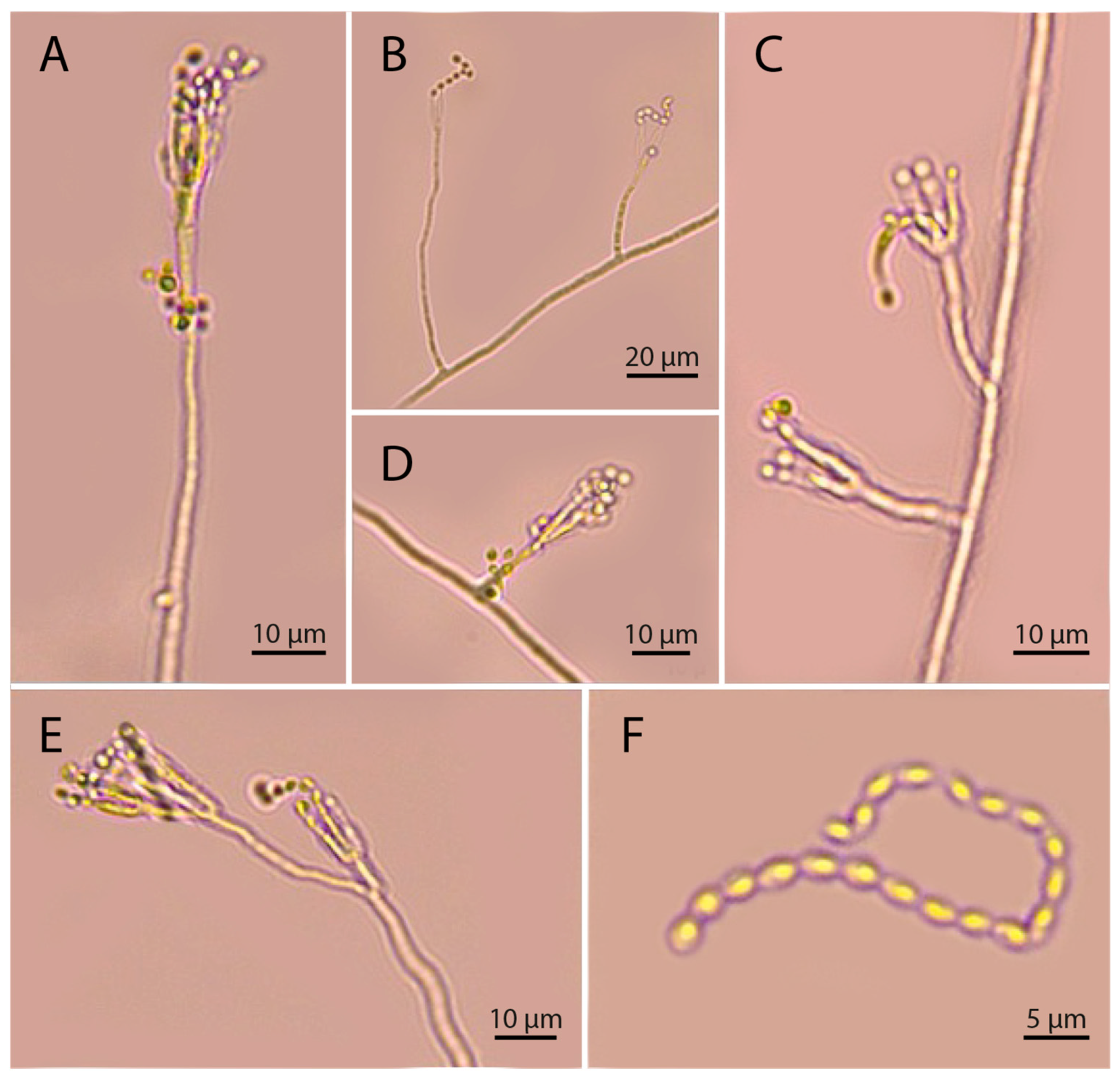
All isolates exhibited radial growth (Figure 2A,B). During the first 3–5 days, colonies appeared white with a powdery mycelial texture but gradually developed a brown pigmentation (Figure 2A,B). The P. takamizusanense isolates (5M, 11M, and 24M) had thin-walled, smooth hyphae measuring 2.7 ± 0.41 µm in diameter. Conidiophores emerged from the mycelium with verticillate branching at the apex (Figure 3). Typically, more than two phialides were formed at each branching point. Conidia were produced in basipetal chains and had a smooth surface, with an ellipsoidal to circular shape, and measured 2.7 ± 0.35 × 1.9 ± 0.23 µm (Figure 3F). After 17 days, isolate 24M reached radial growth of 3.75 ± 0.16 cm, while isolates 11M and 5M reached 3.62 ± 0.12 cm and 3.48 ± 0.23 cm, respectively. Conidial concentrations were 3.5 × 10⁶, 3.45 × 10⁶, and 3.38 × 10⁶ conidia mL−1 for isolates 24M, 11M, and 5M, respectively. Conidial viability was 99.6 ± 0.5%, 99.5 ± 0.51%, and 99.05 ± 1.35%, respectively. Isolate 24M was selected for bioassays based on its growth performance.

3.3. Concentration-Mortality Bioassays

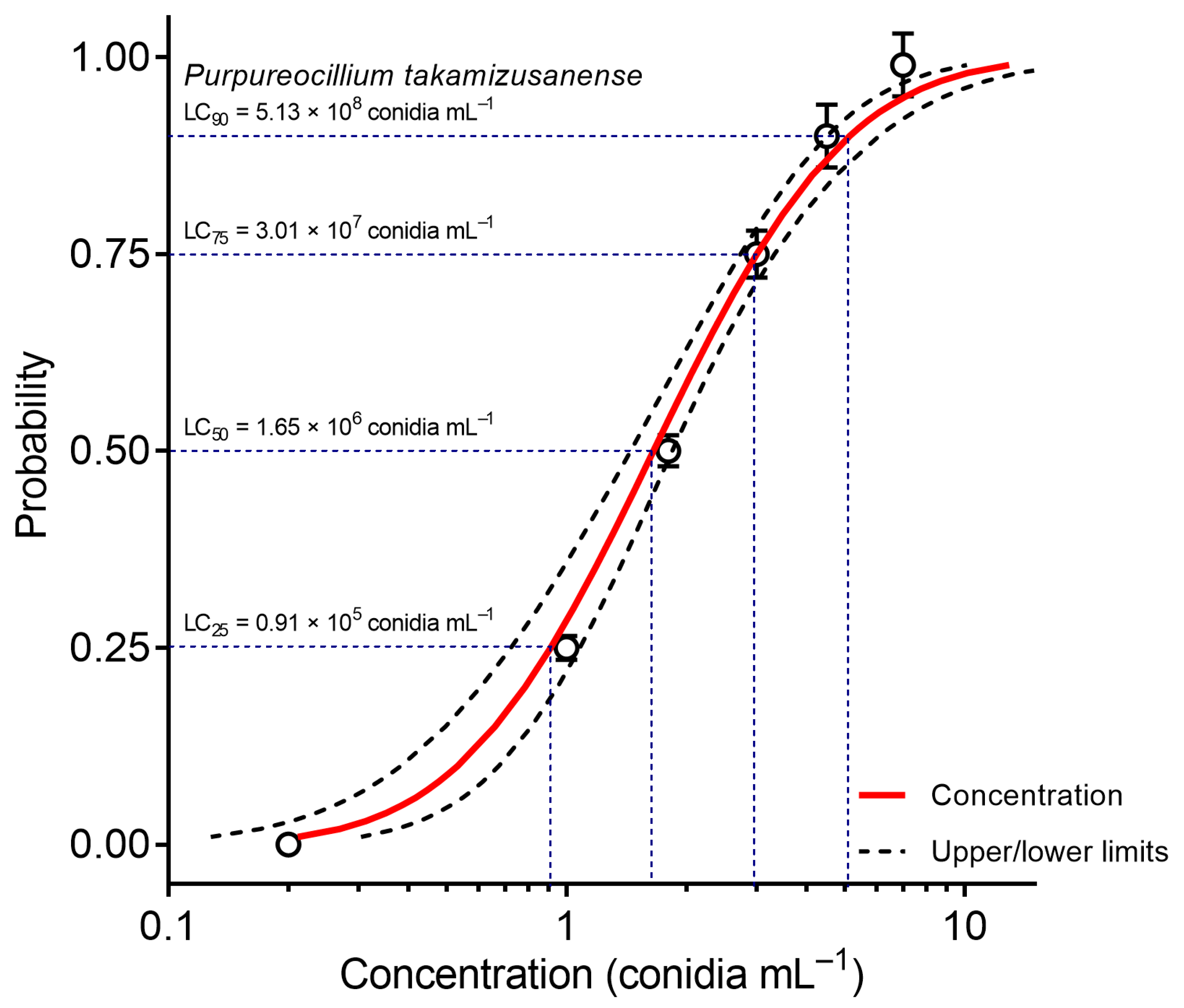
The concentration-mortality data fit the Probit model adequately (n = 1200, DF = 5, χ2 = 1.91, p = 0.10) (Figure 4), confirming the pathogenicity of the P. takamizusanense isolate 24M against A. tripterus nymphs and providing reliable toxicological estimates for future use. The estimated median lethal concentration (LC50) was 1.65 × 10⁶ conidia mL−1 (Confidence interval = 1.44–1.84 × 10⁶ conidia mL−1). Mortality in the control group was less than 1%.

3.4. Time–Mortality Bioassays

The lethal time values of P. takamizusanense against A. tripterus were estimated using Probit analysis (Table 2). The LT50 was 3.08 days, and the LT90 was 7.29 days.

3.5. Field Conditions Pathogenicity Tests

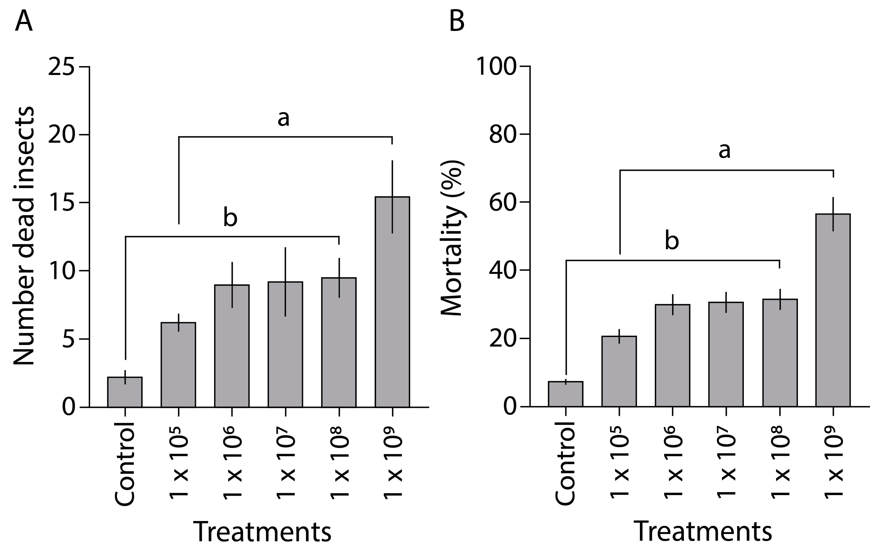
Under field conditions, A. tripterus mortality varied significantly across different P. takamizusanense concentrations (F5,3 = 7.58, p < 0.0019) (Figure 5). After 15 days, the highest mortality was observed at 109 conidia mL−1 (15.50 ± 2.6 nymphs), followed by 108 conidia mL−1 (9.50 ± 1.4 nymphs), 107 conidia mL−1 (9.25 ± 2.5 nymphs), 106 conidia mL−1 (9.00 ± 1.6 nymphs), and 105 conidia mL−1 (6.25 ± 0.6 nymphs) (Figure 5). Mortality in the control group was 2.25 ± 0.4 nymphs. Specifically, mortality rates were 51.6% at 109 conidia mL−1, decreasing to 9.5% at 108 conidia mL−1, 9.2% at 107 conidia mL−1, 9.0% at 106 conidia mL−1, 6.2% at 105 conidia mL−1, and 2.2% in the control group.
Fungal growth was observed on all individuals exposed to the different concentrations (105, 106, 107, 108, and 109 conidia mL−1), confirming infection by P. takamizusanense (Figure 2C,D). This validates that nymph mortality in all the experiments was caused by P. takamizusanense infection.

3.6. FTIR Analysis

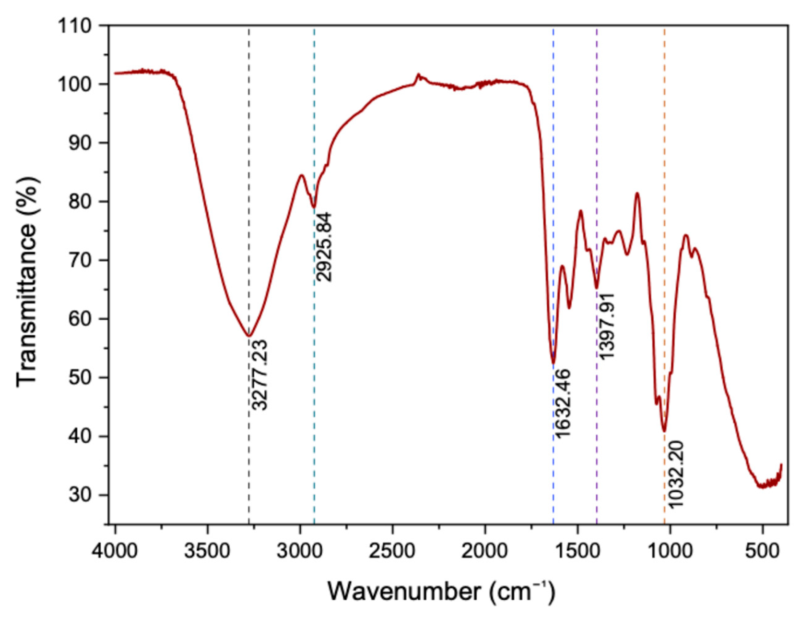
Functional groups in the P. takamizusanense cell wall were identified by FTIR analysis (Figure 6). The broad peak at 3277.23 cm−1 was associated with O-H stretching vibrations, indicating the presence of chitin, glucans, and other polysaccharides in the fungal cell structure. The peak at 2925.84 cm−1 corresponded to C-H vibrations, typical of long hydrocarbon chains, suggesting the presence of lipids and aliphatic compounds such as fatty acids in the cell membrane. The peaks at 1632.46 cm−1 and 1397.91 cm−1 were linked to C=N stretching and C-H bending vibrations, respectively, associated with structural proteins and cell wall polysaccharides. Finally, the peak at 1032.20 cm−1 suggests the presence of sulfoxids (S=O), possibly due to sulfur-containing secondary metabolites.

4. Discussion

Isolates of P. takamizusanense were identified from naturally infected A. tripterus individuals collected in cacao plantations in Amazonas, Peru. The pathogenicity of P. takamizusanense against A. tripterus nymphs was confirmed, and the functional groups present in this fungus were characterized. Purpureocillium takamizusanense was originally discovered from infected cicadas in Japan [39,65,66]. In Thailand, it was identified as an entomopathogen of hemipterans [67], and in Taiwan, it was reported on the giant stink bug T. papillosa [41]. This study is the first to report the presence of P. takamizusanense in the Americas and the first to document its pathogenicity on the cacao black bug A. tripterus. Therefore, our findings expand the known geographic distribution and host range of this entomopathogenic fungus.
The molecular approach is essential for identifying fungal species, particularly within the order Hypocreales, which is known for its morphological complexity [68]. Genetic characterization of isolates 24M, 11M, and 5M, using the rDNA-ITS, LSU, SSU, and TEF markers, enabled accurate molecular identification of P. takamizusanense isolates [67]. These Peruvian isolates exhibit morphological characteristics consistent with those described for the Japanese type strains [69], including the transition from light to brownish coloration when grown on PDA [69].
Isolates of P. takamizusanense (5M, 11M, and 24M) produced conidial concentrations exceeding 1 × 10⁶ conidia mL−1, a level comparable to the conidia production Beauveria and Metarhizium spp., genera recognized for their efficacy in insect pest biocontrol [70,71]. All P. takamizusanense isolates showed conidial viability above 99%, which is equal to or higher than that reported for various Metarhizium spp. and Beauveria spp. isolates (70–99%) [70,72,73]. Conidial germination is a key factor in the virulence of entomopathogenic fungi [74,75]. In vitro germination is influenced by factors such as growth medium [72], geographic origin of isolates [70], relative humidity [76] and temperature [34]. For example, M. anisopliae and B. bassiana isolates show high conidial production and viability at 25 °C, but viability decreases at temperatures below 25 °C or above 30 °C [34,77]. Our results demonstrate that the P. takamizusanense isolates exhibited high conidial viability (>99%) at 28 °C on PDA medium.
Isolate 24M of P. takamizusanense caused mortality in A. tripterus nymphs in a concentration-dependent manner. Similar patterns have been documented for other entomopathogenic fungi, where insect mortality increases with conidial concentration [40,59,78]. The LC50 for P. takamizusanense against A. tripterus was estimated at 1.65 × 10⁶ conidia mL−1. For the white sap-sucking whitefly Aleurodicus dispersus Russell (Hemiptera: Aleyrodidae), previously reported LC50 values were significantly higher than those observed here for A. tripterus. Reported values include 3.085 × 10⁸ for Lecanicillium lecanii (Zimmerm.), 8.189 × 10⁷ for Paecilomyces fumosoroseus (Wize) Brown and Smith, 2.197 × 10⁸ for M. anisopliae (Metschnikoff) Sorokin, and 3.481 × 10⁸ conidia mL−1 for B. bassiana (Balsamo) Vuillemin [79]. Compared with these fungi, P. takamizusanense demonstrated significant lethality against A. tripterus nymphs at relatively low concentrations. This may be attributed to the production of fungal metabolites such as hydrolytic enzymes, mycotoxins, and other compounds involved in the infection process [80,81]. The LC50 value for P. takamizusanense is also comparable to values reported for B. bassiana against Galleria mellonella Linnaeus (Lepidoptera: Pyralidae), which ranged from 0.2 × 10⁶ to 0.6 × 10⁶ conidia mL−1 [82]; 0.4 × 10⁷ against Tetranychus urticae nymphs [83]; and 8.7–14 × 107 against Bemisia tabaci [84]. The variability in LC50 values reported across different studies and pest species indicates the need to screen the pathogenicity of each potential biocontrol agent against specific target pests. Despite these variations, our findings highlight the potential of P. takamizusanense as an effective biocontrol agent for managing A. tripterus in cacao plantations in Latin America.
The Purpureocillium takamizusanense isolate 24M induced 50% (LT50) and 90% (LT90) mortality in A. tripterus nymphs at 3.08 and 7.29 days after exposure, respectively. The time-lethality relation varies among fungal isolates and species, and conidial concentrations [40,85,86,87]. For example, in Leptoglossus occidentalis (Hemiptera: Coreidae), LT50 values for isolates of B. bassiana, Isaria fumosorosea, M. flavoviride, and M. brunneum range from 2.73 to 35 days [86]. LT50 variation among entomopathogenic fungi is influenced by factors such as insect integument structure and chemistry [88,89], as well as the host’s immune response [90,91]. The short lethality times (TL50 and TL90) recorded for P. takamizusanense demonstrate its rapid action against A. tripterus nymphs. In insects, early instar nymphs are generally more susceptible to fungal pathogens than later instars [40,87]. We used third-instar nymphs in our pathogenicity assays, as they are independent of maternal care [92]. The use of later-stage A. tripterus nymphs had some limitations because individuals could reach adulthood during the evaluation period. This may compromise the integrity of the results, as the host’s developmental stage (egg, nymph, larva, pupa, adult) significantly affects fungal virulence [85,86,87]. For example, P. lilacinum (Thom) Luangsa-ard, Houbraken, Hywel-Jones and Samson, causes higher mortality in Tuta absoluta Meyrick (Lepidoptera: Gelechiidae) larvae than in pupae exposed to the same conidia concentration (1 × 10⁸) [40]. Therefore, future pathogenicity assays should consider other life stages of A. tripterus, such as eggs and adults, as well as nymphs under maternal care. Despite this limitation, our findings highlight the potential of P. takamizusanense as a biological control agent against black bug nymphs and its promise as an alternative to chemical insecticides.
Purpureocillium takamizusanense also caused mortality in A. tripterus nymphs on cacao fruits under field conditions, consistent with our in vitro findings. Application of 109 conidia mL−1 resulted in approximately 52% nymphal mortality. This efficiency is lower than the mortality rates achieved by B. bassiana and M. anisopliae (68% and 85%, respectively) against Demotispa neivai Bondar (Coleoptera: Chrysomelidae) populations in oil palm plantations [59]. Compared to laboratory conditions, P. takamizusanense required a higher concentration of conidia to effectively kill A. tripterus nymphs in the field. This discrepancy is likely due to the influence of environmental factors on fungal virulence. During the field bioassays, temperature and relative humidity ranged from 21 °C to 27.5 °C and 68% to 87%, respectively (Figure S1). As reported in previous studies, such variations can influence conidial germination in entomopathogenic fungi [76,93]. Additionally, temperature can affect the expression of proteins associated with fungal virulence [94,95]. Other important climatic factors, such as rainfall and sunlight, may also impact the infection process [76]. On the other hand, nutrient availability is another key factor. Nitrogen sources modulate the pH and affect the growth and sporulation of biocontrol agents [96]. Interactions with environmental factors, as well as formulations and delivery methods, influence outcomes in both laboratory and field settings. In this context, the next step could be the development of a robust, field-ready application system [97,98]. Despite these challenges, the significant mortality observed indicates that P. takamizusanense remains active and pathogenic under natural conditions. Therefore, our results demonstrate its potential as a biological control agent for reducing black bug populations in cacao plantations.
Finally, FTIR analysis showed that the chemical composition of P. takamizusanense includes chitin, glucans, and other polysaccharides, characteristic components of the fungal cell wall. Insect infection involves a complex process of adhesion and interaction with the host cuticle, penetration of the exoskeleton by hydrolytic enzymes, and colonization of the hemocoel [30,31,32]. Chitin and glucans are the main structural components of the cell wall of filamentous fungi, but they also mediate infection during host–pathogen interactions [99]. During infection, these molecules can trigger the insect’s immune response and activate antifungal defense mechanisms [100,101]. However, fungi such as M. acridum express chitin synthase, which disrupts the integrity of their own cell wall to prevent activation of the host immune system [102]. Similarly, Metarhizium spp. and B. bassiana express glucanases that remodel the fungal cell wall by removing glucans, especially β-1,3-glucan, thereby masking pathogen-associated molecular patterns [103]. This prevents immune recognition, allowing penetration of the exoskeleton and colonization of the hemocoel. Fungal cell wall proteins also contribute to the virulence of entomopathogenic fungi [104]. In B. bassiana, cell wall effector proteins bind to chitin, protecting the fungus from host chitinases and promoting colonization and spread within the hemocoel [105]. Likewise, contact with hemolymph can induce the expression of specific proteins in the hyphal bodies of M. anisopliae, preventing phagocytosis and encapsulation by the insect’s immune system [106]. In this way, cell wall proteins facilitate the persistence and establishment of the fungus in the host. Moreover, the presence of lipids and aliphatic chains (such as fatty acids) aligns with previous reports on B. bassiana and Metarhizium spp., which rely on lipid metabolism for growth and infection [107,108]. The results show that functional groups of the P. takamizusanense cell wall are associated with the virulence of A. tripterus. A biochemical analysis of the immune response of P. takamizusanense and A. tripterus, including gene expression during infection, would help clarify the specific functions of the chemical components in the fungal cell wall.

5. Conclusions

The P. takamizusanense isolates (5M, 11M, and 24M) produced conidia with high viability and demonstrated pathogenicity against A. tripterus nymphs. Conidial inoculation with isolate 24M showed high virulence, significantly reducing nymph survival under both in vitro and field conditions. FTIR analysis identified key components of the fungal cell wall, such as chitin and glucans, which are essential for host–pathogen interactions. Therefore, our findings highlight the potential of P. takamizusanense as a biological control agent against black bug infestations in cacao plantations. Future validation experiments under real-life conditions will be conducted, which will help to develop IPM recommendations to farmers. Finally, this study expands the knowledge on the host range and geographic distribution of P. takamizusanense and reports this species for the first time in the Americas.

Supplementary Materials

The following supporting information can be downloaded at https://www.mdpi.com/article/10.3390/agronomy15102315/s1: Figure S1: Average temperature and relative humidity during the field experiment.

Author Contributions

Conceptualization: L.C.M., J.R.D.-V., and M.A.I.-B.; data curation: E.H.-D., L.C.M., L.M.C.-V., S.M.O.-C., A.F.H.-P., K.R., E.E.L.-A., and M.A.I.-B.; formal analysis: E.H.-D., L.C.M., and M.A.I.-B.; funding acquisition: S.M.O.-C. and M.A.I.-B.; investigation: E.H.-D., J.R.D.-V., L.M.C.-V., S.M.O.-C., A.F.H.-P., K.R., E.E.L.-A., and M.A.I.-B.; methodology: E.H.-D., L.C.M., and M.A.I.-B.; validation: L.C.M. and M.A.I.-B.; visualization, E.H.-D., L.C.M., J.R.D.-V., L.M.C.-V., S.M.O.-C., A.F.H.-P., K.R., E.E.L.-A., and M.A.I.-B.; writing—original draft: E.H.-D. and M.A.I.-B.; writing—review and editing: E.H.-D., L.C.M., J.R.D.-V., L.M.C.-V., K.R., and M.A.I.-B. All authors have read and agreed to the published version of the manuscript.

Funding

This work was financially supported by CONCYTEC through the PROCIENCIA program within the framework of the grant “Proyectos Especiales: Proyectos de Incorporación de Investigadores Postdoctorales en Instituciones Peruanas”, contract PE501080952-2022-PROCIENCIA, Project BIOCONTROL; Peruvian Public Investment Project “Creación e Implementación del Centro de Investigación e Innovación Tecnológica en Cacao de la Universidad Nacional Toribio Rodríguez de Mendoza de Amazonas” (CEINCACAO), CUI N° 2315081. The APC was funded by the vice chancellor’s office of Research of the Universidad Nacional Toribio Rodríguez de Mendoza de Amazonas (UNTRM).

Data Availability Statement

Sequences are available in GenBank (https://www.ncbi.nlm.nih.gov/genbank/, (accessed on 30 July 2025)) under accessions numbers: SSU (PV943768; PV943769; PV943770); ITS (PV943735; PV943736; PV943737); LSU (PV943728; PV943729); TEF (PV946954; PV946955; PV946956). All other data generated or analyzed in this research are included in the main text. Additional information or datasets are available from the corresponding author upon reasonable request.

Acknowledgments

The authors thank Elmer Chávez Chacón, Jhon Edler Lopez Merino, Jherson Rojas Vargas, and Ayrtón Ronaldo Armando Chorres Arica for the valuable help in both wet lab and field activities. The authors have reviewed and edited the output and take full responsibility for the content of this publication.

Conflicts of Interest

The authors declare no conflicts of interest.

References

  1. CBI. What Is the Demand for Cocoa on the European Market? Available online: https://www.cbi.eu/market-information/cocoa/what-demand (accessed on 22 June 2025).
  2. Hawkins, D.; Chen, Y. Giant on a Pinhead: A Profile of the Cocoa Sector; Hardman & Co.: London, UK, 2014. [Google Scholar]
  3. Babin, R. Pest Management in Organic Cacao. In Handbook of Pest Management in Organic Farming; CAB International: Wallingford, UK, 2018; pp. 502–518. [Google Scholar]
  4. Franzen, M.; Borgerhoff Mulder, M. Ecological, Economic and Social Perspectives on Cocoa Production Worldwide. Biodivers. Conserv. 2007, 16, 3835–3849. [Google Scholar] [CrossRef]
  5. Abbott, P.C.; Benjamin, T.J.; Burniske, G.R.; Croft, M.C.; Fenton, M.C.; Kelly, C.R.; Lundy, M.M.; Rodriguez Camayo, F.; Wilcox, M.D., Jr. An Analysis of the Supply Chain of Cacao in Colombia; United States Agency for International Development: Washington, DC, USA, 2018. [Google Scholar]
  6. Mestanza, M.; David Hernández-Amasifuen, A.; Jherina Pineda-Lázaro, A.; Eriksson, D.; Carlos Guerrero-Abad, J. Genome Editing for Sustainable Agriculture in Peru: Advances, Potential Applications and Regulation. Front. Genome Ed. 2025, 7, 1611040. [Google Scholar] [CrossRef]
  7. Díaz-Valderrama, J.R.; Leiva-Espinoza, S.T.; Catherine Aime, M. The History of Cacao and Its Diseases in the Americas. Phytopathology 2020, 110, 1604–1619. [Google Scholar] [CrossRef]
  8. Schuh, R.T. On-Line Systematic Catalog of Plant Bugs (Insecta: Heteroptera: Miridae). Available online: http://research.amnh.org/pbi/catalog/ (accessed on 22 June 2025).
  9. Lavabre, E.M. Systematique Des Miridae Du Cacaoyer. In Les Mirides du Cacaoyer. In Les Mirides du Cacaoyer; Lavabre, E.M., Ed.; Institut Français du Cafe et du Cacao: Paris, France, 1977; pp. 47–70. [Google Scholar]
  10. ten Hoopen, G.M.; Deberdt, P.; Mbenoun, M.; Cilas, C. Modelling Cacao Pod Growth: Implications for Disease Control. Ann. Appl. Biol. 2012, 160, 260–272. [Google Scholar] [CrossRef]
  11. Arias, M.; Ninnin, P.; Ten Hoopen, M.; Alvarado, J.; Cabezas Huayllas, O.; Valderrama, B.; Alguilar, G.; Perrier, C.; Dedieu, F.; Bagny Beilhe, L. The American Cocoa Pod Borer, Carmenta Foraseminis, an Emerging Pest of Cocoa: A Review. Agric. Entomol. 2025, 27, 340–356. [Google Scholar] [CrossRef]
  12. ICCO. Annual Report 2012/2013; ICCO: London, UK, 2013. [Google Scholar]
  13. Castillo, P. Insectos Plagas y Sus Enemigos Naturales En El Cultivo de Theobroma Cacao L. (Cacao) En Los Valles de Tumbes y Zarumilla, Perú. Rev. Mangl. 2013, 10, 3–16. [Google Scholar] [CrossRef]
  14. Beilhe, L.B.; Babin, R.; ten Hoopen, M. Insect Pests Affecting Cacao. In Achieving Sustainable Cultivation of Cocoa; Umaharan, P., Ed.; Burleigh Dodds Science Publishing: Cambridge, UK, 2018; pp. 303–326. [Google Scholar]
  15. Castillo-Carrillo, P.S.; Sernaqué-Cortez, A.; Purizaga-Preciado, J.L. Registro Del Chinche Del Cacao Antiteuchus Tripterus (Fabricius, 1787) (Hemiptera: Pentatomidae), En Tumbes-Perú. Bol. Mus. Nac. Hist. Nat. Parag. 2020, 24, 15–20. [Google Scholar]
  16. Valarezo-Cely, O.; Bermúdez, E.C.; Cedeño, B.N. Artrópodos Asociados al Cultivo de Cacao En Manabí. La. Técnica 2012, 7, 34–42. [Google Scholar] [CrossRef]
  17. Quintos-Coronado, C.R. Evaluación Del Daño de Antiteuchus Sp En Frutos de Cacao (Theobroma Cacao L.) En El Valle Del Bajo Mayo, Región San Martín. Bachelor’s thesis, Universidad Nacional de San Martín, Lima, Perú, 2018. [Google Scholar]
  18. Cañarte-Bermúdez, E.; Navarrete-Cedeño, B. Reconocimiento de Artrópodos Plaga y Controladores Biológicos Como Herramienta Para El Manejo Ecológico de Plagas En Cacao, 1st ed.; Instituto Nacional de Investigaciones Agropecuarias: Quito, Ecuador, 2021. [Google Scholar]
  19. Telaumbanua, M.; Haryanto, A.; Wisnu, F.K.; Lanya, B.; Wiratama, W. Design of Insect Trap Automatic Control System for Cacao Plants; EIAETM: Cluj-Napoca, Romania, 2021; Volume 8. [Google Scholar]
  20. Sosan, M.B.; Akingbohungbe, A.E. Occupational Insecticide Exposure and Perception of Safety Measures among Cacao Farmers in Southwestern Nigeria. Arch. Env. Occup. Health 2009, 64, 185–193. [Google Scholar] [CrossRef] [PubMed]
  21. Verzeñassi, D.; Vallini, A.; Fernández, F.; Ferrazini, L.; Lasagna, M.; Sosa, A.J.; Hough, G.E. Cancer Incidence and Death Rates in Argentine Rural Towns Surrounded by Pesticide-Treated Agricultural Land. Clin. Epidemiol. Glob. Health 2023, 20, 101239. [Google Scholar] [CrossRef]
  22. Carvalho, F.P. Pesticides, Environment, and Food Safety. Food Energy Secur. 2017, 6, 48–60. [Google Scholar] [CrossRef]
  23. Romero, F.; Jiao, S.; van der Heijden, M.G.A. Impact of Microbial Diversity and Pesticide Application on Plant Growth, Litter Decomposition and Carbon Substrate Use. Soil. Biol. Biochem. 2025, 208, 109866. [Google Scholar] [CrossRef]
  24. Mesnage, R.; Defarge, N.; Spiroux de Vendômois, J.; Séralini, G.-E. Major Pesticides Are More Toxic to Human Cells Than Their Declared Active Principles. Biomed. Res. Int. 2014, 2014, 179691. [Google Scholar] [CrossRef]
  25. Mojiri, A.; Zhou, J.L.; Robinson, B.; Ohashi, A.; Ozaki, N.; Kindaichi, T.; Farraji, H.; Vakili, M. Pesticides in Aquatic Environments and Their Removal by Adsorption Methods. Chemosphere 2020, 253, 126646. [Google Scholar] [CrossRef]
  26. MIDAGRI. Observatorio de Commodities. Boletín Trimestral No 2021-07002; MIDAGRI: Lima, Perú, 2025. [Google Scholar]
  27. Samal, I.; Bhoi, T.K.; Majhi, P.K.; Murmu, S.; Pradhan, A.K.; Kumar, D.; Saini, V.; Paschapur, A.U.; Raj, M.N.; Ankur; et al. Combatting Insects Mediated Biotic Stress through Plant Associated Endophytic Entomopathogenic Fungi in Horticultural Crops. Front. Plant Sci. 2023, 13, 1098673. [Google Scholar] [CrossRef]
  28. Bhoi, T.K.; Mahanta, D.K.; Samreen;Samal, I.; Dash, S.; Singh, S.; Komal, J. Effect of Endophytic Entomopathogenic Fungi (EEPF) in Biological Control of Odontotermes Obesus (Rambur 1842) and Their Synergistic Impact on Plant Physiological Responses. Microbe 2025, 7, 100411. [Google Scholar] [CrossRef]
  29. Jaber, L.R.; Ownley, B.H. Can We Use Entomopathogenic Fungi as Endophytes for Dual Biological Control of Insect Pests and Plant Pathogens? Biol. Control 2018, 116, 36–45. [Google Scholar] [CrossRef]
  30. Mannino, M.C.; Huarte-Bonnet, C.; Davyt-Colo, B.; Pedrini, N. Is the Insect Cuticle the Only Entry Gate for Fungal Infection? Insights into Alternative Modes of Action of Entomopathogenic Fungi. J. Fungi 2019, 5, 33. [Google Scholar] [CrossRef]
  31. Afifah, L.; Aena, A.C.; Saputro, N.W.; Kurniati, A.; Maryana, R.; Lestari, A.; Abadi, S.; Enri, U. Maize Media Enhance the Conidia Production of Entomopathogenic Fungi Lecanicillium Lecanii Also Its Effective to Control the Weevil Cylas Formicarius (Fabricius) (Coleoptera: Brentidae). Agrivita 2022, 44, 513–525. [Google Scholar] [CrossRef]
  32. Pedrini, N. Molecular Interactions between Entomopathogenic Fungi (Hypocreales) and Their Insect Host: Perspectives from Stressful Cuticle and Hemolymph Battlefields and the Potential of Dual RNA Sequencing for Future Studies. Fungal Biol. 2018, 122, 538–545. [Google Scholar] [CrossRef] [PubMed]
  33. Samal, I.; Bhoi, T.K.; Vyas, V.; Majhi, P.K.; Mahanta, D.K.; Komal, J.; Singh, S.; Kumar, P.V.D.; Acharya, L.K. Resistance to Fungicides in Entomopathogenic Fungi: Underlying Mechanisms, Consequences, and Opportunities for Progress. Trop. Plant Pathol. 2023, 49, 5–17. [Google Scholar] [CrossRef]
  34. Sani, I.; Jamian, S.; Saad, N.; Abdullah, S.; Mohd Hata, E.; Jalinas, J.; Ismail, S.I. Identification and Virulence of Entomopathogenic Fungi, Isaria Javanica and Purpureocillium Lilacinum Isolated from the Whitefly, Bemisia Tabaci (Gennadius) (Hemiptera: Aleyrodidae) in Malaysia. Egypt. J. Biol. Pest Control 2023, 33, 14. [Google Scholar] [CrossRef]
  35. Mascarin, G.M.; Jaronski, S.T. The Production and Uses of Beauveria Bassiana as a Microbial Insecticide. World J. Microbiol. Biotechnol. 2016, 32, 177. [Google Scholar] [CrossRef]
  36. Castillo Lopez, D.; Zhu-Salzman, K.; Ek-Ramos, M.J.; Sword, G.A. The Entomopathogenic Fungal Endophytes Purpureocillium Lilacinum (Formerly Paecilomyces Lilacinus) and Beauveria Bassiana Negatively Affect Cotton Aphid Reproduction under Both Greenhouse and Field Conditions. PLoS ONE 2014, 9, e103891. [Google Scholar] [CrossRef] [PubMed]
  37. Zhai, D.; Lu, H.; Liu, S.; Liu, J.; Zhang, W.; Wu, J.; Li, J.; Bai, R.; Yan, F.; Zhao, C. Fungal Warriors: Effects of Beauveria Bassiana and Purpureocillium Lilacinum on CCYV-Carrying Whiteflies. Biomolecules 2025, 15, 593. [Google Scholar] [CrossRef]
  38. Dahlin, P.; Eder, R.; Consoli, E.; Krauss, J.; Kiewnick, S. Integrated Control of Meloidogyne Incognita in Tomatoes Using Fluopyram and Purpureocillium Lilacinum Strain 251. Crop Prot. 2019, 124, 104874. [Google Scholar] [CrossRef]
  39. Ban, S.; Azuma, Y.; Sato, H.; Suzuki, K.; Nakagiri, A. Isaria Takamizusanensis Is the Anamorph of Cordyceps Ryogamimontana, Warranting a New Combination, Purpureocillium Takamizusanense Comb. Nov. Int. J. Syst. Evol. Microbiol. 2015, 65, 2459–2465. [Google Scholar] [CrossRef]
  40. Bali, G.K.; Singh, S.K.; Maurya, D.K.; Wani, F.J.; Pandit, R.S. Morphological and Molecular Identification of the Entomopathogenic Fungus Purpureocillium Lilacinum and Its Virulence against Tuta Absoluta (Meyrick) (Lepidoptera: Gelechiidae) Larvae and Pupae. Egypt. J. Biol. Pest Control 2022, 32, 86. [Google Scholar] [CrossRef]
  41. Lo, P.H.; Yu, Y.C.; Pai, K.F. First Report of Purpureocillium Takamizusanense as an Omopathogenic Fungus Infecting Tessaratoma Papillosa (Drury) in Taiwan. J. Plant Med. 2019, 61, 27–30. [Google Scholar]
  42. Stuart, B.H. Infrared Spectroscopy of Biological Applications: An Overview. In Encyclopedia of Analytical Chemistry; Wiley: Hoboken, NJ, USA, 2021; pp. 1–16. [Google Scholar]
  43. Łopusiewicz, Ł.; Mazurkiewicz-Zapałowicz, K.; Tkaczuk, C. Chemical Changes in Spores of the Entomopathogenic Fungus Metarhizium Robertsii after Exposure to Heavy Metals, Studied through the Use of Ftir Spectroscopy. J. Elem. 2020, 25, 487–499. [Google Scholar] [CrossRef]
  44. Piekarczyk, J.; Ratajkiewicz, H.; Jasiewicz, J.; Sosnowska, D.; Wójtowicz, A. An Application of Reflectance Spectroscopy to Differentiate of Entomopathogenic Fungi Species. J. Photochem. Photobiol. B 2019, 190, 32–41. [Google Scholar] [CrossRef]
  45. Moussaid, F.Z.; Lahlali, R.; Ezrari, S.; El Barnossi, A.; Housseini, A.I. Biological Control of Alternaria Alternata MW970059, the Causal Agent of Tomato Rot, by Aspergillus Nidulans MW732187 Isolated from Green Household Waste. J. Nat. Pestic. Res. 2025, 13, 100134. [Google Scholar] [CrossRef]
  46. Azrag, A.G.A.; Pirk, C.W.W.; Yusuf, A.A.; Pinard, F.; Niassy, S.; Mosomtai, G.; Babin, R. Prediction of Insect Pest Distribution as Influenced by Elevation: Combining Field Observations and Temperature-Dependent Development Models for the Coffee Stink Bug, Antestiopsis Thunbergii (Gmelin). PLoS ONE 2018, 13, e0199569. [Google Scholar] [CrossRef]
  47. Yun, T.-S.; Park, S.-Y.; Yu, J.; Hwang, Y.; Hong, K.-J. Isolation and Identification of Fungal Species from the Insect Pest Tribolium Castaneum in Rice Processing Complexes in Korea. Plant Pathol. J. 2018, 34, 356–366. [Google Scholar] [CrossRef]
  48. Dummel, D.M.; Badaraco, A.; Kramer, R.; Rohatsch, P.; Agostini, J.P. Avances Sobre La Caracterización “Mal de La Tela” En plantas de Yerba Mate Ilex Paraguariensis A. St. -Hil. Rev. De. Investig. Agropecu. 2022, 48, 188–194. [Google Scholar]
  49. Rehner, S.A.; Minnis, A.M.; Sung, G.-H.; Luangsa-ard, J.J.; Devotto, L.; Humber, R.A. Phylogeny and Systematics of the Anamorphic, Entomopathogenic Genus Beauveria. Mycologia 2011, 103, 1055–1073. [Google Scholar] [CrossRef]
  50. Wang, Y.B.; Wang, Y.; Fan, Q.; Duan, D.E.; Zhang, G.D.; Dai, R.Q.; Dai, Y.D.; Zeng, W.B.; Chen, Z.H.; Li, D.D.; et al. Multigene Phylogeny of the Family Cordycipitaceae (Hypocreales): New Taxa and the New Systematic Position of the Chinese Cordycipitoid Fungus Paecilomyces Hepiali. Fungal Divers. 2020, 103, 1–46. [Google Scholar] [CrossRef]
  51. Edgar, R.C. MUSCLE: Multiple Sequence Alignment with High Accuracy and High Throughput. Nucleic Acids Res. 2004, 32, 1792–1797. [Google Scholar] [CrossRef]
  52. Kumar, S.; Stecher, G.; Li, M.; Knyaz, C.; Tamura, K. MEGA X: Molecular Evolutionary Genetics Analysis across Computing Platforms. Mol. Biol. Evol. 2018, 35, 1547–1549. [Google Scholar] [CrossRef]
  53. Miller, M.A.; Pfeiffer, W.; Schwartz, T. The CIPRES Science Gateway: Enabling High-Impact Science for Phylogenetics Researchers with Limited Resources. In Proceedings of the 1st Conference of the Extreme Science and Engineering Discovery Environment: Bridging from the eXtreme to the Campus and Beyond, Chicago, IL, USA, 16 July 2012; ACM: New York, NY, USA, 2012; pp. 1–8. [Google Scholar]
  54. Rambaut, A. Figtree Ver 1.4.4; Institute of Evolutionary Biology, University of Edinburgh: Edinburgh, UK, 2018. [Google Scholar]
  55. Bugeme, D.M.; Knapp, M.; Boga, H.I.; Wanjoya, A.K.; Maniania, N.K. Influence of Temperature on Virulence of Fungal Isolates of Metarhizium Anisopliae and Beauveria Bassiana to the Two-Spotted Spider Mite Tetranychus Urticae. Mycopathologia 2009, 167, 221–227. [Google Scholar] [CrossRef]
  56. Das, P.; Hazarika, L.K.; Bora, D.S.; Puzari, K.C.; Dutta, P. Effect of Temperature on Radial Growth, Sporulation, Germination, Colony Forming Unit and Biomass Production of Different Strains of Beauveria Bassiana Vuill. (Bals.). Pestology 2011, 35, 50–55. [Google Scholar]
  57. Riaz, M.; Chen, W.-H.; Kafle, L.; Tseng, M.-N. Morphological and Molecular Characterization of Purpureocillium Lilacinum along with Its Biopesticidal Effect against Fall Armyworm (Spodoptera Frugiperda) in Southern Taiwan. Egypt. J. Biol. Pest Control 2024, 34, 60. [Google Scholar] [CrossRef]
  58. Yeo, H.; Pell, J.K.; Alderson, P.G.; Clark, S.J.; Pye, B.J. Laboratory Evaluation of Temperature Effects on the Germination and Growth of Entomopathogenic Fungi and on Their Pathogenicity to Two Aphid Species. Pest Manag. Sci. 2003, 59, 156–165. [Google Scholar] [CrossRef] [PubMed]
  59. Martínez, L.C.; Plata-Rueda, A.; Ramírez, A.; Serrão, J.E. Susceptibility of Demotispa Neivai (Coleoptera: Chrysomelidae) to Beauveria Bassiana and Metarhizium Anisopliae Entomopathogenic Fungal Isolates. Pest Manag. Sci. 2022, 78, 126–133. [Google Scholar] [CrossRef]
  60. Alarcón, M.; Dalmiro, C. Descripción de Los Estadios Inmaduros de Antiteuchus Tripterus (Fabricius, 1787) (Heteroptera: Pentatomidae: Discocephalinae: Discocephalini). Rev. Nicar. De. Entomol. 2022, 261, 3–61. [Google Scholar]
  61. Santos, A.V.; Albuquerque, G.S. Eficiência Do Cuidado Maternal de Antiteuchus Sepulcralis (Fabricius) (Hemiptera: Pentatomidae) Contra Inimigos Naturais Do Estágio de Ovo. Neotrop. Entomol. 2001, 30, 641–646. [Google Scholar] [CrossRef]
  62. de Resende, R.C.; Rodrigues-Silva, A.L.; Pec, M.; Marucci, R.C.; Chagas, P.G.; Sales, F.S.; de Figueiredo, K.G.; Moino Junior, A. Doru Luteipes: Susceptibility to Entomopathogenic Fungi and the Role of Maternal Care in the Protection of Offspring Against Infection. J. Insect Behav. 2025, 38, 10. [Google Scholar] [CrossRef]
  63. Das, P.; Borah, B.; Saikia, P.; Hazarika, L.K.; Sharma, K.K.; Mohanasundaram, A.; Boro, R.C.; Kalita, R.; Gautom, T. Molecular Characterization of an Isaria Fumosorosea (Wize) Native Strain, and Its Pathogenicity on Eublemma Amabilis (Lepidoptera: Noctuidae). Egypt. J. Biol. Pest Control 2022, 32, 122. [Google Scholar] [CrossRef]
  64. Aguirre, E.; Domínguez, J.; Villanueva, E.; Ponce-Ramirez, J.A.; de Fátima Arevalo-Oliva, M.; Siche, R.; González-Cabeza, J.; Rodríguez, G. Biodegradable Trays Based on Manihot Esculenta Crantz Starch and Zea Mays Husk Flour. Food Packag. Shelf Life 2023, 38, 101129. [Google Scholar] [CrossRef]
  65. Kobayashi, Y.; Shimizu, D. Monographic Studies of Cordyceps 2. Group Parasitic on Cicadidae; Bulletin of the National Science Museum: Tokyo, Japan, 1963; Volume 6. [Google Scholar]
  66. Nguyen, N.-H.; Tamura, T.; Shimizu, K. Draft Genome Sequence of Purpureocillium Takamizusanense, a Potential Bioinsecticide. Microbiol. Resour. Announc. 2022, 11, e0026822. [Google Scholar] [CrossRef]
  67. Luangsa-Ard, J.; Houbraken, J.; van Doorn, T.; Hong, S.-B.; Borman, A.M.; Hywel-Jones, N.L.; Samson, R.A. Purpureocillium, a New Genus for the Medically Important Paecilomyces Lilacinus. FEMS Microbiol. Lett. 2011, 321, 141–149. [Google Scholar] [CrossRef]
  68. Dornburg, A.; Townsend, J.P.; Wang, Z. Maximizing Power in Phylogenetics and Phylogenomics: A Perspective Illuminated by Fungal Big Data. In Advances in Genetics; Townsend, J.P., Wang, Z., Eds.; Elsevier: Amsterdam, The Netherlands; Academic Press: Cambridge, MA, USA, 2017; Volume 100, pp. 1–47. [Google Scholar]
  69. Lo, P.-H.; El-Sayid Abdrabo, K.A.; Nai, Y.-S.; Lu, H.; Huang, Y.-T. Versatile Entomopathogenic Activity of Purpureocillium Takamizusanense against Diverse Agricultural Pests. bioRxiv 2025. [Google Scholar] [CrossRef]
  70. Gebremariam, A.; Chekol, Y.; Assefa, F. Phenotypic, Molecular, and Virulence Characterization of Entomopathogenic Fungi, Beauveria Bassiana (Balsam) Vuillemin, and Metarhizium Anisopliae (Metschn.) Sorokin from Soil Samples of Ethiopia for the Development of Mycoinsecticide. Heliyon 2021, 7, e07091. [Google Scholar] [CrossRef]
  71. Schemmer, R.; Chládeková, P.; Medo, J.; Barta, M. Natural Prevalence of Entomopathogenic Fungi in Hibernating Pupae of Cameraria Ohridella (Lepidoptera: Gracillariidae) and Virulence of Selected Isolates. Plant Prot. Sci. 2016, 52, 199–208. [Google Scholar] [CrossRef]
  72. Habtegebriel, B.; Getu, E.; Dawd, M.; Seyoum, E.; Atnafu, G.; Khamis, F.; Hilbur, Y.; Ekesi, S.; Larsson, M. Molecular Characterization and Evaluation of Indigenous Entomopathogenic Fungal Isolates against Sorghum Chafer, Pachnoda Interrupta (Olivier) in Ethiopia. J. Entomol. Nematol. 2016, 8, 34–45. [Google Scholar] [CrossRef]
  73. Mkiga, A.M.; Mohamed, S.A.; Du Plessis, H.; Khamis, F.M.; Akutse, K.S.; Ekesi, S.; Bruck, D. Metarhizium Anisopliae and Beauveria Bassiana: Pathogenicity, Horizontal Transmission, and Their Effects on Reproductive Potential of Thaumatotibia Leucotreta (Lepidoptera: Tortricidae). J. Econ. Entomol. 2020, 113, 660–668. [Google Scholar] [CrossRef]
  74. Dotaona, R.; Wilson, B.A.L.; Stevens, M.M.; Holloway, J.; Ash, G.J. Screening of Tropical Isolates of Metarhizium Anisopliae (Hypocreales: Clavicipitaceae) for Virulence to the Sweet Potato Weevil, Cylas Formicarius (Coleoptera: Brentidae). Int. J. Trop. Insect Sci. 2015, 35, 153–163. [Google Scholar] [CrossRef]
  75. Mar, T.T.; Suwannarach, N.; Lumyong, S. Isolation of Entomopathogenic Fungi from Northern Thailand and Their Production in Cereal Grains. World J. Microbiol. Biotechnol. 2012, 28, 3281–3291. [Google Scholar] [CrossRef]
  76. Abdul Qayyum, M.; Bilal, H.; Naeem Ullah, U.; Ali, H.; Raza, H. Factors Affecting the Epizootics of Entomopathogenic Fungi-A Review. J. Bioresour. Manag. 2021, 8, 78–85. [Google Scholar] [CrossRef]
  77. Moldovan, A.; Munteanu-Molotievskiy, N.; Toderas, I. Temperature Effects on the Entomopathogenic Fungi Beauveria Bassiana Strain CNMN-FE-01: Vegetative Growth, Sporulation, Germination Rate. Curr. Trends Nat. Sci. 2022, 11, 332–338. [Google Scholar] [CrossRef]
  78. Chuquibala-Checan, B.; Torres-De La Cruz, M.; Leiva, S.; Hernandez-Diaz, E.; Rubio, K.; Goñas, M.; Arce-Inga, M.; Oliva-Cruz, M. In Vitro Biological Activity of Beauveria Bassiana, Beauveria Peruviensis, and Metarhizium Sp. against Hypothenemus Hampei (Coleoptera: Curculionidae). Int. J. Agron. 2023, 2023, 4982399. [Google Scholar] [CrossRef]
  79. Thangavel, B.; Palaniappan, K.; Manickavasagam Pillai, K.; Subbarayalu, M.; Madhaiyan, R. Pathogenicity, Ovicidal Action, and Median Lethal Concentrations (LC 50) of Entomopathogenic Fungi against Exotic Spiralling Whitefly, Aleurodicus Dispersus Russell. J. Pathog. 2013, 2013, 393787. [Google Scholar] [CrossRef] [PubMed]
  80. Quesada-Moraga, E.; Carrasco-Díaz, J.-A.; Santiago-Álvarez, C. Insecticidal and Antifeedant Activities of Proteins Secreted by Entomopathogenic Fungi against Spodoptera Littoralis (Lep., Noctuidae). J. Appl. Entomol. 2006, 130, 442–452. [Google Scholar] [CrossRef]
  81. Brown, R.; Priest, E.; Naglik, J.R.; Richardson, J.P. Fungal Toxins and Host Immune Responses. Front. Microbiol. 2021, 12, 643639. [Google Scholar] [CrossRef]
  82. Gençer, D.; Bayramoğlu, Z. Characterization and Pathogenicity of Beauveria Bassiana Strains Isolated from Galleria Mellonella L. (Lepidoptera: Pyralidae) in Turkey. Egypt. J. Biol. Pest Control 2022, 32, 99. [Google Scholar] [CrossRef]
  83. Geroh, M.; Gulati, R.; Tehri, K. Determination of Lethal Concentration and Lethal Time of Entomopathogen Beauveria Bassiana (Balsamo) Vuillemin against Tetranychus Urticae Koch. Int. J. Agric. Sci. 2015, 7, 523–528. [Google Scholar]
  84. Kim, J.; Baek, S.; Kim, J.S. Whitefly-Pathogenic Beauveria Bassiana JEF-507 as a Competitive Biopesticide, Chongchae-Stop. J. Appl. Entomol. 2025, 149, 165–177. [Google Scholar] [CrossRef]
  85. Islam, M.S.; Subbiah, V.K.; Siddiquee, S. Efficacy of Entomopathogenic Trichoderma Isolates against Sugarcane Woolly Aphid, Ceratovacuna Lanigera Zehntner (Hemiptera: Aphididae). Horticulturae 2022, 8, 2. [Google Scholar] [CrossRef]
  86. Eski, A.; Biryol, S.; Acici, O.; Demir, İ. Biocontrol of the Western Conifer Seed Bug, Leptoglossus Occidentalis Heidemann (Heteroptera: Coreidae) Using Indigenous Entomopathogenic Fungi. Egypt. J. Biol. Pest Control 2022, 32, 140. [Google Scholar] [CrossRef]
  87. Lu, Q.; Wang, P.; Ali, A.; Zang, L.S. Molecular Identification and Virulence of Four Strains of Entomopathogenic Fungi Against the Whitefly, Bemisia Tabaci (Hemiptera: Aleyrodidae). J. Econ. Entomol. 2022, 115, 731–738. [Google Scholar] [CrossRef] [PubMed]
  88. Ashida, M.; Brey, P.T. Role of the Integument in Insect Defense: Pro-Phenol Oxidase Cascade in the Cuticular Matrix. Proc. Natl. Acad. Sci. USA 1995, 92, 10698–10702. [Google Scholar] [CrossRef]
  89. Brey, P.T.; Lee, W.J.; Yamakawa, M.; Koizumi, Y.; Perrot, S.; François, M.; Ashida, M. Role of the Integument in Insect Immunity: Epicuticular Abrasion and Induction of Cecropin Synthesis in Cuticular Epithelial Cells. Proc. Natl. Acad. Sci. USA 1993, 90, 6275–6279. [Google Scholar] [CrossRef]
  90. Strand, M.R. The Insect Cellular Immune Response. Insect Sci. 2008, 15, 1–14. [Google Scholar] [CrossRef]
  91. Irving, P.; Ubeda, J.-M.; Doucet, D.; Troxler, L.; Lagueux, M.; Zachary, D.; Hoffmann, J.A.; Hetru, C.; Meister, M. New Insights into Drosophila Larval Haemocyte Functions through Genome-Wide Analysis. Cell Microbiol. 2005, 7, 335–350. [Google Scholar] [CrossRef]
  92. Umaña, E.; Carballo, M. Biología de Antiteuchus Tripterus L. (Hemiptera: Pentatomidae) y Su Parasitoide Trissolcus Radix (Johnson) (Hymenoptera: Scelionidae) En Macadamia. Manejo Integr. De. Plagas 1995, 38, 16–19. [Google Scholar]
  93. Quesada-Moraga, E.; González-Mas, N.; Yousef-Yousef, M.; Garrido-Jurado, I.; Fernández-Bravo, M. Key Role of Environmental Competence in Successful Use of Entomopathogenic Fungi in Microbial Pest Control. J. Pest Sci. 2024, 97, 1–15. [Google Scholar] [CrossRef]
  94. De Croos, J.N.A.; Bidochka, M.J. Cold-Induced Proteins in Cold-Active Isolates of the Insectpathogenic Fungus Metarhizium Anisopliae. Mycol. Res. 2001, 105, 868–873. [Google Scholar] [CrossRef]
  95. Vidhate, R.P.; Dawkar, V.V.; Punekar, S.A.; Giri, A.P. Genomic Determinants of Entomopathogenic Fungi and Their Involvement in Pathogenesis. Microb. Ecol. 2023, 85, 49–60. [Google Scholar] [CrossRef] [PubMed]
  96. Vitale, S.; Salzano, F.; Staropoli, A.; Marra, R.; Turrà, D.; Lorito, M.; Vinale, F. Nitrogen Source Orchestrates PH Modulation and Secondary Metabolism in Trichoderma Harzianum. Chem. Biol. Technol. Agric. 2025, 12, 19. [Google Scholar] [CrossRef]
  97. Nassary, E.K. Fungal Biocontrol Agents in the Management of Soil-Borne Pathogens, Insect Pests, and Nematodes: Mechanisms and Implications for Sustainable Agriculture. Microbe 2025, 7, 100391. [Google Scholar] [CrossRef]
  98. Rodrigues de Miranda, R.P.; Soares, T.K.d.A.; Castro, D.P.; Genta, F.A. General Aspects, Host Interaction, and Application of Metarhizium Sp. in Arthropod Pest and Vector Control. Front. Fungal Biol. 2024, 5, 1456964. [Google Scholar] [CrossRef]
  99. Gow, N.A.R.; Latge, J.-P.; Munro, C.A. The Fungal Cell Wall: Structure, Biosynthesis, and Function. Microbiol. Spectr. 2017, 5. [Google Scholar] [CrossRef] [PubMed]
  100. Stączek, S.; Zdybicka-Barabas, A.; Wojda, I.; Wiater, A.; Mak, P.; Suder, P.; Skrzypiec, K.; Cytryńska, M. Fungal α-1,3-Glucan as a New Pathogen-Associated Molecular Pattern in the Insect Model Host Galleria Mellonella. Molecules 2021, 26, 5097. [Google Scholar] [CrossRef] [PubMed]
  101. Wang, C.; Wang, S. Insect Pathogenic Fungi: Genomics, Molecular Interactions, and Genetic Improvements. Annu. Rev. Entomol. 2017, 62, 73–90. [Google Scholar] [CrossRef] [PubMed]
  102. Zhang, J.; Jiang, H.; Du, Y.; Keyhani, N.O.; Xia, Y.; Jin, K. Members of Chitin Synthase Family in Metarhizium Acridum Differentially Affect Fungal Growth, Stress Tolerances, Cell Wall Integrity and Virulence. PLoS Pathog. 2019, 15, e1007964. [Google Scholar] [CrossRef]
  103. Wang, H.; Lu, Z.; Keyhani, N.O.; Deng, J.; Zhao, X.; Huang, S.; Luo, Z.; Jin, K.; Zhang, Y. Insect Fungal Pathogens Secrete a Cell Wall-Associated Glucanase That Acts to Help Avoid Recognition by the Host Immune System. PLoS Pathog. 2023, 19, e1011578. [Google Scholar] [CrossRef]
  104. Peng, Y.-J.; Ding, J.-L.; Lin, H.-Y.; Feng, M.-G.; Ying, S.-H. A Virulence-Related Lectin Traffics into Eisosome and Contributes to Functionality of Cytomembrane and Cell-Wall in the Insect-Pathogenic Fungus Beauveria Bassiana. Fungal Biol. 2021, 125, 914–922. [Google Scholar] [CrossRef]
  105. Cen, K.; Li, B.; Lu, Y.; Zhang, S.; Wang, C. Divergent LysM Effectors Contribute to the Virulence of Beauveria Bassiana by Evasion of Insect Immune Defenses. PLoS Pathog. 2017, 13, e1006604. [Google Scholar] [CrossRef]
  106. Wang, C.; St. Leger, R.J. A Collagenous Protective Coat Enables Metarhizium Anisopliae to Evade Insect Immune Responses. Proc. Natl. Acad. Sci. USA 2006, 103, 6647–6652. [Google Scholar] [CrossRef]
  107. Tsoupras, A.; Kouvelis, V.N.; Pappas, K.M.; Demopoulos, C.A.; Typas, M.A. Anti-Inflammatory and Anti-Thrombotic Properties of Lipid Bioactives from the Entomopathogenic Fungus Beauveria Bassiana. Prostaglandins Other Lipid Mediat. 2022, 158, 106606. [Google Scholar] [CrossRef]
  108. Litwin, A.; Bernat, P.; Nowak, M.; Słaba, M.; Różalska, S. Lipidomic Response of the Entomopathogenic Fungus Beauveria Bassiana to Pyrethroids. Sci. Rep. 2021, 11, 21319. [Google Scholar] [CrossRef] [PubMed]
Figure 1. Maximum likelihood phylogenetic analysis of concatenated sequences (ITS, TEF, LSU, and SSU) of Purpureocillium species. Isolates 5M, 11M, and 24M were grouped with P. takamizusanense with a support percentage of 87.5%. Sequences from type and reference isolates of species are marked with an asterisk (*). Cordyceps gunnii was used as the outgroup.
Agronomy 15 02315 g001
Figure 2. Morphological characteristics of Purpureocillium takamizusanense (24M). (A,B) Colonies of P. takamizusanense; (C,D) Antiteuchus tripterus nymph colonized by P. takamizusanense.
Agronomy 15 02315 g002
Figure 3. Microscopic features of Purpureocillium takamizusanense. (AD) Conidiophores with monoverticillate growth; (E) conidiophores with biverticillate growth; (F) conidial chain.
Agronomy 15 02315 g003
Figure 4. Mortality caused by the Purpureocillium takamizusanense (24M) isolate against Antiteuchus tripterus nymphs. Lethal concentration values (LC50) were estimated based on concentration-mortality bioassays using probit analysis (intercept = 0.568 ± 0.10; n = 1200, DF = 5, χ2 = 1.91, p = 0.10). The dotted lines indicate 95% confidence intervals.
Agronomy 15 02315 g004
Figure 5. Field efficacy caused by Purpureocillium takamizusanense at different concentrations on Antiteuchus tripterus nymphs. (A) number of dead insects (mean ± SEM). (B) percentage of mortality (mean ± SEM). Different letters indicate statistically significant differences between treatments when compared using analysis of variance followed by Tukey HSD post hoc test (p < 0.05).
Agronomy 15 02315 g005
Figure 6. Functional groups present in Purpureocillium takamizusanense according to FTIR analysis.
Agronomy 15 02315 g006
Table 1. Location of Antiteuchus tripterus sample collection areas in Amazonas region, Peru.
ProvinceDistrictLocaleLatitude
(S)
Longitude (W)Altitude (m.a.s.l.) *Production System
BaguaCopallinLluhuana5°40′37.4″78°24′33.7″917Agroforestry
La PecaLa Tranquilla5°37′44″78°25′06″1060Agroforestry
UtcubambaCajaruroSan José bajo5°42′22.8″78°24′23.4″663Monoculture
CajaruroSan José bajo5°42′28.1″78°23′38.1″700Agroforestry
CajaruroLa Cruz5°41′22.8″78°24′10.9″835Agroforestry
* Meters above sea level.
Table 2. Lethal time (LT50 and LT90) of Purpureocillium takamizusanense (24M) against Antiteuchus tripterus nymphs obtained by Probit analysis (DF = 5).
Lethal TimeEstimated Time (Days)95% Confidence Interval (Days)Slope ± Standard Errorχ2 (p-Value)
LT503.082.86–3.301.671 ± 0.131.89 (0.10)
LT907.296.48–8.48
Disclaimer/Publisher’s Note: The statements, opinions and data contained in all publications are solely those of the individual author(s) and contributor(s) and not of MDPI and/or the editor(s). MDPI and/or the editor(s) disclaim responsibility for any injury to people or property resulting from any ideas, methods, instructions or products referred to in the content.