Article

Rhizosphere-Associated Microbiota Strengthen the Pathogenicity of Meloidogyne incognita on Arabidopsis thaliana

1
State Key Laboratory for Conservation and Utilization of Bio-Resources in Yunnan, Yunnan University, Kunming 650091, China
2
Institute of Tropical Eco-Agricultural Science, Yunnan Academy of Agricultural Sciences, Yuanmou 651300, China
*
Authors to whom correspondence should be addressed.
Agronomy 2024, 14(4), 664; https://doi.org/10.3390/agronomy14040664
Submission received: 5 February 2024 / Revised: 21 March 2024 / Accepted: 22 March 2024 / Published: 25 March 2024
(This article belongs to the Special Issue Soil Microbe and Nematode Communities in Agricultural Systems)

Abstract

:
Microorganisms associated with nematodes or enriched in galls have been reported previously to aid plant-parasitic nematodes (PPNs) in infecting and establishing parasitism in the host plants. However, the rhizosphere-associated microbiota, which strengthens the pathogenicity of PPNs, remains largely unknown. This study illustrated rhizosphere bacteria enhancing Meloidogyne incognita infection on Arabidopsis thaliana by comparing the gall numbers of the treatments between natural soil and the sterile soil or soils drenched with antibiotics. By culture-dependent and pot testing methods, sixteen bacterial combinations from rhizosphere soils of A. thaliana were demonstrated to enhance M. incognita pathogenicity, including the most effective Nocardioides. Single-strain inoculation from the Nocardioides combination significantly resulted in M. incognita forming more galls on roots than the control, in which N. nematodiphilus R-N-C8 was the most effective strain. Strain R-N-C8 could substantially facilitate the M. incognita second-stage juveniles (J2s) moving towards the roots of A. thaliana and infecting the roots by releasing chemoattractant to attract J2s. The chemoattractant from strain R-N-C8 was determined to be L-lysine. This study furnishes vital insights for understanding the infection of root-knot nematodes associated with rhizosphere microbes.

1. Introduction

Plant-parasitic nematodes (PPNs) are among the most economically important pests, which cause USD 358 billion losses annually in a broad range of plants and crops worldwide [1]. Among the PPNs, the root-knot nematodes (RKNs; Meloidogyne spp.) account for more than half of these losses [2]. RKN infection induces root gall formation, resulting in deficits of essential nutrients and impeding plant growth. RKNs are obligate, sedentary endoparasites of >5500 plant species [3]. Over 100 species of RKNs have been reported globally; among them, M. incognita, M. javanica, M. arenaria, and M. hapla are widely recognized as the most significant species due to their broad host range and heavy economic losses [4]. Controlling RKNs, compared to other pests, poses a substantial challenge due to their quick multiplication rate and abbreviated reproductive cycle [5]. In the life cycle of RKNs, the second-stage juvenile (J2) is the only infective or parasitic stage [4]. Thus, the J2 is the main target for studying the parasitism and control of RKNs. As obligate endoparasites, RKNs cannot acquire food outside the plant, and J2s must find and infect an appropriate host plant to obtain food [5]. During the parasitism process of RKNs, J2s first detect and locate hosts through chemotaxis by sensing host-secreted chemoattractants [6]. Once an appropriate host is found, J2s puncture the root cell wall and enter the vasculature. J2s release secretomes to change host defense responses and form giant cells in infected roots [7,8].
In the race to find a host plant, J2s of RKNs detect and locate hosts through chemotaxis by sensing root exudates that serve as a primary guide for orienting nematodes to their destination [9]. Volatile compounds are considered to direct the chemo-orientation and movement of nematodes over relatively long distances to locate roots. In contrast, water-soluble signals are thought to be utilized over shorter distances, possibly directing nematodes to appropriate entry sites in the root [10]. Interestingly, some soil bacteria can exploit nematode chemotaxis to enhance their pathogenicity. This is achieved by releasing certain compounds that serve as attractants, effectively enticing nematodes towards them. Subsequently, bacteria can eliminate the nematodes through direct infection or by releasing nematicidal volatiles. For example, Bacillus nematocida B16 kills nematodes in the “Trojan horse” mode [11]. Strain B16 lures the nematode Caenorhabditis elegans by emitting benzyl benzoate, benzaldehyde, and 2-heptanone. Once the bacteria enter the intestine of nematodes, they secrete two proteases (Bace16 and Bae16), leading to nematode death. Paenibacillus polymyxa KM2501-1 controls M. incognita by the “honey-trap” mode, in which the bacterium releases furfural acetone and 2-decanol functioned as “honey-traps” to attract M. incognita, and then kills it by other volatile organic compounds [12].
It has been demonstrated that particular bacteria associated with nematodes or enriched in galls can aid PPNs in infecting and establishing parasitism in the host plants [13]. Bacteria associated with the pinewood nematode (Bursaphelenchus xylophilus) can promote the nematode to degrade α-pinene and successfully parasitize the pine tree [14]. Cao et al. reported 25 bacterial genera as the ‘core microbiome’ commonly associated with different life stages of M. incognita in tomatoes and suggested that some of these bacteria were related to the RKN parasitism for their importance in nitrogen fixation and cellulose degradation [15]. Tian et al. suggested that the gall-enriched cellulose-degrading bacteria help M. incognitas establish a root-feeding site [16]. Similarly, Yergaliyev et al. revealed an enrichment of root-degrading bacteria in the galls of M. incognita that may help nematodes during feeding [17]. Recently, Li et al. reported that the endophytic nitrogen-fixing bacteria were significantly enriched in RKN-parasitized plants, suggesting their contribution to RKN parasitism [18].
The above exciting results concerning rhizobacteria-associated RKN infection are mainly generated by a culture-independent analysis, such as amplicon and metagenomic sequencing, metaproteomic, and metabolomic analysis. However, the composition and function of these microorganisms still need to be characterized and confirmed by pure strains and culture-dependent experiments. In this study, we aim to describe the rhizosphere microbiota that assists in the infection of M. incognita on Arabidopsis thaliana through culture-dependent methods. Furthermore, we explore the assisting mechanism of a rhizosphere bacterium, Nocardioides nematodiphilus R-N-C8 [19], facilitating M. incognita to infect A. thaliana by releasing chemoattractant to attract J2s. Collectively, for the first time, our study provides evidence that rhizosphere bacteria can help RKNs to infect host plants. Understanding microbial-aided RKN performance is thus pivotal for the efficient management of RKNs.

2. Materials and Methods

2.1. Preparation of Plant Seedings and Nematode Juveniles

Seeds of A. thaliana (ecotype Columbia 0, WT) were kindly provided by Dr. Wang Hou-Ping of Yunnan University, Kunming, China. The seeds were surface sterilized in 5% NaClO for 5 min, then rinsed well with sterile ddH2O four times for surface sterilization. Then, the seeds were resuspended in sterile ddH2O and stored at 4 °C for 3 days to ensure stratification. Approximately 100 seeds were sown on a plate containing M19 medium (Phyto Technology Laboratories, Lenexa, KS, USA) for germination. Plates were sealed with Parafilm and arranged in a chamber with 16 h light/8 h dark photoperiods at 22 °C for 10 days before transplanting. The nematode used in this study was identified previously as M. incognita [20]. The nematode was maintained on a tomato cultivar (cv. Jiabao), which grew in sand and soil (3:1) at 28 °C under a 16 h/8 h light/dark regime in a glasshouse. Egg masses were picked out from the infected roots with tweezers and placed in a filter paper supported on a square wire gauze in a 6 cm diameter. J2s were collected from hatched eggs after incubation at 28 °C for 5–7 d and were disinfected with HgCl2 solution to remove surface microorganisms when needed [21].

2.2. An Analysis of the Influence of Rhizosphere Microorganisms on M. incognita Pathogenicity

The soils used in the pot experiments were collected from the top layer of a tomato-growing field in Eshan, Yunnan, China. Basic physicochemical properties of the soil were determined as follows (unit: g/kg): total N 4.98, total P 1.27, available P 0.02, total K 16.76, available K 8.64, organic matter 260.97, and pH 6.8. After sieving through a sieve (2.5 mm2), the soils were mixed thoroughly with commercial peat moss (SAB Germany Gartengold®, Syke, Germany) in a ratio of 2:1 (v/v) as the natural soils. The natural soils were autoclaved twice for 1 h at 121 °C with a 24 h interval as the sterile soils. Four treatments were designed in the pot experiment: natural soil (T1), sterile soil (T2), natural soil + antibiotics (T3), and sterile soil + antibiotics (T4). In a pot (9 × 9 cm2) containing 55 g substrate, four sterile seedlings of A. thaliana were transplanted. For those treatments with antibiotics, a 50 mL antibiotic solution containing 0.01 mg/mL chloramphenicol, 0.02 mg/mL ampicillin, and 0.02 mg/mL streptomycin, respectively, was sprayed on the substrate and then mixed evenly before the seedling transplant. Each treatment had four replicates, and the experiment was repeated four times. Plants were allowed to grow in an incubator under the 12 h photoperiod, day–night cycle with a 200 μmol/m2·s photosynthetic photon flux density, 22 °C, and 50% relative humidity. Pots were irrigated once a week with half-strength Hoagland nutrient solution (Shanghai Yuanmu Biotechnology Co., Ltd., Shanghai, China) and relocated weekly to avoid position effects. Plants of each pot were infected by approximately 1000 J2s of M. incognita after 10 days of transplanting and were harvested after 40 days of inoculation. Rhizosphere soils were collected from plants of T1 by shaking the plants of each treatment to remove the loosely adhering soil. The soils were brushed off from root surfaces as rhizosphere soil with a sterile brush [22].

2.3. Isolation and Screening the Rhizosphere Bacteria Contributed to M. incognita Pathogenicity in A. thaliana

Four rhizosphere soil samples from plants of T1 and T2 in the repeat pot experiments above were used to isolate rhizosphere bacteria. A subsample of 5 g of soil was taken from each sample and agitated for 30 min in 45 mL of double distilled water (ddH2O) in a 100 mL Erlenmeyer flask using a rotary shaker at 150 rpm. The soil suspension was serially diluted to obtain a suitable 10−4 g/mL concentration. A quantity of 0.1 mL of this dilution was spread on a 9 cm diameter Petri dish containing R2A medium (0.25 g/L tryptone, 0.25 g/L peptone, 0.25 g/L yeast extract, 0.125 g/L malt extract, 0.125 g/L, beef extract, 0.25 g/L Casamino acids, 0.25 g/L soytone, 0.5 g/L glucose, 0.3 g/L soluble starch, 0.2 g/L xylan, 0.3 g/L sodium pyruvate, 0.3 g/L K2HPO4, 0.05 g/L MgSO4, 0.05 g/L CaCl2, and 15 g/L agar). Ten replications were conducted for each sample. After incubating the diluted plates at 37 °C for three days, five bacterial colonies from a plate were randomly isolated and purified using the R2A medium. Bacterial strains were sequenced and identified by the 16S rRNA gene marker, as described by Zhang et al. [19].
The rhizosphere bacteria associated with M. incognita pathogenicity on A. thaliana were screened through a pot experiment. Briefly, those strains only occurring in the rhizosphere soil of nature soil (T1) were selected as the candidates and divided into different combinations at the genus level. Twenty-nine combinations were obtained, with one to six species in a combination (Table S1). The single strain was cultured in R2A broth at 180 rpm and 30 °C for 3 d. The bacterial cells were harvested by centrifuging at 8000 rpm for 10 min. For a combination containing more than one strain, the cells of a single strain were mixed with the rate of 1:1 (w:w), and the mixed cell density was adjusted to OD600 = 0.5 with 1/10 R2A. A. thaliana seedlings were planted on the pots containing natural soils or sterile soils (four seedlings/pot) and grew under the conditions as mentioned above. After 6 days of transplanting, seedlings grown on sterile soils were irrigated with a cell suspension of strain combination (40 mL/pot). An equal volume of 1/10 R2A replaced cell suspension used in sterile or natural soil was taken as the control. After 10 days of transplanting, approximately 1000 J2s of M. incognita were inoculated into a pot, and the gall number of each root was recorded after 40 days of inoculation. The experiment was repeated thrice with 12 replications per treatment.

2.4. Influence of Single Strain in Nocardioides Combination on M. incognita Infection

Based on the results of the above experiment, the Nocardioides combination, which contains strains of N. nematodiphilus R-N-C8, N. baekrokdamisoli RCSC26, and N. simplex RCSC4, was the most effective combination enhancing the pathogenicity of M. incognita on A. thaliana. Here, the contribution of a single strain in this combination was determined using the abovementioned method. An equal volume of 1/10 R2A replaced the bacterial suspension used in sterile soil as the control (CK). Every treatment contained 12 replicates, and the experiment was repeated three times. After 10 days of transplanting, approximately 1000 J2s of M. incognita were inoculated into a pot, and the gall number of each root was recorded after 40 days of inoculation.

2.5. Analysis of N. nematodiphilus R-N-C8 Increasing M. incognita Pathogenicity by Attracting J2s

Based on the results of the above experiment, N. nematodiphilus R-N-C8 from the Nocardioides combination was found to be the most effective strain to enhance M. incognita pathogenicity on A. thaliana. The following chemotaxis assay explored whether N. nematodiphilus R-N-C8 is related to its chemoattractant towards J2s. The roots of five seedlings of A. thaliana were soaked in the cell suspension of strain R-N-C8 (OD600 = 0.5) for 5 min and then were placed on the left area (a) of the plate (diameter = 9 cm) containing 0.8% agar medium. Meanwhile, five seedlings were treated with 1/10 R2A for 5 min as control and placed on the same plate’s right area (b). Then, about 1000 sterile J2s of M. incognita were added to the plate’s bottom area (c). Three duplicates containing 15 seedlings of each treatment were set. All plates were sealed with Parafilm and were incubated in a chamber under 16 h light/8 h dark photoperiods at 22 °C for 24 h. After that, the plates were transferred into a dark chamber at 22 °C for 6 days. The number of J2s within 7 mm of the root terminal [23] was counted under a microscope (Nikon, SMZ18, Tokyo, Japan) at 24 h, 48 h, 72 h, and 96 h after inoculating J2s. Additionally, the number of J2s in each root was counted under the stereoscope after staining the roots with acid fuchsin [24] on day 6 of the J2 inoculation.

2.6. Assay on Chemoattractant from N. nematodiphilus R-N-C8

The genome of strain R-N-C8 was sequenced and deposited in GenBank with accession no. JAIWOY000000000. By the analysis of the biosynthetic gene cluster on the antiSMASH website, strain R-N-C8 contained non-alpha poly-amino acids like the e-Polylysin gene cluster (NAPAA). After the analysis of the amino acid sequences of the core gene by BLASTP, domains of the core gene in NAPAA from strain R-N-C8 were similar to that from Streptomyces albulus NBRC 14147 (Figure S1). Polyamines were reported to attract the RKNs [25]. Thus, we focused on the possible chemoattractant of R-N-C8 on ε-poly-L-lysine (ε-PL) and lysine. Pure chemicals of ε-PL and L-lysine purchased from Sigma-Aldrich were used in the chemotaxis assay according to the methodology described by Wang et al. [26]. Briefly, the Petri dish (diameter 9 cm) containing 1.5% water agar medium was divided into three areas: (A) the neutral area (located in the center of the plate), (B) test area, and (C) control area. Approximately 200 J2s of M. incognita in 10 μL ddH2O were added to the plate’s center (A). A total of 10 µL ε-PL (1 mM) or L-lysine (1 mM) was deposited in position (B), and the same volume of ddH2O was deposited in position (C) as the control. Every treatment contained 12 replicates. Plates were placed at 28 °C in the dark for 1–3 h, and the number of nematodes in each area was quantified separately with an inverted optical microscope. Nematodes that remained in the area (A) were not counted. The chemotaxis index (C.I.) was calculated using the following formula [26].
C.I. = (B − C)/(B + C)
Here, B is the number of J2s in the test area, and C is the number of J2s in the control area. C.I. ≥ 0.2 was highly attractive, 0.1 ≤ C.I. < 0.2 was slightly attractive, −0.1 ≤ C.I. < 0.1 was a random response, −0.2 < C.I. < −0.1 was considered as repellent, and C.I. ≤ −0.2 was highly repellent. The experiment was performed twice, with ten replicates for each treatment. Additionally, the influence of lysine at different concentrations (0.5 mM, 1 mM, 5 mM, 10 mM, and 50 mM) on the chemotaxis of M. incognita was tested following the methodology mentioned above.

2.7. Assay on L-Lysine Production by N. nematodiphilus R-N-C8

Strain R-N-C8 was cultured using R2A medium at 180 rpm and 30 °C for 3 d. The supernatant was harvested by centrifuging the culture broth at 8000 rpm for 10 min at 4 °C and freezing it for 24 h with a freeze dryer (FDU-2100, Eyela, Tokyo, Japan). The remnant was subjected to acid hydrolysis with 6 N HCl and analyzed by using pre-column derivatization with o-phthalaldehyde (OPA) by using High-Performance Liquid Chromatography (HPLC) as described by Tang et al. (2009) [27], and L-lysine (Sigma-Aldrich, St. Louis, MO, USA) was used as the standard sample. Briefly, 10 µL hydrolysis product was dissolved in 30 mL 0.1 M borax buffer. Then, 10 µL OPA (Agilent Technologies, Santa Clara, CA, USA) was added and allowed to react for 50 s at room temperature. The pre-column derivatization with OPA was analyzed using the Agilent 1100 HPLC system, equipped with an Agilent four-unit pump, a 7125 injector, a G1314A UV detector, and a column oven (ABI). ZORBAX Eclipse-AAA (4.66150 mm, 3.5 mm; Agilent) was used for the analytical column, and the wavelength used for UV detection was 338 nm, and elution was carried out at a 1.0 mL/min flow rate at 40 °C. The mobile phase A contained CH3COONa at 0.05 mol/L and 0.3% tetrahydrofuran, and phase B was acetonitrile/methanol (1:1, by vol), by using a gradient elution of 0–50–50% phase B with a linear increase from 0 to 25 to 30 min.

2.8. Statistical Analysis

Data were presented as means ± SD. Statistical analysis was performed using SPSS 23.0 (SPSS, Inc., Chicago, IL, USA) and Office Suites (Microsoft, Redmond, WA, USA). A one-way analysis of variance (ANOVA) with the least significant difference (LSD) test was used to identify the significance of treatment effects. Values were considered statistically significant at p < 0.05.

3. Results

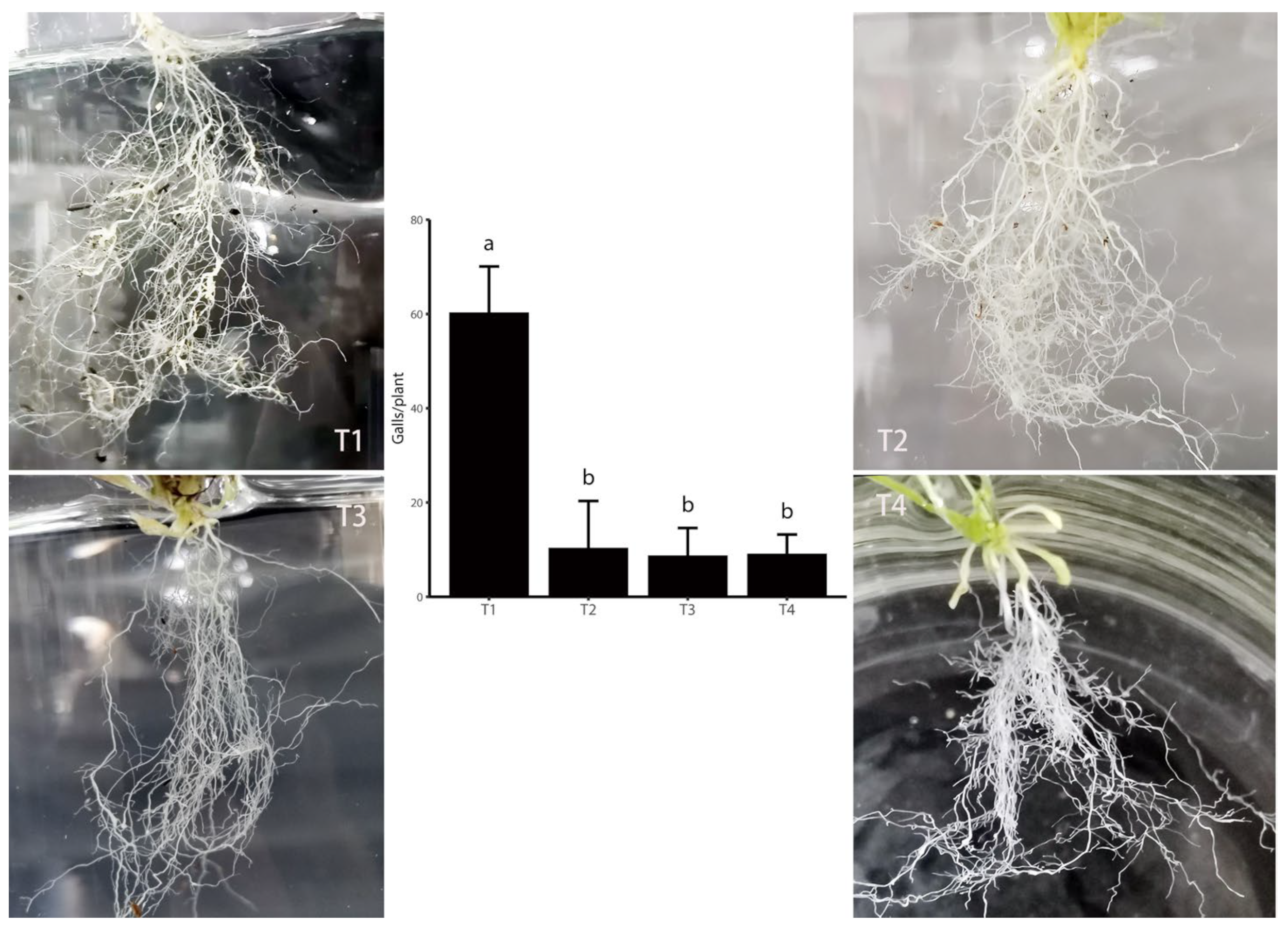
3.1. Elimination or Inhibition of Soil Bacteria Reduced M. incognita Pathogenicity in A. thaliana

At day 40, after inoculation with M. incognita J2s, the gall number on the roots of A. thaliana grown in the natural soil (T1) was 55.3 galls/plant (Figure 1). The galls occurring on the plants grown in the sterile soil (T2), nature soil + antibiotics (T3), and sterile soil + antibiotics (T4) were only 10.4, 8.8, and 9.4 galls/plant, respectively (Figure 1), which were significantly lower than that of T1 (p < 0.05). The results indicated that some rhizobacteria possibly contributed to the M. incognita pathogenicity.

3.2. Sixteen Bacterial Combinations from Rhizosphere Soils Contributed to the M. incognita Pathogenicity in A. thaliana

In a pot experiment, the bacterial combinations at the genus level from the natural soil were investigated for their relationship with M. incognita pathogenicity. Of the 29 combinations tested, 16 combinations generated more galls (15.3–50.8 galls/plant) on the roots of A. thaliana than that of the control (sterile soil without bacterial inoculation, 9.7 galls/plant). The 16 combinations included Methylorubrum, Actinomadura, Muciluginibacter, Sphingopyxis, Microbacterium, Brevundimonas, Pseudomonas, Lysobacter, Kribbella, Rhodanobacter, Mycolicibacterium, Prosthecobacter, Pandoraea, Massilia, Ramlibacter, and Nocardioides (Figure 2). Of them, seven combinations (Rhodanobacter, Mycolicibacterium, Prosthecobacter, Pandoraea, Massilia, Ramlibacter, and Nocardioides) generated the galls significantly higher than the control of the natural soil without bacterial inoculation (22.5 galls/plant).

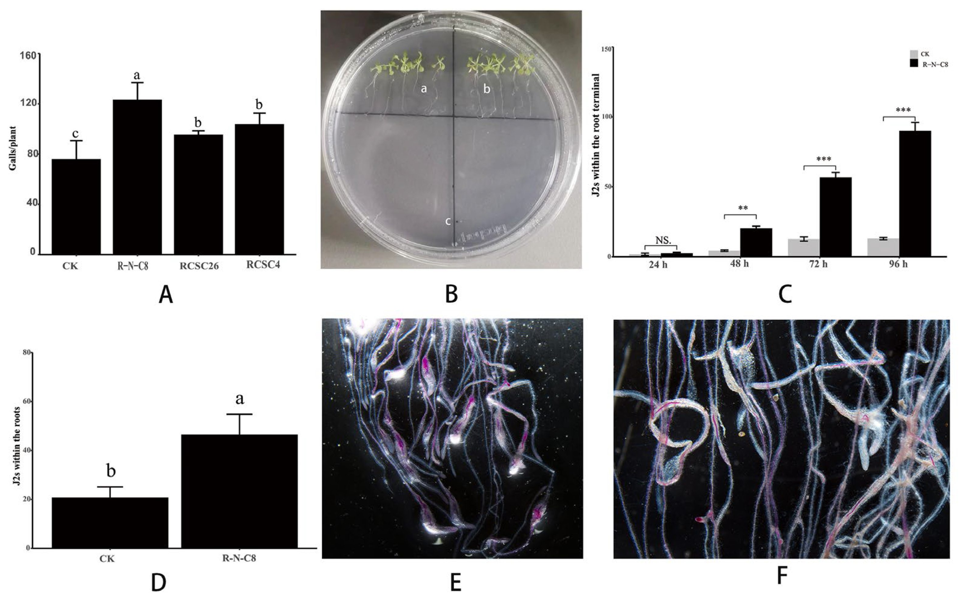
3.3. N. nematodiphilus R-N-C8 Increased M. incognita Pathogenicity in A. thaliana by Attracting M. incognita J2s

Of the above 16 combinations that contributed to M. incognita pathogenicity, Nocardioides was the most efficient combination. The contribution of a single strain from this combination, N. nematodiphilus R-N-C8, N. baekrokdamisoli RCSC26, and N. simplex RCSC4, was determined. The inoculation of N. nematodiphilus R-N-C8, N. baekrokdamisoli RCSC26, and N. simplex RCSC4 produced 123.4, 95.5, and 99.5 galls/plant, respectively, which was significantly higher than that of the control drenched by 1/10 R2A (75.8 galls/plant) (p < 0.05; Figure 3A). Of the three strains, N. nematodiphilus R-N-C8 showed the most decisive contribution to the pathogenicity of M. incognita, as its gall numbers were significantly higher than that of other strains (p < 0.05).
The chemotaxis assay was conducted on a 0.8% agar plate containing A. thaliana seedlings, as shown in Figure 3B. When the roots were treated by the cell suspension of N. nematodiphilus R-N-C8, the J2 number within the root terminal was significantly higher than that of the control after inoculation J2s for 48–96 h (Figure 3C). Additionally, the J2 number within the roots was significantly higher than that of the control after inoculating J2s for 6 d (Figure 3D–F). These results indicated that N. nematodiphilus R-N-C8 could increase M. incognita pathogenicity in A. thaliana by attracting M. incognita J2s towards the host plant and entering the roots.

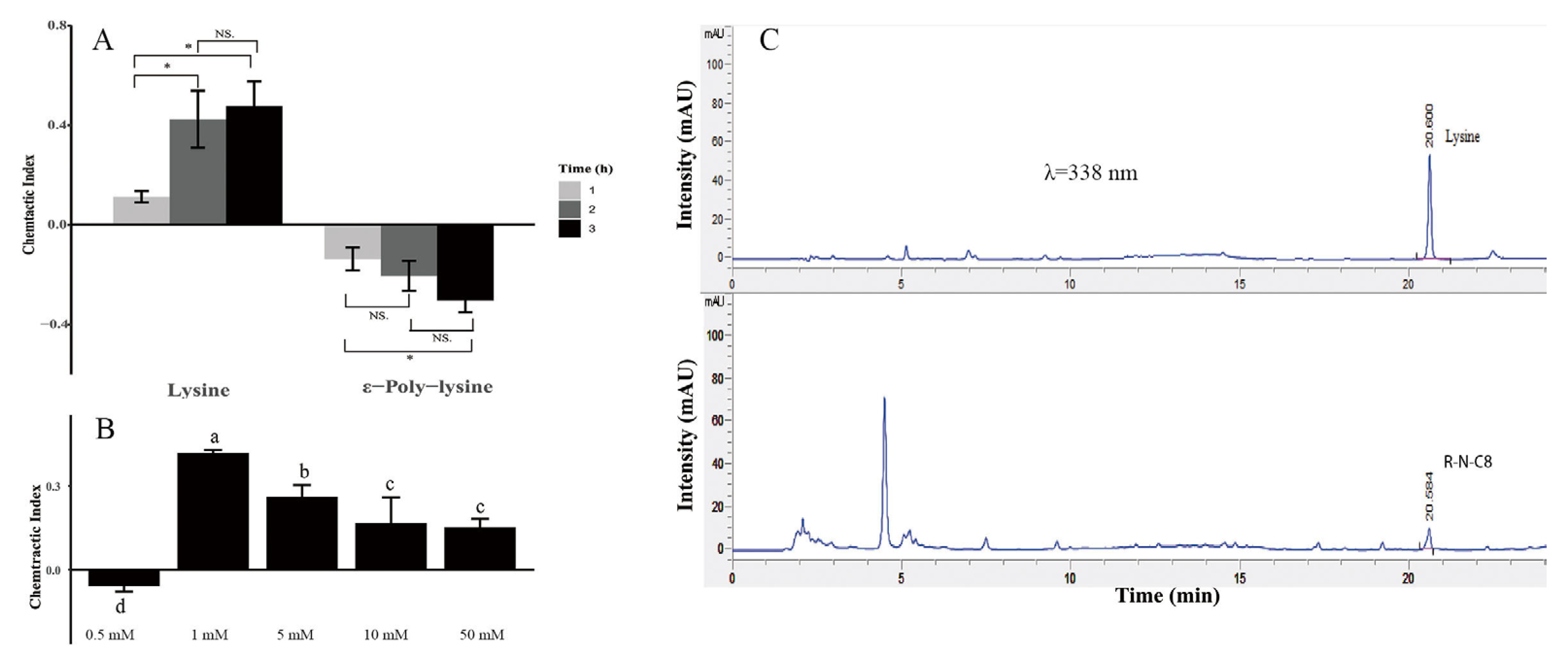
3.4. N. nematodiphilus R-N-C8 Producing L-Lysine to Attract M. incognita

The results of the chemotaxis assay using 1 mM ε-PL and L-lysine as chemoattractants indicated that L-lysine showed highly attractive activity to M. incognita J2s at 2 h and 3 h after the inoculation of J2s, while the C.I. values were recorded as 0.42 and 0.48, respectively (Figure 4A). On the contrary, ε-PL showed a highly repellent response to the J2s at 2 h and 3 h with C.I. values of −0.20 and −0.30, respectively (Figure 4A). From the tested concentrations, 1 mM was the optimal concentration for L-lysine to attract M. incognita J2s in which the C.I. (0.42) was significantly higher than that of the 0.5 mM, 5 mM, 10 mM, and 50 mM (p < 0.05, Figure 4B). To investigate whether N. nematodiphilus R-N-C8 could secrete L-lysine, the supernatant of strain R-N-C8 was analyzed by HPLC with L-lysine as a standard sample. From the HPLC chromatogram, the typical L-lysine standard sample peak was also found in the supernatant of R-N-C8 (Figure 4C). The result indicated that the R-N-C8 strain could secrete L-lysine.

4. Discussion

4.1. Microbes Increased Pathogenicity of RKNs

It has been widely reported that some microorganisms could positively influence the pathogenicity of phytopathogens. Pathogenic fungi Rhizopus spp. caused rice seedling blight by producing the macrocyclic polyketide metabolite rhizoxin from their endosymbiotic bacteria Burkholderia [28,29]. Bacteria Xanthomonas maltophilia, Sphingobacterium multivorum, Enterobacter agglomerans, and Erwinia amylovora are closely associated with phytopathogen Stagonospora nodorum and assisted the S. nodorum to infect wheat by the production of lipases [30]. The plant rhizosphere has diverse microorganisms and is a hot spot habitat for microbe–plant interactions [31,32]. Recently, there has been a growing interest in studying rhizosphere microbes to understand the microbial community associated with the pathogenicity of RKNs. Based on co-occurrence analyses, Castillo et al. revealed that the abundance of α-Proteobacteria Rhodoplanes, Phenylobacterium, and Kaistobacter positively correlated with M. chitwoodi, and the abundance of Bacteroidia and γ-Proteobacteria positively associated with P. neglectus in potato agricultural soils [33]. Sikder et al. reported transplanting the microbiomes from the rhizosphere of the flavonoid over-producing pap1-D A. thaliana mutants to tomato significantly enhanced M. incognita root invasion, suggesting flavonoids indirectly affect RKN invasion by modulating the root-associated microbiome [34]. Gowda et al. reported inoculating the unfiltered microbial slurry from the rhizosphere soil of the RKN-infected plants would increase nematode penetration and reproduction by 8–26% [35]. It has been demonstrated that RKNs acquired most parasitism genes that encode enzyme production from bacteria by horizontal gene transfer [36]. Lu et al. reported that rhizobacteria from the genera of Chitinophaga, Sphingomonas, and Rhizobium were positively correlated with RKN density, possibly due to these bacteria that might destroy plant cell walls and promote the colonization of RKN in tobacco roots by producing cell wall degrading enzymes [37]. The above exciting results concerning rhizosphere bacteria-associated RKN infection were mainly generated by culture-independent analyses, such as amplicon sequencing. However, the microbiota of these rhizobacteria at the species level and their mechanisms largely remain unknown. Hence, further studies are needed to identify the causal microorganisms conclusively.
In this study, by employing culture techniques, we characterized the microbiota of rhizobacteria enhancing the pathogenicity of M. incognita, which includes Nocardioides, Ramlibacter, Massilia, Pandoraea, Prosthecobacter, Rhodanobacter, Mycolicibacterium, Rhodanobacter, Kribbella, Lysobacter, Pseudomonas, Microbacterium, Sphingopyxis, Muciluginibacter, Actinomadura, Methylorubrum, and Brevundimonas (Figure 2). Of the 16 combinations, the genera of Microbacterium and Brevundimonas had also been reported as predominating in the rhizosphere of A. thaliana [38]. Mucilaginibacter was significantly over-represented in the rhizosphere of the carotenoid-deficient Arabidopsis mutant (szl1-1) and might involve Arabidopsis flowering time [39]. Sphingopyxis and Microbacterium were the predominant rhizobacteria of A. thaliana growing in the sterilized soil, which might be involved in the negative plant–soil feedback [40]. Lundberg et al. compared the core microbiome of A. thaliana and found that the Actinobacteria family Streptomycetaceae and several Proteobacteria families were highly enriched in the endophytic compartment [41]. Meanwhile, Acidobacteria, Verrucomicrobia, Gemmatimonadetes, and various proteobacterial families were common in the soil and rhizosphere. This depletion suggested that these taxa were actively excluded by the host immune system and outcompeted by more successful endophytic compartment colonizers. Of the most effective Nocardioides combinations in this study, N. nematodiphilus and N. baekrokdamisoli were respectively isolated from the rhizosphere soil of A. thaliana and the soil of a crater lake and were described as novel species [19,42]. After that, a study was conducted on the two bacteria. N. simplex (also named Rhodococcus erythropolis [43]) possessed the ability to adapt and live under extreme conditions and possessed an extensive array of enzymes enabling cells to carry out an enormous number of biocatalytic reactions and degradations [44]. Until now, no previous reports of the correlations between A. thaliana rhizobacteria and nematode infection exist.

4.2. Host-Seeking and Locating Cues of RKNs

J2s largely influenced the pathogenicity of RKNs to seek and locate host plants, secrete effectors into the hosts, and disrupt plant immunity. In our preliminary experiments of transcriptome analysis, N. nematodiphilus R-N-C8 could not up-regulate the genes encoding effectors of J2s nor down-regulate the genes involved in the plant immunity of A. thaliana. Hence, we focused on the possible mechanism of seeking and locating host plants. It has long been speculated that chemotaxis is the primary way RKNs locate plant roots [45]. RKNs exhibit chemotactic responses to diverse physical and chemical cues, such as rhizospheric gradients in temperature, moisture, pH and redox potentials, plant-derived mineral salts, carbon dioxide, carbohydrates, amino acids, phenolic compounds, chelating compounds, and oxygen [9,46]. Root exudates are the primary plant-derived cues, and RKNs are attracted to plant roots and some root volatiles [47,48]. Many kinds of root exudates, such as lauric acid [49], pinene, limonene, tridecane and 2-methoxy-3-(1-methylpropyl)-pyrazine [50], methyl salicylate, 2-isopropyl 3-methoxypyrazine, and tridecane [51], have been reported to facilitate M. incognita in seeking and locating the host plants. This study identified L-lysine as the infochemical to attract M. incognita [Figure 4]. Strehmel et al. explored the chemical composition of root exudates of A. thaliana, and a total of 103 compounds were detected and annotated but did not include L-lysine [52], which suggests the amino acid releases by N. nematodiphilus R-N-C8. L-lysine as an infochemical can be perceived by chemosensory neurons ASE of M. incognita and lure the nematodes [53,54]. It is well known that the chemotactic behavior of RKNs is also closely linked to the rhizosphere microorganisms. Rhizosphere bacteria Bacillus sp., Brevibacillus brevis, B. cereus, and Chryseobacterium indologens could directly regulate the movement of M. incognita to infect the host [55]. The compound dibenzofuran produced by Streptomyces plicatus could significantly increase the number of M. incognita moving to the tomato plants [26]. The results of the above studies suggest that rhizosphere microorganisms can secrete diverse infochemicals to facilitate the process of RKNs in seeking and locating host plants and then strengthen the RKN pathogenicity.

5. Conclusions and Future Studies

In conclusion, our research firstly highlights the rhizosphere-associated microbiota enhancing the pathogenicity of RKNs on plants by pure strains and culture-dependent experiments. Sixteen bacterial combinations from rhizosphere soils of A. thaliana were found to strengthen M. incognita pathogenicity, which included Methylorubrum, Actinomadura, Muciluginibacter, Sphingopyxis, Microbacterium, Brevundimonas, Pseudomonas, Lysobacter, Kribbella, Rhodanobacter, Mycolicibacterium, Prosthecobacter, Pandoraea, Massilia, Ramlibacter, and Nocardioides. The most effective strain, N. nematodiphilus R-N-C8 from the Nocardioides combination, was taken as a representative to explore the mechanism of rhizobacteria enhancing the pathogenicity of RKNs. This bacterium could significantly facilitate the M. incognita J2s moving towards the roots of A. thaliana and infecting the roots by releasing L-lysine as a chemoattractant to attract J2s. To further advance our understanding of rhizobacteria, strengthen the pathogenicity of RKNs, and manage these pathogens, future research efforts should focus on the following contents: (1) An exploration of the strain specificity. Our study concentrates only on N. nematodiphilus R-N-C8, but the generality of the findings to other bacterial strains or combinations is unclear. Further research could explore the broader influence of rhizosphere microbiota on plant–nematode interactions. (2) An exploration of the expansion mechanisms. While our study identifies L-lysine as a chemoattractant, a further exploration of the underlying mechanisms by which bacteria enhance M. incognita pathogenicity is needed. This could involve investigating changes in plant root exudates, nematode behavior, and gene expression. (3) Elucidating the potential of these rhizobacteria for the managing of RKNs. Presently, J2s and the eggs of RKNs are the targets of most chemical nematicides and microbe agents. Whether the combination of nematocides and the antibiotics targeting these rhizobacteria can improve the efficiency in RKN management deserves further exploration.

Supplementary Materials

The following supporting information can be downloaded at: https://www.mdpi.com/article/10.3390/agronomy14040664/s1, Figure S1. The non-alpha poly-amino acids like e-Polylysin (NAPAA) secondary metabolite biosynthesis gene cluster in N. nematodiphilus R-N-C8. Supplementary Table S1. Bacterial combinations from rhizosphere soils of A. thaliana were used to evaluate their contribution to M. incognita pathogenicity.

Author Contributions

Methodology, X.-K.Z.; formal analysis, Z.-X.Y.; investigation, L.-F.B. and X.-K.Z.; writing—original draft, X.-K.Z. and L.M.; writing—review and editing, Z.-X.Y. and M.-H.M.; supervision, Z.-X.Y. and M.-H.M.; project administration, Z.-X.Y. and M.-H.M.; funding acquisition, M.-H.M. All authors have read and agreed to the published version of the manuscript.

Funding

This research was financially supported by the National Key R&D Program of China (2023YFD1400400), National Natural Science Foundation of China (32170131), Southwest United Graduate School of Yunnan Province (202302CC4040021), Special Funds for Central Guidance of Local Scientific and Technological Development (202307AB110011), Department of Science and Technology of Yunnan Province (202201BC070004; 202001BB050072; 202401AS070123), and State Key Laboratory for Conservation and Utilization of Bio-Resources in Yunnan, Yunnan University (2022KF003).

Data Availability Statement

The data presented in this study are available on request from the corresponding author.

Acknowledgments

The authors gratefully acknowledge Waqar Ahmed of South China Agricultural University for reviewing and editing the manuscript.

Conflicts of Interest

The authors declare no conflicts of interest.

References

  1. Montarry, J.; Mimee, B.; Danchin, E.G.J.; Koutsovoulos, G.D.; Ste-Croix, D.T.; Grenier, E. Recent Advances in Population Genomics of Plant-Parasitic Nematodes. Phytopathology 2021, 111, 40–48. [Google Scholar] [CrossRef]
  2. Kim, J.-H.; Kim, H.J.; Kim, Y.H.; Jeon, J.M.; Song, H.S.; Kim, J.; No, S.Y.; Shin, J.H.; Choi, K.Y.; Park, K.M.; et al. Functional Study of Lysine Decarboxylases from Klebsiella pneumonia in Escherichia coli and Application of Whole Cell Bioconversion for Cadaverine Production. J. Microbiol. Biotechnol. 2016, 26, 1586–1592. [Google Scholar] [CrossRef] [PubMed]
  3. Jaouannet, M.; Perfus-Barbeoch, L.; Deleury, E.; Magliano, M.; Engler, G.; Vieira, P.; Danchin, E.G.J.; Rocha, M.D.; Coquillard, P.; Abad, P.; et al. A Root-Knot Nematode-Secreted Protein Is Injected into Giant Cells and Targeted to the Nuclei. New Phytol. 2012, 194, 924–931. [Google Scholar] [CrossRef]
  4. Elling, A.A. Major Emerging Problems with Minor Meloidogyne species. Phytopathology 2013, 103, 1092–1102. [Google Scholar] [CrossRef] [PubMed]
  5. Adam, M.; Westphal, A.; Hallmann, J.; Heuer, H. Specific Microbial Attachment to Root Knot Nematodes in Suppressive Soil. Appl. Environ. Microbiol. 2014, 80, 2679–2686. [Google Scholar] [CrossRef] [PubMed]
  6. Tsai, A.Y.L.; Iwamoto, Y.; Tsumuraya, Y.; Oota, M.; Konishi, T.; Ito, S.; Kotake, T.; Ishikawa, H.; Sawa, S. Root-knot Nematode Chemotaxis is Positively Regulated by L-galactose Sidechains of Mucilage Carbohydrate Rhamnogalacturonan-I. Sci. Adv. 2021, 7, eabh4182. [Google Scholar] [CrossRef] [PubMed]
  7. Hewezi, T.; Baum, T.J. Manipulation of Plant Cells by Cyst and Root-Knot Nematode Effectors. Mol. Plant Microbe. Interact. 2013, 26, 9–16. [Google Scholar] [CrossRef] [PubMed]
  8. Ali, M.A.; Anjam, M.S.; Nawaz, M.A.; Lam, H.M.; Chung, G. Signal Transduction in Plant–Nematode Interactions. Int. J. Mol. Sci. 2018, 19, 1648. [Google Scholar] [CrossRef]
  9. Curtis, R.H.C. Plant-nematode Interactions: Environmental Signals Detected by the Nematode’s Chemosensory Organs Control Changes in the Surface Cuticle and Behavior. Parasite 2008, 15, 310–316. [Google Scholar] [CrossRef]
  10. Siddique, S.; Coomer, A.; Baum, T.; Williamson, V.M. Recognition and Response in Plant–Nematode Interactions. Annu. Rev. Phytopathol. 2022, 60, 143–162. [Google Scholar] [CrossRef]
  11. Niu, Q.; Huang, X.; Zhang, L.; Xu, J.; Yang, D.; Wei, K.; Niu, X.; An, Z.; Bennett, J.W.; Zou, C.; et al. A Trojan Horse Mechanism of Bacterial Pathogenesis against Nematodes. Proc. Natl. Acad. Sci. USA 2010, 107, 16631–16636. [Google Scholar] [CrossRef] [PubMed]
  12. Cheng, W.; Yang, J.; Nie, Q.; Huang, D.; Yu, C.; Zheng, L.; Cai, M.; Thomashow, L.S.; Weller, D.M.; Yu, Z.; et al. Volatile Organic Compounds from Paenibacillus polymyxa KM2501-1 Control Meloidogyne incognita by Multiple Strategies. Sci. Rep. 2017, 7, 16213. [Google Scholar] [CrossRef]
  13. Topalović, O.; Vestergård, M. Can Microorganisms Assist the Survival and Parasitism of Plant-Parasitic Nematodes? Trends Parasitol. 2021, 37, 947–958. [Google Scholar] [CrossRef] [PubMed]
  14. Cheng, X.Y.; Tian, X.L.; Wang, Y.S.; Lin, R.M.; Mao, Z.C.; Chen, N.; Xie, B.Y. Metagenomic Analysis of the Pinewood Nematode Microbiome Reveals a Symbiotic Relationship Critical for Xenobiotics Degradation. Sci. Rep. 2013, 3, 1869. [Google Scholar] [CrossRef] [PubMed]
  15. Cao, Y.; Tian, B.; Ji, X.; Shang, S.; Lu, C.; Zhang, K. Associated Bacteria of Different Life Stages of Meloidogyne incognita Using Pyrosequencing-Based Analysis. J. Basic Microbiol. 2015, 55, 950–960. [Google Scholar] [CrossRef] [PubMed]
  16. Tian, B.Y.; Cao, Y.; Zhang, K.Q. Metagenomic Insights into Communities, Functions of Endophytes, and Their Associates with Infection by Root-Knot Nematode, Meloidogyne incognita, in Tomato Roots. Sci. Rep. 2015, 5, 17087. [Google Scholar] [CrossRef] [PubMed]
  17. Yergaliyev, T.M.; Alexander-Shani, R.; Dimerets, H.; Pivonia, S.; Bird, D.M.; Rachmilevitch, S.; Szitenberg, A. Bacterial Community Structure Dynamics in Meloidogyne incognita-Infected Roots and Its Role in Worm-Microbiome Interactions. mSphere 2020, 5, 1–18. [Google Scholar] [CrossRef] [PubMed]
  18. Li, Y.; Lei, S.; Cheng, Z.; Jin, L.; Zhang, T.; Liang, L.M.; Cheng, L.; Zhang, Q.; Xu, X.; Lan, C.; et al. Microbiota and Functional Analyses of Nitrogen-fixing Bacteria in Root-Knot Nematode Parasitism of Plants. Microbiome 2023, 11, 48. [Google Scholar] [CrossRef]
  19. Zhang, X.M.; Mo, W.T.; Wei, Y.Q.; Li, X.; Ma, G.M.; Yan, Y.S.; Wang, X.J.; Liu, J.J.; Liu, Z.Y.; Zhou, X.K.; et al. Nocardioides nematodiphilus sp. nov., Isolated from Rhizosphere of Arabidopsis thaliana. Int. J. Syst. Evol. Microbiol. 2022, 72, 005271. [Google Scholar] [CrossRef]
  20. Lu, C.J.; Meng, Y.; Wang, Y.L.; Zhang, T.; Yang, G.F.; Mo, M.H.; Ji, K.F.; Liang, L.M.; Zou, C.G.; Zhang, K.Q. Survival and Infectivity of Second-Stage Root-Knot Nematode Meloidogyne incognita Juveniles Depend on Lysosome-Mediated Lipolysis. J. Biol. Chem. 2022, 298, 101637. [Google Scholar] [CrossRef]
  21. Kyndt, T.; Goverse, A.; Haegeman, A.; Warmerdam, S.; Wanjau, C.; Jahani, M.; Engler, G.; Engler, J.d.A.; Gheysen, G. Redirection of Auxin Flow in Arabidopsis thaliana Roots After Infection by Root-Knot Nematodes. J. Exp. Bot. 2016, 67, 4559–4570. [Google Scholar] [CrossRef] [PubMed]
  22. Dong, H.; Fan, S.; Sun, H.; Chen, C.; Wamg, A.; Jiang, L.; Ma, D. Rhizosphere-Associated Microbiomes of Rice (Oryza sativa L.) Under the Effect of Increased Nitrogen Fertilization. Front. Microbiol. 2021, 12, 730506. [Google Scholar] [CrossRef] [PubMed]
  23. Fudali, S.L.; Wang, C.; Williamson, V.M. Ethylene Signaling Pathway Modulates Attractiveness of Host Roots to the Root-Knot Nematode Meloidogyne hapla. Mol. Plant Microbe Interact. 2013, 26, 75–86. [Google Scholar] [CrossRef] [PubMed]
  24. Teixeira, M.A.; Wei, L.; Kaloshian, I. Root-knot Nematodes Induce Pattern-Triggered Immunity in Arabidopsis thaliana Roots. New Phytol. 2016, 211, 276–287. [Google Scholar] [CrossRef]
  25. Oota, M.; Tsai, A.Y.L.; Aoki, D.; Matsushita, Y.; Toyoda, S.; Fukushima, K.; Saeki, K.; Toda, K.; Perfus-Barbeoch, L.; Favery, B.; et al. Identification of Naturally Occurring Polyamines as Root-Knot Nematode Attractants. Mol. Plant 2020, 13, 658–665. [Google Scholar] [CrossRef] [PubMed]
  26. Wang, P.Y.; Sun, Y.; Yang, L.; Hu, Y.; Li, J.; Wang, J.; Zhang, F.; Liu, Y. Chemotactic Responses of the Root-Knot Nematode Meloidogyne incognita to Streptomyces plicatus. FEMS Microbiol. Lett. 2019, 366, fnz234. [Google Scholar] [CrossRef] [PubMed]
  27. Tang, S.K.; Wang, Y.; Chen, Y.; Lou, K.; Cao, L.L.; Xu, L.H.; Li, W.J. Zhihengliuella alba sp. nov., and Emended Description of the Genus Zhihengliuella. Int. J. Syst. Evol. Microbiol. 2009, 59, 2025–2031. [Google Scholar] [CrossRef]
  28. Iwasaki, S.; Kobayashi, H.; Furukawa, J.; Namikoshi, M.; Okuda, S.; Sato, Z.; Matsuda, I.; Noda, T. Studies on Macrocyclic Lactone Antibiotics. VII. Structure of a Phytotoxin “Rhizoxin” Produced by Rhizopus chinensis. J. Antibiot. 1984, 37, 354–362. [Google Scholar] [CrossRef]
  29. Martinez, L.P.; Hertweck, C. Pathogenic Fungus Harbours Endosymbiotic Bacteria for Toxin Production. Nature 2005, 437, 884–888. [Google Scholar] [CrossRef]
  30. Dewey, F.M.; Wong, Y.L.; Seery, R.; Hollins, T.W.; Gurr, S.J. Bacteria Associated with Stagonospora (Septoria) nodorum Increase Pathogenicity of the Fungus. New Phytol. 1999, 144, 489–497. [Google Scholar] [CrossRef]
  31. Ahmed, W.; Dai, Z.; Zhang, J.; Li, S.; Ahmed, A.; Munir, S.; Liu, Q.; Tan, Y.; Ji, G.; Zhao, Z. Plant-Microbe Interaction: Mining the Impact of Native Bacillus amyloliquefaciens WS-10 on Tobacco Bacterial Wilt Disease and Rhizosphere Microbial Communities. Microbiol. Spectr. 2022, 10, e0147122. [Google Scholar] [CrossRef] [PubMed]
  32. Raaijmakers, J.M.; Paulitz, T.C.; Steinberg, C.; Alabouvette, C.; Moënne-Loccoz, Y. The Rhizosphere: A Playground and Battlefield for Soilborne Pathogens and Beneficial Microorganisms. Plant Soil 2009, 321, 341–361. [Google Scholar] [CrossRef]
  33. Castillo, J.D.; Vivanco, J.M.; Manter, D.K. Bacterial Microbiome and Nematode Occurrence in Different Potato Agricultural Soils. Microb. Ecol. 2017, 74, 888–900. [Google Scholar] [CrossRef]
  34. Sikder, M.M.; Vestergård, M.; Kyndt, T.; Topalović, O.; Kudjordjie, E.N.; Nicolaisen, M. Genetic Disruption of Arabidopsis Secondary Metabolite Synthesis Leads to Microbiome-Mediated Modulation of Nematode Invasion. ISME J. 2022, 16, 2230–2241. [Google Scholar] [CrossRef] [PubMed]
  35. Gowda, M.T.; Prasanna, R.; Rao, U.; Somvanshi, V.S.; Singh, P.K.; Sing, A.K.; Chawla, G. Microbiome Transplant Can Effectively Manage Root-Knot Nematode Infectivity in Tomato. Appl. Soil Ecol. 2023, 190, 105020. [Google Scholar] [CrossRef]
  36. Bird, D.M.; Opperman, C.H.; Davies, K.G. Interactions Between Bacteria and Plant-Parasitic Nematodes: Now and Then. Int. J. Parasitol. 2003, 33, 1269–1276. [Google Scholar] [CrossRef] [PubMed]
  37. Lu, P.; Shi, H.; Tao, J.; Jin, J.; Wang, S.; Zheng, Q.; Liu, P.; Xiang, B.; Chen, Q.; Xu, Y.; et al. Metagenomic Insights into the Changes in the Rhizosphere Microbial Community Caused by the Root-Knot Nematode Meloidogyne incognita in Tobacco. Environ. Res. 2023, 216, 114848. [Google Scholar] [CrossRef]
  38. Wang, R.; Wang, Y.; He, D.; Shi, T.; Zhang, Y.; Liu, S.; Yan, X.; Huang, L. Responses of Plant Immune System and Rhizosphere Soil Microbiome to the Elicitor BAR11 in Arabidopsis thaliana. Sci. Total Environ. 2024, 914, 169920. [Google Scholar] [CrossRef]
  39. Chen, X.; Li, F.L.; Kong, C.H. Rhizosphere Bacteria Mediate Flowering Time of Two Genotypes of Arabidopsis with and without Root-Secreted Signaling (−)-loliolide. Rhizosphere 2023, 27, 100774. [Google Scholar] [CrossRef]
  40. Idbella, M.; Bonanomi, G.; Filippis, F.D.; Foscari, A.; Zotti, M.; Abd-ElGawad, A.M.; Fechtali, T.; Incerti, G.; Mazzoleni, S. Negative Plant-Soil Feedback in Arabidopsis thaliana: Disentangling the Effects of Soil Chemistry, Microbiome, and Extracellular Self-DNA. Microbiol. Res. 2024, 281, 127634. [Google Scholar] [CrossRef]
  41. Lundberg, D.S.; Lebeis, S.L.; Paredes, S.H.; Yourstone, S.; Gehring, J.; Malfatti, S.; Tremblay, J.; Engelbrektson, A.; Kunin, V.; del Rio, T.G.; et al. Defining the Core Arabidopsis thaliana Root Microbiome. Nature 2012, 488, 86–90. [Google Scholar] [CrossRef] [PubMed]
  42. Lee, K.C.; Kim, K.K.; Kim, J.S.; Kim, D.S.; Ko, S.H.; Yang, S.H.; Shin, Y.K.; Lee, J.S. Nocardioides baekrokdamisoli sp. nov., Isolated from Soil of a Crater Lake. Int. J. Syst. Evol. Microbiol. 2016, 66, 4231–4235. [Google Scholar] [CrossRef] [PubMed]
  43. Yoon, J.H.; Lee, J.S.; Shin, Y.K.; Park, Y.H.; Lee, S.T. Reclassification of Nocardioides simplex ATCC 13260, ATCC19565, and ATCC 19566 as Rhodococcus erythropolis. Int. J. Syst. Evol. Microbiol. 1997, 47, 904–907. [Google Scholar]
  44. de Carvalho, C.C.C.R. Adaptation of Rhodococcus erythropolis Cells for Growth and Bioremediationunder Extreme Conditions. Res. Microbiol. 2012, 163, 125–136. [Google Scholar] [CrossRef] [PubMed]
  45. Dam, N.M.V.; Bouwmeester, H.J. Metabolomics in the Rhizosphere: Tapping into Belowground Chemical Communication. Trends Plant Sci. 2016, 21, 256–265. [Google Scholar] [PubMed]
  46. Perry, R.N. Chemoreception in Plant-Parasitic Nematodes. Annu. Rev. Phytopathol. 1996, 34, 181–199. [Google Scholar] [CrossRef]
  47. Farnier, K.; Bengtsson, M.; Becher, P.G.; Witzell, J.; Witzgall, P.; Manduríc, S. Novel Bioassay Demonstrates Attraction of the White Potato Cyst Nematode Globodera pallida (Stone) to Non-Volatile and Volatile Host Plant Cues. J. Chem. Ecol. 2012, 38, 795–801. [Google Scholar] [CrossRef]
  48. Ali, J.G.; Alborn, H.T.; Stelinski, L.L. Constitutive and Induced Subterranean Plant Volatiles Attract both Entomopathogenic and Plant Parasitic Nematodes. J. Ecol. 2011, 99, 26–35. [Google Scholar] [CrossRef]
  49. Dong, L.; Li, X.; Huang, L.; Gao, Y.; Zhong, L.; Zheng, Y.; Zuo, Y. Lauric Acid in Crown Daisy Root Exudate Potently Regulates Root-Knot Nematode Chemotaxis and Disrupts Mi-ffp-18 Expression to Block Infection. J. Exp. Bot. 2014, 65, 131–141. [Google Scholar] [CrossRef]
  50. Kihika, R.; Murungi, L.K.; Coyne, D.; Ng’ang’a, M.; Hassanali, A.; Teal, P.E.A.; Torto, B. Parasitic Nematode Meloidogyne incognita Interactions with Different Capsicum annum Cultivars Reveal the Chemical Constituents Modulating Root Herbivory. Sci. Rep. 2017, 7, 2903. [Google Scholar] [CrossRef]
  51. Murungi, L.K.; Kirwa, H.; Coyne, D.; Teal, P.E.A.; Beck, J.J.; Torto, B. Identification of Key Root Volatiles Signaling Preference of Tomato over Spinach by the Root Knot Nematode Meloidogyne incognita. J. Agric. Food Chem. 2018, 66, 7328–7336. [Google Scholar] [CrossRef]
  52. Strehmel, N.; Böttcher, C.; Schmidt, S.; Scheel, D. Profiling of Secondary Metabolites in Root Exudates of Arabidopsis thaliana. Phytochemistry 2014, 108, 35–46. [Google Scholar] [CrossRef]
  53. Ferkey, D.M.; Sengupta, P.; L’Etoile, N.D. Chemosensory Signal Transduction in Caenorhabditis elegans. Genetics 2021, 217, iyab004. [Google Scholar] [CrossRef]
  54. Shingai, R.; Wakabayashi, T.; Sakata, K.; Matsuura, T. Chemotaxis of Caenorhabditis elegans during Simultaneous Presentation of Two Water-Soluble Attractants, L-lysine and Chloride Ions. Comp. Biochem. Physiol. 2005, 142, 308–317. [Google Scholar] [CrossRef]
  55. Li, B.; Wang, P.; Yang, L.; Rang, X.; Zhou, W.; Liu, Y. Chemotaxis of Meloidogyne incognita Response to Rhizosphere Bacteria. Microorganisms 2023, 11, 2271. [Google Scholar] [CrossRef] [PubMed]
Figure 1. The impact of growing substrates with different treatments on M. incognita pathogenicity. M. incognita generated more galls on the roots of A. thaliana grown in nature soils (T1) than that of the plants grown in sterile soil (T2), nature soil + antibiotics (T3), and sterile soil + antibiotics (T4). The error bars were calculated from four repeated experiments, each with four biological replicates. Different letters on columns indicate significative differences between the treatments (p < 0.05).
Agronomy 14 00664 g001
Figure 2. The effects of different bacterial combinations on the M. incognita pathogenicity in A. thaliana with an equal volume of 1/10 R2A replaced cell suspension used in natural soil and sterile soil as the controls. Different lowercase letters in the graphs indicate significant differences between treatments (p < 0.05) according to the LSD test. Values are means ± SE (n = 12).
Agronomy 14 00664 g002
Figure 3. The impact of Nocardioides strains on the M. incognita pathogenicity in A. thaliana. (A): the effects of N. nematodiphilus R-N-C8, N. baekrokdamisoli RCSC26, and N. simplex RCSC4 from the Nocardioides combination on the M. incognita pathogenicity; (B): the chemotaxis assay using a 0.8% agar plate; (C): strain R-N-C8 attracting more M. incognita J2s within the root terminal than that of the control (CK) after inoculating J2s for 48–96 h; (D): strain R-N-C8 promoting more M. incognita J2s infecting into the root than that of the control (CK) after inoculating J2s for 6 d; (E): an image showing J2s in the roots of inoculation R-N-C8 after staining with acid fuchsin; (F): Image showing J2s in the roots of the control after staining with acid fuchsin. Different lowercase letters in the graphs indicate significant differences between treatments (p < 0.05) according to the LSD test. Values are means ± SE (n = 15). NS, **, and *** denote no significant difference, p < 0.01, and p < 0.001, respectively.
Agronomy 14 00664 g003
Figure 4. The chemotaxis assay using ε-poly-L-lysine and L-lysine as chemoattractants towards M. incognita J2s and the HPLC analysis on L-lysine in the supernatant of N. nematodiphilus R-N-C8. (A): L-lysine shows highly attractive activity to J2s, while ε-PL shows a highly repellent response to the J2s; (B): the impact of concentrations on the attractive activities of L-lysine towards J2s; (C): HPLC profiles showing L-lysine in the supernatant of N. nematodiphilus R-N-C8 when using L-lysine as the standard sample. Different letters on error bars indicate significant differences between the treatments according to the LSD test at p < 0.05. The asterisks represent significant differences at p < 0.05. NS represents no significant difference.
Agronomy 14 00664 g004
Disclaimer/Publisher’s Note: The statements, opinions and data contained in all publications are solely those of the individual author(s) and contributor(s) and not of MDPI and/or the editor(s). MDPI and/or the editor(s) disclaim responsibility for any injury to people or property resulting from any ideas, methods, instructions or products referred to in the content.

