Article

Phytophthora Infection Reorients the Composition of Rhizospheric Microbial Assembly in Khasi Mandarin (Citrus reticulata Blanco)

by
Mridupol Handique
1,
Popy Bora
1,2,*,
Vasileios Ziogas
3,
Anoop Kumar Srivastava
4,
Prasanth Tej Kumar Jagannadham
5 and
Asish Kumar Das
5
1
Program on Bio-Pesticide, Bio-Control Laboratory, Assam Agricultural University, Jorhat 785013, India
2
AAU-Assam Rice Research Institute, Assam Agricultural University, Jorhat 785630, India
3
Institute of Olive Tree, Subtropical Plants and Viticulture, Hellenic Agricultural Organization-DIMITRA (ELGO-DIMITRA), 73134 Chania, Greece
4
ICAR-Indian Agricultural Research Institute, Dhemaji 110012, India
5
ICAR-Central Citrus Research Institute, Nagpur 440033, India
*
Author to whom correspondence should be addressed.
Agronomy 2024, 14(4), 661; https://doi.org/10.3390/agronomy14040661
Submission received: 16 December 2023 / Revised: 8 February 2024 / Accepted: 20 March 2024 / Published: 24 March 2024
(This article belongs to the Special Issue Fruit Growing: Production Practices and Post-Harvest Management)

Abstract

:
Phytophthora gummosis, foot rot, and root rot are considered major challenges to the citrus industry worldwide. Little is known about the Phytophthora–microbiome interaction, despite several studies demonstrating changes in the microbial composition of the rhizosphere following challenges by a pathogen. In the present study, we studied the microbial diversity and community structure in healthy rhizospheres and Phytophthora-infected rhizospheres of Khasi mandarin (Citrus reticulata Blanco), a commercial cultivar extensively grown in the northeast of India. An exploratory study was conducted to identify Phytophthora-infected orchards of Khasi mandarin, and the isolated pathogen was confirmed as P. nicotianae based on its morpho-cultural and molecular characteristics coupled with pathogenicity tests. This study on culturable microbes established the dominance of Trichoderma spp. in the healthy rhizosphere, while the diseased rhizosphere showed the presence of Fusarium spp. A metagenomic study further revealed that the rhizospheres of Phytophthora-infected plants were dominated by species such as Bacteroidia spp., Patescibacteria spp., and Pythium spp., while the healthy Khasi mandarin rhizospheres had a more diverse community predominantly represented by Trichoderma, Penicillium, Linnemannia, Mortierella, Talaromyces, Saitozyma, Bacteroidetes, Pseudomonas, Cytophagia, Cyanobacteria, Bacteroidia, Sphingobacteriia, Burkholderia, Bacillus, and Bradyrhizobium. Terrabacteria and FCB (Fibrobacterota, Chlorobiota, and Bacteroidota groups) were found to exist in higher relative abundance in disease-free soils than in Phytophthora-infected soils, while phylum Proteobacteria showed identical relative abundance in all soil types. The phyla represented by Pseudomonas, Flavobacteriia, Candidatus, Mycobacterium, Rhizobium, Mesorhizobium, Sphingomonas, and Cytophagia were the most common bacterial phyla in all soil samples, but healthy soil exhibited a greater abundance of Bacteroidetes, Pseudomonas, Cytophagia, Cyanobacteria, Bacteroidia, Sphingobacteriia, Burkholderia, Bacillus, and Bradyrhizobium. Our study suggests that the presence of Phytophthora spp. in the rhizosphere alters microbial community structure, having potentially strong implications for plant health and productivity. These rhizosphere microbiome-derived citrus responses shed light on exploring effective management strategies for Phytophthora gummosis disease ailing Khasi mandarin.

1. Introduction

The rhizosphere is “the narrow region of soil that is directly influenced by root secretions and associated soil microorganisms” [1], a critical interface between plant roots and soil microorganisms playing a crucial role in plant growth and health. It is a dynamic and complex environment influenced by various factors such as plant genotype, soil type, and environmental conditions. The concept of the rhizosphere was first introduced by Hiltner in 1904 [2]. The rhizosphere microbiome, comprising diverse microbial communities, is reported to have strong implications for plant health, disease resistance, and the ecosystem [3,4]. Microorganisms in the rhizosphere interact with plants through various chemical signals and mechanisms, altering the soil’s physical and chemical properties, and ultimately influencing plant productivity. Recent studies revealed the role of the rhizosphere in shaping plant-associated microbial communities and their interactions with pathogens [5,6]. For example, a study by Yuan et al. [7] showed that the rhizosphere microbiome of maize plants influenced the colonization and virulence of fungal pathogen Fusarium verticillioides by modulating the expression of plant genes involved in jasmonic acid (JA) and salicylic acid (SA) signalling. Similarly, Berg et al. [8] found that the rhizosphere microbiome of Arabidopsis thaliana promoted the growth and biocontrol activities of fungal endophyte Serendipita indica, and enhanced the plant’s resistance against the foliar pathogen Pseudomonas syringae. Consequently, any alteration in the microbial makeup of the rhizosphere has the potential to influence its ecological roles, relating to the physiological condition of the plant, and ultimately, the plant’s productivity.
Root rot or gummosis is a devastating disease of citrus plants, caused by Phytophthora spp., a group of soilborne pathogens [9] with sensitivity against all major fungicides [10]. This disease is characterized by the formation of gum pockets on the bark, trunk, and branches, eventually leading to the dieback and decline of the tree. The pathogen infects the roots, trunk, and branches of the tree, and the infection is often associated with wounds or injuries caused by mechanical damage or adverse environmental conditions [11]. In recent years, research has focused on the effects of Phytophthora infection on the composition of rhizospheric microbial communities, playing an important role in plant health and productivity. A study by Yang et al. [12] examined the effects of Phytophthora infection on the rhizospheric microbial community composition of citrus plants.
Khasi mandarin (Citrus reticulata Blanco) is an important citrus fruit crop grown in the northeastern region of India, particularly in the states of Meghalaya, Assam, and Arunachal Pradesh [13], having significant economic and cultural value in the region. However, the production of Khasi mandarin is constrained by various biotic and abiotic factors, including pest and disease pressures. Phytophthora spp., a soil-borne pathogen, is a major risk to Khasi mandarin, causing root rot and gummosis in citrus plants. The disease leads to significant yield losses and poses a major constraint on citrus production in the region. For instance, a report from Singh et al. [14] stated that Khasi mandarin is the most preferred citrus fruit in the eastern Himalayan region, the most notable northeastern region of India. However, it was also notable that the crop is vulnerable to various diseases, including Phytophthora root rot, frequently leading to significant losses in fruit yield and quality. Similarly, Khasi mandarin is reported to be susceptible to a range of pests and diseases, including citrus psyllid, citrus greening, foot rot, etc., which could further adversely affect the productivity and quality of the crop [15,16].
Despite these challenges, India has made significant progress towards the development of management strategies for Phytophthora-induced foot and root rot, comprising the development of resistant varieties and the use of integrated disease management approaches to ensure the sustainability of Khasi mandarin production in the region. For example, Deka et al. (2018) [17], in their studies, assessed three modules, the bio-intensive IPM module (BIPM), IPM module, and farmers’ practice (FP), based on previous field studies, which showed IPM displaying significant promise with regard to yielding higher net returns, followed by BIPM. However, the sustainable field management of Phytophthora is still a formidable challenge. Therefore, culturable and metagenomic-based studies of the rhizosphere of citrus are crucial in understanding citrus’ microbial diversity and its functional contribution towards the health and productivity of citrus crops. The objectives of such studies were to identify microbial diversity and its functional roles, assess the effect of soil management practices on the microbial community, and evaluate the interaction between the plant and the microbial community. These studies will pave the way towards developing more effective strategies for the microbial antagonist-mediated management of the Phytophthora disease complex, a problem researchers are as yet clueless about, except for the chemical control strategy.

2. Materials and Methods

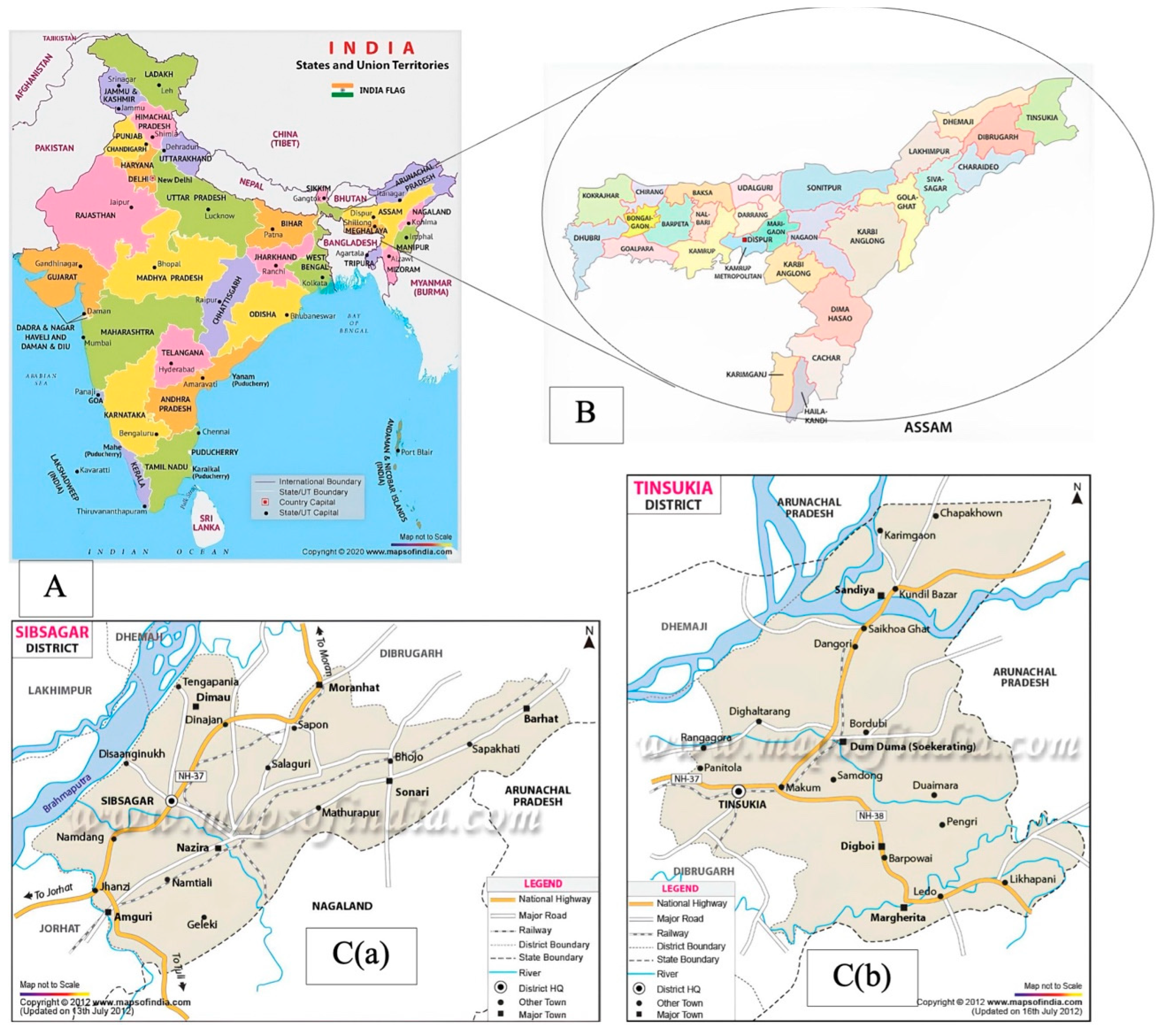
2.1. Rhizospheric Soil Sampling and Isolation of Pathogen

We conducted sampling activity in a 10-year-old Khasi mandarin orchard (showing signs of branch dieback due to root rot) located in Khaman Pathar (Sivasagar), Assam, India, in December 2021. We further confirmed the presence of root rot by visually examining the youngest feeder roots. Briefly, 100 g of soil samples was collected from each of the four sides of the tree canopy using a 25 mm soil auger from inside of the canopy skirt, ensuring the soil contained citrus major feeder roots. The samples were collected with a shovel that was washed with 70% ethanol between each sampling point. Each sample, including rhizospheric soil and feeder roots, individually packed in zip-locked polyethene bags within an ice pack, properly labelled with sample descriptions, were finally brought to the laboratory, in Department of Plant Pathology, Assam Agricultural University, Jorhat (India). Subsequent dilutions were prepared using 1 g of bulked rhizosphere soil and 99 mL of sterile distilled water, and homogenized via vigorous shaking. Dilutions of 1:10 and 1:100 were then streaked onto Petri dishes with Corn Meal Agar-PARPH medium (Cat. 60786; Sigma, St. Louis, MO, USA) in triplicates. Plates were incubated at 24 °C for three days. The purified isolates were examined for their cultural traits, such as colony colour, topography, margin, opacity, and mycelial growth pattern on the PDA medium, five days after inoculation. Morphological features such as sporangial morphology, including shape and size, were studied through microscopic observation (ZEISS Axiolab 5, Oberkochen, Germany).

2.2. Molecular Characterization

DNA extraction was carried out from the isolated Phytophthora spp. following the CTAB protocol originally described by Prabha et al. in 2013 [18] and further amplified using ITS-1 and ITS-4 primers. The DNA was checked for quality and quantity using agarose gel electrophoresis. Amplification was performed with an initial denaturation step at 94 °C for 4 min followed by 30 cycles of denaturation, annealing, and extension. The amplified DNA was visualized using gel electrophoresis. The sequence data were analysed using the Bio-Edit sequence alignment editor and searched against the NCBI database using nucleotide BLAST. A phylogenetic tree was constructed using MEGA version 11 with the maximum likelihood method and 500 bootstrap replicates.

2.3. Pathogenicity Test

The pathogenic potential of identified fungal isolates was assessed as previously described by Das et al. (2017) [19] by taking on detached leaves and excised stems using two methods, the mycelial bit inoculation technique (MBIT) and the pin prick method [20]. Under the MBIT, healthy Khasi mandarin leaves were surface-sterilized and then injured with a sterilized pin. Mycelial discs of 7-day-old Phytophthora spp. were cut and placed on the injured leaves, and kept in a controlled environment for up to 120 h, while under the excised inoculation method, small stems were surface-sterilized and a block of Phytophthora spp. was placed on the exposed portion of the stems kept in a controlled environment for up to 10 days. For comparisons, control leaves and stems were inoculated with sterile water agar discs. Necrotic progression on the leaves and stems was visually monitored every day. Five repetitions were carried out for better visualization.

2.4. Isolation and Identification of Culturable Fungi from Healthy and Diseased Plant Rhizosphere

In order to retrieve the potentially pathogenic and non-pathogenic fungi associated with the rhizosphere, fungal isolations were carried out from the soil samples collected previously from both healthy and diseased Khasi mandarin rhizospheres. For the soil samples, upon serial dilution, 0.5 mL of the soil water sample was added to a Petri plate containing nutrient agar medium, and a dilution factor of 1:100,000 was used to isolate bacteria from the soil samples. Similarly, 1 ml of the 1:1000 dilution soil water sample was added to Petri dishes containing sterilized PDA medium for fungi and incubated upside-down at 25–30 °C in the BOD incubator. Bacterial colony-forming units (CFU) were estimated by counting the number of colonies using the method described by Chowdhry and Varshney (2000) [21]. Fungal morphological observations of mycelia, conidiophores, and conidia were taken using Zeiss Axiolab 5. Recovered rhizospheric microbes from healthy and diseased plant rhizospheres were characterized based on their morphological and reproductive structures, consulting various monographs [21,22,23,24,25]. Purified bacteria were maintained in NA slants for further experiments.
Relative abundance (RA): This method calculates the proportion of individuals in a sample belonging to a particular species or group compared with the total number of individuals in the sample. RA is expressed as a percentage, and represents the contribution of a species to the overall abundance of the community; it is calculated as follows: RA (%) = Ci/Ct × 100, where Ci is the Total number of colonies of individual species and Ct = Total number of colonies of all species.

2.5. Bioinformatics Analysis

2.5.1. Metagenomic Analysis

Metagenomic DNA was extracted from healthy and diseased plants’ rhizospheric soil samples collected from Khaman Pathar using a DNA concentration kit, and purity was determined using NanoDrop Spectrophotometer (Thermo Fischer Scientific, Waltham, MA, USA). Extracted DNA was sent for Metagenome Shotgun Sequencing at Centyle Biotech Private Limited, Rudrapur, Uttarakhand, India. After DNA fragmentation, Illumina sequencing adapters were added, and products were amplified using PCR, during which unique indices were added to each sample. After library amplification, libraries were sequenced using the NovaSeq 6000 system (Illumina, San Diego, CA, USA) following standard methods provided by the manufacturer for pair-end sequencing (2 × 150 bp). Shotgun metagenomic sequencing was performed using the NOVASEQ6000 sequencer (Illumina) in accordance with the standard protocol (https://www.illumina.com/, accessed on 26 February 2022).
Reads’ adapter sequences were filtered and trimmed using Fastp v0.23.2. Reads were treated to have an average Phred score of 20; we removed bases with low complexity, and trimmed poly-tails if there were any. After quality control, the healthy sequences had 39,951,198 metagenomic reads whereas the infected sequences had 35,301,808 metagenomic reads. These were assembled into contigs (metagenomes were assembled individually) using the software MEGAHIT, version 1.2.8 [26], with kmer values from 21 to 127.
We used a protein space-based annotator called Kaiju [27], which essentially performs a 6-frame translation of the datasets and uses a non-redundant taxonomy database comprising bacteria, archaea, viruses, and micro-eukaryotes like fungi and dikarya. To this end, we used a modified Greedy classification approach to classify and assign taxonomies to as many contigs/reads as possible, by using a minimal match of 7 amino acids, and a match score of 50, with an e value of 0.01 [27].

2.5.2. Quantitative Analysis

Shannon–Weiner Diversity Index

This method measures the diversity of a community by taking into account both the number of species present and their relative abundances. The Shannon–Weiner index [28] considers the information content of each species and provides a measure of the uncertainty associated with predicting the identity of a randomly selected individual from the community. The higher the Shannon index value, the greater the diversity of the community.
H = ρ i l n ρ i
where H and ρ i represent the Shannon index and relative abundance, respectively.

Shannon–Weiner Evenness Index

This method measures how evenly individuals are distributed among the species in a community [29]. It considers the relative abundances of species and the total number of species present. The Shannon–Weiner evenness index ranges from 0 to 1, with 1 indicating a perfectly even distribution of individuals among species.
E = ρ iln ρ i ln m
where E, i, and m represent species evenness, relative abundance, and total number of species, respectively.

3. Results

3.1. Identification and Characterization of Pathogen

We conducted a thorough survey of the productive Khasi mandarin orchards (Figure 1).
Phytophthora-induced gummosis disease was identified and confirmed based on field observations of external symptoms including sticky brown fluid and charred bark on the plant’s trunk, brown rot, or sores on the skin of the diseased fruits (Figure 2).

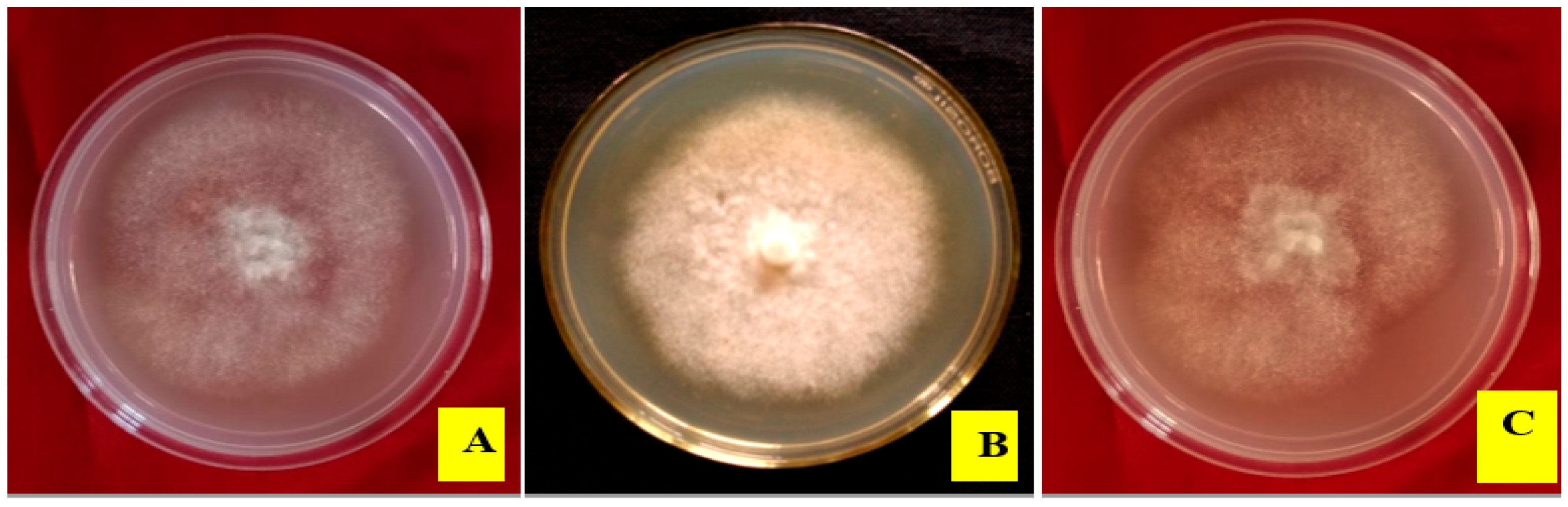
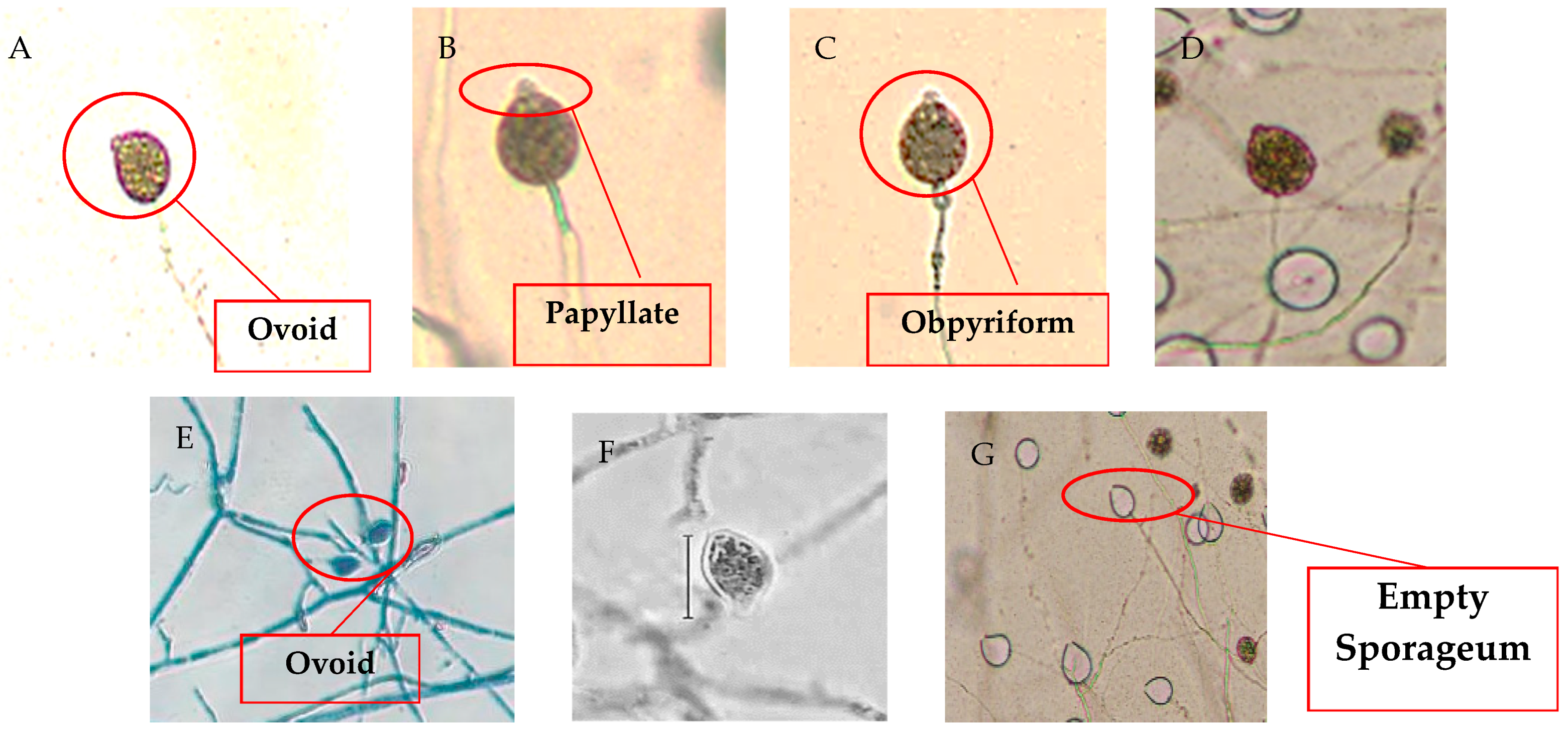
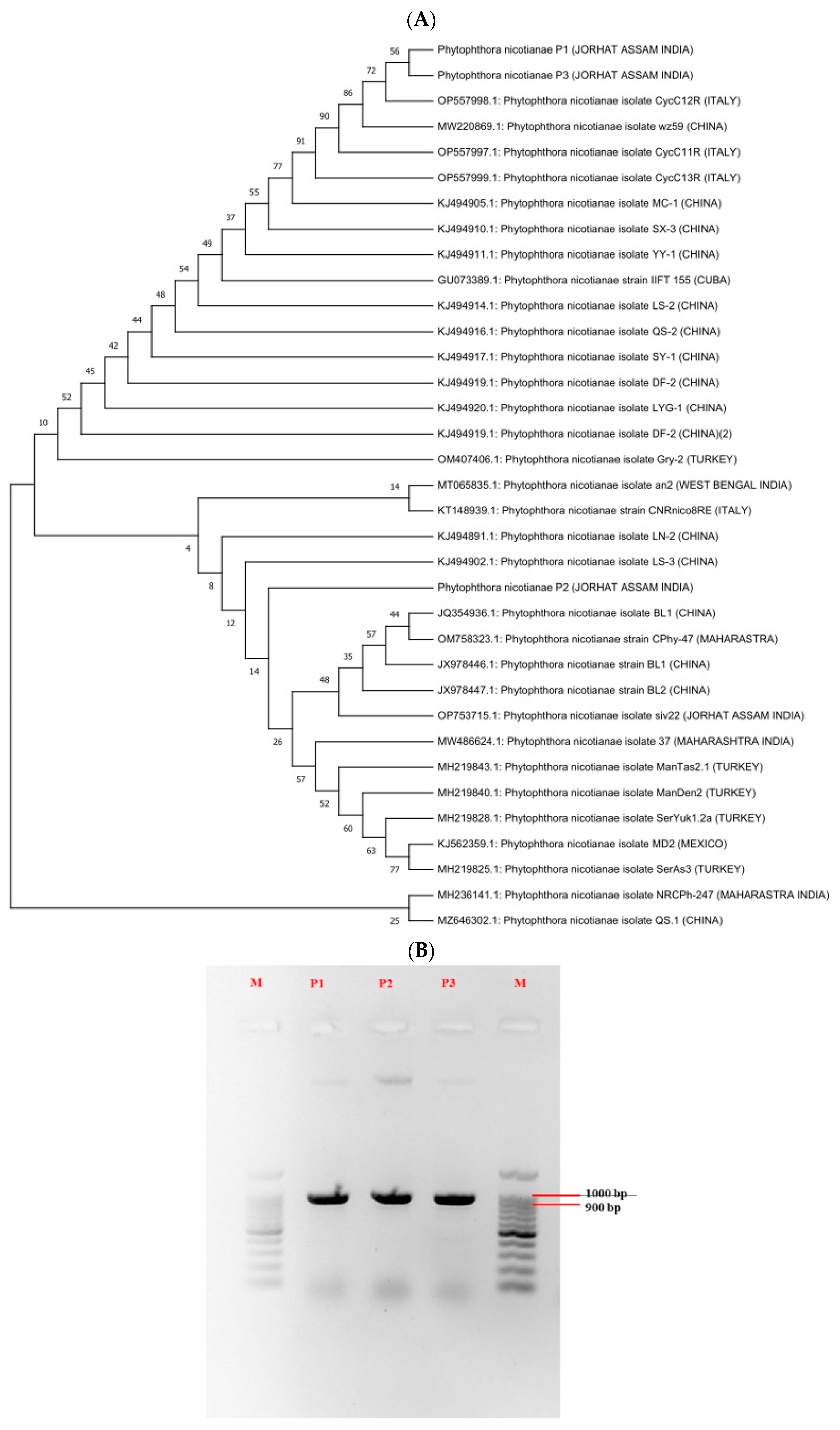
3.2. Morpho-Cultural Characteristics and Molecular Identification of Pathogen

One isolate associated with Phytophthora gummosis was recovered and purified in Corn Meal Agar-PARPH media. The pure cultures showed distinctive fluffy to cottony growth patterns after being subjected to 28 °C incubation on Potato Dextrose Agar media (Figure 3 and Figure 4). Careful observations made under a ZESIS Axiolab 5 microscope revealed distinctive ovoid-to-obpyriform-shaped sporangia (L × B: 44.8–45.3 × 26.6–27.2 microns) that were notably papillate, were occasionally spherical, and often had a distorted shape (Table 1). The morpho-cultural characteristics indicated that the pathogen was Phytophthora spp.
Three PCR products (Takara PCR Master mix) obtained after PCR amplification, labelled as P1, P2, P3, showed a band size of approx. 943 bp aligned with a 100 bp ladder (Takara). The PCR products were sequenced by Eurofins Ltd. (Bothell, WA, USA), and the sequences were used to construct a phylogenetic tree, while 33 closely related species showed the nearest similarity with the Phytophthora nicotianae strains of West Bengal (P2) and Italy (P1 and P3) (Figure 5).

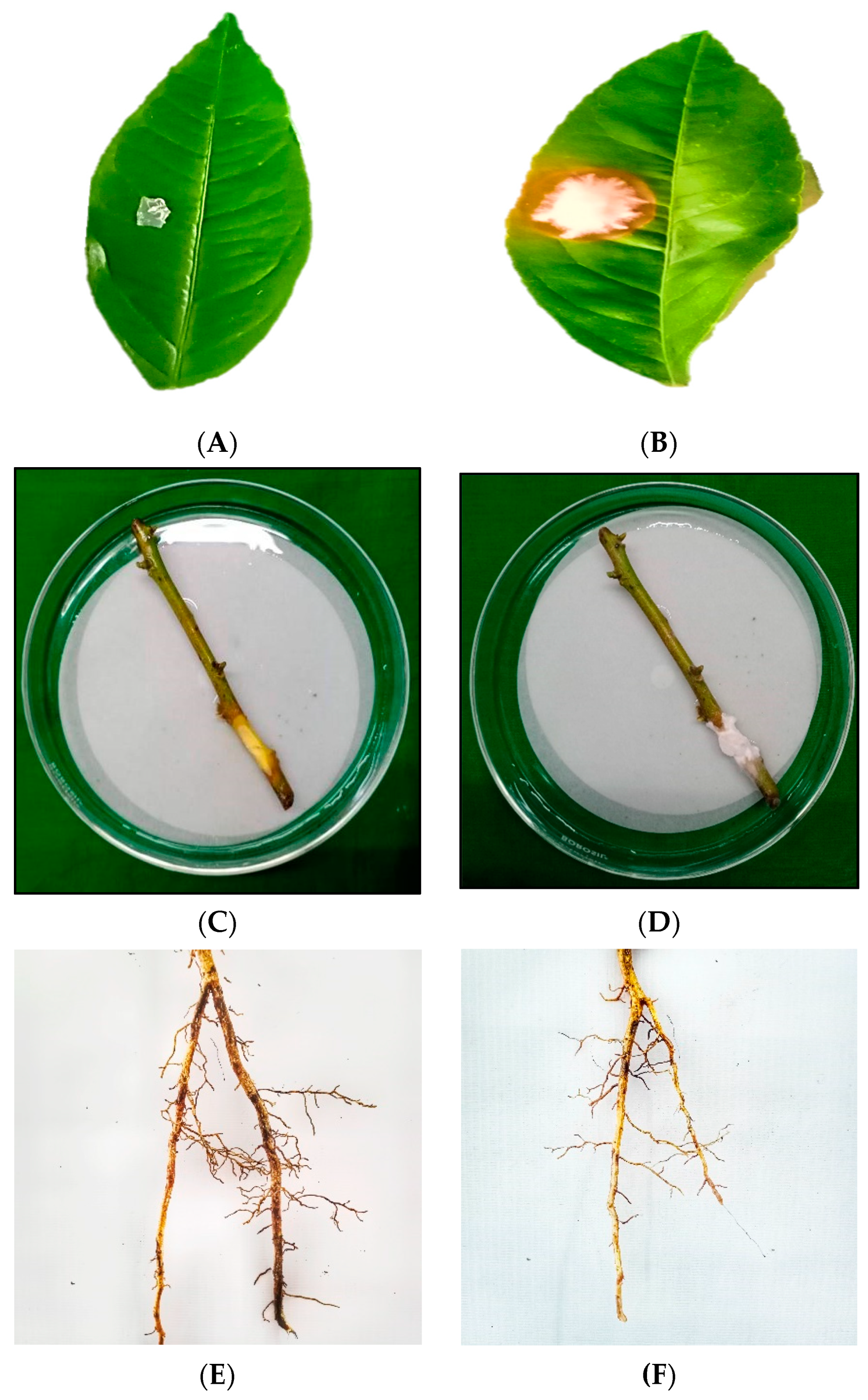
3.3. In Vitro Studies on Pathogenicity on Different Plant Parts

The identified isolate, P. nicotianae, was tested for its pathogenicity using different plant parts: the leaf, excised shoot, and root (Figure 6). Though the symptoms varied, all three plant parts were observed to be affected with the pathogen, displaying its aggressive behavioural polymorphism with regard to the host–pathogen interaction.

3.4. Identification of Microbes from Diseased and Healthy Rhizospheres

Colonies with varied morphology and a dominant growth pattern were isolated from the microbial-rich rhizosphere of Khasi mandarin. In total, 18 fungal isolates and 10 bacterial isolates were isolated from the diseased plant’s rhizosphere, showing typical symptoms of Phytophthora infection. Similarly, from the healthy rhizosphere, 16 isolates of fungi and 28 isolates of bacteria with distinct morphology were isolated for further characterization.

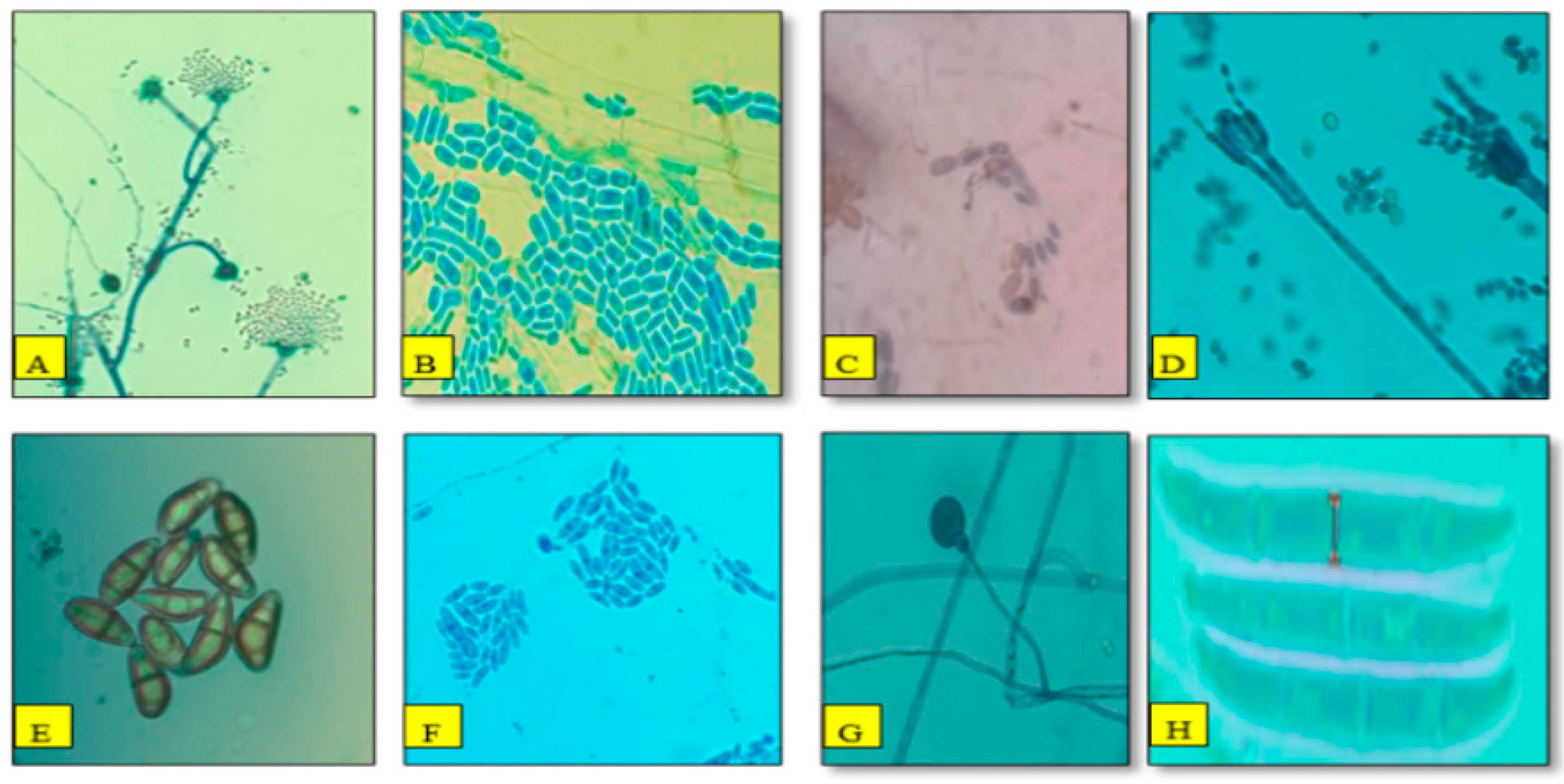
3.5. Morphological Characterization and Relative Abundance of Isolated Fungal Species

The results of the determination of the morphological and reproductive characteristics of the fungal species plus a microscopic study, besides the observations of [22,23,24,25], collectively showed that 16 isolates from the healthy rhizosphere soil belonged to eight different fungal genera, including Fusarium, Penicillium, Aspergillus, Acremonium, Mucor, Geotrichum, Culvularia, and Trichoderma (Figure 7). In contrast, the as many 18 fungal isolates from the diseased rhizosphere were represented by seven different fungal genera: Penicillium, Aspergillus, Geotrichum, Culvularia, Fusarium, and Mucor (Figure 8).
Comparing the relative abundance of different culture-dependent fungal isolates between the health and diseased rhizosphere (Table 2) showed a complete absence of Trichoderma spp. on one hand and the increasing dominance of Aspergillus spp. and Fusarium spp. in the diseased rhizosphere on the other; however, a distinct reduction in the abundance of Penicillium in the healthy rhizosphere was observed compared with that in the diseased rhizosphere. These observations lend strong support in favour of structural and compositional (as well as possibly functional) changes in the rhizospheres, depending upon their status, i.e., healthy or disease-infected.

3.6. Diversity of Rhizospheric Bacterial and Fungal Communities

Soil samples were further evaluated for CFUs (colony forming units) per gram and showed that the CFU counts of microbes varied significantly between healthy and diseased rhizosphere soil. The average CFU values for the bacterial population ranged from (2.3–4.6 × 10⁶) to (4.8–6.1 × 10⁶) in healthy and diseased rhizospheric soil, respectively, while the fungal counts ranged between 2.9–4.1 × 10⁴ and 3.6–4.8 × 10⁴ in healthy and diseased rhizosphere soil, respectively (Table 3).
The microbial diversity in the rhizosphere soils, determined using the Shannon–Weiner index, showed comparatively higher diversity values in the disease-infected rhizosphere, ranging from 1.06 to 1.49 against values of 0.97 to 1.18 in the healthy rhizosphere. Additionally, for the mean species healthy soil samples, these values varied between 0.53 and 0.71. These showed that the diversity index in the diseased soil was far higher compared with that in the healthy soil (Table 4).

3.7. Metagenomics Analysis

From the metagenomic sequencing, raw reads of 6.15 Gbp (DAM_H) and 5.82 Gbp (DAM_D) were obtained from healthy and infected soil after quality control, and totals of 6.01 Gbp (DAM_H) and 5.31 Gbp (DAM_D) were retained with an average of 16.193 Gbp for each sample. The clean reads were assembled into 14,645 contigs (>1000 bp) and 2137 contigs (>1000 bp) for the healthy and infected rizosphere, respectively. The detailed sequencing data and metagenome assembly statistics in each sample are listed (Table 5).

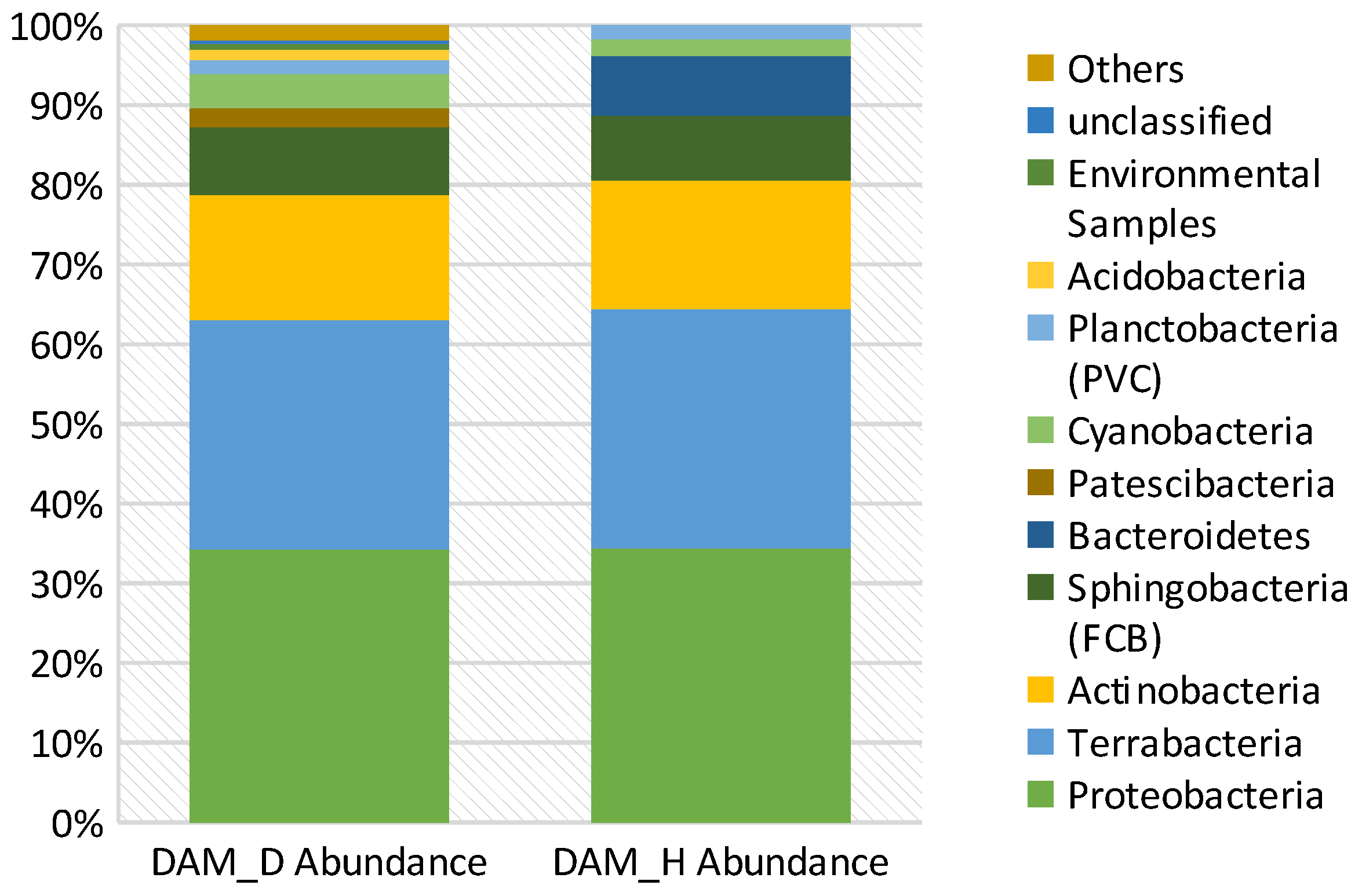
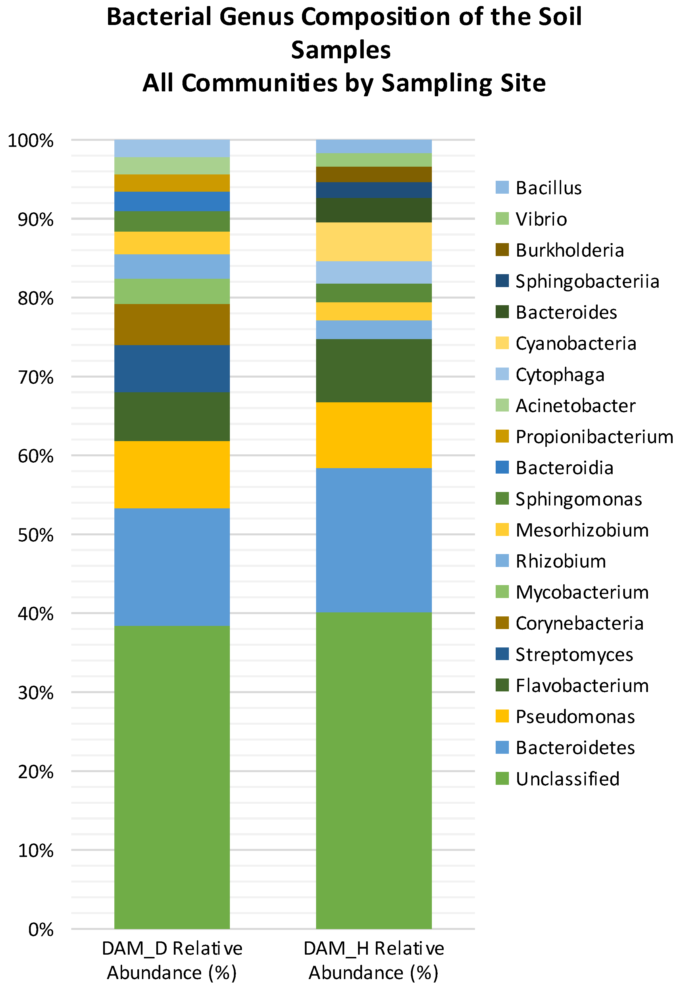
3.8. Diversity and Abundance of Bacterial Phyla in Diseased vs. Healthy Rhizospheres

Metagenomics-related abundance analysis showed the occurrence of nine prominent phyla, including Proteobacteria, Terrabacteria, Actinobacteria, Sphingobacteria, Flavobacteria, Patescibacteria, Cyanobacteria, Planctobacteria, and Acidobacteria, regardless of the nature of the rhizosphere, whether derived from healthy or disease-infected trees, although the proportionate distribution of these phyla differed between the two types of rhizosphere samples (Figure 9).
The diseased rhizosphere soil (DAM_D) had a higher number of bacterial communities represented by seven phyla, while the healthy rhizosphere soil samples (DAM_H) showed a far lower number of bacterial phyla comparatively, marked by the absence of Flavobacteria, Patescibacteria, PVC, and Acidobacteria, providing some strong clues with which to exploit upon such biochemical changes towards microbially sustainable management options. Interestingly, the comparative study of DAM_D (with an abundance range of 0.409–33.118% and a mean value of 8.334% with a standard deviation of 11.196%) versus DAM_H (with an abundance range of 0.000–33.06% and a mean value of 14.076 with a standard deviation of 14.076%) further revealed a marginal difference when parameters such as abundance range and its mean value were coupled with the standard deviation. These observations are indicative of DAM_H displaying a comparatively higher mean abundance variability of bacterial phyla.
At the genus level, the soils of Khaman Pathar had higher occurrences of Bacteroidetes and Pseudomonas. Other notable genera present in the diseased soils included Streptomyces, Corynebacteria, Candidatus, Patescibacteria, Mycobacterium, Rhizobium, Mesorhizobium, Sphingomonas, Bacteroidia, Propionibacteria, Acinetobacter, Cytophagia, Cyanobacteria, Bacteroidia, and Sphingobacter (Figure 10).
The minimum abundance value for DAM_D is 0.000%, and the maximum abundance value is 14.467%, with a mean of 2.145% and a standard deviation of 3.185%. For DAM_H, the minimum value is 0.000%, and the maximum value is 22.276%, with a mean of 2.595% and a standard deviation of 4.937%. The relative abundance in diseased soil (DAM_D) is lower than the relative abundance of other microorganisms in healthy soil (DAM_H). (Figure 11).

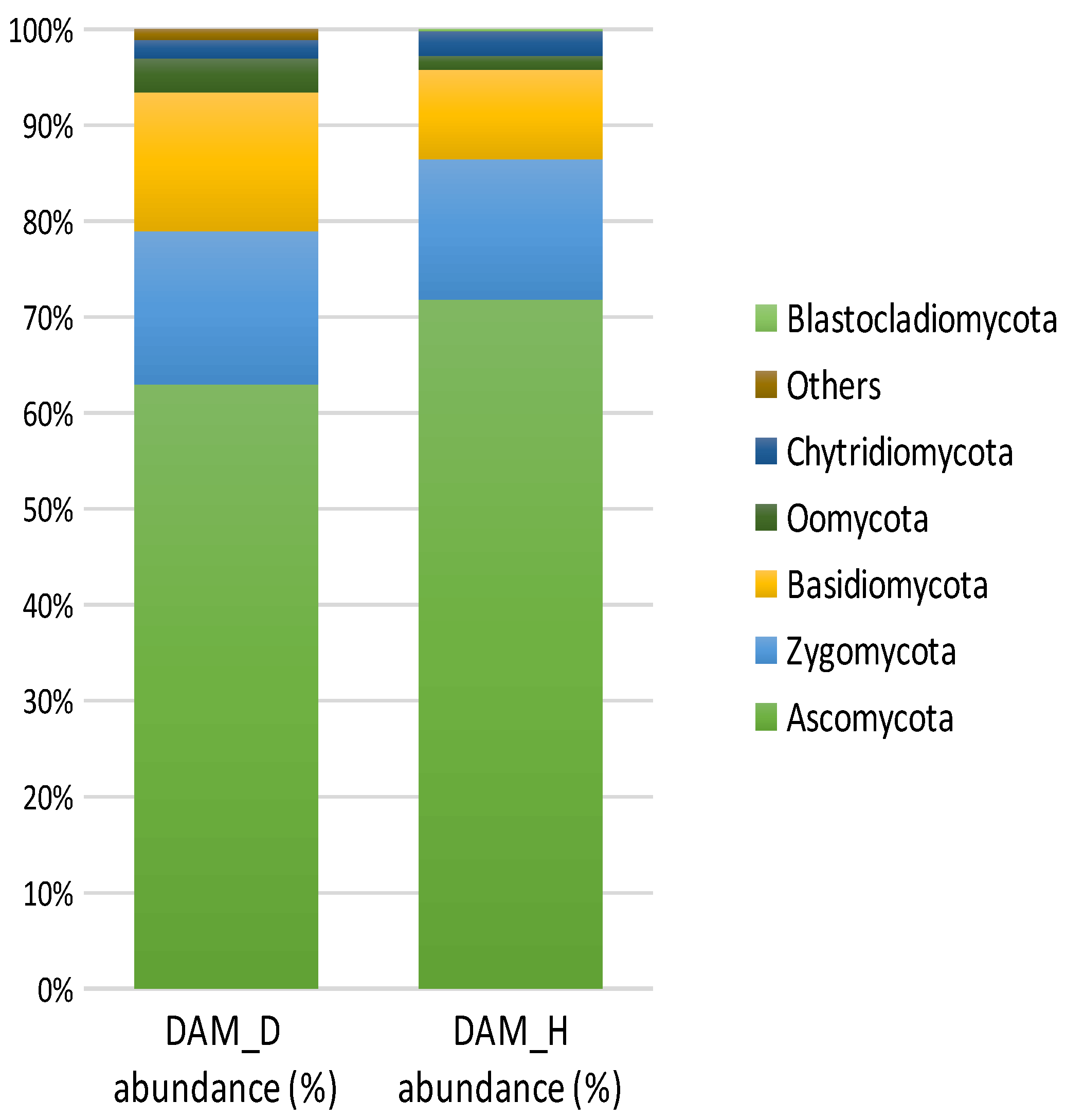
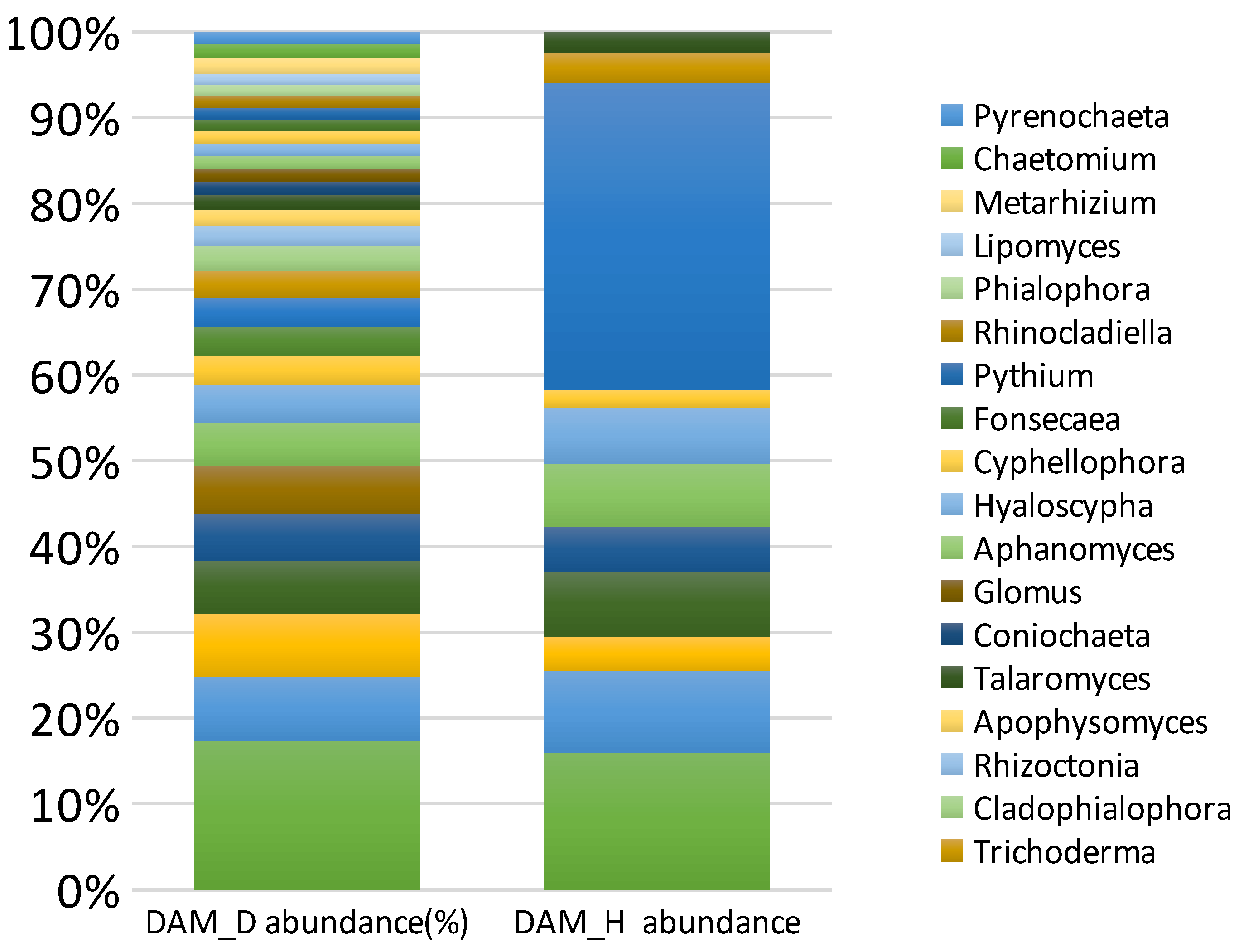
3.9. Diversity and Abundance of Fungi in Soil Samples

Metagenomic analysis of the data based on the relative abundance at the phylum level revealed that Ascomycota, Zygomycota, and Basidiomycota were the dominant phyla (Figure 12).
Over 60% of fungal communities in general belonged to the Ascomycota phylum in all soil samples. The mean value for DAM_D is 8.334, and the standard deviation is 11.196. It is quite noticeable that the mean value for DAM_H is higher than that of DAM_D, with a value of 9.512, and that the former had a higher standard deviation of 14.076, showing a dominant pattern. (Figure 10). Similar with that of the phylum, genus-specific diversity further showed the higher abundance of the Phytophthora genus in the diseased rhizosphere (DAM_D) compared with that in the healthy rhizosphere (DAM_H). The DAM_D samples showed a 0.86% abundance of Phytophthora, while DAM_H samples showed an abundance of 0.30%, thereby showing a net reduction in the presence of the pathogen of 0.56% (Figure 10).
Several other genera showing a higher relative abundance of more than 1% include Fusarium, Aspergillus, Saitozyma, Rhizopus, Exophiala, Rhizophagus, Linnemannia, Mortierella, Colletotrichum, Purpureocillium, Penicillium, Trichoderma, Cladophialophora, Rhizoctonia, Apophysomyces, and Talaromyces, (Figure 11).
It is noteworthy that the metagenome of the diseased soil samples had a much higher content of Fusarium (13.38%) compared with that of the healthy soil samples. The healthy soil sample from Khaman Pathar also had a particularly high abundance of Penicillium (21.02%). The mean for DAM_D is 0.027, and the standard deviation is 0.025. The mean for DAM_H is higher than that of DAM_D, with a value of 0.053, and the former also has a higher standard deviation of 0.034.

4. Discussion

Khasi mandarin is an important fruit crop grown in tropical and subtropical parts of Arunachal Pradesh, Meghalaya, and Assam, India, but it is often challenged by fungal pathogens such as Phytophthora spp., the major cause of gummosis and root rot in growing plantations [19]. Due to the polycyclic nature of the pathogen and the perennial nature of the crop, chemical management strategies could not determine the root cause of the disease [7,8]. However, it was found that some soils of the rhizosphere showed suppressive effect on Phytophthora spp., suggesting that differences in the microbial community structure of healthy vs. pathogen-rich soils may play a pivotal role in finding the microbial source of disease development as well as disease suppressiveness [29]; it may unlock potential strategies for controlling Phytophthora diseases in the crop using beneficial microbes with antagonistic traits.
The major symptoms of Phytophthora spp.-induced disease usually manifest as the oozing of gums followed by the gradual blackening of the trunk [30]. Among the nine surveyed orchards, Khaman Pathar and Motapung (Tinsukia, Assam, India), and Kamalabari (Sivasagar, Assam, India) showed typical symptoms like those mentioned before in these three locations, where we were able to isolate three possible Phytophthora spp., giving them the codes P1, P2, and P3. These kinds of characteristic symptoms of Phytophthora infection were recorded independently in different time periods by Mekonen et al. [29], Das et al. [30], and El-Guilli et al. [31] for sweet orange, Kinnow mandarin, and sour orange, respectively [29].
Morpho-cultural characteristics of these three isolates, P1, P2 and P3, showed a topography of a flat to fluffy colony with a smooth margin, without any pigmentation. These cultural features were also reported earlier by the authors of [32,33], who identified that these characteristics of Phytophthora spp. Were associated with Khasi mandarin as well. Microscopic features revealed that sporangia that were papillate, ovoid, elongated, occasionally spherical, and often distorted in shape, were detected in all the isolates, as reported through earlier studies as well [30,31].
These morphological characteristics were earlier associated with Phytophthora nicotianae [34,35]. Phytophthora causing gummosis, and root and foot rot diseases were recorded in citrus orchards of Maharashtra and Punjab due to P. nicotianae. [19]. The genomic DNA from three Phytophthora isolates, P1, P2, and P3, were amplified with ITS1 and ITS4 primers. Agarose gel amplification showed amplification products measuring ~943 bp in size using a universal primer (Figure 2). After amplification, PCR products were outsourced for sequencing, and the results were checked against the GenBank reference sequence database at the National Centre for Biotechnology Information (NCBI, www.ncbi.nlm.nih.gov, accessed on 15 December 2023). Phylogenetic analysis revealed that all the isolates displayed the closest similarity with Phytophthora nicotianae. (Figure 3, Figure 4 and Figure 5).
Pathogenicity tests showed isolated Phytophthora spp. causing symptoms of brown water-soaked discoloration and white-to-whitish mycelial growth on detached leaves, excised stems, and feeder roots (Figure 6), depending upon the plant parts used. The appearance of these symptoms over a period of 2–7 days following inoculation indicated the ability of the identified pathogen to produce disease symptoms in Khasi mandarin plants. The control leaves showed no discoloration or mycelial growth after inoculation, confirming the specificity of the symptoms against the Phytophthor nicotianae spp. Isolates, with the gummosis, and root and foot rot symptoms observed in infected Khasi mandarin orchards. These results agree with the observations made by Das et al. (2017) during their study [10]. Similarly, excised stems and shoots with inoculation showed brown lesions over the inoculated areas compared with the uninoculated shoots showing whitish-coloured vascular bundles. These symptoms are in accordance with findings from the extensive work of Das et al. [19]. Das et al. [19] reported that within 7 days after treatment, Kinnow mandarin leaves developed brownish-black necrotic lesions, indicating that Phytophthora spp. is the root cause of the citrus gummosis disease. Similarly, the pathogenicity assay employing excised stems conducted by Das et al. [19]. (2017) in citrus revealed that the isolate could cause a progressive necrotic lesion in the Kinnow mandarin stem from the site of inoculation [15].
The population dynamics of microbes in diseased and healthy rhizospheres varied significantly, with the latter maintaining a marginally higher population, comprising both bacterial as well as fungal counts. However, it was not possible to establish which group of microbes modulated the pathosystem or host resistance (Table 4).
We could identify as many as ten (10) bacterial isolates and eighteen (18) fungal isolates in the rhizosphere of diseased-infected plants, while the healthy rhizosphere displayed thirteen (13) fungal isolates and twenty-eight (28) bacterial isolates with distinct morphology. However, the diseased rhizosphere showed a much higher Shannon–Wiener diversity index, indicating its higher diversity index, besides that for mean species evenness due to the presence of opportunistic microorganisms in the diseased rhizosphere in response to plant secretomes carrying root exudates. Root exudates can have a positive or negative effect on fungal community structure, depending upon the nature of substrates or signalling molecules, to support an increase in soil fungal taxa for a higher diversity index [34], while in the case of disease suppressiveness, beneficial microbes are more likely to remain abundant in the root zone of plants, suppressing the growth of harmful and other opportunistic microorganisms, thereby lowering the diversity index of healthy rhizospheric soil [35]. Interestingly, culturable microbes, when compared on the basis of species distribution between healthy and diseased rhizospheres, revealed no differentiating clues, but the pattern of distribution of fungal species led us to identify the source of suppressiveness against Phytophthora nicotianae.
The suppressive ability of different microbial strains is facilitated through a variety of mechanisms of action, including direct antagonistic behaviour, competition for resources, and indirect antagonistic behaviour through the induction of plant resistance [36,37]. However, to fully understand the citrus plant–microbiome interaction and its resultant susceptibility/suppressiveness, a detailed study on the community composition and nature of root exudates is crucial. Some important clues for exploiting the citrus microbiome for disease resistance against soil-borne pathogens can be found in reviews by Srivastava et al. [6] and Zhang et al. [38].
The metagenomic analysis results of the healthy and diseased plants are in sync with the cultural microbiome data establishing the lower intensity of Phytophthora spp. in the rhizosphere of disease-free plants compared with that in the infected Khasi mandarin rhizosphere. This established the presence of Phytophthora spp., leading to the disease. This study also identified some of the microbes and fungal populations varying in accordance with the health status of the plant, further supporting the hypothesis that community composition will affect or becomes affected by the disease in the same field and cultivar. Further analysis such as that on differences in the enzymes activated and the metabolic network will give a better picture of how bacterial community plays a role in disease suppression. With the current data size of each sample (5 GB), despite them revealing the dominating microbes, arriving to any conclusions will be difficult. Taking clues from here, further in-depth metagenomic sequencing will be conducive to gaining insights into either the microbe–microbe interaction or the host–microbe interaction at the molecular and metabolic levels for better elucidations.
Our analysis found that Ascomycota and Basidiomycota were the most prevalent fungal phyla in all the soil samples, but the quantity of Ascomycota was dramatically reduced in diseased soils. The genera Fusarium, Aspergillus, Saitozyma, Rhizopus, Exophiala, Rhizophagus, Collietotrichum, and Purpureocillium were found to have the highest relative abundance in Phytophthora-infected soil, while Trichoderma, Penicillium, Linnemannia, Mortierella, Talaromyces, Globisporangium, Saitozyma, and Serendipita were most abundant in the disease-free rhizosphere, suggesting their putative role in the natural suppression of Phytophthora spp. These studies will strengthen our understanding of the already available community structure associated with Phytophthora [39,40].
Although increasing bacterial diversity is a better predictor of disease suppression than is the presence of a specific bacterial taxon, our study found that the most abundant phyla, such as Proteobacteria, Terrabacteria, Actinobacteria, FCB, Flavobacteria, Patescibacteria, Cyanobacteria, PVC, and Acidobacteria, were present in both types of soil samples. Beneficial effects of the plant microbiota are frequently described as being provided by synergistic interactions between microbes, and it is important to identify and understand the microbiome that has a synergistic effect to explore the maximum expression of crop resilience to disease [41]. However, it is difficult to determine which functional groups contribute to suppressiveness.

5. Conclusions

Our study provided important insights into the changes in the rhizosphere microbiome of Khasi mandarin infected with Phytophthora nicotianae, the causative agent of foot and root rot disease, as established via pathogenicity tests. With the comparatively low depth of our metagenomic data, most of the results are in accordance with some of the metagenomic revelations reported earlier. Regardless of the health status of a rhizosphere (though higher culturable microbial counts prevailed in the healthy rhizosphere compared with the disease-infected rhizosphere), unknown pathogenic species currently referred to as Candidatus predominantly dictated the rhizosphere functions, but a higher abundance of Bacteriodetes and Pseudomonas in the healthy rhizosphere provided strong clues with which to identify the microbial genes, the source of disease resistance. Likewise, identifying functional fungal microbial communities as a source of disease suppression could potentially pave the way for developing newer methods for the consortium-mediated management of Phytophthora-induced diseases in citrus crops, supported by robust screening efforts with focus on the endophytic behaviour of identified microbes. An in-depth metagenomic study of healthy versus disease-infected rhizospheres is further suggested to derive better results with regard to developing microbial antagonists as aids in citrus health management.

Author Contributions

Conceptualization, P.B.; Data curation, M.H.; Writing—original draft, M.H. and P.B.; Writing—review & editing, V.Z., A.K.S., P.T.K.J. and A.K.D.; Supervision, P.B. and A.K.S.; Project administration, P.B. All authors have read and agreed to the published version of the manuscript.

Funding

This research received no external funding.

Data Availability Statement

All the raw data submitted with NCBI are available using the submission ID, SUB14212024.

Acknowledgments

The authors thank the Department of Biotechnology (DBT), Ministry of Science and Technology, Government of India, for funding this research work. This research is funded by DBT project “Exploring rhizosphere microbiome for management of Phytophthora disease of khasi mandarin [Sanction No: BT/PR40089/NER/95/1663/2020; Dated: 08/03/2021 ]” carried out jointly with the Department of Plant Pathology, Assam Agricultural University, Jorhat, Assam, India, and the ICAR-Central Citrus Research Institute in Nagpur, Maharashtra, India.

Conflicts of Interest

The authors declare no conflicts of interest.

References

  1. Lynch, J.M.; Whipps, J.M. Substrate flow in the rhizosphere. Plant Soil 1990, 129, 1–10. [Google Scholar] [CrossRef]
  2. Hiltner, L. Über neuere Erfahrungen und Probleme auf dem Gebiet der Bodenbakteriologie und unter besonderer Berücksichtigung der Gründüngung und Brache. Arb. Dtsch. Landwirtsch. Ges. 1904, 98, 59. [Google Scholar]
  3. Bulgarelli, D.; Rott, M.; Schlaeppi, K.; Ver Loren van Themaat, E.; Ahmadinejad, N.; Assenza, F.; Rauf, P.; Huettel, B. Revealing structure and assembly cues for Arabidopsis root-inhabiting bacterial microbiota. Nature 2012, 488, 91–95. [Google Scholar] [CrossRef] [PubMed]
  4. Mendes, R.; Garbeva, P.; Raaijmakers, J.M. The rhizosphere microbiome: Significance of plant beneficial, plant pathogenic, and human pathogenic microorganisms. FEMS Microbiol. Rev. 2013, 337, 634–663. [Google Scholar] [CrossRef]
  5. Ginnan, N.A.; Dang, T.; Bodaghi, S.; Ruegger, P.M.; McCollum, G.; England, G.; Vidalakis, G.; Borneman, J.; Rolshausen, P.E.; Roper, M.C. Disease-induced microbial shifts in citrus indicate microbiome -derived response to huanglongbing across disease severity spectrum. Phytobiomes J. 2020, 4, 357–387. [Google Scholar] [CrossRef]
  6. Srivastava, A.K.; Das, A.K.; Jagannadham, P.T.K.; Bora, P.; Ansari, F.A.; Bhate, R. Bioprospecting microbiome for soil and plant health management amidst huanglongbing threat in citrus: A review. Front. Plant Sci. 2022, 13, 858842. [Google Scholar] [CrossRef]
  7. Yuan, J.; Zhao, J.; Wen, T.; Zhao, M.; Li, R.; Goossens, P.; Huang, Q.; Bai, Y.; Vivanco, J.M.; Kowalchuk, G.A.; et al. Root exudates drive the soil-borne legacy of aboveground pathogen infection. Microbiome 2018, 6, 156. [Google Scholar] [CrossRef]
  8. Berg, G.; Köberl, M.; Rybakova, D.; Müller, H.; Grosch, R.; Smalla, K. Plant microbial diversity is suggested as the key to future biocontrol and health trends. FEMS Microbiol. Ecol. 2017, 93, fix50. [Google Scholar] [CrossRef]
  9. Ploetz, R.C. (Ed.) Diseases of Tropical Fruit Crops; CABI Publishing: Wallingford, UK, 2003. [Google Scholar]
  10. Van Tran, Q.; Ha, C.V.; Vvedensky, V.V.; Han, V.C. Current status and characterization of Phytophthora-associated with gummosis of citrus in northern Vietnam. J. Phytopathol. 2023, 171, 478–488. [Google Scholar] [CrossRef]
  11. Lai, Z.; Vinod, K.M.; Zheng, Z.; Fan, B.; Chen, Z. Roles of Arabidopsis WRKY3 and WRKY4 transcription factors in plant responses to pathogens. BMC Plant Biol. 2008, 8, 68. [Google Scholar] [CrossRef] [PubMed]
  12. Yang, C.; Ancona, V. Metagenomic analysis reveals reduced beneficial microorganism associations in roots of foot-rot-affected citrus trees. Phytobiomes J. 2021, 5, 305–315. [Google Scholar] [CrossRef]
  13. Srivastava, A.K.; Singh, S. Citrus nutrition research in India; problems and prospects. Indian J. Agric. Sci. 2008, 78, 3–16. [Google Scholar]
  14. Singh, A.K.; Meetei, N.T.; Singh, B.K.; Mandal, N. Khasi mandarin: Its importance, problems and prospects of cultivation in North-eastern Himalayan region. Int. J. Agric. Environ. Biotechnol. 2016, 9, 573–592. [Google Scholar] [CrossRef]
  15. Kakoti, R.K.; Nath, P.D.; Borah, M.; Bora, S. Incidence of citrus greening disease in some major khasi mandarin growing areas in upper Brahmaputra valley zone of Assam. J. Plant Dis. Sci. 2022, 17, 93–100. [Google Scholar] [CrossRef]
  16. Balasubramanian, P.; Kumar, P.G. Insect pests of Khasi Mandarin in east and west Khasi hills district of Meghalaya. Indian J. Entomol. 2022, 85, 238–240. [Google Scholar] [CrossRef]
  17. Deka, S.; Sehgal, M.; Kakoti, R.K.; Barbora, A.C. Module analysis for insect pest management of khasi mandarin (Citrus reticulata Blanco) under climatic conditions of north-eastern India. J. Entomol. Zool. Stud. 2018, 6, 857–861. [Google Scholar]
  18. Prabha, T.R.; Revathi, K.; Vinod, M.S.; Shanthakumar, S.P.; Bernard, P. Shanthakumar, and Paul Bernard. A simple method for total genomic DNA extraction from water moulds. Curr. Sci. 2013, 104, 345–347. [Google Scholar]
  19. Das, A.K.; Nerkar, S.; Thakre, N.; Kumar, A. First report of an atypical strain of Phytophthora inundata causing Kinnow mandarin decline in India. Can. J. Plant Pathol. 2017, 39, 365–372. [Google Scholar] [CrossRef]
  20. Rocha, J.D.R.D.S.; Oliveira, N.T.D.; Menezes, M. Comparison of inoculation methods efficiency for evaluation of Colletotrichum gloeosporioides isolates pathogenicity on passion fruits (Passiflora Edulis). Braz. Arch. Biol. Technol. 1998, 41, 140–148. [Google Scholar] [CrossRef]
  21. Chowdhry, P.N.; Varshney, A. Identification of different Colletotrichum goleosporides species. In Manual on Identification of Plant Pathogenic and Biocontrol Fungi of Agricultural Importance; IARI: New Delhi, India, 2000; pp. 73–78. [Google Scholar]
  22. Subramanian, C.V. Hyphomycetes; an Account of Indian Species Except Cercosporae; Indian Council of Agricultural Research: New Delhi, India, 1971. [Google Scholar]
  23. Barnett, H.L.; Hunter, B.B. Illustrated genera of imperfect fungi. In Illustrated Genera of Imperfect Fungi, 3rd ed.; Burgess Publishing Company: Minneapolis, MN, USA, 1972. [Google Scholar]
  24. Ellis, M.B. Dematiaceous Hyphomycetes; Commonwealth Mycological Institute: Kew, UK, 1971. [Google Scholar]
  25. Domsch, K.H.; Gams, W.; Anderson, T.H. Compendium of Soil Fungi. Volume 1; Academic Press (London) Ltd.: London, UK, 1980. [Google Scholar]
  26. Li, D.; Liu, C.M.; Luo, R.; Sadakane, K.; Lam, T.W. MEGAHIT: An ultra-fast single-node solution for large and complex metagenomics assembly via succinct de Bruijn graph. Bioinformatics 2015, 31, 1674–1676. [Google Scholar] [CrossRef]
  27. Menzel, P.; Ng, K.L.; Krogh, A. Fast and sensitive taxonomic classification for metagenomics with Kaiju. Nat. Commun. 2016, 7, 11257. [Google Scholar] [CrossRef] [PubMed]
  28. Shannon, C.E. A mathematical theory of communication. Bell Syst. Tech. J. 1948, 27, 379–423. [Google Scholar] [CrossRef]
  29. Mekonen, M.; Ayalew, A.; Weldetsadik, K.; Seid, A. Assessing and measuring of Citrus gummosis (Phytophthora spp.) in major citrus growing areas of Ethiopia. J. Hortic. 2015, 2, 1–4. [Google Scholar] [CrossRef]
  30. Das, A.K.; Kumar, A.; Ingle, A.; Nerkar, S. Molecular identification of Phytophthora spp. causing citrus decline in Vidarbha region of Maharashtra. Indian Phytopathol. 2011, 64, 342. [Google Scholar]
  31. El-Guilli, M.; Benyahia, H.; Jrifi, A.; Besri, M. Effect of irrigation water salinity on trunk gummosis symptom severity for citrus affected with Phytophthora citrophthora. Fruits 2000, 55, 181–186. [Google Scholar]
  32. Hall, G. An integrated approach to the analysis of variation in Phytophthora nicotianae and a redescription of the species. Mycol. Res. 1993, 97, 559–574. [Google Scholar] [CrossRef]
  33. Waterhouse, G.M. Phytophthora palmivora and some related species. In Phytophthora Disease of Cocoa; Gregory, P.H., Ed.; Longman: London, UK, 1974. [Google Scholar]
  34. Thomson, S.V.; Hine, R.B. Atypical sporangium-like structures of Phytophthora parasitica. Mycologia 1972, 64, 457–460. [Google Scholar] [CrossRef]
  35. Ho, H.H.; Ann, P.J.; Chang, H.S. The Genus Phytophthora in Taiwan; Institute of Botany, Academia Sinica: Nangang, Taipei, 1995. [Google Scholar]
  36. Bora, P.; Bora, L.C. Disease management in horticultural crops through microbial interventions: An overview. Indian J. Agric. Sci. 2020, 90, 1384–1396. [Google Scholar]
  37. Saikia, S.; Bora, P.; Bora, L.C. Bioagent mediated management of citrus canker. Indian J. Agric. Sci. 2021, 91, 198–201. [Google Scholar] [CrossRef]
  38. Zhang, Y.; Trivedi, P.; Xu, J.; Roper, M.C.; Wang, N. The citrusmicrobiome: From structure and function to microbiome engineering and beyond. Phytobiomes J. 2021, 5, 249–261. [Google Scholar] [CrossRef]
  39. Solís-García, I.A.; Ceballos-Luna, O.; Cortazar-Murillo, E.M.; Desgarennes, D.; Garay-Serrano, E.; Patiño-Conde, V.; Méndez-Bravo, A.; Reverchon, F. Phytophthora root rot modifies the composition of the avocado rhizosphere microbiome and increases the abundance of opportunistic fungal pathogens. Front. Microbiol. 2021, 11, 3484. [Google Scholar] [CrossRef] [PubMed]
  40. Win, T.T.; Bo, B.; Malec, P.; Khan, S.; Fu, P. Newly isolated strain of Trichoderma asperellum from disease suppressive soil is a potential bio-control agent to suppress Fusarium soil borne fungal phytopathogens. J. Plant Pathol. 2021, 103, 549–561. [Google Scholar] [CrossRef]
  41. Bora, P.; Saikia, K.; Hazarkia, H.; Ragesh, G. Exploring potential of bacterial endophytes in disease management. Curr. Hortic. 2019, 7, 32–37. [Google Scholar] [CrossRef]
Figure 1. Surveyed locations. (A) Map of India; (B) map of Assam; (C) map of surveyed districts: (a) Sibsagar and (b) Tinsukia.
Agronomy 14 00661 g001
Figure 2. Symptomatology of Phytophthora-induced gummosis. (A) Infected orchards; (B) crown rot; (C) lesions on trunk; (D) exudated gums; (E) infected Fruits.
Agronomy 14 00661 g002
Figure 3. Pure cultures of the isolates of Phytophthora spp. (AC) with different growth patterns. (A) Light cottony mycelial growth; (B) dense, fluffy, and cottony mycelial growth; and (C) light cottony mycelial growth.
Agronomy 14 00661 g003
Figure 4. Microphotographs of sporangia observed under the microscope (40×). (A,B) P1, (C,D) P2, (E,F) P3, and (G) empty sporangia after releasing zoospores (P1).
Agronomy 14 00661 g004
Figure 5. (A) Phylogenetic positions of isolates. (B) Agarose gel electrophoresis showing amplified DNA products of isolated Phytophthora spp. Lane M: 100 Bp marker; Lane 1: P1; Lane 2: P2; Lane 3: P3. Amplification of its 1/4 region of a primer size of ~943 bp.
Agronomy 14 00661 g005
Figure 6. Pathogenicity of Phytophthora nicotianae on leaf inoculation ((A) as the control; (B) as the treated sample), excised shoot inoculation ((C) as the control; (D) as the treated sample), and root inoculation ((E) as the control; (F) as the treated sample). The effectiveness of P. nicotianae was marked by fungal growth on the inoculated leaf after 7 days. (B) Overlapping mycelial growth on excised shoot after 15 days. (D) Loss of feeder roots after 72 h of pathogen inoculation, (F) marked by the appearance of blackish-brown necrotic lesions over the roots, while the untreated control (E) remained symptomless.
Agronomy 14 00661 g006
Figure 7. Microphotographs (40×) of rhizospheric fungi isolated from healthy rhizosphere. (A) Aspergillus spp., (B) Penicillium spp., (C) Acremonium spp., (D) Mucor spp., (E) Fusarium spp., (F) Geotrichum spp., (G) Culvularia spp., and (H) Trichoderma spp.
Agronomy 14 00661 g007
Figure 8. Microphotographs (40×) of rhizospheric fungi isolated from diseased rhizosphere. (A) Aspergillus spp., (B) Geotrichum spp., (C,D) Penicillium spp., (E) Culvularia spp, (F) Acremonium spp., (G) Mucor spp., and (H) Fusarium spp.
Agronomy 14 00661 g008
Figure 9. Relative abundance of different phylum-based bacterial communities in healthy (DAM_H) versus diseased (DAM_D) rhizosphere soil samples.
Agronomy 14 00661 g009
Figure 10. Relative abundance of bacterial communities arranged by genus in the healthy (DAM_H) versus diseased (DAM_D) rhizosphere.
Agronomy 14 00661 g010
Figure 11. Relative abundance of fungal communities arranged by phylum in the healthy (DAM_H) versus diseased (DAM_D) rhizosphere.
Agronomy 14 00661 g011
Figure 12. Relative abundance of fungal communities arranged by genus in the healthy (DAM_H) versus diseased (DAM_D) rhizosphere.
Agronomy 14 00661 g012
Table 1. Morphological characteristics of Phytophthora isolates.
Morpho-Cultural Characteristics
PigmentationShapeMarginTopographyColony ColourPapillate/Non-PapillateShape (Sporangia)Mean L: B
Ratio
Size (Sporangia B × L) (µm)LocationIsolate
NoneIrregularSmoothFlat to FluffyWhitePapillateOvoid1.6726.6–27.2 × 44.8–45.3Khaman
Pathar
P1
NoneIrregularSmoothFlatWhitePapillateObpyriform1.5229.7–30.5 × 42.8–49.1MotapungP2
NoneIrregularSmoothFluffyWhitePapillateOvoid1.9924.3–25.7 × 49.5–50.3KamalabariP3
Table 2. Percentage relative abundance of isolated rhizospheric fungi in Phytophthora-infected vs. healthy Khasi mandarin rhizospheres.
Relative Abundance (%)Identified Species
Diseased RhizosphereHealthy Rhizosphere
KamalabariMotapungKhaman PatharKamalabariMotapungKhaman Pathar
52.9842.1542.4112.8628.0336.67Fusarium spp.
19.2512.3814.4123.3472.6121.03Penicillium spp.
17.8618.7612.2419.354.817.98Aspergillus spp.
3.373.978.354.2332.068.56Acremonium spp.
4.766.3213.676.2325.616.64Mucor spp.
1.399.716.564.8310.813.04Geotrichum spp.
0.406.712.379.2220.422.59Culvularia spp.
0.010.000.0019.9325.6513.50Trichoderma spp.
Table 3. Population enumeration of culturable rhizospheric microbes in Phytophthora-infected and healthy Khasi mandarin rhizospheres.
Fungal Population
(×103 CFU per Gram Moist Soil)
Bacterial Population
(×103 CFU per Gram Moist Soil)
Location
HealthyInfectedHealthy Diseased
4.6 × 10⁻⁴ ± 0.3674.1 × 10−4 ± 0.6666.1 × 10−6 ± 0.1002.3 × 10−6 ± 0.120 *Motapung
4.8 × 10⁻⁴ ± 0.1674.0 × 10−4 ± 0.5335.2 × 10−6 ± 0.4164.2 × 10−6 ± 0.967Khaman Pathar
3.6 × 10⁻⁴ ± 0.4932.9 × 10−4 ± 0.0334.8 × 10−6 ± 0.2404.6 × 10−6 ± 1.299Kamalabari
* Standard error.
Table 4. Percentage relative abundance of isolated rhizospheric fungi in Phytophthora-infected versus healthy Khasi mandarin rhizospheres.
Relative Abundance (%)Identified Species
Diseased RhizosphereHealthy Rhizosphere
KamalabariMotapungKhaman PatharKamalabariMotapungKhaman Pathar
52.9842.1542.4112.8628.0336.67Fusarium spp.
19.2512.3814.4123.3472.6121.03Penicillium spp.
17.8618.7612.2419.354.817.98Aspergillus spp.
3.373.978.354.2332.068.56Acremonium spp.
4.766.3213.676.2325.616.64Mucor spp.
1.399.716.564.8310.813.04Geotrichum spp.
0.406.712.379.2220.422.59Culvularia spp.
0.010.000.0019.9325.6513.50Trichoderma spp.
Table 5. Contig assembly statistics at a minimal length of 1000 bp/1 kbp.
MetricsDAM_HDAM_D
num_seqs14,6452137
sum_len25,212,0862,700,710
min_len10001000
avg_len1721.51263.8
max_len60,16324,564
med_len12581151
N5015731193
Disclaimer/Publisher’s Note: The statements, opinions and data contained in all publications are solely those of the individual author(s) and contributor(s) and not of MDPI and/or the editor(s). MDPI and/or the editor(s) disclaim responsibility for any injury to people or property resulting from any ideas, methods, instructions or products referred to in the content.

