Article

Overexpressing ATP Sulfurylase Improves Fe-Deficiency Tolerance in Apple Calli and Tobacco

College of Horticulture, Gansu Agricultural University, Lanzhou 730070, China
*
Author to whom correspondence should be addressed.
Agronomy 2024, 14(3), 404; https://doi.org/10.3390/agronomy14030404
Submission received: 4 January 2024 / Revised: 28 January 2024 / Accepted: 30 January 2024 / Published: 20 February 2024
(This article belongs to the Section Crop Breeding and Genetics)

Abstract

:
Iron (Fe) deficiency is one of the most common micronutrient deficiencies limiting crop production globally, especially in arid regions due to decreased availability of Fe in alkaline soils. The ATP sulfurylase (ATPS) gene has been reported to participate in regulating various abiotic stresses. Transcriptome data and qRT-PCR analysis revealed that the ATP sulfurylase gene MhATPS1 was notably induced by Fe-deficiency stress. Consequently, MhATPS1 (103410737) was isolated from Malus halliana, and transgenic tobacco and transgenic apple calli were successfully obtained by genetic transformation. Compared with the wild type (WT), transgenic MhATPS1 lines (transgenic tobacco and transgenic apple calli) displayed stronger resistance to Fe-deficiency treatment. To be specific, transgenic plants exhibited better growth, accumulated more Fe2+ content, had higher ferric chelate reductase (FCR) activity, and a greater active oxygen scavenging capacity. Furthermore, transgenic MhATPS1 lines up-regulated the expression of Fe uptake genes under Fe-deficit stress. Additionally, MhATPS1 transgenic lines secreted more H+ content compared to the WT. In summary, these findings indicate that the MhATPS1 gene may play a positive role in Fe-deficiency stress in both tobacco and apple calli.

1. Introduction

Iron (Fe), as an essential trace element for plant growth and development, is involved in a variety of biological processes, including photosynthesis, respiration, and chlorophyll biosynthesis [1]. Although the total amount of Fe on earth meets the needs of plants, most of it exists in the form of insoluble chelates, especially in saline soils, and cannot be effectively absorbed and utilized by plants [2,3]. Among them, woody fruit trees are susceptible to suffering from Fe deficiency, which triggers chlorosis, resulting in yield reduction, quality decline, and nutritional imbalance [3].
To cope with low Fe availability in the soil, plants have evolved two distinct strategies for Fe acquisition from the soil, including ‘Strategy I’ and ‘Strategy II’ [4]. ‘Strategy I’ is employed by dicotyledons and non-gramineous plants, where the activity of the plasma membrane proton pump and ferric chelate reductase (FCR) in the plasmalemma is increased, and the Fe is transported by IRT1 (Fe-regulated transporter) in the root cells [5,6]. In contrast, graminoid plants utilize ‘Strategy II’, using a chelation mechanism to acquire Fe [7].
The supply of sulfur (S) in a Fe-deficient environment enhances the utilization of Fe by plants. Previous studies conducted on wheat [8], maize [9], and tomatoes [10] also support this perspective. ATP sulfurylase (ATP: sulfate adenylyl transferase; EC 2.7.7.4), serves as the metabolic entry point into the sulfur assimilation pathway. This enzyme catalyzes the formation of adenosine-5-phosphosulfate (APS) and inorganic pyrophosphate (PPi) form sulfate and ATP [11]. Several lines of evidence have found that the ATP sulfurylase gene participates in the stress tolerance response of plants. For instance, overexpression of the ATP sulfurylase gene has been shown to increase the glutathione (GSH) content in plants, thereby enhancing the stress resistance of soybean [12], maize [13], and mustard [11]. This is achieved by maintaining the integrity of the plant cell membrane structure and defending against membrane lipid peroxidation caused by the accumulation of free radicals. Chan et al. (2013) have found that overexpressed ATP sulfurylase gene can improve the drought resistance of plants by regulating the synthesis of osmoregulatory substances, resistance signaling substances, and antioxidants in plants [14]. Furthermore, the overexpression of the ATP sulfurylase gene has been shown to positively influence plant resistance to heavy metal stress. This higher expression level can enhance the plant’s ability to accumulate selenium, which in turn can be utilized to address soil metal pollution [15].
ATP sulfurylase genes have been shown to play a significant role in abiotic stresses in plants [12,15]. However, the mechanism by which they respond to Fe deficiency in plants remains elusive. Therefore, we reveal and study their function in Fe-deficient environments. In this study, we identified an ATP sulfurylase gene, named MhATPS1, that was highly induced by Fe-deficiency stress, and selected it for further investigation. The transgenic lines exhibited increased tolerance to Fe deficit, better growth, enhanced antioxidant enzyme activity, and higher Fe content compared to the WT plants. Additionally, transgenic plants increased the expression of Fe-taking genes under Fe-deficiency stress. Overall, these data might provide valuable insights into stress tolerance mechanisms in perennial woody fruit trees and can contribute to future molecular breeding research.

2. Materials and Methods

2.1. Plant Materials and Treatments

Malus halliana seedlings were selected by culturing them for one month on Murashige and Skoog (MS) medium containing 0.5 mg/L 6-benzyl amino purine (6-BA) (Solarbio, Beijing, China) and 0.2 mg/L naphthalene acetic acid (NAA). Subsequently, they were transferred to root culture with 1/2 MS medium including 0.5 mg/L IBA (Solarbio, Beijing, China). Rooted plants were cultured as previously described by Sun [16] and then treated with different Hoagland nutrient solutions as follows: Fe-sufficient (40 µM Fe, Na-EDTA (Macklin, Shanghai, China)) and Fe-deficient (0 µM Fe, Na-EDTA).
Tobacco (N. tabacum L.) was subcultured at 30-day intervals on Murashige and Skoog (MS) medium at 25 °C with a 16 h/8 h light/dark period. ‘Wanglin’ (Malus Domestica Borkh.) apple calli were subcultured at 20-day intervals on MS medium that contained 1.0 mg/L 2,4-D (Solarbio, Beijing, China) and 0.5 mg/L 6-BA at 26 °C in the dark [17].

2.2. Gene Sequence Analysis

ATP sulfurylase sequences from other plant species were downloaded from the NCBI database (https://www.ncbi.nlm.nih.gov/, accessed on 3 January 2024). The physicochemical properties of the protein were analyzed using ExPASy online tools (https://www.expasy.org/, accessed on 3 January 2024). Their subcellular localization was predicted and analyzed with the WOLF PSORT online tools (genscript.com/wolf-psor, accessed on 3 January 2024), and conserved protein motifs were analyzed using the MEME suite online tool (https://meme-suite.org/tools/meme, accessed on 3 January 2024). Cis-acting promoter elements were predicted and analyzed using the PLACE database (http://www.dna.affrc.go.jp/htdocs/PLACE/, accessed on 3 January 2024). DAMAN 9.0 [18] and MEGA11 software (https://www.megasoftware.net/, accessed on 3 January 2024) were used for multiple sequence alignment and phylogenetic tree construction.

2.3. Quantitative Real-Time PCR

Total RNA was extracted from apple calli and tobacco whole seedlings using TRIzol reagent (Invitrogen, Carlsbad, CA, USA) following the manufacturer’s instructions, and then reverse transcript was synthesized with a PrimeScript 1 Strand cDNA Synthesis Kit (TaKaRa, Dalian, China). Real-time quantitative PCR was carried out on a Roche Light Cycler®96 Instrument PCR System (Applied Biosystems, Roche, Switzerland). The primers are shown in Supplementary Table S1. GAPDH was used as a reference for apple calli, and NtActin was used for tobacco [19,20]. The real-time PCR amplification system was as follows: 2× SYBR® Premix Ex TaqTM II 10 μL, 10 μmol/L upstream primer 0.8 μL, 10 μmol/L downstream primer 0.8 μL, cDNA template 2 μL, and dd H2O 6.4 μL. The amplification procedure was as follows: 95 °C for 30 s, 95 °C for 5 s, 60 °C for 34 s, 95 °C for 15 s, 60 °C for 60 s, and 95 °C for 15 s, with a total of 40 cycles and 3 replicates. Quantitative data were analyzed by the 2−∆∆CT method [21]. Three replicates were performed for each sample.

2.4. Gene Cloning and Vector Construction

ATP sulfurylase genes induced by Fe deficiency were identified in the transcriptome database and compared to sequences at NCBI (https://www.ncbi.nlm.nih.gov/, accessed on 3 January 2024) to obtain the coding sequence of the ATP sulfurylase gene in Malus halliana. The cDNA obtained from leaves from tissue culture seedlings of Malus halliana were used as a template and the primers in Table S1 were used for PCR amplification. The PCR product was obtained by electrophoresis on a 1.5% agarose gel. The target gene fragment was cut out for gel recovery, and the cloned vector pMD19-T (TaKaRa, Dalian, China) was ligated for sequencing. The correctly sequenced MhAPS1 plasmid was extracted, SamI and KpnI (TaKaRa, Dalian, China) restrictases were used to digest the purified plasmid, and the overexpression vector pRI101 (presented by Professor Hao Yujin) was simultaneously digested. T4 ligase (TaKaRa, Dalian, China) was used to ligate the two fragments and then reacted overnight at 16 °C. It was then transformed into an Agrobacterium GV3101 vector (WeiDi, Shanghai, China), which was screened on a medium containing 30 mg/L kanamycin. Positive single colonies were subsequently identified for genetic transformation.

2.5. Agrobacterium-Mediated Transformation of Tobacco and Apple Calli

Based on the method described by Zhang et al. (2022) [18], the Agrobacterium strain GV3101 was transformed into tobacco by leaf disc transformation. Briefly, the leaves were cut into small strips and immersed in Agrobacterium suspension cultures for 15–20 min. After that, the leaves were pre-cultured on an antibiotic-free medium under dark conditions for 2–3 days. Finally, the pieces were dried with sterile filter paper and screened onto a selection medium (containing 250 mg/L cephalosporin and 30 mg/L kanamycin). The regenerated shoot DNA was extracted and identified by PCR. Infection of apple calli was based on the method previously described in [22]. After 15 days, calli of the same culture state were immersed in an infection solution with an OD value between 0.6 and 0.8, cultured in the dark (120 rpm min−1) for 15–20 min, and then filtered. The calli were placed on a solid MS medium for 2 days and spread evenly on 250 mg/L cephalosporin and 30 mg/L kanamycin resistance medium. They were screened for about 30 days on the plate until a transgenic callus was obtained.

2.6. Physiological Index Detection Measurement

The Fe content was determined as described by Gong [23]. Specifically, the test material was washed and then dried in an oven at 65 °C. After grinding, 0.1 g of the sample was weighed added to 5 mL HNO3, and left to stand for 30 min. The material was digested in a microwave digester at 180 °C for 25 min. The Fe content was determined using a plasma spectrometer ICP–mass spectrometer (ELAN DRC-e, Perkin Elmer, Toronto, ON, Canada). Each set of tests was repeated three times.
The chlorophyll concentration of fresh leaves was measured according to the method described by Chazaux et al. [24]. After treatment, 0.1 g of the treated transgenic or WT tobacco leaves were washed with water and dried, the veins were removed and the tissue was cut, and 0.1 g of the fresh leaf was cut into small pieces and homogenized with 15 mL alcohol/acetone (1:1; V/V) for 20 h. Subsequently, the absorptivity of the obtained extracts was determined by spectrophotometry at different wavelengths, including 470, 663, and 646 nm, respectively. The results were calculated as described by Fitter et al. [25]. Each group of tests was repeated three times. Superoxide dismutase (SOD) activity, Catalase (CAT) activity, peroxidase (POD), and ascorbic acid peroxidase (APX) activity were determined relying on spectrophotometry with a kit from Solarbio Biological (Beijing, China). The sample of fresh weight was obtained by using a balance. The determination of relative conductivity was according to the methods described in [26].
The ferric chelate reductase (FCR) activity of apple calli and tobacco was determined by the ferrozine assay based on Schikora et al. [27]. The main steps were as follows: plant tissue was washed in 0.5 mM CaSO4 for 5 min after being rinsed with deionized water, and then transferred to a chromogenic solution (0.5 mM ferrozine, 0.5 mM Fe, Na-EDTA, and 0.5 mM CaSO4 PH 5.8) for 120 min in the dark, with manual rotation every 10 min. The activity of reduction was measured at 562 nm by a spectrophotometer (UV 1800, Shimadzu, Japan), and the assay solution without the experimental material was used as a blank control; the data represent the means of three biological replicates.

2.7. Quantitative Measurement of H2O2 Content and 3,3-Diaminobenzidine (DAB) Staining

The content of H2O2 was determined as described by Sairam et al. [28]. Briefly, fresh tissue was ground in precooled acetone on ice and centrifuged, and the supernatant was transferred into new centrifuge tubes. Next, 5% titanium sulfate and concentrated ammonia water were added to the precipitate in sequence. After three rinses with acetone, the precipitate was redissolved in 2 M H2SO4. The absorbance of the supernatant was measured at 415 nm. Measurements were performed for three replicates per treatment. The amount of H2O2 was determined visually by incubating fresh tissues in 1 mg/mL DAB (pH 3.8) at room temperature overnight in the dark. Leaf pigments that would interfere with the determination were removed with absolute ethanol. The samples obtained by the above method were photographed.

2.8. Quantitative Measurement of O2 Content, and Nitro Tetrazolium Blue Chloride (NBT) Staining

The O2 productivity rate was quantified according to the method described by Liang et al. [29]. In situ, O2 accumulation in leaves was examined via histochemical staining with NBT [30]. Fresh leaves of wild and transgenic tobacco were placed in a final concentration of 1 mg/mL NBT (pH = 7.5) staining solution and then stained for 4 h at 25 °C in the dark, decolored by boiling with anhydrous ethanol, and then photographed for observation.

2.9. Acidification Capacity Determination

Acidification assays were performed as described by Zhao et al. [17]. The key steps were as follows: WT and transgenic apple calli or tobacco lines were grown on Fe-sufficient media for 10 days and then transferred to a Fe-deficient medium for 5 days. They were finally transferred to a 1% agar plate containing 0.006% bromocresol purple and 0.2 mM CaSO4 (pH adjusted to 6.5 with NaOH) for 24–48 h. Acidification was indicated by a yellow color around the tobacco roots or calli.

2.10. Statistical Analyses

Treatment effects were assessed by analysis of variance, and means were compared using Duncan’s test (p < 0.05). Statistical analyses were performed in SPSS version 22.0 (IBM, Armonk, NY, USA), and the figures were prepared using Origin 8.0 software (OriginLab, Hampton, MA, USA).

3. Result

3.1. Analysis of the Physical and Chemical Properties of Apple ATPS Protein

Firstly, we used the ATP sulfurylase protein sequences in Arabidopsis thaliana for BLAST in the apple genome database (http://genomics.research.iasma.it/, accessed on 3 January 2024); secondly, the obtained sequences were analyzed by the SMART (https://smart.embl.de/, accessed on 3 January 2024) and Pfam (http://pfam.xfam.org/null, accessed on 3 January 2024) tools; and, finally, four ATP sulfurylase genes were identified in the whole apple genome.
The physical and chemical properties of four ATP sulfurylase genes were analyzed (Table 1). The numbers of encoded amino acids were relatively equal, with an average of 475.5. The isoelectric points (pIs) of the proteins ranged from 7.01 to 8.82, positive residues ranged from 57 to 61, and aliphatic indices ranged from 83.91 to 89.47. Molecular masses ranged from 52.21 to 54.39 kDa, and negative residues ranged from 57 to 59. The hydrophobicity of the four proteins was negative, indicating that they were hydrophilic, although they varied in the degree of hydrophilicity. Secondary structure analysis of the ATP sulfurylase proteins by the SOPMA program showed that they are mainly comprised of alpha helices (30.54–32.90%) and random coils (47.94–51.61%).

3.2. Subcellular Location Predictions for Four ATPS Proteins

In order to further prove the potential role of ATPS gene in transcriptional regulation, we predicted the localization of its encoded protein. The ATPS proteins were mainly predicted to be located in the chloroplast (Table 2), indicating that they may be present in photosynthetic organs.

3.3. Cloning of the ATP Sulfurylase Gene from Malus halliana

In conjunction with the analysis of Fe-deficiency transcriptome data, MD13G1201500, also known as ATPS1, exhibited the highest expression level in the Malus halliana when compared to the other three genes (Figure 1). The FPKM values of the four genes are presented in Supplementary Table S2. Consequently, ATPS1 was chosen for further investigation.
The deduced amino acid sequence of the ATP sulfurylase gene of Malus halliana was compared with the amino acid sequences of the ATP sulfurylase genes of other species registered in the GenBank database using MEGA11 software. Namely, it was found that MhAPS1 had the highest homology (99.57%) with the ATPS1 encoding protein sequence in Malus sylvestris and the lowest homology with the ATPS1 encoding protein sequence in Prunu mumes (Supplementary Figure S2).
The amino acid sequences of ATP sulfurylase from Malus halliana were compared with each other using the MEGA11 software and found to be highly conserved (Supplementary Figure S1). The amino acid sequences of the ATP sulfurylase gene from different plant species were found to have a highly conserved ATPS domain, which contained three sub-structural domains, denoted as Block I, Block II, and Block III, respectively (Supplementary Figure S3).

3.4. Analysis of Cis-Acting Elements of Promoters

Plant CARE analysis revealed that the MhATPS1 promoter sequence contains several regulatory elements related to stress resistance (Table 3), including the low-temperature element LTR, the light-responsive element (GATA-motif), and multiple hormone-responsive elements, such as abscisic acid responsiveness (ABR1) and the MeJA-responsive cis-acting element (TGACG-motif).

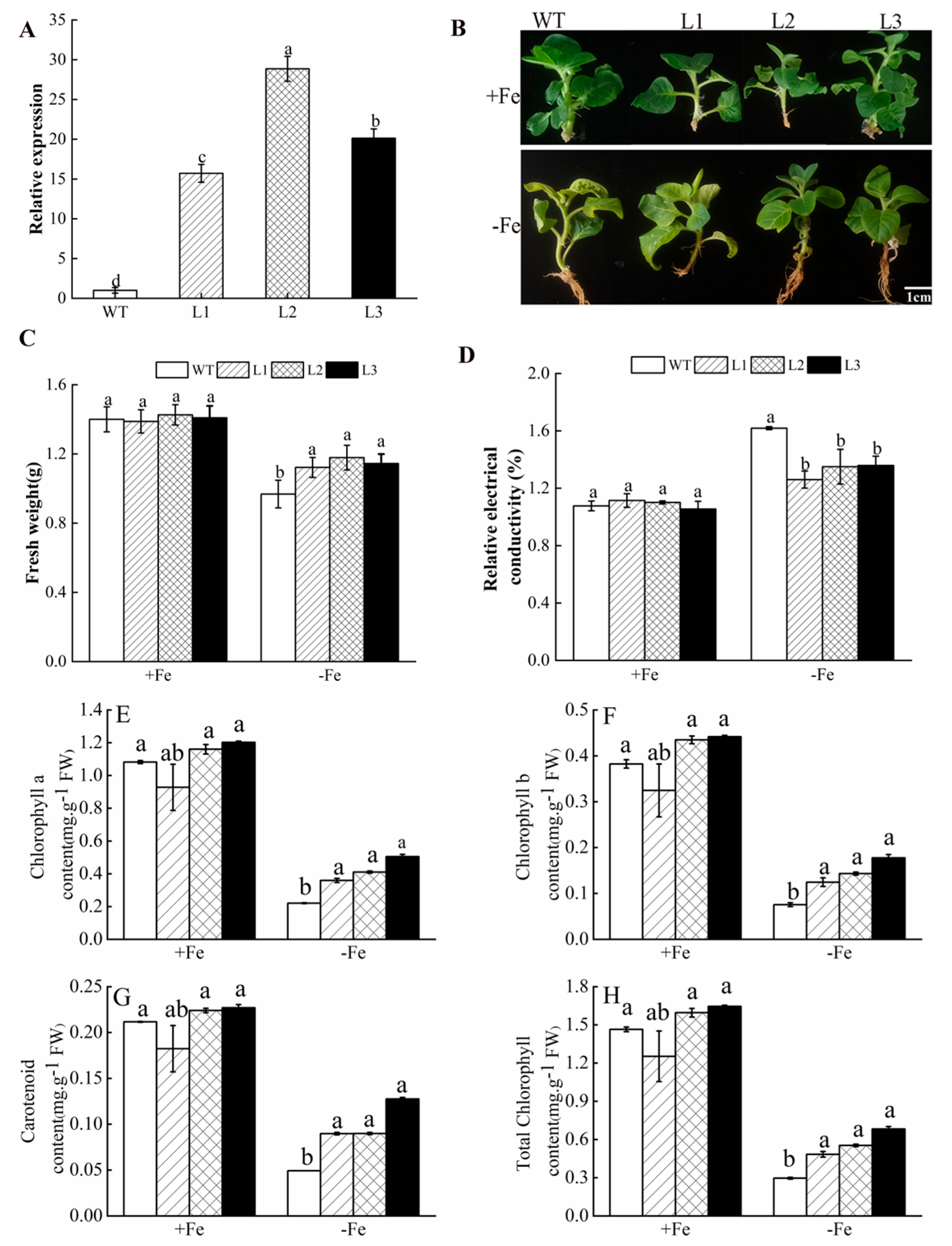
3.5. Overexpression of MhATPS1 in Tobacco Enhances Fe-Deficiency Tolerance

Transgenic tobacco plants overexpressing MhATPS1 were generated to further investigate the role of MhATPS1 in Fe-deficiency stress tolerance. Three homozygous lines (L1, L2, and L3) were selected, and they had higher MhATPS1 expression levels (Figure 2A). Thus, they were selected for further analyses. Subsequently, the seedlings of the wild-type (WT) and transgenic tobacco were exposed to the Fe-sufficient or Fe-deficient surroundings. The results showed that both transgenic and WT tobacco exhibited normal growth when exposed to Fe-sufficient conditions. Under Fe deficiency, the transgenic lines showed better plant growth with more greenish leaves, unlike the WT with obvious chlorotic leaves (Figure 2B). The fresh weight of transgenic tobacco was significantly higher than that of WT, and the REC was significantly lower than that of WT (Figure 2C,D), indicating that overexpression of MhATPS1 enhanced the tolerance of transgenic tobacco to Fe deficiency. In addition, under conditions of Fe-deficiency stress, the transgenic tobacco had higher chlorophyll content compared to the WT tobacco, which is also reflected by the significant decrease in total chlorophyll content, including chlorophyll a and b (Figure 2E–H).

3.6. MhATPS1 Overexpression Enhances Antioxidant Activity in Transgenic Tobacco under Fe Deficiency

When plants are in an Fe-deficient environment, reactive oxygen species (ROS) are produced, and excessive ROS levels lead to impaired cell growth after oxidative stress [1]. To detect that the overexpression of MhATPS1 affects the accumulation of ROS, tissue staining was used to detect the accumulation of O2 and H2O2 in the leaves of transgenic and WT tobacco, and the leaves of transgenic tobacco exhibited a lighter color compared to WT tobacco (Figure 3A,B). Consistent with these results, the O2 and H2O2 content levels for the transgenic plants were lower than those of WT plants under Fe-deficit stress (Figure 3C,D). Next, the activities of SOD, POD, CAT, and APX were measured. The results showed that transgenic lines were not different from WT. However, after Fe-deficiency stress, the activities of SOD, POD, CAT, and APX in transgenic lines were significantly higher than those in WT (Figure 3E–H), which was consistent with the results of ROS staining. These results indicate that overexpression of MhATPS1 reduces the accumulation of reactive oxygen species under Fe-deficiency stress by enhancing the activity of reactive oxygen scavenging enzymes.

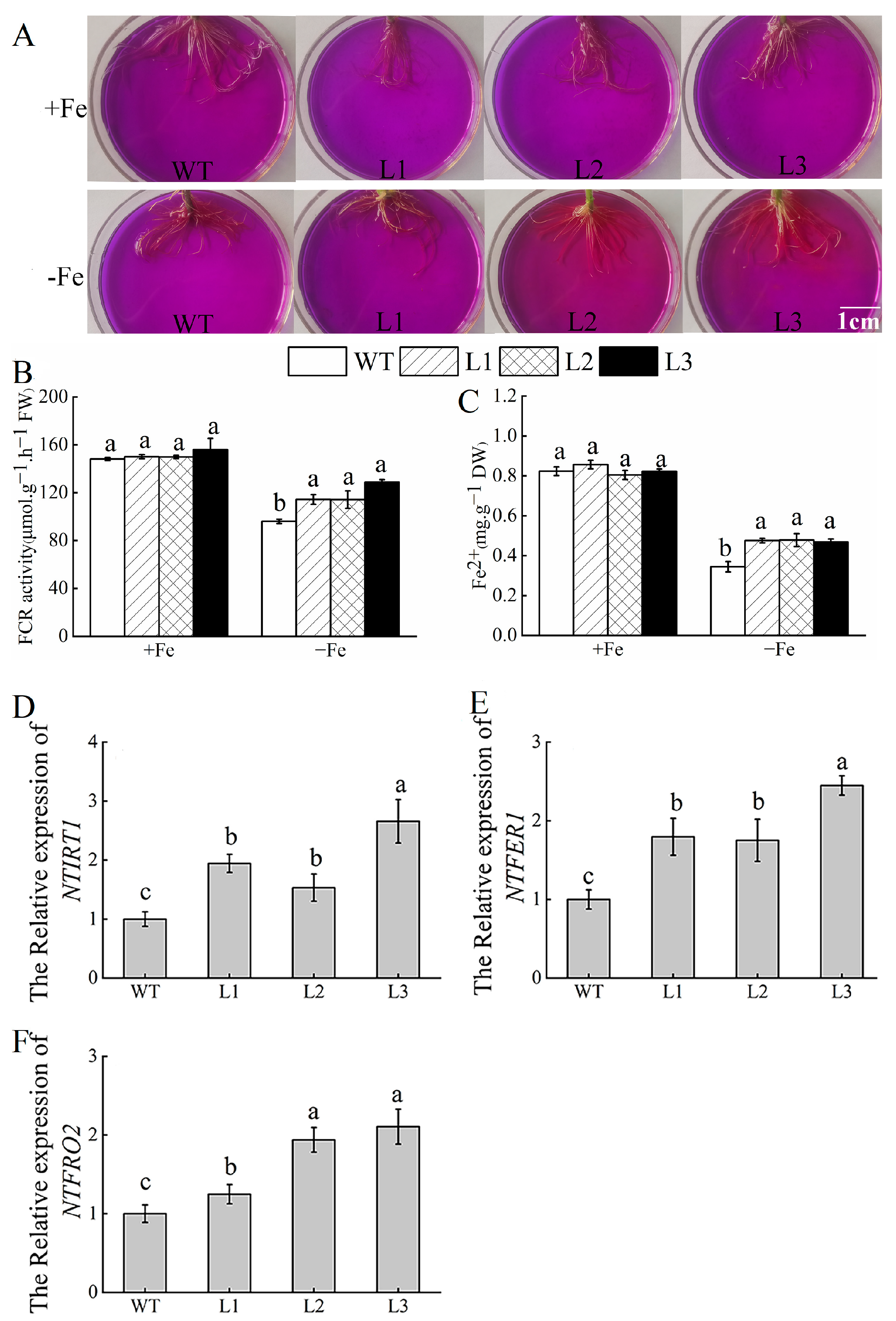
3.7. Overexpression of MhATPS1 Promotes Rhizosphere Acidification and Fe Acquisition in Transgenic Tobacco

When plants are Fe-deficient, their roots secrete protons driven by the PMH+-ATP enzyme to promote the transformation of Fe3+ into Fe2+ in soil, which is then absorbed and transported to corresponding sites by Fe reductase and transporter [4]. As indicated above, compared with WT, transgenic plants showed a greater acidification zone, indicated by the yellow color in agar plates containing the pH indicator bromocresol purple for 24 h (Figure 4A). Further, FCR activity was increased in the transgenic lines under both Fe-sufficient and Fe-deficient conditions (Figure 4B). Fe contents in the young leaves of transgenic lines were significantly higher than in the WT (Figure 4C). To further understand the molecular mechanisms underlying MhATPS1-mediated Fe-deficiency resistance signaling pathways, we examined the transcriptional abundance of the stress response-related genes (NTFRO2, NTFER1, and NTIRT1) by qRT-PCR. The transcription levels of these genes were significantly higher in transgenic tobacco than in WT tobacco under Fe-deficiency conditions (Figure 4E,F).

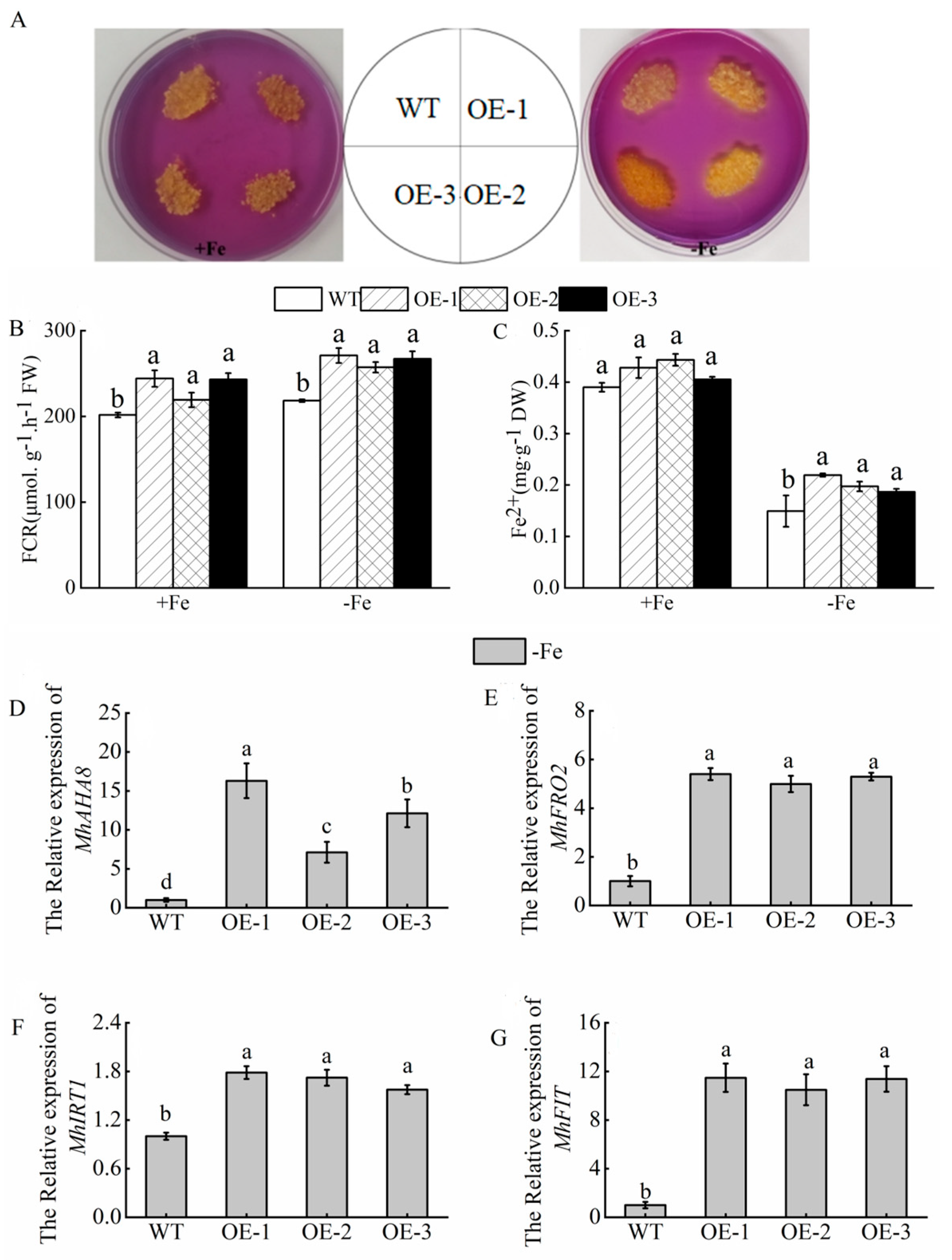
3.8. Tolerance of MhATPS1 Transgenic Apple Calli to Fe Deficiency

As a model system, the apple transgenic calli were used to characterize the function of some genes in modulating stress tolerance [17]. To characterize the functions of MhATPS1, the construct 35S: MhATPS1 was transformed into apple calli (Orin). qRT-PCR analysis indicated that the MhATPS1-OE lines had higher transgenic levels than the WT control (Figure 5A), indicating that transgenic apple calli were obtained. Subsequently, the 15-day-old MhATPS1-OE and WT calli were grown on Fe-sufficient or Fe-deficient medium for another 20 days. Compared with the control, MhATPS1-OE calli exhibited faster growth than the WT control (Figure 5B). Consistent with the phenotype, it was found that compared with WT, the fresh weight of MhATPS1-OE increased and REC decreased in transgenic calli under Fe deficiency (Figure 5C,D). In addition, the activity of antioxidant enzymes in MhATPS1-OE was significantly higher than that in WT calli (Figure 5E–H). Overall, MhATPS1 enhances the tolerance to Fe deficiency in transgenic apple calli.

3.9. Overexpression of MhATPS1 Promotes Rhizosphere Acidification and Fe Acquisition in Transgenic Calli

We verified whether overexpression of MhATPS1 can also promote the absorption of Fe ions in apple calli. As depicted in Figure 6A, the rhizosphere acidification experiment revealed that the yellow coloration around the transgenic apple calli was more prominent than that around the WT calli under Fe-deficient conditions. This observation suggests that the transgenic calli have a higher capacity for pumping out H+ content in comparison to the WT calli. Additionally, the activity of FCR, responsible for Fe reduction during Fe-deficiency treatment, was measured. The results indicated that the FCR activity of the overexpressing calli was significantly higher than that of the WT calli in both Fe-sufficient and Fe-deficient media; and Fe2+ content was significantly higher in transgenic calli than in WT calli under both normal and Fe-deficient conditions (Figure 6B,C). The expression of genes induced by Fe deficiency, namely AHA8, FRO2, FIT, and IRT1, was also examined. As shown, the transcription expression of FRO2 was higher in transgenic calli compared to WT calli under Fe-deficient media. Similarly, the expression of IRT1, a key transporter delivering rhizosphere Fe2+ into cells, was higher in the transgenic calli than in WT calli, while the expression of FIT was highly expressed in transgenic calli (Figure 6D–G).

4. Discussion

Plant growth and development are affected by a range of biological and abiotic stresses. Fe-deficiency stress is one of the major abiotic stresses of plants, which negatively impacts plant growth, development, and crop yield [1,30]. To survive, higher plants have developed a set of Fe signal regulation systems, which strictly control the absorption, transportation, distribution, and storage of Fe. Studies have revealed that plants activate various stress-related genes when faced with Fe-deficiency stress in order to support their normal growth and development [3,31]. In a previous report, the ATPS gene is up-regulated under cold stress [32]. In our study, we observed that the transcript level of ATP sulfurylase genes was induced by Fe-deficiency stress (Figure 1), suggesting that it may have a role in regulating Fe-deficiency stress. ATP sulfurylase genes have been confirmed to participate in various biological processes [33,34]. However, no ATPS genes have been reported to play an essential role in Fe-deficiency tolerance. Thus, we obtained transgenic tobacco and transgenic apple calli through genetic transformation to explore its function in Fe-deficiency stress. In our study, Fe-deficiency treatment and qRT-PCR detection of transgenic tobacco and apple calli showed that the expression level of transgenic plants was significantly higher than that of WT (Figure 2A and Figure 5A). In this study, the resistance of transgenic lines to Fe-deficiency stress was analyzed. MhATPS1 overexpression plants responded better than WT lines (Figure 2B and Figure 5B). Therefore, the MhAPS1 gene may play a role in Fe-deficiency resistance. Under the condition of Fe deficiency, most of the Fe in the chloroplast matrix of plants will be reabsorbed and utilized, and the Fe on the thylakoid membrane and the structure outside the chloroplast will be lost, which will directly affect the chlorophyll synthesis of plants [35]. The degree of chlorosis caused by Fe deficiency in crops can be expressed in terms of chlorophyll content, and therefore it can be used as a direct indicator of how plants are affected by Fe-deficiency stress [36]. Consistent with previous studies [37], the content of chlorophyll in both transgenic tobacco and WT tobacco decreased under Fe-deficiency stress. However, the values of chlorophyll content were still higher in the transgenic tobacco compared to the WT (Figure 2E–H).
Reactive oxygen species (ROS), including O2 and H2O2, can cause damage to cell growth by oxidizing proteins, lipids, and DNA, or by acting as signal molecules that mediate tolerance to various stresses [37,38]. Staining and measurement results showed that transgenic tobacco has lower levels of ROS compared to WT tobacco (Figure 3A–D), indicating that overexpression of MhATPS1 caused plants to accumulate less ROS under Fe-deficiency stress. Fe is an important cofactor involved in the biosynthesis of antioxidant enzymes such as POD, SOD, and CAT. Its deficiency leads to oxidative damage in plants, as observed in Poncirus trifoliata and Pisum sativum [39,40]. Considering the significance of ROS in stress tolerance, we investigated whether the regulation of ROS scavenging ability is influenced by MhATPS1. In our study, the activities of important ROS-scavenging enzymes (POD, SOD, CAT, and APX) were higher in the transgenic tobacco plants and transgenic apple calli than in WT (Figure 3E–H and Figure 5E–H), suggesting that transgenic lines have a stronger ability to scavenge active oxygen species than the WT.
When non-gramineous plants, such as Arabidopsis, tobacco, and woody fruit trees, are exposed to Fe deficiency, they can reduce soil pH and increase available Fe content, thereby facilitating Fe uptake [41]. Therefore, to test whether the overexpression of MhATPS1 affects rhizosphere pH in response to Fe-deficiency stress, transgenic and WT tobacco plants were cultured under normal conditions for 10 days and then subjected to an Fe-deficient medium for 5 days. Afterward, they were transferred to a medium containing the pH indicator bromocresol purple for staining. As indicated above, compared with WT, transgenic plants showed a greater acidification zone, indicated by a yellow color in agar plates containing the pH indicator bromocresol purple for 24 h (Figure 4A). The transgenic calli also exhibited a similar trend (Figure 6A). These findings suggest that overexpression of MhATPS1 enhances the acidification response of plants, which is likely the initial step taken by the transgenic lines. Next, non-gramineous plants increase Fe absorption by increasing FCR activity. To confirm this, FCR activity was measured in transgenic tobacco and WT tobacco. The results demonstrated that FCR activity increased in transgenic lines under Fe-deficient conditions, with transgenic lines showing significantly higher FCR activity compared to WT (Figure 4B). The transgenic calli also exhibited the same trend (Figure 6B). Most of the Fe in plant species is in the leaves, so the accumulation of Fe in plants can be judged by the chlorophyll level [42]. In this experiment, it was observed that under Fe-deficiency conditions, the chlorophyll content of transgenic tobacco leaves was significantly higher compared to WT tobacco. This finding indicates that transgenic tobacco accumulated more Fe than WT tobacco. Then, we determined the content of Fe2+ in transgenic tobacco and WT tobacco. The results showed that the content of Fe2+ in transgenic tobacco leaves was higher than that in WT tobacco leaves under Fe-deficiency conditions. It is speculated that the overexpression of the MhATPS1 gene promoted the accumulation of Fe2+ in the leaves, leading to increased chlorophyll synthesis during growth (Figure 5C). Additionally, transgenic calli exhibited higher Fe2+ accumulation compared to WT calli under Fe-deficiency stress (Figure 6C). Subsequently, several key genes involved in the absorption of Fe by the plants come into play, including AHA2, FRO2, IRT1, and FIT, which are responsible for H+-ATPase acidification, FCR reduction, and IRT1 transfer. AHA2, known for its role in regulating plant root tumor acidification in Arabidopsis [43], was found to have its closest homolog, AHA8, in woody fruit trees, according to Zhao et al. [17]. Therefore, the study of MhATPS1’s function necessitates the verification of these aforementioned genes. In transgenic tobacco, the expression of genes (NTFRO2, NTIRT1, and NTFER1) associated with the three steps of Fe absorption significantly increased, leading to an upward trend in the levels of Fe2+ and chlorophyll in the leaves (Figure 4D–F). Similar results were observed in transgenic calli (MhAHA8, MhFRO2, MhIRT1, and MhFIT) (Figure 6D–G). The above findings suggested up-regulation of Fe uptake in the transgenic plants. These results indicate that MhATPS1 promoted Fe acquisition.

5. Conclusions

In summary, our study shows that MhATPS1 regulates Fe acquisition in transgenic plants, and its overexpression promotes the enhancement of antioxidant enzyme activity and Fe absorption in transgenic lines, thereby enhancing the tolerance of transgenic lines to Fe deficiency. This study also provides a theoretical basis for the tolerance of plants to Fe deficiency and provides a useful strategy for crops to obtain Fe.

Supplementary Materials

The following supporting information can be downloaded at: https://www.mdpi.com/article/10.3390/agronomy14030404/s1, Table S1. Primer sequence for fluorescence quantitative PCR. Table S2. FPKM values of four ATPS genes in Fe-deficient transcriptome at 0 h, 12 h and 3 d at 0 h, 12 h, and 3 d in Mulas halliana. Figure S1. Conserved motifs of four putative ATPS family proteins analyzed with MEME online tools. Figure S2. Phylogenetic tree of the deduced amino acid sequences of ATPS in different plants. The phylogenetic tree of ATPS from different plants was determined by the MEGAX program using the neighbor-joining method. The numbers on the branches represent bootstrap support for 1000 replicates. Figure S3. Homologous sequence comparison of the MhATPS1 protein with the ATPS1 of other species. Three boxes in the ATP sulfurylase domain represent three motifs: Block I, Block II, and Block III.

Author Contributions

J.C. designed and conceived the experiments and wrote the manuscript. Z.Z. and Y.G. provided helpful comments on the manuscript. J.C. and Y.D. performed most of the experiments described in the manuscript. C.L., X.X. and L.D. carried out the experiments. Y.W. guided the whole study and valuable discussions. All authors have read and agreed to the published version of the manuscript.

Funding

This work was supported by the National Natural Science Foundation of China (Project Number 31960581 and 32160696).

Data Availability Statement

The datasets used and/or analyzed during the current study are available to readers as in the manuscript and from the corresponding author upon reasonable request.

Conflicts of Interest

The authors declare no conflicts of interest.

References

  1. Curie, C.; Panaviene, Z.; Loulergue, C.; Dellaporta, S.L.; Briat, J.F.; Walker, E.-L. Maize yellow stripe1 encodes a membrane protein directly involved in Fe (III) uptake. Nature 2001, 409, 346–349. [Google Scholar] [CrossRef]
  2. Li, L.; Gao, W.W.; Peng, Q.; Zhou, B.; Kong, Q.H.; Ying, Y.H.; Shou, H.X. Two soybean bHLH factors regulate response to iron deficiency. J. Integr. Plant Biol. 2018, 60, 608–622. [Google Scholar] [CrossRef] [PubMed]
  3. Li, D.Y.; Sun, Q.; Zhang, G.F.; Zhai, L.M.; Li, K.T.; Feng, Y.; Zhang, X.Z.; Xu, X.F.; Wang, Y.; Han, Z.H. MxMPK6-2-bHLH104 interaction is involved in reactive oxygen species signaling in response to iron deficiency in apple rootstock. J. Exp. Bot. 2021, 72, 1919–1932. [Google Scholar] [CrossRef] [PubMed]
  4. Santi, S.; Schmidt, W. Dissecting iron deficiency-induced proton extrusion in Arabidopsis roots. New Phytol. 2009, 183, 1072–1084. [Google Scholar] [CrossRef]
  5. Waters, B.M.; Blevins, D.G.; Eide, D.J. Characterization of FRO1, a pea ferric-chelate reductase involved in root iron acquisition. Plant Physiol. 2002, 129, 85–94. [Google Scholar] [CrossRef] [PubMed]
  6. Vert, G.; Grot, N.; Dedaldechamp, F.; Gaymard, F.; Guerinot, M.L.; Briat, J.F.; Curie, C. IRT1, an Arabidopsis transporter essential for iron uptake from the soil and for plant growth. Plant Cell. 2003, 14, 1223–1233. [Google Scholar] [CrossRef] [PubMed]
  7. Lee, S.; Chiecko, J.C.; Kim, S.A.; Walker, E.L.; Lee, Y.; Guerinot, M.L.; An, G. Disruption of OsYSL15 leads to iron inefficiency in rice plants. Plant Physiol. 2009, 150, 786–800. [Google Scholar] [CrossRef] [PubMed]
  8. Zuchi, S.; Cesco, S.; Astolfifi, S. High S supply improves Fe accumulation in durum wheat plants grown under Fe limitation. Environ. Exp. Bot. 2012, 77, 25–32. [Google Scholar] [CrossRef]
  9. Bouranis, D.L.; Chorianopoulou, S.N.; Protonotarios, V.E.; Siyannis, V.F.; Hopkins, L.; Hawkesford, M.J. Leaf response of young iron- inefficient maize plants to sulfur deprivation. J. Plant Nutr. 2003, 26, 1189–1202. [Google Scholar] [CrossRef]
  10. Zuchi, S.; Cesco, S.; Varanini, Z.; Pinton, R.; Astolfi, S. Sulphur deprivation limits Fe-deficiency responses in tomato plants. Planta 2009, 230, 85–94. [Google Scholar] [CrossRef]
  11. Khan, N.A.; Anjum, N.A.; Nazar, R.; Iqbal, N. Increased activity of ATP-sulfurylase and increased contents of cysteine and glutathione reduce high cadmium-induced oxidative stress in mustard cultivar with high photosynthetic potential. Russ. J. Plant Physl. 2009, 56, 670–677. [Google Scholar] [CrossRef]
  12. Logan, H.; Cathala, N.; Grignon, C.; Davidian, J. Cloning of a cDNA encoded by a member of the Arabidopsis thaliana ATP Sulfurylase multigene family expression studies in yeast and in relation to plant sulfur nutrition. J. Biol. Chem. 1996, 271, 12227. [Google Scholar] [CrossRef]
  13. Bolchi, A.; Petrucco, S.; Tenca, P.L.; Foroni, C.; Ottonello, S. Coordinate modulation of maize sulfate permease and ATP sulfurylase mRNAs in response to variations in sulfur nutritional status: Stereospecific down-regulation by L-cysteine. Plant Mol. Biol. 1999, 39, 527–537. [Google Scholar] [CrossRef] [PubMed]
  14. Chan, K.X.; Wirtz, M.; Phua, S.Y.; Estavillo, G.M.; Pogson, B.J. Balancing metabolites in drought: The sulfur assimilation conundrum. Trends Plant Sci. 2013, 18, 18–29. [Google Scholar] [CrossRef] [PubMed]
  15. Leduc, D.L.; Abdelsamie, M.; Montes-Bayon, M.; Wu, C.P.; Reisinger, S.J.; Terry, N. Overexpressing both ATP sulfurylase and selenocysteine methyltransferase enhances selenium phytoremediation traits in Indian mustard. Environ. Pollut. 2006, 144, 70–76. [Google Scholar] [CrossRef] [PubMed]
  16. Sun, C.H.; Wu, T.; Zhai, L.M.; Li, D.Y.; Zhang, X.Z.; Xu, X.F.; Ma, H.Q.; Wang, Y.; Han, Z.H. Reactive oxygen species function to mediate the Fe deficiency response in an Fe-efficient apple genotype: An early response mechanism for enhancing reactive oxygen production. Front. Plant Sci. 2016, 7, 1726. [Google Scholar] [CrossRef] [PubMed]
  17. Zhao, Q.; Ren, Y.R.; Wang, Q.J.; Yao, Y.X.; You, C.X.; Hao, Y.J. Overexpression of MdbHLH104 gene enhances the tolerance to iron deficiency in apple. Plant Biotechnol. J. 2016, 14, 1633–1645. [Google Scholar] [CrossRef] [PubMed]
  18. Zhang, Z.Z.; Cheng, J.; Wang, S.C.; Gao, Y.L.; Xian, X.L.; Li, C.L.; Wang, Y.X. Molecular cloning and functional characterization of MhHEC2-like genes in Malus halliana reveals it enhances Fe (iron) deficiency tolerance. Funct. Integr. Genom. 2022, 22, 1283–1295. [Google Scholar] [CrossRef] [PubMed]
  19. Zhu, L.F.; Yang, C.Q.; You, Y.; Liang, W.; Wang, N.; Ma, F.W.; Li, C.Y. Validation of reference genes for qRT-PCR analysis in peel and flesh of six apple cultivars (Malus domestica) at diverse stages of fruit development. Sci. Hortic. 2019, 244, 165–171. [Google Scholar] [CrossRef]
  20. Zhao, Q.; Fan, Z.H.; Qiu, L.N.; Che, Q.Q.; Wang, Y.Z. MdbHLH130, an Apple bHLH transcription factor, confers water stress resistance by regulating stomatal closure and ROS homeostasis in transgenic tobacco. Front. Plant Sci. 2020, 11, 543696. [Google Scholar] [CrossRef]
  21. Li, T.Y.; Wang, Y.; Zhang, X.Z.; Han, Z.H. Isolation and characterization of ARRO-1 genes from apple rootstocks in response to auxin treatment. Plant Mol. Biol. Rep. 2012, 30, 1408–1414. [Google Scholar] [CrossRef]
  22. Xie, B.X.; Li, S.; Zhang, R.F.; Zhao, J.; Chen, Y.C.; Zhao, Q.; Hao, Y.J. The bHLH transcription factor MdbHLH3 promotes anthocyanin accumulation and fruit colouration in response to low temperature in apples. Plant Cell Environ. 2012, 35, 884–1897. [Google Scholar] [CrossRef]
  23. Gong, J.M.; Lee, D.A.; Schroeder, J.I. Long-distance root-to-shoot transport of phytochelatins and cadmium in Arabidopsis. Proc. Natl. Acad. Sci. USA 2003, 100, 10118–10123. [Google Scholar] [CrossRef]
  24. Chazaux, M.; Schiphorst, C.; Lazzari, G.; Caffarri, S. Precise estimation of chlorophyll a, b and carotenoid content by deconvolution of the absorption spectrum and new simultaneous equations for Chl determination. Plant J. 2022, 109, 1630–1648. [Google Scholar] [CrossRef]
  25. Fitter, D.W.; Martin, D.J.; Copley, M.J.; Scotland, R.W.; Langdale, J.A. GLK gene pairs regulate chloroplast development in diverse plant species. Plant J. 2002, 31, 713–727. [Google Scholar] [CrossRef] [PubMed]
  26. Ying, Y.Q.; Song, L.L.; Jacobs, D.F.; Mei, L.; Liu, P.; Jin, S.H.; Wu, J.S. Physiological response to drought stress in Camptotheca acuminata seedlings from two provenances. Front. Plant Sci. 2015, 6, 361. [Google Scholar] [CrossRef] [PubMed]
  27. Schikora, A.; Schmidt, W. Iron stress-induced changes in root epidermal cell fate are regulated independently from physiological responses to low iron availability. Plant Physiol. 2001, 125, 1679–1687. [Google Scholar] [CrossRef] [PubMed]
  28. Sairam, R.K.; Srivastava, G.C. Changes in antioxidant activity in sub-cellular fractions of tolerant and susceptible wheat genotypes in response to long term salt stress. Plant Sci. 2002, 162, 897–904. [Google Scholar] [CrossRef]
  29. Liang, Y.; Ma, F.; Li, B.; Guo, C.; Hu, T.; Zhang, M.; Zhan, X. A bHLH transcription factor, SlbHLH96, promotes drought tolerance in tomato. Hortic. Res. 2022, 9, uhac198. [Google Scholar] [CrossRef] [PubMed]
  30. Zhao, Q.; Ren, Y.R.; Wang, Q.J.; Wang, X.F.; You, C.X.; Hao, Y.J. Ubiquitination-related MdBT scaffold proteins target a bHLH transcription factor for iron homeostasis. Plant Physiol. 2016, 172, 1973–1988. [Google Scholar] [CrossRef] [PubMed]
  31. Rampey, R.A.; Woodward, A.W.; Hobbs, B.N.; Tierney, M.P.; Lahner, B.; Salt, D.E.; Bartel, B. An Arabidopsis basic helix-loop-helix leucine zipper protein modulates metal homeostasis and auxin conjugate responsiveness. Genetics 2006, 174, 1841–1857. [Google Scholar] [CrossRef]
  32. Phartiyal, P.; Kim, W.S.; Cahoon, R.E.; Jez, J.M.; Krishnan, H.B. Soybean ATP sulfurylase, a homodimeric enzyme involved in sulfur assimilation, is abundantly expressed in roots and induced by cold treatment. Arch. Biochem. Biophys. 2006, 450, 20–29. [Google Scholar] [CrossRef]
  33. Pilon-Smits, E.; Hwang, S.; Mel-Lytle, C.; Zhu, Y.L.; Tai, J.C.; Chen, Y.C.; Leustek, T.; Terry, N.; Bravo, R.C. Overexpression of ATP sulfurylase in Indian mustard leads to increased selenate uptake, reduction, and tolerance. Plant Physiol. 1999, 119, 123–132. [Google Scholar] [CrossRef]
  34. Takahashi, H.; Watanabe-Takahashi, A.; Smith, F.W.; Blake-Kalff, M.; Hawkesford, M.J.; Saito, K. The roles of three functional sulphate transporters involved in uptake and translocation of sulphate in Arabidopsis thaliana. Plant J. 2000, 23, 171–182. [Google Scholar] [CrossRef] [PubMed]
  35. Khan, N.A.; Nazar, R.; Anjum, N.A. Growth, photosynthesis and antioxidant metabolism in mustard (Brassica juncea L.) cultivars differing in ATP-sulfurylase activity under salinity stress. Sci. Hotic. 2009, 122, 455–460. [Google Scholar] [CrossRef]
  36. Halliwell, B.; Gutteridge, J. Oxygen toxicity, oxygen radicals, transition metals and disease. Biochem. J. 1984, 219, 1–14. [Google Scholar] [CrossRef] [PubMed]
  37. Fan, W.; Wang, H.; Wu, Y.; Yang, N.; Yang, J.; Zhang, P. H+-pyrophosphatase IbVP1 promotes efficient iron use in sweet potato [Ipomoea batatas (L.) Lam.]. Plant Biotechnol. J. 2017, 15, 698–712. [Google Scholar] [CrossRef] [PubMed]
  38. Yamauch, I.M.; Peng, X.X. Iron toxicity and stress-induced ethylene production in rice leaves. Plant Soil. 1995, 173, 21–28. [Google Scholar] [CrossRef]
  39. Huang, X.S.; Wang, W.; Zhang, Q.; Liu, J.H. A basic helix-loop-helix transcription factor, PtrbHLH, of Poncirus trifoliata confers cold tolerance and modulates peroxidase-mediated scavenging of hydrogen peroxide. Plant Physiol. 2013, 162, 1178–1194. [Google Scholar] [CrossRef] [PubMed]
  40. Jelali, N.; Donnini, S.; Dell‘Orto, M.; Abdelly, C.; Gharsalli, M.; Zocchi, G. Root antioxidant responses of two Pisum sativum cultivars to direct and induced Fe deficiency. Plant Biol. 2014, 16, 607–614. [Google Scholar] [CrossRef]
  41. Wu, T.; Zhang, H.T.; Wang, Y.; Jia, W.S.; Xu, X.F.; Zhang, X.Z.; Han, Z.H. Induction of root Fe (lll) reductase activity and proton extrusion by iron deficiency is mediated by auxin-based systemic signalling in Malus xiaojinensis. J. Exp. Bot. 2012, 63, 859–870. [Google Scholar] [CrossRef] [PubMed]
  42. Sun, Y.; Luo, J.; Feng, P.; Yang, F.; Liu, Y.; Lisng, J.; Wang, H.; Zou, Y.; Ma, F.; Zhao, T. MbHY5-MbYSL7 mediates chlorophyll synthesis and iron transport under iron deficiency in Malus baccata. Front. Plant Sci. 2022, 13, 1035233. [Google Scholar] [CrossRef] [PubMed]
  43. Palmgren, M.G. Plant plasma membrane H+-ATPases: Powerhouses for nutrient uptake. Annu. Rev. Plant Biol. 2001, 52, 817–845. [Google Scholar] [CrossRef] [PubMed]
Figure 1. FPKM values of four genes in the transcriptome under Fe deficiency conditions at 0 h, 12 h, and 3 d in Mulus halliana.
Agronomy 14 00404 g001
Figure 2. Effect of MhATPS1 on Fe-deficiency tolerance in transgenic tobacco. (A) Expression analysis of MhATPS1 in WT and transgenic tobacco by qRT-PCR. (B) Phenotypes of the WT and MhATPS1 transgenic lines; the WT and transgenic tobacco lines under Fe-sufficient (+Fe) and Fe-deficient (−Fe) stress surroundings. (C,D) Fresh weights and REC of WT and transgenic tobacco under the control or Fe-deficient treatment conditions. (EH) Determination of chlorophyll content in transgenic tobacco and WT tobacco. Error bars represent the means ± SD taken from three independent biological replicates (p < 0.05). The letters indicate significant differences according to one-way ANOVA (Tukey’s test; p < 0.05).
Agronomy 14 00404 g002
Figure 3. ROS accumulation and activity of antioxidant enzymes in transgenic tobacco and WT tobacco under Fe-sufficient (+Fe) and Fe-deficient (−Fe) conditions. (A) H2O2 and O2 contents in the leaves. (B) NBT and DAB tissue staining. (C) O2 productivity. (D) H2O2 content. (EH) Enzymatic activity of SOD, POD, APX, and CAT in transgenic and WT tobacco under Fe-sufficient or Fe-deficient conditions. The letters indicate significant differences according to one-way ANOVA (Tukey’s test; p < 0.05).
Agronomy 14 00404 g003
Figure 4. Fe acquisition analysis of tobacco in response to Fe deficiency. (A) Rhizosphere acidification assay using agar plates containing bromocresol purple for 24 h. (B) FCR activity. (C) Fe2+ content. (DF) Expression of genes related to Fe uptake and transport in transgenic and WT tobacco in a Fe-deficient environment (NTIRT1, NTFER1, and NTFRO2). The letters indicate significant differences according to one-way ANOVA (Tukey’s test; p < 0.05).
Agronomy 14 00404 g004
Figure 5. Effect of MhATPS1 on Fe-deficiency tolerance in transgenic apple calli. (A) Expression analysis of MhATPS1 in WT and MhATPS1-OE transgenic calli by qRT-PCR. (B) The growth status of transgenic and WT calli under +Fe or −Fe conditions. (C,D) Fresh weights and REC of WT and transgenic calli. (EH). Determination of antioxidant enzyme activity in transgenic and WT calli under Fe-sufficient of Fe-deficient surroundings. The letters indicate significant differences according to one-way ANOVA (Tukey’s test; p < 0.05).
Agronomy 14 00404 g005
Figure 6. Transgenic calli respond to Fe-deficiency stress. (A) Acidification assay using agar plates containing bromocresol purple for 24 h under Fe-sufficient or Fe-deficient surroundings. (B) Determination of FCR activity. (C) Determination of Fe2+ content. (DG) Expression of the genes involved in Fe transport and intake in the transgenic and WT calli. The letters indicate significant differences according to one-way ANOVA (Tukey’s test; p < 0.05).
Agronomy 14 00404 g006
Table 1. List of ATPS-family genes and information on their encoded proteins.
Accession NoGene NameConserved
Domain
Amino AcidMolecular
Mass (kDa)
pIPositive
Residues
Negative
Residues
Aliphatic IndexProtein
Hydrophobicity
Alpha
Helix (%)
Random
Coil (%)
InstabilityGrand
Average Hydropathicity
MD13G1201500LOC103410737ATPS46552.288.82615784.75−0.39732.90%49.89%48.71−0.397
MD15G1014100LOC103450392ATPS48654.207.01575889.47−0.34030.86%47.94%48.09−0.340
MD16G1201400LOC103403953ATPS46552.217.81595883.91−0.37530.54%51.61%48.96−0.375
MD08G1014800LOC103440334ATPS48654.397.34595988.87−0.37030.86%48.56%48.25−0.370
Table 2. Subcellular location predictions of ATPS-family proteins.
GeneCytoplasmicChloroplastNuclearMitochondrialPeriplastNuclear and
Cytoplasmic
Golgi
Apparatus
Nuclear and
Plasma Membrane
MD13G1201500 11
MD15G1014100111
MD16G1201400 10
MD08G1014800 5 8
The subcellular location values indicate the possibility of being located in corresponding organelles.
Table 3. Cis-elements in the genomic sequence 2000 bp upstream of the MhATPS1.
Regulator
Sequence
PositionMatrix ScoreSequenceFunction of Site
GATA-motif2449AAGGATAAGGpart of a light-responsive element
TGA-box578TGACGTAAPart of an auxin-responsive element
LTR968;1382;1335;14296CCGAAACis-acting element involved in low-temperature responsiveness
ABR115627TACGGTCCis-acting element involved in the abscisic acid responsiveness
MBSI190711TTTTTACGGTTAMYB binding site involved in flavonoid biosynthetic gene regulation
TGACG-motif60;73;1487;605TGACGCis-acting regulatory element involved in the MeJA-responsiveness
TATC-box13597TATCCCACis-acting element involved in gibberellin-responsiveness
Disclaimer/Publisher’s Note: The statements, opinions and data contained in all publications are solely those of the individual author(s) and contributor(s) and not of MDPI and/or the editor(s). MDPI and/or the editor(s) disclaim responsibility for any injury to people or property resulting from any ideas, methods, instructions or products referred to in the content.

