Article

Heat Stress Resistance in Chlorella vulgaris Enhanced by Hydrolyzed Whey Proteins

by
Wolfram Manuel Brück
1,*,
Esteban Alfonso
2,
Markus Rienth
2 and
Wilfried Andlauer
1
1
Institute of Life Sciences, University of Applied Sciences Western Switzerland Valais-Wallis, 1950 Sion, Switzerland
2
University of Sciences and Art Western Switzerland, CHANGINS College for Viticulture and Enology, Route de Duillier 60, 1260 Nyon, Switzerland
*
Author to whom correspondence should be addressed.
Agronomy 2024, 14(12), 2854; https://doi.org/10.3390/agronomy14122854
Submission received: 21 October 2024 / Revised: 27 November 2024 / Accepted: 27 November 2024 / Published: 28 November 2024
(This article belongs to the Section Plant-Crop Biology and Biochemistry)

Abstract

:
Plant biologists have long used various model organisms, such as Arabidopsis thaliana, to study biological processes. The emergence of algal model organisms in recent years greatly facilitated plant research due to their compact genomes with a very low level of functional redundancy and their physiological properties. Here, the green microalga Chlorella vulgaris has been used as a model organism to study the effect of whey hydrolysates against heat stress in vascular plants. Cell density in culture, as well as chlorophyll and carotenoid concentrations, were monitored during an initial growth phase, followed by a heat stress phase and a subsequent recovery phase. Results showed that whey hydrolysates promoted the recovery of heat-stressed C. vulgaris and showed improved growth rates and generation times after heat stress compared to cells grown in BG-11 only. Similarly, the production of chlorophyll a, chlorophyll b, total chlorophyll and total carotenoids was improved in comparison to cells grown only in BG-11. The results may aid the development of novel biostimulants that protect crops from climate change due to higher throughput and shorter study times compared to traditional plant models.

1. Introduction

Microalgae have emerged as essential models for studying plant biology due to their simple structure, rapid growth and adaptability to diverse environmental conditions. Algae such as Chlamydomonas reinhardtii, Dunaliella salina, Scenedesmus obliquus and Chlorella vulgaris are frequently used, especially in studies related to photosynthesis and stress physiology [1,2]. Chlamydomonas reinhardtii has been shown to be a valuable model for investigating chloroplast biogenesis, photosynthetic electron transport and stress responses [1,3]. Thus, Chlamydomonas has been instrumental in uncovering the molecular mechanisms of photosynthesis and in understanding how various environmental factors, such as light and temperature, influence pigment synthesis and overall cellular metabolism [4]. Dunaliella salina has been used to study carotenoid biosynthesis, which accumulates under stress conditions. The algae have provided important insights into how plants and algae modulate pigment production to protect themselves from oxidative damage caused by environmental stressors such as high salinity, light and temperature [2]. Scenedesmus obliquus is commonly used to study nutrient stress and photosynthetic efficiency [5]. This species is known for its resilience to various abiotic stresses, including high temperature and nutrient deprivation, making it a useful model for understanding how plants and algae regulate pigment composition and energy metabolism under adverse conditions [6]. Chlorella vulgaris is recognized for its well-conserved photosynthetic machinery that closely mirrors that of higher plants. This makes C. vulgaris a particularly useful model organism for studying photosynthetic processes, as well as for exploring the effects of environmental stresses, including temperature, light and nutrient availability [7,8]. The chlorophyll content of C. vulgaris and, in particular, chlorophyll a (ChlA) and chlorophyll b (ChlB) are important indicators of the organism’s stress response and photosynthetic efficiency [9]. Chlorophylls a and b play distinct yet complementary roles in capturing and transferring light energy during photosynthesis. Chlorophyll a is the primary pigment involved in capturing light and converting it into chemical energy, whereas chlorophyll b broadens the spectrum of light absorption, allowing the organism to harvest light energy more efficiently under suboptimal conditions [10]. Total chlorophyll content is often used as a proxy for the physiological status of both algae and higher plants, especially under stress conditions like heat, drought or nutrient deficiency [11]. Carotenoids, another important class of pigments in both algae and plants, complement chlorophyll by providing photoprotection and stabilizing the photosynthetic apparatus [12]. In addition to their role in light harvesting, carotenoids quench reactive oxygen species (ROS) produced during photosynthesis, particularly under environmental stresses such as heat or excessive light [13]. Heat stress significantly affects the growth and productivity of both algae and higher plants by disrupting photosynthetic processes, impairing chlorophyll stability and increasing oxidative stress. In higher plants, heat stress typically results in a reduction of total chlorophyll content and damage to the photosystems, thereby decreasing photosynthetic efficiency [14]. Furthermore, heat stress impacts the function and structure of photosystem II (PSII) and, in particular, the transcription of two key proteins, D1 and CP43, which are encoded by psbA and psbC, respectively [15]. Studies have shown that psbA expression increases under heat stress as part of the plant’s attempt to enhance D1 protein synthesis and facilitate repair. However, extreme heat can overwhelm this mechanism, reducing overall PSII activity [16]. Similarly, psbC expression is often upregulated as part of the plant’s protective response, attempting to stabilize PSII and mitigate the effects of heat-induced denaturation of its proteins [17]. The interaction between psbA (D1) and psbC (CP43) with ROS is a primary cause of photoinhibition under heat stress conditions [15]. In algae, including C. vulgaris, similar patterns of chlorophyll degradation have been observed under elevated temperatures. Furthermore, temperatures above the optimal range for Chlorella (typically between 25 and 30 °C) were also shown to impair cell division, resulting in lower biomass accumulation. For instance, C. vulgaris exposed to high temperatures (35–40 °C) exhibited a reduced content of photosynthetic pigments, limited chlorophyll synthesis and a decrease in cell density due to damage to its photosystems [18]. However, the impact of heat stress may be mitigated by an increase in carotenoid production, which helps to protect the photosynthetic apparatus from oxidative damage by stabilizing thylakoid membranes [12,19]. This interplay between chlorophylls and carotenoids under heat stress conditions is not only crucial for understanding stress responses in algae but also provides valuable insights into how similar mechanisms may operate in higher plants. By studying these processes in model organisms like C. vulgaris, researchers are exploring potential strategies for enhancing heat tolerance in crop plants, such as the addition of biostimulants, which are of increasing importance in the context of global climate change.
Biostimulants are natural substances that positively affect plant physiology, resulting in increased biomass production, improved nutrient uptake, enhanced crop quality and yield, and greater tolerance to both abiotic and biotic stresses [20,21]. As such, a plant biostimulant may be any substance or microorganism applied to a plant to improve its nutrient efficiency, stress tolerance and crop quality, regardless of its nutrient content [22]. One particularly promising group of biostimulants is protein hydrolysates, which have been shown to improve plant performance, fruit quality and stress resilience [23]. Protein hydrolysates contain mainly peptides and free amino acids [24]. However, their application in agriculture remains in its early stages due to a lack of systematic, reproducible studies and a limited understanding of their modes of action [25]. Some indications show that protein hydrolysates have hormonal activities [22]. The mitigation of environmental stress may be due to the antioxidant activity of nitrogenous compounds. Further evidence suggests that protein hydrolysates affect the plant-associated microbiota, leading to an increase in microbial biomass favoring known decomposer species [24,25].
This study suggests that whey protein hydrolysates enhance the resistance of Chlorella vulgaris to heat stress and promote improved recovery after the heat stress phase, serving as a model for similar responses in higher plants. To observe the effect solely of the protein hydrolysates on the algae, axenic cultures were used.

2. Materials and Methods

2.1. Whey Hydrolysates

Whey hydrolysates were obtained from raw milk raclette production from the Fromagerie d’Etiez, AOP & Artisan Cheese in Vollèges, Switzerland (fromagerieetiez.ch). The whey was centrifuged (CLARA 20, Alfa Laval, Hamburg, Germany: 50 °C, 11,130 G, 1.3 bar, 100 L/h) and pasteurized (plate heat exchanger Rosista APV: 72 °C, 30 s). To increase protein and reduce mineral and lactose levels, the whey was ultrafiltered twice using a pilot unit (SW25 MMS, Tami membrane UF 3 kDa: 50 °C, TMP 1.5–2.5 bar, 400 L/h, flux 8–9 L/m2h). In the first step, 20 L of whey was concentrated to 1.5 L, then diluted with 20 L of demineralized water and reconcentrated to 2.7 L in the second step. The protein content of the whey concentrate was analyzed using the Kjeldahl method, following the ISO 8968-3:2007/IDF 20-3:2007 standard and applying a nitrogen-to-protein conversion factor of 6.38 [26]. The concentrate was hydrolyzed using the endopeptidase Alcalase® Pure 2.4 L and the exopeptidase Flavourzyme® 1000 L. The enzymes were provided free of charge by Novozymes AG (Bagsvaerd, Denmark). Whey concentrates (900 mL, equivalent to 8.3 g of protein) were incubated with 225 mg of Flavourzyme (225 LAPU) = 180 μL of Flavourzyme enzyme preparation and 225 mg of Alcalase (21.6 Anson units/g = 4.86 Anson units = 2673 IU) = 180 μL of Alcalase enzyme preparation. A carbonate/bicarbonate buffer (90 mL, 0.1 mol/L, pH 7.5) was added, and the temperature was maintained at 50 °C. The pH was kept at 7.5 using NaOH (1 moL/L) with an automated pump. Hydrolyzed whey was collected and characterized after 0 (unhydrolyzed control), 4 h and 24 h.

2.2. Cultured Reference Strains

Cultures of Chlorella vulgaris (SAG 211-12) were obtained from the “Sammlung von Algenkulturen der Universität Göttingen” (SAG). Sample strains were transferred and adapted under axenic culture conditions to Blue–Green Medium (BG-11) growth media and maintained under non-limiting conditions using LED-assisted natural sunlight illumination.

2.3. Growth Assays

For heat stress assays, 200 mL cultures containing 8.5 × 10⁵ cells/mL C. vulgaris (SAG 211-12) in BG-11 medium containing 1 ml of a 0.5% (v/v) solution of either unhydrolyzed whey concentrate (W0 h), whey hydrolyzed for 4 h (W4 h) or whey hydrolyzed for 24 h (W24 h). Thus, the working concentration of the whey hydrolysates in 200 mL cultures was 0.0025% (v/v). A non-treatment control containing only BG-11 medium (100× base: CaCl2·2H2O 36.7 mg/L; C6H8O7 5.6 mg/L; K2HPO4 31.4 mg/L; C10H12MgN2Na2O8 1 mg/L; C6H11FeNO7+3 6 mg/L; MgSO4 36 mg/L; Na2CO3 20 mg/L; NaNO3 1.5 g/L; trace metal mix: H3BO3 46 mM; MnCl24H2O 9mM; ZnSO4·7H2O 0.77mM; Na2MoO4·2H2O 1.6mM; CuSO4·5H2O 0.3mM; Co(NO3)2·6H2O 0.17mM; final pH 7.1, Merck (Schweiz) AG, Zug, Switzerland) was included in all experiments. All experiments were performed in triplicate. Initially, all cultures were incubated for 7 d under optimal (25 ± 1 °C) temperature conditions. Afterward, cultures were subjected to heat stress at 30 ± 1 °C for 7 d, followed by a recovery period for 7 d at 25 ± 1 °C. Cultures were continually shaken at 130 rpm and illuminated with 100 μmol/m2·s white LED light and a 14/10 h light/dark photoperiod in a customized shaking incubator with water-cooled LED illumination underneath each incubator position (TB2000 light incubator, FutureLED, Berlin, Germany). All cultures were continually sparged with air supplemented with CO2 at a flow rate of 1.0 Nl/min. The gas passed through a 0.22 μm Hydrophilic Polyvinylidene Fluoride (PVDF) filter (Merck (Schweiz) AG, Zug, Switzerland) before entering the reactor. Ten ml samples of all cultures were taken after 0 d, 8 d (start of heat stress period), 15 d (start of recovery period) and 21 d (end of experiment). Cell counts (counts/mL) were determined using a Neubauer-improved counting chamber (chamber dept = 0.02 mm, Assistent, Glaswarenfabrik Karl Hecht GmbH & Co KG, Sondheim vor der Rhön, Germany). Furthermore, variations of cell sizes from C. vulgaris under the various culture conditions were estimated by taking the average cell size of 10 cells in a sample under a bright field at 200× magnification (Zeiss Axioplan 2, Carl Zeiss Vision Swiss AG, Hombrechtikon, Switzerland) equipped with an Olympus XC10 camera running Olympus Stream (vers. 1.7) image analysis software (Olympus Schweiz AG, Wallisellen, Switzerland). Besides cell counts and productivity (g/L/d) as dry weight (dw) as described by [27], the following growth parameters were calculated according to [28]:
Generation   time   [ d ] :   g = 1 v
Growth   rate   [ d 1 ] :   K = l n ( N 2 / N 1 ) t 2 t 1
Cell   divisions   [ d 1 ] :   v = K ln 2
N1 and N2 are equal to the biomass at time 1 (t1) and time 2 (t2), respectively [29]. Generation time (g) is expressed in d, whereas growth rate (K’) and cell divisions (v) are expressed in d−1.

2.4. Chlorophyll and Carotenoid Determination

Chlorophyll was extracted from C. vulgaris cultures using Dimethylsulfoxide (DMSO, Sigma, Buchs, Switzerland) as described in [30]. A Triplicate 2 mL sample of the culture was placed into a 2 mL tube and centrifuged (Heraeus Pico 17, ThermoFisher, Reinach, Switzerland) at 7500 g for 3 min. After discarding the supernatant, the pellet was resuspended in 2 mL of preheated (60 °C) DMSO and vortexed for 10 min, followed by another centrifugation at 7500 g for 3 min. The supernatant, containing the chlorophyll extract, was transferred to a new centrifuge tube and diluted with DMSO to a concentration that did not give an OD that exceeded a value of 1. The optical densities at 480, 649 and 665 nm were measured using a Biowave 3 CT UV/VIS Diode Array Spectrophotometer (Labgene Scientific SA, Châtel-Saint-Denis, Switzerland). Chlorophyll and carotenoid concentrations were calculated using the following formulas [25]:
Chlorophyll a (ChlA) (mg/L) = 12.47(OD665) − 3.62(OD649)
Chlorophyll b (ChlB) (mg/L) = 25.06(OD649) − 6.5(OD665)
Total carotenoid (mg/L) = [1000(OD480) − 1.29(ChlA) − 53.78(ChlB)]/220
Total chlorophyll (mg/L) = (ChlA) + (ChlB) + (Total carotenoid)

2.5. qRT-PCR of psbA and psbC

The transcription levels of two photosynthesis-related genes, psbA and psbC, from the chloroplast-encoded photosystem II (PSII), were analyzed using qRT-PCR of TRIzol-extracted RNA following Barati et al. [31]. Table 1 shows the primers used for amplification. A NanoDrop 2000c Spectrophotometer (Thermo Fisher Scientific, Basel, Switzerland) was used to determine the yield and purity of the RNA extracts. Samples were analyzed using a QuantiTect Multiplex RT-PCR Kit and a Rotor-Gene Q (QIAGEN AG-Switzerland Hombrechtikon, Switzerland) with a reverse transcription step for 20 min at 50 °C. Cycling steps were as follows: initial denaturation at 95 °C for 15 min, followed by 40 cycles of 95 °C for 45 s and 60 °C for 75 s. A no-template control was added for each analysis run and consisted of all the reaction components except for the sample. ΔCt values were calculated by subtracting the Ct of the H3 gene from the Cts of either psbA or psbC. ΔΔCt values were calculated by subtracting the ΔCts from the control and stressed samples, and fold changes were calculated using the 2−ΔΔCt method. Histones H3 and H4 (H3), previously reported to show stable expression during abiotic stress, were used as internal controls [32].

2.6. Statistical Analysis

The obtained results were expressed by the mean values ± standard deviation. The significance of the results was evaluated using the analysis of variance (one-way ANOVA) and Tukey–Kramer HSD (Honestly Significant Difference) tests with a significance level α of 0.05 [33].

3. Results

3.1. C. vulgaris Growth Under Heat Stress

In this study, C. vulgaris cultures were initially grown with a 7 d heat stress phase either at 40 °C or 35 °C. However, none of the cultures survived. Hence, cells were subjected to a maximum temperature of 30 °C.
During the initial (base) phase at 25 °C, the cultures containing 0.5% (v/v) whey hydrolysates or BG-11 medium had similar cell densities (Log10 cells/mL) (Figure 1), with generation times between 1.32 and 1.55 d (A in Table 2). When the algae were heat stressed for 7 d at 30 °C (Heat stress), cell densities (Log10 cells/mL) remained either stable or only minimally increased, as also seen with generation times increasing more than 10-fold. Generally, whey hydrolysates supported faster growth (g = 17.58–23.99 d) compared to cultures in BG-11 medium only (g = 61.12 d). The difference between the initial incubation and the incubations at 30 °C were significantly different (p ≤ 0.01). During the recovery phase (Recovery) at 25 °C for 7 d, all cultures recovered but had a noticeably declined growth. However, generation times were significantly better (p ≤ 0.01) than during the heat stress phase. C. vulgaris cultures with whey hydrolyzed for 4 h (W4 h) performed significantly (p ≤ 0.05) better than either culture with whey hydrolyzed for 24 h (W24 h) or BG-11 medium only. The productivity (g/L/d) of C. vulgaris was also improved (p ≤ 0.05) during base and recovery periods (B in Table 2). Cultures containing unhydrolyzed whey and whey hydrolyzed for 4 h (W4 h) had significantly greater (p ≤ 0.05) productivity than cultures in BG-11 medium only. During heat stress, productivity was significantly reduced (p ≤ 0.01) in all treatments.
Similar observations were made for growth rate and number of cell divisions per day (Figure 2). Growth rates and cell divisions of C. vulgaris at 25 °C were initially comparable between the different whey hydrolysates, with only BG-11 medium being significantly (p ≤ 0.05) lower. During heat stress treatment, both variables were significantly suppressed (p ≤ 0.01) but were able to recover when the temperature was decreased again to 25 °C after 7 d (Recovery). Nonetheless, the parameters did not return to the previous values. During recovery, C. vulgaris cultures in BG-11 medium containing whey hydrolyzed for 4 h (W4 h) recovered significantly better (p ≤ 0.05) than cultures BG-11 only.
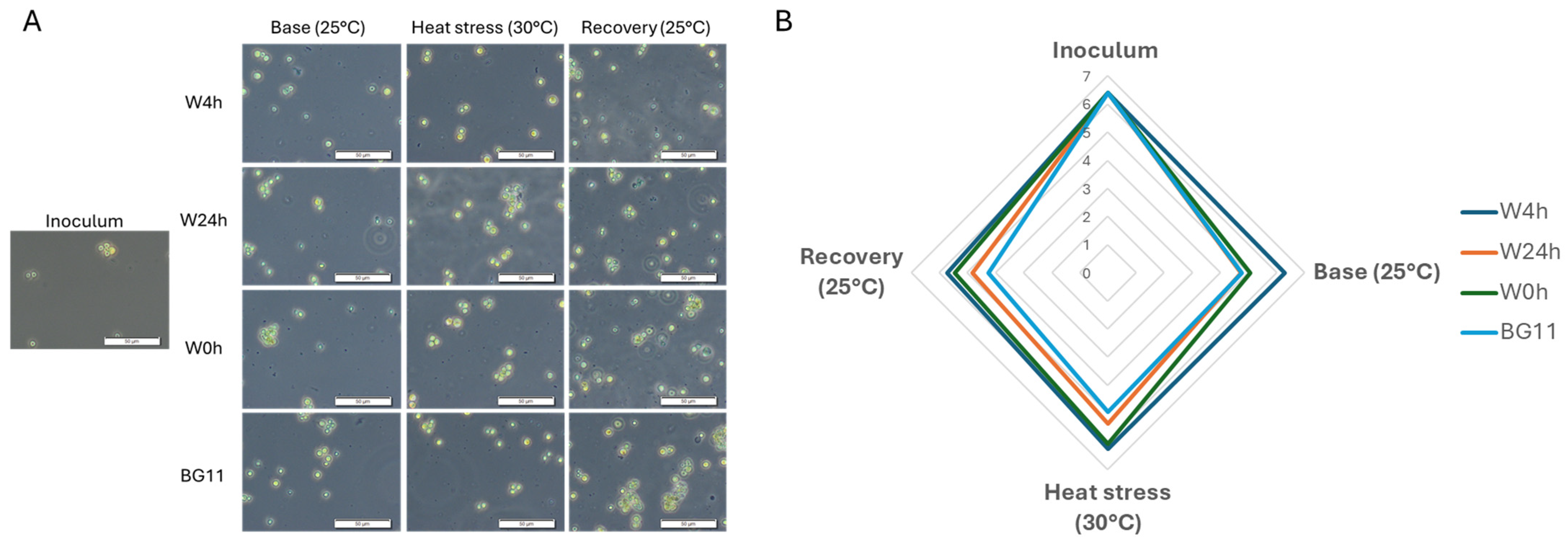
Microscopic examination of C. vulgaris cell size variation during the different phases of the experiment showed that cells generally were spherical and between 4 and 6 µm diameter. Bright-field and phase contrast observations at 100×, 200× and 400× magnification showed no significant changes in cellular morphology or coloration between cells at inoculation, nor during the various temperature treatments or different whey hydrolysate additions (Figure 3A). However, a fluctuation in cell size during the different temperature cycles was observed in BG-11 medium and BG-11 medium containing 0.0025% (v/v) of unhydrolyzed whey concentrate (W0 h) and whey hydrolyzed for 24 h (W24 h) (Figure 3B). Cells in W24 h and BG-11 only tended to be smaller after inoculation, at an average cell size of 4 µm diameter. Algae in W0 h initially decreased in cell diameter but then increased again to 6 µm diameter average. Size variations were not significant.

3.2. Heat Stress Induced Changes in Chlorophyll and Carotenoid Content

The values of ChlA at the control (base) temperature (25 °C) fluctuated between 13.38 and 17.36 mg/L. At the stress-inducing temperature (30 °C), the initial concentrations in the cultures reached 21.23 to 32.67 mg/L, with cultures in BG-11 containing whey hydrolyzed for 4 h (W4 h) and unhydrolyzed whey (W0 h) having more ChlA than cells in whey hydrolyzed for 24 h (W24 h) or in BG-11 medium only. For the difference between W4 h and W24 h and BG-11, only the difference was significant at p ≤ 0.05 and p ≤ 0.01, respectively. During the recovery period, ChlA showed an increasing trend, with W4 h values being significantly higher than cultures with other whey hydrolysates or the control. Comparable trends were observed with ChlB, where initial concentrations were stable between 6.83 and 7.53 mg/L. During heat stress, ChlB in W4 h cultures was significantly higher (p ≤ 0.05) at 15.70 mg/L compared to cultures in BG-11 only (11.70 mg/L). This trend was observed again during recovery, with W4 h cultures showing a ChlB concentration of 38.59 mg/L compared to control cultures (BG-11 only), which had only 23.04 mg/L of ChlB. This difference was highly significant (p ≤ 0.01). Analogous to ChlA and ChlB, total carotenoids showed similar trends, with W4 h having a significantly higher concentration of carotenoids during heat stress and recovery periods than the control (p ≤ 0.01). W4 h also had higher total chlorophyll concentrations during heat stress and recovery periods (Figure 4).
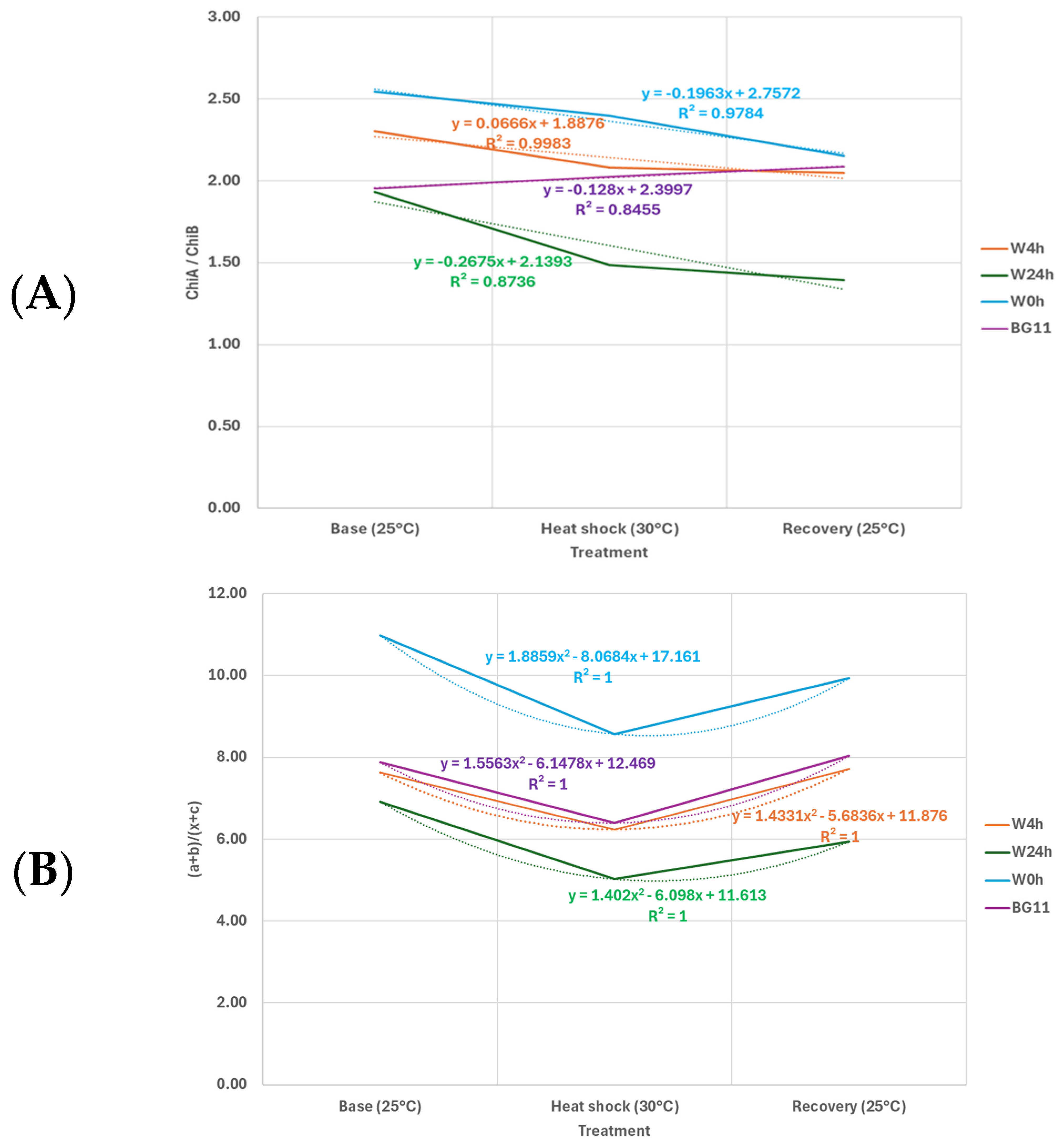
ChlA to ChlB ratios stayed between 1.40 and 2.54 throughout the experiments (Figure 5A). However, W24 h cultures had a significantly lower (p ≤ 0.05) ratio during heat stress compared to W0 h and a significantly lower ratio (p ≤ 0.05) during the recovery phase compared to other treatments. The ratio of ChlA and ChlB to carotenoids varied between 5.03 and 10.98 throughout the experiments (Figure 5B). W0 h had a significantly higher (p ≤ 0.05) ratio during the initial 7 d (base) phase compared to W24 h. During heat stress, all treatments decreased their ratio, indicating that carotenoids had a greater increase during the heat stress phase compared to ChlA and ChlB. However, ratios increased again during recovery and were comparable to the ratios at the start of the experiments.

3.3. Heat Stress Induced Changes of psbA and psbC

The results indicated that the mRNA transcript levels of psbA and psbC remained unchanged under the various temperature conditions applied in this study.

4. Discussion

4.1. C. vulgaris Growth Under Heat Stress

Global warming has led to rising temperatures on Earth, creating significant abiotic stress that threatens plants and other photosynthetic organisms [14]. To combat these and other environmental stressors, solutions such as biostimulants are needed to alleviate stress and prevent reductions in productivity. More efficient modeling systems are essential for rapidly screening these biostimulants, as they offer higher throughput than traditional plant models or field trials. This makes Chlorella vulgaris an important model organism for studying photosynthesis and examining the impact of environmental stressors like temperature [7,8].
Chlorella vulgaris is a unicellular green microalga with a size range of 2 μm to 10 μm and a reported temperature range of 25–35 °C [34,35]. The optimal temperature range for many commercially cultivated microalgae varies between 20 °C and 30 °C [36]. Variations in temperature may influence the growth of C. vulgaris by altering the mass transfer rate and solubility of CO2 and O2 in the medium, as well as the equilibrium and kinetics of reactions. Additionally, temperature plays a critical role in enzymatic processes and metabolism, thus influencing cellular regulation and ultimately determining cell composition and structure. At higher temperatures, this eventually leads to cell death [37,38,39]. The present findings suggest that using the physiological conditions applied, our strain of C. vulgaris did not survive temperatures superior to 30 °C. This corresponds with previous reports estimating the optimal growth temperature of C. vulgaris in a photobioreactor to be 23.3 °C, with no observable growth beyond 30 °C [40]. A significant decrease in the growth rate started at 28 °C [37]. The authors attributed the lack of growth at higher temperatures to short acclimation periods in photobioreactors. Longer acclimation periods for abiotic stressors may have a less pronounced effect on cell survival and productivity [41]. Exposure to elevated temperatures can severely impact microalgal cell growth and productivity, leading to the accumulation of reactive oxygen species (ROS), damage to the photosynthetic system and irreversible oxidative harm to DNA, proteins and lipids [42].
In the present study, we decided to subject C. vulgaris to a heat stress phase at 30 °C that did not result in complete viability loss during heat exposure to assess the cell’s ability to recover when exposed again to optimal temperatures. Besides changes in productivity (reduced biomass) and generation times, exposure to heat also resulted in changes in cellular morphology. Most pronounced was a small reduction of cell diameter in cells grown in BG-11 medium and BG-11 containing whey protein hydrolyzed for 24 h (W24 h). While the differences were not significant and cell diameter remained within the normal cell range reported, our finding corresponds to reports that indicate a relationship between the morphology (size and shape) of algae and temperature fluctuations [43]. It is assumed that the reduction in cellular diameter in response to increasing temperatures is provoked by changes in metabolic processes and resource availability [44]. While the mechanisms driving size reductions at both intraspecific and community levels may vary, temperature increases and nutrient limitations would promote smaller algae and increased sedimentation [45,46]. Similar changes are also frequently observed in vascular plants, where morphological changes manifest as reductions in anthesis, leaf area, plant size, seed quality and others, all of which hinder crop productivity [47].

4.2. Heat Stress Induced Changes in Chlorophyll and Carotenoid Content

Photosynthesis acts as a global sensor of environmental stress, triggering cellular energy imbalances and loss of pigment content [48]. It is particularly sensitive to high temperature stress and is often inhibited before other cellular functions are affected [14]. ChlA and ChlB are essential pigments in photosynthesis, with ChlA being the primary pigment participating in the light reactions, specifically capturing light in the blue-violet and red regions of the spectrum. ChiB, an accessory pigment, absorbs light in the blue and red-orange regions, expanding the range of light wavelengths available for photosynthesis and transferring the captured energy to ChlA for more efficient photosynthesis [49,50]. Carotenoids, in turn, act as accessory pigments in photosynthesis, absorbing blue–green and violet light and transferring energy to chlorophyll for photosynthesis. Additionally, they protect the plant by quenching excess energy and preventing damage from reactive oxygen species under stress conditions, acting as antioxidants and photoprotective agents [13,51]. Carotenoids help dissipate this excess energy via the xanthophyll cycle, converting high-energy molecules like violaxanthin to zeaxanthin, which protects the plant by safely dissipating excess light energy as heat, reducing ROS formation [12,13]. Additionally, carotenoids stabilize photosynthetic membranes and support the integrity of photosystem II during thermal stress. When plants and algae experience heat stress, reports show that chlorophylls are generally more rapidly degraded than carotenoids [12]. This increased carotenoid stability was also reflected in the decreased chlorophyll/carotenoid ratio observed during heat stress. This reduction occurs because chlorophylls, particularly ChlA, are more susceptible to degradation under stress conditions [1,3,52]. In the present study, unhydrolyzed whey (W0 h) appeared to protect against chlorophyll degradation in C. vulgaris compared to the other treatments applied to the culture. However, contents of chlorophylls and carotenoids were generally lower in W0 h cultures than in other treatments except for algae grown in BG-11 only. Like the chlorophyll/carotenoid ratio, the ChlA/ChlB ratio is an important indicator of the photosynthetic apparatus’s efficiency and adaptation to environmental conditions, including heat stress. Under oxidative stress conditions, ChlA is oxidized at the methyl group on ring II, resulting in the formation of aldehyde groups and leading to the production of ChlB [53]. Consequently, the ChlA/ChlB ratio is more sensitive to changes than the total chlorophyll/carotenoid ratio. Beyond the decrease in ChlA/ChlB ratio, cellular heat stress may also be observed as damage to photosystem II (PSII). ChlB, being primarily associated with light-harvesting complexes, helps absorb and transfer light energy to ChlA, and its relative stability under stress can maintain some photosynthetic function [54,55]. The molar ratios of ChlA/ChlB in the thalli of marine green algae were reported to be between 1.5 and 2.2 [56]. In this study, ChlA to ChlB ratios stayed between 1.40 and 2.54 throughout the experiments. However, a slight decrease in ratio was observed in cultures containing whey proteins hydrolyzed for 4 h and 24 h (W4 h, W24 h). The lowest ratio was seen in W24 h cultures during the recovery phase, suggesting that highly hydrolyzed whey protein had a detrimental effect on the cultures. W0 h cultures had the highest ChlA/ChlB ratio throughout the different temperature cycles, suggesting that the efficiency of the photosynthetic apparatus in those cultures was the most stable. Cultures with whey protein hydrolyzed for 4 h (W4 h) had the largest amount of chlorophyll and carotenoids compared to all cultures, which resulted in ratios of ChlA/ChlB and chlorophyll/carotenoid being comparable to those in cultures in BG-11 medium only. Damage to Photosystem II in our study was not recorded by semi-quantitative RT-PCR of the light-harvesting intrinsic transmembrane proteins psbA and psbC. No transcriptional changes between the initial phase (base 25 °C), heat stress phase (30 °C) and recovery phase (25 °C) were observed. These results agree with those described previously, suggesting that PSII may be more stable against transcriptional changes due to temperature fluctuations [57]. Alternatively, the algae in this study were insufficiently stressed to induce the expected transcriptional changes.

5. Conclusions

Our data suggest that unhydrolyzed whey and whey hydrolyzed for 4 h using the endopeptidase Alcalase® Pure 2.4 L and the exopeptidase Flavourzyme® 1000 L in combination may protect C. vulgaris cultures against morphological changes and aid their recovery after abiotic temperature stress. This protective effect also impacts photosynthesis by protecting against the degradation of ChlA, as observed in improved ChlA/ChlB and chlorophyll/carotenoid ratios. The results from this algal model organism may be applied to plants of agricultural importance as the cellular reactions to abiotic stress and well-conserved photosynthetic machinery of C. vulgaris closely mirror those of higher plants. However, the mechanism of action for the whey protein biostimulants should be further elucidated, especially since qRT-PCR analysis remained inconclusive. Hence, future studies should include a direct survey of the ROS and ROS-related markers relevant to plant biostimulant action. In addition, RNA expression profiles should be further evaluated using high throughput methods such as RNA-seq. Furthermore, this study also highlights the potential of whey protein biostimulants for their potential to enhance the tolerance of microalgae cultures to heat stress. Whey protein hydrolyzed with trypsin for four hours was previously shown to act through antioxidant activity and overall improvement in growth and biomass production [58,59]. As microalgae biotechnology continues to expand, the use of protein hydrolysates as biostimulants offers a cost-effective and sustainable approach to enhancing the productivity and resilience of microalgae cultures, especially under challenging environmental conditions like heat stress [15,24].

Author Contributions

All authors W.M.B., E.A., M.R. and W.A. contributed equally to the funding acquisition, writing, editing, and writing of the draft manuscript. W.M.B. and W.A. also contributed equally to the conceptualization, methodology, validation, formal analysis, investigation, resources, data curation. W.M.B.; supervision, W.A.; project administration. All authors have read and agreed to the published version of the manuscript.

Funding

Financial support was provided by the University of Applied Sciences and Arts Western Switzerland (BiostimVine, AGP 114’957).

Data Availability Statement

The raw data of this article is available in Zenodo (https://zenodo.org/) under https://doi.org/10.5281/zenodo.13973798.

Acknowledgments

The authors would like to acknowledge the support of Nancy Nicolet, Martine Baudin, Bruno Lehner and Martine Emery Mabillard from the Microbiological and Analytical and Bioanalytical Platforms of the Institute of Life Technologies, University of Applied Sciences and Arts Western Switzerland (HES-SO Valais Wallis) in carrying out the laboratory work.

Conflicts of Interest

The authors declare no conflicts of interest.

References

  1. Merchant, S.S.; Prochnik, S.E.; Vallon, O.; Harris, E.H.; Karpowicz, S.J.; Witman, G.B.; Terry, A.; Salamov, A.; Fritz-Laylin, L.K.; Maréchal-Drouard, L.; et al. The Chlamydomonas genome reveals the evolution of key animal and plant functions. Science 2007, 318, 245–250. [Google Scholar] [CrossRef]
  2. Borowitzka, M.A.; Vonshak, A. Stress physiology and cell division in Dunaliella: Implications for commercial production. J. Appl. Phycol. 2017, 29, 69–82. [Google Scholar]
  3. Harris, E.H.; Stern, D.B.; Witman, G.B. The Chlamydomonas Sourcebook, 2nd ed.; Academic Press: Cambridge, MA, USA, 2009. [Google Scholar]
  4. Levine, R.P.; Goodenough, U.W. The genetics of photosynthesis and of the chloroplast in Chlamydomonas reinhardtii. Ann. Rev. Genet. 1970, 4, 397–408. [Google Scholar] [CrossRef] [PubMed]
  5. Zhang, Y.; Ren, L.; Chu, H.; Zhou, X.; Yao, T.; Zhang, Y. Optimization for Scenedesmus obliquus Cultivation: The Effects of Temperature, Light Intensity and pH on Growth and Biochemical Composition. Microbiol. Biotechnol. Lett. 2019, 47, 614–620. [Google Scholar] [CrossRef]
  6. Schambach, J.Y.; Kruse, C.P.S.; Kitin, P.; Mays, W.; Hunt, C.G.; Starkenburg, S.R.; Barry, A.N. Metabolism of Scenedesmus obliquus cultivated with raw plant substrates. Front. Plant Sci. 2022, 13, 992702. [Google Scholar] [CrossRef] [PubMed]
  7. Zheng, G.; Tian, B.; Zhang, F.; Yin, J. Effects of nitrogen on growth and chlorophyll fluorescence of microalga Chlorella vulgaris in thebiomass production. Environ. Sci. Pollut. Res. 2011, 18, 563–570. [Google Scholar]
  8. Kumar, K.; Mishra, S.K.; Shrivastav, A.; Park, M.S.; Yang, J.W. Recent trends in the mass cultivation of algae in raceway ponds. Renew. Sustain. Energy Rev. 2011, 15, 3462–3470. [Google Scholar] [CrossRef]
  9. Porra, R.J.; Thompson, W.A.; Kriedemann, P.E. Determination of accurate extinction coefficients and simultaneous equations for assaying chlorophyll a and b extracted with four different solvents: Verification of the concentration of chlorophyll standards by atomic absorption spectroscopy. Biochim. Biophys. Acta—Bioenerg. 1997, 975, 384–394. [Google Scholar] [CrossRef]
  10. Lichtenthaler, H.K.; Buschmann, C. Chlorophylls and carotenoids: Measurement and characterization by UV-VIS spectroscopy. Curr. Prot. Food Anal. Chem. 2001, 1, F4.3.1–F4.3.8. [Google Scholar] [CrossRef]
  11. Xu, H.; Liu, G.; Liu, G.; Yan, B.; Duan, W.; Wang, L.; Li, S. Comparison of investigation methods of heat injury in grapevine (Vitis) and assessment to heat tolerance in different cultivars and species. BMC Plant Biol. 2014, 14, 156. [Google Scholar] [CrossRef]
  12. Havaux, M. Carotenoids as membrane stabilizers in chloroplasts. Trends Plant Sci. 1998, 3, 147–151. [Google Scholar] [CrossRef]
  13. Demmig-Adams, B.; Adams, W.W. Photoprotection and other responses of plants to highlight stress. Ann. Rev. Plant. Biol. 1992, 43, 599–626. [Google Scholar] [CrossRef]
  14. Mathur, S.; Agrawal, D.; Jajoo, A. Photosynthesis: Response to high temperature stress. J. Photochem. Photobiol. B Biol. 2014, 137, 116–126. [Google Scholar] [CrossRef]
  15. Aro, E.M.; McCaffery, S.; Anderson, J.M. Photoinhibition and D1 protein degradation in vivo: Kinetics and dose dependence. Proc. Natl. Acad. Sci. USA 1993, 90, 7356–7360. [Google Scholar]
  16. Baniulis, D.; Janda, T.; Žukauskaitė, G. Oxidative stress in plants under environmental conditions. Environ. Exp. Bot. 2008, 63, 52–63. [Google Scholar]
  17. Haldimann, P.; Feller, U. Inhibition of photosynthesis by heat stress: Effects on chloroplast proteins and electron transport. J. Exp. Bot. 2005, 56, 3037–3047. [Google Scholar]
  18. Zhou, J.; Jiang, X.; Wei, L. Impact of heat stress on Chlorella vulgaris: Photosynthesis and growth responses. Biotech. Biofuels 2017, 10, 45–53. [Google Scholar]
  19. Cheng, D.; He, Q.; Qiu, C. Responses of Chlorella vulgaris to heat stress and role of carotenoids in protecting PSII. J. Appl. Phycol. 2016, 28, 2081–2090. [Google Scholar]
  20. Calvo, P.; Nelson, L.; Kloepper, J.W. Agricultural uses of plant biostimulants. Plant Soil 2014, 383, 3–41. [Google Scholar] [CrossRef]
  21. Van Oosten, M.J.; Pepe, O.; DePascale, S.; Silletti, S.; Maggio, A. The role of biostimulants and bioeffectors as alleviators of abiotic stress in crop plants. Chem. Biol. Technol. Agric. 2017, 4, 5. [Google Scholar] [CrossRef]
  22. du Jardin, P. Plant biostimulants: Definition, concept, main categories and regulation. Sci. Hortic. 2015, 196, 3–14. [Google Scholar] [CrossRef]
  23. Meggio, F.; Trevisan, S.; Manoli, A.; Ruperti, B.; Quaggiotti, S. Systematic Investigation of the Effects of a Novel Protein Hydrolysate on the Growth, Physiological Parameters, Fruit Development and Yield of Grapevine (Vitis vinifera L., cv Sauvignon Blanc) under Water Stress Conditions. Agronomy 2020, 10, 1785. [Google Scholar] [CrossRef]
  24. Colla, G.; Hoagland, L.; Ruzzi, M.; Cardarelli, M.; Bonini, P.; Canaguier, R.; Rouphael, Y. Biostimulant Action of Protein Hydrolysates: Unraveling Their Effects on Plant Physiology and Microbiome. Front. Plant Sci. 2017, 8, 2202. [Google Scholar] [CrossRef] [PubMed]
  25. Roche, D.; Rickson, J.R.; Pawlett, M. Moving towards a mechanistic understanding of biostimulant impacts on soil properties and processes: A semi-systematic review. Front. Agron. 2024, 6, 1271672. [Google Scholar] [CrossRef]
  26. ISO 8968-3:2007/IDF 20-3:2007; Milk. Determination of Nitrogen Content. International Organization for Standardization: Geneva, Switzerland, 2007.
  27. Metsoviti, M.N.; Papapolymerou, G.; Karapanagiotidis, I.T.; Katsoulas, N. Effect of Light Intensity and Quality on Growth Rate and Composition of Chlorella vulgaris. Plants 2019, 9, 31. [Google Scholar] [CrossRef]
  28. Fogg, G.E.; Thake, B. Algal Cultures and Phytoplankton Ecology; University of Wisconsin Press: Madison, WI, USA, 1987. [Google Scholar]
  29. Levasseur, M.; Thompson, P.A.; Harrison, P.J. Physiological acclimation of marine phytoplankton to different nitrogen sources. J. Phycol. 1993, 29, 587–595. [Google Scholar] [CrossRef]
  30. Chai, S.; Shi, J.; Huang, T.; Guo, Y.; Wei, J.; Guo, M.; Li, L.; Dou, S.; Liu, L.; Liu, G. Characterization of Chlorella sorokiniana growth properties in monosaccharide-supplemented batch culture. PLoS ONE 2018, 13, e0199873. [Google Scholar] [CrossRef]
  31. Barati, B.; Lim, P.E.; Gan, S.Y.; Poong, S.W.; Phang, S.M. Gene expression profile of marine Chlorella strains from different latitudes: Stress and recovery under elevated temperatures. J. Appl. Phycol. 2018, 30, 3121–3130. [Google Scholar] [CrossRef]
  32. Chatelain, P.; Blanchard, C.; Astier, J.; Klinguer, A.; Wendehenne, D.; Jeandroz, S.; Rosnoblet, C. Reliable reference genes and abiotic stress marker genes in Klebsormidium nitens. Sci. Rep. 2022, 12, 18988. [Google Scholar] [CrossRef]
  33. Abdi, H.; Williams, L.J. Tukey’s Honestly Significant Difference (HSD) Test. In Encyclopedia of Research Design; Salkind, N.J., Ed.; Sage: Thousand Oaks, CA, USA, 2010. [Google Scholar]
  34. Van der Wiel, J.B.; Mikulicz, J.D.; Boysen, M.R.; Hashemi, N.; Kalgren, P.; Nauman, L.M.; Baetzold, S.J.; Powell, G.G.; He, Q.; Hashemi, N.N. Characterization of Chlorella vulgaris and Chlorella protothecoides using multi-pixel photon counters in a 3D focusing optofluidic system. RSC Adv. 2017, 7, 4402–4408. [Google Scholar] [CrossRef]
  35. Cassidy, K.O. Evaluating Algal Growth at Different Temperatures. Master’s Thesis, Biosystems and Agricultural Engineering, University of Kentucky, Lexington, KY, USA, 2011. [Google Scholar]
  36. Sánchez-Luna, L.D.; Bezerra, R.P.; Matsudo, M.C.; Sato, S.; Converti, A.; Carvalho, J.C.M. Influence of pH, temperature, and urea molar flowrate on Arthrospira platensis fed-batch cultivation: A kinetic and thermodynamic approach. Biotechnol. Bioeng. 2007, 96, 702–711. [Google Scholar] [CrossRef] [PubMed]
  37. Serra-Maia, R.; Bernard, O.; Gonçalves, A.; Bensalem, S.; Lopes, F. Influence of temperature on Chlorella vulgaris growth and mortality rates in a photobioreactor. Algal. Res. 2016, 18, 352–359. [Google Scholar] [CrossRef]
  38. Das, K.; Roychoudhury, A. Reactive oxygen species (ROS) and response of antioxidants as ROS-scavengers during environmental stress in plants. Front. Environ. Sci. 2014, 2, 53. [Google Scholar] [CrossRef]
  39. Suzuki, N.; Mittler, R. Reactive oxygen species and temperature stresses: A delicate balance between signaling and destruction. Physiol. Plant. 2006, 126, 45–51. [Google Scholar] [CrossRef]
  40. Converti, A.; Casazza, A.A.; Ortiz, E.Y.; Perego, P.; Borghi, M. Effect of temperature and nitrogen concentration on the growth and lipid content of Nannochloropsis oculata and Chlorella vulgaris for biodiesel production. Chem. Eng. Process. 2009, 48, 1146–1151. [Google Scholar] [CrossRef]
  41. Zlotnik, I.; Dubinsky, Z. The effect of light and temperature on DOC excretion by phytoplankton. Limnol. Oceanogr. 1989, 34, 831–839. [Google Scholar] [CrossRef]
  42. Song, K.; Zhou, Z.; Huang, Y.; Chen, L.; Cong, W. Multi-omics insights into the mechanism of the high-temperature tolerance in a thermotolerant Chlorella sorokiniana. Bioresour. Technol. 2023, 390, 129859. [Google Scholar] [CrossRef]
  43. Ahmad, S.; Kothari, R.; Shankarayan, R.; Tyagi, V.V. Temperature dependent morphological changes on algal growth and cell surface with dairy industry wastewater: An experimental investigation. 3 Biotech 2020, 10, 24. [Google Scholar] [CrossRef]
  44. Ras, M.; Steyer, J.P.; Bernard, O. Temperature effect on microalgae: A crucial factor for outdoor production. Rev. Environ. Sci. Biotech. 2013, 12, 153–164. [Google Scholar] [CrossRef]
  45. Peter, K.H.; Sommer, U. Interactive effect of warming, nitrogen and phosphorus limitation on phytoplankton cell size. Ecol. Evol. 2015, 5, 1011–1024. [Google Scholar] [CrossRef]
  46. Piontek, J.; Handel, N.; Langer, G.; Wohlers, J.; Riebesell, U.; Engel, A. Effects of rising temperature on the formation and microbial degradation of marine diatom aggregates. Aquat. Microb. Ecol. 2009, 54, 305–318. [Google Scholar] [CrossRef]
  47. Lal, M.K.; Tiwari, R.K.; Gahlaut, V.; Mangal, V.; Kumar, A.; Singh, M.P.; Paul, V.; Kumar, S.; Singh, B.; Zinta, G. Physiological and molecular insights on wheat responses to heat stress. Plant Cell Rep. 2022, 41, 501–518. [Google Scholar] [CrossRef] [PubMed]
  48. Zuppini, A.; Andreoli, C.; Baldan, B. Heat stress: An inducer of programmed cell death in Chlorella saccharophila. Plant Cell Physiol. 2007, 48, 1000–1009. [Google Scholar] [CrossRef] [PubMed]
  49. Lichtenthaler, H.K. Chlorophylls and carotenoids: Pigments of photosynthetic biomembranes. Meth. Enzymol. 1987, 148, 350–382. [Google Scholar]
  50. Gitelson, A.A. Wide dynamic range vegetation index for remote quantification of biophysical characteristics of vegetation. J. Plant. Physiol. 1999, 161, 165–173. [Google Scholar] [CrossRef] [PubMed]
  51. Fraser, P.D.; Bramley, P.M. The biosynthesis and nutritional uses of carotenoids. Prog. Lipid Res. 2004, 43, 228–265. [Google Scholar] [CrossRef]
  52. Napaumpaiporn, P.; Sirikhachornkit, A. Effects of high temperature on Carotenoid Accumulation and Gene Expression in the Model Green Alga Chlamydomonas reinhardtii. Chiang Mai J. Sci. 2016, 43, 453–461. [Google Scholar]
  53. Chettri, M.K.; Cook, C.M.; Vardaka, E.; Sawidis, T.; Lanaras, T. The effect of Cu, Zn, and Pb on the chlorophyll content of the lichens Cladonia convoluta and Cladonia rangiformis. Environ. Exp. Bot. 1988, 39, 1–10. [Google Scholar] [CrossRef]
  54. Yamane, Y.; Kashino, Y.; Koike, H.; Satoh, K. Increases in the fluorescence Fo level and reversible inhibition of photosystem II reaction center by high-temperature treatments in higher plants. Photosynth. Res. 1997, 52, 57–64. [Google Scholar] [CrossRef]
  55. Xu, Q.; Huang, B.; Liu, Y. Differential accumulation of chlorophyll and alteration of chlorophyll a/b ratio in response to drought and heat stress in creeping bentgrass. J. Am. Soc. Hortic. Sci. 2017, 142, 295–302. [Google Scholar]
  56. Nakamura, K.; Ogawa, T.; Shibata, K. Chlorophyll and peptide compositions in the two photosystems of marine green algae. Biochim. Biophys. Acta 1976, 423, 227–237. [Google Scholar] [CrossRef] [PubMed]
  57. Kizilkaya, I.T.; Akcaalan, S.; Unal, D. Determination of Photosynthesis-Related and Ascorbate Peroxidase Gene Expression in the Green Algae (Chlorella vulgaris) Under High-Temperature Conditions. Int. J. Second Metab. 2021, 8, 59–69. [Google Scholar] [CrossRef]
  58. Elwazuru, E.; Ismail, H.; Abou El-Khairi, E.-S.; Al_Qahtani, S.M.; Al-Harbi, N.-A.; Abd El-Gawad, H.G.; Omar, W.A.; Abdelaal, K.; Osman, A. Biostimulant application of whey protein hydrolysates and potassium fertilization enhances the productivity and tuber quality of sweet potato. Not. Bot. Horti. Agrobo. 2023, 51, 13122. [Google Scholar] [CrossRef]
  59. Riseh, R.A.; Vazvani, M.G.; Hassanisaadi, M.; Thakur, V.K.; Kennedy, J.F. Use of whey protein as a natural polymer for the encapsulation of plant biocontrol bacteria: A review. Int. J. Biol. Macromol. 2023, 234, 123708. [Google Scholar] [CrossRef]
Figure 1. C. vulgaris cell counts (Log10 cells7 mL) at inoculation (0 d), at the start of the heat stress period with (8 d), at the start of the recovery period (15 d) and at the end of the experiment (21 d). BG-11 medium containing 0.0025% (v/v) of unhydrolyzed whey concentrate (W0 h), whey hydrolyzed for 4 h (W4 h) and whey hydrolyzed for 24 h (W24 h). BG11: control without whey. Error bars are ±SD of triplicate experiments.
Agronomy 14 02854 g001
Figure 2. (A,B): (A) C. vulgaris growth rate (K’)/d, (B) C. vulgaris cell divisions/d. Base: 7 d culture under optimal (25 ± 1 °C) temperature conditions; heat stress: follow-up 7 d culture at heat stress conditions (30 ± 1 °C); recovery: follow-up 7 d culture under optimal (25 ± 1 °C) conditions. BG-11 medium containing 0.0025% (v/v) of unhydrolyzed whey concentrate (W0 h), whey hydrolyzed for 4 h (W4 h) and whey hydrolyzed for 24 h (W24 h). BG11: control without whey. Error bars are ± SD of triplicate experiments. Meaning of character abbreviations: * = p ≤ 0.05, *** = p ≤ 0.01.
Agronomy 14 02854 g002
Figure 3. (A,B): Phenotypic changes in Chlorella vulgaris cells following optimal temperatures, heat stress and recovery periods (A) C. vulgaris cellular appearance (200× magnification at phase contrast, bar = 50 µm), (B) C. vulgaris average (n = 10) cell size (µm). Inoculum: cells before start of experiments; base: 7 d culture under optimal (25 ± 1 °C) temperature conditions; heat stress: follow-up 7 d culture at heat stress conditions (30 ± 1 °C); recovery: follow-up 7 d culture under optimal (25 ± 1 °C) conditions. BG-11 medium containing 0.0025% (v/v) of unhydrolyzed whey concentrate (W0 h), whey hydrolyzed for 4 h (W4 h) and whey hydrolyzed for 24 h (W24 h). BG11: control without whey.
Agronomy 14 02854 g003
Figure 4. (AD): C. vulgaris (A) ChlA, (B) ChlB, (C) total carotenoids and (D) total chlorophyll concentrations (mg/L). Base: 7 d culture under optimal (25 ± 1 °C) temperature conditions; heat stress: follow-up 7 d culture at heat stress conditions (30 ± 1 °C); recovery: follow-up 7 d culture under optimal (25 ± 1 °C) conditions. BG-11 medium containing 0.0025% (v/v) of unhydrolyzed whey concentrate (W0 h), whey hydrolyzed for 4 h (W4 h) and whey hydrolyzed for 24 h (W24 h). BG11: control without whey. Meaning of character abbreviations: * = p ≤ 0.05, *** = p ≤ 0.01.
Agronomy 14 02854 g004
Figure 5. (A,B): (A) Values of the ratio ChlA/ChlB and (B) the ratio chlorophylls/carotenoids of C. vulgaris cultures. Base: 7 d culture under optimal (25 ± 1 °C) temperature conditions; heat stress: follow-up 7 d culture at heat stress conditions (30 ± 1 °C); recovery: follow-up 7 d culture under optimal (25 ± 1 °C) conditions. BG-11 medium containing 0.0025% (v/v) of unhydrolyzed whey concentrate (W0 h), whey hydrolyzed for 4 h (W4 h) and whey hydrolyzed for 24 h (W24 h). BG11: control without whey.
Agronomy 14 02854 g005
Table 1. List of genes and primers used for qRT-PCR.
Primer Sequence (5′–3′)
FunctionGene SymbolForwardReverseAmplicon Size (bp)Reference
D1 synthesispsbAGGTGGTCCTTACCAACTTATCGTTTGGGTCCTTACCAACTTATCGTTTG98[29]
PSII subunitpsbCGAACATCACCACCACCAGGACGGTGCTTGGCTTTTAGTTTG79[29]
Endogenous controlH3GAGATCCGCAAGTACCAGAAGGGTCTTGAAGTCCTGGGC93[29]
Table 2. Generation time (d) ± SD and productivity (g/L/d) ± SD of C. vulgaris cultures in BG-11 medium containing 0.0025% (v/v) of unhydrolyzed whey concentrate (W0 h), whey hydrolyzed for 4 h (W4 h) and whey hydrolyzed for 24 h (W24 h). BG11: control without whey.
A Base (25 ± 1 °C)Heat Stress (30 ± 1 °C)Recovery (25 ± 1 °C)
Generation time (d) ± SDW4 h1.32 ± 0.3923.99 ± 0.743.24 ± 0.33
W24 h1.37 ± 0.4617.58 ± 0.874.40 ± 0.62
W0 h1.32 ± 0.5417.63 ± 0.994.09 ± 0.52
BG11 1.55 ± 0.4361.12 ± 0.474.59 ± 0.35
B
Productivity (g/L/d) ± SDW4 h0.081 ± 0.0230.012 ± 0.0260.320 ± 0.055
W24 h0.053 ± 0.0190.011 ± 0.0320.147 ± 0.042
W0 h0.082 ± 0.0220.020 ± 0.0420.274 ± 0.066
BG110.056 ± 0.0030.001 ± 0.0340.152 ± 0.038
Disclaimer/Publisher’s Note: The statements, opinions and data contained in all publications are solely those of the individual author(s) and contributor(s) and not of MDPI and/or the editor(s). MDPI and/or the editor(s) disclaim responsibility for any injury to people or property resulting from any ideas, methods, instructions or products referred to in the content.