Article

Regulation of Fumonisin B1 Production and Pathogenicity in Fusarium verticillioides by Histone Deacetylases

1
Fujian Universities Key Laboratory for Plant-Microbe Interaction, College of Life Science, Fujian Agriculture and Forestry University, Fuzhou 350002, China
2
Key Laboratory of Biopesticide and Chemical Biology of Education Ministry, Fujian Agriculture and Forestry University, Fuzhou 350002, China
3
Marine Biotechnology Center, Fuzhou Institute of Oceanography, Minjiang University, Fuzhou 350108, China
*
Authors to whom correspondence should be addressed.
These authors contributed equally to this work.
Current address: Department of Molecular Genetics and Microbiology, Duke University Medical Center, Durham, NC 27710, USA.
Agronomy 2024, 14(10), 2196; https://doi.org/10.3390/agronomy14102196
Submission received: 30 July 2024 / Revised: 14 September 2024 / Accepted: 20 September 2024 / Published: 24 September 2024
(This article belongs to the Section Pest and Disease Management)

Abstract

:
Transcriptional regulation mediated by the balance of histone acetylation and deacetylation is fundamental in responding to environmental cues by impacting chromatin remodeling. Histone deacetylases (HDACs) are enzymes that remove acetyl groups from histone and non-histone proteins, thus restoring a tight chromatin structure. In pathogenic fungi, HDACs have been implicated in growth, secondary metabolite biosynthesis, and virulence. However, the role of HDACs in the mycotoxin fumonisin B1 (FB1)-producing fungus Fusarium verticillioides is poorly understood. In this study, we systematically characterized six F. verticillioides HDACs. An increased level of H4K16ac was observed in the deletion mutant of FvHOS2, which was associated with vegetative growth, conidiation, and virulence when infecting sugarcane and maize. FvRpd3 appeared to be essential for vegetative growth, while FvHda1 promoted growth, and both contributed to conidiation and pathogenicity. In contrast, FvSirt4 displayed a negative correlation with these processes. Additionally, the FB1 production was positively affected by FvHos2 and FvRpd3, but negatively impacted by Fvhda1, FvSir2, FvHst2, and FvSirt4 through the regulation of different key fumonisin biosynthetic (FUM) genes. Further findings indicate an association between FvSirt4 and FvSkb1, which is a histone methylase that positively regulates FB1 and pathogenicity. Moreover, as a global transcriptional regulator, over 2365 genes (~15% of the genome) enriched in multiple metabolic pathways were significantly downregulated in the ΔFvhos2 mutants relative to the wild type. Overall, our results suggest distinct roles of HDACs in regulating the growth, virulence, mycotoxin FB1 production, and gene expression in F. verticillioides.

1. Introduction

In eukaryotes, nucleosomes are composed of DNA and histone proteins, which serve as the basic and fundamental repeating units of chromatin. Chromatin architecture and transcription can be heavily affected by distinct chemical modifications on the histone tail that extends from the nucleosome complex. For example, the acetylation of histone tails tends to relax condensed chromatin, which leads to higher gene expression by facilitating access to DNA for transcriptional machinery [1]. The acetyl groups deposited by histone acetyltransferase can also be removed by a specific family of enzymes (known as histone deacetylases (HDACs)), which leads to chromatin condensation and transcriptional repression [2]. With the discovery of the first HDAC, namely, reduced potassium dependency 3 (Rpd3), three decades ago, HDACs have been widely identified and studied in multiple organisms and systems [3,4,5]. In yeast, there are three HDACs classes: class I, which encompasses the founding member Rpd3 and Histone deacetylase-A (HDA) one similar 1, 2, and 3 (Hos1, 2, and 3) types [6]; class II, which includes the histone deacetylase 1 (Hda1) type; and class III, which consists of the Silent information regulator 2 (Sir2) and Homologues of Sir two 1, 2, 3, and 4 (Hst1, 2, 3, and 4) types, which are structurally different from class I and II HDACs [7,8,9]. HDACs have been associated with various functions and regulatory patterns in chromatin remodeling and transcriptional control. Rpd3, along with co-factors [10,11], forms two independent complexes with distinct sizes (Rpd3S complex with 0.6 MDa and Rpd3L complex with 1.2 MDa) and localizes in different parts of the genome for transcriptional regulation. For example, the promoter of INO1, which is a gene involved in inositol biosynthesis, attracts significant enrichment of the Rpd3L complex, which leads to inhibition of the gene expression [12], while Rpd3S is mainly responsible for suppressing cryptic expression in the gene body [13]. Additionally, the Hda1 complex exclusively deacetylates lysine residues in the histones’ H3 and H2B amino-terminal tails, spares H4 and H2A [14], and antagonizes and competes with Gcn5 acetyltransferase for the promoter region [15]. This complex mobilizes to its target promoters via the Tup1 repressor [16]. Furthermore, Hos2 physically associates with the coding region of actively transcribed genes, specifically deacetylates histones H3 and H4 in vivo, and is directly required for gene activation via deacetylated H4K16ac [17,18]. In contrast to class I and II HDACs, the deacetylase function of class III HDACs is dependent on the level of cellular nicotine adenine dinucleotide (NAD+) [19] and causes transcription silencing in multiple loci of the genome, such as the telomere, mating type locus, and rDNA array [20,21,22,23].
In fungi, HDACs can facilitate diverse biological functions. The disruption of class I Rpd3 homologs in various filamentous fungi, such as Aspergillus nidulans, A. fumigatus, Botrytis cinerea, and Magnaporthe oryzae, appear to be lethal and indicate the essentiality [24,25,26]. Class II Hda1 is associated with the production of secondary metabolism in Fusarium fujikuroi [27] and spore development in Ustilgo maydis [28]. The Su(var)3-9, which comprise an enhancer-of-zeste and trithorax 3 complex (Set3C) of Saccharomyces cerevisiae, which includes Hos2 and Hst1 deacetylases, act as inhibitive factors specific to meiosis of the spore formation [29]. S. cerevisiae Set3C can suppress genes that encode primary regulatory elements for metaphase spore formation genes (IME1, IME2, NTD80) [29]. Meanwhile, the Set3C in human fungal pathogen Candida albicans dampens cAMP-PKA signaling, thereby inhibiting the yeast-to-filament transition [30], and C. albicans Sir2 plays a crucial role in controlling the phenotypic transformation [31]. M. oryzae Sir2 is crucial for biotrophic growth due to its function in neutralizing host ROS production [32]. Moreover, Beauveria bassiana Sir2 modulates a unique set of cellular targets, thereby influencing conidiation, carbon utilization, stress responses, and blastospore development [33].
HDACs have also been identified as critical regulators of major virulence traits in diverse pathogenic fungal species [34]. For instance, in the opportunistic human fungal pathogen Candida glabrata, Sir2 inhibits the expression of EPA1, which encodes the primary epithelial adhesin that is vital for the survival and proliferation of the fungus in the host environment [35]. In C. albicans, the Set3C complex plays an important role in virulence [30]. In the global human fungal pathogen Cryptococcus neoformans, the loss of Sir2 reduces the replicative lifespan, weakens fitness, and lessens the virulence [36]. The overexpression of RPD3 in B. cinerea and M. oryzae leads to a severe impairment in infection structure formation, oxidative stress response, and virulence [25,37]. Moreover, B. bassiana Sir2 is capable of targeting an extensive array of cytoplasmic proteins, including a benzoquinone oxidoreductase indirectly involved in the detoxification of cuticular compounds, as well as two critical fungal LysM effectors for virulence [33].
F. verticillioides is a globally distributed hemibiotrophic fungal pathogen with a broad range of plant hosts and causes diseases such as stalk rot, ear rot in maize, and pokkah boeng in sugarcane. Consequently, it induces substantial yield loss, along with the production of the highly toxic fumonisin FB1 during the infection process, which severely threatens the security of the infected food [38].
In this study, we identified 11 HDACs within F. verticillioides, with class I including two types (FvHos2 and FvRpd3), class II containing two types (FvHda1 and FvHos3), and class III harboring seven types (FvSir2, FvHst2-4, FvSirt4-6 (Figure S1a)). Among them, six histone-deacetylase-encoding genes—FvHOS2, FvHDA1, FvSIR2, FvHST2, FvSIRT4, and FvRPD3—were selected to study their function through gene deletion or overexpression. The deacetylation function and biological roles were then examined. This investigation provides an inclusive understanding of the functional roles of the canonical HDAC family members in the plant fungal pathogen F. verticillioides.

2. Materials and Methods

2.1. Bioinformatics Analysis

The HDAC protein sequences of S. cerevisiae S288C, Ustilago maydis 521, and Neurospora crassa OR74A were retrieved from the NCBI database. The F. verticillioides HDAC protein sequences were obtained by comparing the protein sequences of S. cerevisiae or U. maydis by NCBI Blast, or the protein by annotation as deacetylase from F. verticillioides genome (https://www.ncbi.nlm.nih.gov/datasets/genome/?taxon=117187, accessed on 6 July 2011) (Table S1). Subcellular localization was predicted by using Euk-mPLoc 2.0 (http://www.csbio.sjtu.edu.cn/bioinf/Cell-PLoc-2/, accessed on 22 May 2023). The domain structures of HDAC were analyzed through SMART (http://smart.embl-heidelberg.de), with the corresponding schematic diagrams drawn using IBS 2.0 software. Multiple HDAC protein sequence alignments were subsequently performed using Cluster W. A phylogenetic tree was constructed via the maximum likelihood method using MEGA 7.0 software (http://www.megasoftware.net, accessed on 22 May 2023), with 1000 bootstrap analyses performed to evaluate the reliability [39].

2.2. Strains and Growth Condition

All the tested strains were cultured at 25 °C in either the CM II solid medium (2% sucrose, 0.05% magnesium sulfate heptahydrate, 0.1% potassium dihydrogen phosphate, 0.05% potassium chloride, 0.2% nitrate potassium acid, 0.1% hydrolyzed casein, 0.2% peptone, 0.1% Wolfe’s Vitamin Solution [Coolaber], 1.8% agar) [40] or liquid medium at 150 rpm. The mycelium growth of the investigated strains was measured after a three-day incubation at 25 °C, and the quantity of conidia was counted after five days of cultivation, as previously described [41]. Six agar plugs (0.5 cm in diameter) were collected in 5 mL of sterile distilled water, and the conidia were counted with a hemocytometer [41]. To assay the stress response, the strain plugs were inoculated onto MM solid medium (with supplementary 2% sucrose) containing different additional stress agents, such as hydrogen peroxide (H2O2, 5 mM), Congo red (CR, 100 µg mL−1), calcofluor white (CFW, 100 µg mL−1), sodium dodecyl sulfate (SDS, 200 µg mL−1), sodium chloride (1.0 M), and potassium chloride (1.0 M).

2.3. Generation of Deletion Mutants and Complementary Stains

All the strains generated in the study were derived from wild-type (WT) F. verticillioides Fv7600 (UniProt ID: UP000009096; NCBI Taxonomy ID: 334819) [42]. Homologous recombination-based gene manipulation was utilized to generate deletion mutants for HOS2 (FVEG_04189), HDA1 (FVEG_11339), SIR2 (FVEG_04063), and HST2 (FVEG_03890), as described previously [41]. The mutants were confirmed through PCR and Southern blot assays, following previous descriptions [41]. The primers used in this study can be found in Supplementary Table S2.
For the Southern blots, the genomic DNA of the WT and ΔFvhos2 candidate transformants were probed using the upstream A fragment of the FvHOS2 gene. Subsequently, they were digested with the Eco RI. Utilizing different restriction endonucleases as required, Southern blotting was applied for the validation of the three other gene deletion mutants: ∆Fvhda1, ∆Fvsir2, and ΔFvhst2. Digestion was undertaken by using Sal I for ΔFvhda1 and WT, Eco RV was used for ΔFvsir2 and WT, and Bam HI was employed for ΔFvhst2 and WT.
In addition, double-deletion mutants of ∆Fvsir2hst2 were generated in the background of the ∆Fvsir2 mutant. Candidate ∆Fvsir2hst2 mutants were screened by performing PCR and q-PCR. Complementation strains were constructed by first integrating FvHOS2, FvHDA1, FvSIR2, and FvHST2, with the respective native promoters, into the pKNT vectors to create fusion plasmids, which were then transferred to the corresponding deletion mutant via protoplast transformations, respectively. The validation of complementary strains was confirmed through PCR and q-PCR assays.

2.4. Generation of Overexpression Mutants, Fluorescence Fusion Strains, and Fluorescence Microscopy

Mutant strains of the overexpressed FvRPD3 (FVEG_00400) and FvSIRT4 (FVEG_07020) were produced by first cloning the respective coding sequences (CDSs) into a constitutive pRP27:KNTG vector. The pRP27:FvRPD3:KNTG and pRP27:FvSIRT4:KNTG fusion vectors were then each transferred into a WT protoplast. These overexpression transformants were primarily selected using 100 µg mL−1 of hygromycin B and screened for a GFP signal through a fluorescence microscope following a final confirmation by q-PCR to assay the expression level of the target gene.
Fluorescent fusion strains were constructed for each HDAC type to examine their subcellular localizations. The respective CDSs of FvRPD3, along with FvSIRT4, FvHOS2, FvHDA1, FvSIR2, and FvHST2, with their corresponding native promoters, were cloned into pKNTG vectors. The pFvRPD3:KNTG and pFvSIRT4:KNTG fusion vectors were each transferred into a WT protoplast, while pFvHOS2:KNTG, pFvHDA1:KNTG, pFvSIR2:KNTG, and pFvHST2:KNTG were introduced into their respective selected mutant strains via protoplast transformation. Subcellular localizations were observed via a Nikion A1 Plus confocal microscope (Tokyo, Japan). To confirm the nuclear localization of HDACs, all the GFP-tagged strains were incubated for two days at 25 °C and subsequently stained with 4′,6-diamidino-2-phenylindole (DAPI) to check the co-localization (Invitrogen, Waltham, MA, USA).

2.5. RNA Extraction and Real-Time qPCR (RT-qPCR)

Total RNA from fresh 2-day-old mycelium (collected from liquid CMII) was extracted for RT-qPCR analysis of the transcription level of the target gene in overexpression mutants, complementary strains, and double-deletion mutants. A total RNA Trizol extraction kit (Invitrogen, Carlsbad, CA, USA) was used in accordance with the instructions. The relative abundance of the target gene was calculated using the 2−ΔΔCt method, as described previously [41], where each treatment underwent three biological replicates.

2.6. RNA-Seq Analysis

Maize kernel infected with WT Fv7600 or ∆Fvhos2 strains for 10 days were harvested, and the total RNA was extracted and subjected to library preparation RNA sequencing. The Illumina high-throughput sequencing platform NovaSeq 6000 was used with double-ended (PE) sequencing and a data size of 4 GB. The resulting RNA-seq data was subsequently analyzed similarly to the process as described previously [41]. Differentially expressed genes (DEGs) were identified using absolute log2 ratios of the FPKM values > 1.2 and a p-value < 0.05. The transcriptome DEGs of the WT and ∆Fvhos2 mutant were analyzed and functionally annotated using Gene Ontology (GO) annotation and Kyoto Encyclopedia of Genes and Genomes (KEGG) annotation [43].

2.7. Western Blot Analysis

The total proteins were extracted from 2-day-old fresh mycelium from liquid CM II, as described previously [41]. The collected mycelium was finely ground and suspended in a lysis buffer (10 mM Tris/Cl, pH 7.5, 150 mM NaCl, 0.5 mM EDTA, 0.1% SDS, 1% Tritonx-100, 0.01 mM PMSF, 0.01% cocktail), and then the supernatant was collected after centrifugation. The histone-containing supernatant was mixed with a quarter volume of trichloroacetic acid and precipitated for 1 h at −20 °C. Following another round of centrifugation, the resulting pellets were washed with ice-cold acetone and dried using vacuum conditions. The purified histone total proteins were dissolved in a Tris buffer (pH 8), and the protein concentrations were measured with a nanodrop spectrophotometer (Thermo Fisher Scientific, Waltham, MA, USA). Specifically, 10 µg protein from each sample was subjected to SDS–polyacrylamide gel electrophoresis and immunoblotting using antibodies against histone 4 lysine 16ac (H4K16ac antibody, Product #13534, Cell Signaling Technology), and H4ac (H4ac antibody, Product #39043, Active Motif) served as the loading control. The chemiluminescence was detected using the Goat Anti-Rabbit IgG (H+L) Superclonal™ Secondary Antibody, HRP conjugate (Product #A27036), after being probed with Anti-Histone H4K16ac Rabbit Monoclonal Antibody. The signal intensities were quantified and analyzed using Image J software https://imagej.en.softonic.com/?ex=RAMP-2081.4, accessed on 22 May 2023 [44].

2.8. Yeast Two-Hybrid Experiment

Yeast two-hybrid experiments were conducted following our previous protocol [45]. Briefly, FvHOS2 cDNA was cloned into pGBKT7 to create the bait vector, and FvSDA1 cDNA was cloned into pGADT7, which served as the prey vector. For the self-activation assay, the plasmids pGBKT7-FvHOS2 and pGADT7 were co-transferred into yeast AH109. Additionally, a pair of plasmids—pGBKT7-P53 and pGADT7-T—served as a positive control, whereas pGBKT7-Lam and pGADT7-T, which were utilized as the negative control, were co-transferred into yeast.

2.9. Pathogenicity Assays and Detection of Fumonisin FB1

To inoculate the maize cob, a syringe needle was saturated in the spore suspension (5.0 × 10⁵ spores mL−1) [41], and then inserted into a kernel at a depth of 2 mm and removed. The maize cobs were then placed in a plastic box with water-soaked tissue at the bottom to moisturize at 25 °C. After 5 days of incubation, the lesions were detected and photographed.
For the sugarcane stem inoculation, a 1.8 cm deep hole was pricked into the stem with a syringe needle, and then a toothpick soaked in the spore solution (5.0 × 10⁵ spores mL−1) [46,47] was inserted into the hole, which was then sealed with parafilm. After 5–7 days of moist incubation at 25 °C, the sugarcane was longitudinally split along the toothpick inoculation point and lesions were photographed. The lesion area was calculated using Image J software. In addition, the surface-sterilized leaves were inoculated with mycelial plugs from a WT or FvRpd3: OE mutant strain and incubated at 25 °C for 3 days to observe the lesions.
For the FB1 assay, a 2 µL spore suspension (5.0 × 10⁵ spores mL−1) was inoculated into surface-sterilized B73 maize kernels for 10 days. The FB1 amount was quantified according to the instructions for the FB1 ELISA reagent kit (Finder Biotech, Shenzhen, China), as described previously [41]. The mycelial DNA of the infected kernel was extracted, and the amount of β-tubulin (TUB2) was measured with RT-qPCR. The quantity of F. verticillioides in the sample was calculated based on the TUB2 standard curve. The total mycelial RNA of the infected maize kernel was extracted as well to measure the expression of FB1 toxin-related genes of F. verticillioides by RT-qPCR. Each experiment was repeated three times.

3. Results

3.1. Identification and Subcellular Localization of Histone Deacetylases (HDACs) in F. verticillioides

To obtain a comprehensive understanding of histone deacetylases in F. verticillioides, a BlastP program was performed by using the known HDAC query sequences from S. cerevisiae and the maize smut fungus Ustilago maydis, along with additional proteins annotated as a deacetylase from the F. verticillioides genome. A total of 11 HDAC homologs of F. verticillioides across three classes (I, II, and III) were obtained (Table S1, Figure S1a,b). As expected, the HDAC domain was predicted in both the class I and II HDACs, including FvRpd3, FvHos2, FvHos3, and FvHda1. In contrast, the class III HDACs FvSir2; FvHst2, -3, -4; and FvSirt4, -5, and -6 featured a deacetylated domain (Sir2), while FvHst3 contained three Sir2 domains (Figure S1a). According to the Euk-mPLoc 2.0 analysis, all F. verticillioides HDACs were predicted to be localized within the nucleus, with the exception of FvHda1, which was expected in the cytoplasm, and FvSirt4, which was predicted to be localized in the mitochondria (Table S1). To validate the predicted subcellular localizations, several HDAC homologs from F. verticillioides, along with the native promoters, were each fused with a GFP reporter. Consistently, the nuclear localizations for FvRpd3, FvHos2, and FvSir2 were confirmed by the co-localization of the GFP signal and 4′,6-diamidino-2-phenylindole (DAPI) staining (Figure 1). FvSirt4 was potentially located in the mitochondria, which needed further verification through mitochondrial tracking staining (Figure 1). Contrary to the prediction, FvHda1 was observed to localize in the nuclei, while FvHst2 was found in the cytoplasm (Figure 1).

3.2. The Class I HDACs FvHos2 and FvRpd3 Were Crucial for the Vegetative Growth, Conidiation, Osmotic Stress Response, Pathogenicity, and Mycotoxin Production in F. verticillioides

To investigate the biological function of class I HDACs in F. verticillioides, four independent ∆Fvhos2 deletion mutants were generated by replacing the gene with hygromycin B phosphotransferase (HPH) through homologous recombination and verified using PCR and Southern blot assays (Figure S2a–c). The canonical deacetylation site of yeast Hos2 was histone 4 lysine 16 (H4K16ac) [17]. In order to test whether FvHos2 was involved in H4K16ac deacetylation, we assessed the acetylation level of H4K16 by conducting a Western blot analysis on the total histone proteins extracted from the WT and the ∆Fvhos2 mutants (Figure 2a). The level of histone H4K16 acetylation in the ∆Fvhos2 mutant was found to be significantly higher than that in the WT, which indicated the conserved function of FvHos2 in H4K16ac deacetylation (Figure 2a,b).
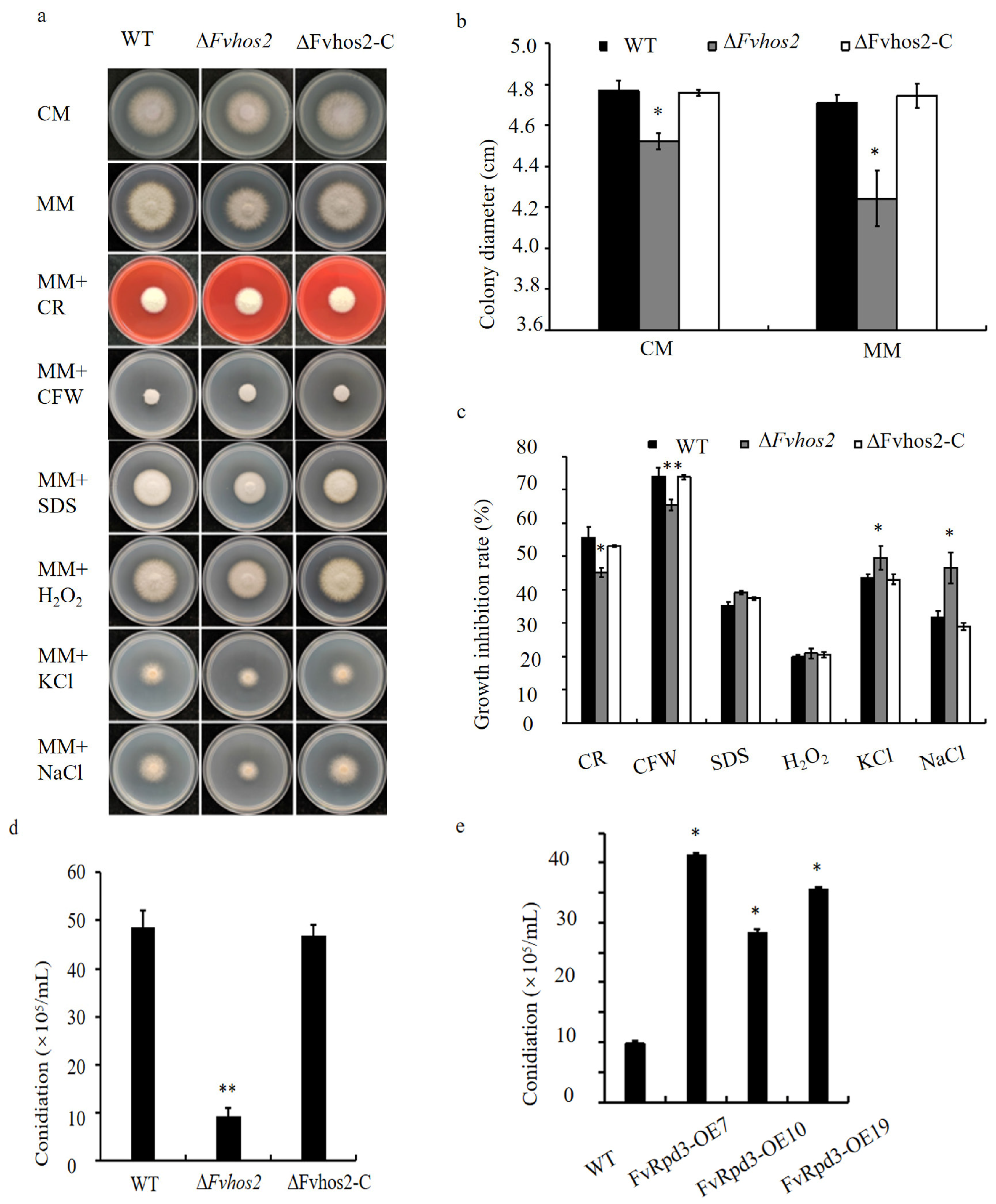
Furthermore, one of the ∆Fvhos2 mutants (∆Fvhos2-16) was randomly selected for the subsequent studies. FvHos2 was found to impact vegetative growth (Figure 3a,b), where the colonies of ∆Fvhos2 had significantly decreased compared with the WT on both nutrient-rich CM and nutrient-deficient MM. Moreover, ∆Fvhos2 exhibited diverse responses to various abiotic stresses. The ∆Fvhos2 deletion mutant exhibited increased sensitivity to high osmotic stress, (e.g., on MM that contained highly concentrated KCl and NaCl), but decreased sensitivity to the medium with CR and CFW (Figure 3a,c). Additionally, significantly reduced production of conidia was observed in the ∆Fvhos2 deletion mutant (Figure 3d). These defects were restored in complementary strains (hereinafter referred to as ∆Fvhos2-C) (Figure 3a–d). Our results highlight the critical role of FvHos2 in the growth, stress response, and conidiation in F. verticillioides.
In addition to FvHOS2, we attempted to generate deletion mutants of FvRPD3, which is the other canonical class I HDAC, in F. verticillioides multiple times, but none were obtained, which led to the hypothesis that FvRPD3 is an essential gene in F. verticillioides. To further explore its function, we inserted an ectopic copy of FvRPD3 under the constitutive promoter RP27, along with a GFP reporter. FvRPD3 overexpressed strains (hereinafter referred to as FvRPD3-OE) displayed with green fluorescence, which indicated the proper expression of the overexpression construct (Figure S3a). Subsequently, the overexpression levels were verified using RT-qPCR, and FvRPD3 was upregulated approximately 25, 30, and 50 folds in the three FvRPD3-OE strains, respectively (Figure S3b). Interestingly, the overexpression of FvRPD3 did not impact the vegetative growth (Figure S3c), while it positively influenced the conidium production (Figure 3e).
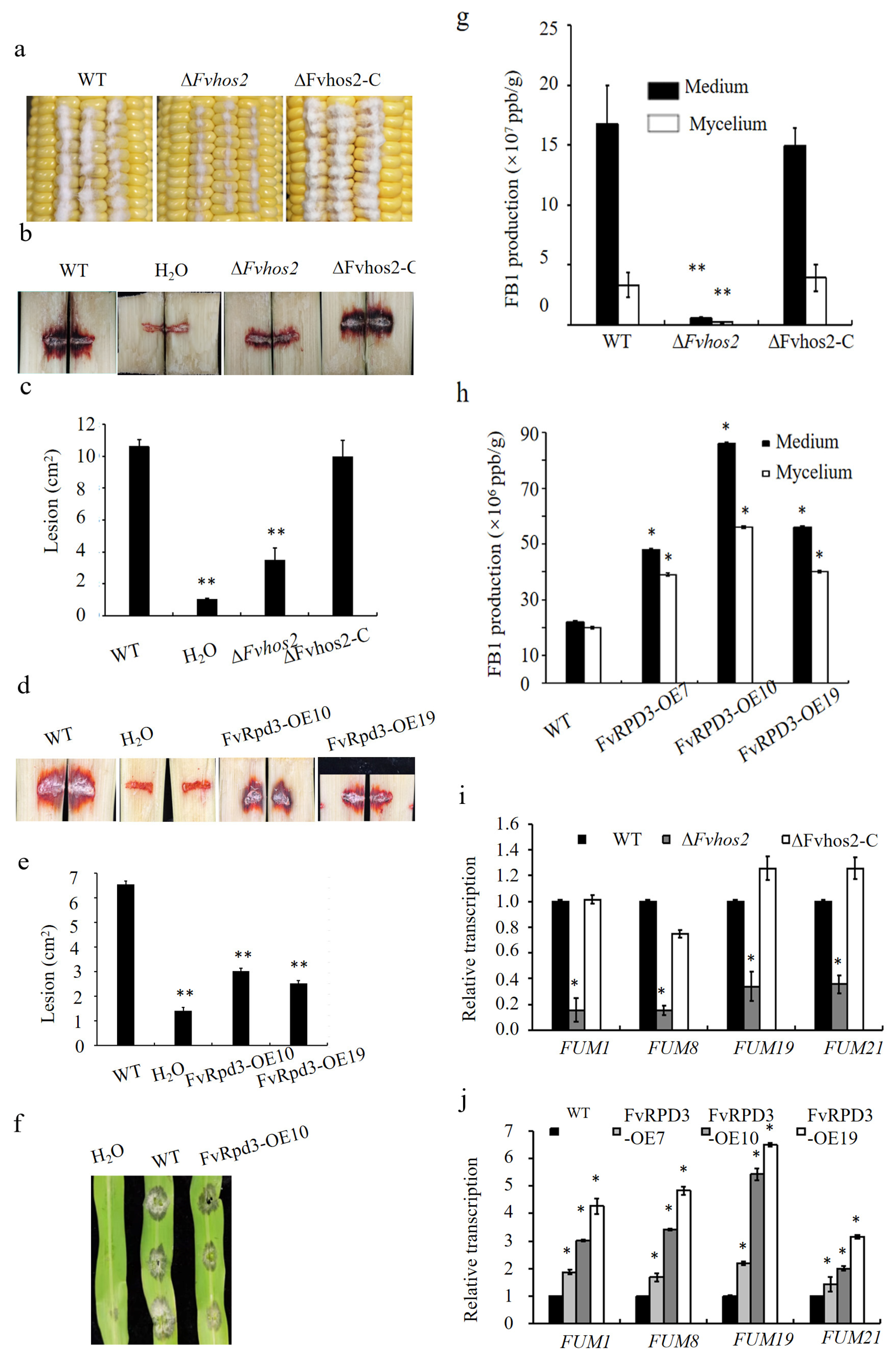
To determine the roles of FvHos2 and FvRpd3 in the pathogenicity, we inoculated the spore suspensions into sugarcane stalk and maize cob. Compared with the WT and ∆Fvhos2-C strains, the pathogenicity of the ∆Fvhos2 mutant was obviously reduced in the maize cob (Figure 4a) and sugarcane stem (Figure 4b). The quantification of the lesion area in the sugarcane stalk was undertaken using Image J software revealed a significant decrease in the deletion mutant (Figure 4c). Similarly, significant reductions in the lesion size were observed for two independent FvRPD3-OE strains in the sugarcane stem (Figure 4d,e) and on maize leaves (Figure 4f). These findings indicate that FvHos2 exerted a positive effect on the pathogenicity toward two different plant hosts, while FvRpd3 negatively regulated the pathogenicity, considering the similar effects observed in both ∆Fvhos2 and FvRPD3-OE strains. Fumonisin B1 (FB1), which is one of the most destructive mycotoxins produced by F. verticillioides, was measured in ∆Fvhos2 and FvRPD3-OE. Both the levels of FB1 production (measured in the mycelium) and secretion (measured in the medium) were significantly reduced in the ∆Fvhos2 deletion mutant (Figure 4g), while the opposite appearance was found in the FvRPD3-OE strains (Figure 4h). To explore the mechanism by which FvHOS2 and FvRPD3 affected the FB1 production, qRT-PCR was used to quantify the expression of the FUM gene cluster, which is known for controlling the biosynthesis of FB1. Four tested FUM genes, i.e., FUM1, FUM8, FUM19, and FUM21, displayed significantly reduced expressions in the ∆Fvhos2 strain (Figure 4i), but higher expressions in the FvRPD3-OE strains (Figure 4j). To gain a more comprehensive understanding of FvHos2 in the transcriptional regulation of the FUM gene cluster, a comparative RNA sequencing analysis between the wild type and ∆Fvhos2 from infected maize kernel was performed, and all the FUM genes expression in the ∆Fvhos2 mutant were found to be downregulated compared with the WT (Table S3).
These observations support the positive impact of both FvHos2 and FvRpd3 on the production of fumonisin FB1 via regulating the FUM genes in F. verticillioides (Figure 4g–j, Table S3). Additionally, to gain more insights into the mechanism by which FvHos2 regulated the FB1 production, we screened for and identified known FB1 regulators from differentially expressed genes (DEGs) between WT and ∆Fvhos2. From our RNA-seq data, we found that FvSda1, which was a previously identified negative regulator of FB1 [48], was significantly upregulated in ∆Fvhos2 (Table S3). Furthermore, qRT-PCR assays validated an approximate 4-fold upregulation of FvSDA1 in the ∆Fvhos2 mutant (Figure S4a). However, a yeast two-hybrid (Y2H) analysis revealed no direct interaction between FvHos2 and FvSda1 (Figure S4b).
Moreover, to investigate the impact of FvHos2 on the gene transcription during maize infection, an RNA-seq analysis was conducted on maize kernels infected with either the WT or ∆Fvhos2 deletion mutant. Relative to the WT, 2365 genes were downregulated, while 234 genes were upregulated in the ∆Fvhos2 mutant (Figure S5a). KEGG analysis showed that the upregulated genes were predominantly enriched in six carbon metabolism pathways, as well as a ribosome biogenesis pathway and a disease-related pathway (Figure S5b), and the downregulated genes were enriched in two metabolic types: the biosynthesis of secondary metabolites (58 genes, such as FUM cluster genes of the FB1 toxin in Table S3) and generic metabolic pathways (189 genes) (Figure S5c). In additions, the GO annotation revealed an upregulation in molecular categories (Figure S5d) and the downregulated genes were significantly enriched in three functional categories, especially in catalytic activity of molecular functionalities category (Figure S5e). Overall, these findings highlight the critical roles and complex regulatory mechanisms of two class I HDACs in F. verticillioides. FvHos2 was involved in vegetative growth, conidiation, virulence, and FB1 production via transcriptional regulation. In contrast, FvRpd3 appeared to be essential, and its overexpression led to defects in virulence but an increased level of conidiation and mycotoxin production.

3.3. The Class II HDAC Hda1 Regulated the Growth, Spore Production, Osmotic Stress Sensitivity, Pathogenicity, and FB1 Synthesis in F. verticillioides

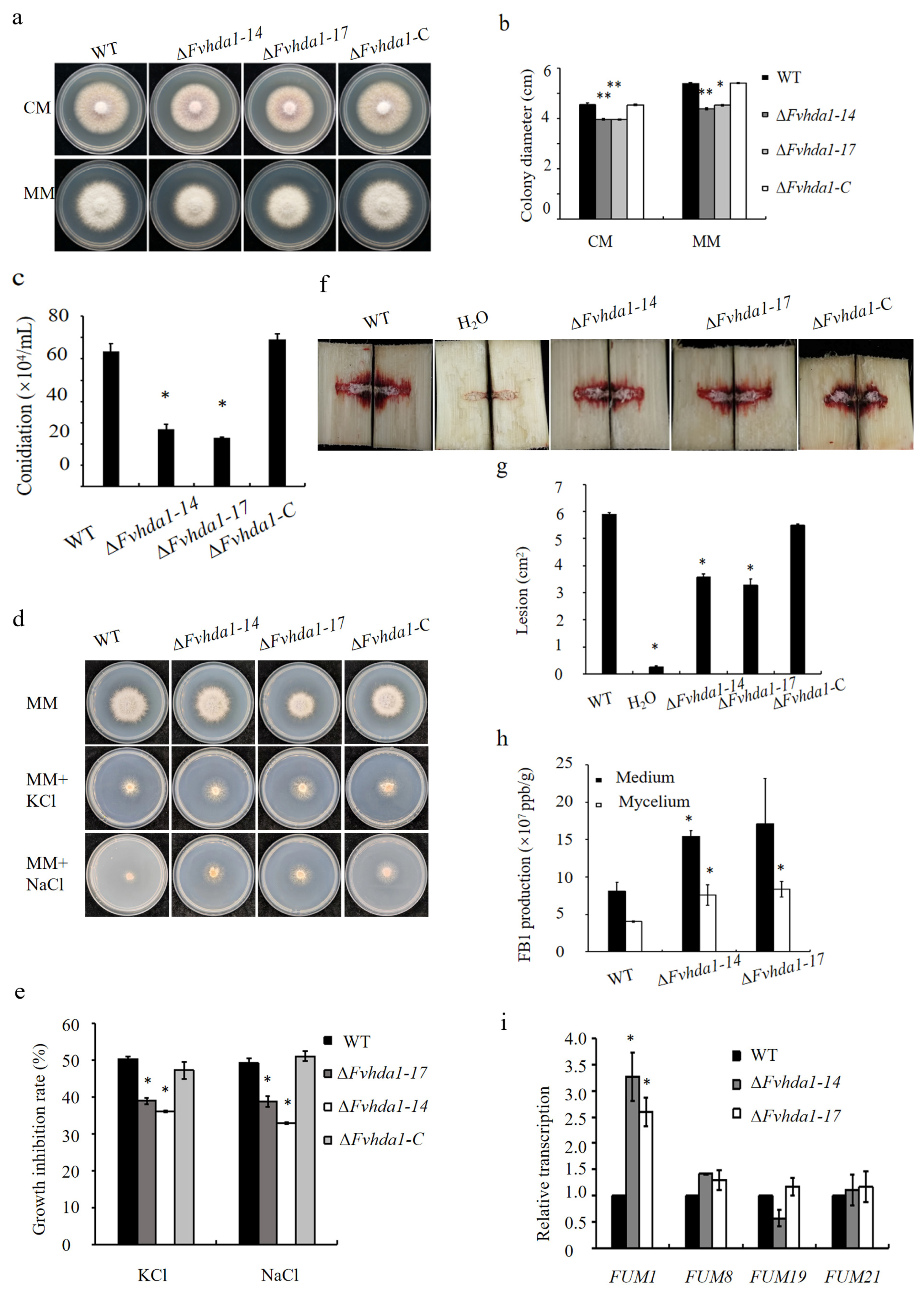
To test the role of class II HDACs in F. verticillioides, we first generated ∆Fvhda1 deletion mutants by employing a homologous recombination strategy (Figure S6a). Three ∆Fvhda1 deletion mutants were then validated by PCR screening and Southern blot hybridization (Figure S6b,c). Compared with the WT and the complemented strain ∆Fvhda1-C, the colony diameter of the ∆Fvhda1 mutants significantly decreased on CM and MM (Figure 5a,b). A significant decrease in conidia production was also observed (Figure 5c), which implied that FvHda1 was involved in the growth and regulation of spore production. The ∆Fvhda1 deletion mutants exhibited significantly decreased sensitivity to high osmotic pressure (e.g., highly concentrated KCl and NaCl) (Figure 5d,e).
To understand the role of FvHda1 in the pathogenesis of F. verticillioides, the conidia of the WT and ∆Fvhda1 mutants were inoculated into a sugarcane stem. The analysis of diseased lesions using Image J software revealed a significant reduction in the ∆Fvhda1 mutants compared with the WT (Figure 5f,g). Furthermore, the restoration of pathogenicity was observed in the ∆Fvhda1-C, which underlines the crucial role of FvHda1 in the regulation of F. verticillioides pathogenicity (Figure 5f,g). Subsequently, the conidia suspensions of WT and ∆Fvhda1 mutants were each inoculated onto a solid maize powder medium. After 10 days, the FB1 content was assessed using an FB1 ELISA Kit, which revealed a higher content in the ∆Fvhda1 compared with the WT (Figure 5h). Additionally, the qRT-PCR revealed an increase in the expression of FUM1 in the ∆Fvhda1 mutants, although not in FUM8, FUM19, and FUM21 (Figure 5i). These findings suggest a negative regulatory role of FvHda1 in the synthesis of FB1 that was potentially mediated by FUM1.
Taken together, FvHDA1 appeared to affect the growth, pathogenicity, transcription of FUM1, and FB1 production in F. verticillioides.

3.4. The Class III HDACs FvSir2, FvHst2, and FvSirt4 Regulated the Growth, Pathogenicity, and FB1 Production in F. verticillioides

To test the function of the class III HDACs, we attempted to generate deletion mutants of FvSIR2, FvHST2, and FvSIRT4. Except for FvSIRT4, the deletion mutants of FvSIR2 and FvHSTt2 were obtained and confirmed by preliminary PCR, followed by Southern blotting (Figure S7). To explore the possible functional redundancy of FvSir2 and FvHst2, a ∆Fvsir2Fvhst2 double-deletion mutant was constructed on the background of the ∆Fvsir2 mutant (Figure S7). Additionally, FvSirt4: OE strains were constructed to study its function. qRT-PCR confirmed that the expression level of FvSIRT4 increased by 7 and 11 times in the two FvSirt4: OE strains, respectively (Figure S7j).
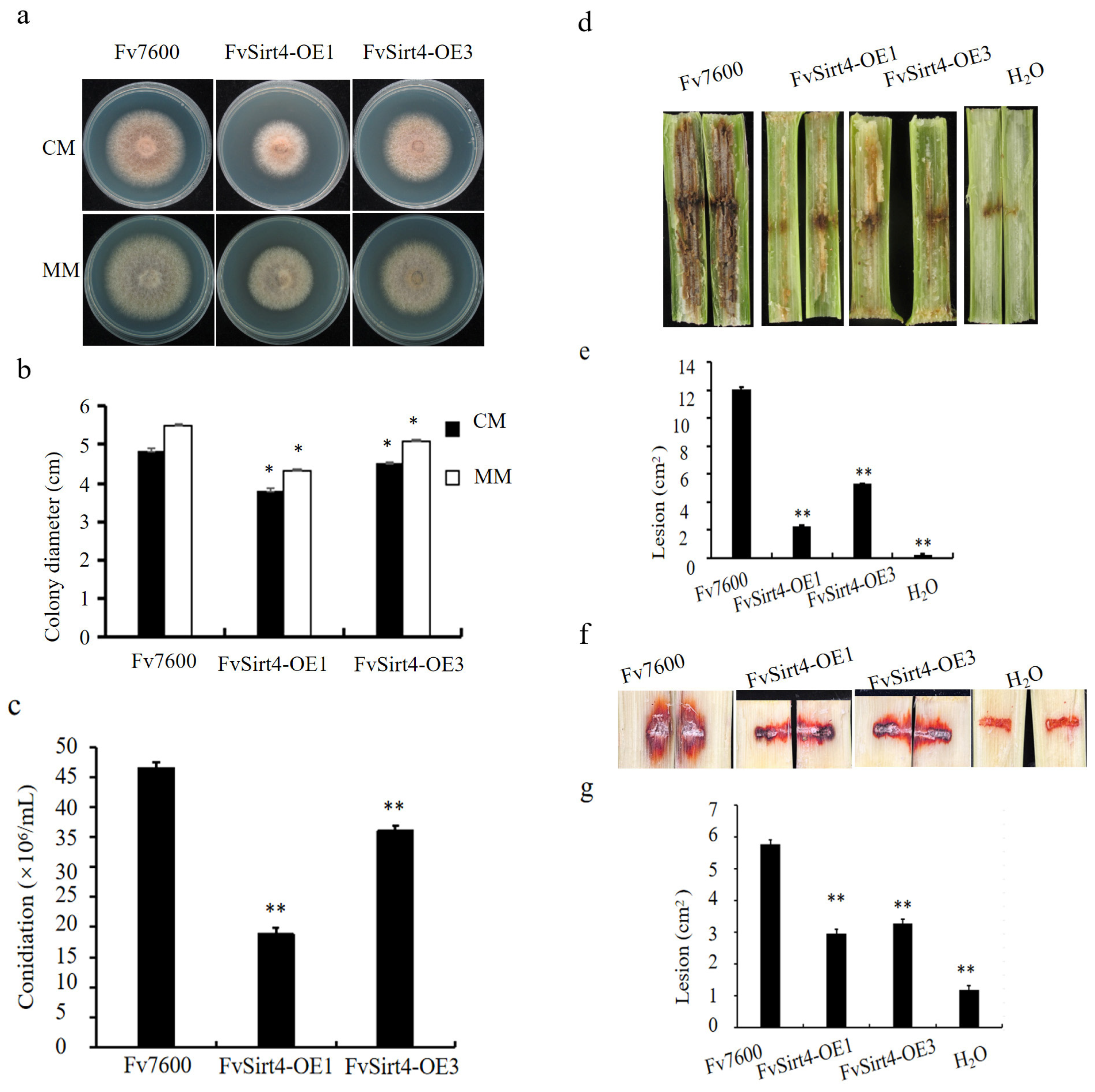
Growth assays on the CMII and MM demonstrated significant inhibitory effects on the FvSirt4-OE growth compared with the WT (Figure 6a,b). A significant decrease in the conidia production on CMII medium was also observed for the FvSirt4-OE strain in comparison with the WT (Figure 6c). However, no discernible effects on the mycelium growth were observed for the ∆Fvsir2, ∆Fvhst2, and ∆Fvsir2hst2 strains when compared with the WT (Figure S8a). A virulence investigation on the sugarcane (Figure 6d) and maize (Figure 6e) stems revealed a significant decrease in the pathogenicity of FvSirt4-OE compared with the WT (Figure 6f,g). However, the ∆Fvsir2, ∆Fvhst2, and ∆Fvsir2hst2 strains exhibited no pathogenicity on sugarcane stems compared with the WT (Figure S8b). The impact of the three class III HDAC genes on FB1 toxin production was assessed in F. verticillioides. The FvSirt4-OE strain displayed decreased FB1 toxin production (Figure 7a), while ∆Fvsir2 presented an increase in toxin production (Figure 7b). Furthermore, ∆Fvsir2hst2, which was the double knockout mutant, demonstrated a higher increase in FB1 toxin production than ∆Fvsir2, which was the single FvSIR2 deleted mutant, which suggests an additive effect of FvHst2 and FvSir2 on the FB1 production pathway (Figure 7c). The analysis of key FUM genes expression involved in FB1 toxin synthesis revealed significant decreases in FUM1, FUM8, and FUM19 in the FvSirt4-OE strain (Figure 7d). However, in the ∆Fvsir2, ∆Fvhst2 single-deletion mutant, and ∆Fvsir2hst2 double-deletion mutant, only a significant increase in FUM19 was observed (Figure 7e,f). Although only FvSirt4 exerted a negative effect on the pathogenicity and growth, all the tested three class III HDACs, i.e., FvSir2, FvHst2, and FvSirt4-OE, negatively influenced the FB1 production, either independently or synergistically.
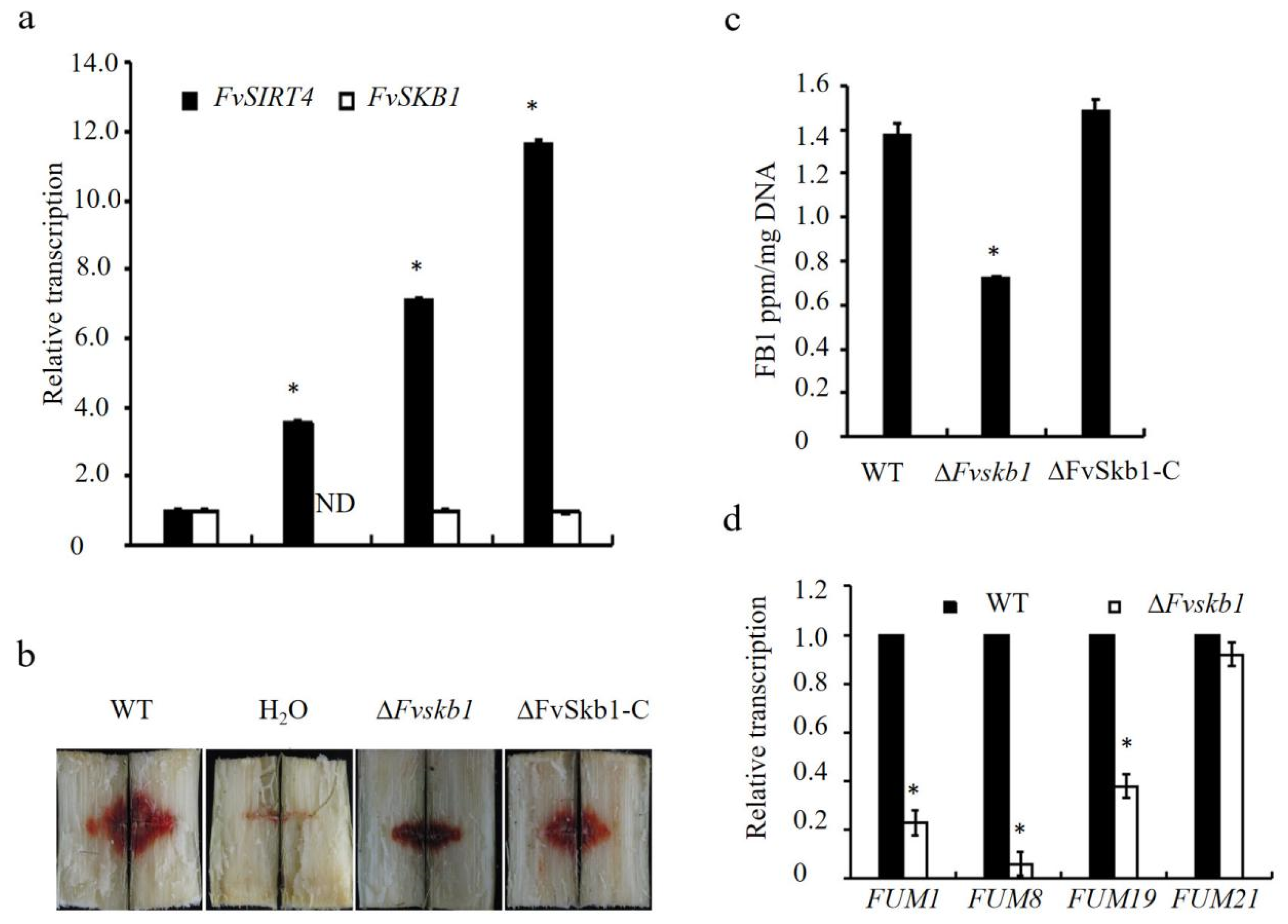
In addition to histones, HDACs are also involved in the deacetylation and interaction with non-histone proteins [49]. Our previous study, which was based on transcriptome co-expression analysis, identified a sub-network module that contained two hub genes [41,50]: FvSIRT4 and an annotated histone methylase FvSKB1 (Figure S9a,b). To dissect the regulatory network of FvSIRT4 and the interaction between the two hub genes, FvSKB1 was deleted via a similar homologous recombination-based approach. The generated transformants were further confirmed with PCR screening and Southern blotting (Figure S9c–e). Additionally, the expression of FvSKB1 was indeed not detected in the ΔFvskb1 mutants, while it was detected in the complemented strain ΔFvSkb1-C by a qRT-PCR assay (Figure S9f). Although there were no observed variances in the expressions of FvSKB1 within the FvSirt4: OE mutants, a significant increase in the expressions of FvSIRT4 within the ΔFvskb1 mutants was revealed (Figure 8a), which was consistent with our predicted network (Figure S9a). Moreover, the disruption of FvSKB1 displayed a significant decrease in pathogenicity (Figure 8b) and FB1 toxin production, along with the downregulation of three FUM genes, i.e., FUM 1, FUM 8, and FUM 19 (Figure 8c,d), which suggests that FvSkb1 might function as a negative regulator of FvSirt4 in terms of pathogenicity and FB1 production.

4. Discussion

Two primary families of HDACs were demonstrated [49]: the traditional histone deacetylase family and the Sir2 regulator lineage (class III). The traditional family is characterized as classes I and II in yeast and classes I, II, and IV in humans. Sirtuins, which are conserved across eukaryotes, prokaryotes, and archaea, are divided into subclasses I through IV and U [51]. In F. verticillioides, 11 HDACs were identified, which are analogous to those in yeast or humans, including class I (FvHos2 and FvRpd3-type), class II (FvHda1 and FvHos3-type), and class III (FvSir2, FvHst2-4, FvSirt4-6-type). Notably, F. verticillioides lacks Hos1 and Hst1, but has three additional Sirt proteins compared with yeast, which suggests a possible evolutionary adaptation in this phytopathogenic fungus.
The subcellular localization of HDACs in F. verticillioides indicates their diverse functional roles. Most HDACs are nuclear, except for FvHst2, which is cytoplasmic, and FvSirt4, which is potentially mitochondrial. This localization pattern is consistent with their homologs in humans and yeast. For example, human Sirt2 and yeast Hst2 move to and associate with chromatin during the G2/M transition of the cell cycle, where they perform functions related to histone deacetylation and chromatin modification [52]. The mitochondrial localization of FvSirt4 aligns with its human counterpart [49], which highlights a conserved functional role. Further investigation is needed to determine whether FvHst2 shows a similar cell-cycle-dependent localization pattern to that of human Sirt2.
HDACs in F. verticillioides have specific roles in histone and non-histone protein deacetylation. On one hand, HDACs can target specific histone residues, such as the deacetylation of H4K16ac by Sir2, which initiates a cascade of deacetylation events [53]. Our analyses suggest a direct role for FvHos2 in the deacetylation of histone H4K16ac. However, some HDACs have no site-specific histones substrates, such as Hda1, deacetylating histones H2B and H3 [14], and the Rpd3 deacetylating, which are all lysines within the core histones H3, H4, H2A, and H2B, with the exception of H4K16ac [54]. Notably, the failure to delete FvRpd3 indicates its essential role in the organism, which may be due to its impact on innumerable histone deacetylation sites, thereby affecting a vast array of gene expression and function. On the other hand, the regulatory role of HDACs in F. verticillioides extends beyond histone modification to non-histone proteins. FvSirt4 and FvHst2, also known as human Sirt4 and Sirt5 [49], can interact with non-histone substrates. FvSirt4 is known to deacetylate non-histone targets within mitochondria, which influences the cellular metabolism and stress responses. Similarly, FvHst2, which is localized in the cytoplasm, may interact with cytoskeletal proteins, which affects the cell structure and mobility. These interactions highlight the multifaceted roles of HDACs in cellular function and regulation.
HDACs significantly impact fungal pathogenicity and growth. In F. verticillioides, class I HDACs, like FvHos2, and class II HDACs, like FvHda1, promote growth, while class I HDACs, like FvRpd3, and class III HDACs, like FvSirt4, negatively impact pathogenicity. The overexpression of FvRpd3 impairs infection capabilities, which is similar to findings in other fungi such as B. cinerea and M. oryzae, where Rpd3 influences infection structure formation and virulence [25,26]. Additionally, in U. maydis, Hda1 acts as a repressor of the biotrophic marker gene mig1 during infection [55], which further illustrates the critical roles of HDACs in fungal virulence and adaptability.
Regarding secondary metabolites, HDACs regulate various biosynthetic pathways in fungi. In F. fujikuroi, the deletion of Hda1 affects secondary metabolites, like gibberellic acid and fusaric acid, and disrupts bikaverin biosynthesis regulation [27,56]. In F. graminearum, Hdf1, which is a homolog of Hos2, is involved in deoxynivalenol production [57]. Our studies in F. verticillioides revealed that different HDACs uniquely regulated FB1 biosynthesis. Class I HDACs, like FvHos2 and FvRpd3, enhanced the FB1 production, while class II HDACs, like FvHda1, negatively regulated it. Class III HDACs, such as FvSir2, FvHst2, and FvSirt4, negatively impacted the FB1 production through different regulatory mechanisms. This intricate regulation of secondary metabolites highlights the multifaceted roles of HDACs in fungal pathogenicity and survival.
HDAC regulates gene transcription through two patterns—regulating chromatin dynamics or regulating the acetylation status of specific genes. The hypoacetylation of histones is generally associated with heterochromatin (HM) formation and gene silencing, while hyperacetylation is linked to euchromatin formation and gene activation [58]. Approximately 10% of gene transcription is governed by HDACs through global expression profiling experiments [49]. However, our study identified FvHos2 as a negative regulator of ribosomal protein genes expression based on RNA-seq data, as Hos2 in yeast exhibits a preference for the deacetylation of ribosomal protein genes [59]. An increased expression of FUM1 and FUM21 genes has been linked with a hyperacetylated state of histones, with a corresponding increase in acetylation levels in the promoter regions of these genes under fumonisin B1 (FB1)-inducing conditions in F. verticillioides [60]. Moreover, in F. verticillioides, class I HDACs, like FvHos2 and FvRpd3, positively regulate key fumonisin-related genes, such as FUM1, FUM8, and FUM19, which enhances FB1 production, whereas FvSirt4 negatively regulates their expression, along with FvHda1, which only negatively regulates FUM1.
Additionally, the interplay between HDACs and other epigenetic regulators in F. verticillioides is crucial for understanding their comprehensive roles. For instance, FvSirt4 upregulation in ΔFvskb1 mutants suggests a compensatory mechanism between these proteins, which indicates a complex network of gene regulation. The observed reduction in FB1 toxin production and pathogenicity in these mutants further supports the interconnected roles of HDACs and histone methylases in regulating fungal virulence and secondary metabolism.
The involvement of HDACs in regulating key pathways related to stress response, development, and secondary metabolism is evident in various fungal pathogens [14,56,59,61]. In F. verticillioides, the failure to delete FvRpd3 results in a lethal phenotype is consistent with the observations in other fungi, such as M. oryzae, A. fumigatus, and B. cinerea [24,25,26]. Similarly, the role of Hda1 in normal germination and vegetative growth, as seen in F. fujikuroi, underscores its importance in fungal development [27]. U. maydis Hos2, directly controls the expression of mating-type genes through H4K16 deacetylation, and hence, acts as a prerequisite for the dimorphic switch [62]. In addition, the HDAC complex Sum1-Rfm1-Hst1 regulated biological processes, which was identified as being able to repress sporulation genes during vegetative growth [63,64]. Some genes containing Hda1-affected subtelomeric (HAST) domains are deacetylated by the Hda1 complex, which is involved in crucial survival pathways, such as gluconeogenesis, alternative carbon-source utilization, and tolerance of adverse conditions, including osmotic shock, starvation, and anaerobic growth [14,59]. These genes are typically repressed in a nutrient-rich medium via a mechanism implicating the Tup1 repressor [65]. ΔFvHos2 and ΔFvHda1 showed increased sensitivity to osmotic shock induced by high salt concentrations. These examples highlight the essential functions of HDACs in maintaining cellular homeostasis and adaptability.
Overall, our findings highlight the complex roles of HDACs in F. verticillioides, which impacted both the pathogenicity and secondary metabolite production. While some HDACs were essential for fungal growth and virulence, others modulated specific pathways and processes. Further investigation into the comprehensive acetylation mechanisms and the interplay between different HDACs will enhance our understanding of their roles in fungal biology and could lead to the development of novel strategies for managing fungal diseases and toxin production.

5. Conclusions

In this study, we identified three classes of HDACs with 11 distinct types and characterized six of these types—FvRpd3, FvHos2, FvHda1, Fvhst2, FvSir2, and FvSirt4—for their biological function in the phytopathogenic fungus F. verticillioides (Table 1). Overall, the class I and II HDACs appeared to play more significant roles compared with those in class III, which demonstrated a phenomenon of functional redundancy. FvRpd3, FvHos2, FvHda1, and FvSirt4 were associated with pathogenicity, growth, and conidia. These six HDAC proteins had different effects on the production of FB1. These findings contribute to the development of effective strategies for disease and toxin control by precisely regulating the acetylation levels.

Supplementary Materials

The following supporting information can be downloaded from https://www.mdpi.com/article/10.3390/agronomy14102196/s1, Figure S1: Identification of HDACs in F. verticillioides; Figure S2: Molecular validation of ∆Fvhos2 deletion mutants; Figure S3: Verification of overexpression mutants of FvRpd3-OE and the colony growth of FvRpd3-OE mutants; Figure S4: FvHos2 indirectly negatively regulated FvSda1; Figure S5: The RNA-seq analysis of the ∆Fvhos2 mutant; Figure S6: Molecular validation of ∆Fvhda1 deletion mutants; Figure S7: Identified gene knockout mutants of ∆Fvsir2, ∆Fvhst2, and ∆Fvsir2Fvhst2, and overexpression mutants of FvSirt4-OE; Figure S8: FvSir2 and FvHst2 had no effects on mycelial growth or pathogenicity; Figure S9: Prediction of negative regulation of FvSIRT4 by FvSkb1, which is a histone methylase and the identification of ∆Fvskb1 and ∆Fvskb1-C; Table S1: Classification and related complexes of HDACs; Table S2: The primers used in this study; Table S3: The expressions of FUM genes in ∆Fvhos2.

Author Contributions

Conceptualization, W.Y., G.L. and Z.W.; methodology, W.Y., J.W., M.W., G.W. and J.L.; analysis, X.C.; writing W.Y. and J.H.; supervision, J.H. and Z.W. All authors have read and agreed to the published version of the manuscript.

Funding

This work was supported by the Fujian Province Science and Technology Major Special Project (2022NZ0106) and by the Science and Technology Innovation Funding of FAFU (CXZX2020044A).

Data Availability Statement

The original contributions presented in the study are included in the article/Supplementary Material; further inquiries can be directed to the corresponding author/s.

Conflicts of Interest

The authors declare no conflicts of interest.

References

  1. Nitsch, S.; Zorro Shahidian, L.; Schneider, R. Histone acylations and chromatin dynamics: Concepts, challenges, and links to metabolism. EMBO Rep. 2021, 22, e52774. [Google Scholar] [CrossRef] [PubMed]
  2. Verdin, E.; Ott, M. 50 years of protein acetylation: From gene regulation to epigenetics, metabolism and beyond. Nat. Rev. Mol. Cell Biol. 2015, 16, 258–264. [Google Scholar] [CrossRef]
  3. Vidal, M.; Gaber, R.F. RPD3 encodes a second factor required to achieve maximum positive and negative transcriptional states in Saccharomyces cerevisiae. Mol. Cell. Biol. 1991, 11, 6317–6327. [Google Scholar]
  4. Taunton, J.; Hassig, C.A.; Schreiber, S.L. A mammalian histone deacetylase related to the yeast transcriptional regulator Rpd3p. Science 1996, 272, 408–411. [Google Scholar] [CrossRef] [PubMed]
  5. Li, X.; Egervari, G.; Wang, Y.; Berger, S.L.; Lu, Z. Regulation of chromatin and gene expression by metabolic enzymes and metabolites. Nat. Rev. Mol. Cell Biol. 2018, 19, 563–578. [Google Scholar] [CrossRef] [PubMed]
  6. Rundlett, S.E.; Carmen, A.A.; Kobayashi, R.; Bavykin, S.; Turner, B.M.; Grunstein, M. HDA1 and RPD3 are members of distinct yeast histone deacetylase complexes that regulate silencing and transcription. Proc. Natl. Acad. Sci. USA 1996, 93, 14503–14508. [Google Scholar] [CrossRef]
  7. Frye, R.A. Phylogenetic classification of prokaryotic and eukaryotic Sir2-like proteins. Biochem. Biophys. Res. Commun. 2000, 273, 793–798. [Google Scholar] [CrossRef]
  8. Kurdistani, S.K.; Grunstein, M. Histone acetylation and deacetylation in yeast. Nat. Rev. Mol. Cell Biol. 2003, 4, 276–284. [Google Scholar] [CrossRef]
  9. Dai, Y.; Faller, D.V. Transcription regulation by Class III histone deacetylases (HDACs)-Sirtuins. Transl. Oncogenomics 2008, 3, 53–65. [Google Scholar]
  10. Patel, A.B.; Qing, J.; Tam, K.H.; Zaman, S.; Luiso, M.; Radhakrishnan, I.; He, Y. Cryo-EM structure of the Saccharomyces cerevisiae Rpd3L histone deacetylase complex. Nat. Commun. 2023, 14, 3061. [Google Scholar] [CrossRef]
  11. Yeheskely-Hayon, D.; Kotler, A.; Stark, M.; Hashimshony, T.; Sagee, S.; Kassir, Y. The roles of the catalytic and noncatalytic activities of Rpd3L and Rpd3S in the regulation of gene transcription in yeast. PLoS ONE 2013, 8, e85088. [Google Scholar] [CrossRef] [PubMed]
  12. Kurdistani, S.K.; Robyr, D.; Tavazoie, S.; Grunstein, M. Genome-wide binding map of the histone deacetylase Rpd3 in yeast. Nat. Genet. 2002, 31, 248–254. [Google Scholar] [CrossRef] [PubMed]
  13. Carrozza, M.J.; Li, B.; Florens, L.; Suganuma, T.; Swanson, S.K.; Lee, K.K.; Shia, W.J.; Anderson, S.; Yates, J.; Washburn, M.P.; et al. Histone H3 methylation by Set2 directs deacetylation of coding regions by Rpd3S to suppress spurious intragenic transcription. Cell 2005, 123, 581–592. [Google Scholar] [CrossRef] [PubMed]
  14. Wu, J.; Carmen, A.A.; Kobayashi, R.; Suka, N.; Grunstein, M. HDA2 and HDA3 are related proteins that interact with and are essential for the activity of the yeast histone deacetylase HDA1. Proc. Natl. Acad. Sci. USA 2001, 98, 4391–4396. [Google Scholar] [CrossRef]
  15. Islam, A.; Turner, E.L.; Menzel, J.; Malo, M.E.; Harkness, T.A. Antagonistic Gcn5-Hda1 interactions revealed by mutations to the anaphase promoting complex in yeast. Cell Div. 2011, 6, 13. [Google Scholar] [CrossRef]
  16. Wu, J.; Suka, N.; Carlson, M.; Grunstein, M. TUP1 utilizes histone H3/H2B-specific HDA1 deacetylase to repress gene activity in yeast. Mol. Cell 2001, 7, 117–126. [Google Scholar] [CrossRef]
  17. Wang, A.; Kurdistani, S.K.; Grunstein, M. Requirement of Hos2 histone deacetylase for gene activity in yeast. Science 2002, 298, 1412–1414. [Google Scholar] [CrossRef]
  18. Wirén, M.; Silverstein, R.A.; Sinha, I.; Walfridsson, J.; Lee, H.M.; Laurenson, P.; Pillus, L.; Robyr, D.; Grunstein, M.; Ekwall, K. Genomewide analysis of nucleosome density histone acetylation and HDAC function in fission yeast. EMBO J. 2005, 24, 2906–2918. [Google Scholar] [CrossRef]
  19. North, B.J.; Verdin, E. Sirtuins: Sir2-related NAD-dependent protein deacetylases. Genome Biol. 2004, 5, 224. [Google Scholar] [CrossRef]
  20. Guarente, L. Sir2 links chromatin silencing, metabolism, and aging. Genes. Dev. 2000, 14, 1021–1026. [Google Scholar] [CrossRef]
  21. Kobayashi, T.; Horiuchi, T.; Tongaonkar, P.; Vu, L.; Nomura, M. SIR2 regulates recombination between different rDNA repeats, but not recombination within individual rRNA genes in yeast. Cell 2004, 117, 441–453. [Google Scholar] [CrossRef] [PubMed]
  22. Xu, F.; Zhang, Q.; Zhang, K.; Xie, W.; Grunstein, M. Sir2 deacetylates histone H3 lysine 56 to regulate telomeric heterochromatin structure in yeast. Mol. Cell 2007, 27, 890–900. [Google Scholar] [CrossRef] [PubMed]
  23. Li, M.; Fine, R.D.; Dinda, M.; Bekiranov, S.; Smith, J.S. A Sir2-regulated locus control region in the recombination enhancer of Saccharomyces cerevisiae specifies chromosome III structure. PLoS Genet. 2019, 15, e1008339. [Google Scholar] [CrossRef]
  24. Bauer, I.; Misslinger, M.; Shadkchan, Y.; Dietl, A.M.; Petzer, V.; Orasch, T.; Abt, B.; Graessle, S.; Osherov, N.; Haas, H. The lysine deacetylase RpdA is essential for virulence in Aspergillus fumigatus. Front. Microbiol. 2019, 10, 2773. [Google Scholar] [CrossRef]
  25. Zhang, N.; Yang, Z.; Zhang, Z.; Liang, W. BcRPD3-mediated histone deacetylation is involved in growth and pathogenicity of Botrytis cinerea. Front. Microbiol. 2020, 11, 1832. [Google Scholar] [CrossRef]
  26. Lee, S.H.; Farh, M.E.; Lee, J.; Oh, Y.T.; Cho, E.; Park, J.; Son, H.; Jeon, J. A histone deacetylase, Magnaporthe oryzae RPD3, regulates reproduction and pathogenic development in the rice blast fungus. mBio 2021, 12, e0260021. [Google Scholar] [CrossRef] [PubMed]
  27. Studt, L.; Schmidt, F.J.; Jahn, L.; Shevchenko, A.; Tekotte, H.; Wilm, M.; Rigaut, G.; Séraphin, B.; Aasland, R.; Stewart, A.F. Two histone deacetylases, FfHda1 and FfHda2, are important for Fusarium fujikuroi secondary metabolism and virulence. Appl. Environ. Microbiol. 2013, 79, 7719–7734. [Google Scholar] [CrossRef]
  28. Reichmann, M.; Jamnischek, A.; Weinzierl, G. The histone deacetylase Hda1 from Ustilago maydis is essential for teliospore development. Mol. Microbiol. 2002, 46, 1169–1182. [Google Scholar] [CrossRef]
  29. Pijnappel, W.W.; Schaft, D.; Roguev, A.; Ladendorf, O.; Huber, S.; Kahmann, R.; Kämper, J. The S. cerevisiae SET3 complex includes two histone deacetylases, Hos2 and Hst1, and is a meiotic-specific repressor of the sporulation gene program. Genes Dev. 2001, 15, 2991–3004. [Google Scholar] [CrossRef]
  30. Hnisz, D.; Majer, O.; Frohner, I.E.; Komnenovic, V.; Kuchler, K. The Set3/Hos2 histone deacetylase complex attenuates cAMP/PKA signaling to regulate morphogenesis and virulence of Candida albicans. PLoS Pathog. 2010, 6, e1000889. [Google Scholar] [CrossRef]
  31. Pérez-Martín, J.; Uría, J.A.; Johnson, A.D. Phenotypic switching in Candida albicans is controlled by a SIR2 gene. EMBO J. 1999, 18, 2580–2592. [Google Scholar] [CrossRef] [PubMed]
  32. Fernandez, J.; Marroquin-Guzman, M.; Nandakumar, R.; Shijo, S.; Cornwell, K.M.; Li, G.; Wilson, R.A. Plant defence suppression is mediated by a fungal sirtuin during rice infection by Magnaporthe oryzae. Mol. Microbiol. 2014, 94, 70–88. [Google Scholar] [CrossRef]
  33. Cai, Q.; Tian, L.; Xie, J.T.; Huang, Q.Y.; Feng, M.G.; Keyhani, N.O. A fungal sirtuin modulates development and virulence in the insect pathogen, Beauveria bassiana. Environ. Microbiol. 2021, 23, 5164–5183. [Google Scholar] [CrossRef]
  34. Brandão, F.A.; Derengowski, L.S.; Albuquerque, P.; Nicola, A.M.; Silva-Pereira, I.; Poças-Fonseca, M.J. Histone deacetylases inhibitors effects on Cryptococcus neoformans major virulence phenotypes. Virulence 2015, 6, 618–630. [Google Scholar] [CrossRef] [PubMed]
  35. Gallegos-García, V.; Pan, S.J.; Juárez-Cepeda, J.; Ramírez-Zavaleta, C.Y.; Martin-del-Campo, M.B.; Martínez-Jiménez, V.; Castaño, I.; Cormack, B.; De Las Peñas, A. A novel downstream regulatory element cooperates with the silencing machinery to repress EPA1 expression in Candida glabrata. Genetics 2012, 190, 1285–1297. [Google Scholar] [CrossRef] [PubMed]
  36. Arras, S.D.M.; Chitty, J.L.; Wizrah, M.S.I.; Erpf, P.E.; Schulz, B.L.; Tanurdzic, M.; Fraser, J.A. Sirtuins in the phylum Basidiomycota: A role in virulence in Cryptococcus neoformans. Scientificreports 2017, 7, 46567. [Google Scholar] [CrossRef] [PubMed]
  37. Lin, C.; Cao, X.; Qu, Z.; Zhang, S.; Naqvi, N.I.; Deng, Y.Z. The histone deacetylases MoRpd3 and MoHst4 regulate growth, conidiation, and pathogenicity in the rice blast gungus Magnaporthe oryzae. mSphere 2021, 6, e0011821. [Google Scholar] [CrossRef]
  38. Ponce-García, N.; Serna-Saldivar, S.O.; Garcia-Lara, S. Fumonisins and their analogues in contaminated corn and its processed foods—A review. Food Addit. Contam. Part A 2018, 35, 2183–2203. [Google Scholar] [CrossRef]
  39. Kumar, S.; Stecher, G.; Tamura, K. MEGA7: Molecular evolutionary genetics analysis version 7.0 for bigger datasets. Mol. Biol. Evol. 2016, 33, 1870–1874. [Google Scholar] [CrossRef]
  40. Yu, W.Y.; Lin, M.; Peng, M.H.; Yan, H.J.; Wang, J.J.; Zhou, J.; Lu, G.D.; Wang, Z.H.; Shim, W.B. Fusarium verticillioides FvPex8 is a key component of the peroxisomal Docking/Translocation module that serves important roles in fumonisin biosynthesis but not in virulence. Mol. Plant Microbe Interact. 2021, 34, 803–814. [Google Scholar] [CrossRef]
  41. Yu, W.Y.; Lin, M.; Yan, H.J.; Wang, J.; Zhang, S.; Lu, G.D.; Wang, Z.H.; Shim, W.B. The peroxisomal matrix shuttling receptor Pex5 plays a role of FB1 production and virulence in Fusarium verticillioides. J. Integr. Agric. 2022, 21, 2957–2972. [Google Scholar] [CrossRef]
  42. Ma, L.J.; van der Does, H.C.; Borkovich, K.A.; Coleman, J.J.; Daboussi, M.J.; Di Pietro, A.; Dufresne, M.; Freitag, M.; Grabherr, M.; Henrissat, B.; et al. Comparative genomics reveals mobile pathogenicity chromosomes in Fusarium. Nature 2010, 464, 367–373. [Google Scholar] [CrossRef] [PubMed]
  43. Ye, J.; Zhang, Y.; Cui, H.; Liu, J.; Wu, Y.; Cheng, Y.; Xu, H.; Huang, X.; Li, S.; Zhou, A.; et al. WEGO 2.0: A web tool for analyzing and plotting GO annotations, 2018 update. Nucleic Acids Res. 2018, 46, W71–W75. [Google Scholar] [CrossRef] [PubMed]
  44. Gallo-Oller, G.; Ordoñez, R.; Dotor, J. A new background subtraction method for Western blot densitometry band quantification through image analysis software. J. Immunol. Methods 2018, 457, 1–5. [Google Scholar] [CrossRef]
  45. Lin, M.; Abubakar, Y.S.; Wei, L.J.; Wang, J.J.; Lu, X.G.; Lu, G.D.; Wang, Z.H.; Zhou, J.; Yu, W.Y. Fusarium verticillioides Pex7/20 mediates peroxisomal PTS2 pathway import, pathogenicity, and fumonisin B1 biosynthesis. Appl. Microbiol. Biotechnol. 2022, 106, 6595–6609. [Google Scholar] [CrossRef]
  46. Hilton, A.; Zhang, H.; Yu, W.; Shim, W.B. Identification and characterization of pathogenic and endophytic fungal species associated with pokkah boeng disease of sugarcane. Plant Pathol. J. 2017, 33, 238–248. [Google Scholar] [CrossRef]
  47. Gallan, D.Z.; Henrique, M.O.; Silva-Filho, M.C. The phytopathogen fusarium verticillioides modifies the intestinal morphology of the sugarcane borer. Pathogens 2023, 12, 443. [Google Scholar] [CrossRef] [PubMed]
  48. Malapi-Wight, M.; Smith, J.; Campbell, J.; Bluhm, B.H.; Shim, W.B. Sda1, a Cys2-His2 zinc finger transcription factor, is involved in polyol metabolism and fumonisin B1 production in Fusarium verticillioides. PLoS ONE 2013, 8, e67656. [Google Scholar] [CrossRef]
  49. Seto, E.; Yoshida, M. Erasers of histone acetylation: The histone deacetylase enzymes. Cold Spring Harb. Perspect. Biol. 2014, 6, a018713. [Google Scholar] [CrossRef]
  50. Kim, M.S.; Zhang, H.; Yan, H.; Yoon, B.J.; Shim, W.B. Characterizing co-expression networks underpinning maize stalk rot virulence in Fusarium verticillioides through computational subnetwork module analyses. Sci. Rep. 2018, 8, 8310. [Google Scholar] [CrossRef]
  51. Costantini, S.; Sharma, A.; Raucci, R.; Costantini, M.; Autiero, I.; Colonna, G. Genealogy of an ancient protein family: The Sirtuins, a family of disordered members. BMC Evol. Biol. 2013, 13, 60. [Google Scholar] [CrossRef] [PubMed]
  52. Vaquero, A.; Scher, M.B.; Lee, D.H.; Sutton, A.; Cheng, H.L.; Alt, F.W.; Serrano, L.; Sternglanz, R.; Reinberg, D. SirT2 is a histone deacetylase with preference for histone H4 Lys 16 during mitosis. Genes. Dev. 2006, 20, 1256–1261. [Google Scholar] [CrossRef] [PubMed]
  53. Vermeulen, M.; Carrozza, M.J.; Lasonder, E.; Workman, J.L.; Logie, C.; Stunnenberg, H.G. In vitro targeting reveals intrinsic histone tail specificity of the Sin3/histone deacetylase and N-CoR/SMRT corepressor complexes. Mol. Cell. Biol. 2004, 24, 2364–2372. [Google Scholar] [CrossRef] [PubMed]
  54. Suka, N.; Suka, Y.; Carmen, A.A.; Wu, J.; Grunstein, M. Highly specific antibodies determine histone acetylation site usage in yeast heterochromatin and euchromatin. Mol. Cell 2001, 8, 473–479. [Google Scholar] [CrossRef]
  55. Torreblanca, J.; Stumpferl, S.; Basse, C.W. Histone deacetylase Hda1 acts as repressor of the Ustilago maydis biotrophic marker gene mig1. Fungal Genet. Biol. 2003, 38, 22–32. [Google Scholar] [CrossRef]
  56. Shwab, E.K.; Bok, J.W.; Tribus, M.; Galehr, J.; Graessle, S.; Keller, N.P. Histone deacetylase activity regulates chemical diversity in Aspergillus. Eukaryot. Cell 2007, 6, 1656–1664. [Google Scholar] [CrossRef]
  57. Li, Y.; Wang, C.; Liu, W.; Wang, G.; Kang, Z.; Kistler, H.C.; Xu, J.R. The HDF1 histone deacetylase gene is important for conidiation, sexual reproduction, and pathogenesis in Fusarium graminearum. Mol. Plant Microbe Interact. 2011, 24, 487–496. [Google Scholar] [CrossRef] [PubMed]
  58. Trojer, P.; Brandtner, E.M.; Brosch, G.; Loidl, P.; Galehr, J.; Linzmaier, R.; Haas, H.; Mair, K.; Tribus, M.; Graessle, S. Histone deacetylases in fungi: Novel members, new facts. Nucleic Acids Res. 2003, 31, 3971–3981. [Google Scholar] [CrossRef]
  59. Robyr, D.; Suka, Y.; Xenarios, I.; Kurdistani, S.K.; Wang, A.; Suka, N.; Grunstein, M. Microarray deacetylation maps determine genome-wide functions for yeast histone deacetylases. Cell 2002, 109, 437–446. [Google Scholar] [CrossRef]
  60. Visentin, I.; Montis, V.; Döll, K.; Alabouvette, C.; Tamietti, G.; Karlovsky, P.; Cardinale, F. Transcription of genes in the biosynthetic pathway for fumonisin mycotoxins is epigenetically and differentially regulated in the fungal maize pathogen Fusarium verticillioides. Eukaryot. Cell 2012, 11, 252–259. [Google Scholar] [CrossRef]
  61. Roze, L.V.; Arthur, A.E.; Hong, S.Y.; Chanda, A.; Linz, J.E. The initiation and pattern of spread of histone H4 acetylation parallel the order of transcriptional activation of genes in the aflatoxin cluster. Mol. Microbiol. 2007, 66, 713–726. [Google Scholar] [CrossRef] [PubMed]
  62. Elías-Villalobos, A.; Fernández-Álvarez, A.; Moreno-Sánchez, I.; Helmlinger, D.; Ibeas, J.I. The Hos2 histone deacetylase controls Ustilago maydis virulence through direct regulation of mating-type genes. PLoS Pathog. 2015, 11, e1005134. [Google Scholar] [CrossRef] [PubMed]
  63. McCord, R.; Pierce, M.; Xie, J.; Wonkatal, S.; Mickel, C.; Vershon, A.K. Rfm1, a novel tethering factor required to recruit the Hst1 histone deacetylase for repression of middle sporulation genes. Mol. Cell. Biol. 2003, 23, 2009–2016. [Google Scholar] [CrossRef] [PubMed]
  64. Xie, J.; Pierce, M.; Gailus-Durner, V.; Wagner, M.; Winter, E.; Vershon, A.K. Sum1 and Hst1 repress middle sporulation-specific gene expression during mitosis in Saccharomyces cerevisiae. EMBO J. 1999, 18, 6448–6454. [Google Scholar] [CrossRef]
  65. Ha, S.D.; Ham, S.; Kim, M.Y.; Kim, J.H.; Jang, I.; Lee, B.B.; Lee, M.K.; Hwang, J.T.; Roh, T.Y.; Kim, T. Transcription-dependent targeting of Hda1C to hyperactive genes mediates H4-specific deacetylation in yeast. Nat. Commun. 2019, 10, 4270. [Google Scholar] [CrossRef]
Figure 1. Subcellular localization of six representative histone deacetylases (HDACs) in Fusarium verticillioides. Subcellular localization of four HDAC-GFP fusions in the respective corresponding deletion mutants (∆Fvhos2, ∆Fvhda1, ∆Fvsir2, and ∆Fvhst2) and the other two HDAC-GFP fusions (FvRpd3 and FvHst2) in the wild-type strain. Nuclear localization was determined with DAPI staining. Bars: 10 µm.
Agronomy 14 02196 g001
Figure 2. Fvhos2 regulated the H4K16 acetylation activity. (a) Western blot analysis was used to assess the H4K16 acetylation in F. verticillioides and ∆Fvhos2 mutants using an H4 antibody as a loading control. (b) The relative quantification of H4K16ac levels to histone H4 was executed using Image J software. The statistical significance was tested using a t-test (* p < 0.05).
Agronomy 14 02196 g002
Figure 3. Roles of FvHos2 and FvRpd3 in vegetative growth, stress sensitivity, and conidiation of F. verticillioides. (a) Photographs of the colonial morphology of the ∆Fvhos2 mutant in complete medium (CM), minimal medium (MM) supplemented with sugar, and MM supplemented with sugar and various stresses. (b) Quantification of colony diameters of the ∆Fvhos2 mutant in CM and MM. (c) The growth inhibition of the ∆Fvhos2 mutant under varied stress conditions. The mycelial growth inhibition rate (%) was calculated using the following formula: [(Diameter of WT strain—Diameter of mutant strain)/Diameter of WT strain] × 100%. (d) The conidiation was reduced in the ∆Fvhos2 mutant compared with the WT. (e) The conidiation was increased in the FvRpd3-OE mutants compared with the WT. All experiments were conducted in triplicate and the standard deviations of the three replicates are depicted by the error bars. The statistical significance was tested using a t-test (* p < 0.05, ** p < 0.01).
Agronomy 14 02196 g003
Figure 4. Roles of FvHos2 and FvRpd3 in the pathogenicity and fumonisin B1 (FB1) production of F. verticillioides. (a) Photographs of the lesion of maize cobs infected with the ∆Fvhos2. (b,c) The lesion of sugarcane stalks infected with the ∆Fvhos2, with photos (in (b)) and quantification with Image J software (in (c)). Sugarcane (Badila) stems were inoculated in the internodal region with spore solutions (5 × 10⁵ spores mL−1) and incubated at room temperature for 7 days. Sterile water was served as the negative control. Image J was used for the quantification. (d,e) The lesion of sugarcane (Badila) stems infected with the FvRpd3-OE mutants, with photos (in (d)) and quantification (in (e)). (f) Photographs of the lesion of maize leaves infected with the FvRpd3-OE mutants were taken. (g,h) Quantification of the FB1 production for the indicated strains ∆Fvhos2 (in (g)) and FvRpd3-OE mutants (in (h)) were quantified by an FB1 ELISA reagent kit. Relative FB1 levels in samples were measured by calculating the FB1 (ppm)/TUB2 DNA (ng) ratio. Samples were harvested in either a medium or mycelium. (i,j) Relative transcriptions of FUM1, FUM8, FUM19, and FUM21 for the indicated strains on maize kernel powder medium. β-tubulin2 (FvTUB2) served as an endogenous normalization reference. All experiments were conducted in triplicate and the standard deviations are depicted by the error bars. The statistical significance was determined by t-tests (* p < 0.05, ** p < 0.01).
Agronomy 14 02196 g004
Figure 5. Impact of FvHda1 on mycelial growth, sporulation, pathogenicity, and FB1 production. (a) Photographs of the colonial morphology of each indicated strain ∆FvHda1 mutant on CMII and MM after 3 days of inoculation. (b) The colony diameter of each indicated strain on CMII and MM. (c) The conidiation of each indicated strain on CMII plates. (d) The colonial morphology of each indicated strain under high ionic stress. (e) The growth inhibition rate of each indicated strain under high ionic stress. The mycelial growth inhibition rate was calculated as described in Figure 3c. (f,g) The lesions of sugarcane (Badila) stems infected with each ∆FvHda1 mutant, with photos (in (f)) and quantification with Image J software (in (g)). (h) The FB1 production was measured in each indicated strain using FB1 (ppm)/TUB2 DNA (ng) ratio as noted in Figure 4g. (i) The normalized expression levels of FUM1, FUM8, FUM19, and FUM21 in the indicated mutants, referenced to FvTUB2, were assayed using RT-PCR on maize kernel powder medium. All experiments were conducted in triplicate and the standard deviations are depicted by error bars. The statistical significance was assessed via t-tests (* p < 0.05,** p < 0.01).
Agronomy 14 02196 g005
Figure 6. Effects of FvSirt4 on the mycelial growth, sporulation, pathogenicity. (a) Photographs of the colonial morphology of each indicated strain FvSirt4-OE mutant on the CMII and MM after 3 days of inoculation. (b) The colony diameter of each indicated strain on the CMII and MM. (c) The conidiation of each indicated strain on the CMII. (d,e) The lesions of maize (B73) stems infected with WT and the FvSirt4-OE strains, with photos (in (d)) and the related quantification by Image J software (in (e)). The results were observed as described in Figure 4b. (f,g) The lesions of sugarcane (Badila) stems infected with each indicated strain, with photos (in (f)) and the related quantification by Image J software (in (g)). The results were observed as described in Figure 4b. All the experiments were conducted with three biological replicates, and the standard errors are presented as error bars. The statistical significance was assessed via t-tests (* p < 0.05; ** p < 0.01).
Agronomy 14 02196 g006
Figure 7. Impacts of FvSirt4, FvSir2, and FvHst2 on the FB1 production. (ac) The FB1 production in each indicated strain—FvSirt4-OE mutant (in (a)), ∆FvSir2 mutant and ∆Fvhst2 (in (b)), ∆FvSir2FvHst2 mutant (in (c))—using the FB1 (ppm)/TUB2 DNA (ng) proportion, as noted in Figure 4g. (df) The normalized expression levels of FUM1, FUM8, FUM19, and FUM21 of FvSirt4-OE (in (d)), ∆FvSir2 and ∆Fvhst2 (in (e)), and ∆FvSir2Fvhst2 (in (f)) mutants were evaluated via RT-PCR performed on a B73 maize kernel powder medium. FvTUB2 was used as the endogenous reference for normalization. Three biological replicates were included for each experiment, and the averages are denoted by bars that represent the standard deviations from the replicates. The statistical significance was assessed via t-tests (* p < 0.05).
Agronomy 14 02196 g007
Figure 8. FvSkb1 probably regulated the production of FB1 and pathogenicity through FvSirt4. (a) qRT-PCR was performed to assess the FvSIRT4 and FvSKB1 gene expression on maize kernel powder medium in each indicated strain ∆Fvscb1 mutant and FvSIRT4-OE mutant. The expression levels were normalized to FvTUB2. (b) Photographs of the lesion of maize (B73) stems infected with the ∆Fvscb1 mutant. (c) The FB1 level was decreased in the ∆Fvscb1 mutant using FB1 (ppm)/TUB2 DNA (ng) proportion as noted in Figure 4g. (d) The expression levels of FUM1, FUM8, FUM19, and FUM21 of the ∆Fvskb1 mutant were assayed by RT-PCR on a maize kernel powder medium using FvTUB2 as the endogenous reference for normalization. All experiments were conducted with three biological replicates and the bars represent the standard errors from the replicates. The statistical significance was assessed via t-tests (* p < 0.05).
Agronomy 14 02196 g008
Table 1. Eleven deacetylases of F. verticillioides and characterization of the biological functions of six genes.
Class IClass IIClass III
Sirtuins
IdentifyRpd3Hos2Hda1Hos3Sir2Hst2Hst3Hst4Sirt4Sirt5Sirt6
Mutants OE 1KO 2KO KOKO OE
Pathogenic+ 3++ 4 +
Affect different
genes on FB1
FUM1, 8, 19, 21FUMs,
FvSda1 5
FUM1, 8, 19, 21 FUM19 6 FUM1, 8, 19, 21
FvSkb1 7
1 OE: overexpression mutant; 2 KO: gene knockout mutant; 3 +: this gene contributed to pathogenicity; 4 −: this gene did not contribute to pathogenicity; 5 FvSda1: a negative regulatory transcription factors of FB1; 6 Sir2 and Hst2 additive effect on FUM19; 7 FvSkb1: a histone methylase.
Disclaimer/Publisher’s Note: The statements, opinions and data contained in all publications are solely those of the individual author(s) and contributor(s) and not of MDPI and/or the editor(s). MDPI and/or the editor(s) disclaim responsibility for any injury to people or property resulting from any ideas, methods, instructions or products referred to in the content.