Article

Chemical Profile of Cell Cultures of Kalanchoë gastonis-bonnieri Transformed by Agrobacterium rhizogenes

by
María Guadalupe Barrera Núñez
1,
Mónica Bueno
2,
Miguel Ángel Molina-Montiel
3,
Lorena Reyes-Vaquero
4,
Elena Ibáñez
2 and
Alma Angélica Del Villar-Martínez
1,*
1
Centro de Desarrollo de Productos Bióticos, Instituto Politécnico Nacional, Yautepec de Zaragoza 62731, Morelos, Mexico
2
Laboratory of Foodomics, Institute of Food Science Research, CIAL, CSIC, 28049 Madrid, Spain
3
Servicio Nacional de Sanidad, Inocuidad y Calidad Agroalimentaria, Jiutepec 62550, Morelos, Mexico
4
Conahcyt—Centro de Investigación y Asistencia en Tecnología y Diseño del Estado de Jalisco, Subsede Sureste, Mérida 97302, Yucatán, Mexico
*
Author to whom correspondence should be addressed.
Agronomy 2024, 14(1), 189; https://doi.org/10.3390/agronomy14010189
Submission received: 20 December 2023 / Revised: 8 January 2024 / Accepted: 11 January 2024 / Published: 15 January 2024
(This article belongs to the Special Issue Plant Tissue Culture and Plant Somatic Embryogenesis)

Abstract

:
Kalanchoë gastonis-bonnieri Raym.-Hamet & Perrier is a plant used for medicinal purposes in the treatment of several ailments. The aim of this study was to analyze the chemical profile of extracts from K. gastonis-bonnieri embryogenic calli, generated from genetically transformed roots by Agrobacterium rhizogenes. Putative transformants were verified by PCR. Hydroalcoholic extracts were obtained and the chemical profile was analyzed by LC-ESI-MS/MS. Root formation was obtained from 80% of infected seedlings. Fifteen root lines were isolated, and two lines showed prominent longitudinal growth and profuse branching in the B5 semi-solid medium. In all lines, the formation of nodules and later embryogenic callus was observed. Putative transgenic root lines were cultivated in free-plant growth regulators B5 medium. In the two selected lines, the PCR amplification of rolA, rolB, rolC, rolD, and aux1 genes was detected. The extract of embryogenic calli showed 60 chemical compounds tentatively identified, such as ferulic acid, quinic acid, neobaisoflavone, and malic acid, among others, and the chemical profile was different in comparison to wild-type extracts. This is the first study reporting the analysis of the chemical profile of hairy root extracts derived from Kalanchoë gastonis-bonnieri. This work displays the great potential for obtaining chemical compounds of pharmacological importance from hairy roots and facilitates the identification of new useful drugs against human chronic-degenerative diseases.

1. Introduction

The main drawback of obtaining bioactive compounds from plants is the variation in the accumulation of secondary metabolites due to development, plant growth cycles, and diversity of environmental conditions [1]. The biosynthesis and accumulation of chemical compounds of interest in highly specialized tissues occurs at specific stages of development [2]. The hairy roots induction through Agrobacterium rhizogenes infection is a biotechnological alternative to obtain specific secondary metabolites in vitro cultures free of plant growth regulators [3]. A. rhizogenes is a Gram-negative bacterium of the Rhizobiaceae family that induces hairy roots disease by infecting higher plants and inserting root loci (rol genes) from the root-inducing plasmid (Ri) into the plant genome [4]. From hairy roots, induction of embryogenic calli and regeneration of whole plants has been observed in species such as Hypericum perforatum [5], Tylophora indica [6], Gentiana utriculosa [7], and Pentalinon andrieuxii [8].
It has been reported that hairy roots synthesize chemical compounds that are not detected in wild plants [9]. Furthermore, some authors have reported that both shoots and transgenic plants from hairy roots accumulate higher levels of metabolites in comparison to wild plants. Tusevski et al. (2014) [5], reported the accumulation of naphthodiatrons and specific phenolic compounds in transgenic shoots of Hypericum perforatum from hairy roots. Vinterhalter et al. (2019) [10], reported a higher accumulation of xanthones in transgenic plants of Gentiana utriculosa regenerated by somatic embryogenesis derived from hairy roots. Hiebert-Giesbrecht et al. (2021) [8], reported the accumulation of terpenoids in leaf of transgenic plants, obtained from hairy roots, compared to wild Pentalinon andrieuxii.
The species of the genus Kalanchoë (Crassulaceae) are succulent plants, used in traditional medicine to treat several health conditions such as gastric ulcers, asthma, infections, tumors, and blood glucose regulation [11]. Additionally, it is an important ornamental plant. Various studies have been developed around the Kalanchoe genus, which provide knowledge on human health [12]. The importance of these plants is found in the diversity of chemical compounds that accumulate which represent the interest in the medicine industry, and as an ornamental plant due to the diversity of the colors and leaf shapes, these characteristics contribute to the economic importance of the Kalanchoe genus [13]. In any case, agronomic management for plant production is extremely important because chemical compounds accumulate at different parts of the plant and in response to environmental factors. The production of plants in controlled environments represents an option for maintaining germplasm and keeping plant diversity with all its benefits [14,15]. The application of biotechnological tools to develop technologies that can provide interesting metabolites and generate new plant materials with specific characteristics represents the option to make the most of natural resources. Thirukkumaran et al. [16], reported the application of technologies allowing the production of transgenic plants without selectable marker genes and described that marker-free transgenic K. blossfeldiana could be produced using ipt-type MAT vector system carrying the chimeric ipt. The transformation of Kalanchoe pinnata by Agrobacterium tumefaciens with ZsGreen1 by Cho et al. [17] selected optimum succulent species for future genetic transformation efforts and the development of an efficient transformation method using a novel fluorescent gene, was accomplished. This method achieves new cultivars of succulents with eye-catching colors or patterns in the leaves and flowers. The potential to develop new cultivars with predictable traits in a reduced period is a great advantage of the genetic transformation approach.
Kalanchoë gastonis-bonnieri Raym.-Hamet & H. Perrier is used in traditional Latin American medicine as a contraceptive and in the treatment of genital and urinary infections, diabetes, kidney infections, gastric ulcers, leishmaniasis, and cancer [18,19,20,21]. Few studies have reported the chemical profile of K. gastonis-bonnieri [19,21,22]. The aim of this study was to analyze the chemical profile of extracts from the embryogenic calli of Kalanchoë gastonis-bonnieri generated from hairy roots.

2. Materials and Methods

2.1. Plant Material

Kalanchoë gastonis-bonnieri was collected in Centro de Desarrollo de Productos Bióticos-IPN, Yautepec, Morelos, México (18°49′53″ N, 99°05′37.40″ W at 1064 m.a.s.l.). The in vitro plants were obtained from vegetative shoots (size: 2–3 cm) that grew from meristematic tissue, at the tip of acuminated adult plant leaves from wild-growing plants that were gently washed with tap water, then with a Tween 20 (Sigma Company, St. Louis, MO, USA) solution (1%) for 1 min, ethanol (70%) for 2 min and NaOCl (0.5%) during 17 min, and rinsed 3 times with sterile distilled water between each solution [23]. Vegetative shoots were cultivated on semi-solid MS (Sigma Company, St. Louis, MO, USA) medium [24], to which a sterile medium was added with 30 g/L sucrose (Sigma Company, St. Louis, MO, USA) and 3 g/L Phytagel (Sigma Company, St. Louis, MO, USA) and the pH was adjusted to 5.8 before autoclaving at 125 °C for 15 min. In glass Gerber-type containers with 20 mL of medium, cultures were incubated in a growth chamber at 25 ± 2 °C with a photoperiod of 16 h light/8 h dark, at 30 μmol/m2s provided by cool white fluorescent tubes, for 35 d.

2.2. Induction of Transformed Roots

The A. rhizogenes strain A4 was used and cultured in YMB medium (2.8 g/L) with 3 g/L of phytagel (Sigma-Aldrich®, St. Louis, MO, USA) and incubated at 29 °C [25] for 3 days. Subsequently, it was kept at 4 °C and reseeded every 30 d. In vitro seedlings of K. gastonis-bonnieri were inoculated in the internodal zone by a longitudinal scalpel wound with the A. rhizogenes strain A4 and incubated in semi-solid MS medium [24] free of growth regulators added with 30 g/L of sucrose (Sigma Company, St. Louis, MO, USA), and 2.6 g/L of Phytagel (Phytotech, St. Lenexa, KS, USA) [26]. The transformation frequency was determined [27]. The bacterium was eliminated from the plant culture with cefotaxime (Phytotech, St. Lenexa, KS, USA) according to Tavassoli and Safipour-Afshar [28], and the cultures were maintained in semisolid MS medium [24] with cefotaxime for 30 days with subcultures every 7 days to eliminate A. rhizogenes residues. The bacteria-free cultures were transferred to a B5 liquid medium for subsequent analyses. Root segments were individualized and transferred to semi-solid B5 medium [29] phytohormones free, supplemented with 30 g/L sucrose, 2 g/L polyvinylpyrrolidone, and 2.6 g/L phytagel (Sigma Company, St. Louis, MO, USA).
Finally, after 55 days, the selected lines were transferred to liquid B5 medium, and the cultures were maintained at the above-mentioned conditions at 100 rpm and sub-cultured every 30 days.

2.3. Morphological Description of In Vitro Cultures

The analysis of the culture development was carried out as previously described [30]. The specific growth rate (μ) and the doubling time (T2) were determined as follows:
μ = ln (XX0/tt0) × 100       T2 = ln2/(μ)
were, X: Final dry weight, X0: Initial dry weight, t: Final time, t0: Initial time. The plant material morphology was observed in a stereoscopic microscope (Nikon, SMZ-1500, Tokyo, Japan), coupled to a PC (Data image, DS33, Tokio, Japan) with a video camera, controller, and integrated interface. The plant material was disaggregated, and the samples were kept hydrated with B5 liquid culture medium. The samples were displayed in triplicate [31].

2.4. DNA Extraction and PCR Analysis

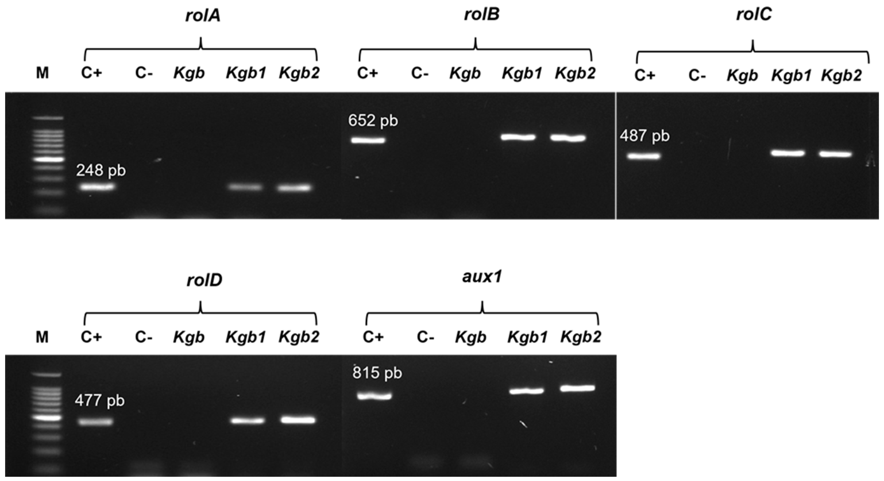
Genomic DNA from plant material was extracted using the method of Doyle and Doyle [32]. Plasmid DNA of A. rhizogenes was extracted with Wizard® Plus SV Minipreps DNA Purification System kit (Promega Corporation, A1460 Madison, WI, USA), following the manufacturer’s protocol. DNA from the A. rhizogenes A4 strain was used as a positive control, DNA from wild-type K. gastonis-bonnieri plants was used as a negative control, and sterile distilled H2O was used as a negative control, to develop the polymerase chain reaction (PCR). The amplification was carried out in a thermal cycler (Applied Biosystems, Gene Amp PCR Systems 9700, Waltham, MA, USA), using specific primers according to reports in each case. rolA: 5′-CGTTGTCGGAATGGCCCAGACC-3′ and 3′-CGTAGGTCTGAATATTCCGGTCC-5′ to amplify a 248 bp fragment, rolB: 5′-ACTATAGCAAACCCCTCCTGC-3′ and 3′-TTCAGGTTTACTGCAGCAGGC-5′, to amplify a 652 bp fragment [33], rolC: 5′-TGTGACAAGCAGCGATGAGC-3′ and 3′-GATTGC AAACTTGCACTCGC-5′ to amplify a 487 bp fragment, rolD: 5′-CCTTACGAATTCTCTTAGCGGCACC-3′ and 3′-GAGGTACACTGGACTGAATCTGCAC-5′ to amplify a 477 bp fragment [34] and aux1: 5′-CCAAGCTTGTCAGAAAACTTCAGGG-3′ and 3′-CCGGATCCAATACCCAGCGCTTT-5′ to amplify a 815 bp fragment [35]. DNA electrophoresis was performed in a 1.5% agarose gel at 95V for 60 min. The visualization of the amplified fragments was mixed with a SYBR ®Green (Lonza, Hayward, CA, USA) solution. Electrophoresis was analyzed in a photo-documenter (ChemiDoc™ MP Imaging System BIO-RAD, 170-01402, Hercules, CA, USA).

2.5. Ultrasound Assisted Extraction (UAE)

Metabolites were extracted in an ultrasound bath Branson (Branson Ultrasonics™, 2510R-MTH, Brookfield, CT, USA) with automatic control of time and temperature and ultrasound frequency of 40 kHz. A total of 50 mg of dry and ground biomass were mixed in 2 mL of ethanol (80%, v/v), and were sonicated at 40 ± 5 °C for 30 min. Samples were centrifuged at 3500 rpm for 5 min. The supernatants were recovered and filtered through cellulose membranes (0.22 μm) (MILLEX® GS). Samples were dryness at 25 ± 2 °C and the dried extracts were stored at 4 °C until analyzed [36].

2.6. Chemical Characterization of Kalanchoë Gastonis-Bonnieri Extracts

The chemical profile of extracts was obtained by LC-ESI-MS/MS. Samples were solubilized in 500 µL of MeOH, HPLC grade (Sigma-Aldrich®) and filtered through nylon membrane, (0.45 µm, Agilent Technologies, Santa Clara, CA, USA). The mobile phase for gradient elution consists of two solvents: solvent A (0.1% formic acid (FA) Sigma-Aldrich® in H2O) and solvent B (0.1% FA in CH3CN/MeOH (1:1; v/v) Sigma-Aldrich®. The linear gradient profile was as follows: 95% A (5 min), 95–90% A (10 min), 90–50% A (55 min), 50–95% A (65 min), and 95% A (70 min). The injection volume was 10 µL. The flow rate (0.6 mL/min) was split 1:1 before the MS interface. Electrospray ionization analysis (ESI) was performed using a micrOTOF-Q II mass spectrometer (Bruker Daltonics, Bremen, Germany). The mass spectrometer was operated in negative ion mode with a capillary potential of 2.5 kV, gas temperature of 180 °C, drying gas flow of 6 L/min, and nebulizer gas pressure of 1.0 Bar. Detection was performed at 50–3000 m/z.
The tentative identification of compounds was based on the comparison of the MS fragmentation profile obtained by the analytical equipment, with the mass spectra of MassBank of North America (https://mona.fiehnlab.ucdavis.edu accessed on 15 July 2021) and Competitive Fragmentation Modeling for Metabolite Identification (http://cfmid3.wishartlab.com accessed on 30 July 2021).

2.7. Statistical Analysis

Relative abundance data of tentatively identified metabolites in K. gastonis-bonnieri extracts were analyzed by clustered color mapping, using a Pearson distance measurement mean bond clustering method, using the Heatmapper software (http://www.heatmapper.ca/, accessed on 15 September 2021).

3. Results

3.1. Morphology of the Kgb1 and Kgb2 Cultures and Molecular Analysis

K. gastonis-bonnieri seedlings were obtained from in vitro cultures. The infection with A. rhizogenes A4 strain was accomplished and the root formation at the infection site was observed after 15 d with a transformation efficiency of 80%. Fifteen root lines were individualized in a semi-solid B5 medium, most of the lines were characterized by slow growth and poor branching, and the formation of cell aggregates was observed 30 d after isolation from the initial explant. In vitro cultures of K. gastonis-bonnieri were successfully initiated from aseptically vegetative shoots isolated from wild-growing plants. The in vitro shoots were subjected to Agrobacterium-mediated transformation (Figure 1). Figure 1a shows an uninfected explant (negative control), demonstrating that mechanical injury did not result in root formation. In Figure 1b–d, the response of different infected seedlings and the development of hairy roots that emerge from the infection site with plagiotropic growth is shown; Figure 1b shows abundant proliferation of hairy roots, while in Figure 1c, d explants with few roots were obtained; Figure 1d shows callus formation and few roots scarcely hairy developed from the infection site.
A total of 15 lines were individualized and 2 lines, Kgb1 and Kgb2, were selected due to accelerated growth and abundant secondary roots. Figure 2 shows the follow-up of the development of Kgb1 and Kgb2 cell cultures at 9 d (Figure 2a), 18 d (Figure 2b), and 25 days (Figure 2c) of the subculture. The images were captured 90 days after remaining in liquid B5 medium. The asynchronous growth in different stages of somatic embryogenesis was observed. The globular (GB), torpedo (T), heart-shaped (H), and embryo (SE), structures were identified. Embryogenic calli cultures were morphologically heterogeneous, with asynchronous development, meaning that the growth of dedifferentiated cells, the cell differentiation, and the development of embryos occur at uncoordinated times, hence the cellular aggregates in Kgb-1 as Kgb-2 show different stages of embryogenesis through the 25-days of subculture. Figure 2d shows the initial embryogenic aggregates, which were observed since the roots were isolated from the infected explant and remained in a liquid medium. These structures were observed in all stages of culture. The lines currently remain stable with the same characteristics.
Figure 3 shows the amplified fragments of the rolA, rolB, rolC, rolD, and aux1 genes from the DNA of Kgb1 and Kgb2 lines; none of the analyzed genes was amplified from K. gastonis-bonnieri wild-type plants. These results suggest that the Kgb1 and Kgb2 lines were induced in the infection mediated by A. rhizogenes A4 strain. In this work, it is suggested that both TL-DNA and TR-DNA of A. rhizogenes A4 strain were inserted into Kgb1 and Kgb2 genomes. It has been reported that the response of a plant species to genetic transformation by A. rhizogenes is in the function of the integration and combined expression of rolA, rolB, rolC, and rolD genes [31,32].

3.2. Analysis of the Chemical Profile of Kgb1 and Kgb2 Extracts

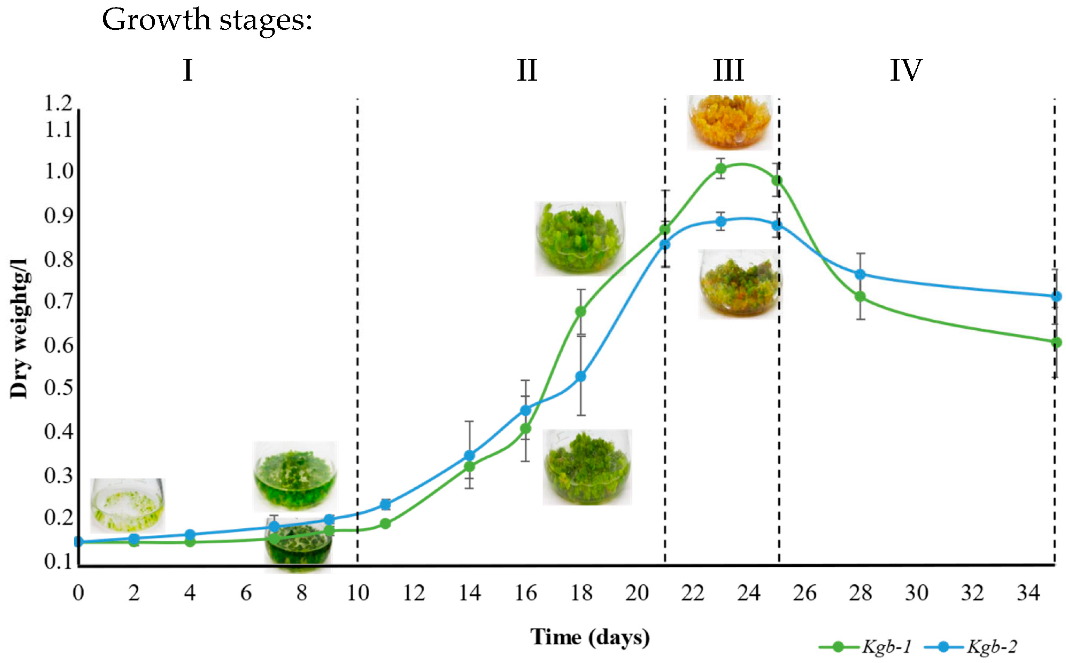
Figure 4 shows changes during the cell growth of Kgb1 and Kgb2 lines. The stages were defined as follows: stage (I) 1–10 days of culture, as an adaptation period, and changes in cell growth were observed, stage (II) 11–21 d, increase accelerated biomass with doubling time for Kgb1 (T2) = 3.31 d and cell growth speed (μ) = 0.20 d−1; while for Kgb2, T2 = 4.15 d and μ = 0.16 d−1, stage (III) 21–25 d, a decrease in biomass growth was observed, for Kgb1 the T2 = 20.3 d and μ = 0.03 d−1; finally, for Kgb2 T2 = 48.5 d, and μ = 0.01 d−1; stage (IV) 25–35 d, considerable decrease in cell growth was observed in both lines. The chemical profile of Kgb1 and Kgb2 was analyzed at 9, 18 and 25 days of culture, which were selected taking quantity biomass as a selection criteria.
Table 1 shows 60 tentatively identified metabolites in the extracts of Kgb1, Kgb2 lines at 9, 18, 25 daysof culture, and the wild plant of K. gastonis-bonnieri; among them, 18 flavonoids, 11 fatty acids, 5 coumarins, 4 phenolic acids, 3 phenolic compounds, 3 terpenes, 4 carboxylic acids, 1 alkaloid, 2 amino acids, 1 carbohydrate and 8 compounds grouped as others, based on chemical structure. The highest number of flavonoids was identified in the wild type, while fatty acids and carbohydrate were mainly identified in Kgb1 and Kgb2 lines.
The metabolites were identified according to LC-ESI-MS/MS parameters: retention time, match factor values database, molecular formula, and monoisotopic mass. The tentative identification of compounds was based on the comparison of the MS fragmentation profile obtained by the analytical equipment, with the mass spectra of MassBank of North America and Competitive Fragmentation Modeling for Metabolite Identification.
The Kgb1 and Kgb2 lines showed a differential chemical profile compared to the wild plant. Particularly, in Kgb1 and Kgb2 extracts the following chemical compounds were detected: ribose-1-arsenate (0.7), 3-(4-hydroxy-3,5-dimethoxyphenyl)-1-(2,4,6-trihydroxy-3-methoxyphenyl)propane-1,2-dione (RT = 0.9), 2-(1,3-dihydroxy4-oxocyclohex-2-en-1-yl)-5-hydroxy-3,6,7-trimethoxy-4H-chromen-4-one (RT = 1.0), sarmentosin epoxide (RT = 1.1), 4-hydroxycoumarin (RT = 1.4), sarmentosin epoxide (RT = 1.1) D-glutamine (RT = 1.8), 1, 6-dihydroxy-3,7-dimethoxy-2-(3-methyl-2-butenyl)-8-(3-hydroxy-3-methyl-1E-butenyl)-xanthone (RT = 5.1), verbasoside (RT = 5.4), 4-coumaric acid (RT = 5.9), ferulic acid (6.0), hallactone B (RT = 6.3), 6,7,3’,4’-tetrahydroxyflavanone (RT = 6.6), epiafzelechin (2R,3R)(-) (RT = 6.9), naringenin (RT = 7.0), 3-hydroxytetradecanedioic acid (RT = 7.6), (9S,10E,12S,13S)-9,12,13-trihydroxy-10-octadecenoic acid (RT = 8.4), 13-HOTrE (RT = 11.3), ipecoside (RT = 12.3), altamisic acid (12.4), a-linolenic acid (RT = 12.8), linoleic acid (RT = 13.3), and heneicosanoic acid (RT = 14.9), p-coumaraldehyde (RT = 15.2). Although, neobavaisoflavone, malic acid, heneicosanoic acid, heliannuol A, 3-(4-hydroxy-3,5-dimethoxyphenyl)-1-(2,4,6-trihydroxy-3-methoxyphenyl)propane-1,2-dione,sarmentosin epoxide, were major compounds in the Kgb1 and Kgb2 cell lines, in comparison to wild plant.
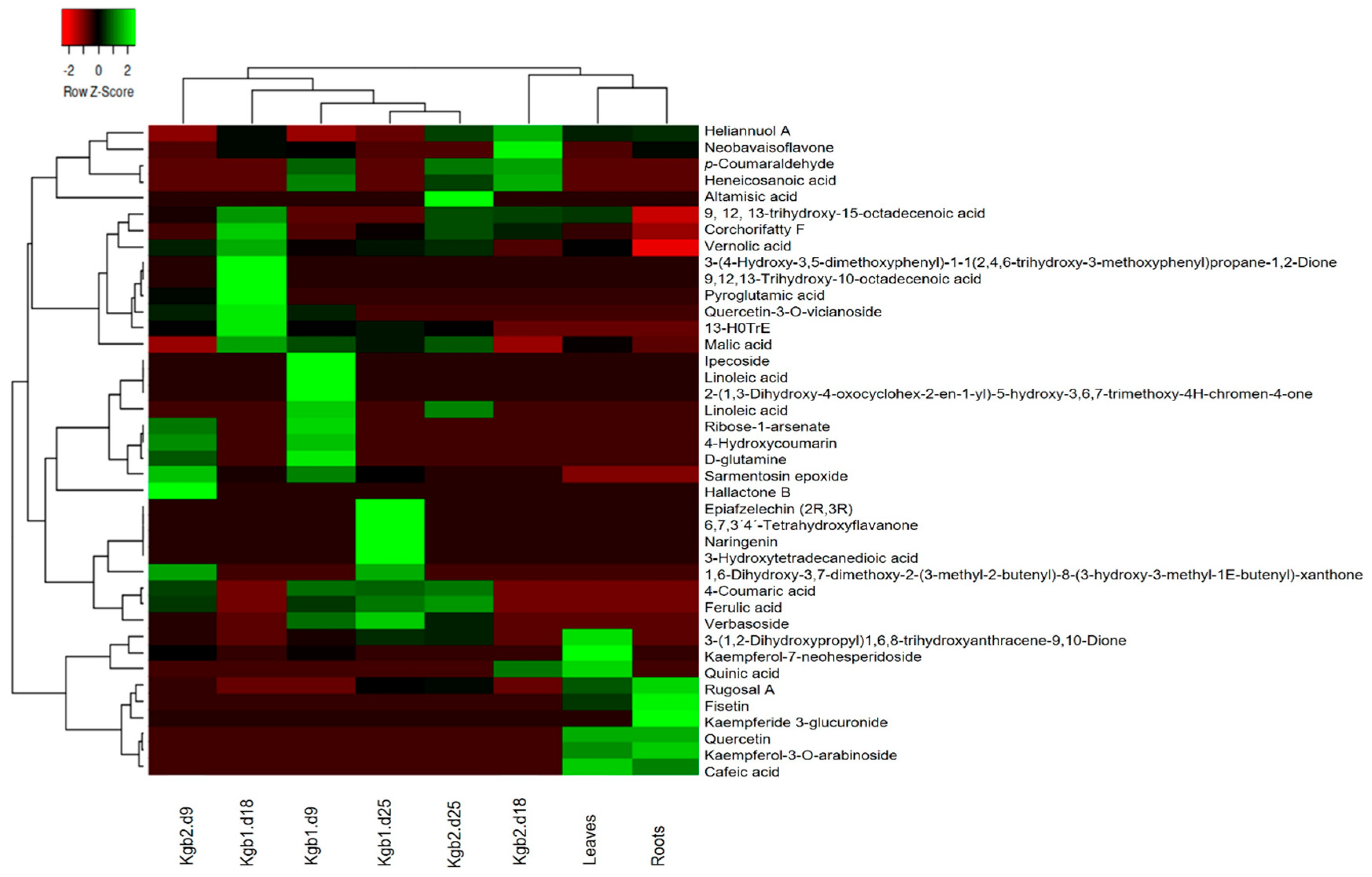
Figure 5 shows the comparison of the relative abundance of the identified metabolites; the lowest concentration in red light, while the highest in green light. According to the row dendrogram, it is observed that the relative abundance of the compounds is different between extracts of the transformed lines Kgb1, Kgb2, leaf, and wild type root extracts. Column dendrogram allows for visualizing the formation of two main groups: on the left the extracts obtained from Kgb1 of 9, 18, and 25 days of culture and Kgb2 of 9 and 25 days, and on the right side the leaf and wild root extracts. Subgroups are formed between the Kgb1 and Kgb2 lines, showing the chemical compound diversity between the different culture days analyzed. Regarding the relative abundance of 4-coumaric acid, ferulic acid, verbasoside, and 3-(1,2-dihydroxypropyl)-1,6,8-trihydroxyanthracene-9,10-dione. They were similar at 9 and 25 days between Kgb1 and Kgb2, while the wild type of leaf and root extracts were similar in relation to the abundance of rugosal A, fisetin, quercetin, kaempferol-3-O-arabonoside, and y caffeic acid. Dendrogram also shows the identification of specific compounds in Kgb1 and Kgb2 extracts in the different culture periods. In the Kgb1-d9 extract, particular compounds were identified as ipecoside, linoleic acid, 2-(1,3-dihydroxy-4-oxocyclohex-2-en-1-yl)-5-hydroxy-3,6,7-trimethoxy-4H-chromen-4-one, and linolenic acid. In the Kgb1-d18 extract, specific compounds were identified as 3-(4-hydroxy-3,5-dimethoxyphenyl)-1-(2,4,6-trihydroxy-3-methoxyphenyl) propane-1,2-dione, 9,12,13-trihydroxy-10-octadecenoic acid, pyroglutamic acid, quercetin-3-O-vicianoside, and y 13-HOTrE, while in the Kgb1-d25 extract, the specific identified compounds were epiafzelechin (2R,3R), 6,7,3’,4’-tetrahydroxyflavanone, and naringenin y 3-hydroxytetradecanedioic acid.
Likewise, specific compounds were observed in the leaf and/or root extracts: 3-(1,2-dihydroxypropyl)-1,6,8-trihydroxyanthracene-9,10-dione, kaempferol-7-neohesperidoside, rugosal A, fisetin kaempferide 3-glucuronide, quercetin, kaempferol-3-O-arabinoside, and caffeic acid. Similar relative compound abundance was observed for Kgb1 and Kgb2 extracts at 9 days of culture: ribose-1-arsenate, 4-hydroxycoumarin, D-glutamine, sarmentosin epoxide, 4-coumaric acid, and ferulic acid; at 25 days of culture: 4-coumaric acid, ferulic acid, and verbasoside.

4. Discussion

Some data on transformation efficiency have been reported: 72% in leaf explants of Solanum erianthum D. Don. [36]; 65% in shoot explants and 60 in leaf explants of Althaea officinalis [28]; 56% in leaf explants and 29% in seedlings’ inter nodal explants of Salvia bulleyana [37]. According to this information, the transformation efficiency of A. rhizogenes strain A4 is diverse; in this work, A4 strain was capable of infecting K. gastonis-bonnieri tissue which led to genetically modified embryogenic calli. Also, Chaudhuri et al. [38] and Tavassoli and Safipour-Afshar [28] reported that transformation efficiency was obtained as a function of culture conditions, Agrobacterium-host interaction, age, and explant type. It has also been associated with actively dividing cells showing higher transformation rates.
In this work, 15 lines were individualized, and 2 lines, Kgb1 and Kgb2, were selected, due to accelerated growth and abundant secondary roots. Kgb1 and Kgb2 showed spontaneous callus formation. Vinterhalter et al. [10] reported calli and somatic embryo formation at 35 d of culture, from roots induced with the A. rhizogenes A4M70GUS strain, in Gentiana utriculosa. Hiebert-Giesbrecht et al. (2021) [8] reported in Pentalinon andrieuxii the formation of embryogenic callus 6 months after obtaining hairy roots with the A. rhizogenes ATCC15834 strain in leaves and hypocotyls. Results obtained in this work represent the possibility of whole plant regeneration; moreover, is an interesting tool to carry out advanced studies on the secondary metabolism of K. gastonis-bonnieri transgenic culture. The response of K. gastonis-bonnieri agrees with previous reports on the spontaneous formation of embryogenic calli from hairy roots obtained by Agrobacterium infection, on different plant species such as Gentiana utriculosa with the A. rhizogenes A4M70GUS strain [1,10], Pentalinon andrieuxii with the A. rhizogenes ATCC15834 strain [8]; all these strains carrying the same agropine-like Ri plasmid [39]. It has been suggested that abnormal morphological features of hairy roots can be a result of the combined participation of rol genes in plant cells since each gene might be associated with specific phenotypic alterations in Kalanchoë species; in addition, the response of each plant species against infection by A. rhizogenes is diverse [38]. In this work, it is suggested that both TL-DNA and TR-DNA of A. rhizogenes A4 strain were inserted into Kgb1 and Kgb2 genomes. It has been reported that the response of a plant species to genetic transformation by A. rhizogenes is in function of the integration and combined expression of rolA, rolB, rolC, and rolD genes [40,41].
The observed difference in the chemical profile of the transformed embryogenic calli extracts is possibly due to asynchronous growth in the different culture periods [42]. There is wide variability in the compounds reported in K. gastonis-bonnieri, which could be attributed to the analyzed plant material that includes the plant development stage, the collection season, and extraction conditions, even among the transformed cell lines analyzed in this work.
Kgb1 and Kgb2, at 9 days of culture, showed a greater number of detected compounds, in contrast to culture analyzed at 18 and 25 days, which could be related to the adaptation of subculture towards the beginning of a new cycle [43]; a lower number of compounds were detected at 18 days of culture which could be attributed to accelerated cell growth [43,44], therefore the cell metabolism is redirected toward multiplication and cell growth. Finally, at 25 days of culture, the increase of some compounds was observed, which could be attributed to the accumulation of compounds related to the embryogenic process. The accumulation and changes in metabolites detected are related to different stages of development and growth in the in vitro culture, which is also observed at different developmental stages of wild plants [45,46,47] as a part of plant development or in response to epigenetic factors.
The extracts of Kgb1 and Kgb2 lines and leaves showed corchorifatty F acid, a compound identified in rice and other cereals, with antibacterial activity [48]. In Kgb1 and Kgb2 extracts, sarmentosine epoxide was detected, which has shown antihepatotoxic activity [49]. In this work, malic acid, caffeic acid, rugosal A, campferol 3-O-arabinoside, quercetin, fisetin, and heliannuol were detected in leaves and roots of K. gastonis bonnieri which have not been previously reported in wild plants.
Interestingly, hairy root cultures synthesize compounds that have not been detected in wild plants [9]. Furthermore, some authors have reported that shoots and transgenic plants obtained from hairy roots accumulate specific metabolites compared to the wild plant. Tusevski et al. [5], reported an accumulation of naphthodiatrons and specific phenolic compounds in transgenic shoots obtained of hairy roots from Hypericum perforatum. Vinterhalter et al. [7] reported xanthones in transgenic Gentiana utriculosa plants regenerated by somatic embryogenesis from hairy roots. Hiebert-Giesbrecht et al. [8] reported terpenoids accumulation in leaf extracts of transgenic plants, which were obtained from hairy roots of Pentalinon andrieuxii.
The phenotypic changes showed in transformed cultures, could be due to (a) position and (b) number of copies of the T-DNA inserted in the genome of the host cell, (c) the regulation of gene expression, and (d) protein biosynthesis encoded by rol genes in the plant cell, among others [7,8].
The biological activity of some of compounds identified in this work has been reported, quinic and malic acid inhibit the growth of S. aureus and P. aeruginosa [50]; the neobavaisoflavone is a compound that has antioxidant, anti-inflammatory, and anticancer properties [51]; ferulic acid has a wide variety of effects, especially on oxidative stress, inflammation, vascular endothelial injury, fibrosis, apoptosis, and platelet aggregation [52]. The compounds detected in Kgb1 and Kgb2 suggest the effect of genetic transformation through the differential biosynthesis of chemical compounds. It has been reported that highly specialized plant organs such as roots and leaves are needed for the biosynthesis of phytochemicals [26]; therefore, Kgb1 and Kgb2 do not develop highly specialized organs, which could explain the limited accumulation of flavonoids.

5. Conclusions

This is the first report of the chemical profile of transformed cell cultures of K. gastonis-bonnieri. Specific compounds that have not been reported in K. gastonis-bonnieri wild-type plants were identified. In vitro, culture of genetically transformed embryogenic calli could be an alternative to produce metabolites of commercial interest in the pharmacological industry as treatment against various diseases, such as cancer. In addition, obtaining transgenic K. gastonis-bonnieri plants from embryogenic callus could be an opportunity in the ornamental industry since genetic modification could produce plants with different phenotype.

Author Contributions

M.G.B.N.: Conceptualization, Investigation, Methodology, Formal analysis, Writing (Revision—original and final draft. M.B.: Formal analysis, Writing (Revision—original and final draft). M.Á.M.-M.: Methodology, Formal analysis, Revision final draft. L.R.-V.: data curation. E.I.: Conceptualization, Methodology, Investigation, Methodology, Revision—final draft. A.A.D.V.-M.: Conceptualization, Investigation, Methodology, Writing (Revision—original and final draft), Formal analysis, Project administration, Supervision, Funding acquisition. All authors have read and agreed to the published version of the manuscript.

Funding

This work was supported by Instituto Politécnico Nacional, México (IPN/SIP 20220810, 20231185).

Data Availability Statement

The original contributions presented in the study are included in the article, further inquiries can be directed to the corresponding author.

Acknowledgments

MGBN (744161) acknowledge study grant from CONACYT.

Conflicts of Interest

The authors declare that they have no conflict of interest.

References

  1. Chung, I.-M.; Rekha, K.; Rajakumar, G.; Thiruvengadam, M. Production of glucosinolates, phenolic compounds and associated gene expression profiles of hairy root cultures in turnip (Brassica rapa ssp. rapa). 3 Biotech 2016, 6, 175. [Google Scholar] [CrossRef] [PubMed]
  2. Li, Y.; Kong, D.; Fu, Y.; Sussman, M.R.; Wu, H. The effect of developmental and environmental factors on secondary metabolites in medicinal plants. Plant Physiol. Biochem. 2020, 148, 80–89. [Google Scholar] [CrossRef]
  3. Abhyankar, G.; Suprasanna, P.; Pandey, B.N.; Mishra, K.P.; Rao, K.V.; Reddy, V.D. Hairy root extract of Phyllanthus amarus induces apoptotic cell death in human breast cancer cells. Innov. Food Sci. Emerg. Technol. 2010, 11, 526–532. [Google Scholar] [CrossRef]
  4. Rekha, K.; Thiruvengadam, M. Secondary metabolite production in transgenic hairy root cultures of cucurbits. In Transgenesis and Secondary Metabolism; Reference Series in, Phytochemistry; Jha, S., Ed.; Springer: Cham, Switzerland, 2017. [Google Scholar] [CrossRef]
  5. Tusevski, O.; Stanoeva, J.P.; Stefova, M.; Pavokovic, D.; Simic, S.G. Identification and quantification of phenolic compounds in Hypericum perforatum L. transgenic shoots. Acta Physiol. Plant 2014, 36, 2555–2569. [Google Scholar] [CrossRef]
  6. Roychowdhury, D.; Halder, M.; Jha, S. Agrobacterium rhizogenes-mediated transformation in medicinal plants: Genetic stability in long-term culture. In Transgenesis and Secondary Metabolism; Reference Series in, Phytochemistry; Jha, S., Ed.; Springer: Cham, Switzerland, 2017. [Google Scholar] [CrossRef]
  7. Vinterhalter, B.; Savić, J.; Zdravković-Korać, S.; Banjac, N.; Vinterhalter, D.; Krstić-Milošević, D. Agrobacterium rhizogenes-mediated transformation of Gentiana utriculosa L. and xanthones decussatin-1-O-primeveroside and decussatin accumulation in hairy roots and somatic embryo-derived transgenic plants. Ind. Crops Prod. 2019, 130, 216–229. [Google Scholar] [CrossRef]
  8. Hiebert-Giesbrecht, M.R.; Avilés-Berzunza, E.; Godoy-Hernández, G.; Peña-Rodriguez, L.M. Genetic transformation of the tropical vine Pentalinon andrieuxii (Apocynaceae) via Agrobacterium rhizogenes produces plants with an increased capacity of terpenoid production. In Vitro Cell Dev. Biol-Plant 2021, 57, 21–29. [Google Scholar] [CrossRef]
  9. Huang, S.H.; Vishwakarma, R.K.; Lee, T.T. Establishment of hairy root lines and analysis of iridoids and secoiridoids in the medicinal plant Gentiana scabra. Bot. Stud. 2014, 55, 17. [Google Scholar] [CrossRef] [PubMed]
  10. Vinterhalter, B.; Krstić-Milošević, D.; Janković, T.; Pljevljakusic, D.; Ninković, S.; Smigocki, A.; Vinterhalter, D. Gentiana dinarica Beck. hairy root cultures and evaluation of factors affecting growth and xanthone production. Plant Cell Tiss. Organ. Cult. 2015, 121, 667–679. [Google Scholar] [CrossRef]
  11. Stefanowicz-Hajduk, J.; Asztemborska, M.; Krauze-Baranowska, M.; Godlewska, S.; Gucwa, M.; Moniuszko-Szajwaj, B.; Stochmal, A.; Ochocka, J.R. Identification of flavonoids and bufadienolides and cytotoxic effects of Kalanchoë daigremontiana extracts on human cancer cell lines. Planta Med. 2020, 86, 239–246. [Google Scholar] [CrossRef]
  12. Hernández-Caballero, M.E.; Sierra-Ramírez, J.A.; Villalobos Valencia, R.; Seseña-Méndez, E. Potential of Kalanchoe pinnata as a Cancer Treatment Adjuvant and an Epigenetic Regulator. Molecules 2022, 27, 6425. [Google Scholar] [CrossRef]
  13. Christensen, B.; Sriskandarajah, S.; Serek MMüller, R. Transformation of Kalanchoe blossfeldiana with rol-genes is useful in molecular breeding towards compact growth. Plant Cell Rep. 2008, 27, 1485–1495. [Google Scholar] [CrossRef]
  14. Fkiara, A.; Barba-Espín, G.; Bahij, R.; Müller, R.; Christensen, L.P.; Lütken, H. Elicitation of Flavonoids in Kalanchoe pinnata by Agrobacterium rhizogenes-mediated Transformation and UV-B Radiation. In Medicinal Plants: Biodiversity, Sustainable Utilization and Conservation; Khasim, S., Long, C., Thammasiri, K., Lütken, H., Eds.; Springer: Cham, Switzerland, 2020; pp. 395–403. [Google Scholar] [CrossRef]
  15. Ramasamy, M.; Dominguez, M.M.; Irigoyen, S.; Padilla, C.S.; Mandadi, K.K. Rhizobium rhizogenes-mediated hairy root induction and plant regeneration for bioengineering citrus. Plant Biotechnol. J. 2023, 21, 1728–1730. [Google Scholar] [CrossRef]
  16. Thirukkumaran, G.; Khan, R.S.; Chin, D.P.; Nakamura, I.; Mii, M. Isopentenyl transferase gene expression offers the positive selection of marker-free transgenic plant of Kalanchoe blossfeldiana. Plant Cell Tiss. Organ. Cult. 2009, 97, 237–242. [Google Scholar] [CrossRef]
  17. Cho, K.H.; Vieira, A.E.; Kim, J.; Clark, D.G.; Colquhoun, T.A. Transformation of Kalanchoe pinnata by Agrobacterium tumefaciens with ZsGreen1. Plant Cell Tissue Organ. Cult. 2021, 146, 401–407. [Google Scholar] [CrossRef]
  18. Yukes, J.; Balick, M. Dominican Medicinal Plants: A Guide for Health Care Providers, 2nd ed.; New York Botanical Garden: Bronx, NY, USA, 2010; pp. 7–14. [Google Scholar]
  19. Costa, S.S.; Corrêa, M.F.P.; Casanova, L.M. A new triglycosyl flavonoid isolated from leaf juice of Kalanchoë gastonis-bonnieri (Crassulaceae). Nat. Prod. Commun. 2015, 10, 433–436. [Google Scholar] [CrossRef] [PubMed]
  20. Abdalla, S.L.; Costa, S.S.; Gioso, M.A.; Casanova, L.M.; Coutinho, M.A.S.; Silva, M.F.A.; Botelho, M.C.D.S.N.; Díaz, R.S.G. Efficacy of a Kalanchoë gastonis-bonnieri extract to control bacterial biofilms and dental calculus in dogs. Pesqui. Vet. Bras. 2017, 37, 859–865. [Google Scholar] [CrossRef]
  21. Palumbo, A.; Casanova, L.M.; Corrêa, M.F.P.; Da Costa, N.M.; Nasciutti, L.E.; Costa, S.S. Potential therapeutic effects of underground parts of Kalanchoë gastonis-bonnieri on benign prostatic hyperplasia. Evid.-Based Complement. Altern. Med. 2019, 2019, 6340757. [Google Scholar] [CrossRef]
  22. Siems, K.; Jas, G.; Arriaga-Giner, F.J.; Wollenweber, E.; Dörr, M. On the chemical nature of epicuticular waxes in some succulent Kalanchoë and Senecio species. Z. Naturforsch. 1995, C 50, 451–454. [Google Scholar] [CrossRef]
  23. Vanegas, P.E.; Cruz-Hernández, A.; Valverde, M.E.; Paredes-López, O. Plant regeneration via organogenesis in marigold. PCTOC 2002, 69, 279–283. [Google Scholar] [CrossRef]
  24. Murashige, T.; Skoog, F. A revised medium for rapid growth and bioassays with tobacco tissue cultures. Physiol. Plant 1962, 15, 472–497. [Google Scholar] [CrossRef]
  25. Hooykas, P.J.J.; Klapwijk, P.M.; Nuti, M.P.; Schilperoort, R.A.; Rörsch, A. Transfer of the Agrobacterium tumefaciens TI plasmid to avirulent Agrobacteria and to Rhizobium ex planta. J. Gen. Microbiol. 1977, 98, 477–484. [Google Scholar] [CrossRef]
  26. Sharifi, S.; Sattari, T.N.; Zebarjadi, A.; Majd, A.; Ghasempour, H. The influence of Agrobacterium rhizogenes on induction of hairy roots and ß-carboline alkaloids production in Tribulus terrestris L. Physiol. Mol. Biol. Plants 2014, 20, 69–80. [Google Scholar] [CrossRef]
  27. Torres-García, B.E.; Morales-Domínguez, J.F.; Fraire-Velázquez, S.; Pérez-Molphe-Balch, E. Generación de cultivos de raíces transformadas de la planta medicinal Bidens odorata Cav (Compositae) y análisis fitoquímico preliminar. Polibotánica 2018, 46, 241–257. [Google Scholar] [CrossRef]
  28. Tavassoli, P.; Safipour-Afshar, A. Influence of different Agrobacterium rhizogenes strains on hairy root induction and analysis of phenolic and flavonoid compounds in marshmallow (Althaea officinalis L.). 3 Biotech 2018, 8, 351. [Google Scholar] [CrossRef] [PubMed]
  29. Gamborg, O.L.; Miller, R.A.; Ojima, K. Nutrient requirements of suspension cultures of soybean root cells. Exp. Cell Res. 1968, 50, 151–158. [Google Scholar] [CrossRef]
  30. Kieran, P.; MacLoughlin, P.; Malone, D. Plant cell suspension cultures: Someengineering considerations. J. Biotechnol. 1997, 59, 39–52. [Google Scholar] [CrossRef]
  31. Urquiza-López, A.; Álvarez-Rivera, G.; Ballesteros-Vivas, D.; Cifuentes, A.; Del Villar-Martínez, A.A. Metabolite profiling of rosemary cell lines with antiproliferative potential against human HT-29 colon cancer cells. Plant Foods Hum. Nutr. 2021, 76, 319–325. [Google Scholar] [CrossRef] [PubMed]
  32. Doyle, J.J.; Doyle, J.L. A rapid DNA isolation procedure for small quantities of fresh leaf tissue. Phytochem. Bull. 1987, 19, 11–15. [Google Scholar]
  33. Petrova, M.; Zayova, E.; Vlahova, M. Induction of hairy roots in Arnica montana L. by Agrobacterium rhizogenes. Cent. Eur. J. Biol. 2013, 8, 470–479. [Google Scholar] [CrossRef]
  34. Savić, J.; Nikolić, R.; Banjac, N.; Zdravković-Korać, S.; Stupar, S.; Cingel, A.; Ćosić, T.; Raspor, M.; Smigocki, A.; Ninković, S. Beneficial implications of sugar beet proteinase inhibitor BvSTI on plant architecture and salt stress tolerance in Lotus corniculatus L. J. Plant Physiol. 2019, 243, 153055. [Google Scholar] [CrossRef]
  35. Rana, M.M.; Han, Z.-X.; Song, D.-P.; Liu, G.-F.; Li, D.-X.; Wan, X.-C.; Karthikeyan, A.; Wei, S. Effect of medium supplements on Agrobacterium rhizogenes mediated hairy root induction from the callus tissues of Camellia sinensis var. sinensis. Int. J. Mol. Sci. 2016, 17, 1132. [Google Scholar] [CrossRef] [PubMed]
  36. Sarkar, J.; Misra, A.; Banerjee, N. Genetic transfection, hairy root induction and solasodine accumulation in elicited hairy root clone of Solanum erianthum D. Don. J. Biotechnol. 2020, 323, 238–245. [Google Scholar] [CrossRef]
  37. Wojciechowska, M.; Owczarek, A.; Kiss, A.K.; Grąbkowska, R.; Olszewska, M.A.; Grzegorczyk-Karolak, I. Establishment of hairy root cultures of Salvia bulleyana Diels for production of polyphenolic compounds. J. Biotechnol. 2020, 318, 10–19. [Google Scholar] [CrossRef]
  38. Chaudhuri, K.N.; Ghosh, B.; Tepfer, D.; Jha, S. Spontaneous plant regeneration in transformed roots and calli from Tylophora indica: Changes in morphological phenotype and tylophorine accumulation associated with transformation by Agrobacterium Rhizogenes. Plant Cell Rep. 2006, 25, 1059–1066. [Google Scholar] [CrossRef]
  39. Bahramnejad, B.; Naji, M.; Bose, R.; Jha, S. A critical review on use of Agrobacterium rhizogenes and their associated binary vectors for plant transformation. Biotechnol. Adv. 2019, 37, 107405. [Google Scholar] [CrossRef] [PubMed]
  40. Ozyigit, I.I.; Dogan, I.; Artam Tarhan, E. Agrobacterium rhizogenes-mediated transformation and its biotechnological applications in crops. In Crop improvement; Springer: Boston, MA, USA, 2013; pp. 1–48. [Google Scholar]
  41. Sarkar, S.; Ghosh, I.; Roychowdhury, D.; Jha, S. The effects of rol genes of Agrobacterium rhizogenes on morphogenesis and secondary metabolite accumulation in medicinal plants. In Biotechnological Approaches for Medicinal and Aromatic Plants; Kumar, N., Ed.; Springer: Singapore, 2018. [Google Scholar] [CrossRef]
  42. Rattan, S.; Kumar, D.; Warghat, A.R. Growth kinetics, metabolite yield, and expression analysis of biosynthetic pathway genes in friable callus cell lines of Rhodiola imbricata (Edgew). Plant Cell Tiss. Organ Cult. 2021, 146, 149–160. [Google Scholar] [CrossRef]
  43. Chiavegatto, R.B.; Castro, A.H.F.; Marçal, M.G.; Padua, M.S.; Alves, E.; Techio, V.H. Cell Viability, Mitotic Indexand Callus Morphology of Byrsonima verbascifolia (Malpighiaceae). TropicalPlant Biol. 2015, 8, 87–97. [Google Scholar] [CrossRef]
  44. Sathish, S.; Venkatesh, R.; Safia, N.; Sathishkumar, R. Studies on growth dynamics ofembryogenic cell suspension cultures of commercially important Indica rice cultivars ASD16 and Pusa basmati. 3 Biotech 2018, 8, 1–9. [Google Scholar] [CrossRef]
  45. Pan, Y.; Li, L.; Xiao, S.; Chen, Z.; Sarsaiya, S.; Zhang, S.; ShangGuan, Y.; Liu, H.; Xu, D. Callus growth kinetics and accumulation of secondary metabolites of Bletilla striata Rchb.f. using a callus suspension culture. PLoS ONE 2020, 15, e0220084. [Google Scholar] [CrossRef] [PubMed]
  46. Partap, M.; Kumar, P.; Ashrita; Kumar, P.; Kumar, D.; Warghat, A.R. Growth kinetics, metabolites production and expression profiling of picrosides biosynthetic pathway genes in friable callus culture of Picrorhiza kurroa Royle ex Benth. Appl. Biochem. Biotechnol. 2020, 192, 1298–1317. [Google Scholar] [CrossRef] [PubMed]
  47. Wahyuni, D.K.; Rahayu, S.; Zaidan, A.H.; Ekasari, W.; Prasongsuk, S.; Purnobasuki, H. Growth, secondary metabolite production, and in vitro antiplasmodial activity of Sonchus arvensis L. callus under dolomite [CaMg(CO3)2] treatment. PLoS ONE 2021, 16, e0254804. [Google Scholar] [CrossRef] [PubMed]
  48. Ang, H.; Mak, K.; Lum, M. Liquid chromatography mass spectrometry based high-throughput, unbiased profiling of upland and lowland rice varieties cultivated in Sabah. Trans. Sci. Technol. 2020, 7, 137–146. [Google Scholar]
  49. Nahrstedt, A.; Walther, A.; Wray, V. Sarmentosin epoxide, a new cyanogenic compound from Sedum cepaea. Phytochemistry 1982, 21, 107–110. [Google Scholar] [CrossRef]
  50. Adamczak, A.; Ożarowski, M.; Karpiński, T.M. Antibacterial activity of some flavonoids and organic acids widely distributed in plants. J. Clin. Med. 2020, 9, 109. [Google Scholar] [CrossRef] [PubMed]
  51. Maszczyk, M.; Rzepka, Z.; Rok, J.; Beberok, A.; Wrześniok, D. Neobavaisoflavone may modulate the activity of topoisomerase inhibitors towards U-87 MG cells: An in vitro study. Molecules 2021, 26, 4516. [Google Scholar] [CrossRef]
  52. Li, D.; Rui, Y.-X.; Guo, S.-D.; Luan, F.; Liu, R.; Zeng, N. Ferulic acid: A review of its pharmacology, pharmacokinetics and derivatives. Life Sci. 2021, 284, 119921. [Google Scholar] [CrossRef] [PubMed]
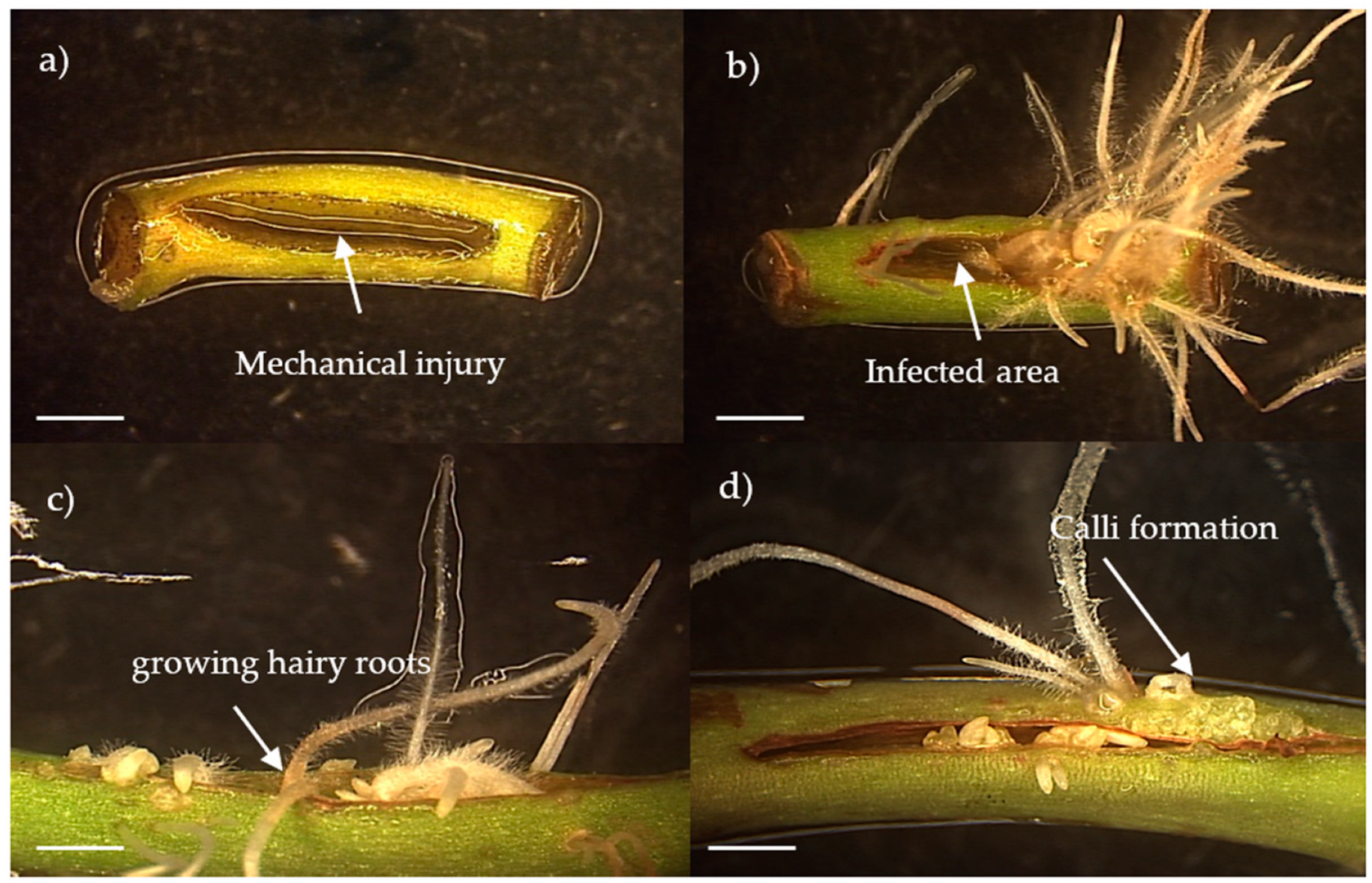
Figure 1. Hairy roots induction and appearance of embryogenic calli of Kalanchoë gastonis-bonnieri. in the internodal segment 15 days after infection. (a) Control: explant cut out, (bd) Infected internodal segments. Reference bar = 1 mm.
Agronomy 14 00189 g001
Figure 2. Transgenic embryogenic calli of Kalanchoë gastonis-bonnieri, Kgb1 and Kgb2. (a) 9, (b) 18, (c) 25 days of a subculture in a liquid B5 medium, (d) embryogenic aggregates, (CA) calli, (H) heart-shaped, (T) torpedo, (SE) somatic embryo, (R) transgenic root, and (GB) globular-shape. Reference bar = 1 mm.
Agronomy 14 00189 g002
Figure 3. Amplification of rolA, rolB, rolC, rolD, and aux1 genes of A. rhizogenes from genomic DNA of embryogenic calli (Kgb1 and Kgb2 lines). (M) molecular weight marker 1Kb; (C+) DNA from A. rhizogenes A4 strain (positive control); (C−) water (negative control); (Kgb) DNA from wild-type K. gastonis-bonnieri (negative control); (Kgb1 and Kgb2) embryogenic callus lines.
Agronomy 14 00189 g003
Figure 4. Changes during the cell growth of Kgb1 and Kgb2 lines in liquid B5 medium. The dotted line indicates the transition between growth stages.
Agronomy 14 00189 g004
Figure 5. Heat map showing the relative abundance of the compounds tentatively identified in extracts of Kgb1, Kgb2, wild leaves and roots of Kalanchoë gastonis-bonnieri. Color code: light green (highest relative abundance); light red (lower relative abundance).
Agronomy 14 00189 g005
Table 1. Tentatively identified compounds in Kgb1 and Kgb2 cell lines extracts of Kalanchoe gastonis-bonnieri (- means no detected, while +, ++ and +++ refer to different peak intensity).
RT (min)Tentative IdentificationMatch FactorMonoisotopic MassMain Fragments (m/z)Parent Ion
[M-H]-
Kgb1Kgb2Wild Type Plants
Days of CultureLeavesRoots
9182591825
Flavonoids
5.93,7-Dihydroxy-3’,4’-dimethoxyflavone79314.0863637.1386313.0684------+-
638.1423
653.1331
6.1Syringetin-3-O-glucoside78508.1272477.1005507.1111------+-
508.1177
535.2139
6.2Quercetin-3-O-pentosyl (1-2) acetilpentosida80608.1295327.0844607.1259------+-
623.1247
623.2683
6.23,4-dimethoxy-myricetin-3-O-dideoxyhexosyl(1-2)-dideoxyhexoside80638.1445521.2012637.1383------+-
623.1227
638.1445
6.3Guaijaverin79434.0762434.0762433.0762------+-
491.0744
519.2203
6.5Apigenin-6-C-glucoside-7-O-glucoside90594.1500461.1064593.1466------+-
506.0978
594.1500
6.66,7,3’,4’-Tetrahydroxyflavanone86288.0149146.9666287.1465--+-----
288.1511
309.1295
6.7Kaempferol-3-O-arabinoside95418.0852418.0852417.0791------++
491.1174
607.2728
6.9Epiafzelechin (2R,3R)(-)80274.1671187.0955273.1671--+-----
289.1635
607.2757
7.0Naringenin80272.1627112.9843271.1528--+-----
289.1627
607.2727
7.1Kaempferide 3-glucuronide95476.0914597.2469475.0873-------+
607.2738
608.2776
7.2Diosmine89608.1664475.0857607.1641------+-
608.1683
643.1410
7.7Quercetin90302.0368146.9683301.0317------++
157.0095
302.0331
8.22’,7-Dihydroxy-4’-methoxy-8-prenylflavan 2’,7-diglucoside83664.2705653.2342663.2626------+-
680.2472
707.2874
8.6Fisetin80286.0392112.9833285.0360------++
286.0392
315.0477
10.5Kaempferol-7-neohesperidoside93594.2728197.9606593.2716+--+--+-
201.0350
594.2728
11.1Quercetin-3-O-vicianoside94596.2922197.9612595.2873++-+----
596.2922
723.3794
11.9Neobavaisoflavone90322.1733322.1762321.1724++--+++--+
406.1516
421.0987
Fatty acids
6.3Deacetoxy (7)-7-Oxokhivorinic acid78520.2216509.1912519.2210------+-
520.2216
536.2101
7.63-Hydroxytetradecanedioic acid83274.171146.9682273.1676--+-----
173.9999
274.1710
8.4(9S,10E,12S,13S)-9,12,13-Trihydroxy-10-octadecenoic acid81330.2366174.0007329.2320-+------
201.0377
330.2366
8.7Corchorifatty acid F83328.2277201.0351327.2141+++++++-
263.1309
328.2158
9.2(Z)-9,12,13-trihydroxyoctadec-15-enoic acid80330.9998157.0100329.2300++++++++-
330.2365
397.2198
11.313-HOTrE81294.2131275.2000293.2095++++-+--
294.2131
613.9943
11.7Vernolic acid83296.2289116.9269295.2255+++++++-
296.2289
363.2136
12.4Altamisic acid80 112.9851279.1628-----+--
135.9701
309.1725
12.8α-Linolenic acid80278.7812135.9699277.2160+----+--
278.2192
400.2108
13.3Linoleic acid80280.2351278.7270279.2322+-------
280.2351
325.1854
14.9Heneicosanoic acid90326.1831326.1873325.1829+++---+++++--
339.2004
340.2043
Coumarins
1.44-Hydroxycoumarin83162.0495117.0193161.0451+--+----
128.0353
292.1378
4.93,4,5-Trihydroxy-6-{[2-oxo-6-(3-oxobutyl)-2H-chromen-7-yl]oxy}oxane-2-carboxylate85408.0981341.0851407.0945------+-
408.0981
443.0710
6.3Hallactone B80440.1090112.9848439.1031---+----
174.0002
440.1090
11.4Rugosal A87266.1482134.8628265.1446--++-++++++
135.9684
817.1497
11.8Corylifol A85390.2340321.1705389.2295------+-
390.2340
411.2125
Phenolic acids
5.4Caffeic acid93180.0365135.0425179.0325------++
180.0365
755.1999
5.71-Caffeoyl-4-deoxyquinic acid90338.0910191.0536337.0887------+-
338.0910
359.0706
5.94-Coumaric acid81164.0467164.0467163.0378+-++-+--
279.0519
475.1830
6.0Ferulic acid86194.0537194.0537193.0486+-+++-++--
309.0612
311.1114
Phenolic compounds
5.2Syringate81198.0453198.0453197.0428------+-
313.0536
431.1863
5.5Verbasoside83462.1709415.1603461.1640+-++-+--
451.1389
462.1709
15.2p-Coumaraldehyde81147.8759178.8796146.9643+---++--
220.9484
231.9439
Terpenes
6.4Kanokoside D89624.2753577.2622623.2694---+---+
613.2428
624.2753
10.3Heliannuol A94250.1536248.9578249.1497++++++++++++++++
250.1536
251.1476
12.3Ipecoside80565.3214116.9282564.3191+-------
554.2898
581.3077
Carboxylic acids
1.0Malic acid80134.0215128.0336133.0119+++++++++--++++++
341.1061
377.0831
1.0DL-Pyroglutamic acid80129.0345290.0858128.0336-+-+----
310.0687
403.1352
1.2Citrate90192.0301111.0072191.0175------+-
128.0334
173.0081
1.3D-(-)-Quinic acid91192.0579157.0341191.0520----++-+++-
377.0813
379.0805
Alkaloids
6.1Voacristine80384.0108384.0108383.0066-------+
481.1331
563.2139
Amino acids
0.8D-Glutamine87146.0324179.0556145.0598+--+----
215.0324
307.1131
4.3Tryptophan82204.0841204.0841203.0800------+-
261.0370
271.0673
Carbohydrate
1.1Sarmentosin epoxide90291.0908133.0134290.0859++++++++++++++--
200.0561
632.2048
Others
0.7Ribose-1-arsenate89273.9598158.9785272.9565++--+----
273.9598
274.9575
0.93-(4-hydroxy-3,5-dimethoxyphenyl)-1-(2,4,6-trihydroxy-3-methoxyphenyl)propane-1,2-dione90378.0889341.1077377.0854-+++------
379.0830
404.1046
1.02-(1,3-dihydroxy-4-oxocyclohex-2-en-1-yl)-5-hydroxy-3,6,7-trimethoxy-4H-chromen-4-one79378.0822341.1083377.0846+-------
404.1043
470.1521
4.4Cusparine82307.1179112.9835306.1163------+-
296.0879
350.1422
5.11,6-Dihydroxy-3,7-dimethoxy-2-(3-methyl-2-butenyl)-8-(3-hydroxy-3-methyl-1E-butenyl)-xanthone86440.1819393.1746439.1785--++----
429.1495
440.1819
5.81-(2H-1,3-benzodioxol-5-yl)-2-[2,6-dimethoxy-4-(prop-2-en-1-yl)phenoxy]propyl benzoate83476.1831429.1758475.1800------+-
476.1831
521.1996
8.53-(1,2-dihydroxypropyl)-1,6,8-trihydroxyanthracene-9,10-dione83330.2334285.0364329.2298+-++-++-
286.0402
330.2334
8.7Isocyclocalamin85502.2151491.1836501.2109-------+
493.1828
518.2009
Disclaimer/Publisher’s Note: The statements, opinions and data contained in all publications are solely those of the individual author(s) and contributor(s) and not of MDPI and/or the editor(s). MDPI and/or the editor(s) disclaim responsibility for any injury to people or property resulting from any ideas, methods, instructions or products referred to in the content.