Article

Magnesium Oxide Nanoparticles: An Influential Element in Cowpea (Vigna unguiculata L. Walp.) Tissue Culture

1
Department of Field Crops, Faculty of Agriculture, Ataturk University, 25240 Erzurum, Türkiye
2
Department of Field Crops, Faculty of Agriculture, Necmettin Erbakan University, 42310 Konya, Türkiye
3
Seed and Plant Improvement Institute, Agricultural Research, Education and Extension Organization (AREEO), Karaj P.O. Box 3158854119, Iran
4
Department of Food Security and Public Health, Khabat Technical Institute, Erbil Polytechnic University, Erbil 44001, Iraq
5
Department of Plant Physiology and Metabolomics, Agricultural Institute, Centre for Agricultural Research, 2462 Martonvásár, Hungary
6
Department of Nursing, Faculty of Health Sciences, Sakarya University of Applied Sciences, 54187 Sakarya, Türkiye
7
Department of Food Technology, Vocational College of Technical Sciences, Ataturk University, 25240 Erzurum, Türkiye
8
Department of Nano-Science and Nano-Engineering, Institute of Science, Ataturk University, 25240 Erzurum, Türkiye
*
Authors to whom correspondence should be addressed.
These authors contributed equally to this work.
Agronomy 2023, 13(6), 1646; https://doi.org/10.3390/agronomy13061646
Submission received: 10 May 2023 / Revised: 14 June 2023 / Accepted: 17 June 2023 / Published: 19 June 2023
(This article belongs to the Special Issue Plant Tissue Culture and Plant Somatic Embryogenesis)

Abstract

:
Nanotechnology is a rapidly growing field of science and technology that deals with the development of new solutions by understanding and controlling matter at the nanoscale. Since the last decade, magnesium oxide nanoparticles (MgO-NPs) have gained tremendous attention because of their unique characteristics and diverse applications in materials sciences and because they are non-toxic and relatively cheaply available materials. MgO-NPs can improve plant growth and contribute to plant tolerance of heavy metal toxicity. The effects of MgO-NPs on cowpea (Vigna unguiculata L. Walp.) plants were surveyed under in vitro conditions to find the optimum combination for cowpea tissue culture. The MgO-NPs used in the study were synthesized using walnut shell extract by the green synthesis method. MgO nanoparticles with 35–40 nm size was used in this research. When the size distribution of the MgO-NPs’ structure was examined, two peaks with 37.8 nm and 78.8 nm dimensions were obtained. The zeta potential of MgO-NPs dispersed in water was measured around −13.3 mV on average. The results showed that different doses of MgO-NPs applied to cowpea plant on all in vitro parameters significantly affected all measured parameters of cowpea plantlets under in vitro condition in a positive way. The best results in morphogenesis were MS medium supplemented with high MgO-NP applications (555 mg/L), resulting in a 25% increase in callus formation. The addition of Mg-NPs in the induction medium at concentrations at 370 mg/L increased shoot multiplication. The highest root length with 1.575 cm was obtained in MS medium containing 370 mg/L MgO. This study found that MgO-NPs greatly influenced the plantlets’ growth parameters and other measured traits; in addition, our results indicate that the efficiency of tissue culture of cowpea could be improved by increased application of MgO in the form of nanoparticles. In conclusion, the present work highlights the possibility of using MgO-NPs in cowpea tissue culture.

1. Introduction

Nanotechnology, which is a multidisciplinary science, uses nano-sized materials [1], which are defined as substances smaller than 100 nanometers in size, and controls them at the atomic level and makes them useful [2]. Nanotechnology covers many different fields, including pesticide distribution, nano sensors, pesticide degradation, use of micronutrients in agriculture, and plant protection and nutrition [3]. Nanotechnology offers effective methods to protect soil health and conditions by helping to minimize agricultural waste and environmental pollution [4,5]; therefore, it can greatly improve the functioning of precision agriculture [6]. Nanotechnology is useful in the agricultural sector, in the form of nano-pesticides and nano-fertilizers [7]. Among various methods of NP synthesis, the plant tract-mediated method is preferred due to its cost-effective nature [8]. MgO-NPs have applications in various fields and have earlier been synthesized using plant extract [9].
Magnesium plays an important role in plant growth and development, serves as a component of the chlorophyll molecules, and regulates the activity of key photosynthetic enzymes in the chloroplast [10]. Magnesium is a macronutrient that activates more enzymes than other nutrients [11] and has structural and regulatory functions related to nucleophilic ligands in plants [10,12]. It is one of the essential elements in the function and synthesis of nucleic acids and ATP [13]. Magnesium deficiency suppresses plant growth and decreases yield [14]. Magnesium oxide is an important inorganic material with a wide band range [15]. This material is used in many applications, such as catalysis, catalyst supports, toxic waste reclamation, refractory materials and adsorbents, additives in heavy fuel oils, reflective and anti-reflective coatings, substrate such as superconducting and ferroelectric thin films, superconductors, and lithium-ion batteries [16,17]. Nano MgO, on the other hand, has many special physical and chemical properties brought about by its nano size. With its size, nanoparticles can be used more by plant cells, induce plant growth, and have antimicrobial, antifungal, and antiviral effects against pests [18]. The small size of nanoparticles allows them to penetrate into the plant cell. Hence, we assessed the impact of MgO-NPs on the legume, cowpea plant.
Cowpea (Vigna unguiculata L. Walp.) is a very common annual plant, especially in Africa, South America, Asia, and the United States, and is one of the most important legumes worldwide [19]. It is a good pre-plant that has the ability to grow in poor soils and increases the yield of the next product with the help of nitrogen fixation [20,21]. Cowpea, which is considered as a green vegetable and dry grain in human nutrition and as a fodder in animal nutrition, belongs to the legume family and contains 2.0–4.3% protein in fresh beans and 4.5–5.0% in fresh grains. Protein content in dry cowpea grains that have reached maturity varies between 20.42 and 34.60%, depending on the variety and environmental conditions. In addition, its grains contain rich source of essential amino acids, except cysteine and methionine [22]. The protein in its seeds is rich in Lysine and Tryptophan amino acids compared to cereal seeds, and is insufficient in terms of Methionine and Cystine compared to animal proteins [23].
Nanoparticles, due to their special physical and chemical properties, may lead to unpredictable changes in the morphological characteristics of the plant [24]. The synthesized nanomaterials may provide protection that is effective in controlling pests and pathogens that significantly affect the yield of the plant [25]. Toxic effects of NPs for plants and animals have been reported, but there is no report documented until now that showed the harmful effects of NPs on tissue culture plants [26]. There have been many studies on the use of NPs in plant tissue culture systems [26,27,28,29]. Wide applications of NPs in plant tissue culture include the elimination of microbial contaminants from explants, callus induction, organogenesis, somatic embryogenesis, somaclonal variation, genetic transformation, and secondary metabolite development. Compounds can be developed by integrating the concept of nanotechnology into plant tissue culture techniques, synthesis, purification, and desired plant-derived yield [30]. There is limited information available in the literature regarding the effect of NPs on in vitro regeneration characters on cowpea plant, which has a great importance in human and animal nutrition. Therefore, the aim of this study is to determine the possible effects of callus formation on morphogenesis and plant regeneration by applying different doses of MgO-NPs of cowpea plant under plant tissue culture condition.

2. Materials and Methods

2.1. Synthesis of Mg Nanoparticles (MgO-NPs)

MgO-NPs were synthesized using walnut shell extract. The walnut shell extraction used for green synthesis was prepared with distilled water, and for this purpose, 25 g walnut shells were washed and crushed using the freeze–thaw technique in liquid nitrogen; 250 mL of distilled water was added and mixed in a magnetic stirrer for 1–2 h. The extract was obtained by first filtering through cheesecloth and then filter paper, before being kept at −25 °C until use. An amount of 0.1 M Mg (NO3)2 solution was actively used for conversation of MgO in plants. The formation of the synthesis was followed qualitatively and quantitatively by UV-Vis spectrophotometer (Epoch). After the method optimization, the obtained NPs were characterized. For this purpose, different morphological and molecular detection methods, such as scanning electron microscopy (SEM; Zeiss Sigma 300), Fourier-transform infrared spectroscopy (FT-IR; VERTEX 70 v FT-IR Spectrometer, Billerica, MA, USA), and X-ray diffraction (XRD; Malvern Panalytical B.V., Almelo, The Netherlands), were used. The obtained MgO-NPs were washed under vacuum using distilled water and ethanol and were used in the reaction after drying in an oven. In the application range, after MgO-NPs were weighed and homogenized in pure water with the help of an ultrasonicator, they were used in plant in vitro experiments [31,32].

2.2. Plant Material

The cowpea cultivar, registered as “Ulkem”, used in this study was obtained from Ondokuz Mayıs University and is usually used for forage purposes. Forage cowpea seeds were washed with tap water and surface-sterilized with 70% ethanol for 5 min, treated for 25 min with solution containing 1% sodium hypochlorite with a few drops of Tween 20 with constant stirring, and rinsed three times with sterile distilled water thereafter. The seeds were imbibed in sterile water for 24 h in the dark. Plumula parts were aseptically dissected and used as explants in the experiment.

2.3. Tissue Culture Applications

MS medium, mineral salts, and vitamins [33] were used in the experiments. Magnesium and MgO-NPs in MS medium were exchanged with different concentrations. In the experiment, there were 5 treatments with different magnesium content: 0 mg/L (MS medium without MgSO4.7H2O and MgO-NPs), MgSO4.7H2O, which is used commonly in MS medium 370 mg/L as macronutrient elements, and three treatment with removed MgSO4.7H2O from the MS medium and replaced with a nanoparticle version of these elements: 1/2X (185 mg/L MgO-NPs), 1X (370 mg/L MgO-NPs), 2X (555 mg/L MgO-NPs) concentrations. Plumules were cultured on MS medium containing MgO-NPs previously prepared. The explants were kept in the dark at 24 ± 1 °C. In shoot regeneration medium (MS salt and vitamins + 1.0 mg/L BAP) containing different Mg types and concentrations, explant were kept for 8 weeks, and they were then placed in root formation medium (MS salt and vitamins + 0.5 mg/L BAP) containing different MgO-NPs types and concentrations and kept under white fluorescent light (Preheat Daylight-42 µmol photons m−2s−1) for 4 weeks at 24 ± 1 °C in a 16-h light photo period. Morphogenesis and callus formation measurements were made after 30 days. The shoot formation rate, number of shoots, number of shoots per explant, shoot length, root formation rate, number of roots per explant, and root length were calculated after 60 days. This study was carried out in a complete randomized experimental design arrangement with four replications. Each petri dish was considered as an experimental unit, and 10 cowpea plumula were cultured in each petri dish. Analysis of variance and Duncan multiple comparison tests were computed with SPSS statistical analysis program (Version 20).

3. Results

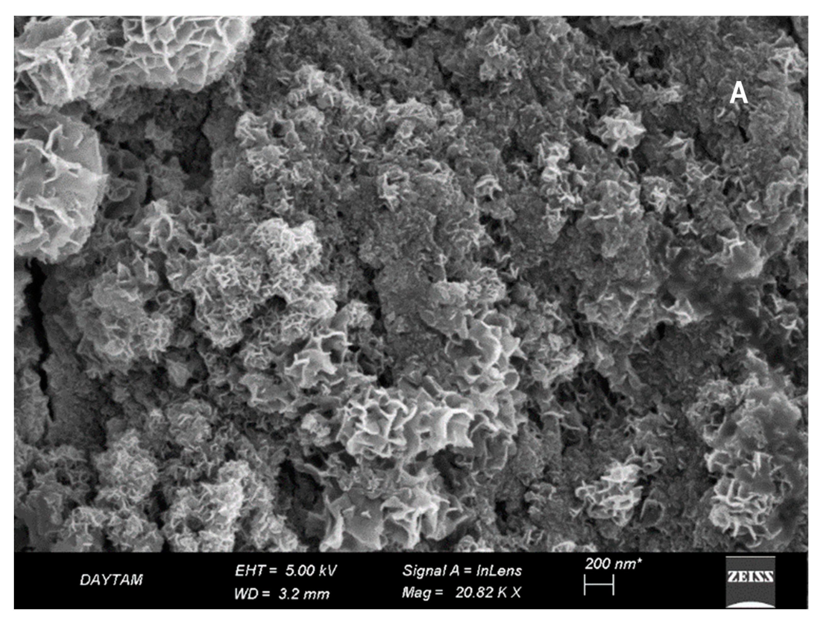
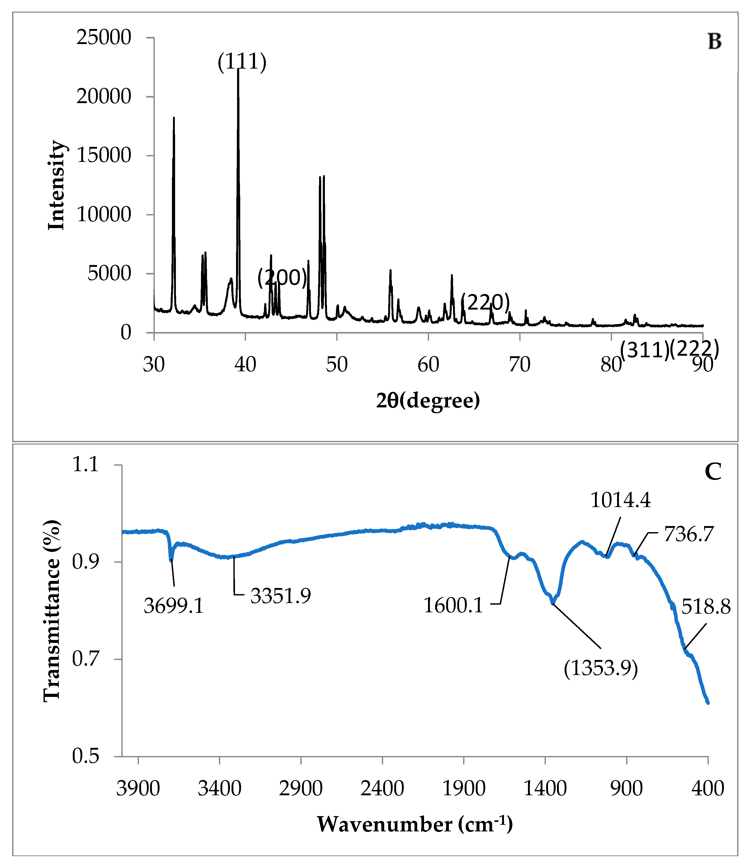
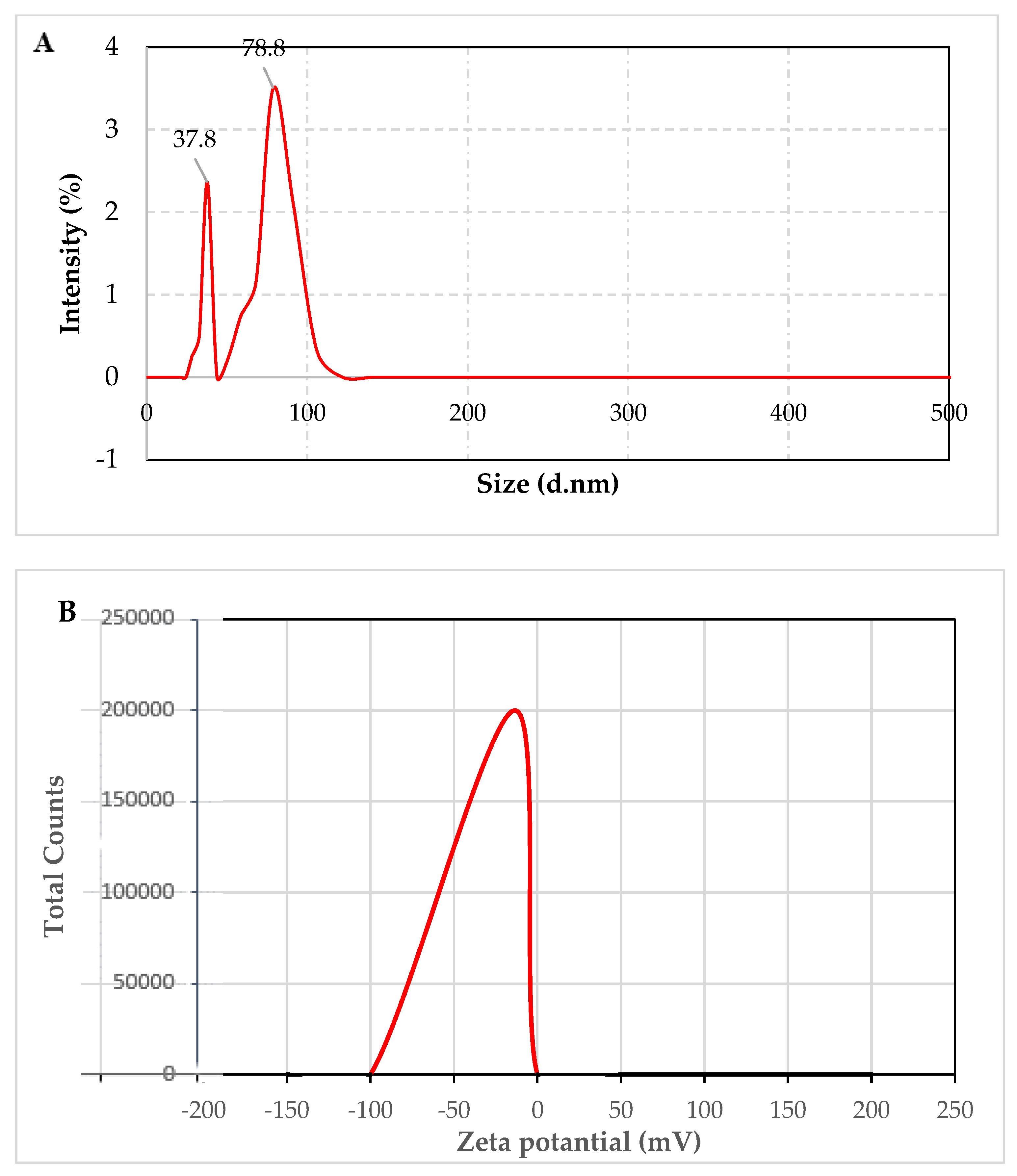
3.1. Surface Morphological Characterization of MgO-NPs

Surface characterization of MgO-NPs was performed using SEM, FT-IR, and XRD analyzes, and the results are given in Figure 1. SEM analysis determined that the MgO-NPs obtained by green synthesis were well dispersed and cubic (Figure 1A), while the peaks at 39.2° (111), 62.53° (220), 77.8° (311), and 81.7° (222) 2θ° in the XRD graph in Figure 1B belong to Mg (OH)2. FT-IR analysis is an effective technique used to identify possible peaks of MgO-NPs and extract used for the reduction of metal. FT-IR analysis of MgO-NPs obtained by green synthesis is given in Figure 1C. As seen from the FT-IR diagram, the wavelength between 400 and 4000 cm−1 was scanned. From the findings, it was determined that intense absorption peaks occurred at 3699, 3351, 2293, 1600, 1354, 1014, 763, and 519 cm−1. While the peak observed at 3699 cm−1 belongs to the -OH band, the wide peak band observed at 3351 cm−1 indicates the presence of -NH2 and -OH groups in the medium. The peaks seen around 1600 cm−1 indicate the presence of peaks defined as the primary amine group (N-H) overlapping with the amide and carboxylate group. The peak at 1354 cm–1 is matched with the Mg-OH group, while all bands between 400 and 736 confirm the presence of MgO-NPs. The size of MgO-NPs was determined to be 35–40 nm as a result of measurements and calculations. The obtained spectrum showed that the walnut shell extract had a high ability to reduce and stabilize MgO-NPs.
When the size distribution of the MgO NPs structure was examined, two peaks with 37.8 nm and 78.8 nm dimensions were obtained. These peaks showed that the MgO NP structure did not have much agglomeration, but there was a small amount of agglomeration. However, the structure was smaller than 100 nm, and the findings support the SEM image (Figure 2A). The zeta potential analysis is an important indicator of the surface charges of chemical compounds. The zeta potential of MgO NPs dispersed in water was measured around −13.3 mV on average. It shows a negative potential value (Figure 2B). The negative value obtained is due to the oxygen atoms in the MgO structure (10.1002/cbdv.201900608 (13 June 2023)).

3.2. Morphogenesis

The averages of the characters determined by the treatments of different concentrations of MgO-NPs to the cowpea and the related variance analysis results are given in Table 1. In this study, morphogenesis basically refers to any changes in the explant, such as elongation, contraction, color, and structure changes, during the course of in vitro culture, except for the formation of callus, shoots, roots, or whole plants.
It has been observed that different MgO-NP applications have significant effects on morphogenesis (p < 0.01). These changes began to be observed after the first week of culture initiation. While the average number of explants showing morphogenesis was 9.25 from the original 10 explants at control (MS medium without MgO-NPs) and MS medium containing 370 mg/L MgO applications, this number was increased in Mg-NP applications (185 mg/L, 370 mg/L and 555 mg/L MgO-NPs). A significant 8.11% increase (p < 0.05) in the number of morphogenesis was observed in MS medium containing 185 mg/L, 370 mg/L, and 555 mg/L MgO-NP applications compared to the control. Morphological changes such as tissue growth and tissue swelling were observed in the cultured cowpea explants (Figure 3).

3.3. Callus Formation

It has been observed that different MgO-NP applications have significant effects on callus formation. Under the present experimental conditions, where 10 explants were originally started, the average callus formation depending on the MgO-NPs varied between 8 and 10. Callus formation reached the highest value with 10 in the application of MS medium containing 555 mg/L MgO-NPs, and this value was statistically significant from the control plants. This application was followed by 9.5 in MS medium containing 370 mg/L MgO-NPs and 9 in MS medium containing 370 mg/L MgSO4.7H2O applications. Different Mg treatments significantly increased callus formation compared to control. The highest increase was observed with 25% in MS medium supplemented with 555 mg/L MgO-NP application, followed by 18.75% with MS medium including 370 mg/L MgO-NPs, 12.50% in MS medium comprising 370 mg/L MgSO4.7H2O and 6.25% in MS medium comprising185 mg/L MgO-NP applications (Table 1).

3.4. Shoot Formation

Different MgO (with or without NPs) treatments led to significant differences in shoot formation (p < 0.01). According to the effect of different doses of magnesium NPs applied to the cowpea, shoot formation rates varied between 20.83% and 37.50%. The highest value in shoot formation rate was obtained from MS medium containing 555 mg/L MgO-NP application with 82.50%, and the lowest value was obtained from MS medium containing 185 mg/L MgO-NP application with 30%. Shoot formation rate was 60% from the control application, 42.50% from the MS medium containing 370 mg/L MgSO4.7H2O application, and 72.50% from the MS medium containing 370 mg/L MgO-NP application (Table 1).

3.5. Number of Shoots

The highest number of shoots was obtained from MS medium containing 370 mg/L MgO-NP application (61.25), followed by 36.25 in MS medium containing 555 mg/L MgO-NP application. Control and 370 mg/L MgSO4.7H2O applications gave the same shoot number value as 21.75, while MS medium containing 185 mg/L MgO-NP application gave the lowest value with 3.75, but this did not differ significantly from the control (Table 1).

3.6. Number of Shoots per Explant

The mean values of the number of shoots per explant determined in the cowpea at different doses varied between 1.25 and 17.50. In terms of applications, the highest number of shoots per explant (17.50) was obtained from the application of MS medium comprising 370 mg/L MgO-NPs, and this value was statistically higher than that found in the control treatment (6.5). The lowest value (1.25) was obtained from the MS medium comprising 185 mg/L MgO-NP application. The number of shoots per explant obtained from other applications in our study was determined as 6.75 in the MS medium comprising 370 mg/L MgSO4.7H2O application and 10 in the 555 mg/L MgO-NPs, but they were not statistically different from the control value (Table 1).

3.7. Shoot Length

In terms of shoot length, different MgO applications created significant differences. The longest shoot length was 2.07 cm in the MS medium containing 370 mg/L MgO-NP application, and the shortest shoot was 0.175 cm in the MS medium containing 185 mg/L MgO-NP application. MS medium supplemented with 370 mg/L MgO-NP application was followed by MS medium comprising 555 mg/L MgO-NPs (1.450 cm), MS medium comprising 370 mg/L MgSO4.7H2O (1.200 cm), and control (0.475 cm) applications. Except for the MS medium comprising 185 mg/L MgO-NP application, the other MS medium comprising 370 mg/L MgSO4.7H2O, MS medium comprising 370 mg/L MgO-NPs, and MS medium comprising 555 mg/L MgO-NP applications showed an increasing effect compared to the control. The highest increase was obtained from the administration of MS medium supplemented with 370 mg/L MgO-NPs, with 336.84% (Table 1).

3.8. Root Formation Rate

Different Mg applications had significant effects on root formation rate. The average root formation rate of the applied magnesium NPs varied between 10 and 27.5%. In terms of root formation rate, the highest value among magnesium NPs was obtained from control and MS medium containing 370 mg/L MgSO4.7H2O applications with 27.50%; this application was followed by 22.50% in MS medium containing 370 mg/L MgO-NP application and 19.75% with MS medium containing 185 mg/L MgO-NP applications. The lowest value of 10% was obtained in MS medium containing 555 mg/L MgO-NP application. The highest value was recorded in the application of MS medium containing 555 mg/L MgO-NPs, with a 63.64% decrease in the variation of magnesium NPs compared to the control (Table 1).

3.9. Number of Roots per Explant

Different MgO-NP applications had significant effects in terms of root number per explant. The maximum number of roots per explant determined in cowpea was obtained as 6.75 in the application of MS medium containing 370 mg/L MgO-NPs; this application was followed by 2.75 in the control application. The applied magnesium nanoparticles at different doses caused significant reductions compared to the control, which was statistically significant at 185 mg/L MgO-NPs (Table 1).

3.10. Root Length

According to the analysis of variance, root length was significantly affected by certain MgO-NP applications (Table 1). It was determined that the root length values obtained from the cowpea with different doses of magnesium NPs varied between 0.015 cm (MS medium supplementary with 185 mg/L MgO-NPS) and 157.50 cm (MS medium supplementary with 370 mg/L MgO-NPS). These extreme values were significant from the control; the other treatments used in this experiment were not (Table 1).

4. Discussion

NPs have begun to be used extensively in plant tissue culture studies. It is known that the kind, type, concentration, and size of NPs can be effective in studies conducted within the scope of plant tissue culture. The present study clearly showed the beneficial effects of MgO-NPs on the in vitro parameters of cowpea. In this study, MgO-NPs were synthesized using walnut shell extract by the green synthesis method. The size of MgO nanoparticles was determined to be 35–40 nm as a result of measurements and calculations. SEM analysis determined that the MgO-NPs obtained by green synthesis were well dispersed and cubic. The diffraction peaks are points that represent cubic MgO-NPs at 42.76° (200)2θ and 62.6° (220)2θ [34]. FT-IR analysis is an effective technique used to identify possible peaks of MgO-NPs and the extract used for the reduction of metal. As seen from the FT-IR diagram, the wavelength between 400 and 4000 cm−1 was scanned. Similar findings were found in the literature in the FT-IR analyzes of MgO-NPs obtained using some plant extracts, and they support our study [35,36,37,38,39,40,41].
Morphological changes such as tissue growth and tissue swelling were observed in the cultured cowpea explants. Our findings showed that positive effects of MgO-NPs on callus induction, shoot regeneration, and explant growth were observed. The results showed that the highest value of callus formation was obtained in MS medium containing with 555 mg/L MgO-NPs. Our result showed that NPs not only overcome negative effects but also improve callus formation. It seems that MgO-NPs may play a role similar to plant hormones such as cytokinins and gibberellins owing to their ability to induce plant cell division and stimulate cellular expansion; however, the mechanism of its action in darkness is still unknown. A similar finding was obtained by Mandeh et al. [27]. Several studies have shown positive effects of NPs on callus induction. Different concentrations of silver or gold NPs alone or combined with naphthalene acetic acid (NAA) were evaluated for callus culture growth in Prunella vulgaris L. The silver (30 μg L−1), silver and gold (1:2), and silver and gold (2:1) NPs in combination with NAA (2.0 mg L−1) enhanced callus proliferation (100%) as compared to the control (95%) [42]. The number and size of calli increased when barley mature embryos were grown in MS medium supplemented with 20 mg L−1 2,4-D and 60 mg mL−1 TiO2-NPs [27].
The explants were cultured on shoot multiplication medium supplemented with MS medium supplementary with 370 mg/L Mg-NPs. These media were optimum for the formation and development of shoot. Our result clearly showed that 370 mg/L Mg-NPs had a stimulation effect on the growth of shoot parameters, whiles 185 mg/L decreased NS and NSE and the 555 mg/L Mg-NPs concentrations induced an inhibitory effect. One reason for this may be that Mg-NPs block ethylene signaling and trigger shoot growth. Similar results have been found with AgNPs in banana plants by Do et al. [43]. Sharma et al. [44] also reported that AgNPs increased plant growth processes such as shoot and root lengths, area of the leaf, and biochemical parameters, such as carbohydrate and protein contents of common bean and corn.
The explants were also cultured on rooting medium supplemented with MS medium supplementary with 370 mg/L MgSO4.7H2O. These media were optimum for the formation and development of roots. Our result suggested that 370 mg/L MgSO4.7H2O had a stimulation effect on the growth of root parameters, while the application of NPs induced an inhibitory effect for root formation, especially in higher concentrations of NPs; this is very clear and obvious. These results are in agreement with earlier findings. For example, Helaly et al. [45] reported that root lengths were increased when Zn and nano ZnO-NPs were added to the MS medium. Zhang et al. [46] stated that copper nanoparticles inhibit primary root elongation and enhance lateral root emergence. Auxins, especially indole-3-acetic acid (IAA), have an important role in root development in plants. Regulation of auxin levels in different cells of the root is involved in many root functions, including growth, lateral root elongation, and root hair formation. There is a sharp toxicity threshold because at higher doses the toxic effects of ion exposure are manifest, with such responses as root shortening [47]. The reduction in elongation in cowpea roots exposed to high doses of Mg-NPs may possibly be related to the abnormal distribution of auxin. These results are in conflict with those obtained in barley [27]. However, this is not surprising given that nanoparticles can explain their effects depending on the size and/or shape of the particles, the concentrations applied, the particular experimental conditions, plant species, and uptake mechanisms [48]. Sotoodehnia-Korani [49] emphasized the belief that nanomaterials such as MgO-NPs have the ability to improve the efficiency of tissue cultures in vitro and boost agricultural yield.

5. Conclusions

The effects of various metal and metal oxide NPs on plants are well documented in vivo. Such NPs can be used to promote or enhance the morphogenetic potential of explants obtained from different plant species. NPs have been used widely in plant tissue culture studies. The influence of different concentrations and combinations of NPs on different media (shoot induction, shoot propagation, and rooting media) should also be investigated to gain a clear understanding of the underlying mechanisms behind the role of NPs in plant tissue culture. Given the potential for future research, it is vital to understand the role of MgO-NPs in callogenesis performance, micropropagation, and cell culture elicitation. The MgO-NPs used in the study were synthesized using walnut shell extract by green synthesis method. When the size distribution of the MgO-NPs structure was examined, two peaks with 37.8 nm and 78.8 nm dimensions were obtained. The zeta potential of MgO-NPs dispersed in water was measured at approximately −13.3 mV on average. The present study provides the first evidence of Mg-NPs effects on the in vitro culture of cowpea, showing the possibility of using MgO-NPs in cowpea tissue culture. The results showed that different doses of MgO-NPs applied to cowpea plants on all in vitro parameters significantly affected all measured parameters of cowpea plantlets under in vitro condition in a positive way. The best results in morphogenesis were MS medium supplemented with 555 mg/L MgO-NP applications, resulting in a 25% increase in callus formation. The addition of Mg-NPs in the induction medium at concentrations of 370 mg/L increased shoot multiplication. The highest root length with 1.575 cm was obtained in MS medium containing 370 mg/L MgO. However, for a clear understanding of the mechanisms underlying the role of MgO-NPs in cowpea tissue culture, it is recommended to investigate in detail the actual mechanisms of the promoting or inhibitory effects of MgO-NPs on each parameter.

Author Contributions

Conceptualization, M.O. and K.H.; methodology, K.H., A.T. and M.O.; software, M.O.; validation, K.H., H.N., A.A. and A.T.; formal analysis, M.O., K.H., A.T. and R.K.; investigation, K.H. and A.T.; resources, K.H., M.O., A.A. and H.N.; data curation, K.H. and A.T.; writing—original draft preparation, M.O. and A.T.; writing—review and editing, K.H., A.T., A.A., H.N., A.P.-A., B.J. and T.J.; visualization, M.O., K.H. and A.T.; supervision, M.O.; project administration, K.H., A.A. and H.N.; funding acquisition, T.J. All authors have read and agreed to the published version of the manuscript.

Funding

This work was funded by a grant from the National Research, Development, and Innovation Office (grant No. K142899).

Data Availability Statement

All data supporting the conclusions of this article are included in this article.

Conflicts of Interest

The authors declare no conflict of interest.

References

  1. Arnall, A.H. Future Technologies, Today’s Choices-Nanotechnology, Artificial Intelligence and Robotics. A technical, political and institutional map of emerging technologies. AHS 2003, 56, 1329–1332. [Google Scholar]
  2. Bergeson, L.L.; Cole, M.F. Regulatory implications of nanotechnology. Biointeract. Nanomater. 2014, 315. [Google Scholar] [CrossRef]
  3. Ghormade, V.; Deshpande, M.V.; Paknikar, K.M. Perspectives for nano-biotechnology enabled protection and nutrition of plants. Biotechnol. Adv. 2011, 29, 792–803. [Google Scholar] [CrossRef]
  4. Duhan, J.S.; Kumar, R.; Kumar, N.; Kaur, P.; Nehra, K.; Duhan, S. Nanotechnology: The new perspective in precision agriculture. Biotechnol. Rep. 2017, 15, 11–23. [Google Scholar] [CrossRef]
  5. Raliya, R.; Saharan, V.; Dimkpa, C.; Biswas, P. Nanofertilizer for precision and sustainable agriculture: Current state and future perspectives. J. Agric. Food Chem. 2017, 66, 6487–6503. [Google Scholar] [CrossRef]
  6. Panpatte, D.G.; Jhala, Y.K. Nanotechnology for Agriculture: Crop Production & Protection; Springer: Berlin/Heidelberg, Germany, 2019. [Google Scholar]
  7. Bratovcic, A.; Hikal, W.M.; Said-Al Ahl, H.A.; Tkachenko, K.G.; Baeshen, R.S.; Sabra, A.S.; Sany, H. Nanopesticides and nanofertilizers and agricultural development: Scopes, advances and applications. Open J. Ecol. 2021, 11, 301–316. [Google Scholar] [CrossRef]
  8. Sharma, P.; Gautam, A.; Kumar, V.; Guleria, P. In vitro exposed magnesium oxide nanoparticles enhanced the growth of legume Macrotyloma uniflorum. ESPR 2022, 29, 13635–13645. [Google Scholar] [CrossRef] [PubMed]
  9. Kumar, V.; Jain, A.; Wadhawan, S.; Mehta, S.K. Synthesis of biosurfactant-coated magnesium oxide nanoparticles for methylene blue removal and selective Pb2+ sensing. IET Nanobiotechnol. 2018, 12, 241–253. [Google Scholar] [CrossRef]
  10. Shaul, O. Magnesium transport and function in plants: The tip of the iceberg. Biometals 2002, 15, 307–321. [Google Scholar] [CrossRef]
  11. Epstein, E.; Bloom, A. Mineral Nutrition of Plants: Principles and Perspectives, 2nd ed.; Sinauer Associates Inc.: Sunderland, UK, 2005. [Google Scholar]
  12. Cakmak, I.; Kirkby, E.A. Role of magnesium in carbon partitioning and alleviating photooxidative damage. Physiol. Plant. 2008, 133, 692–704. [Google Scholar] [CrossRef] [Green Version]
  13. Igamberdiev, A.U.; Kleczkowski, L.A. Optimization of ATP synthase function in mitochondria and chloroplasts via the adenylate kinase equilibrium. Front. Plant Sci. 2015, 6, 10. [Google Scholar] [CrossRef] [Green Version]
  14. Stagnari, F.; Pisante, M. The critical period for weed competition in French bean (Phaseolus vulgaris L.) in Mediterranean areas. Crop. Prot. 2011, 30, 179–184. [Google Scholar] [CrossRef]
  15. Al-Gaashani, R.; Radiman, S.; Al-Douri, Y.; Tabet, N.; Daud, A.R. Investigation of the optical properties of Mg(OH)2 and MgO nanostructures obtained by microwave-assisted methods. J. Alloys Compd. 2012, 521, 71–76. [Google Scholar] [CrossRef]
  16. Mirzaei, H.; Davoodnia, A. Microwave assisted sol-gel synthesis of MgO nanoparticles and their catalytic activity in the synthesis of hantzsch 1,4-dihydropyridines. Chin. J. Catal. 2012, 33, 1502–1507. [Google Scholar] [CrossRef]
  17. Ouraipryvan, P.; Sreethawong, T.; Chavadej, S. Synthesis of crystalline MgO nanoparticle with mesoporous-assembled structure via a surfactant-modified sol–gel process. Mater. Lett. 2009, 63, 1862–1865. [Google Scholar] [CrossRef]
  18. Ramezani Farani, M.; Farsadrooh, M.; Zare, I.; Gholami, A.; Akhavan, O. Green Synthesis of Magnesium Oxide Nanoparticles and Nanocomposites for Photocatalytic Antimicrobial, Antibiofilm and Antifungal Applications. Catalysts 2023, 13, 642. [Google Scholar] [CrossRef]
  19. Xiong, H.; Shi, A.; Mou, B.; Qin, J.; Motes, D.; Lu, W.; Ma, J.; Weng, Y.; Yang, W.; Wu, D. Genetic diversity and population structure of cowpea (Vigna unguiculata L. Walp.). PLoS ONE 2016, 11, e0160941. [Google Scholar] [CrossRef] [Green Version]
  20. Miller, B.; Oplinger, E.; Rand, R.; Peters, J.; Weis, G. Effect of planting date and plant population on sunflower performance 1. J. Agron. 1984, 76, 511–515. [Google Scholar] [CrossRef]
  21. Pemberton, I.; Smith, G.; Miller, J. Inheritance of ineffective nodulation in cowpea. Crop Sci. 1990, 30, 568–571. [Google Scholar] [CrossRef]
  22. Affrifah, N.S.; Phillips, R.D.; Saalia, F.K. Cowpeas: Nutritional profile, processing methods and products—A review. Legume Sci. 2022, 4, e131. [Google Scholar] [CrossRef]
  23. Davis, D.; Oelke, E.; Oplinger, E.; Doll, J.; Hanson, C.; Putnam, D. Cowpea. In Alternative Field Crops Manual; University of Wisconsin Cooperative or Extension Service: Madison, WI, USA, 1991. [Google Scholar]
  24. Kumar, V.; Guleria, P.; Ranjan, S. Phytoresponse to nanoparticle exposure. Nanotoxicol. Nanoecotoxicol. 2021, 1, 251–286. [Google Scholar]
  25. Prasad, R.; Bhattacharyya, A.; Nguyen, Q.D. Nanotechnology in sustainable agriculture: Recent developments, challenges, and perspectives. Front. Microbiol. 2017, 8, 1014. [Google Scholar] [CrossRef] [PubMed] [Green Version]
  26. Zafar, H.; Ali, A.; Ali, J.S.; Haq, I.U.; Zia, M. Effect of ZnO nanoparticles on Brassica nigra seedlings and stem explants: Growth dynamics and antioxidative response. Front. Plant Sci. 2016, 7, 535. [Google Scholar] [CrossRef] [PubMed] [Green Version]
  27. Mandeh, M.; Omidi, M.; Rahaie, M. In vitro influences of TiO2 nanoparticles on barley (Hordeum vulgare L.) tissue culture. Biol. Trace Elem. Res. 2012, 150, 376–380. [Google Scholar] [CrossRef]
  28. Nalci, O.B.; Nadaroglu, H.; Hosseinpour, A.; Gungor, A.A.; Haliloglu, K. Effects of ZnO, CuO and γ-Fe3O4 nanoparticles on mature embryo culture of wheat (Triticum aestivum L.). PCTOC 2019, 136, 269–277. [Google Scholar] [CrossRef]
  29. Anwaar, S.; Maqbool, Q.; Jabeen, N.; Nazar, M.; Abbas, F.; Nawaz, B.; Hussain, T.; Hussain, S.Z. The effect of green synthesized CuO nanoparticles on callogenesis and regeneration of Oryza sativa L. Front. Plant Sci. 2016, 7, 1330. [Google Scholar] [CrossRef] [Green Version]
  30. Kim, D.H.; Gopal, J.; Sivanesan, I. Nanomaterials in plant tissue culture: The disclosed and undisclosed. RSC Adv. 2017, 7, 36492–36505. [Google Scholar] [CrossRef] [Green Version]
  31. Gultekin, D.D.; Nadaroglu, H.; Gungor, A.A.; Kishali, N.H. Biosynthesis and characterization of copper oxide nanoparticles using Cimin grape (Vitis vinifera cv.) extract. IJSM 2017, 4, 77–84. [Google Scholar] [CrossRef] [Green Version]
  32. Nadaroglu, H.; Güngör, A.A.; Selvi, İ. Synthesis of nanoparticles by green synthesis method. JIRR 2017, 1, 6–9. [Google Scholar]
  33. Murashige, T.; Skoog, F. A revised medium for rapid growth a bioassays with tobacco tissue cultures. Physiol. Plant 1962, 15, 473–497. [Google Scholar] [CrossRef]
  34. Nguyen, D.T.C.; Dang, H.H.; Vo, D.-V.N.; Bach, L.G.; Nguyen, T.D.; Van Tran, T. Biogenic synthesis of MgO nanoparticles from different extracts (flower, bark, leaf) of Tecoma stans (L.) and their utilization in selected organic dyes treatment. JHM Lett. 2021, 404, 124146. [Google Scholar] [CrossRef] [PubMed]
  35. Aasim, M.; Bakhsh, A.; Khawar, K.M.; Ozcan, S. Past, present and future of tissue culture and genetic transformation research on cowpea (Vigna unguiculata L.). COBIOT 2011, 22, S131. [Google Scholar] [CrossRef]
  36. Asami, H.; Tokugawa, M.; Masaki, Y.; Ishiuchi, S.-I.; Gloaguen, E.; Seio, K.; Saigusa, H.; Fujii, M.; Sekine, M.; Mons, M. Effective strategy for conformer-selective detection of short-lived excited state species: Application to the IR spectroscopy of the N1H Keto tautomer of guanine. J. Phys. Chem. 2016, 120, 2179–2184. [Google Scholar] [CrossRef]
  37. Dobrucka, R. Synthesis of MgO nanoparticles using Artemisia abrotanum herba extract and their antioxidant and photocatalytic properties. Iran. J. Sci. Technol. Trans. A Sci. 2018, 42, 547–555. [Google Scholar] [CrossRef] [Green Version]
  38. Karimi, B.; Khorasani, M.; Vali, H.; Vargas, C.; Luque, R. Palladium nanoparticles supported in the nanospaces of imidazolium-based bifunctional PMOs: The role of plugs in selectivity changeover in aerobic oxidation of alcohols. ACS Catal. 2015, 5, 4189–4200. [Google Scholar] [CrossRef]
  39. Saied, E.; Eid, A.M.; Hassan, S.E.-D.; Salem, S.S.; Radwan, A.A.; Halawa, M.; Saleh, F.M.; Saad, H.A.; Saied, E.M.; Fouda, A. The catalytic activity of biosynthesized magnesium oxide nanoparticles (MgO-NPs) for inhibiting the growth of pathogenic microbes, tanning effluent treatment, and chromium ion removal. Catalysts 2021, 11, 821. [Google Scholar] [CrossRef]
  40. Somanathan, T.; Krishna, V.M.; Saravanan, V.; Kumar, R.; Kumar, R. MgO nanoparticles for effective uptake and release of doxorubicin drug: pH sensitive controlled drug release. JNN 2016, 16, 9421–9431. [Google Scholar] [CrossRef]
  41. Suresh, J.; Pradheesh, G.; Alexramani, V.; Sundrarajan, M.; Hong, S.I. Green synthesis and characterization of hexagonal shaped MgO nanoparticles using insulin plant (Costus pictus D. Don) leave extract and its antimicrobial as well as anticancer activity. Adv. Powder Technol. 2018, 29, 1685–1694. [Google Scholar] [CrossRef]
  42. Ewais, E.A.; Desouky, S.A.; Elshazly, E.H. Evaluation of callus responses of Solanum nigrum L. exposed to biologically synthesized silver nanoparticles. J. Nanosci. Nanotechnol. 2015, 5, 45–56. [Google Scholar]
  43. Do, D.G.; Dang, T.K.T.; Nguyen, T.H.T.; Nguyen, T.D.; Tran, T.T.; Hieu, D.D. Effects of nano silver on the growth of banana (Musa spp.) cultured in vitro. J. Vietnam. Environ. 2018, 10, 92–98. [Google Scholar] [CrossRef]
  44. Salama, H.M. Effects of silver nanoparticles in some crop plants, common bean (Phaseolus vulgaris L.) and corn (Zea mays L.). Int. Res. J. Biotechnol. 2012, 3, 190–197. [Google Scholar]
  45. Helaly, M.N.; El-Metwally, M.A.; El-Hoseiny, H.; Omar, S.A.; El-Sheery, N.I. Effect of nanoparticles on biological contamination of ‘in vitro’ cultures and organogenic regeneration of banana. Aust. J. Crop Sci. 2014, 8, 612–624. [Google Scholar]
  46. Zhang, Z.; Ke, M.; Qu, Q.; Peijnenburg, W.; Lu, T.; Zhang, Q.; Ye, Y.; Xu, P.; Du, B.; Sun, L. Impact of copper nanoparticles and ionic copper exposure on wheat (Triticum aestivum L.) root morphology and antioxidant response. Environ. Pollut. 2018, 239, 689–697. [Google Scholar] [CrossRef]
  47. Pena, L.B.; Méndez, A.A.; Matayoshi, C.L.; Zawoznik, M.S.; Gallego, S.M. Early response of wheat seminal roots growing under copper excess. Plant Physiol. Biochem. 2015, 87, 115–123. [Google Scholar] [CrossRef] [PubMed]
  48. Ruffini Castiglione, M.; Giorgetti, L.; Geri, C.; Cremonini, R. The effects of nano-TiO2 on seed germination, development and mitosis of root tip cells of Vicia narbonensis L. and Zea mays L. J. Nanopart Res. 2011, 13, 2443–2449. [Google Scholar] [CrossRef]
  49. Sotoodehnia-Korani, S.; Iranbakhsh, A.; Ebadi, M.; Majd, A.; Oraghi-Ardebili, Z. Efficacy of magnesium nanoparticles inmodifying growth, antioxidant activity, nitrogen status, and expression of WRKY1 And BZIP transcription factors in pepper (Capsicum annuum L.); an in vitro biological assessment. Russ. J. Plant Physiol. 2023, 70, 39. [Google Scholar] [CrossRef]
Figure 1. (A) SEM image of MgO-NPs synthesized by walnut shell extract. (B) XRD pattern of MgO nanoparticles. (C) The FT-IR spectrum of green synthesized MgO-NPs using walnut shell extract.
Agronomy 13 01646 g001aAgronomy 13 01646 g001b
Figure 2. (A) Size distribution of MgO NPs. (B) Zeta potential analysis of MgO-NPs using walnut shell extract.
Agronomy 13 01646 g002
Figure 3. (A) Cultured explants. (B) Morphogenesis. (C,D) Shoot and root formation. (E) Callus formation and (F) regenerated cow pea plantlets in MS medium supplementary with 370 mg/L MgO-NPs.
Agronomy 13 01646 g003
Table 1. Average values and analysis of variance of the parameter ratios of magnesium nanoparticles at different doses examined in cowpea plant.
MgMorphogenesisCallus FormationShoot FormationNumber of ShootsNumber of Shoots per ExplantShoot LengthRoot Formation RateNumber of Roots per ExplantRoot Length
Number% 1Number%%%Number%Number%cm%%%Number%cm%
Control9.25 ab 2-8.00 b-60.00 ab 21.75 b-6.50 bc-0.475 d-27.50 a-2.750 b-1.0750 b-
370 mg/L MgSO4.7H2O9.25 ab-9.00 ab12.5042.50 bc−29.1721.75 b-6.75 bc3.851.200 c152.6327.50 a-6.750 a145.451.5750 a46.51
185 mg/L MgO-NPs10.00 a8.118.50 ab6.2530.00 c−503.75 c−82.761.25 c−80.770.175 d−63.1619.75 b−28.180.0009 c−99.960.00015 c−99.96
370 mg/L MgO-NPs10.00 a8.119.50 ab18.7572.50 a20.8361.25 a181.6117.50 a169.232.075 a336.8422.50 ab−18.180.7500 bc−72.720.2750 bc−74.41
555 mg/L MgO-NPs10.00 a8.1110.00 a2582.50 a37.5036.25 ab66.6710.00 ab53.851.450 b205.2610.00 c−63.640.7500 bc−72.720.2000 bc−81.39
Variation Sources1.350 * 31837.5 **3.459 *5.675 **88.901 **1.905 *18.030 **2.679 *20.402 **
Error15
1 Percent compared to control groups. 2 a–d—Mean values with the same letter are not significantly different (p < 0.05). 3 ** and *: Significant at the 0.01 and 0.05 probability levels, respectively.
Disclaimer/Publisher’s Note: The statements, opinions and data contained in all publications are solely those of the individual author(s) and contributor(s) and not of MDPI and/or the editor(s). MDPI and/or the editor(s) disclaim responsibility for any injury to people or property resulting from any ideas, methods, instructions or products referred to in the content.

