Article

Elicitation and Enhancement of Phenolics Synthesis with Zinc Oxide Nanoparticles and LED Light in Lilium candidum L. Cultures In Vitro

by
Piotr Pałka
1,*,
Bożena Muszyńska
2,
Agnieszka Szewczyk
2 and
Bożena Pawłowska
1
1
Faculty of Biotechnology and Horticulture, Department of Ornamental Plants and Garden Art, University of Agriculture in Krakow, 29 Listopada 54, 31-425 Kraków, Poland
2
Faculty of Pharmacy, Department of Pharmaceutical Botany, Jagiellonian University Medical College, ul. Medyczna 9, 30-688 Kraków, Poland
*
Author to whom correspondence should be addressed.
Agronomy 2023, 13(6), 1437; https://doi.org/10.3390/agronomy13061437
Submission received: 17 April 2023 / Revised: 19 May 2023 / Accepted: 19 May 2023 / Published: 23 May 2023
(This article belongs to the Special Issue Plant Tissue Culture and Plant Somatic Embryogenesis)

Abstract

:
In this study, we identified and determined the content of phenolic compounds in Lilium candidum adventitious bulbs formed in vitro. HPLC analysis revealed the presence of four phenolic acids: chlorogenic, caffeic, p-coumaric, and ferulic acid. Phenolic acid content was assessed in adventitious bulbs formed in vitro on media supplemented with zinc oxide nanoparticles (ZnO NPs at 25, 50, and 75 mg/L) under fluorescent light (FL) or in darkness (D). The second experiment analyzed the effects of light-emitting diodes (LEDs) of variable light spectra on the formation of adventitious bulbs and their contents of phenolic acids. Spectral compositions of red (R; 100%), blue (B; 100%), red and blue (RB; 70% and 30%, respectively), a mix of RB and green (RBG) in equal proportions (50%), and white light (WLED, 33.3% warm, neutral, and cool light, proportionately) were used in the study. FL and D conditions were used as controls for light spectra. Bulbs grown in soil served as control samples. The most abundant phenolic acid was p-coumaric acid. Treatment with LED light spectra, i.e., RB, RBG, WLED, and B, translated into the highest p-coumaric acid concentration as compared with other treatments. Moreover, all the bulbs formed in light, including those grown on the media supplemented with ZnO NPs and under FL light, contained more p-coumaric acid than the bulbscales of the control bulbs grown in soil. On the other hand, control bulbs grown in soil accumulated about two to three times higher amounts of chlorogenic acid than those formed in vitro. We also found that the levels of all examined phenolics decreased under FL, R, and D conditions, while the bulblets formed in vitro under RB light showed the highest phenolic content. The use of ZnO NPs increased the content of p-coumaric, chlorogenic, and caffeic acid in the bulblets formed under FL as compared with those grown in darkness.

1. Introduction

Lilium candidum L., commonly known as Madonna lily or white lily, is a bulb geophyte that occurs naturally in Mediterranean countries. The species produces pure white flowers with a strong and pleasant scent [1]. Flower and bulb extracts of Madonna lily have been used in folk medicine to treat ulcers, wounds, burns, and muscle pains [2,3,4]. These properties have been confirmed in studies on burn treatment [5]; moreover, previous experiments have also demonstrated anti-inflammatory, antioxidant, anticancer, antidiabetic, and hepatoprotective properties of these extracts [3,4]. These properties, as well as the significant ornamental potential of Madonna lily, have contributed to the spread of its cultivation in many countries. They have also encouraged the species’ harvest in its natural sites, which leads to the depletion of the environment [1]. Moreover, the generative multiplication rate of L. candidum is low, due to which the species was taken under protection in the countries of its natural occurrence [6].
Little information is available in the literature on the micropropagation of Madonna lily [6,7,8,9,10,11,12,13,14,15]. Available studies mainly concern the elimination of contaminations that occur during in vitro culture initiation [9,10,12], the selection of mother plant explant [7,11,14], and growth regulators used during organogenesis [8,15]. Our previous work [6] investigated the effects of LED light on adventitious organogenesis in L. candidum. The use of LED lighting has numerous energy-saving advantages. Moreover, LED lamps allow for the use of light of a specific wavelength so that experiments can be conducted under strictly controlled conditions [16,17,18]. LED light spectrum quality was demonstrated to affect the direction and performance of organogenesis and metabolite production in in vitro cultures of white lily [6] and other species of this genus [19,20].
One of the methods of influencing plant metabolism is the use of elicitors, and nanoparticles (NPs) can serve this purpose [21]. Elicitors stimulate biosynthetic pathways of compounds responsible for defense against stress associated with pathogen and pest attacks [22,23,24]. Nanoparticles are defined as materials with a size range between 1 and 100 nm. Because of their physical and chemical characteristics in the nanoscale, NPs show properties different from the bulk material [21]. Living organisms may react differently to NPs than to their bulk counterparts [25]. Zinc (Zn) is involved in numerous enzymatic reactions and physiological processes, which makes it an essential micronutrient [26]. This element is a component of enzymes, and it is involved in the synthesis of chlorophyll, proteins, carbohydrates, and nucleic acids, as well as the metabolism of these compounds [27]. Zinc oxide nanoparticles (ZnO NPs) have the form of a white, inorganic powder and can be chemically synthesized or obtained from plant extracts [28,29,30,31]. NPs can be used as a fertilizer [32], and they also exhibit antibacterial and antifungal properties [30,33,34]. To date, only a few studies have reported on the use of ZnO NPs in in vitro cultures [35,36,37,38,39,40,41,42], and some researchers have used ZnO NPs in ex vitro conditions [43,44].
Phenolic compounds are a widespread group of compounds found in living organisms, mainly in plants. Altogether, more than 8000 of these compounds have been distinguished, which greatly vary in their chemical structure. All phenols share a common feature, which is the presence of at least one aromatic ring with one hydroxyl group. Among the most important phenolic compounds are phenolic acids that contain a single phenyl group in their structure substituted by one carboxylic group and at least one hydroxy group [45]. In plants, phenols play a crucial role in the regulation of their growth and development, with antioxidant, protective, signaling, and structural functions [46]. In in vitro cultures, phenolic content has been demonstrated to be highly dependent on species, organ, and culture conditions, including light [47,48,49].
Chlorogenic acid is a phenolic compound commonly detected in plant tissues, and it is also an important component of the human diet [50]. To date, its presence has been demonstrated in numerous traditionally used medicinal plants [51]. Human consumption of foods containing chlorogenic acid may have health benefits related to its antioxidant properties [50]. This compound can also act as a free radical scavenger. Moreover, it shows a wide array of other functions, inter alia, and acts antivirally, antimicrobially, and antipyretically; it is a cardio- and hepatoprotective chemical, as well as a stimulator of the central nervous system [52]. It has been demonstrated to be effective against fungal pathogens of plants [53] and insect herbivores [54].
Caffeic acid is also widespread in the plant kingdom and is therefore often found in food and medicinal products of plant origin [55]. This compound has anticancer [56,57], antioxidant, and antibacterial properties [58], as well as the potential to prevent the development of cardiovascular diseases [59]. Studies have shown that caffeic acid exhibits even greater antioxidant potential in many lipid systems in combination with other phenolic acids, such as chlorogenic acid [55].
Another phenolic acid widely distributed in plants is p-coumaric acid, which occurs either in free form or conjugated with other chemicals, such as amines, alcohols, lignin, and mono- and oligosaccharides. Conjugates of p-coumaric acid exhibit particularly wide biological activity and are the subject of intense study. Moreover, they occur in plants in higher concentration than the free form of the compound [60]. Its antioxidant [61,62], antibacterial [63,64], anticancer [65,66,67], wound-healing [68,69], and skin discoloration leveling [70] effects have also been proven.
Ferulic acid occurs in numerous plant species used in traditional medicine and in vegetables and fruits used for food. The compound is rarely found in its free form, but it forms conjugates with other chemicals [71,72,73]. It has been shown to have antioxidant [72,74], anti-inflammatory [75,76], anticancer [77], and antidiabetic [78] effects. Ferulic acid is easily assimilated and remains in the blood longer than other compounds with antioxidant activity [72]. Because of its negligible toxicity and strong antioxidant properties, ferulic acid is approved as a food and cosmetic additive [71], and as it exhibits protective effects on the skin (i.e., inhibits melanin production and accelerates wound healing), it is used in cosmetics, including sunscreens [74].
Our study aimed to identify phenolic compounds in L. candidum adventitious bulbs formed in vitro. We also assessed the effect of ZnO NPs added to the culture media on the content of phenolic acids in adventitious bulbs of L. candidum formed under either dark or light conditions. Our second experiment analyzed the effects of different LED light spectra used during bulb formation on phenolic acid content in the bulbs and the intensity of adventitious organogenesis.

2. Materials and Methods

2.1. Plant Material

Adventitious bulbs of L. candidum L. from the in vitro collection of the Department of Ornamental Plants and Garden Art, University of Agriculture in Kraków, were used as the experimental material. The cultures were formed on bulbscales of lilies grown in the field collection. The in vitro-formed adventitious bulbs were stored at 4 °C for 12 months. Bulbs with 11 to 12 individual bulbscales were used as explants.

2.2. Experimental Conditions

Individual bulbscales were placed on Petri dishes with Murashige and Skoog (MS) [79] medium containing 3% sucrose, pH 5.7, solidified with 0.5% BioAgar (BIOCORP, Warszawa, Poland).
In the first experiment, ZnO NPs (≤40 nm average particle size) (Sigma-Aldrich, St. Louis, MO, USA) suspended in distilled water were added to the medium at three concentrations: 25 mg/L (Zn25), 50 mg/L (Zn50), and 75 mg/L (Zn75). The nanoparticles were added before sterilization of the medium. To disperse the NPs, they were placed in a Sonic 3 ultrasonic stirrer (Polsonic, Poland) for one hour. The cultures were maintained either under florescent light (FL Zn) (OSRAM LUMILUX Cool White L 36W/840) or in darkness (D Zn). A total of six factor combinations were tested: D Zn25 and FL Zn25, D Zn50 and FL Zn50, and D Zn75 and FL Zn75.
In the second experiment, the medium did not contain ZnO NPs. The cultures were maintained under six combinations of LED light quality (i.e., different wavelengths) [80]: 100% red at 670 nm (R); 100% blue at 430 nm (B); a mix of 70% red and 30% blue (RB); 50% RB and 50% green at 528 nm (RBG); 33.3% warm white (2700 K), 33.3% neutral white (4500 K), and 33.3% cool white (5700 K) (WLED); as well as fluorescent lamp light (FL) and darkness (D). A total of seven combinations were tested in this experiment.
The cultures were maintained in a culture room at 23/21 °C (day/night), 80% relative humidity, and 16 h photoperiod (16 h day/8 h night).

2.3. Data Collection

After eight weeks of culture, biometric observations of the formed bulbs and roots (i.e., number of bulbs, bulb diameter, bulb weight, and number of roots) were performed. The phenolic compound content was analyzed in the obtained bulblets. For each combination, three weighed amounts of three grams each were prepared. The material was then frozen at −80 °C until further analyses. Next, the material was lyophilized (Freezone 4.5, Labconco, Kansas City, MO, USA). From the lyophilized material, weighed amounts of one gram each were prepared and homogenized in an agate mortar and extracted with methanol in an ultrasonic bath at 49 kHz (Sonic-2, Polsonic, Warszawa, Poland). The obtained extracts were evaporated. The whole procedure was carried out a total of four times. The resulting filtrate was left to evaporate at room temperature to obtain dry extracts. The dry extracts were washed with an appropriate amount of HPLC-grade methanol. The samples were brought to final volumes, then filtered through syringe filters (Millex, Millipore Corporation, Burlington, MA, USA) into HPLC vials. Bulbscales obtained from plants growing in soil in the university collection served as control samples.

2.4. High-Performance Liquid Chromatography Analysis (HPLC) of Phenolic Compounds

Determination of phenolic compounds was carried out by RP-HPLC using an HPLC VWR Hitachi-Merck instrument with an L2200 autosampler, an L-2130 pump, an RP-18e LiChrospher column (4 mm × 250 mm, 5 μm) thermostated at 25 °C, an L-2350 column oven, and an L-2455 diode array detector operating in the UV wavelength range of 200–400 nm. The mobile phase consisted of solvent A (a mixture of methanol and 0.5% acetic acid solution by volume (1:4)) and solvent B (methanol). The gradient was as follows: 100:0 for 0–25 min, 70:30 for 35 min, 50:50 for 45 min, 0:100 for 50–55 min, and 100:0 for 57–67 min. Comparison of UV spectra and retention times with standard compounds enabled identification of phenolic compounds present in the analytical samples. Quantitative analysis of free phenolic acids was carried out using a calibration curve, assuming a linear relationship of the area under the curve to the concentration of the standard. Results were expressed in mg/100 g dry weight (dw).

2.5. Statistical Analysis

All the study findings were analyzed statistically (ANOVA) using Statistica 13.3 software (StatSoft, TIBCO Software Inc., Palo Alto, CA, USA). A post hoc multiple range Duncan test was used. Significantly different means were separated at p ≤ 0.05.

3. Results and Discussion

3.1. Adventitious Bulblet and Root Formation

The experiment yielded correctly formed bulblets and adventitious roots (Figure 1). The organs differed in their size and greenness, depending on the factor combinations. The bulblets obtained under red LED (R) (Figure 1g) and in darkness (D and D Zn) (Figure 1d,e,h) were white due to their low chlorophyll content, as confirmed by literature data [6,81,82,83,84,85,86].
ZnO NP medium concentration above 25 mg/L inhibited the formation of adventitious bulbs. It was also the only concentration that allowed for obtaining more than two bulblets, both in the light and in the dark (Table 1). Chamani et al. [35] obtained the greatest number of Lilium ledebourii bulblets from a culture maintained under a fluorescent lamp on a medium supplemented with 50 mg/L ZnO NPs, but higher concentrations of NPs decreased the regeneration efficiency.
In the second experiment, individual explants maintained under the analyzed light combinations yielded 1.3 to 2.2 bulblets. The greatest number of bulblets was formed under blue (B), red–blue (RB), and white LED light (WLED) (Table 2). These findings are similar to those from our previous study [6].
The diameter of all the resulting bulblets was greater than 4 mm. The largest bulblets (above 5 mm) were formed on Zn50 medium under fluorescent light (Table 1). Bulblets of this size were also formed on the bulbscales placed on ZnO NPs-free medium under RBG light (Table 2), which confirmed our previous results [6]. The mixture of red and blue (RB) light increased the bulblet weight, which was even two times greater than that under the remaining light combinations. The bulblets produced under RBG light were also characterized by considerable weight (Table 2). Under fluorescent light, increasing concentrations of ZnO NPs in the medium enhanced the bulblet weight from 0.10 to 0.22 g. Such a trend was not observed in the dark (Table 1). Mosavat et al. [36] reported improved callus growth in species of the Thymus genus at higher concentrations of ZnO NPs in the culture medium. In contrast, in a study by Garcia-Lopez et al. [87], the dry weight of Capsicum annuum seedlings germinated ex vitro did not depend on the applied concentration of ZnO NPs. Nanoparticles may lower plant biomass due to their phytotoxicity [88]. This happened in our study in the cultures maintained in the light.
We found that increasing concentrations of ZnO NPs were associated with a tendency to form more numerous adventitious roots (Table 1). A greater number of roots, along with increasing concentration of ZnO NPs in the medium, was also reported in the cultures of Phoenix dactylifera [42]. Lilium ledebourii produced the longest roots in the medium supplemented with 50 mg/L ZnO NPs, but higher concentrations of NPs inhibited root growth [35]. The effect of Zn on root development may be associated with its role in the biosynthesis of tryptophan, which is an indispensable component in the biosynthesis of IAA responsible for adventitious root formation [89].
The bulblets formed in our second experiment under the light of different quality also produced adventitious roots, and their number was the highest under RB, RBG, and R light and in the dark (D) (Table 2), which again confirmed our previous results [6].

3.2. Identification of Phenolic Acids in the Bulblets

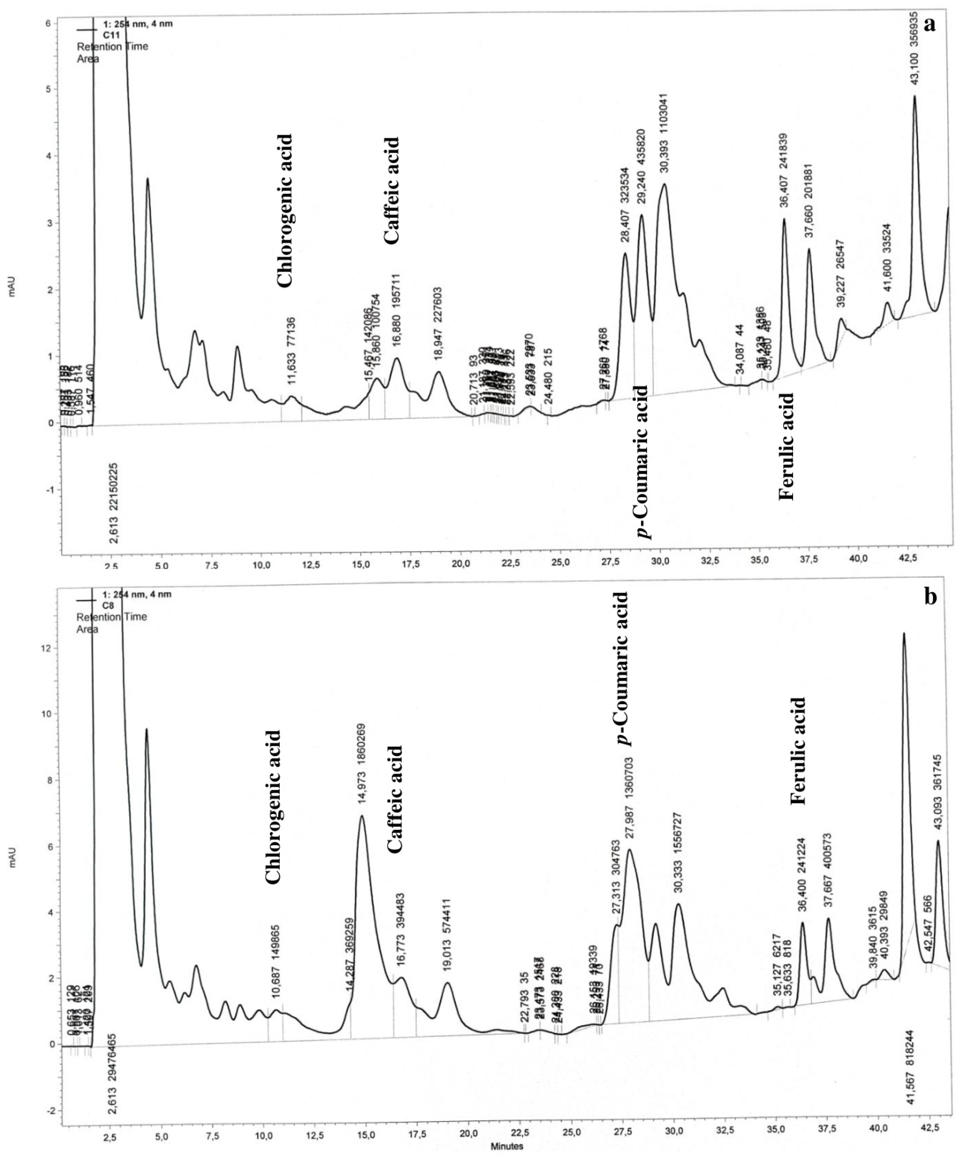
Chromatographic analysis revealed the peaks typical of four phenolic acids: chlorogenic, caffeic, p-coumaric, and ferulic acid. Example chromatograms are shown in Figure 2.

3.3. Effect of Zinc Oxide Nanoparticles on Phenolic Acid Content

Chlorogenic, caffeic, p-coumaric, and ferulic acids were detected in the adventitious bulblets maintained on the media containing ZnO NPs. One of the acids, namely p-coumaric acid, occurred at the highest concentrations, usually exceeding 15 mg/100 g dw (except for Zn combination in the dark) (Figure 3a).
The content of p-coumaric acid was about two times higher (19.92 to 25.14 mg/100 g dw) in the bulblets formed in the light than in the dark, and the presence of 50 mg/L ZnO NPs promoted its accumulation under both conditions (D Zn50 and FL Zn50). The concentration of p-coumaric acid in the bulbscales of the control bulbs growing in soil reached about 15 mg/100 g dw (Figure 3a).
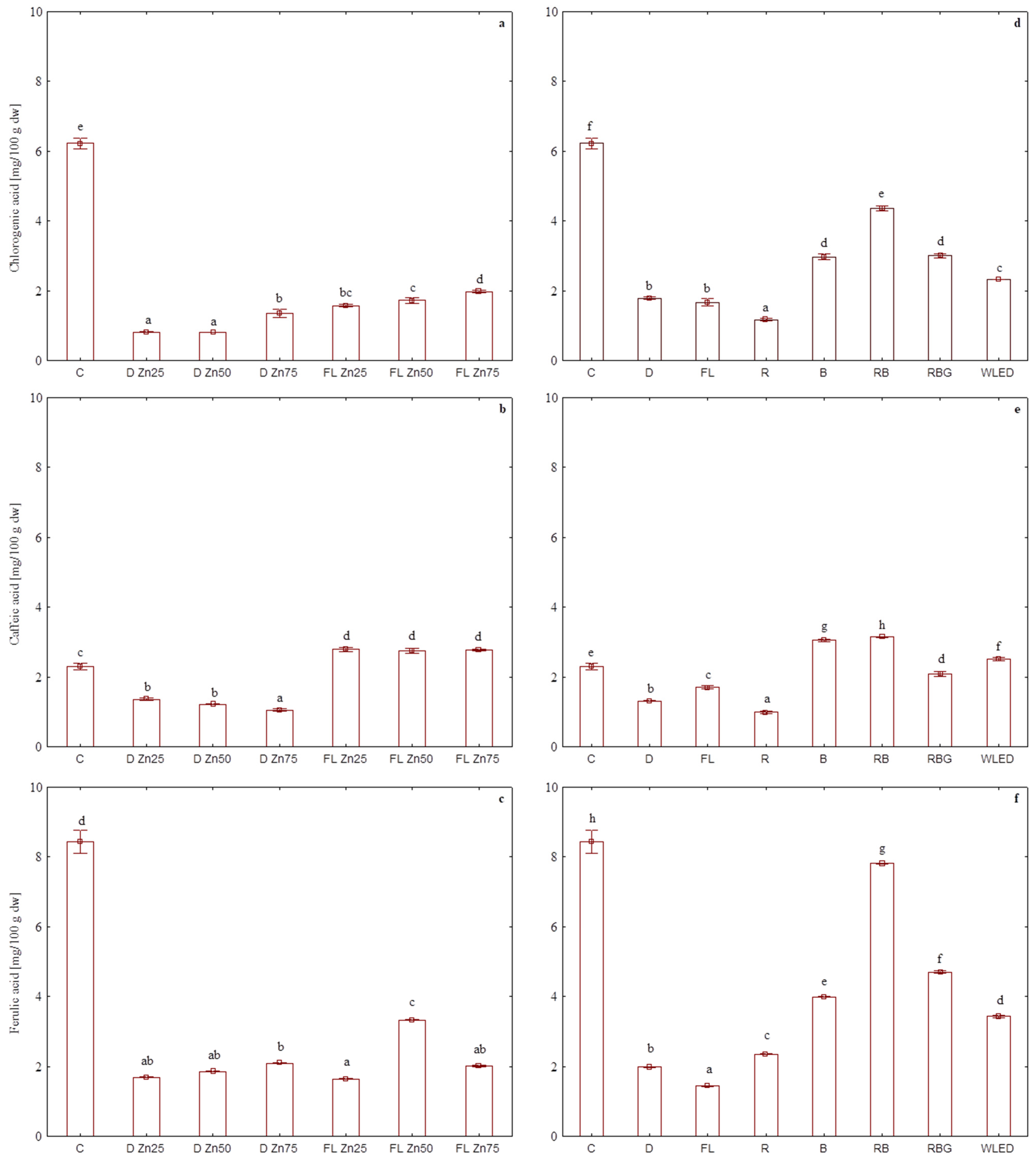
The content of chlorogenic acid in the bulblets formed on the media supplemented with ZnO NPs was the lowest in the dark, but the highest concentration of NPs (D Zn75) increased its accumulation up to 1.35 mg/100 g dw. A similar response was observed in the bulblets formed in the light, where the content of this acid was the highest for the FL Zn75 variant among all bulblets grown in vitro (Figure 4a) but three times lower than in the bulbscales of the control bulbs (C) that contained 6.22 mg/100 g dw chlorogenic acid (Figure 4a).
The highest concentrations of caffeic acid (2.74–2.78 mg/100 g dw) were found in the bulblets formed in the light, and they were independent of ZnO NP content in the medium. The bulblets grown in the dark accumulated two times less caffeic acid, and its content dropped with increasing concentration of ZnO NPs. The control bulbs growing in the field had an average content of this acid at a level of 2.3 mg/100 g dw (Figure 4b).
The content of ferulic acid in the bulblets grown in vitro was four times lower than that in the control bulbs, where it reached 8.43 mg/100 g dw. No differences were found between the cultures maintained in the dark and in the light. For all investigated combinations, the content of ferulic acid was similar (1.64–2.09 mg/100 g dw), except for FL Zn50, where it reached 3.33 mg/100 g dw (Figure 4c).
Statistical analysis examining the influence of light conditions (D, FL) and the presence of ZnO NPs in the medium (regardless of their concentration) revealed that the bulbs formed in the light on the medium with nanoparticles usually had the highest content of the investigated acids. The addition of NPs to the cultures carried out in the dark (D Zn) had an inhibitory effect on the synthesis of phenolic acids. For example, the content of p-coumaric acid was two times lower in the bulblets treated with NPs and growing in the dark, while in those growing under a fluorescent lamp (FL Zn), it increased by 30% as compared with the control. For chlorogenic acid, the most significant drop was found in the bulblets formed in the dark (D Zn), and for caffeic acid, the most significant change was a rise in its content in the bulblets grown in the light and in the presence of NPs (FL Zn). Ferulic acid was the only acid whose concentration was the highest in the bulblets formed in the dark (D) as compared with those grown under FL. Medium supplementation with ZnO NPs did not affect its content in the bulblets formed in the dark (D Zn), while those grown under FL (FL Zn) were the most abundant in ferulic acid (2.33 mg/100 g dw) (Table 3).
The literature lacks data on the analysis of phenolic acids investigated in this work, typical for tissues formed in the presence of nanoparticles. The available information focuses solely on total content of phenols. Callus cultures of Fagonia indica showed increased total phenolic content following elicitation with iron-doped zinc oxide nanoparticles. This increase depended on the concentration of the nanoparticles and the duration of the culture [90]. The content of thymol and carvacrol in Thymus tissues rose a few times as compared with the control after application of ZnO NPs at 150 mg/L [36].
The effect of the application of ZnO NPs on the concentration of phenolic compounds ex vitro depended on plant species. For example, in the cultures of Brassica nigra [91] and Solanum tuberosum [92], ZnO NPs enhanced total phenolic content. In contrast, their contents in Capsicum annuum seedlings generally dropped in the presence of ZnO NPs, except for radicle, where they were clearly boosted [87]. While the effect of Zn NPs on plant physiology and biochemistry has been proven, their influence on secondary metabolism [93], especially of phenolic compounds, is still unclear [94].

3.4. Effect of LED Light on Phenolic Acid Content

The second experiment revealed that fluorescent light (FL), red LED light (R), and darkness (D) decreased the content of four of the investigated phenolic acids in the bulblets formed on the media not supplemented with ZnO NPs (Figure 3b and Figure 4d–f). Similarly, as on the media supplemented with ZnO NPs, the most abundant phenolic acid was p-coumaric acid. The bulblets formed under RB light had the highest content of p-coumaric acid (38.73 mg/100 g dw). Its concentration was lower in the bulblets exposed to RBG and WLED, followed by B light. The bulblets subjected to the other combinations featured a lower content of p-coumaric acid, which was least abundant in the dark, where its content matched that of the control bulbs from field cultivation (Figure 3b).
The highest content of chlorogenic acid was found in the bulblets growing under RB LED (4.35 mg/100 g dw), but it was still lower than that in the control bulbs (by about two units). This acid was also abundant in the bulblets exposed to B and RBG light, but its content under red LED light (R) was almost three times lower (1.16 mg/100 g dw), which was the lowest result of all tested combinations (Figure 4d). Chen et al. [95] also reported on variable content of chlorogenic acid in the tissues of Peucedanum japonicum exposed to LED light of different quality. In this species, the content of chlorogenic acid was the highest in the plants acclimatized to ex vitro conditions (it was also 17 times higher than in the herbal raw material). The callus grown in vitro under a mixture of blue, red, and far-red LED light also featured high (three times higher than in the raw herbal material) content of chlorogenic acid.
The highest content of caffeic acid, also in comparison with control bulbscales of the field-cultivated bulbs, was detected in the bulblets formed under RB (3.14 mg/100 g dw), B (3.05 mg/100 g dw), and WLED light (2.51 mg/100 g dw). In the bulblets exposed to R light, the content of caffeic acid was below 1 mg/100 g dw (Figure 4e). The tissues of Protea cynaroides exposed to a fluorescent lamp accumulated nearly two times more caffeic acid (15.9 mg/g) than those exposed to LED light. Red and blue light triggered similar accumulation of this acid (8.4–8.0 mg/g), and a mixture of red and blue LED light slightly increased the acid content (9.0 mg/g) [96]. There are no literature data on the effects of LED light on the content of individual phenolic acids, but there is information on its effect on the accumulation of total phenolics. Many researchers have observed the inhibiting effect of red light and the stimulating effect of blue light on the synthesis of phenolics [97,98,99]. This was also reported in our previous paper in L. candidum [6].
Our experiment revealed high contents (7.80 mg/100 g dw) of ferulic acid in the bulblets maintained under RB LED light. This value was only by 0.6 mg/100 g dw lower than that in the control bulbs (C). Values half as high as that in the control were achieved under blue (B) and white LED light (WLED), as well as when the spectrum also contained green light (RBG). The lowest content of ferulic acid (below 1.5 mg/100 g dw) was detected under the fluorescent lamp (FL) (Figure 4f). Wu and Lin [96] reported the highest content of this acid (9.7 mg/g) in the tissues of P. cynaroides exposed to fluorescent light. Ferulic acid concentration was lower under blue light (8.2 mg/g) and a mixture of red and blue LED light (8.7 mg/g) and the lowest under red light (7.4 mg/g). This confirmed a previously described relationship between total content of phenolics and the presence of blue and red light. The addition of blue light to the spectrum increases the content of phenolic metabolites, which is in accordance with literature data [100,101,102,103,104]. One should, however, keep in mind that the response to different stimuli, including light, is strongly species-dependent, even in in vitro cultures [105]. Photoreceptors and auxin-responsive factors indirectly affect gene expression and cellular responses, but the exact mechanisms have not been fully explained yet [106].

4. Conclusions

The bulblets of Lilium candidum formed during adventitious organogenesis contained the following phenolic acids: p-coumaric, chlorogenic, caffeic, and ferulic acid. Their contents depended on the concentration of ZnO NPs in the medium and the light quality during bulblet formation. p-Coumaric acid was the most abundant acid, especially in the samples exposed to LED light (RB, RBG, WLED, and B), and on the media without ZnO NPs. All bulbs formed in the light, including those maintained on the media supplemented with ZnO NPs, contained more p-coumaric acid than the bulbscales of control bulbs grown in soil.
The control bulbs always accumulated about two to three times higher amounts of chlorogenic acid than the bulblets formed in vitro. The bulblets exposed to R light accumulated the highest amounts of this acid, although 30% lower than in the control.
We found that FL, R, and darkness decreased the levels of all examined phenolics, the contents of which were the highest in the bulblets formed in vitro under RB light.
The use of ZnO NPs increased the contents of p-coumaric, chlorogenic, and caffeic acid in the bulblets formed under FL as compared with those grown in darkness.
The reported presence of phenolic acids in the bulblets confirmed the medicinal properties of Lilium candidum and the use of this species described in folk medicine sources. Moreover, the compounds we investigated act synergistically with each other, which makes Madonna lily a valuable object of future research. The methods of in vitro elicitation described in this work yield natural compounds of chemical and microbiological purity that can be produced with high efficiency.

Author Contributions

Conceptualization, P.P. and B.P.; methodology, B.P.; validation, P.P. and B.M.; formal analysis, P.P.; investigation, P.P.; resources, B.M., A.S. and B.P.; data curation, A.S.; writing—original draft preparation, P.P.; writing—review and editing, B.P.; visualization, P.P.; supervision, B.M. and B.P.; project administration, B.P. All authors have read and agreed to the published version of the manuscript.

Funding

This project was supported by the Polish Ministry of Science and Higher Education.

Data Availability Statement

The data presented in this study are available upon request from the corresponding author.

Acknowledgments

This project was supported by the Polish Ministry of Science and Higher Education.

Conflicts of Interest

The authors declare no conflict of interest.

Abbreviations

B: 100% blue LED light; D: darkness; D Zn: culture medium supplemented with zinc oxide nanoparticles in darkness; D Zn25: culture medium supplemented with 25 mg/L zinc oxide nanoparticles in darkness; D Zn50: culture medium supplemented with 50 mg/L zinc oxide nanoparticles in darkness; D Zn75: culture medium supplemented with 75 mg/L zinc oxide nanoparticles in darkness; FL: fluorescent lamp; FL Zn: culture medium supplemented with zinc oxide nanoparticles under fluorescent lamp; FL Zn25: culture medium supplemented with 25 mg/L zinc oxide nanoparticles under fluorescent lamp; FL Zn50: culture medium supplemented with 50 mg/L zinc oxide nanoparticles under fluorescent lamp; FL Zn75: culture medium supplemented with 75 mg/L zinc oxide nanoparticles under fluorescent lamp; g dw: gram of dry weight; LED: light-emitting diode; MS: Murashige and Skoog culture medium; R: 100% red LED; RB: 70% red + 30% blue LED; RBG: 35% red + 15% blue + 50% green LED; WLED: 33.3% warm + 33.3% neutral + 33.3% cool white LED.

References

  1. Özen, F.; Temeltaş, H.; Aksoy, Ö. The anatomy and morphology of the medicinal plant, Lilium candidum L. (Liliaceae) distributed in Marmara region of Turkey. Pak. J. Bot. 2012, 44, 1185–1192. [Google Scholar]
  2. Pieroni, A. Medicinal plants and food medicines in the folk traditions of the upper Lucca Province, Italy. J. Ethnopharmacol. 2000, 70, 235–273. [Google Scholar] [CrossRef] [PubMed]
  3. Patocka, J.; Navratilova, Z. Bioactivity of Lilium candidum L.: A mini review. Biomed. J. Sci. Technol. Res. 2019, 8, 13859–13862. [Google Scholar] [CrossRef]
  4. Zaccai, M.; Yarmolinsky, L.; Khalfin, B.; Budovsky, A.; Gorelick, J.; Dahan, A.; Ben-Shabat, S. Medicinal properties of Lilium candidum L. and its phytochemicals. Plants 2020, 9, 959. [Google Scholar] [CrossRef]
  5. Momtaz, S.; Dibaj, M.; Abdollahi, A.; Amin, G.; Bahramsoltani, R.; Abdollahi, M.; Mahdaviani, P.; Abdolghaffari, A.H. Wound healing activity of the flowers of Lilium candidum L. in burn wound model in rats. J. Med. Plants 2020, 19, 109–118. [Google Scholar] [CrossRef]
  6. Pałka, P.; Cioć, M.; Hura, K.; Szewczyk-Taranek, B.; Pawłowska, B. Adventitious organogenesis and phytochemical composition of Madonna lily (Lilium candidum L.) in vitro modeled by different light quality. Plant Cell Tissue Organ Cult. (PCTOC) 2023, 152, 99–114. [Google Scholar] [CrossRef]
  7. Khawar, K.M.; Cocu, S.; Parmaksiz, I.; Sarihan, E.O.; Özcan, S. Mass proliferation of Madonna Lily (Lilium candidum L.) under in vitro conditions. Pak. J. Bot. 2005, 37, 243–248. [Google Scholar]
  8. Sevimay, C.S.; Khawar, K.M.; Parmaksız, I.; Cocu, S.; Sancak, C.; Sarihan, E.; Özcan, S. Prolific in vitro bulblet formation from bulb scales of meadow lily (Lilium candidum L.). Period. Biol. 2005, 107, 107–111. [Google Scholar]
  9. Altan, F.; Bürün, B.; Şahin, N. Fungal contaminants observed during micropropagation of Lilium candidum L. and the effect of chemotherapeutic substances applied after sterilization. Afr. J. Biotechnol. 2010, 9, 991–995. [Google Scholar] [CrossRef]
  10. Burun, B.; Sahin, O. Micropropagation of Lilium candidum L.: A rare and native bulbous flower of Turkey. Bangladesh J. Bot. 2013, 42, 185–187. [Google Scholar] [CrossRef]
  11. Saadon, S.; Zaccai, M. Lilium candidum bulblet and meristem development. Vitr. Cell. Dev. Biol. Plant 2013, 49, 313–319. [Google Scholar] [CrossRef]
  12. Altan, F.; Bürün, B. The effect of some antibiotic and fungucide applications on the micropropagation of Lilium candidum L. Mugla J. Sci. Technol. 2017, 3, 86–91. [Google Scholar] [CrossRef]
  13. Daneshvar Royandazagh, S. Efficient approaches to in vitro multiplication of Lilium candidum L. with consistent and safe access throughout year and acclimatization of plant under hot-summer mediterranean (Csa Type) climate. Not. Bot. Horti Agrobot. 2019, 47, 734–742. [Google Scholar] [CrossRef]
  14. Tokgoz, H.B.; Altan, F. Callus induction and micropropagation of Lilium candidum L. using stem bulbils and confirmation of genetic stability via SSR-PCR. Int. J. Second. Metab. 2020, 7, 286–296. [Google Scholar] [CrossRef]
  15. Akshay, M.P.; Pooja, P.G.; Sonali, D. In vitro micropropagation of Lilium candidum bulb by application of multiple hormone concentrations using plant tissue culture technique. Int. J. Res. Appl. Sci. Biotechnol. 2021, 8, 244–253. [Google Scholar] [CrossRef]
  16. Bornwaßer, T.; Tantau, H.J. Evaluation of LED lighting systems in in vitro cultures. Acta Hort. 2012, 956, 555–562. [Google Scholar] [CrossRef]
  17. Gupta, S.D.; Jatothu, B. Fundamentals and applications of lightemitting diodes (LEDs) in in vitro plant growth and morphogenesis. Plant Biotechnol. Rep. 2013, 7, 211–220. [Google Scholar] [CrossRef]
  18. Bantis, F.; Smirnakou, S.; Ouzounis, T.; Koukounaras, A.; Ntagkas, N.; Radoglou, K. Current status and recent achievements in the field of horticulture with the use of light-emitting diodes (LEDs). Sci. Hortic. 2018, 235, 437–451. [Google Scholar] [CrossRef]
  19. Lian, M.L.; Murthy, H.N.; Paek, K.Y. Effects of light emitting diodes (LED) on the in vitro induction and growth of bulblets of Lilium oriental hybrid ‘Pesaro’. Sci. Hortic. 2002, 94, 365–370. [Google Scholar] [CrossRef]
  20. Prokopiuk, B.; Cioć, M.; Maślanka, M.; Pawłowska, B. Effects of light spectra and benzyl adenine on in vitro adventitious bulb and shoot formation of Lilium regale E. H. Wilson. Propag. Ornam. Plants 2018, 18, 12–18. [Google Scholar]
  21. Rivero-Montejo, S.J.; Vargas-Hernandez, M.; Torres-Pacheco, I. Nanoparticles as novel elicitors to improve bioactive compounds in plants. Agriculture 2021, 11, 134. [Google Scholar] [CrossRef]
  22. Namdeo, A.G. Plant cell elicitation for production of secondary metabolites: A review. Pharmacogn. Rev. 2007, 1, 69–79. [Google Scholar]
  23. Shitan, N. Secondary metabolites in plants: Transport and self-tolerance mechanisms. Biosci. Biotechnol. Biochem. 2016, 80, 1283–1293. [Google Scholar] [CrossRef] [PubMed]
  24. Jafari, S.M.; McClements, D.J. Chapter One—Nanotechnology approaches for increasing nutrient bioavailability. In Advances in Food and Nutrition Research, 1st ed.; Toldrá, F., Ed.; Academic Press: London, UK, 2017; Volume 81, pp. 1–30. [Google Scholar] [CrossRef]
  25. Iranbakhsh, A.; Oraghi Ardebili, Z.; Oraghi Ardebili, N. Synthesis and characterization of zinc oxide nanoparticles and their impact on plants. In Plant Responses to Nanomaterials; Singh, V.P., Singh, S., Tripathi, D.K., Prasad, S.M., Chauhan, D.K., Eds.; Nanotechnology in the Life Sciences; Springer: Cham, Switzerland, 2021; pp. 33–93. [Google Scholar] [CrossRef]
  26. Misra, A.; Srivastava, A.K.; Srivastava, N.K.; Khan, A. Zn-acquisition and its role in growth, photosynthesis, photosynthetic pigments, and biochemical changes in essential monoterpene oil(s) of Pelargonium graveolens. Photosynthetica 2005, 43, 153–155. [Google Scholar] [CrossRef]
  27. Eisvand, H.R.; Kamaei, H.; Nazarian, F. Chlorophyll fluorescence, yield and yield components of bread wheat affected by phosphatebio-fertilizer, zinc and boron under late-season heat stress. Photosynthetica 2018, 56, 1287–1296. [Google Scholar] [CrossRef]
  28. Raliya, R.; Tarafdar, J.C. ZnO nanoparticle biosynthesis and its effect on phosphorus-mobilizing enzyme secretion and gum contents in clusterbean (Cyamopsis tetragonoloba L.). Agric. Res. 2013, 2, 48–57. [Google Scholar] [CrossRef]
  29. Sabir, S.; Arshad, M.; Chaudhari, S.K. Zinc oxide nanoparticles for revolutionizing agriculture: Synthesis and applications. Sci. World J. 2014, 2014, 925494. [Google Scholar] [CrossRef]
  30. Singh, A.; Singh, N.B.; Afzal, S.; Singh, T.; Hussain, I. Zinc oxide nanoparticles: A review of their biological synthesis, antimicrobial activity, uptake, translocation and biotransformation in plants. J. Mater. Sci. 2018, 53, 185–201. [Google Scholar] [CrossRef]
  31. Wojnarowicz, J.; Chudoba, T.; Lojkowski, W. A Review of Microwave Synthesis of Zinc Oxide Nanomaterials: Reactants, Process Parameters and Morphologies. Nanomaterials 2020, 10, 1086. [Google Scholar] [CrossRef]
  32. Prasad, T.N.V.K.V.; Sudhakar, P.; Sreenivasulu, Y.; Latha, P.; Munaswamy, V.; Raja Reddy, K.; Sreeprasad, T.S.; Sajanlal, P.R.; Pradeep, T. Effect of nanoscale zinc oxide particles on the germination, growth and yield of peanut. J. Plant Nutr. 2012, 35, 905–927. [Google Scholar] [CrossRef]
  33. Helaly, M.N.; El-Metwally, M.A.; El-Hoseiny, H.; Omar, S.A.; El-Sheery, N.I. Effect of nanoparticles on biological contamination of in vitro cultures and organogenic regeneration of banana. Aust. J. Crop Sci. 2014, 8, 612–624. [Google Scholar]
  34. Raskar, S.V.; Laware, S.L. Effect of zinc oxide nanoparicles on cytology and seed germination in onion. Int. J. Curr. Microbiol. Appl. Sci. 2014, 3, 467–473. [Google Scholar]
  35. Chamani, E.; Ghalehtaki, S.K.; Mohebodini, M.; Ghanbari, A. The effect of zinc oxide nano particles and humic acid on morphological characters and secondary metabolite production in Lilium ledebourii Bioss. Iran. J. Genet. Plant Breed. 2015, 4, 11–19. [Google Scholar]
  36. Mosavat, N.; Golkar, P.; Yousefifard, M.; Javed, R. Modulation of callus growth and secondary metabolites in different Thymus species and Zataria multiflora micropropagated under ZnO nanoparticles stress. Biotechnol. Appl. Biochem. 2019, 66, 316–322. [Google Scholar] [CrossRef]
  37. Ahmad, M.A.; Javed, R.; Adeel, M.; Rizwan, M.; Ao, Q.; Yang, Y. Engineered ZnO and CuO nanoparticles ameliorate morphological and biochemical response in tissue culture regenerants of candyleaf (Stevia rebaudiana). Molecules 2020, 25, 1356. [Google Scholar] [CrossRef] [PubMed]
  38. El-Mahdy, M.T.; Elazab, D. Impact of zinc oxide nanoparticles on pomegranate growth under in vitro conditions. Russ. J. Plant Physiol. 2020, 67, 162–167. [Google Scholar] [CrossRef]
  39. Mazaheri-Tirani, M.; Dayani, S. In vitro effect of zinc oxide nanoparticles on Nicotiana tabacum callus compared to ZnO micro particles and zinc sulfate (ZnSO4). Plant Cell Tissue Organ Cult. (PCTOC) 2020, 140, 279–289. [Google Scholar] [CrossRef]
  40. Tymoszuk, A.; Wojnarowicz, J. Zinc oxide and zinc oxide nanoparticles impact on in vitro germination and seedling growth in Allium cepa L. Materials 2020, 13, 2784. [Google Scholar] [CrossRef]
  41. Zaeem, A.; Drouet, S.; Anjum, S.; Khurshid, R.; Younas, M.; Blondeau, J.P.; Tungmunnithum, D.; Giglioli-Guivarc’h, N.; Hano, C.; Abbasi, B.H. Effects of biogenic zinc oxide nanoparticles on growth and oxidative stress response in flax seedlings vs. in vitro cultures: A comparative analysis. Biomolecules 2020, 10, 918. [Google Scholar] [CrossRef] [PubMed]
  42. Al-Mayahi, A.M.W. The effect of humic acid (HA) and zinc oxide nanoparticles (ZnO-NPS) on in vitro regeneration of date palm (Phoenix dactylifera L.) cv. Quntar. Plant Cell Tissue Organ Cult. 2021, 145, 445–456. [Google Scholar] [CrossRef]
  43. Hezaveh, T.A.; Rahmani, F.; Alipour, H.; Pourakbar, L. Effects of foliar application of ZnO nanoparticles on secondary metabolite and micro-elements of camelina (Camelina sativa L.) under salinity stress. J. Stress Physiol. Biochem. 2020, 16, 54–69. [Google Scholar]
  44. Sharifi-Rad, R.; Bahabadi, S.E.; Samzadeh-Kermani, A.; Gholami, M. The effect of non-biological elicitors on physiological and biochemical properties of medicinal plant Momordica charantia L. Iran. J. Sci. Technol. Trans. A Sci. 2020, 44, 1315–1326. [Google Scholar] [CrossRef]
  45. Rosa, L.A.; Moreno-Escamilla, J.O.; Rodrigo-García, J.; Alvarez-Parrilla1, E. Chapter 12 Phenolic compounds. In Postharvest Physiology and Biochemistry of Fruits and Vegetables; Yahia, E.M., Ed.; Woodhead Publishing: Sawton, UK, 2018; pp. 253–271. [Google Scholar] [CrossRef]
  46. Babenko, L.M.; Smirnov, O.E.; Romanenko, K.O.; Trunova, O.K.; Kosakivska, I.V. Phenolic compounds in plants: Biogenesis and functions. Ukr. Biochem. J. 2019, 91, 5–18. [Google Scholar] [CrossRef]
  47. Ghasemzadeh, A.; Jaafar, H.Z.E.; Rahmat, A.; Wahab, P.E.M.; Halim, M.R.A. Effect of different light intensities on total phenolics and flavonoids synthesis and anti-oxidant activities in young ginger varieties (Zingiber officinale Roscoe). Int. J. Mol. Sci. 2010, 11, 3885–3897. [Google Scholar] [CrossRef]
  48. Chang, H.P.; Kim, N.S.; Park, J.S.; Lee, S.Y.; Lee, J.W.; Park, S.U. Effects of light-emitting diodes on the accumulation of glucosinolates and phenolic compounds in sprouting canola (Brassica napus L.). Foods 2019, 8, 76. [Google Scholar] [CrossRef]
  49. Hsie, B.S.; Bueno, A.I.S.; Bertolucci, S.K.V.; Carvalho, A.A.; Cunha, S.H.B.; Martins, E.R.; Pinto, J.E.B.P. Study of the influence of wavelengths and intensities of LEDs on the growth, photosynthetic pigment, and volatile compounds production of Lippia rotundifolia Cham in vitro. J. Photochem. Photobiol. B Biol. 2019, 198, 111577. [Google Scholar] [CrossRef]
  50. Upadhyay, R.; Rao, L.J.M. An outlook on chlorogenic acids—Occurrence, chemistry, technology, and biological activities. Crit. Rev. Food Sci. Nutr. 2013, 53, 968–984. [Google Scholar] [CrossRef]
  51. Marques, V.; Farah, A. Chlorogenic acids and related compounds in medicinal plants and infusions. Food Chem. 2009, 113, 1370–1376. [Google Scholar] [CrossRef]
  52. Naveed, M.; Hejazi, V.; Abbas, M.; Kamboh, A.A.; Khan, G.J.; Shumzaid, M.; Ahmad, F.; Babazadeh, D.; Xia, F.; Modarresi-Ghazani, F.; et al. Chlorogenic acid (CGA): A pharmacological review and call for further research. Biomed. Pharmacother. 2018, 97, 67–74. [Google Scholar] [CrossRef]
  53. Martínez, G.; Regente, M.; Jacobi, S.; Del Rio, M.; Pinedo, M.; de la Canal, L. Chlorogenic acid is a fungicide active against phytopathogenic fungi. Pestic. Biochem. Physiol. 2017, 140, 30–35. [Google Scholar] [CrossRef]
  54. Kundu, A.; Vadassery, J. Chlorogenic acid-mediated chemical defence of plants against insect herbivores. Plant Biol. 2019, 21, 185–189. [Google Scholar] [CrossRef] [PubMed]
  55. Magnani, C.; Isaac, V.L.B.; Correa, M.A.; Salgado, H.R.N. Caffeic Acid: A review of its potential use for medications and cosmetics. Anal. Methods 2014, 6, 3203–3210. [Google Scholar] [CrossRef]
  56. Greenwald, P. Clinical trials in cancer prevention: Current results and perspectives for the future. J. Nutr. 2004, 134, 3507S–3512S. [Google Scholar] [CrossRef] [PubMed]
  57. Bouzaiene, N.N.; Jaziri, S.K.; Kovacic, H.; Chekir-Ghedira, L.; Ghedira, K.; Luis, J. The effects of caffeic, coumaric and ferulic acids on proliferation, superoxide production, adhesion and migration of human tumor cells in vitro. Eur. J. Pharmacol. 2015, 766, 99–105. [Google Scholar] [CrossRef]
  58. Sanchez-Moreno, C.; Jimenez-Escrig, A.; Saura-Calixto, F. Study of low-density lipoprotein oxidizability indexes to measure the antioxidant activity of dietary polyphenol. Nutr. Res. 2000, 20, 941–953. [Google Scholar] [CrossRef]
  59. Vinson, J.A.; Teufel, K.; Wu, N. Red wine, dealcoholized red wine, and especially grape juice, inhibit atherosclerosis in a hamster model. Atherosclerosis 2001, 156, 67–72. [Google Scholar] [CrossRef]
  60. Pei, K.; Ou, J.; Huang, J.; Ou, S. p-Coumaric acid and its conjugates: Dietary sources, pharmacokinetic properties and biological activities. J. Sci. Food Agric. 2016, 96, 2952–2962. [Google Scholar] [CrossRef]
  61. Zang, L.Y.; Cosma, G.; Gardner, H.; Shi, X.; Castranova, V.; Vallyathan, V. Effect of antioxidant protection by p-coumaric acid on low-density lipoprotein cholesterol oxidation. Am. J. Physiol. Cell Physiol. 2000, 279, C954–C960. [Google Scholar] [CrossRef]
  62. Kiliç, I.; Yeşiloğlu, Y. Spectroscopic studies on the antioxidant activity of p-coumaric acid. Spectrochim. Acta A Mol. Biomol. Spectrosc. 2013, 115, 719–724. [Google Scholar] [CrossRef]
  63. Lou, Z.; Wang, H.; Rao, S.; Sun, J.; Ma, C.; Li, J. p-Coumaric acid kills bacteria through dual damage mechanisms. Food Control 2012, 25, 550–554. [Google Scholar] [CrossRef]
  64. Boz, H. p-Coumaric acid in cereals: Presence, antioxidant and antimicrobial effects. Int. J. Food Sci. Technol. 2015, 50, 2323–2328. [Google Scholar] [CrossRef]
  65. Janicke, B.; Hegardt, C.; Krogh, M.; Onning, G.; Akesson, B.; Cirenajwis, H.M.; Oredsson, S.M. The antiproliferative effect of dietary fiber phenolic compounds ferulic acid and p-coumaric acid on the cell cycle of Caco-2 cells. Nutr. Cancer. 2011, 63, 611–622. [Google Scholar] [CrossRef] [PubMed]
  66. Jaganathan, S.K.; Supriyanto, E.; Mandal, M. Events associated with apoptotic effect of p-Coumaric acid in HCT-15 colon cancer cells. World J. Gastroenterol. 2013, 19, 7726–7734. [Google Scholar] [CrossRef] [PubMed]
  67. Roy, N.; Narayanankutty, A.; Nazeem, P.A.; Valsalan, R.; Babu, T.D.; Mathew, D. Plant phenolics ferulic acid and p-coumaric acid inhibit colorectal cancer cell proliferation through EGFR down-regulation. Asian Pac. J. Cancer Prev. 2016, 17, 4019–4023. [Google Scholar] [PubMed]
  68. Contardi, M.; Alfaro-Pulido, A.; Picone, P.; Guzman-Puyol, S.; Goldoni, L.; Benítez, J.J.; Heredia, A.; Barthel, M.J.; Ceseracciu, L.; Cusimano, G.; et al. Low molecular weight epsilon-caprolactone-p-coumaric acid copolymers as potential biomaterials for skin regeneration applications. PLoS ONE 2019, 14, e0214956. [Google Scholar] [CrossRef]
  69. Contardi, M.; Heredia-Guerrero, J.A.; Guzman-Puyol, S.; Summa, M.; Benítez, J.J.; Goldoni, L.; Caputo, G.; Cusimano, G.; Picone, P.; Di Carlo, M.; et al. Combining dietary phenolic antioxidants with polyvinylpyrrolidone: Transparent biopolymer films based on p-coumaric acid for controlled release. J. Mater. Chem. B 2019, 7, 1384–1396. [Google Scholar] [CrossRef]
  70. Boo, Y.C. p-Coumaric acid as an active ingredient in cosmetics: A review focusing on its antimelanogenic effects. Antioxidants 2019, 8, 275. [Google Scholar] [CrossRef]
  71. Ou, S.; Kwok, K. Ferulic acid: Pharmaceutical functions, preparation and applications in foods. J. Sci. Food Agric. 2004, 84, 1261–1269. [Google Scholar] [CrossRef]
  72. Srinivasan, M.; Sudheer, A.R.; Menon, V.P. Ferulic acid: Therapeutic potential through its antioxidant property. J. Clin. Biochem. Nutr. 2007, 40, 92–100. [Google Scholar] [CrossRef]
  73. Li, D.; Rui, Y.; Guo, S.; Luan, F.; Liu, R.; Zeng, N. Ferulic acid: A review of its pharmacology, pharmacokinetics and derivatives. Life Sci. 2021, 284, 119921. [Google Scholar] [CrossRef]
  74. Zduńska, K.; Dana, A.; Kolodziejczak, A.; Rotsztejn, H. Antioxidant properties of ferulic acid and its possible application. Skin Pharmacol. Physiol. 2018, 31, 332–336. [Google Scholar] [CrossRef] [PubMed]
  75. Sakai, S.; Ochiai, H.; Nakajima, K.; Terasawa, K. Inhibitory effect of ferulic acid on macrophage inflammatory protein-2 production in a murine macrophage cell line, RAW264.7. Cytokine 1997, 9, 242–248. [Google Scholar] [CrossRef] [PubMed]
  76. Ou, L.; Kong, L.; Zhang, X.; Niwa, M. Oxidation of ferulic acid by Momordica charantia peroxidase and related anti-inflammation activity changes. Biol. Pharm. Bull. 2003, 26, 1511–1516. [Google Scholar] [CrossRef] [PubMed]
  77. Kawabata, K.; Yamamoto, T.; Hara, A.; Shimizu, M.; Yamada, Y.; Matsunaga, K.; Tanaka, T.; Mori, H. Modifying effects of ferulic acid on azoxymethane-induced colon carcinogenesis in F344 rats. Cancer Lett. 2000, 157, 15–21. [Google Scholar] [CrossRef]
  78. Balasubashini, M.S.; Rukkumani, R.; Viswanathan, P.; Menon, V.P. Ferulic acid alleviates lipid peroxidation in diabetic rats. Phytother. Res. 2004, 18, 310–314. [Google Scholar] [CrossRef]
  79. Murashige, T.; Skoog, F. A revised medium for rapid growth and bioassays with tobacco tissue cultures. Physiol. Plant. 1962, 15, 473–497. [Google Scholar] [CrossRef]
  80. Pawłowska, B.; Żupnik, M.; Szewczyk-Taranek, B.; Cioć, M. Impact of LED light sources on morphogenesis and levels of photosynthetic pigments in Gerbera jamesonii grown in vitro. Hortic. Environ. Biotechnol. 2018, 59, 115–123. [Google Scholar] [CrossRef]
  81. Li, H.; Xu, Z.; Tang, C. Effect of light-emitting diodes on growth and morphogenesis of upland cotton (Gossypium hirsutum L.) plantlets in vitro. Plant Cell Tissue Organ Cult. 2010, 103, 155–163. [Google Scholar] [CrossRef]
  82. Fan, X.X.; Zang, J.; Xu, Z.G.; Guo, S.R.; Jiao, X.L.; Liu, X.Y.; Gao, Y. Effects of different light quality on growth, chlorophyll concentration and chlorophyll biosynthesis precursors of non-heading Chinese cabbage (Brassica campestris L.). Acta Physiol. Plant. 2013, 35, 2721–2726. [Google Scholar] [CrossRef]
  83. Habiba, A.U.; Kazuhiko, S.; Ahasan, M.; Alam, M. Effects of different light quality on growth and development of protocorm-like bodies (PLBs) in Dendrobium kingianum cultured in vitro. Bangladesh Res. Publ. J. 2014, 10, 223–227. [Google Scholar]
  84. Bello-Bello, J.J.; Martínez-Estrada, E.; Caamal-Velázquez, J.H.; Morales-Ramos, V. Effect of LED light quality on in vitro shoot proliferation and growth of vanilla (Vanilla planifolia Andrews). Afr. J. Biotechnol. 2016, 15, 272–277. [Google Scholar] [CrossRef]
  85. Coelho, A.D.; Souza, C.K.; Bertolucci, S.K.V.; Carvalho, A.A.; Santos, G.C.; Oliveira, T.; Marques, E.A.; Salimena, J.P.; Pinto, J.E.B.P. Wavelength and light intensity enhance growth, phytochemical contents and antioxidant activity in micropropagated plantlets of Urtica dioica L. Plant Cell Tissue Organ Cult. (PCTOC) 2021, 145, 59–74. [Google Scholar] [CrossRef]
  86. Klimek-Szczykutowicz, M.; Prokopiuk, B.; Dziurka, K.; Pawłowska, B.; Ekiert, H.; Szopa, A. The influence of different wavelengths of LED light on the production of glucosinolates and phenolic compounds and the antioxidant potential in in vitro cultures of Nasturtium ofcinale (watercress). Plant Cell Tissue Organ Cult. (PCTOC) 2022, 149, 113–122. [Google Scholar] [CrossRef]
  87. García-López, J.I.; Zavala-García, F.; Olivares-Sáenz, E.; Lira-Saldívar, R.H.; Barriga-Castro, E.D.; Ruiz-Torres, N.A.; Ramos-Cortez, E.; Vázquez-Alvarado, R.; Niño-Medina, G. Zinc oxide nanoparticles boosts phenolic compounds and antioxidant activity of Capsicum annuum L. during germination. Agronomy 2018, 8, 215. [Google Scholar] [CrossRef]
  88. Kim, D.H.; Gopal, J.; Sivanesan, I. Nanomaterials in plant tissue culture: The disclosed and undisclosed. RSC Adv. 2017, 7, 6492–36505. [Google Scholar] [CrossRef]
  89. Garcia-Lópes, J.; Nino-Medina, G.; Olivares-Sàenz, E.; LiraSaldivar, R.; Barriga-Costro, E.; Vàzques-Alvarado, R.; Rodriguez-Salinas, P.; Zavala-Garcia, F. Foliar application of zinc oxide nanoparticles and zinc sulfate boosts the content of bioactive compound in Habanero peppers. Plants 2019, 8, 254. [Google Scholar] [CrossRef]
  90. Khan, A.U.; Khan, T.; Khan, M.A.; Nadhman, A.; Aasim, M.; Khan, N.Z.; Ali, W.; Nazir, N.; Zahoor, M. Iron-doped zinc oxide nanoparticles-triggered elicitation of important phenolic compounds in cell cultures of Fagonia indica. Plant Cell Tissue Organ Cult. (PCTOC) 2021, 147, 287–296. [Google Scholar] [CrossRef]
  91. Zafar, H.; Alli, A.; Ali, J.S.; Haq, I.U.; Zia, M. Effect of ZnO nanoparticles on Brassica nigra seedlings and stem explants: Growth dynamics and antioxidative response. Front. Plant Sci. 2016, 7, 535. [Google Scholar] [CrossRef]
  92. Raigond, P.; Raigond, B.; Kaundal, B.; Singh, B.; Joshi, A.; Dutt, S. Effect of zinc nanoparticles on antioxidative system of potato plants. J. Environ. Biol. 2017, 38, 435–439. [Google Scholar] [CrossRef]
  93. Marslin, G.; Sheeba, C.J.; Franklin, G. Nanoparticles alter secondary metabolism in plants via ROS burst. Front. Plant Sci. 2017, 8, 832. [Google Scholar] [CrossRef]
  94. Michalak, A. Phenolic compounds and their antioxidant activity in plants growing under heavy metal stress. Pol. J. Environ. 2006, 15, 523–530. [Google Scholar]
  95. Chen, C.; Agrawal, D.C.; Lee, M.; Lee, R.; Kuo, C.; Wu, C.; Tsay, H.; Chang, H. Influence of LED light spectra on in vitro somatic embryogenesis and LC–MS analysis of chlorogenic acid and rutin in Peucedanum japonicum Thunb.: A medicinal herb. Bot. Stud. 2016, 57, 9. [Google Scholar] [CrossRef] [PubMed]
  96. Wu, H.; Lin, C. Red light-emitting diode light irradiation improves root and leaf formation in difficult-to-propagate Protea cynaroides L. plantlets in vitro. HortScience 2012, 47, 1490–1494. [Google Scholar] [CrossRef]
  97. Guo, B.; Liu, Y.; Yan, Q.; Liu, C. Spectral composition of irradiation regulates the cell growth and flavonoids biosynthesis in callus cultures of Saussurea medusa Maxim. Plant Growth Regulat. 2007, 52, 259–263. [Google Scholar] [CrossRef]
  98. Urbonaviciute, A.; Samuoliene, G.; Brazaityte, A.; Duchovskis, P.; Ruzgas, V.; Zukauskas, A. The effect of variety and lighting quality on wheatgrass antioxidant properties. ZemdirbysteAgriculture 2009, 96, 119–128. [Google Scholar]
  99. Leal-Costa, M.V.; dos Santos Nascimento, L.B.; dos Santos Moreira, N.; Reinert, F.; Costa, S.; Lage, C.L.S.; Tavares, E.S. Influence of blue light on the leaf morphoanatomy of in vitro Kalanchoe pinnata (Lamarck) Persson (Crassulaceae). Microsc. Microanal. 2010, 16, 576–582. [Google Scholar] [CrossRef]
  100. Szopa, A.; Ekiert, H. The importance of applied light quality on the production of lignans and phenolic acids in Schisandra chinensis (Turcz.) Baill. cultures in vitro. Plant Cell Tissue Organ Cult. 2016, 127, 115–121. [Google Scholar] [CrossRef]
  101. Kawka, B.; Kwiecień, I.; Ekiert, H. Infuence of culture medium composition and light conditions on the accumulation of bioactive compounds in shoot cultures of Scutellaria laterifora L. (American Skullcap) grown in vitro. Appl. Biochem. Biotechnol. 2017, 183, 1414–1425. [Google Scholar] [CrossRef]
  102. Kubica, P.; Szopa, A.; Ekiert, H. Production of verbascoside and phenolic acids in biomass of Verbena ofcinalis L. (vervain) cultured under diferent in vitro conditions. Nat. Prod. Res. 2017, 31, 1663–1668. [Google Scholar] [CrossRef]
  103. Szopa, A.; Kokotkiewicz, A.; Bednarz, M.; Luczkiewicz, M.; Ekiert, H. Studies on the accumulation of phenolic acids and favonoids in diferent in vitro culture systems of Schisandra chinensis (Turcz.) Baill. using a DAD-HPLC method. Phytochem. Lett. 2017, 20, 462–469. [Google Scholar] [CrossRef]
  104. Szopa, A.; Starzec, A.; Ekiert, H. The importance of monochromatic lights in the production of phenolic acids and favonoids in shoot cultures of Aronia melanocarpa, Aronia arbutifolia and Aronia × prunifolia. J. Photochem. Photobiol. B 2018, 179, 91–97. [Google Scholar] [CrossRef] [PubMed]
  105. Samuoliene, G.; Brazaityte, A.; Vaštakaitė-Kairienė, V. Light-emitting diodes (LEDs) for improved nutritional quality. In Light-Emitting Diodes for Agriculture; Dutta Gupta, S., Ed.; Springer: New York, NY, USA, 2017; pp. 149–190. [Google Scholar] [CrossRef]
  106. Zielińska, S.; Piątczak, E.; Kozłowska, W.; Bohater, A.; Jezierska-Domaradzka, A.; Kolniak-Ostek, J.; Matkowski, A. LED illumination and plant growth regulators’ efects on growth and phenolic acids accumulation in Moluccella laevis L. in vitro cultures. Acta Physiol. Plant. 2020, 42, 72. [Google Scholar] [CrossRef]
Figure 1. Adventitious organogenesis on Lilium candidum bulbscales in vitro depending on elicitation factor: on medium suplemented with zinc oxide nanoparticles (ZnO NP) (Zinc oxide nanoparticle clicitation): in darkness with (d) 25 (D Zn25); (e) 75 mg/L (D Zn75) and under fluorescent lamp with (a) 25 (Fl Zn25); (b) 50 (Fl Zn50); (c) 75 mg/L (Fl Zn75). Under different light quality in vitro (Light elicitation): (h) darkness (D); (j) fluorescent lamp (Fl); under LED light (%): (f) 100 blue (B); (g) 100 red (R); (k) 35 R + 15 B + 50 green (RBG) and (i) white: 33.3 warm, 33.3 neutral + 33.3 cool (Wled). Bar = 1 cm.
Agronomy 13 01437 g001
Figure 2. HPLC chromatographic separation of phenolic acids from Lilium candidum L. bulblets in vitro on medium supplemented with 25 mg/L zinc oxide nanoparticles in (a) darkness and (b) under a fluorescent lamp.
Agronomy 13 01437 g002
Figure 3. p-Coumaric acid content in Lilium candidum L. bulblets in vitro: (a) on medium with different concentration of zinc oxide nanoparticles: 25 mg/L (D Zn25 and FL Zn25); 50 mg/L (D Zn50 and FL Zn50) and 75 mg/L (D Zn75 and FL Zn75) in the darkness (D) and under fluorescent lamp (FL) and (b) under different light quality: in the darkness (D); under fluorescence lamp (FL); under LED light (%): 100 red of 670 nm (R); 100 blue of 430 nm (B); mix of 70 red and 30 blue (RB); 50 RB and 50 green of 528 nm (RBG); 33.3 warm white (2700 K), 33.3 neutral white (4500 K), and 33.3 cool white (5700 K) (WLED). Data are presented as means ± standard deviations. Different letters indicate significant differences between values according to Duncan’s multiple range test at p ≤ 0.05. Statistical analysis was performed for each experiment separately.
Agronomy 13 01437 g003
Figure 4. Chlorogenic acid, caffeic acid, and ferulic acid contents in Lilium candidum L. bulblets in vitro: (ac) on medium with different concentrations of zinc oxide nanoparticles: 25 mg/L (D Zn25 and FL Zn25); 50 mg/L (D Zn50 and FL Zn50) and 75 mg/L (D Zn75 and FL Zn75) in the darkness (D) and under fluorescent lamp (FL) and (df) under different light quality: in the darkness (D); under fluorescence lamp (FL); under LED light (%): 100 red of 670 nm (R); 100 blue of 430 nm (B); mix of 70 red and 30 blue (RB); 50 RB and 50 green of 528 nm (RBG); 33.3 warm white (2700 K), 33.3 neutral white (4500 K), and 33.3 cool white (5700 K) (WLED). Data are presented as means ± standard deviations. Different letters indicate significant differences between values according to Duncan’s multiple range test at p ≤ 0.05. Statistical analysis was performed for each experiment separately.
Agronomy 13 01437 g004
Table 1. Adventitious organogenesis on bulbscales of Lilium candidum under different light and zinc oxide nanoparticle treatments in vitro.
Culture ConditionBulblets per Regenerating BulbsacaleBulblet Diameter (mm)Bulblet Weight (g)Root per Bulblet
D a1.70 ± 0.1 a–c b4.21 ± 0.3 a0.14 ± 0.0 ab0.92 ± 0.1 ab
FL1.97 ± 0.1 bc4.23 ± 0.3 a0.14 ± 0.0 ab0.59 ± 0.1 a
D Zn252.07 ± 0.2 c4.29 ± 0.2 a0.22 ± 0.0 c0.60 ± 0.1 a
D Zn501.33 ± 0.1 a4.26 ± 0.3 a0.18 ± 0.0 bc0.84 ± 0.1 ab
D Zn751.59 ± 0.1 ab4.32 ± 0.2 a0.20 ± 0.0 c0.91 ± 0.1 ab
FL Zn252.01 ± 0.1 c4.18 ± 0.3 a0.10 ± 0.0 a0.51 ± 0.1 a
FL Zn501.74 ± 0.1 bc5.28 ± 0.4 a0.19 ± 0.0 bc1.02 ± 0.2 b
FL Zn751.78 ± 0.2 bc4.68 ± 0.4 a0.22 ± 0.0 c0.88 ± 0.1 ab
Source of variation
Culture condition ***n.s.***n.s.
Significant effect: *** p ≤ 0.001; n.s. not significant. a Culture in darkness (D) and under fluorescent lamp (FL) on medium supplemented with: 25 mg/L (D Zn25 and FL Zn25); 50 mg/L (D Zn50 and FL Zn50) and 75 mg/L (D Zn75 and FL Zn75) zinc oxide nanoparticles. b Means ± standard deviations within a column followed by the same letter are not significantly different according to Duncan’s multiple range test at p ≤ 0.05.
Table 2. Adventitious organogenesis on bulbscales of Lilium candidum under different light quality conditions in vitro.
LightBulblets per Regenerating BulbsacaleBulblet Diameter (mm)Bulblet Weight (g)Root per Bulblet
D a1.70 ± 0.1 ab b4.21 ± 0.3 a0.14 ± 0.0 ab0.92 ± 0.1 bc
FL1.97 ± 0.1 bc4.23 ± 0.3 a0.14 ± 0.0 ab0.59 ± 0.1 ab
R1.34 ± 0.1 a4.20 ± 0.4 a0.16 ± 0.0 bc0.81 ± 0.1 bc
B2.17 ± 0.1 bc4.06 ± 0.2 a0.11 ± 0.0 a0.39 ± 0.1 a
RB2.14 ± 0.2 bc4.63 ± 0.3 a0.24 ± 0.0 d0.98 ± 0.1 c
RBG1.99 ± 0.1 bc5.71 ± 0.2 b0.19 ± 0.0 c0.89 ± 0.1 bc
WLED2.34 ± 0.3 c4.20 ± 0.2 a0.11 ± 0.0 a0.67 ± 0.1 a-c
Source of variation
Light************
Significant effect: *** p ≤ 0.001; n.s. not significant. a Darkness (D); fluorescent lamp (FL); LED lights (%): 100 red (R); 100 blue (B); 70 red + 30 blue (RB); 35 R + 15 B + 50 green (RBG) and white: 33.3 warm, 33.3 neutral + 33.3 cool (WLED). b Means ± standard deviations within a column followed by the same letter are not significantly different according to Duncan’s multiple range test at p ≤ 0.05.
Table 3. Phenolic acid contents (mg/100 g dw) in Lilium candidum bulblets depending on light and the presence of zinc oxide nanoparticles, regardless of the concentration of nanoparticles.
Culture ConditionChlorogenic AcidCaffeic Acidp-Coumaric AcidFerulic Acid
D a1.79 ± 0.1 b b1.31 ± 0.0 a15.40 ± 0.2 b1.98 ± 0.0 ab
FL1.67 ± 0.2 b1.70 ± 0.1 b16.85 ± 0.1 b1.44 ± 0.0 a
D Zn0.99 ± 0.3 a1.25 ± 0.1 a6.82 ± 1.4 a1.88 ± 0.2 ab
FL Zn1.75 ± 0.2 b2.77 ± 0.1 c22.19 ± 2.3 c2.33 ± 0.8 b
Source of variation
Culture condition ***n.s.***n.s.
Significant effect: *** p ≤ 0.001; n.s. not significant. a Culture in darkness (D) and under fluorescent lamp (FL) on medium without zinc oxide nanoparticles and in darkness (D Zn) and under fluorescent lamp (FL Zn) on medium supplemented with zinc oxide nanoparticles. b Means ± standard deviations within a column followed by the same letter are not significantly different according to Duncan’s multiple range test at p ≤ 0.05.
Disclaimer/Publisher’s Note: The statements, opinions and data contained in all publications are solely those of the individual author(s) and contributor(s) and not of MDPI and/or the editor(s). MDPI and/or the editor(s) disclaim responsibility for any injury to people or property resulting from any ideas, methods, instructions or products referred to in the content.