Article

Integrated Anatomical and Transcriptomic Analysis Revealed the Molecular Mechanism of the Healing Process in Homografted and Heterografted Seedlings of Acanthopanax senticosus

1
College of Traditional Chinese Medicine, Jilin Agricultural University, Changchun 130118, China
2
State Local Joint Engineering Research Center of Ginseng Breeding and Application, Changchun 130118, China
3
College of Traditional Chinese Medicine, Jilin Agricultural Science and Technology University, Jilin 132101, China
4
College of Horticulture, Jilin Agricultural University, Changchun 130118, China
*
Authors to whom correspondence should be addressed.
Agronomy 2023, 13(6), 1527; https://doi.org/10.3390/agronomy13061527
Submission received: 17 April 2023 / Revised: 23 May 2023 / Accepted: 25 May 2023 / Published: 31 May 2023
(This article belongs to the Section Crop Breeding and Genetics)

Abstract

:
Grafting is a widely used technique, and graft compatibility between the rootstock and scion is a prerequisite for grafting. To date, the underlying reasons for the success of healing after heterografting remain largely unknown. Here, using Acanthopanax senticosus (A. senticosus) grafted onto more vigorously grown Acanthopanax sessiliflorus (A. sessiliflorus) rootstocks, and self-grafting A. senticosus as controls, anatomical analysis was used to conduct studies on the healing process of grafted plants and transcriptome analysis was also performed on the healing union at 16 days after grafting (DAGs). In total, 10,215 significantly differentially expressed genes were detected between the transcriptomes of heterografts and homografts at 16 DAGs. Go and KEGG analyses showed that a number of metabolic, physiological and hormonal responses are involved in the healing process of heterografted seedlings, including metabolic processes, cellular processes, responses to stimulus, plant hormone signal transduction, the plant–pathogen interaction, the MAPK signaling pathway of the plant, transcription factors and defense responses. This study advances our understanding of the molecular mechanism of the grafting healing process in heterografts and provides a useful reference for elucidating the molecular mechanism of the healing process in homografted and heterografted systems and the candidate genes for functional analysis.

1. Introduction

Acanthopanax senticosus (Rupr. Maxim.) Harms (A. senticosus), also known as Eleutherococcus senticosus, is a traditional Chinese medicine and is widely used in the field of traditional Chinese medicine [1]. It has various effects, such as anti-stress, anti-tumor, hypoglycemic and anti-arrhythmic [2,3,4,5]. For a long time, people have been interested in the medicinal value of the root of A. senticosus, leading to the widespread use of the root and a gradual decrease in wild resources. A. senticosus is mainly based on sexual reproduction, which requires high conditions for pollination and has a low fruit set rate, which is not conducive to its reproduction. In recent years, in the continuous exploration of the medicinal parts of A. senticosus, it has been found that its leaves are rich in chemical components, such as flavonoids, organic acids, phenols, triterpenoid phenylpropanoids, polysaccharides and other compounds [6,7,8], and they have attracted the attention of more and more researchers because of their renewable ability. Grafting, an ancient agricultural practice, is widely used to maintain the quality of good seeds, encourage rapid breeding, adjust growth, improve resistance, increase yield and improve quality [9,10,11,12,13]. At present, grafting is mostly focused on fruits, forest trees, vegetables and other cash crops and can greatly improve their productivity, disease resistance, etc. Acantopanax sessiliflorus (A. sessiliflorus) is traditionally used both as an herbal medicine and as a food ingredient, and has stronger growth potential and more vigorous plant growth than A. senticosus. A. senticosus was grafted onto the more vigorous A. sessiliflorus to improve the leaf yield and quality of A. senticosus, making it an alternative resource to the roots of A. senticosus. Grafting union healing is the key to the survival of grafted seedlings, which is an important problem faced when attempting to successfully achieve grafting-improved traits [14]. Grafting different families, genera and species can cause grafting failure due to differences in the anatomical structure, growth characteristics and genetic characteristics of the scion and rootstock, which may cause the grafting union to fail to heal and vascular bundles fail to connect [15,16]. In order to improve the success rate of grafting healing, it is crucial to study the process. In the past decade or so, reports on plant grafting have mostly focused on physiological processes and histological characteristics [17,18,19,20]. Tissue section observation and scanning electron microscopy (SEM) techniques are the best methods to observe the structural changes that occur early in the grafting process and between rootstocks and scions, which can increase the understanding of grafting healing [21,22]. Early studies on graft morphology have shown that the reconnection of vascular bundles between rootstocks and scions is an important feature of graft healing affinity [19]. In addition, grafting healing may involve a complex signaling system. Some researchers have reported that plant hormone signal transduction is related to grafting healing [23,24]. For example, auxin participates in the growth of plant vascular tissue by promoting cell division, elongation and development, and can be synthesized in plants [25]. Nanda and Melnyk [26] reviewed the important role of plant hormones in the reconnection of vascular bundles during grafting. These studies have revealed potential mechanisms for regulating plant grafting development and stress response. However, the molecular mechanisms involved in grafting are still not fully understood, especially between homografted and heterografted combinations. RNA-Seq is the most powerful tool available to compare transcriptomes at present and it can provide opportunities for the genome analysis and gene function research of model organisms and non-model organism [27,28]. Therefore, in order to successfully establish the grafting system of A. senticosus and A. sessiliflorus, and to clarify the changes in the grafting healing process of homografts and heterografts, the scions of A. senticosus were grafted onto the rootstock of A. sessiliflorus in this study, and the self-grafting of A. senticosus was used as the control to observe the graft survival rate, plant growth status, tissue changes in the healing process and the vascular bundle connectivity of the heterografted seedlings according to the time course of grafting healing. RNA-Seq analysis was also performed to determine the molecular mechanism of healing during the critical period in heterografted seedlings. This study provides a useful reference for revealing the tissue variation and molecular mechanism of the healing process in homografted and heterografted systems.

2. Materials and Methods

2.1. Plant Material

One-year-old seedlings of A. senticosus and A. sessiliflorus were purchased from Zhengyang Nursery, Liaoning, China. All samples were identified by Prof. Jun Ai, College of Horticulture, Jilin Agricultural University, Changchun, China. The corresponding voucher specimens were stored in the State Local Joint Engineering Research Center of Ginseng Breeding and Application, China. The study was conducted at the agricultural base facility of Jilin Agricultural University, China. The one-year-old plants of A. senticosus and A. sessiliflorus were placed in a nutrient bowl with organic substrates (Vloam:Vsand:Vpeatmoss = 3:1:1). Field management was handled according to conventional cultivation techniques.

2.2. Grafting Method

The grafted seedlings in the experiment were divided into two groups: “A. senticosus/A. senticosus” (homografted seedlings) and “A. senticosus/A. sessiliflorus” (heterografted seedlings), and the homografted seedlings were used as the control. Semi-lignified softwood twigs with the same diameter were taken from the scions and rootstocks, and the splitting method was used for grafting. There were 60 plants grafted in each group, and three replications were performed. We tracked the complete growth of all groups of grafted plants. Grafted seedlings were planted in a greenhouse in the agricultural base facility of Jilin Agricultural University, China, at a temperature of 20 ± 1 °C and 80–90% relative humidity. All the plants were watered twice a week.

2.3. Microscopic Examination of Anatomical Structures

2.3.1. Scanning Electron Microscopy Examination

To detect plasmodesmata, the healed parts of plants at 4, 5 and 6 DAGs were selected for scanning electron microscopy (SEM). The fresh sample was fixed with 5% pentanal for 2 h and then rinsed 3 times with deionized water for 5 min each time. The samples in ethanol were dehydrated step by step (50%, 70%, 80%, 90% and 100%, every 10 min interval, 100% replacement three times). Finally, the dried sample was placed onto the stub, sprayed with gold, and SEM (MIRA3, TESCAN ORSAY HOLDING, a.s., Shanghai, China) was used to obtain SEM images.

2.3.2. Histological Sectioning Examination

For histological analysis, the graft union was collected every 4 days from 4–24 DAGs plants and placed in an FAA fixative for 48 h. Then, the samples were dehydrated in 50%, 70%, 85%, 95% and 100% ethanol for 90 min, and the ethanol in the samples was replaced with xylenes, followed by paraffin embedding. The samples were sectioned horizontally into 5 µm thick sections (RM2265, Leica, Hong Kong, China) and mounted on glass slides. For staining, the sections were dewaxed and stained in 1% Safranin O solution for 2 h and in 0.5% Fast Green solution for 5 min. The stained samples were rinsed with water and then briefly rinsed once with 95% ethanol. Then, the sections were imaged with a fluorescence microscope (DM6B, Leica Microsystems, Shanghai, China).

2.3.3. Vascular Reconnection Analysis

Xylem and phloem connectivity was measured with acid fuchsin and 8-hydroxypyrene-1,3,6-trisulfonic acid, trisodium salt (HPTS) loading, respectively. Grafted seedlings were taken at 24 DAGs and the roots were immersed in 0.1% (m/v) acidic perilla solution. Six hours later, the staining of xylem, true leaf veins and ground new leaf veins were observed [29]. They were observed and photographed with a microscope (S9D, Leica Microsystems, China). Referring to the HPTS staining method [30,31], the above-ground leaves of grafted seedlings were immersed in 1 mmol/L HPTS solution and incubated for 12 h at room temperature in the dark before obtaining freehand sections. Fluorescence signals were observed and photographed with a fluorescence microscope (DM6B, Leica Microsystems, Shanghai, China). For each loading site of grafted seedlings (scion, rootstock and healing union, respectively), three experiments with 11–15 individual grafts were conducted.

2.4. RNA Extraction, cDNA Library Construction and Illumina Sequencing

The total RNA of homografted and heterografted unions was extracted using the mirVanaTM miRNA isolation kit (Ambion-1561, Thermo Fisher Scientific, Shanghai, China) and plant RNA kit according to the manufacturer’s instructions. RNA integrity was assessed using an Agilent 2100 Bioanalyzer (Agilent Technologies, Santa Clara, CA, USA). Equal amounts of RNA from three biological replicates of each sample were used for cDNA library preparation.
The specific steps of transcriptome library construction and sequencing are as follows: According to the manufacturer’s instructions, the cDNA library was constructed using the A kit through a series of operations such as mRNA enrichment, fragment homogenization, synthesis of first-strand cDNA and second-strand cDNA, purification and end repair of double-strand DNA, adding A tails and connecting sequencing connectors, fragment size selection and PCR amplification. After the constructed library passed the quality inspection, the prepared libraries were sequenced in the sequencing platform (HiSeqTM 2500).

2.5. De Novo Transcriptome Assembly and Functional Annotation

Transcriptome sequencing and analysis were performed by OE Biotechnology Co., LTD. (Shanghai, China). Trimmomatic was used to process the raw data (raw read) [32] and the aptamers and low-quality sequences were removed to obtain high-quality clean reads. The clean reads were spliced with the Trinity software (version: 2.4) paired-end splicing method to obtain the transcript sequence [33]. The longest transcript sequence was selected as the unigene according to sequence similarity and length. Unigene sequences were compared to the NCBI non-redundant (NR), clusters of orthologous groups of proteins (COG/KOG) and Swissprot databases by using Blastx [34], and the threshold E-value was set to 10−5. The proteins with the highest sequence similarity to the unigene were used for functional annotation. Based on the results of Swissprot, the Swissprot ID was mapped to the GO term to obtain the GO annotation of the unigene. Finally, the unigene was compared to the KEGG database to obtain the pathway annotation information [35].

2.6. Screening and Enrichment of DEGs

The FPKM and read count values of unigenes were analyzed using bowtie2 and eXpress software [36,37,38,39]. Differentially expressed genes (DEGs) were analyzed by DESeq, and the screening criteria were false discovery rate (FDR) < 0.01 and fold change ≥ 2. The GO and KEGG enrichment analysis of the differential gene was performed. At the same time, unsupervised hierarchical clustering was carried out to explore the expression pattern of DEGs among different samples.

2.7. Quantitative Real-Time PCR Analysis

After extracting the total RNA, cDNA was synthesized using FastQuant cDNA Synthesis Kit (Tiangen, Beijing, China) according to the manufacturer’s instructions. Primer 5.0 software (PREMIER Biosoft International, CA, USA) was used to design specific primers (Supplementary Table S1). The GeneAmp® PCR System 9700 (Applied Biosystems, Waltham, MA, USA) and a LightCycler® 480 II Real-time PCR Instrument (Roche, Basel, Switzerland) were used for qRT-PCR. The amplification conditions were 94 °C for 30 s, followed by 45 cycles of 94 °C for 5 s and 60 °C for 30 s. Each sample was analyzed in triplicate. Finally, the passed 2−ΔΔCt method was used to calculate the relative expression level of unigenes [40]. The GAPDH gene was used as a reference gene.

2.8. Statistical Analysis

The data represent the average value and its standard deviation of at least three repeated samples. SPSS 26.0 (IBM, Armonk, NY, USA) was used to perform one-way ANOVA on the data through Duncan’s multiple range tests.

3. Results

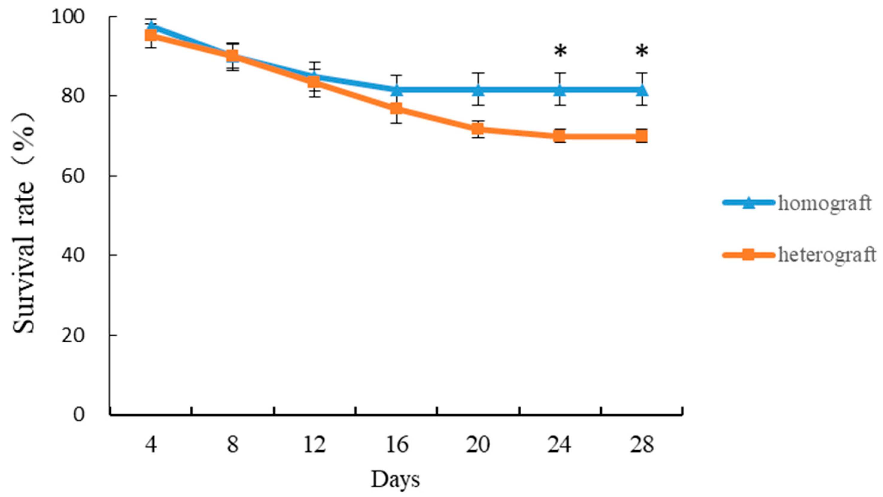
3.1. Survival Rate of Homografted and Heterografted Seedlings

From the continuous observation for 28 DAGs, it was found that the mortality of homografted and heterografted seedlings was not obvious at the beginning of grafting, but gradually increased with time. The survival rate of homografted and heterografted seedlings was maintained at above 90% for eight DAGs. However, as the healing process of grafting continues, there are constantly grafted seedlings that die, resulting in a declining survival rate of grafted seedlings. At 16 DAGs, the survival rate of homografted and heterografted seedlings and the mortality rate of heterografted seedlings gradually increased. At 24 DAGs, the survival rate of homografted seedlings was 82% and that of heterografted seedlings was 70%. There was a significant difference between the two treatments (Figure 1). Therefore, the graft healing process was completed at 24 DAGs, and the time point for the completion of graft healing has been tentatively determined. According to the statistical observation, the survival rate of homografted seedlings is higher than that of heterografted seedlings.

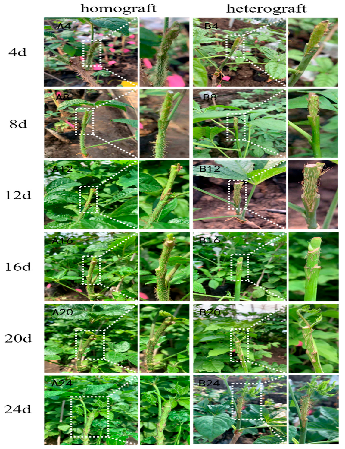
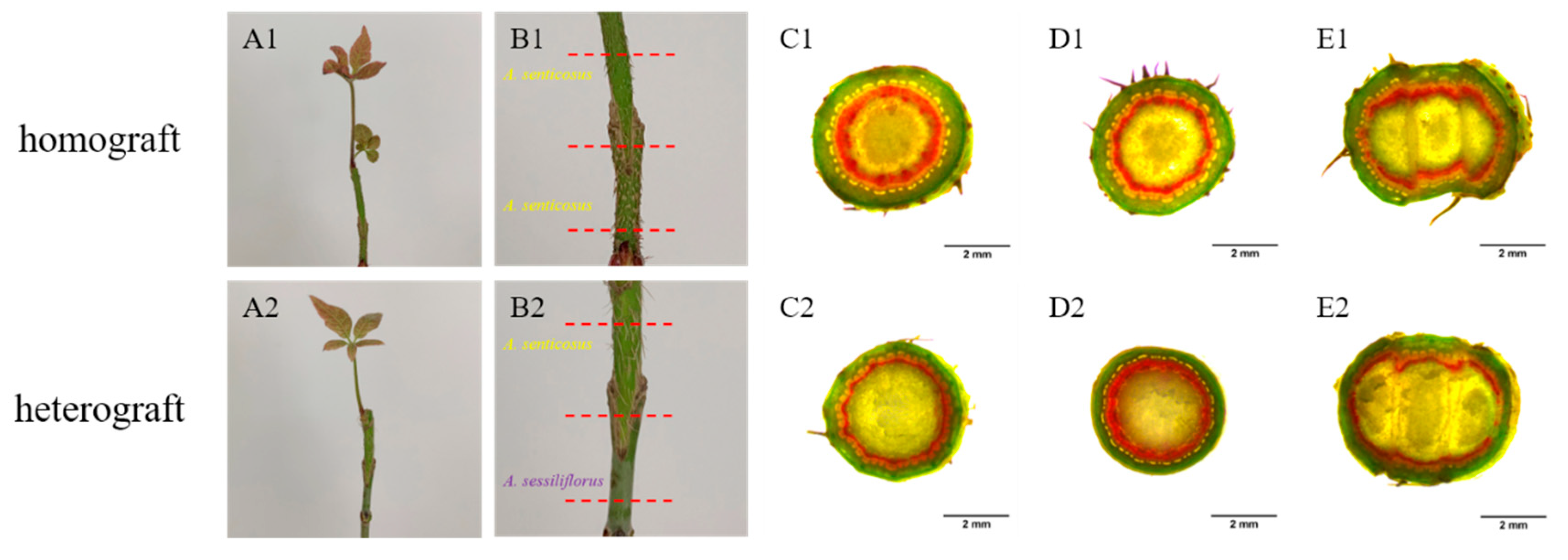
3.2. Morphological Observation of Homografted and Heterografted Seedlings

The observation of the external morphology of homografted and heterografted seedlings showed that the grafted union of rootstock and scion secreted a small amount of mucus at four DAGs, which caused the rootstock and scion to initially bond together. At eight DAGs, the tightness of the grafted union of the seedlings increased, the adhesion between root and stocks increased and the callus began to form in large quantities. At 12 DAGs, the grafted union was enlarged, filled with yellow callus and more tightly connected and new shoots begin to sprout at the scion. At 16 DAGs, it was observed that the nascent epidermis at the grafted union resulted in a better fit of rootstock and scion and gradual bud growth. With time, the color of the nascent epidermis of the grafted union changed from light brown to dark brown, the tightness of the rootstock and scion increased and the callus basically disappeared. At 24 DAGs, the color of the epidermis of the grafted union changed to dark brown, the healing process was basically completed, the rootstock and scion were closely connected together and the leaves were developing and growing (Figure 2). Overall, the development of heterografted seedlings was slower than that of homografted seedlings.

3.3. Microscopic Observation of Homografted and Heterografted Seedlings

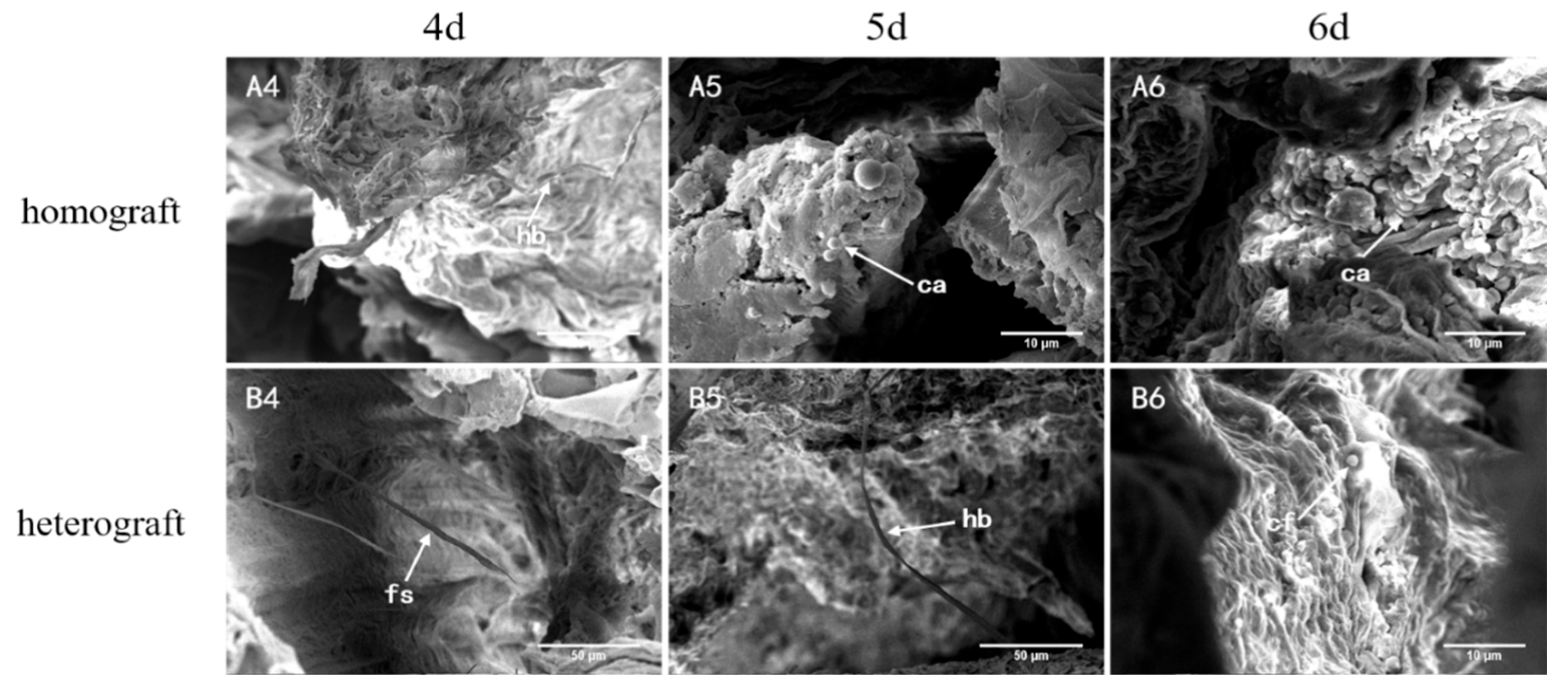
3.3.1. Scanning Electron Microscope Observation

The healing process of homografted and heterografted seedlings was observed by scanning electron microscopy at four, five, and six DAGs during the early stages of graft healing (Figure 3). At four DAGs of homografted seedlings, plasmodesmata and several horizontal bridges appeared (Figure 3, panel A4); at five DAGs, the adjacent tissues at the grafted union produced a large amount of callus for rootstock and scion connection (Figure 3, panel A5); and at six DAG, with the development of the grafted union, cell division and differentiation of the callus took place, gradually achieving complete connection of tissues between rootstocks and scion (Figure 3, panel A6). In contrast, at four DAGs of heterografted seedlings, a small amount of plasmodesmata was produced between rootstocks and scion (Figure 3, panel B4); at five DAGs, the first horizontal bridge was formed, constituting the first contact between rootstocks and scion (Figure 3, panel B5); and at six DAGs, the adjacent tissues at the grafted union showed the proliferation of callus (Figure 3, panel B6). From the observations of scanning electron microscopy, the first to be produced in the early stages of grafting are plasmodesmata for cell-to-cell material transport.

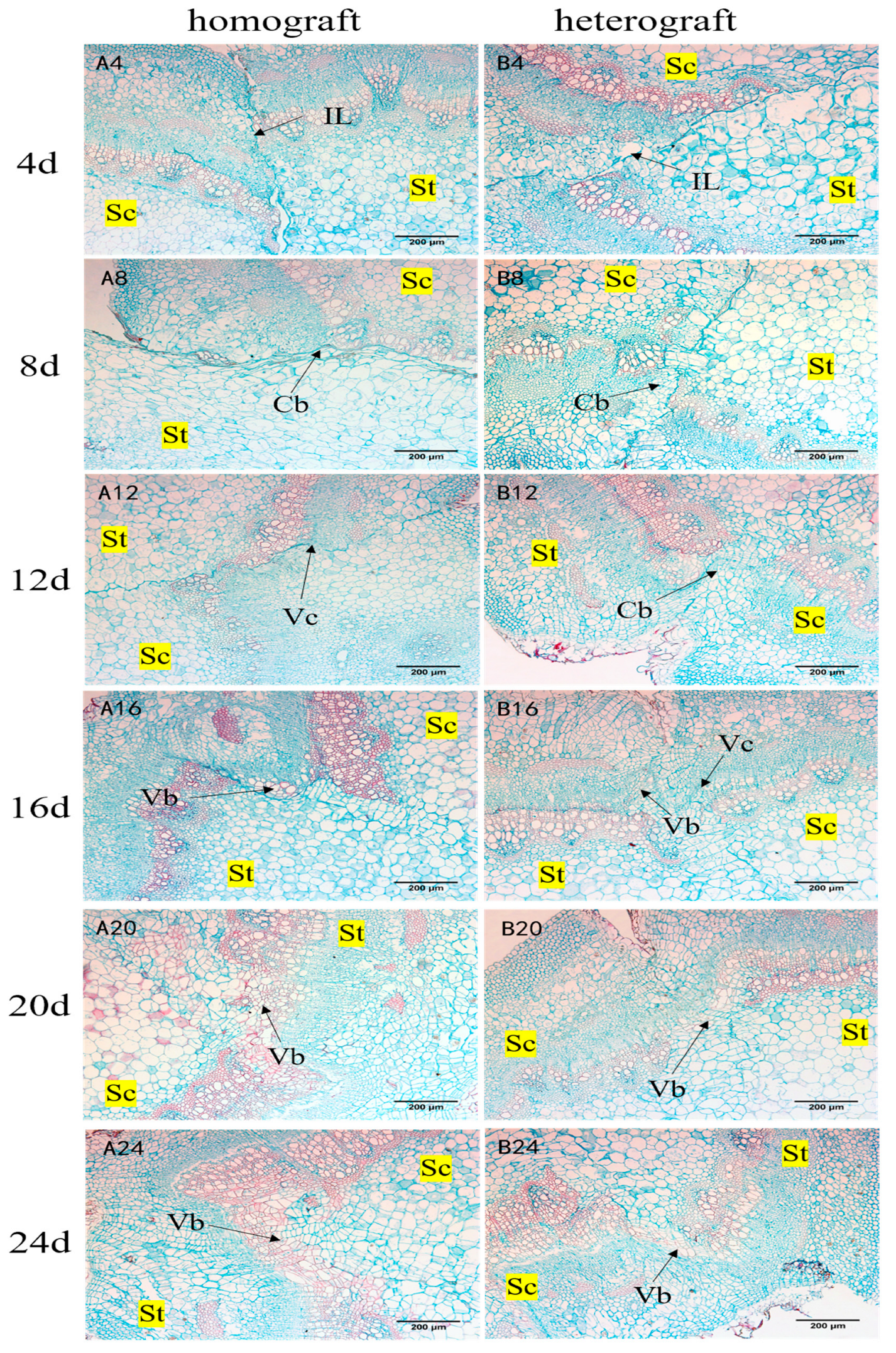
3.3.2. Paraffin Section Observation

The tissue structure formation was observed by fluorescence microscopy throughout the developmental stages of the graft healing process (Figure 4). At four DAGs, coalescence occurred because the protoplasm of dead and damaged thin-walled cells at the wound where the rootstock and scion met was broken, thus producing a thin isolation layer at the union where the rootstock and scion touched, and the rootstock and scion initially adhered through the isolation layer (Figure 4, panel A4,B4). At eight DAGs, the cells at the wound where the rootstock and scion were in contact showed strong activity, and the thin-walled cells of the cortex, xylem and bast of the rootstock and scion divided vigorously and dedifferentiated to produce large amounts of callus, prompting the isolation layer to thicken and the gap between the rootstock and scion to be filled, forming a callus bridge (Figure 4, panel A8,B8). At 12 DAGs, thin-walled cells at the vascular cambium of rootstock and scion continued to differentiate into the new vascular cambium, which crossed the callus bridge to reconnect the vascular cambium of rootstock and restore the vascular cambium ring (Figure 4, panel A12), whereas the differentiation process of the vascular cambium of heterografted seedlings was slower than that of homografted seedlings (Figure 4, panel B12). At 16 DAGs, as the newly attached vascular cambium continued to differentiate, new xylem began to differentiate inward and the new bast outward (Figure 4, panel A16,B16). The connection of vascular tissues is a crucial step for the survival of grafting. At 20 DAGs, the rootstock and scion xylem ducts and bast sieve tubes of homografted seedlings were connected and vascular tissues were re-established and penetrated as a whole (Figure 4, panel A20), while heterografted seedlings were still in the vascular tissue differentiation stage (Figure 4, panel B20). At 24 DAGs, the vascular tissues of homografted seedlings were heavily formed (Figure 4, panel A24), while those of heterografted seedlings were just reconnected (Figure 4, panel B24). Although the cell development process of the heterografted seedlings was slightly later than that of the homografted seedlings, they possessed complete cellular tissue changes, indicating that the heterografted seedlings were able to complete the process of graft healing and the grafting of A. senticosus and A. sessiliflorus was feasible. Combined with the survival rate, it was found that the mortality rate of heterografted seedlings gradually increased at 16 DAGs compared with homografted seedlings, suggesting that vascular tissue formation and differentiation are crucial for grafting success. At 24 DAGs, the vascular tissue of both homografted and heterografted seedlings was re-established, and the survival rate of grafted seedlings was basically unchanged, which marked the end of the grafting healing process and meant that the grafted seedlings were alive.

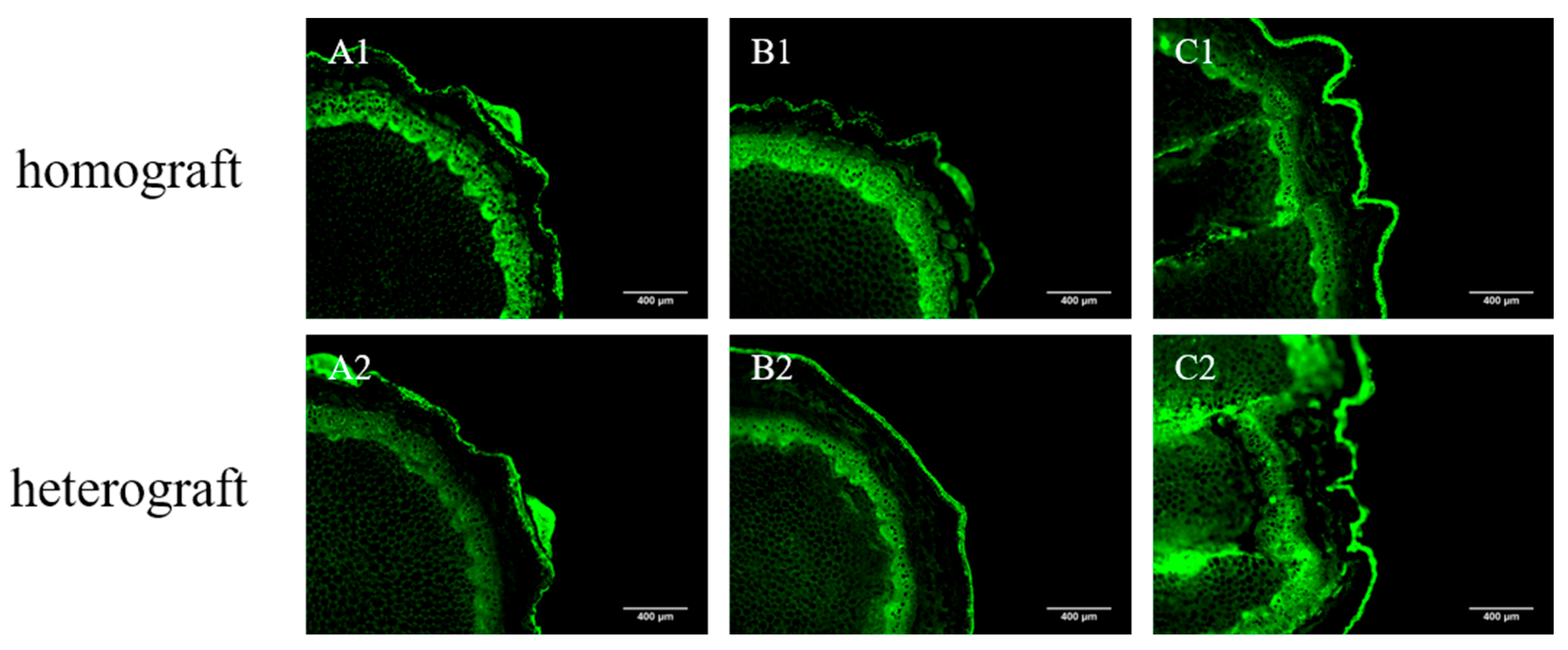
3.4. Vascular Tissue Connectivity of Homografted and Heterografted Seedlings

To test the connectivity of the vascular tissues of grafted seedlings, connectivity tests were conducted on the xylem and bast of grafted seedlings. Xylem attachment in homografted and heterografted seedlings was examined at 24 DAGs. After 6 h, it was found that the xylem of the rootstock, graft union, scion and leaf transport tissue of different grafted seedlings turned red (Figure 5), indicating that the acidic magenta solution was transported from the rootstock to scion with transpiration, and the xylem of homografted and heterografted seedlings had been connected.
At 24 DAGs, the bast connectivity of homografted and heterografted seedlings was examined. The results showed that HPTS could be transported from the scion leaves to the rootstock via the coplasm after 12 h (Figure 6), demonstrating that the bast of grafted seedlings of different combinations were connected at 24 DAGs.

3.5. RNA Sequence Data Analysis and Functional Annotation

The transcriptome was sequenced using the Illumina HiSeq™ 2500 (Illumina, San Diego, CA, USA) high-throughput sequencing platform on the grafted unions of homograft and heterograft at 16 DAGs. After processing, 39.47 Gb of clean reads was obtained. The average clean bases of each sample were 6.6 Gb, Q30 was more than 92.95% (sequencing error rate < 0.1%) and the GC content was more than 43.55% (Supplementary Table S2). A total of 39,489 unigenes were obtained, with an average length of 1017.86 bp and an N50 length of 1389 bp, indicating that the construction quality of the sequencing library was good, and the sequencing data were accurate and reliable. The diamond software was used to compare the assembled the unigene to NR, eggNOG, SwissProt, GO, KOG and KEGG databases, and the HMMER software was used to compare Pfam databases for the functional analysis of unigenes. The functional annotations gave 28,834 (73.02%), 27,028 (68.44%), 21,523 (54.50%), 19,033 (48.20%), 16,090 (40.75%) and 6527 (16.53%) unigenes, respectively, compared to the NR, eggNOG, SwissProt, GO, KOG and KEGG databases (Supplementary Table S3).

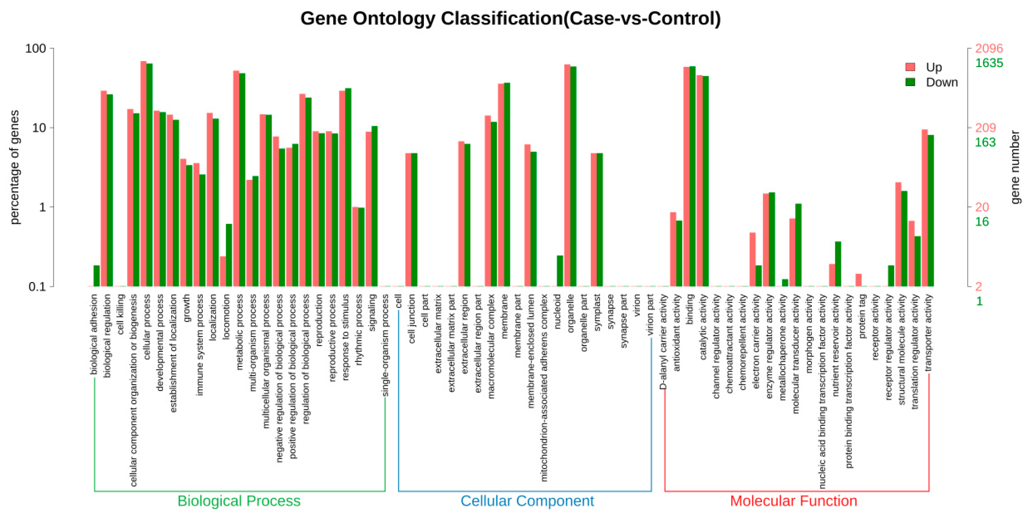
3.6. GO Enrichment Analysis of DEGs

The expression of unigenes was screened by the FPKM method (fold change > 2 or <0.5, Q value ≤ 0.05), and 10,215 differentially expressed genes (DEGs) were obtained, among which 5504 were upregulated, accounting for 53.88% of the total DEGs; 4471 were downregulated, accounting for 43.77% of the total number of DEGs (Supplementary Figure S1). For the functional classification of DEGs, GO enrichment analysis was conducted. A total of 10,215 unigenes have been annotated in the GO database, which can be classified into three major categories of biological process (BP), cellular component (CC) and molecular function (MF), and further subdivided into 64 functional groups. The BP items were divided into 23 functional groups, among which “cellular processes”, “metabolic processes”, “response to stimuli” and “bioregical regulation” were significantly enriched. The CC items were divided into 20 functional groups, among which “organelles”, “membranes”, “macromolecular complexes” and “extracellular regions” were significantly enriched. The MF were divided into 21 functional groups, among which “catalytic activity”, “protein binding” and “transport activity” were significantly enhanced. Most of the identified DEGs are responsible for essential processes related to bioregulation and metabolism (Figure 7).

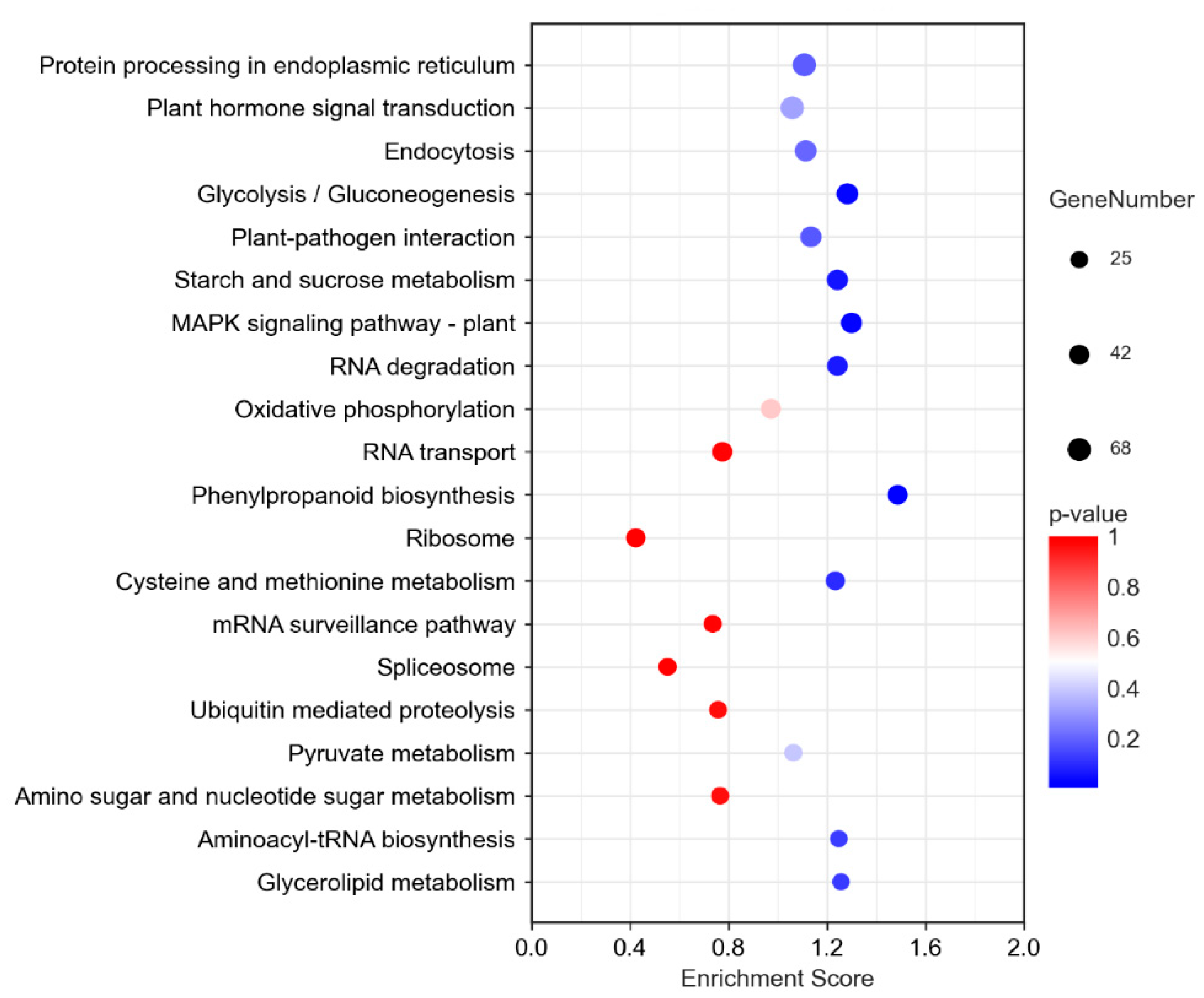
3.7. KEGG Enrichment Analysis of DEGs

By comparison with the KEGG database, DEGs were enriched in a total of 122 KEGG pathways, which are divided into four major categories: metabolism, genetic information processing, environmental information processing and cellular processes. The main KEGG pathways enriched by DEGs were protein processing in endoplasmic reticulum, plant hormone signal transduction, endocytosis, glycolysis/gluconeogenesis, plant–pathogen interaction, starch and sucrose metabolism and MAPK signaling pathway—plant (Figure 8).

3.8. Analysis of DEGs in MAPK Signaling Pathway

The upregulated DEGs in this study were mainly genes associated with the MAPK signaling pathway. The transcriptome results showed that the gene WRKY33, associated with pathogen infection, was significantly upregulated, causing flg22 to bind to the LRR receptor-like serine/threonine protein kinase FLS2, inducing phosphorylation of the associated mitogen-activated protein kinases MKK4/5 and MPK1/2, followed by the further activation of upregulation of downstream-associated protein PR1 expression, causing plant cells to produce a specific set of physiological and biochemical responses that began to generate defense mechanisms. Meanwhile, the expression of many genes related to multiple hormone regulation was upregulated in the MAPK signaling pathway, with significant changes in the signaling pathways related to ethylene, jasmonic acid and abscisic acid. Among them, the most significant changes in abscisic-acid-related signaling pathways were observed, with the kinase signaling pathway MPK3/MPK6 cascade reaction promoting abscisic acid biosynthesis through PYR/PYL, PP2C and SnPK2 upregulation. In addition, grafting wounds induced the upregulation of CaM4 and MPK8 expression, thereby inhibiting ROS production (Supplementary Table S4).

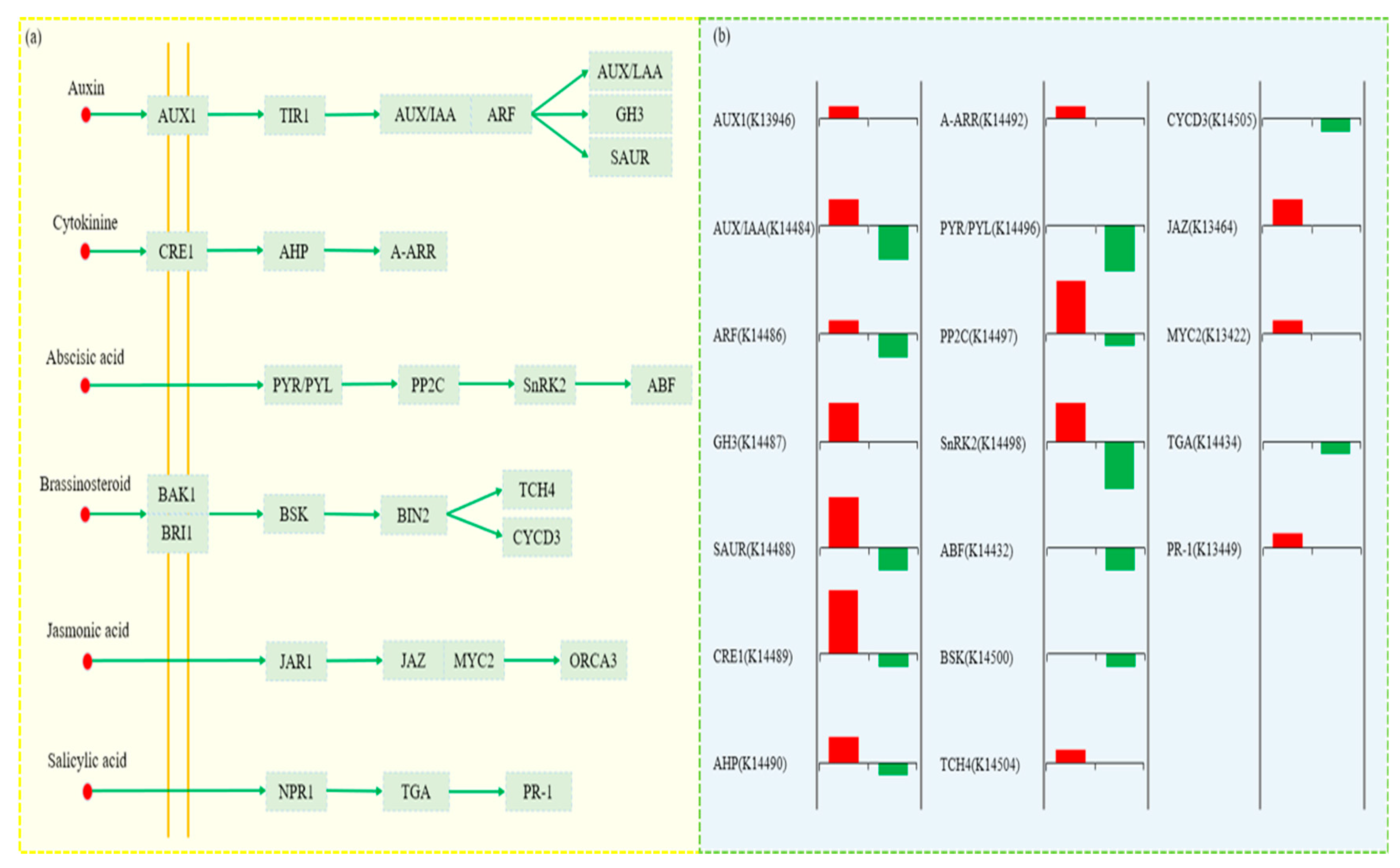
3.9. Analysis of DEGs in Plant Hormone Signaling Pathways

Among the phytohormone signaling pathways, those related to growth hormone, cytokinin, abscisic acid, oleurosterol, jasmonic acid and salicylic acid were all significantly altered (Figure 9a,b). The transcript levels of most of the genes encoding growth hormone were significantly altered. For example, the AUX1 family gene TRINITY_DN30497_c2_g1_i1 and the TIR1 family gene TRINITY_DN22536_c1_g1_i1 were upregulated, which led to the upregulation of the GH3 family genes TRINITY_DN16509_c0_g1_i1, TRINITY_DN32008_c0_g1_i1 and TRINITY_DN8599_c0_g1_i1. The most important role of cytokinin is to promote cell division, and the expression of CRE1, AHP and A-ARR genes associated with cytokinin signaling is upregulated. In addition, the PP2C gene related to abscisic acid signaling, the TCH4 gene related to brassinosteroid signaling and the JAZ gene related to jasmonic acid signaling were all upregulated in expression. We also identified one upregulated gene, TGA, and one downregulated gene, PR-1, in the salicylic acid signaling pathway (Supplementary Table S5).

3.10. Transcription Factor Analysis

Transcription factor analysis was performed on the DEGs in homografted and heterografted unions. The results showed that 227 DEGs were identified, belonging to 65 transcription factor families, most of which belonged to AP2/ERF-ERF (17), MYB (17), bHLH (14), MYB-related (13), NAC (13), C2H2 (12), bZIP (12), GRAS (11), GARP-G2-like (10) and WRKY (10) (Supplementary Table S6).

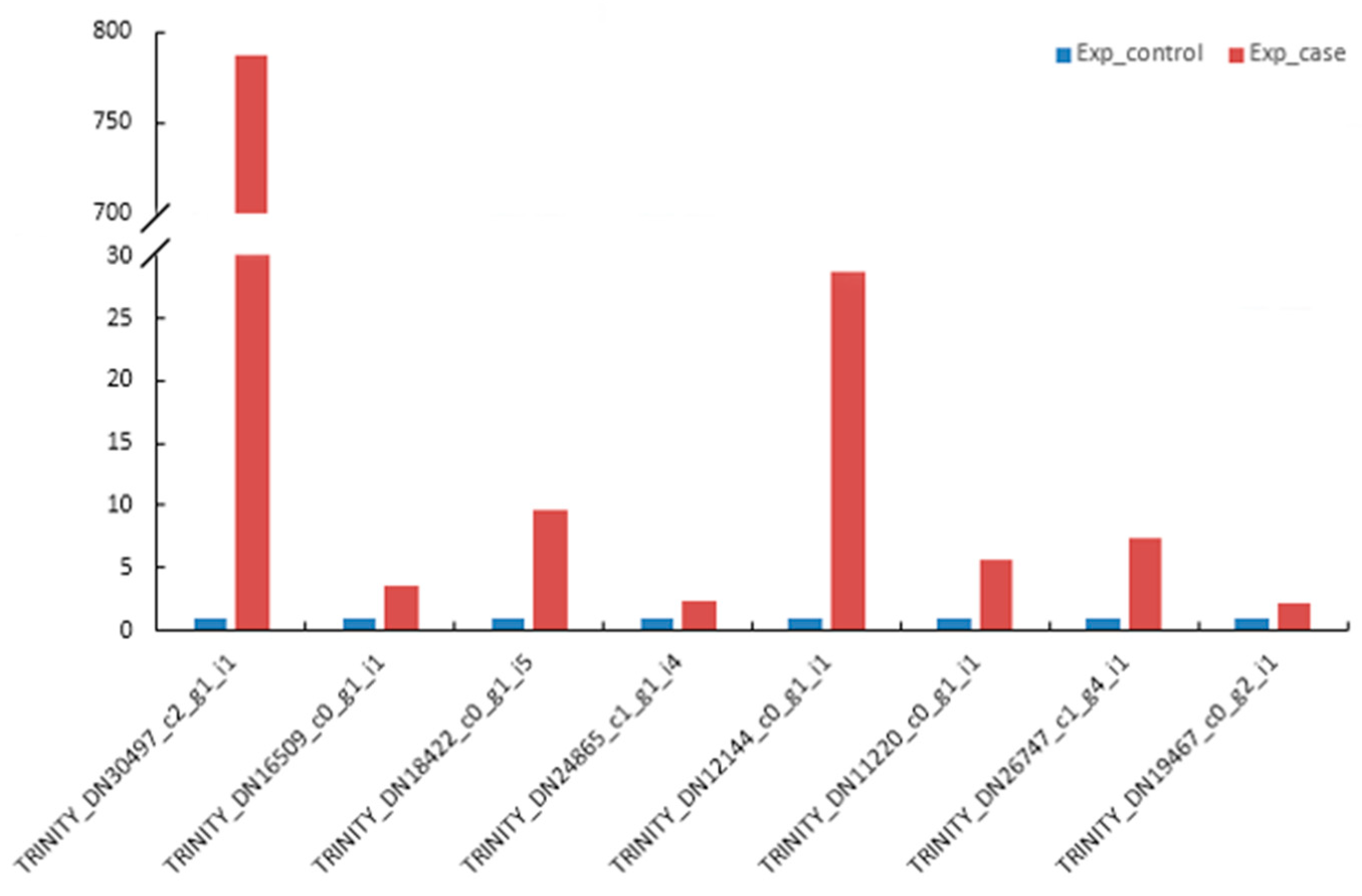
3.11. qRT-PCR Validation of DEGs

To verify the accuracy and reproducibility of the RNA-seq data, we selected eight DEGs for qRT-PCR analysis. The validation results were consistent with the trend of RNA-seq sequencing results, indicating that the transcriptome analysis results were true and reliable (Figure 10).

4. Discussion

The graft healing process between different species undergoes several developmental stages. There are three main stages in the success of the graft healing process, which is a complex process. In the first stage, the scion and rootstock are in close contact with each other, forming a callus that allows them to adhere to each other. In the second stage, cells continue to differentiate and a continuous vascular cambium is formed between the scion and rootstock. In the third stage, the vascular tissue is re-established between the scion and rootstock [22,41,42,43]. However, most of the previous studies did not observe in detail the first stage of grafting, i.e., before the formation of healing tissue. In this study, the healing process in the first stage after grafting was analyzed by SEM. It was found that plasmodesmata were produced before the formation of the callus, thus constituting the first connection between the scion and rootstock and providing a channel for intercellular material transport and information transfer. This agrees with the findings of Pina et al. [44]. In addition, vascular tissue reconstruction is also considered an important indicator of grafting success [19,45], but the time of reconstruction is more dependent on the environment and plant species. Yang et al. [46] showed that the healing process was completed when the vascular tissues of the scion and rootstock were joined at five DAGs, as shown by an anatomical study on the formation of the healing union in watermelon and bottle cucurbits grafting. Observations of the grafted healing union of cucumber and pumpkin showed that the vascular tissue between the scion and rootstock had started to connect at 6 DAGs, and more vascular tissue connections could be observed at 9 DAGs, indicating that the healing process of cucumber and pumpkin took 6–9 days [47]. Studies on the healing process of tomato grafting showed that vascular bridges connecting rootstock and scion appeared 11 DAGs, but the full formation of vascular bundles on grafted tomato rootstock generally took 7–14 days depending on plant growth conditions [21]. In contrast, woody plants, such as grape [48] and American pecan [49], require approximately ten days for vascular tissue formation after grafting. In this study, we observed the healing process of homografted and heterografted seedlings of A. senticosus at different times and found that the healing process was the same in both, but there were temporal differences in the developmental stages. The xylem and bast of the homografted and heterografted seedlings were connected at 24 DAGs, indicating that the healing process of homografted and heterografted seedlings was completed at 24 DAGs. The greater healing time than herbaceous plants may be because woody plants do not have the same regeneration capacity as herbaceous plants.
By observing the anatomical changes in the graft healing process at six time points and combining the survival rates of homografted and heterografted seedlings, we found that the healing process of homografted and heterografted seedlings was similar, but the survival rate of heterografted seedlings was significantly lower at 20 DAGs, indicating that the vascular tissue differentiation stage (vascular cambium stage) is a critical stage for the survival of heterografted seedlings. Therefore, we selected homografted and heterografted grafted unions for RNA sequencing at 16 DAGs to detect their differences in transcript levels. GO and KEGG analyses showed that a number of metabolic, physiological and hormonal responses are involved in the healing process of heterografted seedlings, including metabolic processes, cellular processes, responses to stimulus, plant hormone signal transduction, plant–pathogen interaction, MAPK signaling pathway—plant, transcription factors and defense responses. This finding suggested that the transcript levels of a large number of genes related to cell rearrangement, cell division and metabolic patterns were all altered in heterografted seedlings.
The MAPK cascade is a ubiquitous signal transduction module in eukaryotes. The MAPK signaling pathway in plants plays an important role in a wide range of activities, including hormonal and developmental signaling and responses to biotic and abiotic challenges, such as pathogenic infections, trauma, hypothermia and reactive oxygen species [50]. Kinases of the MAPK pathway are triggered when plants are exposed to a variety of stress stimuli, including trauma, temperature, drought, salinity, high osmotic pressure, ozone and reactive oxygen species, linking extracellular stimuli to a broad range of cellular responses [51]. In the current study, the expression of these kinase genes of the MAPK pathway associated with pathogen infection, phytohormones and wounding increased rapidly in heterografted seedlings, suggesting that these kinases are closely associated with tolerance to oxidative stress, with mechanical damage caused by grafting, and with the healing process of heterografted seedlings. It also indicated that more genes and transcription factors associated with defense responses were produced in heterografted seedlings in response to heterografting. The present data have demonstrated the involvement of oxidative stress in the regulation of graft healing and suggest that antioxidant defense systems are activated in heterografted seedlings. Furthermore, given that both the MAPK signaling pathway and the phytohormone signaling pathway are involved in resistance responses in many crops, we speculate that these changes may be related to the ability of heterografted seedlings to recover from wounding [52,53,54]. This also explains how heterografted seedlings are able to maintain normal metabolism and growth.
Plant hormone signal transduction is associated with graft healing, for example growth hormone and cytokinin which are necessary for tissue regeneration during vegetative graft healing [55]. RNA-Seq data from this study showed changes in the expression levels of genes associated with auxin, cytokinine, abscisic acid, brassinosteroid and jasmonic and salicylic acid signaling, which play key roles in heterografted seedlings. Auxin, in particular, has a crucial role in heterografted seedlings compared to homografted seedlings because it promotes vascular growth, xylem and phloem production and the healing of the graft union [55,56,57,58]. Studies have shown that auxin is important for the formation of the vascular system and the differentiation of the xylem and phloem [59,60,61]. Melnyk CW et al. [55] found that rootstock is not passive in graft formation, it is able to induce the connection with the scion by using different pathways, reflecting the importance of auxin in vascular reconstruction. The expression of the AUX1 family gene (TRINITY_DN30497_c2_g1_i1) and GH3 protein family genes (TRINITY_DN16509_c0_g1_i1, TRINITY_DN32008_c0_g1_i1 and TRINITY_DN8599_c0_g1_i1) was upregulated in heterografted seedlings compared to homografted seedlings, inducing plant growth response and promoting vascular development and xylem and phloem formation by regulating transcript levels of the auxin signal pathway. Cytokinins have been reported to contribute to the formation of healing tissue [62]. The upregulated expression of CRE1 genes (TRINITY_DN16988_c1_g1_i1, TRINITY_DN22133_c0_g1_i5, TRINITY_DN29587_c0_g3_i1, TRINITY_DN30286_c1_g2_i2 and TRINITY_DN30286_c1_g3_i1) and the A-ARR gene (TRINITY_DN27306_c0_g1_i6) was associated with cytokinin signaling, indicating that CRE1 and A-ARR can promote cell division and activate and induce healing tissue formation during healing in heterografted seedlings. Abscisic acid is an important hormone that plays a role in abiotic stress signals. Studies have shown that genes associated with abscisic acid may play a role in the adaptation to graft-induced stresses such as water deficit [63]. Ren et al. [64] also found that CmRNF5 may regulate the graft healing process through the abscisic acid signaling pathway. PP2C are genes related to abscisic acid signaling, and in our study, their expression was upregulated in heterografted seedlings. Other hormones, such as brassinosteroid as an emerging steroid hormone with high physiological activity, play important roles in plant growth and development, including stem and leaf growth, the differentiation of vascular tissues and the defense against environmental stresses. The expression of the gene encoding TCH4 is upregulated in the brassinosterol signaling pathway. During wound healing, jasmonic acid also promotes wound healing by activating primary and secondary metabolism through signal transduction [65]. The JAZ gene expression in the jasmonic acid signaling pathway is upregulated. In addition, the PR-1 gene obtained in the salicylate signaling pathway was upregulated in expression. By a comparative analysis of DEGs at the homografted and heterografted unions, we identified many DEGs encoding hormonal signaling, suggesting that the healing process of heterografted seedlings is regulated by more complex hormonal signaling pathways.
In the plant’s innate immune system, transcription factors (TFs) initiate the transcription of downstream defense genes. Many plant transcription factors have been shown to confer resistance to various stresses by binding to specific cis elements in the promoter regions of several genes [51]. TFs have been reported to be induced by a variety of stress conditions. In our results, the expression levels of some TFs were different at the homografted and heterografted unions, suggesting that they may play an important role in the healing process of heterografted seedlings. WRKY and ERF TFs are induced by various biotic and abiotic stresses [66]. Plant AP2/ERF TFs are a superfamily of TFs widely present in plants that play an important role in regulating plant growth and development in response to adversity stress. Some ERF TFS can bind to the GCC-box present in the promoters of pathogen-related (PR) genes (e.g., PR1, PR2, PR3, PR5) and mediate the critical role of these genes in the plant response to adversity stress [67,68]. The NtERF5 gene in tobacco is activated by trauma and pathogen infection [69]. The overexpression of SlERF5 also leads to the accumulation of higher levels of the defense gene PR5 in tomato plants and shows better tolerance to the bacterial pathogen Ralstonia solanacearum [70]. Various plant growth regulators, such as abscisic acid, salicylic acid and jasmonic acid, also converge on ERF proteins through complex interactions [71,72]. Yang et al. [73] showed that AtERF4 is able to regulate ethylene and abscisic acid responses, as well as the transcription of many ethylene/abscisic acid-dependent defense genes. In addition, AtERF1 has been reported to integrate defense signals from the ethylene and jasmonate pathways and induce downstream defense-related genes [74]. Significantly upregulated TFs of the AP2/ERF family contribute to plant adaptation to adversity stress [75]. The WRKY family was initially shown to play a role in plant defense responses. AtWRKY8 is involved in the defense response to the Tobacco Mosaic Virus infection in cruciferous plants and mediates crosstalk between ethylene and abscisic acid signaling [76]. The overexpression of cotton GhWRKY39 in N. benthamiana regulates the reactive oxygen system through multiple signaling pathways and enhances its resistance to infection by bacterial and fungal pathogens, as well as tolerance to salt and oxidative stress [77], and current findings suggest that other members of this gene family also have important roles in regulating plant growth and development, morphogenesis and metabolism [78]. MYB TFs are also capable of playing a defensive role in plant pathogen invasion [79,80]. The overexpression of some MYB transcription factors can activate the expression of plant PR genes and trigger systemic acquired resistance (SAR). Such a response enhances the ability of plants to resist bacterial, fungal and viral pathogens [81,82]. NAC genes, known as a family of plant-specific TFs, are widely involved in the biosynthesis of plant secondary walls and tolerance to cold, salt and drought stresses [66]. They have key regulatory functions in the formation of plant meristematic tissues and organ boundaries, root development, the growth of secondary plant cell walls, plant senescence, hormone regulation and stress response. NAC101/VND6 and NAC030/VND7 have been reported to be key regulators of xylem development [83] and NAC020, NAC045 and NAC086 are involved in the development of the bast [84,85]. NAC071 and NAC096 deliver auxin and ethylene signals that trigger xylan endoglucosidase/hydrolase (XTH) in cell wall tissues, promoting the formation of damaged stem cells [86,87]. The current findings confirm that this NAC TFs has a wide range of functions in graft healing.

5. Conclusions

Successful grafting can cause scions to inherit the good traits of rootstocks. Future research will focus on grafting different species of plants, which will allow more rootstocks with excellent characteristics to provide strong genetic resources to scions and produce excellent new varieties. This study is the first report on the homograft and heterograft of A. senticosus. Through Illumina sequencing, 10,215 significant DEGs were detected in homografted and heterografted seedlings, of which 5504 upregulated genes and 4471 downregulated genes were detected. Most of the identified differential genes were responsible for essential processes related to bioregulation and metabolism, as they were involved in primary and secondary metabolism, endoplasmic reticulum protein processing, phytohormone signaling, plant–pathogen interactions and plant MAPK signaling pathways according to the GO and KEGG analyses. Some TFs, including those in the WRKY, AP2/ERF and NAC families, were upregulated. The expression patterns of these genes indicated that related biological pathways may be essential in promoting vascular tissue formation and improving graft survival. This research identified numerous candidate genes for future functional analysis, provided new data resources and a theoretical basis for an in-depth understanding of the molecular mechanism of heterografted union formation, and provided a reference for the efficient use of grafting technology for variety improvement.

Supplementary Materials

The following supporting information can be downloaded at https://www.mdpi.com/article/10.3390/agronomy13061527/s1, Figure S1: Summary of up- and downregulated DEGs in the graft interface of homografted and heterografted seedlings; Table S1: Sequences of oligonucleotide primers used for gene-specific qRT-PCR amplification; Table S2: Throughput and quality of RNA-seq reads from grafted tissues; Table S3: Results of annotation from seven public databases; Table S4: Data of selected differentially expressed genes (DEGs) associated with the MAPK signaling pathway detected in graft unions of homograft and heterograft; Table S5: Data of selected differentially expressed genes (DEGs) associated with the plant hormone signal transduction pathway detected in graft unions of homograft and heterograft; Table S6: Differentially expressed transcription factor genes in graft unions of homograft and heterograft.

Author Contributions

Y.R. and N.Z. conceived the study. Q.W., J.A. and Y.W. (Yingping Wang) designed the experiments. Q.W., Y.W. (Yougui Wang) and K.D. performed the experiments. Q.W. and K.D. analyzed the data. Q.W. and K.D. wrote the initial draft of the manuscript. All authors have read and agreed to the published version of the manuscript.

Funding

This research was supported by the key R&D project in the field of medicine and health of Jilin Provincial Science and Technology Department (20200404087YY).

Institutional Review Board Statement

Our collection of A. senticosus and A. sessiliflorus complies with relevant institutions, national and international guidelines and legislation, as well as the IUCN Policy Statement on Research Involving Species at Risk of Extinction and the Convention on the Trade in Endangered Species of Wild Fauna and Flora.

Data Availability Statement

The authors declare that all the data and plant materials will be available without restrictions. The original FASTQ files generated in the current study have been stored in the NCBI Sequence Read Archive (https://dataview.ncbi.nlm.nih.gov/object/PRJNA906004?reviewer=igouek0lq5t6crn61dq1dpdbnj accessed on 31 December 2024.).

Acknowledgments

We thank the State Local Joint Engineering Research Center of Ginseng Breeding and Application for its technical support and Jilin Agricultural University of China for providing the experimental site at its facility’s agriculture base.

Conflicts of Interest

The authors declare no conflict of interest.

References

  1. Lee, D.; Park, J.; Yoon, J.; Kim, M.-Y.; Choi, H.-Y.; Kim, H. Neuroprotective effects of Eleutherococcus senticosus bark on transient global cerebral ischemia in rats. J. Ethnopharmacol. 2012, 139, 6–11. [Google Scholar] [CrossRef]
  2. Hibasami, H.; Fujikawa, T.; Takeda, H.; Nishibe, S.; Satoh, T.; Fujisawa, T.; Nakashima, K. Induction of apoptosis by Acanthopanax senticosus HARMS and its component, sesamin in human stomach cancer KATO III cells. Oncol. Rep. 2000, 7, 1213–1216. [Google Scholar] [CrossRef] [PubMed]
  3. Kimura, Y.; Sumiyoshi, M. Effects of various Eleutherococcus senticosus cortex on swimming time, natural killer activity and corticosterone level in forced swimming stressed mice. J. Ethnopharmacol. 2004, 95, 447–453. [Google Scholar] [CrossRef] [PubMed]
  4. Park, S.H.; Lee, S.G.; Kang, S.K.; Chung, S.H. Acanthopanax senticosus reverses fatty liver disease and hyperglycemia in ob/ob mice. Arch. Pharmacal Res. 2006, 29, 768–776. [Google Scholar] [CrossRef]
  5. Maslov, L.N.; Guzarova, N.V. Cardio protective and antiarrhythmic properties of preparations from Leuzea carthamoides, Aralia mandshurica, and Eleutherococcus senticosus. Eksperimentalnaia I Klin. Farmakol. 2007, 70, 48–54. [Google Scholar]
  6. Zhang, Y.Z.; Zhang, A.H.; Zhang, Y.; Sun, H.; Meng, X.C.; Yan, G.L.; Wang, X.J. Application of Ultra-performance Liquid Chromatography with Time-of-Flight Mass Spectrometry for the Rapid Analysis of Constituents and Metabolites from the Extracts of Acanthopanax senticosus Harms Leaf. Pharmacogn. Mag. 2016, 12, 145–152. [Google Scholar] [CrossRef]
  7. Jin, Z.X.; Xu, S.Y.; Sun, S.Q.; Zhou, Q. Analysis of Acanthopanax senticosus harms for different parts using fourier transform infrared spectroscopy. Spectrosc. Spectr. Anal. 2008, 28, 2859–2863. [Google Scholar] [CrossRef]
  8. Zhang, X.N.; Sun, Y.; Cao, F. Extraction and GC/MS analysis of volatile constituents from Acanthopanax senticosus (Rupr.et Maxim.) Harms leaf. Chin. J. New Drugs 2011, 20, 936–939. [Google Scholar]
  9. Dubey, A.K.; Sharma, R.M. Effect of rootstocks on tree growth, yield, quality and leaf mineral composition of lemon (Citrus limon (L.) Burm.). Sci. Hortic. 2016, 200, 131–136. [Google Scholar] [CrossRef]
  10. Gao, Q.H.; Wu, Y.; Jia, S.S.; Huang, S.H.; Liu, X.M. Effect of rootstock on the growth, photosynthetic capacity and osmotic adjustment of eggplant seedlings under chilling stress and recovery. Pak. J. Bot. 2016, 48, 461–468. [Google Scholar]
  11. Deng, W.W.; Han, J.Y.; Fan, Y.B.; Tai, Y.L.; Zhu, B.Y.; Lu, M.Q.; Wang, R.J.; Wang, X.C.; Zhang, Z.Z. Uncovering tea-specific secondary metabolism using transcriptomic and metabolomic analyses in grafts of Camellia sinensis and C. oleifera. Tree Genet. Genomes 2018, 14, 23. [Google Scholar] [CrossRef]
  12. Kyriacou, M.C.; Rouphael, Y.; Colla, G.; Zrenner, R.; Schwarz, D. Vegetable grafting: The implications of a growing agronomic imperative for vegetable fruit quality and nutritive value. Front. Plant Sci. 2017, 8, 741. [Google Scholar] [CrossRef]
  13. Duan, X.; Bi, H.G.; Li, T.; Wu, G.X.; Li, Q.; Ai, X.Z. Root characteristics of grafted peppers and their resistance to Fusarium solani. Biol. Plant. 2016, 61, 579–586. [Google Scholar] [CrossRef]
  14. Wang, H.; Zhou, P.; Zhu, W.; Wang, F. De-novo Comparative Transcriptome Analysis of Genes Differentially Expressed in the Scion of Homografted and Heterografted Tomato Seedlings. Sci. Rep. 2019, 9, 20240. [Google Scholar] [CrossRef] [PubMed]
  15. Bahar, E.; Korkutal, I.; Carbonneau, A.; Akcay, G. Using magnetic resonance imaging technique (MRI) to investigate graft connection and its relation to reddeningdiscoloration in grape leaves. J. Food Agric. Environ. 2010, 8, 293–297. [Google Scholar] [CrossRef]
  16. Yin, H.; Yan, B.; Sun, J.; Jia, P.F.; Zhang, Z.J.; Yan, X.S.; Chai, J.; Ren, Z.Z.; Zheng, G.C.; Liu, H. Graft-union development: A delicate process that involves cell-cell communication between scion and stock for local auxin accumulation. J. Exp. Bot. 2012, 63, 4219–4232. [Google Scholar] [CrossRef] [PubMed]
  17. Aloni, B.; Cohen, R.; Karni, L.; Aktas, H.; Edelstein, M. Hormonal signalling in rootstock-scion interactions. Sci. Hortic. 2010, 127, 119–126. [Google Scholar] [CrossRef]
  18. Taylor, S.A. Flowering in Pisum: Identifcation of a new ppd allele and its physiological action as revealed by grafing. Physiol. Plant. 2010, 97, 719–723. [Google Scholar] [CrossRef]
  19. Flaishman, M.A.; Loginovsky, K.; Golobowich, S.; Lev-Yadun, S. Arabidopsis thalianaas a model system for graf union development in homografs and heterografs. J. Plant Growth Regul. 2008, 27, 231–239. [Google Scholar] [CrossRef]
  20. Lee, E.J.; Malik, A.; Pokharel, S.; Ahmad, S.; Mir, B.A.; Cho, K.H.; Kim, J.; Kong, J.C.; Lee, D.M.; Chung, K.Y.; et al. Identifcation of Genes Diferentially Expressed in Myogenin Knock-Down Bovine Muscle Satellite Cells during Diferentiation through RNA Sequencing Analysis. PLoS ONE 2014, 9, e92447. [Google Scholar] [CrossRef]
  21. Fan, J.; Yang, R.; Li, X.; Zhao, W.; Zhao, F.; Wang, S. The processes of graf union formation in tomato. Hortic. Environ. Biotechnol. 2015, 56, 569–574. [Google Scholar] [CrossRef]
  22. Trinchera, A.; Pandozy, G.; Rinaldi, S.; Crinò, P.; Temperini, O.; Rea, E. Graf union formation in artichoke grafing onto wild and cultivated cardoon: An anatomical study. J. Plant Physiol. 2013, 170, 1569–1578. [Google Scholar] [CrossRef]
  23. Xie, L.; Dong, C.; Shang, Q. Gene co-expression network analysis reveals pathways associated with graf healing by asymmetric profling in tomato. BMC Plant Biol. 2019, 19, 373. [Google Scholar] [CrossRef]
  24. Cui, Q.; Xie, L.; Dong, C.; Gao, L.; Shang, Q. Stage-specifc events in tomato graf formation and the regulatory efects of auxin and cytokinin. Plant Sci. 2021, 304, 110803. [Google Scholar] [CrossRef] [PubMed]
  25. Overvoorde, P.; Fukaki, H.; Beeckman, T. Auxin control of root development. J. Cold Spring Harb. Perspect. Biol. 2010, 2, a001537. [Google Scholar] [CrossRef] [PubMed]
  26. Nanda, A.K.; Melnyk, C.W. The role of plant hormones during grafing. J. Plant Res. 2018, 131, 49–58. [Google Scholar] [CrossRef] [PubMed]
  27. Seo, J.K.; Kim, M.K.; Kwak, H.R.; Choi, H.S.; Nam, M.; Choe, J.; Choia, B.; Hana, S.J.; Kanga, J.H.; Jung, C. Molecular dissection of distinct symptoms induced by tomato chlorosis virus and tomato yellow leaf curl virus based on comparative transcriptome analysis. Virology 2018, 516, 1–20. [Google Scholar] [CrossRef] [PubMed]
  28. Nookaew, I.; Papini, M.; Pornputtapong, N.; Scalcinati, G.; Fagerberg, L.; Uhlén, M.; Nielsen, J. A comprehensive comparison of RNA-Seq based transcriptome analysis from reads to diferential gene expression and cross-comparison with microarrays: A case study in Saccharomyces cerevisiae. Nucleic Acids Res. 2012, 40, 10084–10097. [Google Scholar] [CrossRef] [PubMed]
  29. Marsch-Martínez, N.; Franken, J.; Gonzalez-Aguilera, K.L.; Folter, S.D.; Angenent, G.; Alvarez-Buylla, E.R. An efficient flat-surface collar-free grafting method for Arabidopsis thaliana seedlings. Plant Methods 2013, 9, 14. [Google Scholar] [CrossRef]
  30. Wright, K.M.; Oparka, K.J. The fluorescent probe HPTS as a phloem-mobile, symplastic tracer: An evaluation using confocal laser scanning microscopy. J. Exp. Bot. 1996, 47, 439–445. [Google Scholar] [CrossRef]
  31. Zhang, W.; Kollwig, G.; Stecyk, E.; Apelt, F.; Dirks, R.; Kragler, F. Graft-transmissible movement of inverted-repeat-induced siRNA signals into flowers. Plant J. 2014, 80, 106–121. [Google Scholar] [CrossRef]
  32. Bolger, A.M.; Lohse, M.; Usadel, B. Trimmomatic: A flexible trimmer for Illumina sequence data. Bioinformatics 2014, 30, 2114–2120. [Google Scholar] [CrossRef]
  33. Grabherr, M.G.; Haas, B.J.; Yassour, M.; Levin, J.Z.; Thompson, D.A.; Amit, I.; Adiconis, X.; Fan, L.; Raychowdhury, R.; Zeng, Q.D.; et al. Full-length transcriptome assembly from RNA-Seq data without a reference genome. Nat. Biotechnol. 2011, 29, 644–652. [Google Scholar] [CrossRef]
  34. Altschul, S.F.; Gish, W.; Miller, W.; Myers, E.W.; Lipman, D.J. Basic local alignment search tool. J. Mol. Biol. 1990, 215, 403–410. [Google Scholar] [CrossRef]
  35. Kanehisa, M.; Araki, M.; Goto, S.; Hattori, M.; Hirakawa, M.; Itoh, M.; Katayama, T.; Kawashima, S.; Okuda, S.; Tokimatsu, T.; et al. KEGG for linking genomes to life and the environment. Nucleic Acids Res. 2007, 36, D480–D484. [Google Scholar] [CrossRef] [PubMed]
  36. Trapnell, C.; Williams, B.A.; Pertea, G.; Mortazavi, A.; Kwan, G.; van Baren, M.J.; Salzberg, S.L.; Wold, B.J.; Pachter, L. Transcript assembly and quantification by RNA-Seq reveals unannotated transcripts and isoform switching during cell differentiation. Nat. Biotechnol. 2010, 28, 511–515. [Google Scholar] [CrossRef] [PubMed]
  37. Langmead, B.; Salzberg, S.L. Fast gapped-read alignment with Bowtie 2. Nat. Methods 2012, 9, 357–359. [Google Scholar] [CrossRef] [PubMed]
  38. Roberts, A.; Pachter, L. Streaming fragment assignment for real-time analysis of sequencing experiments. Nat. Methods 2012, 10, 71–73. [Google Scholar] [CrossRef]
  39. Anders, S.; Huber, W. Differential expression of RNA-Seq data at the gene level-the DESeq package. Heidelb. Ger. Eur. Mol. Biol. Lab. 2013, 10, f1000research. [Google Scholar]
  40. Livak, K.J.; Schmittgen, T.D. Analysis of relative gene expression data using real-time quantitative PCR and the 2−ΔΔCt Method. Methods 2001, 25, 402–408. [Google Scholar] [CrossRef]
  41. Moore, R.; Walker, D.B. Study of vegetative compatibility-incompatibility in higher plants. I. A structural studies of a compatible autograft in Sedum telephoides (Crassulaceae). Am. J. Bot. 1981, 68, 820–830. [Google Scholar] [CrossRef]
  42. Tiedemann, R. Graft union development and symplastic phloem contact in the heterograft Cucumis sativus on Curcubita ficifolia. J. Plant Physiol. 1989, 134, 427–440. [Google Scholar] [CrossRef]
  43. Hartmann, H.T.; Kester, D.E.; Davies, F.T.; Geneve, R.L. Plant Propagation. Principles and Practices, 7th ed.; Prentice Hall: Upper Saddle River, NJ, USA, 2002; pp. 199–220. [Google Scholar]
  44. Pina, A.; Errea, P.; Martens, H.J. Graft union formation and cell-to-cell communication via plasmodesmata in compatible and incompatible stem unions of Prunus spp. Sci. Hortic. 2012, 143, 144–150. [Google Scholar] [CrossRef]
  45. Yousef, A.F.; Ali, M.M.; Rizwan, H.M.; Gad, A.G.; Liang, D.; Binqi, L.; Kalaji, H.M.; Wróbel, J.; Xu, Y.; Chen, F. Light quality and quantity affect graft union formation of tomato plants. Sci. Rep. 2021, 11, 9870. [Google Scholar] [CrossRef]
  46. Yang, X.; Hu, X.; Zhang, M.; Xu, J.; Ren, R.; Liu, G.; Yao, X.F.; Chen, X.H. Effect of low night temperature on graft union formation in watermelon grafted onto bottle gourd rootstock. Sci. Hortic. 2016, 212, 29–34. [Google Scholar] [CrossRef]
  47. Miao, L.; Li, Q.; Sun, T.S.; Chai, S.; Wang, C.L.; Bai, L.Q.; Sun, M.T.; Li, Y.S.; Qin, X.; Zhang, Z.H.; et al. Sugars promote graft union development in the heterograft of cucumber onto pumpkin. Hortic. Res. 2021, 8, 146. [Google Scholar] [CrossRef]
  48. Cookson, S.J.; Clemente Moreno, M.J.; Hevin, C.; Nyamba Mendome, L.Z.; Delrot, S.; Trossat-Magnin, C.; Ollat, N. Graft union formation in grapevine induces transcriptional changes related to cell wall modification, wounding, hormone signalling, and secondary metabolism. J. Exp. Bot. 2013, 64, 2997–3008. [Google Scholar] [CrossRef]
  49. Mo, Z.H.; Feng, G.; Su, W.C.; Liu, Z.Z.; Peng, F.R. Transcriptomic Analysis Provides Insights into Grafting Union Development in Pecan (Carya illinoinensis). Genes 2018, 9, 71. [Google Scholar] [CrossRef]
  50. Gerszberg, A.; Hnatuszko-Konka, K. Tomato tolerance to abiotic stress: A review of most often engineered target sequences. Plant Growth Regul. 2017, 83, 175–198. [Google Scholar] [CrossRef]
  51. Ren, Y.; Xu, Q.; Wang, L.W.; Guo, S.R.; Shu, S.; Lu, N.; Sun, J. Involvement of metabolic, physiological and hormonal responses in the graft-compatible process of cucumber/pumpkin combinations was revealed through the integrative analysis of mRNA and miRNA expression. Plant Physiol. Biochem. 2018, 129, 368–380. [Google Scholar] [CrossRef]
  52. Kovtun, Y.; Chiu, W.L.; Tena, G.; Sheen, J. Functional analysis of oxidative stress-activated mitogen-activated protein kinase cascade in plants. Proc. Natl. Acad. Sci. USA 2000, 97, 2940–2945. [Google Scholar] [CrossRef] [PubMed]
  53. Raja, V.; Majeed, U.; Kang, H.; Andrabi, K.I.; John, R. Abiotic stress: Interplay between ROS, hormones and MAPKs. Environ. Exp. Bot. 2017, 137, 142–157. [Google Scholar] [CrossRef]
  54. Agrawal, G.K.; Tamogami, S.; Iwahashi, H.; Agrawal, V.P.; Rakwal, R. Transient regulation of jasmonic acid-inducible rice MAP kinase gene (OsBWMK1) by diverse biotic and abiotic stresses. Plant Physiol. Bioch. 2003, 41, 355–361. [Google Scholar] [CrossRef]
  55. Melnyk, C.W.; Schuster, C.; Leyser, O.; Meyerowitz, E.M. A Developmental Framework for Graft Formation and Vascular Reconnection in Arabidopsis thaliana. Curr. Biol. 2015, 25, 1306–1318. [Google Scholar] [CrossRef]
  56. Asahina, M.; Azuma, K.; Pitaksaringkarn, W.; Yamazaki, T.; Mitsuda, N.; Ohme-Takagi, M.; Yamaguchi, S.; Kamiya, Y.; Okada, K.; Nishimura, T.; et al. Spatially selective hormonal control of RAP2.6L and ANAC071 transcription factors involved in tissue reunion in Arabidopsis. Proc. Natl. Acad. Sci. USA 2011, 108, 16128–16132. [Google Scholar] [CrossRef]
  57. Shen, C.J.; Yue, R.Q.; Sun, T.; Zhang, L.; Yang, Y.J.; Wang, H.Z. OsARF16, a transcription factor regulating auxin redistribution, is required for iron defificiency response in rice (Oryza sativa L.). Plant Sci. 2015, 231, 148–158. [Google Scholar] [CrossRef] [PubMed]
  58. Yu, C.L.; Sun, C.D.; Shen, C.J.; Wang, S.K.; Liu, F.; Liu, Y.; Chen, Y.L.; Qian, Q.; Aryal, B.; Geisler, M.; et al. The auxin transporter, OsAUX1, is involved in primary root and root hair elongation and in Cd stress responses in rice (Oryza sativa L.). Plant J. 2015, 83, 818–830. [Google Scholar] [CrossRef]
  59. Scarpella, E. Control of leaf vascular patterning by polar auxin transport. Genes Dev. 2006, 20, 1015–1027. [Google Scholar] [CrossRef]
  60. Aloni, R. Differentiation of vascular tissues. Annu. Rev. Plant Physiol. 1987, 38, 179–204. [Google Scholar] [CrossRef]
  61. Heo, J.O.; Roszak, P.; Furuta, K.M.; Helariutta, Y. Phloem development: Current knowledge and future perspectives. Am. J. Bot. 2014, 101, 1393–1402. [Google Scholar] [CrossRef]
  62. Iwase, A.; Mitsuda, N.; Koyama, T.; Hiratsu, K.; Kojima, M.; Arai, T.; Inoue, Y.; Seki, M.; Sakakibara, H.; Sugimoto, K.; et al. The AP2/ERF Transcription Factor WIND1 Controls Cell Dedifferentiation in Arabidopsis. Curr. Biol. 2011, 21, 508–514. [Google Scholar] [CrossRef] [PubMed]
  63. Kumari, A.; Kumar, J.; Kumar, A.; Chaudhury, A.; Singh, S.P. Grafting triggers difffferential responses between scion and rootstock. PLoS ONE 2015, 10, e0124438. [Google Scholar] [CrossRef] [PubMed]
  64. Ren, Y.; Guo, S.R.; Shu, S.; Xu, Y.; Sun, J. Isolation and expression pattern analysis of CmRNF5 and CmNPH3L potentially involved in graft compatibility in cucumber/pumpkin graft combinations. Sci. Hortic. 2018, 227, 92–101. [Google Scholar] [CrossRef]
  65. Jacobo-Velázquez, D.A.; González-Agüero, M.; Cisneros-Zevallos, L. Cross-talk between signaling pathways: The link between plant secondary metabolite production and wounding stress response. Sci. Rep. 2015, 5, 8608. [Google Scholar] [CrossRef]
  66. Erpen, L.; Devi, H.S.; Grosser, J.W.; Dutt, M. Potential use of the DREB/ERF, MYB, NAC and WRKY transcription factors to improve abiotic and biotic stress in transgenic plants. Plant Cell Tissue Organ Cult. 2017, 132, 1–25. [Google Scholar] [CrossRef]
  67. Sakuma, Y.; Liu, Q.; Dubouzet, J.G.; Abe, H.; Shinozaki, K.; Yamaguchi-Shinozaki, K. DNA-binding specificity of the ERF/AP2 domain of Arabidopsis DREBs, transcription factors involved in dehydration-and cold-inducible gene expression. Biochem. Biophys. Res. Commun. 2002, 290, 998–1009. [Google Scholar] [CrossRef]
  68. Zarei, A.; Körbes, A.P.; Younessi, P.; Montiel, G.; Champion, A.; Memelink, J. Two GCC boxes and AP2/ERF-domain transcription factor ORA59 in jasmonate/ethylene-mediated activation of the PDF1.2 promoter in Arabidopsis. Plant Mol. Biol. 2011, 75, 321–331. [Google Scholar] [CrossRef] [PubMed]
  69. Xu, Z.S.; Chen, M.; Li, L.C.; Ma, Y.Z. Functions of the ERF transcription factor family in plants. Botany 2008, 86, 969–977. [Google Scholar] [CrossRef]
  70. Li, C.W.; Su, R.C.; Cheng, C.P.; You, S.J.; Hsieh, T.H.; Chao, T.C.; Chan, M.T. Tomato RAV transcription factor is a pivotal modulator involved in the AP2/EREBP-mediated defense pathway. Plant Physiol. 2011, 156, 213–227. [Google Scholar] [CrossRef]
  71. Zhang, H.; Huang, Z.; Xie, B.; Chen, Q.; Tian, X.; Zhang, X.; Zhang, H.; Lu, X.; Huang, D.; Huang, R. The ethylene-, jasmonate-, abscisic acid- and NaCl-responsive tomato transcription factor JERF1 modulates expression of GCC box-containing genes and salt tolerance in tobacco. Planta 2004, 220, 262–270. [Google Scholar] [CrossRef]
  72. Rehman, S.; Mahmood, T. Functional role of DREB and ERF transcription factors: Regulating stress-responsive network in plants. Acta Physiol. Plant. 2015, 37, 178. [Google Scholar] [CrossRef]
  73. Yang, Z.; Tian, L.N.; Latoszek-Green, M.; Brown, D.; Wu, K.Q. Arabidopsis ERF4 is a transcriptional repressor capable of modulating ethylene and abscisic acid responses. Plant Mol. Biol. 2005, 58, 585–596. [Google Scholar] [CrossRef] [PubMed]
  74. Lorenzo, O.; Piqueras, R.; Sanchez-Serrano, J.J.; Solano, R. Ethylene response factor 1 integrates signals from ethylene and jasmonate pathways in plant defense. Plant Cell 2003, 15, 165–178. [Google Scholar] [CrossRef] [PubMed]
  75. Liu, Q.; Li, P.T.; Cheng, S.; Zhao, Z.L.; Liu, Y.L.; Wei, Y.Y.; Lu, Q.W.; Han, J.P.; Cai, X.Y.; Zhou, Z.L.; et al. Protoplast Dissociation and Transcriptome Analysis Provides Insights to Salt Stress Response in Cotton. Int. J. Mol. Sci. 2022, 23, 2845. [Google Scholar] [CrossRef]
  76. Chen, L.; Zhang, L.; Li, D.; Wang, F.; Yu, D. WRKY8 transcription factor functions in the TMV-cg defense response by mediating both abscisic acid and ethylene signaling in Arabidopsis. Proc. Natl. Acad. Sci. USA 2013, 110, 1963–1971. [Google Scholar] [CrossRef] [PubMed]
  77. Shi, W.N.; Liu, D.D.; Hao, L.L.; Wu, C.A.; Guo, X.Q.; Li, H. GhWRKY39, a member of the WRKY transcription factor family in cotton, has a positive role in disease resistance and salt stress tolerance. Plant Cell Tissue Organ Cult. 2014, 118, 17–32. [Google Scholar] [CrossRef]
  78. Shinozaki, K.; Yamaguchi-Shinozaki, K. Gene networks involved in drought stress response and tolerance. J. Exp. Bot. 2006, 58, 221–227. [Google Scholar] [CrossRef]
  79. Dubos, C.; Stracke, R.; Grotewold, E.; Weisshaar, B.; Martin, C.; Lepiniec, L. MYB transcription factors in Arabidopsis. Trends Plant Sci. 2010, 15, 573–581. [Google Scholar] [CrossRef]
  80. Stracke, R.; Werber, M.; Weisshaar, B. The R2R3-MYB gene family in Arabidopsis thaliana. Curr. Opin. Plant Biol. 2001, 4, 447–456. [Google Scholar] [CrossRef]
  81. Bostock, R.M. Signal crosstalk and induced resistance: Straddling the line between cost and benefit. Annu. Rev. Phytopathol. 2005, 43, 545–580. [Google Scholar] [CrossRef]
  82. Durrant, W.; Dong, X. Systemic acquired resistance. Annu. Rev. Phytopathol. 2004, 42, 185–209. [Google Scholar] [CrossRef] [PubMed]
  83. Kubo, M.; Udagawa, M.; Nishikubo, N.; Horiguchi, G.; Yamaguchi, M.; Ito, J.; Mimura, T.; Fukuda, H.; Demura, T. Transcription switches for protoxylem and metaxylem vessel formation. Gene. Dev. 2005, 19, 1855–1860. [Google Scholar] [CrossRef] [PubMed]
  84. Furuta, K.M.; Hellmann, E.; Helariutta, Y. Molecular control of cell specifification and cell difffferentiation during procambial development. Annu. Rev. Plant Biol. 2014, 65, 607–638. [Google Scholar] [CrossRef]
  85. Kondo, Y.; Nurani, A.M.; Saito, C.; Ichihashi, Y.; Saito, M.; Yamazaki, K.; Mitsuda, N.; Ohme-Takagi, M.; Fukuda, H. Vascular cell induction culture system using Arabidopsis leaves (VISUAL) reveals the sequential difffferentiation of sieve element-like cells. Plant Cell 2016, 28, 1250–1262. [Google Scholar] [CrossRef] [PubMed]
  86. Pitaksaringkarn, W.; Matsuoka, K.; Asahina, M.; Miura, K.; Sage-Ono, K.; Ono, M.; Yokoyama, R.; Nishitani, K.; Lshii, H.; Satoh, S. XTH20 and XTH19 regulated by ANAC071 under auxin flflow are involved in cell proliferation in incised Arabidopsis inflflorescence stems. Plant J. 2014, 80, 604–614. [Google Scholar] [CrossRef]
  87. Matsuoka, K.; Matsuoka, K.; Sato, R.; Matsukura, Y.; Kawajiri, Y.; Iino, H.; Nozawa, N.; Shibata, K.; Kondo, Y.; Satoh, S.; et al. Wound-inducible ANAC071 and ANAC096 transcription factors promote cambial cell formation in incised Arabidopsis flflowering stems. Commun. Biol. 2021, 4, 369. [Google Scholar] [CrossRef]
Figure 1. The survival rate of homografted and heterografted seedlings (*: p-value < 0.05).
Agronomy 13 01527 g001
Figure 2. (A,B) Morphological observation of homografted and heterografted seedlings. The enlarged area is within the dashed box.
Agronomy 13 01527 g002
Figure 3. (A,B) SEM images of the graft union on 4 DAGs, 5 DAGs and 6 DAGs. P: plasmodesmata; Hb: horizontal bridge; Ca: callus. SEM was performed at EHT: 10 kV; pressure: 1.0 Pa; WD: 25–15 mm.
Agronomy 13 01527 g003
Figure 4. (A,B) Anatomical observation of homografted and heterografted seedlings. Sc: scion; St: stock; IL: isolated layer; Cb: callus bridge; Vc: vascular cambium; Vb: vascular tissue.
Agronomy 13 01527 g004
Figure 5. Identification of xylem system connectivity of grafted seedlings by acid fuchsin staining. (A). Different grafted seedlings. (B). The dot-ted line represents the cross-sectional part. (C): The upper part of the graft union; (D): The underside of the graft union; (E): Graft union.
Agronomy 13 01527 g005
Figure 6. Identification of phloem system connectivity of grafted seedlings by HTPS method. (A): The upper part of the graft union; (B): The underside of the graft union; (C): Graft union.
Agronomy 13 01527 g006
Figure 7. GO enrichment analysis of DEGs in homografted and heterografted unions.
Agronomy 13 01527 g007
Figure 8. Bubble chart of the DEGs in enriched KEGG pathway. The enrichment score is the ratio between the number of DEGs in a pathway and all the annotated genes in this pathway. The larger the bubble, the more differential genes there are in the pathway.
Agronomy 13 01527 g008
Figure 9. Changes in the expression levels of hormonal-signaling-related DEGs in homografted and heterografted unions of A.senticosus. (a) Plant hormone signal transduction pathway. (b) DEGs related to hormone signaling pathway. The red bar indicates upregulation and the green bar indicates downregulation.
Agronomy 13 01527 g009
Figure 10. Validation of differentially expressed genes between homografted and heterografted unions by qRT-PCR.
Agronomy 13 01527 g010
Disclaimer/Publisher’s Note: The statements, opinions and data contained in all publications are solely those of the individual author(s) and contributor(s) and not of MDPI and/or the editor(s). MDPI and/or the editor(s) disclaim responsibility for any injury to people or property resulting from any ideas, methods, instructions or products referred to in the content.

