Article

Higher Aluminum Tolerance of Lespedeza bicolor Relative to Lespedeza cuneata Is Associated with Saccharide Components of Root Tips

1
School of Environmental Science and Engineering, Hubei Polytechnic University, Huangshi 435003, China
2
State Key Laboratory of Soil and Sustainable Agriculture, Institute of Soil Science, Chinese Academy of Sciences, Nanjing 210008, China
3
Hubei Key Laboratory of Mine Environmental Pollution Control & Remediation, Hubei Polytechnic University, Huangshi 435003, China
4
University of Chinese Academy of Sciences, Beijing 100049, China
*
Author to whom correspondence should be addressed.
Agronomy 2023, 13(3), 629; https://doi.org/10.3390/agronomy13030629
Submission received: 19 January 2023 / Revised: 14 February 2023 / Accepted: 17 February 2023 / Published: 22 February 2023
(This article belongs to the Special Issue Crop and Vegetable Physiology under Environmental Stresses)

Abstract

:
Aluminum (Al) toxicity is the primary factor limiting agricultural productivity in acid soils. The cell wall is mainly composed of saccharides and the first barrier for aluminum (Al) to enter plant root cells, but it is unknown whether and how root saccharide components are involved in regulating the Al tolerance of Lespedeza that is well adapted to acid soils. Here, we used Al-tolerant Lespedeza bicolor and Al-sensitive Lespedeza cuneata to examine the association of root saccharide components with Lespedeza Al tolerance through analyzing the saccharide changes of roots exposed to Al toxicity. Al-sensitive Lespedeza accumulated more Al and pectin but less hemicellulose in the root cell walls than Al-tolerant Lespedeza. Al treatment decreased the amounts of total sugar secreted from and within the roots of only Al-tolerant Lespedeza. Al treatment decreased the content of root monosaccharides including glucose and mannose in both Lespedeza species, but increased the xylose contents in only Al-sensitive Lespedeza. Taken together, less cell wall pectin rather than hemicellulose is responsible for less root Al accumulation, and Al-decreased root saccharide contents may enhance root organic-acid secretion to chelate toxic Al, both of which contribute to Lespedeza Al tolerance.

1. Introduction

Lespedeza plays important roles in land reclamation, forage production, and soil conservation. Lespedeza is also capable of improving soil fertility by rapidly increasing soil organic matter and nitrogen content by symbiotic N2 fixation in agricultural production. It has been known since the early twentieth century that Lespedeza adapts well to acid infertile soils [1,2]. Aluminum (Al) toxicity is the primary factor limiting plant growth in acid soils [3]. It is critical to elucidate the mechanisms of Lespedeza in response to Al toxicity in order to find out new information on plant Al tolerance and to make full use of Lespedeza in acid soils. Plants generally detoxify Al by excluding toxic Al from roots and tolerating toxic Al within plants [4,5,6]. The secretion of organic acids from roots under Al toxicity is the main mechanism of Al exclusion in most plants [7,8,9]. Our previous study indicated that the secretion of malate and citrate from roots under Al toxicity was one of the Al-exclusion mechanisms in Lespedeza [10]. Furthermore, we found that efficient phosphorus use and ammonium supply were also involved in the Al tolerance of Lespedeza [11,12]. However, these mechanisms are still not enough to explain the extremely high Al tolerance of Lespedeza, because some Al-sensitive plants such as wheat (Triticum aestivum L.) and barley (Hordeum vulgare L.) can also secret organic acids from roots under Al toxicity [13,14].
The apoplast of plant roots represents the major site for plants to perceive and express Al toxicity [15,16]. The protection of the root apoplast is a prerequisite for plant Al tolerance [17]. The cell wall in the root apoplast plays an important role in the tolerance of plants to Al toxicity [18,19,20,21,22,23]. Most of Al entering the root apex is bound to the cell wall of roots [9,18,21,24]. The changes in the size and quantity of polysaccharides within the root cell wall regulate the stretching of the cell wall [9,25,26]. Aluminum causes the accumulation of cell wall polysaccharides and thus makes the cell wall thick and rigid, thereby inhibiting the growth of plant roots [27]. In addition, the cell wall polysaccharides play an important role in modulating the Al-binding capacity of the cell wall [8,20,21]. Aluminum inhibits xyloglucan endotransglucosylase that catalyzes the splitting of xyloglucan chains and the linking of the newly generated reducing end to the non-reducing end of another xyloglucan chain in the cell wall [23]. Therefore, the binding of Al in the cell wall can further change the mechanical and chemical properties of the cell wall. This supports that theory that Al inhibits root elongation initially through the interaction of Al with the apoplastic side of the cell wall–plasma-membrane–cytoskeleton continuum [6,18,19]. Although the importance of the root cell wall and apoplast for Al tolerance has been reported in some plants, as described above, it is still unknown whether they also play important roles in the Al tolerance of Lespedeza, a strongly Al-tolerant plant.
In this context, we used Al-tolerant and Al-sensitive Lespedeza and examined the Al injury site and distribution, root sugar secretion, and cell wall components when exposed to Al toxicity. The objective of this study was to address the possible role of the root cell wall in regulating Lespedeza Al tolerance, thereby providing new information on the explanation of Al tolerance mechanisms in this strongly Al-tolerant plant.

2. Materials and Methods

2.1. Plant Material and Growth Conditions

Lespedeza bicolor (Turcz. cv. Jiangxi) and L. cuneata (Dum.-Cours. G. Don cv. Zhejiang), which are, respectively, Al-tolerant and Al-sensitive, were used in this study [10,11]. The seed coat of L. bicolor is relatively hard and needed to be softened by 98% (m/m) sulfuric acid, but the seed coat of L. cuneata is soft and did not need such pretreatment. Seeds of L. bicolor were surface-softened in 98% (m/m) sulfuric acid for 20 min, washed with deionized water, soaked in deionized water overnight, and then germinated in a plastic Petri dish with filter paper saturated with deionized water, in darkness, at 25 °C for two days. Seeds of L. cuneata were soaked in deionized water for 2 h, and then germinated in a plastic Petri dish for two days according to the above-described method. Germinated seeds were transplanted to plastic net suspended on a plastic container (2.5 L) with 0.5 mM CaCl2 solution (pH 4.5). The solution was renewed every day. Five-day-old seedlings were treated with different concentrations of Al (AlCl3·6H2O) in 0.5 mM CaCl2 solutions (pH 4.5) for subsequent experiments.

2.2. Monitoring and Energy-Spectrum Analysis of Elements on Root Surface

We performed the monitoring and energy-spectrum analysis of elements on the root surface, as described previously [28]. Five-day-old seedlings were subjected to 0.5 mM CaCl2 solution (pH 4.5) with or without 200 μM Al for 24 h. Root tips were taken from 1 to 5 mm behind the apex, stored in the refrigerator at −20 °C for 18 h, and then put into the freeze dryer (Thermo Savant, Hyannis, MA, USA) for 24 h. Subsequently, the dried complete root tips were fixed on the adhesive table with double-sided adhesive tape. After gold-plating with an ion-coating machine (Leica EM SCD 005, Sunnyvale, CA, USA; time 30 s, current 40 mA), the scanning pictures with a Philips field emission scanning electron microscope were obtained (Sirion 200, FEI, Amsterdam, The Netherlands). At the same time, the root surface energy spectrum was scanned with an X-ray energy-spectrum analyzer (Genesis, EDAX Inc., Berwyn, IL, USA).

2.3. Morin Staining and Fluorescence Microscopy

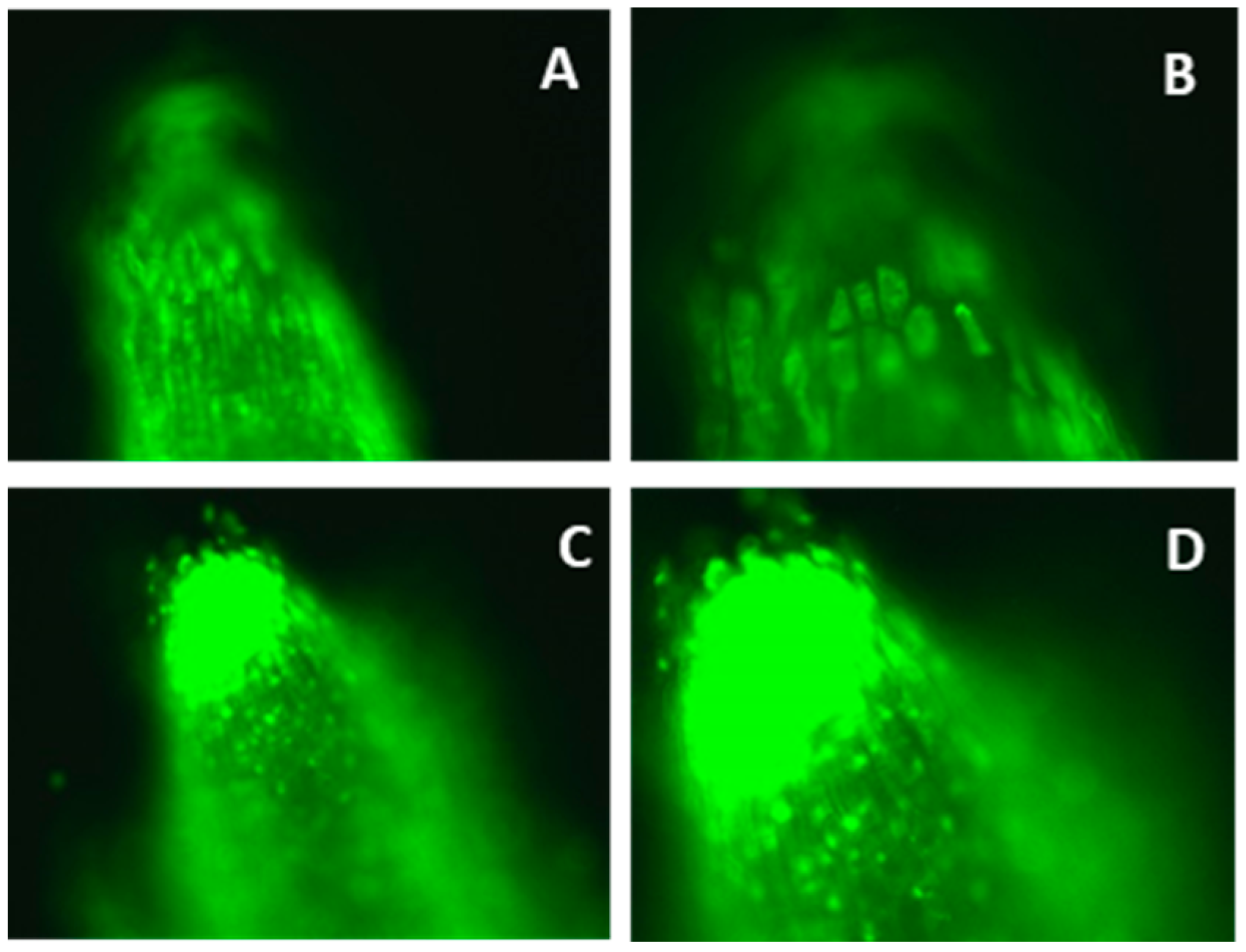
Morin was used to indicate the presence of Al in root tips and apoplast. Morin staining was performed as described previously [29]. Five-day-old seedlings were subjected to 0.5 mM CaCl2 solution (pH 4.5) with or without 100 μM Al for 24 h. Root tips were cut, washed in 5 mM NH4OAc buffer (pH 5.0) for 10 min, and then transferred to 100 μM morin solution plus 5 mM NH4OAc (pH 5.0) for 1 h. Finally, these stained roots were washed 3 times in 5 mM NH4OAc buffer (pH 5.0) for 10 min each time. The root tips were observed under a fluorescence microscope (excitation wavelength 420 nm, emission wavelength 510 nm, axivoert 40 CFL, Carl Zeiss, Jena, Oberkochen, Germany). The images were analyzed by Axio Vision-AC software.

2.4. Al Distribution in Apoplast and Symplast

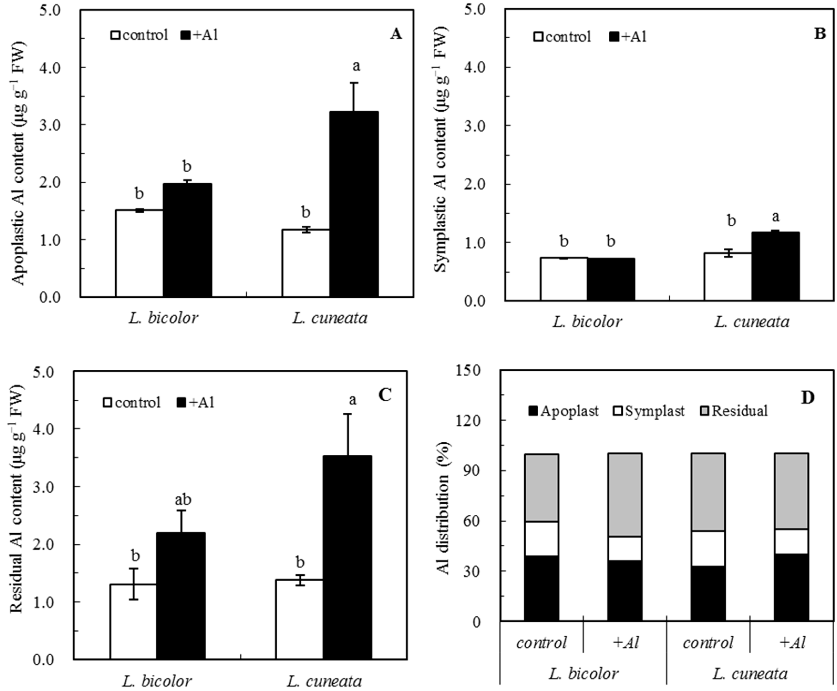
The roots of five-day-old seedlings were exposed to 0.5 mM CaCl2 solution (pH 4.5) with or without 50 μM Al for 24 h. Seedling roots were rinsed with ultrapure water, and the attached water in the root surface was blotted with filter paper. After that, 70 root tips (0–1.0 cm) were excised and placed in 50 mL polyethylene tubes. Fresh root tips were weighed and then refrigerated at 4 °C for fractionation testing. The method of continuous extraction of Al in apoplast and symplast was modified from a previous study [29]. The fractionation of Al was as follows: (1), root tips were sequentially washed 6 times on the oscillator for 60 min per cycle with 10 mL desorbing solution (5 mM CaCl2 + 100 μM sodium citrate); (2), cell membranes were ruptured by freezing and sonication, where freezing duration was approximately 18 h at −20 °C and sonication was conducted with a sonicator with a 3 mm diameter head for 1 min at high power; (3), the same method as (1) and (2) was used 4 times to separate the root tip residues; (4), root tip residues were digested in 1.0 mL concentrated HNO3 (trace metal grade) at 100 °C water bath for 1 h, and digested solutions were diluted to 10 mL with the ultrapure water. Al in wash and digest solutions was, respectively, apoplastic and symplastic Al, and analyzed spectro-fluorometrically using 8-hydroxyquinoline with butyl acetate extraction [30].

2.5. Determination of Total Sugar Secreted from and within Root Tips

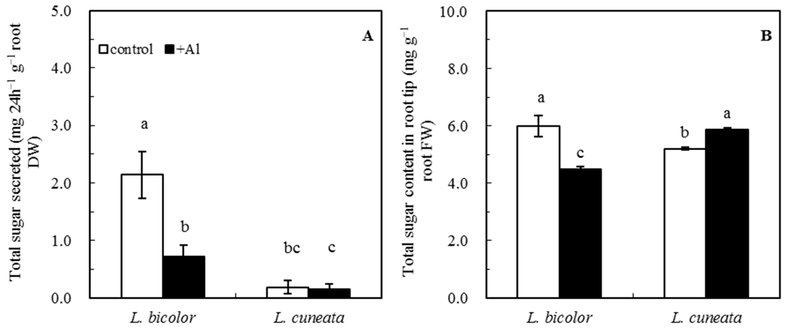
Five-day-old seedlings were cultured for 30 d in the modified Hoagland’s solution as described previously [11]. Then, the plants were exposed to a 0.5 mM CaCl2 solution (pH 4.5) with or without 50 µM Al for 24 h. Thereafter, the original solution was filtered and evaporated to dryness at 40 °C on a rotary evaporator. The sample was fixed to 10 mL, centrifuged at 3000× g for 10 min, and the supernatant was used for the determination of total sugar secreted from root tips. In addition, the excised root apices (0–1.0 cm) were stored in the freezer at −20 °C for total sugar extraction. The frozen root tips were homogenized with 5% (v/v) HClO4 in an ice bath, and then the combined material was centrifuged at 12,500× g for 10 min. These supernatants constituted the total sugar contents of the root apices. Total sugar in the supernatants was determined by anthrone colorimetry.

2.6. Cell Wall Extraction

Five-day-old seedlings were exposed to a 0.5 mM CaCl2 solution (pH 4.5) with or without 50 µM Al (pH 4.5) for 24 h. Root tips (0–2.0 cm) were washed with ultrapure water and the excess liquid was blotted with filter paper. After weighing, the samples were temporarily stored at −20 °C for cell wall extraction. The method of cell wall extraction was modified from a previous study [31]. The frozen root tips were homogenized with a mortar in an ice bath. The homogenate was subsequently rinsed twice with 10 × volume of 80% (v/v) ice ethanol, transferred into a centrifugal tube, and kept in ice bath for 2 h. The homogenate was then centrifuged at 12,500× g for 10 min, and the supernatant was decanted. The same amount of 80% (v/v) ice ethanol was added, and the above step was repeated once. After that, the precipitate was washed sequentially with 10 mL ultrapure water, 10 mL a methanol:chloroform mixture (1:1 [v/v]), and 10 mL phenol: acetic acid:water mixture (2:1:1 [v/v/v]), and this step was repeated once. Finally, the insoluble material was resuspended in water with α-amylase (10 μg mL−1) for 3 h at 37 °C, and was washed twice with 20 mL ultrapure water. Centrifugation was repeated after each wash. After frozen-drying, this material was stored at 4 °C for further use.

2.7. Measurement of Cell Wall Polysaccharides

Cell wall polysaccharides were measured according to the previous report [20]. Extracted cell walls were fractionated into three fractions: pectin, hemicellulose 1 (HC1), and hemicellulose 2 (HC2). To remove pectin, the pellet was resuspended in 5.0 mL of 0.5% (m/v) ammonium oxalate containing 0.1% (m/v) NaBH4 (pH 4.0) for 1 h in a boiling water bath. This material was centrifuged and washed twice with 0.5% (m/v) ammonium oxalate. These combined supernatants were the pectin fraction. The resulting pellet was extracted three times with 5.0 mL 4% (m/v) KOH containing 0.1% (m/v) NaBH4 (pH 4.0) on a water bath shaker (25 °C) for a total time of 24 h. Then, similar extraction with 24% (m/v) KOH containing 0.1% (m/v) NaBH4 (pH 4) was repeated. The collected supernatants from the 4% and 24% KOH extraction thus yielded the HC1 and HC2 fractions, respectively.
The pectin extract was hydrolyzed for 1 h in a boiling water bath under the action of 3 M sulfuric acid. HC1 and HC2 extract was hydrolyzed for 1 h in a boiling water bath. Polysaccharide content in each fraction was determined by the phenol−sulfuric acid method [32]. Glucose was used as a calibration standard. All treatments consisted of at least three repetitions within each experiment.

2.8. Pretreatment and Determination of Monosaccharide in Root Tips

Root tips (0–1.0 cm) of 5-day-old seedlings were homogenized in 0.5 mL 5% HClO4 (v/v) in an ice bath. The homogenate was then rinsed with 0.5 mL 5% (v/v) HClO4 and transferred into a 1.5 mL Eppendorf tube. Subsequently, the homogenate was centrifuged at 12,500× g for 10 min, filtered by 0.45 μm membrane, and used to determine the monosaccharide content of root tips.
The concentration of monosaccharide in the solution was determined by high performance liquid chromatography (HPLC; LC-10AT VP, Shimadzu, Tokyo, Japan) with gradient elution and post-column derivatization fluorescence detection using a Shim-pack ISA-07 column (4.0 mm i.d. × 25 cm). The mobile phase included A solution of 0.1 mM boric acid (adjusted to pH 8.0 with KOH) and B solution of 0.4 M boric acid (adjusted to pH 9.0 with KOH). The mobile phase was eluted from 100% (v/v) of A solution to 100% (v/v) of B solution in a linear gradient of 2% (v/v)/min. The total flow rate was 0.6 mL min−1, and the column temperature was 65 °C. The post-column derivatization reagent was 1% (m/v) L-arginine containing 3% (m/v) boric acid. The flow rate was 0.5 mL min−1, and reaction temperature was 150 °C. The wavelengths for fluorescence detection were 320 nm (excitation) and 430 nm (emission).

2.9. Statistical Analysis

All experiments and observations were repeated three times. One-way ANOVA was used and a least significant difference (Tukey) test was applied for multiple means’ comparisons at a significance level of p < 0.05. For the statistical analyses, the software SPSS (version 13.0, Chicago, IL, USA) was used.

3. Results

3.1. Aluminum-Induced Element Exclusion from Lespedeza Roots

Al exposure induced irregular curve and crack formation in the meristematic zone of two species (Figure 1A,B). The energy-spectrum analysis of elements showed that Al, potassium (K), calcium (Ca), and iron (Fe) were detected on the root surface of Al-sensitive L. cuneata (Figure 1D,F), whereas only Al was detected on that of Al-tolerant L. bicolor (Figure 1C,E).

3.2. Aluminum Distribution in Apoplast and Symplast of Roots

In order to explore the Al distribution in the Lespedeza root apex, morin, as a fluorochrome, was used to localize Al in roots. The Al accumulation in the root tips of Al-tolerant L. bicolor was much less than that of Al-sensitive L. cuneata (Figure 2). When exposed to Al toxicity, Al-sensitive L. cuneata accumulated more Al in both apoplast and symplast of root tips than did Al-tolerant L. bicolor (Figure 3). About 38%, 15%, and 47% of the accumulated Al were distributed in the apoplastic, symplastic, and residual fractions, respectively.

3.3. Total Sugar Secreted from and within Root Tips

Aluminum toxicity reduced the amounts of total sugar secreted from and within the roots of only Al-tolerant L. bicolor (Figure 4). In addition, Al-tolerant L. bicolor secreted much more total sugar than Al-sensitive L. cuneata regardless of Al toxicity (Figure 4A). The contents of total sugar within the root tips of Al-tolerant L. Bicolor were higher than those of Al-sensitive L. cuneata under control treatment, but this was reversed under Al treatment (Figure 4B).

3.4. Polysaccharides of Root Cell Walls

As the Al bound in the apoplast can cause changes in the cell wall polysaccharides, the cell wall fractions of root tips were isolated and analyzed to determine whether cell wall composition was changed by Al toxicity in two Lespedeza species (Figure 5). Al treatment did not affect the pectin contents of root tips of two Lespedeza species, whereas the contents of pectin in the root tips of Al-sensitive L. cuneata were significantly higher than those of Al-tolerant L. bicolor (Figure 5A). Al treatment increased the contents of HC1 and HC2 in root tips of Al-tolerant L. bicolor but not in Al-sensitive L. cuneata, and those contents were higher in Al-tolerant L. bicolor than in Al-sensitive L. cuneata under Al toxicity (Figure 5B,C).

3.5. Monosaccharides of Root Tips

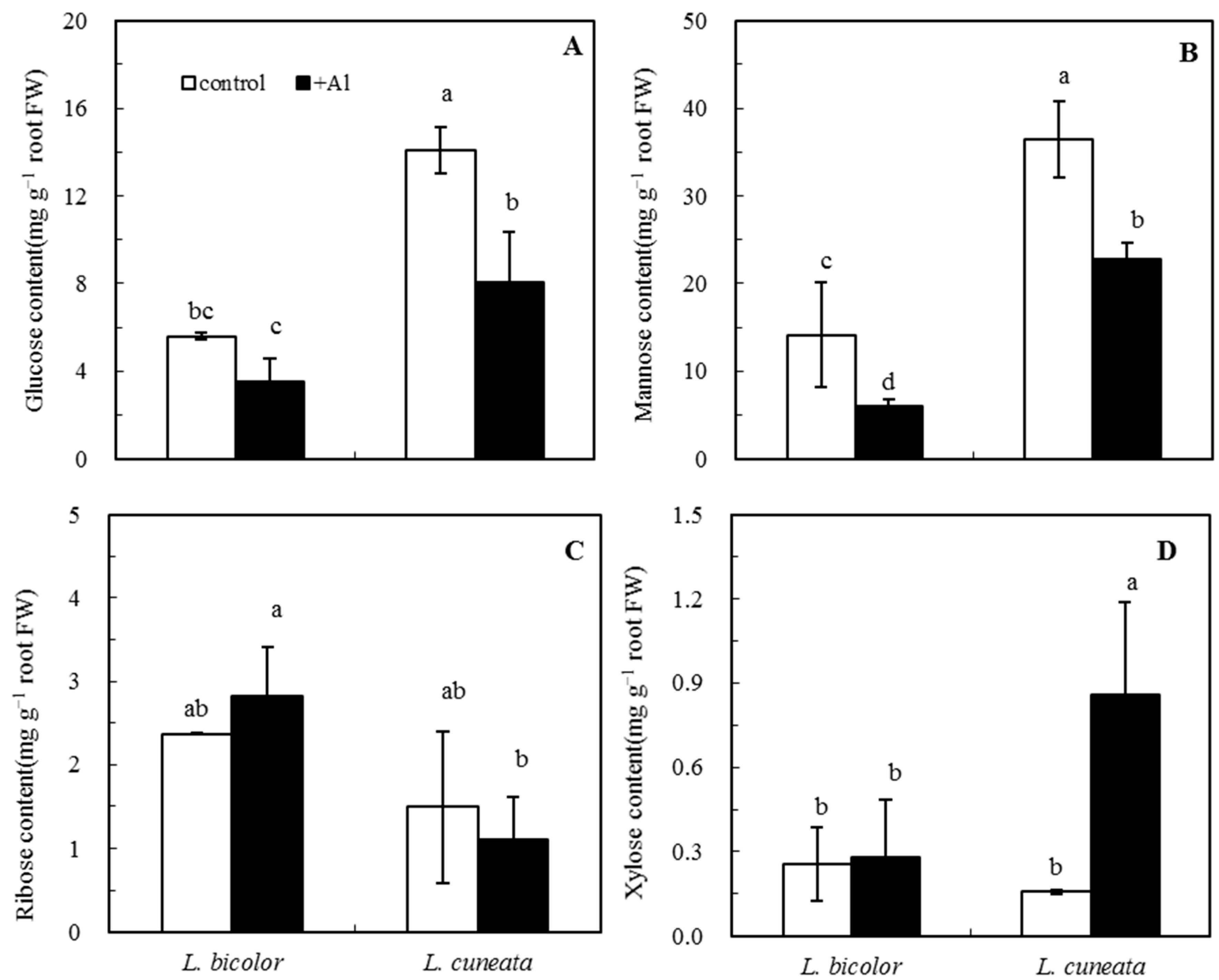
The monosaccharides of root apices were mainly composed of glucose and mannose (Figure 6). The contents of glucose and mannose of roots were significantly decreased under Al toxicity, and were higher in Al-sensitive L. cuneata than in Al-tolerant L. bicolor (Figure 6A,B). There was no significant difference in the ribose content between the control and Al treatment, and between the two species (Figure 6C). Al treatment substantially increased the contents of xylose in root tips of only Al-sensitive L. cuneata being higher than those of Al-tolerant L. bicolor under Al toxicity (Figure 6D).

4. Discussion

Root growth inhibition is the most dramatic visual symptom of Al toxicity in various plants, and the location of Al toxicity is mainly at the root tips [11,19,33,34,35,36]. Our previous research suggests that the occurrence of Al toxicity to Lespedeza is mainly in the 0–5 mm root tips [11,37]. Here, the scanning images further revealed that Al toxicity affected the morphology of root tips including the irregular curve and crack formation, and the morin staining results further suggested that Al was accumulated much more in the root tips of the Al-sensitive Lespedeza than in those of the Al-tolerant one. The analysis of energy spectrum elements showed that the destroyed cell structure of root tips of the Al-sensitive Lespedeza due to Al toxicity resulted in the release of several nutrients such as K, Ca, and Fe from the cytosol to the root surface. However, this loss of nutrients did not occur in the root tips of the Al-tolerant Lespedeza. We infer that Al toxicity may result in much bigger damage to root cell integrity and cause more nutrient leakage from roots in Al-sensitive Lespedeza compared with Al-tolerant Lespedeza. These results suggest that root tips play a decisive role in the expression of Al toxicity in Lespedeza, which is in accordance with previous reports on other plants [6,19,38]. In the root tips, the apoplast accumulated more Al than did the symplast, and the Al-sensitive Lespedeza had much more apoplastic Al than did the Al-tolerant one. This is also in accordance with previous reports on other plants [19,23]. Although the apoplast occupies 5% or less of the total tissue volume in higher plants, more than 70% of Al is accumulated in the cell wall of root apoplast [18,23,37,39]. The protection of the root-tip apoplast represents an important way by which Al-tolerant Lespedeza adapts to Al toxicity.
As the first barrier for all ions entering the cell, the cell wall plays a major role in regulating the normal growth and development of plants under Al stress [5,9,23]. The apoplast is mainly composed of cell walls which contain large amounts of saccharides, so the responses of cell wall saccharides to Al toxicity were further examined in this study. Al accumulation in roots is closely positively correlated with the pectin contents of root tips of maize and faba bean [19]. Our study showed that the Al-sensitive Lespedeza accumulated more Al and pectin in the cell walls of root tips than did the Al-tolerant one, which is consistent with previous studies in other plants [20,26]. The experimental evidence has proved that the modification of the root cell wall might contribute to Al tolerance [8,18,19,20,21,23,26,27,40]. Al accumulation in the root apoplast could modify the mechanical components and chemical properties of the cell walls [6,19,20,21]. In our study, Al treatment did not affect the cell wall pectin contents of root tips in either Lespedeza species. However, previous studies showed that Al treatment induced a significant increase in the pectin contents of cell walls of tea and rice roots [23,41]. In addition, the enhancement of HC1 and HC2 contents of root tips by Al toxicity was found in the Al-tolerant Lespedeza but not in the Al-sensitive. However, previous studies showed that Al addition also increased the HC contents of root tips of Al-sensitive species [20,26,41]. These inconsistencies may be attributed to the specific properties of cell walls of Lespedeza differing from other plant species [8,20,21,23].
The fundamental constituents of pectin are homogalacturonan (HGA) and rhamnogalacturonan I (RG I), while the major parts of HCs are xyloglucans and mixed-linkage β-(1/3),(1/4)-D-glucans [21,33]. The increase in the HCs was attributed to the increases in arabinose, xylose, and glucose [26]. Our results showed that the contents of the root monosaccharides including glucose and mannose were decreased in Al-tolerant Lespedeza by Al treatment. This suggested that Al might promote the synthesis of monosaccharides into polysaccharides such as HCs, thereby increasing the contents of HCs of root tips of Al-tolerant Lespedeza. For the Al-sensitive Lespedeza, Al treatment decreased the contents of the root monosaccharides including glucose and mannose but increased the root xylose (xyloglucan monomer) contents, which may be the reason for the lack of effect of Al treatment on the contents of HCs of root tips of Al-sensitive Lespedeza.
Subjected to Al toxicity, plants might activate multiple mechanisms of Al tolerance to maintain their normal growth. Our previous study showed that Al-tolerant Lespedeza secreted large amounts of organic acids to alleviate Al toxicity while Al-sensitive Lespedeza did not [10]. In the present study, Al-tolerant Lespedeza secreted more sugars than Al-sensitive Lespedeza, indicating that sugar secretion could also provide some donors to alleviate the effect of Al toxicity. Meanwhile Al treatment decreased the amounts of sugars secreted from Al-tolerant Lespedeza but not from Al-sensitive Lespedeza, which proved that Al exposure might hasten the intracellular physiological process and transform sugars into more organic acids in Al-tolerant species. In addition, Al decreased the amounts of sugars in the roots of Al-tolerant Lespedeza but increased those of Al-sensitive Lespedeza. Based these results, we infer that Al-tolerant Lespedeza might transform more sugars into organic acids that can chelate toxic Al3+ and alleviate Al phytotoxicity, but Al-sensitive Lespedeza could not. Consequently, Al-tolerant Lespedeza secrets much more organic acid to chelate toxic Al in the rhizosphere and is more tolerant to Al than Al-sensitive Lespedeza [10,11]. The specific mechanisms for the transformation of sugars into organic acids under Al toxicity in Lespedeza will be an interesting research topic in the future.

5. Conclusions

Less accumulation of pectin rather than hemicellulose in the cell walls of root tips is responsible for the less Al accumulation in root tips and the high Al tolerance of Al-tolerant L. bicolor relative to Al-sensitive L. cuneata. Furthermore, the decreased amounts of sugars secreted from and within the roots by Al toxicity may contribute to the increased secretion of organic acids from the roots of Al-tolerant L. bicolor, thereby increasing Lespedeza Al tolerance through chelating toxic Al in the rhizosphere. These findings update the physiological mechanisms of Lespedeza Al tolerance and supply the reference for understanding Al tolerance mechanisms in other plants. In the future, it would be worth uncovering the molecular mechanism of saccharide metabolism related to Lespedeza Al tolerance.

Author Contributions

Conceptualization, X.-Q.Z. and R.-F.S.; methodology, data curation, writing—original draft, Q.-B.S.; supervision, R.-F.S.; writing—review and editing, X.-Q.Z., R.-F.S., C.-Q.Y., X.-Y.D. and H.Z.; funding acquisition, X.-Q.Z., H.Z. and Q.-B.S.; All authors have read and agreed to the published version of the manuscript.

Funding

This work was financially supported by the National Key Research and Development Program of China (No. 2021YFD1901201-04), the National Natural Science Foundation of China (No. 42107088), and The Foundation of Central Guidance on Local Science and Technology Development (No. 2022BGE252).

Data Availability Statement

All data analyzed in this study are included in this article.

Conflicts of Interest

The authors declare no conflict of interest.

References

  1. Hyland, H.L. Comparison of legume growth in different soil types at varying acidity levels. J. Am. Soc. Agron. 1938, 30, 111–121. [Google Scholar] [CrossRef] [Green Version]
  2. Campbell, T.A.; Nuernberg, N.J.; Foy, C.D. Differential responses of Sericea Lespedeza to aluminum stress. J. Plant Nutr. 1991, 14, 1057–1066. [Google Scholar] [CrossRef]
  3. Zhao, X.Q.; Chen, R.F.; Shen, R.F. Coadaptation of plants to multiple stresses in acidic soils. Soil Sci. 2014, 179, 503–513. [Google Scholar] [CrossRef]
  4. Kochian, L.V.; Piñeros, M.A.; Hoekenga, A.O. The physiology, genetics and molecular biology of plant aluminum resistance and toxicity. Plant Soil 2005, 274, 175–195. [Google Scholar] [CrossRef]
  5. Zhang, X.; Long, Y.; Huang, J.J.; Xia, J.X. Molecular mechanisms for coping with Al toxicity in plants. Int. J. Mol. Sci. 2019, 20, 1551. [Google Scholar] [CrossRef] [PubMed] [Green Version]
  6. Rahman, R.; Upadhyaya, H. Aluminium toxicity and its tolerance in plant: A review. J. Plant Biol. 2020, 64, 101–121. [Google Scholar] [CrossRef]
  7. Kochian, L.V.; Piñeros, M.A.; Liu, J.; Magalhaes, J.V. Plant adaptation to acid soils: The molecular basis for crop aluminum resistance. Annu. Rev. Plant Biol. 2015, 66, 571–598. [Google Scholar] [CrossRef]
  8. Zhu, X.F.; Wan, J.X.; Sun, Y.; Shi, Y.Z.; Braam, J.; Li, G.X.; Zheng, S.J. Xyloglucan endotransglucosylase-hydrolase17 interacts with xyloglucan endotransglucosylase-hydrolase31 to confer xyloglucan endotransglucosylase action and affect aluminum sensitivity in Arabidopsis. Plant Physiol. 2014, 165, 1566–1574. [Google Scholar] [CrossRef] [Green Version]
  9. Liu, H.; Zhu, R.; Shu, K.; Lv, W.; Wang, S.; Wang, C. Aluminum stress signaling, response, and adaptive mechanisms in plants. Plant Signal Behav. 2022, 17, 2057060. [Google Scholar] [CrossRef]
  10. Dong, X.Y.; Shen, R.F.; Chen, R.F.; Zhu, Z.L.; Ma, J.F. Secretion of malate and citrate from roots is related to high Al-resistance in Lespedeza bicolor. Plant Soil 2008, 306, 139–147. [Google Scholar] [CrossRef]
  11. Sun, Q.B.; Shen, R.F.; Zhao, X.Q.; Chen, R.F. P enhances Al tolerance in Al-resistant Lespedeza but not in Al-sensitive Lespedeza. Ann. Bot. 2008, 102, 795–804. [Google Scholar] [CrossRef] [Green Version]
  12. Chen, Z.C.; Zhao, X.Q.; Shen, R.F. The alleviating effect of ammonium on aluminum toxicity in Lespedeza bicolor results in decreased aluminum-induced malate secretion from roots compared with nitrate. Plant Soil 2010, 337, 389–398. [Google Scholar] [CrossRef]
  13. Wang, J.Q.; Yu, X.F.; Ding, Z.J.; Ding, Z.J.; Zhang, X.K.; Luo, Y.P.; Xu, X.M.; Xie, Y.; Li, X.X.; Yuan, T.; et al. Structural basis of ALMT1-mediated aluminum resistance in Arabidopsis. Cell Res. 2022, 32, 89–98. [Google Scholar] [CrossRef] [PubMed]
  14. Hajiboland, R.; Panda, C.K.; Lastochkina, O.; Gavassi, M.A.; Habermann, G.; Pereira, J.F. Aluminum toxicity in plants: Present and future. J. Plant Growth Regul. 2022. [Google Scholar] [CrossRef]
  15. Yang, Z.B.; Rao, I.M.; Horst, W.J. Interaction of aluminium and drought stress on root growth and crop yield on acid soils. Plant Soil 2013, 372, 3–25. [Google Scholar] [CrossRef] [Green Version]
  16. Zheng, S.J.; Yang, J.L. Target sites of aluminum phytotoxicity. Biol. Plantarum 2005, 49, 321–331. [Google Scholar] [CrossRef]
  17. Horst, W.J.; Kollmeier, M.; Schmohl, N.; Sivaguru, M.; Wang, Y.; Felle, H.H.; Hedrich, R.; Schröder, W.; Staß, A. Significance of the root apoplast for aluminium toxicity and resistance of maize. In The Apoplast of Higher Plants: Compartment of Storage, Transport and Reactions 49; Springer: Berlin/Heidelberg, Germany, 2007; pp. 49–66. [Google Scholar]
  18. He, H.; He, L.; Gu, M. Signal transduction during aluminum-induced secretion of organic acids in plants. Biol. Plant 2015, 59, 601–608. [Google Scholar] [CrossRef]
  19. Horst, W.J.; Wang, Y.; Eticha, D. The role of the root apoplast in aluminium-induced inhibition of root elongation and in aluminium resistance of plants: A review. Ann. Bot. 2010, 106, 185–197. [Google Scholar] [CrossRef]
  20. Yang, J.L.; Li, Y.Y.; Zhang, Y.J.; Zhang, S.S.; Wu, Y.R.; Wu, P.; Zheng, S.J. Cell wall polysaccharides are specifically involved in the exclusion of aluminum from the rice root apex. Plant Physiol. 2008, 146, 602–611. [Google Scholar] [CrossRef]
  21. Yang, J.L.; Zhu, X.F.; Peng, Y.X.; Zheng, C.; Li, G.X.; Liu, Y.; Zheng, S.J. Cell wall hemicellulose contributes significantly to Al adsorption and root growth in Arabidopsis. Plant Physiol. 2011, 155, 1885–1892. [Google Scholar] [CrossRef] [Green Version]
  22. Ma, B.H.; Gao, L.; Zhang, H.X.; Cui, J.; Shen, Z.G. Aluminum-induced oxidative stress and changes in antioxidant defenses in the roots of rice varieties differing in Al tolerance. Plant Cell Rep. 2012, 31, 687–696. [Google Scholar] [CrossRef]
  23. Safari, M.; Ghanati, F.; Safarnejad, M.R.; Chashmi, N.A. The contribution of cell wall composition in the expansion of Camellia sinensis seedlings roots in response to aluminum. Planta 2018, 247, 381–392. [Google Scholar] [CrossRef] [PubMed]
  24. Chauhan, D.K.; Yadav, V.; Vaculík, M.; Gassmann, W.; Pike, S.; Arif, N.; Singh, V.P.; Deshmukh, R.; Sahi, S.; Tripathi, D.K. Aluminum toxicity and aluminum stress-induced physiological tolerance responses in higher plants. Crit. Rev. Biotechnol. 2021, 41, 715–730. [Google Scholar] [CrossRef] [PubMed]
  25. Braybrook, S.A.; Peaucelle, A. Mechano-chemical aspects of organ formation in Arabidopsis thaliana: The relationship between auxin and pectin. PLoS ONE 2013, 8, e57813. [Google Scholar] [CrossRef] [PubMed] [Green Version]
  26. Lan, T.; You, J.F.; Kong, L.N.; Yu, M.; Liu, M.H.; Yang, Z.M. The interaction of salicylic acid and Ca2+ alleviates aluminum toxicity in soybean (Glycine max L.). Plant Physiol. Biochem. 2016, 98, 146–154. [Google Scholar] [CrossRef]
  27. Tabuchi, A.; Matsumoto, H. Changes in cell-wall properties of wheat (Triticum aestivum L.) roots during aluminum-induced growth inhibition. Biol. Plantarum 2001, 112, 353–358. [Google Scholar] [CrossRef]
  28. Sun, Q.B.; Zhao, X.Q.; Shen, R.F. Application of three microscopic techniques to research on Al toxicity in plants. Acta Pedol. Sin. 2009, 46, 1026–1032. [Google Scholar]
  29. Tice, K.R.; Parker, D.R.; DeMason, D.A. Operationally defined apoplastic and symplastic aluminum fractions in root tips of aluminum-intoxicated wheat. Plant Physiol. 1992, 100, 309–318. [Google Scholar] [CrossRef] [Green Version]
  30. Bloom, P.R.; Weaver, R.M.; McBride, M.B. The spectrophotometric and fluorometric determination of aluminum with 8-hydroxyquinoline and butyl acetate extraction. Soil Sci. Soc. Am. J. 1978, 42, 713–716. [Google Scholar] [CrossRef]
  31. Heim, D.R.; Skomp, J.R.; Waldron, C.; Larrinua, I.M. Differential response to isoxaben of cellulose biosynthesis by wild-type and resistant strains of Arabidopsis thaliana. Pest. Biochem. Physiol. 1991, 39, 93–99. [Google Scholar] [CrossRef]
  32. Dubois, M.; Gilles, K.A.; Hamilton, J.K.; Rebers, P.A.; Smith, F. Colorimetric method for determination of sugars and related substances. Anal. Chem. 1956, 28, 350–356. [Google Scholar] [CrossRef]
  33. Buchana, B.B.; Gruissem, W.; Jones, R.L. The cell wall. In Biochemistry and Molecular Biology of Plants 52; Elsevier: New York, NY, USA, 2000; pp. 52–98. [Google Scholar]
  34. Mariano, E.D.; Pinheiro, A.S.; Garcia, E.E.; Keltjens, W.G.; Jorge, R.A.; Menossi, M. Differential aluminium-impaired nutrient uptake along the root axis of two maize genotypes contrasting in resistance to aluminium. Plant Soil 2015, 388, 323–335. [Google Scholar] [CrossRef]
  35. De Souza, L.C.; Nogueira, D.C.S.; Machado, L.C.; Costa, T.C.; Martins, T.S.; Mendes, C.A.P.; Pires, N.M.C.; Neto, C.F.O.; Conceição, S.S.; Brito, A.E.A. Nitrogen compounds, proteins and amino acids in corn subjected to doses of aluminium. Afr. J. Agric. Res. 2016, 11, 1519–1524. [Google Scholar]
  36. De Rosmaninho, C.L.B.; Dias, A.S.; da Silva, M.F.; de Vasconcelos, A.; Santos, W.O.; Perez, C.E.A.; Vergutz, L.; Cardoso, L.G. Performance of Crambe submitted to aluminum stress: An important oilseed plant. J. Agric. Sci. 2019, 11, 454–463. [Google Scholar] [CrossRef]
  37. Sun, Q.B.; Shen, R.F.; Yin, C.Q.; Zhao, X.Q. Analysis of variations and factors affecting callose formation in response to Al stress in Lespedeza root tips. Acta Ecol. Sin. 2016, 36, 1073–1082. [Google Scholar]
  38. Brito, D.S.; Neri-Silva, R.; Ribeiro, K.V.G.; Peixoto, P.H.P.; Ribeiro, C. Effects of aluminum on the external morphology of root tips in rice. Braz. J. Bot. 2020, 43, 413–418. [Google Scholar] [CrossRef]
  39. Grignon, C.; Sentenac, H. pH and ionic conditions in the apoplast. Ann. Rev. Plant Physiol. Mol. Biol. 1991, 42, 103–128. [Google Scholar] [CrossRef]
  40. Hossain, A.K.M.Z.; Hossain, M.A.; Asgar, M.A.; Tosaki, T.; Koyama, H.; Hara, T. Changes in cell wall polysaccharides and hydroxycinnamates in wheat roots by aluminum stress at higher calcium supply. J. Plant Nutr. 2006, 29, 601–613. [Google Scholar] [CrossRef]
  41. Wang, W.; Zhao, X.Q.; Chen, R.F.; Dong, X.Y.; Lan, P.; Ma, J.F.; Shen, R.F. Altered cell wall properties are responsible for ammonium-reduced aluminum accumulation in rice roots. Plant Cell Environ. 2015, 38, 1382–1390. [Google Scholar] [CrossRef]
Figure 1. Scanning images, energy-spectrum analysis, and element percentage in the meristematic zone of two Lespedeza species. (A,C,E), L. bicolor; (B,D,F), L. cuneata. Seedlings (five-day-old) of two Lespedeza species were subjected to 0.5 mM CaCl2 solutions (pH 4.5) with 200 µM Al for 24 h. Scanning images (A,B) were observed by the scanning electron microscopy microscope. The image magnification was 750 under 20 kV voltage. All pictures were taken under the same conditions. Energy-spectrum analysis (C,D) was carried out using energy-dispersive X-ray spectroscopy, and element percentages (E,F) were obtained from the energy-spectrum analysis.
Agronomy 13 00629 g001
Figure 2. Aluminum distribution in the roots of two Lespedeza species using morin staining. (A,B), L. bicolor; (C,D), L. cuneata. (A,C), ×200; (B,D), ×320. The roots were exposed to a 0.5 mM CaCl2 solution (pH 4.5) with 100 µM Al for 1 h, and then stained with 100 µM morin. The sections were taken from the root zone between 0 and 3 mm behind the root apex of Lespedeza seedlings.
Agronomy 13 00629 g002
Figure 3. Distribution of Al in the root apices of two Lespedeza species. (A) apoplastic Al, (B) symplastic Al, (C) residual Al, and (D) percentage Al distribution in different fractions. Five-day-old seedlings were exposed to 0.5 mM CaCl2 solutions (pH 4.5) containing 0 (control) or 50 μM Al (+Al) for 24 h. The apoplastic, symplastic, and residual Al contents of root apices were analyzed. Significant differences between mean values are indicated by different letters at the p < 0.05 level (Tukey test). Error bars represent ±SD (n = 3).
Agronomy 13 00629 g003
Figure 4. Effects of Al treatments on the contents of total sugar secreted from and within root tips of two Lespedeza species. (A) total sugar secreted from roots and (B) total sugar within root apices. Lespedeza seedlings were exposed to 0.5 mM CaCl2 solutions (pH 4.5) with or without 50 µM AlCl3 for 24 h. Thereafter, the contents of total sugar in the culture solution and root tips (0–1.0 cm) were both determined by anthrone colorimetry. Significant differences between mean values are indicated by different letters at the p < 0.05 level (Tukey test). Values shown are means ± SD (n = 3).
Agronomy 13 00629 g004
Figure 5. Effect of Al treatments on the contents of polysaccharides in root tips of two Lespedeza species. (A) pectin, (B) HC1, and (C) HC2. Lespedeza seedlings were exposed to 0.5 mM CaCl2 solutions (pH 4.5) with or without 50 μM Al for 24 h. Root apices (0–1 cm) were cut and cell wall polysaccharides were fractionated into pectin, HC1, and HC2 for glucose-content measurement. Significant differences between mean values are indicated by different letters at the p < 0.05 level (Tukey test). Error bars represent ±SD (n = 3).
Agronomy 13 00629 g005
Figure 6. Effects of Al treatments on the contents of monosaccharides in root apices of two Lespedeza species. (A) glucose, (B) mannose, (C) ribose, and (D), xylose. Five-day-old seedlings were exposed to 0.5 mM CaCl2 solutions (pH 4.5) with or without 50 μM Al for 24 h. Significant differences between mean values are indicated by different letters at the p < 0.05 level (Tukey test). Values shown are means ± SD (n = 3).
Agronomy 13 00629 g006
Disclaimer/Publisher’s Note: The statements, opinions and data contained in all publications are solely those of the individual author(s) and contributor(s) and not of MDPI and/or the editor(s). MDPI and/or the editor(s) disclaim responsibility for any injury to people or property resulting from any ideas, methods, instructions or products referred to in the content.